spinal meningiomas: a comprehensive overview and own ... · spinal menigiomas 118 eur assoc...
TRANSCRIPT

Neurology · Neurosurgery · Medical Oncology · Radiotherapy · Paediatric Neuro-
oncology · Neuropathology · Neuroradiology · Neuroimaging · Nursing · Patient Issues
THE EUROPEAN ASSOCIATION OF
NEUROONCOLOGY
Volume 3 (2013) // Issue 3 // e-ISSN 2224-3453
Member of the
Homepage:Homepage:
www.kup.at/journals/eano/index.html
Online Database Featuring Author, Key Word and
Full-Text Search
Online Database Featuring Author, Key Word and
Full-Text Search
Spinal Meningiomas: A
Comprehensive Overview and Own
Experience
Perneczky G, Loyoddin M
Schappelwein H, Sherif C
European Association of
NeuroOncology Magazine 2013; 3 (3)
118-121
Spinal Menigiomas
118 EUR ASSOC NEUROONCOL MAG 2013; 3 (3)
Introduction
Approximately 2/3 of all intraspinal neoplasms are intraduralextramedullar tumours. Among those, neurinomas followedby meningiomas are the most common histologic entities [1].Spinal menigiomas occur less frequently than intracranialmeningiomas. They constitute only for 7.5–12.7 % of allmeningiomas [2].
Comparable to intracranial meningiomas their incidence is2–3 times higher in women than in men. These lesions are atypical disease of the middle or older age [3, 4].
The purpose of this article is an update of this disease basedon a literature review and our own experience. Additionally,we present our most recent own consecutive case series col-lected between 2010 and 2012.
Localization
More than 2/3 of all spinal meningiomas are located in thethoracic spine (67–84 %), followed by 14–27 % in the cervi-cal spine and 2–14 % in the lumbar spine. Typically, they arefound purely intradurally in 86–95 %. Only 5–14 % have anadditional extradural part [3–7]. On rare occasions, spinalmeningiomas occur completely extradurally (3–9 %) [4, 7].In the latter, in 2 locations they are predominantly dumb-bell tumours causing an enlargement of the intervertebralforamen.
Received on May 1, 2013; accepted after revision on May 5, 2013; Pre-PublishingOnline on June 25, 2013
From the Neurosurgical Department, Krankenanstalt Rudolfstiftung, Vienna, Austria
Correspondence to: Gedeon Perneczky, MD, Neurosurgical Department, Kranken-anstalt Rudolfstiftung, Juchgasse 25, 1030 Vienna, Austria;e-mail: [email protected]
Spinal Meningiomas: A Comprehensive Overviewand Own Experience
Gedeon Perneczky, Michel Loyoddin, Horst Schappelwein, Camillo Sherif
Risk Factors
Most publications focus on risk factors for the development ofmeningiomas in general [8]. Only a few papers refer directlyto spinal meningiomas.
Ionizing Radiation
Hiroshima and Nagasaki survivors showed an elevated risk ofdeveloping intracranial meningiomas. Their risk depended ontheir vicinity to the epicentre of the nuclear explosion [9–11].Several US studies reported a significant correlation betweenX-ray dosage prior to the 20th year of life with the risk of me-ningioma development [12–14]. Also acute lymphoblasticleukaemia (ALL) patients showed an elevated risk of menin-gioma development after a latency of decades [15–20]. Thelatter lesions are more frequently multifocal, atypical, ormalign [21, 22]. It is unclear whether this risk is caused byirradiation of the whole neuroaxis alone or whether additionalfactors such as chemotherapy are causative.
Genetics
Changes or complete or partial loss of chromosome 22 mayplay a role in the development of meningiomas. Otherchanges in the gene loci are also associated with carcinoge-nesis and could play a role in the development of spinal me-ningiomas [23]. Contradictorily, Ketter et al documented aseries of 23 spinal meningiomas, all of which showed a regu-lar set of chromosomes or a monosomy 22 [24]. Additionally,neurofibromatosis type 2 with mutation of chromosome 22Q12is an autosomally recessive, hereditary disease with elevatedrisk of developing meningiomas or schwannomas [25]. In avery recent publication, changes in the gene SMARCE1 couldbe identified in relation to an increased incidence of familialspinal meningiomas [26].
Gender
Women have a 2–3 times higher incidence of meningiomadevelopment. Additionally, the gender-related risk is slightly
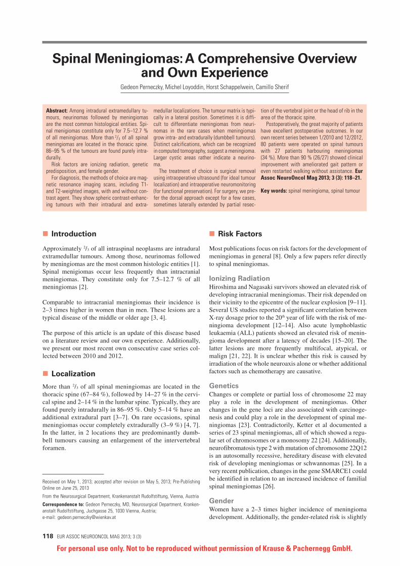
medullar localizations. The tumour matrix is typi-cally in a lateral position. Sometimes it is diffi-cult to differentiate meningiomas from neuri-nomas in the rare cases when meningiomasgrow intra- and extradurally (dumbbell tumours).Distinct calcifications, which can be recognizedin computed tomography, suggest a meningioma.Larger cystic areas rather indicate a neurino-ma.
The treatment of choice is surgical removalusing intraoperative ultrasound (for ideal tumourlocalization) and intraoperative neuromonitoring(for functional preservation). For surgery, we pre-fer the dorsal approach except for a few cases,sometimes laterally extended by partial resec-
Abstract: Among intradural extramedullary tu-mours, neurinomas followed by meningiomasare the most common histological entities. Spi-nal menigiomas constitute only for 7.5–12.7 %of all meningiomas. More than 2/3 of all spinalmeningiomas are located in the thoracic spine.86–95 % of the tumours are found purely intra-durally.
Risk factors are ionizing radiation, geneticpredisposition, and female gender.
For diagnosis, the methods of choice are mag-netic resonance imaging scans, including T1-and T2-weighted images, with and without con-trast agent. They show spheric contrast-enhanc-ing tumours with their intradural and extra-
tion of the vertebral joint or the head of rib in thearea of the thoracic spine.
Postoperatively, the great majority of patientshave excellent postoperative outcomes. In ourown recent series between 1/2010 and 12/2012,80 patients were operated on spinal tumourswith 27 patients harbouring meningiomas(34 %). More than 90 % (26/27) showed clinicalimprovement with ameliorated gait pattern oreven restarted walking without assistance. EurAssoc NeuroOncol Mag 2013; 3 (3): 118–21.
Key words: spinal meningioma, spinal tumour
For personal use only. Not to be reproduced without permission of Krause & Pachernegg GmbH.
EUR ASSOC NEUROONCOL MAG 2013; 3 (3)
Spinal Menigiomas
119
higher in women who take contraceptives or receive hormonereplacement therapy [27–29]. The coincidence of breast car-cinoma and meningiomas has been observed for many years[30]. It may be due to a joint risk profile (age, genetics, envi-ronmental factors in interaction) [31, 32].
Symptoms
At the beginning of the disease, mostly sensation disorders, adiscrete spasticity of extremities, and gait disturbance are ob-served. Due to the slow growth tendency of these tumours,their symptoms remain often untypical for a long period oftime. Because of the higher patient age (> 50 a) the altered gaitpattern is often misinterpreted as ordinary joint pain. Due tothese non-characteristic clues the correct diagnosis is oftensignificantly delayed, especially in the most frequent locationof the thoracic spine. Diagnosis remains unclear until the typi-cal vesical and rectal disorders and progressive paraparesisemerge. With the help of magnetic resonance imaging spinalmeningiomas are diagnosed earlier than several years ago.
Management
Diagnosis and Operative PlanningThe methods of choice are magnetic resonance imaging(MRI) scans, including T1- and T2-weighted images, withand without contrast agent (Figure 1). They show spheric con-trast-enhancing tumours with their intradural and extra-medullar localization [33]. The tumour matrix is in a lateralposition in most of spinal meningiomas, more often dorsola-teral than ventrolateral. Extensive growth and infiltration ofthe pia are significantly less frequently observed than in in-tracranial meningiomas. It is sometimes difficult to differenti-ate meningiomas from neurinomas in the rare cases whenmeningiomas grow intra- and extradurally (dumbbell tu-mours). Distinct calcifications, which can only be recognizedin computed tomography (CT), suggest a meningioma. Lar-ger cystic areas rather indicate a neurinoma.
Cystic changes are very rare in spinal meningiomas in con-trast to calcifications. The latter may influence the surgical
approach especially in ventrally positioned tumours. For thisreason, we believe that it makes sense to perform a CT scan inventrally positioned tumours to estimate the extent of calcifi-cations prior to surgery. In central calcified tumours that arecompletely covered by the spinal cord total removal via a dor-sal or dorsolateral approach is very difficult and may only becarried out at an elevated neurological risk.
Surgical Technique
We prefer the dorsal approach except for a few cases, some-times laterally extended by partial resection of the vertebraljoint or the head of rib in the area of the thoracic spine [2, 5,33]. The rare ventral approach is discussed in the literature asan alternative mainly for purely ventral tumour locationscompletely covered by the spinal cord. The intention is tominimize manipulations at the spinal cord. The advantage ofthe ventral approach is a lower neurological risk for the spinalcord and better chance of radical removal in ventral tumours.The disadvantages are complications caused by the larger ap-proach with vertebral body resection and the need for sta-bilization.
The application of intraoperative ultrasound [34] improves theprecise localization and helps avoid unnecessarily large ap-proaches with multi-level laminectomy. Depending on the lon-gitudinal extension of the tumour we try to remove the vertebralarch only at one level. In many cases, surgery can be performedvia an extended interlaminar approach with partial preservationof the vertebral arches. In younger patients, the vertebral archshould be preferably restored by laminoplasty, especially in thelumbar and cervical spine. The dura is opened paramediallyvertically depending on the lateralization of the tumour. Inventrally positioned tumours, the incision can be enlarged later-ally by way of a dural flap. Resection of the denticulate liga-ments allows for a better view, especially of ventral tumours.Opening the dura directly at the tumour site instead of choosingthe common median incision leaves the spinal cord mostly cov-ered by the dura during surgery. This reduces the risk of spinalcord impingement in the slit-like area of the dural opening.Comparable to intracranial meningiomas, spinal tumour de-
Figure 1. Pre- and intraoperative findings: (a) T2-weighted native sagittal MR image: the tumour can be nicely delineated (red arrow). (b, c) Axial T1-weighted contrast-enhanced sequence: inhomogeneous contrast enhancement is due to intratumoural calcifications (red arrow). (d) Sagittal contrast-enhanced T1-weighted image: again, thecalcifications can be seen as contrast agent-free areas within the tumour (red arrow). (e) Intraoperative findings: the spinal cord is carefully mobilized under IOM. Thetumour (blue arrow) can be seen at the left ventral dura.
Spinal Menigiomas
120 EUR ASSOC NEUROONCOL MAG 2013; 3 (3)
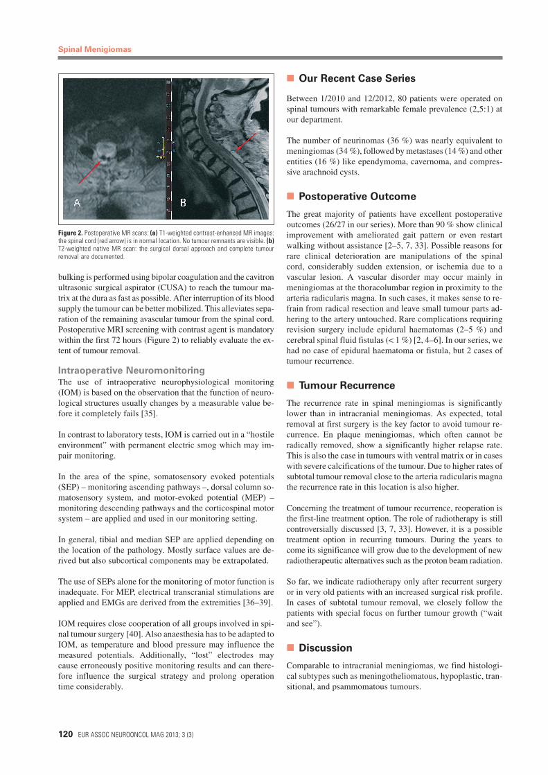
bulking is performed using bipolar coagulation and the cavitronultrasonic surgical aspirator (CUSA) to reach the tumour ma-trix at the dura as fast as possible. After interruption of its bloodsupply the tumour can be better mobilized. This alleviates sepa-ration of the remaining avascular tumour from the spinal cord.Postoperative MRI screening with contrast agent is mandatorywithin the first 72 hours (Figure 2) to reliably evaluate the ex-tent of tumour removal.
Intraoperative Neuromonitoring
The use of intraoperative neurophysiological monitoring(IOM) is based on the observation that the function of neuro-logical structures usually changes by a measurable value be-fore it completely fails [35].
In contrast to laboratory tests, IOM is carried out in a “hostileenvironment” with permanent electric smog which may im-pair monitoring.
In the area of the spine, somatosensory evoked potentials(SEP) – monitoring ascending pathways –, dorsal column so-matosensory system, and motor-evoked potential (MEP) –monitoring descending pathways and the corticospinal motorsystem – are applied and used in our monitoring setting.
In general, tibial and median SEP are applied depending onthe location of the pathology. Mostly surface values are de-rived but also subcortical components may be extrapolated.
The use of SEPs alone for the monitoring of motor function isinadequate. For MEP, electrical transcranial stimulations areapplied and EMGs are derived from the extremities [36–39].
IOM requires close cooperation of all groups involved in spi-nal tumour surgery [40]. Also anaesthesia has to be adapted toIOM, as temperature and blood pressure may influence themeasured potentials. Additionally, “lost” electrodes maycause erroneously positive monitoring results and can there-fore influence the surgical strategy and prolong operationtime considerably.
Our Recent Case Series
Between 1/2010 and 12/2012, 80 patients were operated onspinal tumours with remarkable female prevalence (2,5:1) atour department.
The number of neurinomas (36 %) was nearly equivalent tomeningiomas (34 %), followed by metastases (14 %) and otherentities (16 %) like ependymoma, cavernoma, and compres-sive arachnoid cysts.
Postoperative Outcome
The great majority of patients have excellent postoperativeoutcomes (26/27 in our series). More than 90 % show clinicalimprovement with ameliorated gait pattern or even restartwalking without assistance [2–5, 7, 33]. Possible reasons forrare clinical deterioration are manipulations of the spinalcord, considerably sudden extension, or ischemia due to avascular lesion. A vascular disorder may occur mainly inmeningiomas at the thoracolumbar region in proximity to thearteria radicularis magna. In such cases, it makes sense to re-frain from radical resection and leave small tumour parts ad-hering to the artery untouched. Rare complications requiringrevision surgery include epidural haematomas (2–5 %) andcerebral spinal fluid fistulas (< 1 %) [2, 4–6]. In our series, wehad no case of epidural haematoma or fistula, but 2 cases oftumour recurrence.
Tumour Recurrence
The recurrence rate in spinal meningiomas is significantlylower than in intracranial meningiomas. As expected, totalremoval at first surgery is the key factor to avoid tumour re-currence. En plaque meningiomas, which often cannot beradically removed, show a significantly higher relapse rate.This is also the case in tumours with ventral matrix or in caseswith severe calcifications of the tumour. Due to higher rates ofsubtotal tumour removal close to the arteria radicularis magnathe recurrence rate in this location is also higher.
Concerning the treatment of tumour recurrence, reoperation isthe first-line treatment option. The role of radiotherapy is stillcontroversially discussed [3, 7, 33]. However, it is a possibletreatment option in recurring tumours. During the years tocome its significance will grow due to the development of newradiotherapeutic alternatives such as the proton beam radiation.
So far, we indicate radiotherapy only after recurrent surgeryor in very old patients with an increased surgical risk profile.In cases of subtotal tumour removal, we closely follow thepatients with special focus on further tumour growth (“waitand see”).
Discussion
Comparable to intracranial meningiomas, we find histologi-cal subtypes such as meningotheliomatous, hypoplastic, tran-sitional, and psammomatous tumours.
Figure 2. Postoperative MR scans: (a) T1-weighted contrast-enhanced MR images:the spinal cord (red arrow) is in normal location. No tumour remnants are visible. (b)T2-weighted native MR scan: the surgical dorsal approach and complete tumourremoval are documented.
EUR ASSOC NEUROONCOL MAG 2013; 3 (3)
Spinal Menigiomas
121
The first 2 types are predominant in spinal meningiomas. Inter-estingly, the histological type does not seem to influence prog-nosis. Compared to intracranial meningiomas spinal tumoursless frequently belong to WHO grades II and III [1–7]. Never-theless, spinal meningioma represents an entity of its own.
Surgery is always the therapy of choice in spinal meningi-omas. In the vast majority of patients, the operation results insignificant improvement of the preoperative neurologicaldeficits [2, 4–6, 33].
In rare tumours exclusively located ventrally or in close prox-imity to the arteria radicularis magna, the risk of complete re-moval has to be evaluated against the preservation of functionon a case-by-case basis. In these patients, age plays an impor-tant role for the decision.
Concerning our surgical philosophy we prefer dorsal ap-proaches whenever possible. Also, ventral tumours normallydisplace the spinal cord and thus create enough space for sur-gical manipulation using the dorsal or dorsolateral approach.In very rare cases of ventral tumours located exactly in themidline, the spinal cord may cover the tumour bilaterally.Only in these cases a ventral approach with vertebral bodyresection is necessary.
Conclusions
During the last 3 decades the prognosis of spinal menin-giomas has improved for the following 3 reasons:– Significantly earlier diagnosis because of magnetic reso-
nance imaging and, consequently, better neurological sta-tus at the time of surgery.
– Reduction of surgical trauma and improvement of func-tional outcomes due to improved localization with the helpof intraoperative ultrasound and the use of CUSA dissec-tion and intraoperative neuromonitoring techniques.
– Avoidance of secondary defects (instabilities and postop-erative deformities after years) with the help of lamino-plasty when applying the dorsal approach and improvedstabilizing techniques including spinal body replacementwhen applying the ventral approach.
Conflict of Interest
No author has a conflict of interest related to this paper.
References:
1. Helseth A, Mork SJ. Primary intraspinalneoplasms in Norway, 1955 to 1986. Apopulation-based survey of 467 patients. JNeurosurg 1989; 71: 842–5.
2. Solero CL, Fornari M, Giombini S, et al.Spinal meningiomas: review of 174 oper-ated cases. Neurosurgery 1989; 25: 153–60.
3. Gezen F, Kahraman S, Canakci Z, et al.Review of 36 cases of spinal cord meningi-
oma. SpinMomas: a 20-year review. Br JNeurosurg 1998; 12: 521–6.
5. Klekamp J, Samii M. Surgical results forspinal meningiomas. Surg Neurol 1999; 52:552–62.
6. Levy WJ Jr, Bay J, Dohn D. Spinal cordmeningioma. J Neurosurg 1982; 57: 804–12.
7. Roux FX, Nataf F, Pinaudeau M, et al. In-traspinal meningiomas: review of 54 caseswith discussion of poor prognosis factorsand modern therapeutic management. SurgNeurol 1996; 46: 458–64.
8. Saraf S, McCarthy BJ, Villano JL. Updateon meningiomas. Oncologist 2011; 16:1604–13.
9. Thompson DE, Mabuchi K, Ron E, et al.Cancer incidence in atomic bomb survivors.Part II: solid tumors, 1958–1987. Radiat Res1994; 137 (Suppl): S17–S67.
10. Sadamori N, Shibata S, Mine M, et al.Incidence of intracranial meningiomas inNagasaki atomic-bomb survivors. Int J Can-cer 1996; 67: 318–22.
11. Shintani T, Hayakawa N, Hoshi M, et al.High incidence of meningioma among Hiro-shima atomic bomb survivors. J Radiat Res(Tokyo) 1999; 40: 49–57.
12. Preston-Martin S, Henderson BE, Bern-stein L. Medical and dental x rays as riskfactors for recently diagnosed tumors of thehead. Natl Cancer Inst Monogr 1985; 69:175–9.
13. Preston-Martin S, Mack W, HendersonBE. Risk factors for gliomas and meningi-omas in males in Los Angeles County. Can-cer Res 1989; 49: 6137–43.
14. Longstreth WT Jr, Dennis LK, McGuireVM, et al. Epidemiology of intracranial me-ningioma. Cancer 1993; 72: 639–48.
15. Al-Mefty O, Topsakal C, Pravdenkova S,et al. Radiation-induced meningiomas: Clini-cal, pathological, cytokinetic, and cytoge-netic characteristics. J Neurosurg 2004;100: 1002–13.
16. Harrison MJ, Wolfe DE, Lau TS, et al.Radiation-induced meningiomas: Experienceat the Mount Sinai Hospital and review ofthe literature. J Neurosurg 1991; 75: 564–74.
17. Mack EE, Wilson CB. Meningiomas in-duced by high-dose cranial irradiation. JNeurosurg 1993; 79: 28–31.
18. Moss SD, Rockswold GL, Chou SN, et al.Radiation-induced meningiomas in pediatricpatients. Neurosurgery 1988; 22: 758–61.
19. Hijiya N, Hudson MM, Lensing S, et al.Cumulative incidence of secondary neo-plasms as a first event after childhood acutelymphoblastic leukemia. JAMA 2007; 297:1207–15.
20. Neglia JP, Robison LL, Stovall M, et al.New primary neoplasms of the central ner-vous system in survivors of childhood cancer:A report from the Childhood Cancer SurvivorStudy. J Natl Cancer Inst 2006; 98: 1528–37.
21. Rubinstein AB, Shalit MN, Cohen ML, etal. Radiation-induced cerebral meningioma:A recognizable entity. J Neurosurg 1984; 61:966–71.
22. Soffer D, Pittaluga S, Feiner M, et al.Intracranial meningiomas following low-dose irradiation to the head. J Neurosurg1983; 59: 1048–53.
23. Arslantas A, Artan S, Oner U, et al. De-tection of chromosomal imbalances in spi-nal meningiomas by comparative genomic
hybridization. Neurol Med Chir 2003; 43:12–9.
24. Ketter R, Henn W, Niedermayer I, et al.Predictive value of progression-associatedchromosomal aberrations for the prognosisof meningiomas: a retrospective study of198 cases. J Neurosurg 2001; 95: 601–7.25. Martuza RL, Eldridge R. Neurofibromato-sis 2 (bilateral acoustic neurofibromatosis).N Engl J Med. 1988; 318: 684–8.
26. Smith MJ, O’Sullivan J, Bhaskar SS, etal. Loss-of-function mutations in SMARCE1cause an inherited disorder of multiple spi-nal meningiomas. Nat Genet 2013; 45: 295–8.27. Barnholtz-Sloan JS, Kruchko C. Meningi-omas: Causes and risk factors. NeurosurgFocus 2007; 23: E2.
28. Custer B, Longstreth WT Jr, Phillips LE,et al. Hormonal exposures and the risk ofintracranial meningioma in women: A popu-lation-based case-control study. BMC Can-cer 2006; 6: 152.29. Wigertz A, Lönn S, Mathiesen T, et al.Risk of brain tumors associated with expo-sure to exogenous female sex hormones.Am J Epidemiol 2006; 164: 629–36.
30. Helseth A, Mørk SJ, Glattre E. Neo-plasms of the central nervous system inNorway. V. Meningioma and cancer of othersites An analysis of the occurrence of multi-ple primary neoplasms in meningioma pa-tients in Norway from 1955 through 1986.APMIS 1989; 97: 738–44.31. Custer BS, Koepsell TD, Mueller BA. Theassociation between breast carcinoma andmeningioma in women. Cancer 2002; 94:1626–35.
32. Schoenberg BS, Christine BW, WhisnantJP. Nervous system neoplasms and primarymalignancies of other sites. The unique as-sociation between meningiomas and breastcancer. Neurology 1975; 25: 705–12.
33. Gottfried ON, Gluf W, Qinones-HinojosaA, et al. Spinal meningiomas: surgical man-agement and outcome. Neurosurg Focus2003: 14: 1–7.34. Mimatsu K, Kawakami N, Kato F, et al.Intraoperative ultrasonography of extra-medullaryspinal tumours. Neuroradiology1992; 34: 440–3.
35. Grundy BL. Intraoperative monitoring ofsensory evoked potentials. Anesthesiology1983; 58: 72–87.36. MacDonald DB. Intraoperative motorevoked potential monitoring: overview andupdate. J Clin Monit Comput 2006; 20: 347–77.
37. Sutter M, Eggspühler A, Grob D, et al.The diagnostic value of multimodal intra-operative monitoring (MIOM) during spinesurgery: a prospective study of 1017 cases.Eur Spine J 2007; 16 (Suppl 2): S162–S170.
38. Tamaki T, Kubota S. History and develop-ment of intraoperative monitoring duringspine surgery. Eur Spine J 2007; 16 (Suppl 2):S140–S146.39. Kothbauer KF. Motor evoked potentialmonitoring for intramedullary spinal cordtumor surgery. In: Deletis V, Shils JL (eds).Neurophysiology in Neurosurgery. AcademicPress, Amsterdam-Boston, 2002; 73–92.40. Sloan TB, Heyer EJ. Anesthesia forintraoperative neurophysiologic monitoringof the spinal cord. J Clin Neurophysiol 2002;19: 430–43.