spinal neuronavigation and 3d-printed tubular guide for
TRANSCRIPT
Landi et al., J Spine 2015, 4:5DOI: 10.4172/2165-7939.1000e118
Editorial Open Access
Volume 4 • Issue 5 • 1000e118J Spine, an open access journalISSN: 2165-7939
Spinal Neuronavigation and 3D-Printed Tubular Guide for Pedicle Screw Placement: A Really New Tool to Improve Safety and Accuracy of the Surgical Technique?Alessandro Landi*, Cristina Mancarella, Fabrizio Gregori and Roberto DelfiniDepartment of Neurology and Psychiatry, Division of Neurosurgery, University of Rome “Sapienza”, Rome, Italy
IntroductionDuring the last years in spine surgery has become more urgent
the need for a safe method to improve pedicle screw placement. A safe and correct placement can have many important consequences, both clinical (development of new symptoms) and medico-legal. The most widespread and used tool is neuronavigation. In literature, many studies have compared the precision of screw placement between free-hand, fluoroscopic guided technique and neuronavigation [1]. Data from the literature seem to show a higher precision of neuronavigation, if compared to the other two classic techniques, even if some studies seem to show a lower precision in the thoracic spine [2]. A recent developed technology is based on the use of tubular guides realized with a 3D printer. This new technology has been currently developed, and only few patients have been treated, so that is too early to express a definitive judgment. In this editorial, we will be analysing the characteristics of both systems, with both their pros and cons.
NeuronavigationSpinal Neuronavigation has shown to be a useful tool for planning
and performing spinal procedures in degenerative, traumatic and oncological disease. Surgical navigation system is a system that processes medical images by computer graphics and image processing techniques and reconstructs 2-D and/or 3-D medical image models. It optimizes pre-operative planning, clarifies and secures screw placement, and reduces overall surgical morbidity. The neuronavigation system consists of a pointer to achieve image guidance during surgery. A 3D CT scan is performed before surgery and then the images are transferred onto the neuronavigation computer workstation. A surface-matching and paired-point technique (Figure 1) are used to mark the characteristic anatomical landmarks of the vertebrae. The frame of the navigator is then fixed onto a spinous process of a vertebra within the surgical area. The accuracy is usually within 0.5-1.0 mm, and the angular accuracy is typically within 1°.
*Corresponding author: Alessandro Landi MD, PhD, Department ofNeurology and Psychiatry, division of Neurosurgery, University of Rome“Sapienza” Viale del Policlinico 155, 00161 Rome, Italy, Tel: +390649979105;E-mail: [email protected]
Received September 13, 2015; Accepted September 15, 2015; Published September 17, 2015
Citation: Landi A, Mancarella C, Gregori F, Delfini R (2015) Spinal Neuronavigation and 3D-Printed Tubular Guide for Pedicle Screw Placement: A Really New Toolto Improve Safety and Accuracy of the Surgical Technique? J Spine 4: e118.doi:10.4172/21657939.1000e118
Copyright: © 2015 Landi A, et al. This is an open-access article distributed underthe terms of the Creative Commons Attribution License, which permits unrestricted use, distribution, and reproduction in any medium, provided the original author and source are credited.
Figure 1: Intraoperative surface matching for neuronavigation.
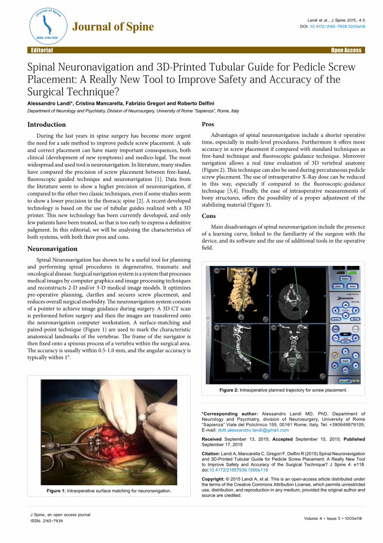
Figure 2: Intraoperative planned trajectory for screw placement.
Pros
Advantages of spinal neuronavigation include a shorter operative time, especially in multi-level procedures. Furthermore it offers more accuracy in screw placement if compared with standard techniques as free-hand technique and fluoroscopic guidance technique. Moreover navigation allows a real time evaluation of 3D vertebral anatomy (Figure 2). This technique can also be used during percutaneous pedicle screw placement. The use of intraoperative X-Ray dose can be reduced in this way, especially if compared to the fluoroscopic-guidance technique [3,4]. Finally, the ease of intraoperative measurements of bony structures, offers the possibility of a proper adjustment of the stabilizing material (Figure 3).
Cons
Main disadvantages of spinal neuronavigation include the presence of a learning curve, linked to the familiarity of the surgeon with the device, and its software and the use of additional tools in the operative field.
Journal of Spine
ISSN: 2165-7939
Journal of Spine

Citation: Landi A, Mancarella C, Gregori F, Delfini R (2015) Spinal Neuronavigation and 3D-Printed Tubular Guide for Pedicle Screw Placement: A Really New Tool to Improve Safety and Accuracy of the Surgical Technique? J Spine 4: e118.doi:10.4172/21657939.1000e118
Page 2 of 3
Volume 4 • Issue 5 • 1000e118J Spine, an open access journalISSN: 2165-7939
D Printed Tubular Guides
They constitute a customized solution for the patient. On the basis of preoperative imaging study, a thin slice CT scan (as for neuronavigation), and the surgeon elaborates the preoperative planning. Angulation and dimensions of the screws can be chosen and visualized in the three axes (axial, coronal and sagittal) to verify the alignment, the convergence, their position into the pedicle and the length of the screws within the vertebral body. After the planning has been accepted, it receives the approval by the surgeon and is elaborated by the owner firm that, in about 20 days, delivers the guides to the hospital. The material delivered for surgery includes: customized tubular guides for each vertebra needing instrumentation and a 3D model of the posterior elements of the same vertebra (Figure 4). The 3D model is useful for the surgeon so that he can visualize the anatomic landmarks where the guides are in contact with the vertebra. Usual anatomic landmarks are the spinous process, the laminae, the pars interarticularis and the transverse processes. The surgical exposure of the anatomical landmarks has to be really accurate, avoiding soft tissue between the bone and the tubular guides, in order to maximize the precision of the system (Figure 5). The technique requires an adequate exposure: the surgical incision should allow the
Figure 3: Postoperative CT scan in patient with neuronavigation-assisted pedicle screw placement.
Figure 4: Tubular guides and vertebra model printed with 3D technology.
Figure 5: Intraoperative placement of the tubular guides.

Citation: Landi A, Mancarella C, Gregori F, Delfini R (2015) Spinal Neuronavigation and 3D-Printed Tubular Guide for Pedicle Screw Placement: A Really New Tool to Improve Safety and Accuracy of the Surgical Technique? J Spine 4: e118.doi:10.4172/21657939.1000e118
Page 3 of 3
Volume 4 • Issue 5 • 1000e118J Spine, an open access journalISSN: 2165-7939
exposition and visualization of all the anatomical landmarks matching with the tubular guides. For this reason the surgical incision needs to be longer if compared to the one needed for a standard intervention: the spinous processes cranial to the first and caudal to the last vertebra to instrument have to be exposed and visualized. The placement of the pedicle screws is assisted by the 3D printed tubular guides and by some adapters, allowing the sequential use of pedicle awl, pedicle probe and finally the screw. At every moment during the procedure, the surgeon can change the dimensions of the screws (length and diameter). Their orientation cannot be modified, but the intervention can be converted in a standard free hand or fluoroscopic guided technique. A system similar to the one described has been developed by Kaneyama [5] and Sugawara[6].
Pros
This new technique offers several advantages. In the first place there is the possibility to plan the surgical intervention and visualize the screws in the three planes (axial, coronal and sagittal). The surgeon can accurately plan the screw dimensions, its relation with the medial and lateral cancellous bone of the pedicle, compare the size of the screw with the dimensions of the pedicle and measure the distance between the tip of the screw and the anterior cancellous bone of the vertebral body (Figure 6). The maximal potential of the technique is expressed in those cases in which the normal anatomic landmarks are altered, as in degenerative deformity. Another advantage is the margin of error in screw placement. The neuronavigation system offers a margin of error that is at least of 0.5 mm. The tubular guides system allows screw placement with a maximal margin of error of 0.5 mm. To reach this precision, no soft tissues must be placed between the tubular guides and the anatomic landmarks. At last, the use of the tubular guides reduces the exposure to ionizing radiations.
Cons
The tubular guides system has three main disadvantages. The first one is the time needed for the 3D print of the guides (about 20 days). This gap makes the system not suitable for trauma surgery (i.e. fractures), but can be used only for elective surgery. The second disadvantage is the use limited to the thoracolumbar spine. Cervical spine is not suitable at the moment. Neuronavigation allows exploring all the segments of the spine. The last disadvantage is that the system does not allow placing screws in percutaneous fashion.
ConclusionsBoth techniques seem to offer some advantages if compared to the
aforementioned standard techniques, in terms of accuracy of the screw placement. This reduces the post-operative morbidity and shortens the length of hospital stay. Furthermore, both techniques reduce the exposure to ionizing radiations, both for the patient and for the staff. The increased precision in screw placement can permit the reduction of medico legal issues through the reduction of the complications related to their misplacement. The expenses for both systems are higher than a surgical intervention performed with a standard technique. Neuronavigation requires a high initial expense that can be written off in time with the number of interventions performed and the multidisciplinary of its use. The expense for the tubular guides is higher than a standard stabilization, due to the expense for the 3D print of the tubular guides and of the vertebral model. Both neuronavigation and tubular guides seems to be a valid alternative to standard technique for a safe screw placement, each one with its own characteristics and its own intrinsic limits.
References
1. Gelalis ID, Paschos NK, Pakos EE, Politis AN, Arnaoutoglou CM, et al. (2012)“Accuracy of pedicle screw placement: a systematic review of prospectivein vivo studies comparing free hand, fluoroscopy guidance and navigation techniques.” Eur Spine J 21: 247-255.
2. Kosmopoulos V, Schizas C (2007) Pedicle screw placement accuracy: a meta-analysis. Spine (Phila. Pa. 1976) 32: E111-E120.
3. Laine T, Schlenzka D, Mäkitalo K, Tallroth K, Nolte LP, et al. (1997) Improvedaccuracy of pedicle screw insertion with computer-assisted surgery. Aprospective clinical trial of 30 patients 22: 1254-1258.
4. Ryang YM, Villard J, Obermüller T, Friedrich B, Wolf P, et al. (2015) Learningcurve of 3D fluoroscopy image-guided pedicle screw placement in the thoracolumbar spine. Spine J 15: 67-476.
5. Kaneyama S, Sugawara T, Sumi M (2015) Safe and accurate midcervicalpedicle screw insertion procedure with the patient-specific screw guide template system. Spine (Phila. Pa. 1976) 40: E341-348.
6. Sugawara T, Higashiyama N, Kaneyama S, Takabatake M, Watanabe N, et al.(2013) Multistep pedicle screw insertion procedure with patient-specific lamina fit-and-lock templates for the thoracic spine. J Neurosurg Spine 19: 185-90.
Figure 6: Preoperative planning and post-operative CT scan of pedicle screws placed with tubular guides technique.