spinal tuberculosis diagnosis and · pdf filespinal tuberculosis diagnosis physical...
TRANSCRIPT

SPINAL
TUBERCULOSIS
DIAGNOSIS &
MANAGEMENT
Presenter:Presenter:Dr. Shailesh Jain
Moderators :Dr. P.S.Chandra
Dr. Rajinder Kumar

INTRODUCTION
• Evidence of spinal TB dates back to Egyptian times and has
been documented in 5000-year-old mummies.
• In 1779, Percival Pott published the first modern description • In 1779, Percival Pott published the first modern description
of spinal deformity and paraplegia resulting from spinal TB.
• According to WHO(2006), about one third of the world’s
population is infected by Mycobacterium TB, and 9 million
individuals develop TB each year.

• One fifth of TB population is in India.
• Three percent are suffering from skeletal TB.
• Vertebral TB is the most common form of skeletal TB and
accounts for 50% of all cases of skeletal TB.
• Almost 50% are from pediatric group. • Almost 50% are from pediatric group.
• Every day 1000 die of tuberculosis in India.
• Neurological complications are the most crippling
complications of spinal TB ( Incidence : 10 to 43%).

SPINAL TUBERCULOSIS
REGIONAL DISTRIBUTION
1 Cervical 12%
2 Cervicodorsal 5%
3 Dorsal 42%3 Dorsal 42%
4 Dorsolumbar 12%
5 Lumbar 26%
6 Lumbosacral 3%

SPINAL TUBERCULOSIS
Pathology• Spinal tuberculosis is usually a secondary infection from a primary
site in the lung or genitourinary system.
• Spread to the spine is hematogenous in most instances.• Spread to the spine is hematogenous in most instances.
• Delayed hypersensitivity immune reaction.
• Initially : a pre-pus inflammatory reaction with Langerhan’s giant
cells, epithelioid cells, and lymphocytes.
• The granulation tissue proliferates, producing thrombosis of vessels.
SPINAL TUBERCULOSIS
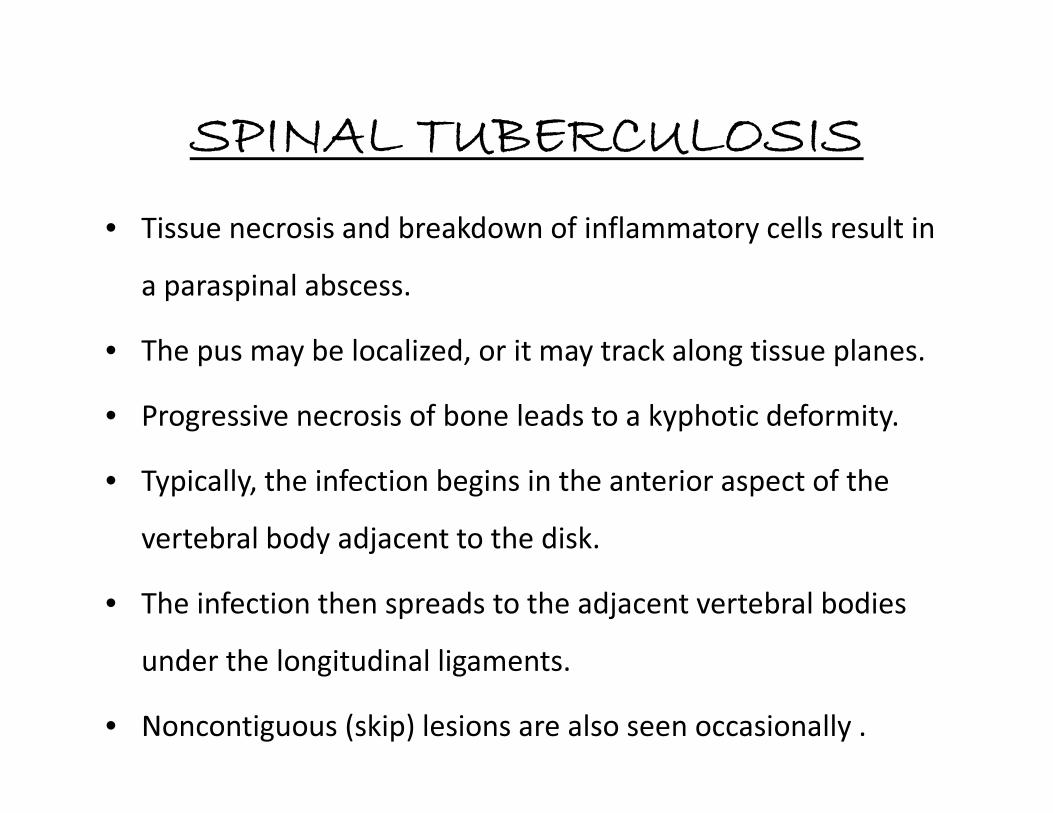
• Tissue necrosis and breakdown of inflammatory cells result in
a paraspinal abscess.
• The pus may be localized, or it may track along tissue planes.
• Progressive necrosis of bone leads to a kyphotic deformity.• Progressive necrosis of bone leads to a kyphotic deformity.
• Typically, the infection begins in the anterior aspect of the
vertebral body adjacent to the disk.
• The infection then spreads to the adjacent vertebral bodies
under the longitudinal ligaments.
• Noncontiguous (skip) lesions are also seen occasionally .

SPINAL TUBERCULOSIS
SIGNS & SYMPTOMS
Spine Neurological Constitutional Local
Spine Deformity
Kyphosis Scolosis
Neurological
Pain
Local / Radicular / Dysesthetic
Motor deficits SpasticitySensory deficits
Bladder involvement
Constitutional Local
Cold abscess / Sinuses

SPINAL TUBERCULOSIS
DIAGNOSIS
� HISTORY
� Presentation depends on :
• Stage of disease,
• Site• Site
• Presence of complications such as neurologic deficits, abscesses, or sinus
tracts.
� Average duration of symptoms at the time of diagnosis is 3 – 4 months.
� Back pain is the earliest & most common symptom.
� Constitutional symptoms.
� Neurologic symptoms (50 % of cases).

SPINAL TUBERCULOSIS
DIAGNOSIS
�Physical examination of the spine :
• Localised tenderness and paravertebral muscle spasm,
• Kyphotic deformity,• Kyphotic deformity,
• Cold abscess swelling / sinus tract
• Cervical spine TB is a less common presentation, characterized
by pain & stiffness with dysphagia / stridor more common in
lower cervical spine involvement

SPINAL TUBERCULOSIS
DIAGNOSIS
LAB STUDIES
• Mantoux / Tuberculin skin test ( purified protein derivative {PPD})
A positive test can be observed, one to 3 months after infection.
Positive in 84 – 95 % of patients who are HIV negative
Negative in almost 20 per cent patients with active disease if the disease is disseminated, or if the Negative in almost 20 per cent patients with active disease if the disease is disseminated, or if the
patient is immunocompromised or suffering from exanthematous fever.
• ESR may be markedly elevated (neither specific nor reliable).
• ELISA : for antibody to mycobacterial antigen-6 , sensitivity of 60 – 80%.
• PCR : sensitivity of 40% only.
• Brucella complement fixation test (useful in endemic areas as brucella can clinically mimic tuberculosis).

SPINAL TUBERCULOSIS
DIAGNOSIS
Microbiology studies to confirm diagnosis :
• Ziehl-Neelsen staining:
a quick and inexpensive method.
• Obtain bone tissue or abscess samples to stain for acid-fast bacilli (AFB), & isolate
organisms for culture & drug susceptibility.
• Culture results are available only after a few weeks.
• Positive only in 50% of cases.

SPINAL TUBERCULOSIS
DIAGNOSIS
LAB STUDIES
• IFN – Release assays (IGRAs)
Recently, two in vitro assays that measure T-cell
release of IFN – in response to stimulation with the highly
specific tuberculosis antigens ESAT- 6 & CFP-10 have become
commercially available.

SPINAL TUBERCULOSIS
DIAGNOSIS
• RADIOLOGICAL DIAGNOSIS
• 1. PLAIN RADIOGRAPH
• 2. CT SCAN• 2. CT SCAN• 3. MRI SPINE• 4.BONE SCAN
TB bacilli are rarely found in CSF, therefore imaging plays pivotal
role in suggesting the diagnosis.

PLAIN RADIOGRAPH
• More than 50% of bone has to be destroyed before a lesion can be seen on X-ray.
This process takes approximately six months.
• The classic roentgen triad in spinal tuberculosis is primary vertebral lesion, disc
space narrowing and paravertebral abscess.
• Typical tubercular spondylitic features in long standing paraspinal abscesses =
a} produce concave erosions around the anterior margins of the vertebral bodiesa} produce concave erosions around the anterior margins of the vertebral bodies
producing a scalloped appearance called the Aneurysmal phenomenon.
b} fusiform paraspinal soft tissue shadow with calcification in few .
• Skip lesions as involvement of non contiguous vertebrae (7 – 10 % cases).
• DEFORMITIES:
1. Anterior wedging
2. Gibbous deformity.
3. Vertebra plana = single collapsed vertebra .


CT SCAN
• Patterns of bony destruction.
• Calcifications in abscess (pathognomic for Tb)
• Regions which are difficult tovisualize on plain films, like :
1. Cranio-vertebral junction (CVJ)
MRI• Lack of ionizing radiation, high
contrast resolution & 3D imaging.
• Detect marrow infiltration invertebral bodies, leading to earlydiagnosis.
• Changes of discitis1. Cranio-vertebral junction (CVJ)
2. Cervico-dorsal region,
3. Sacrum
4. Sacro-iliac joints.
5. Posterior spinal tuberculosis
because lesions less than 1.5cm
are usually missed due to
overlapping of shadows on x rays.
• Assessment of extradural abscesses / subligamentous spread.
• Skip lesions
• Spinal cord involvement.
• Spinal arachanoiditis.

LOCATION OF VERTEBRAL BODY DESTRUCTION
PATTERNS OF VERTEBRAL BODY DESTRUCTION ON CT:

MRI
• MRI is the modality of choice as delineates leptomeningeal
disease better, direct evaluation of intramedullary lesions,
associated osseous signal change and epidural abscesses.
• Typical (spondylo-discitis) and atypical (spondylitis without
discitis) types.

EXTRA-DURAL INVOLVEMENT
Patterns of Vertebral Involvement
• The primary focus of infection in the spine can be
either in the vertebral body or in the posterior either in the vertebral body or in the posterior
elements.
• Four patterns :
� Paradiscal ( Commonest)
� Central
� Anterior, &
� Appendiceal
SPINAL TUBERCULOSIS
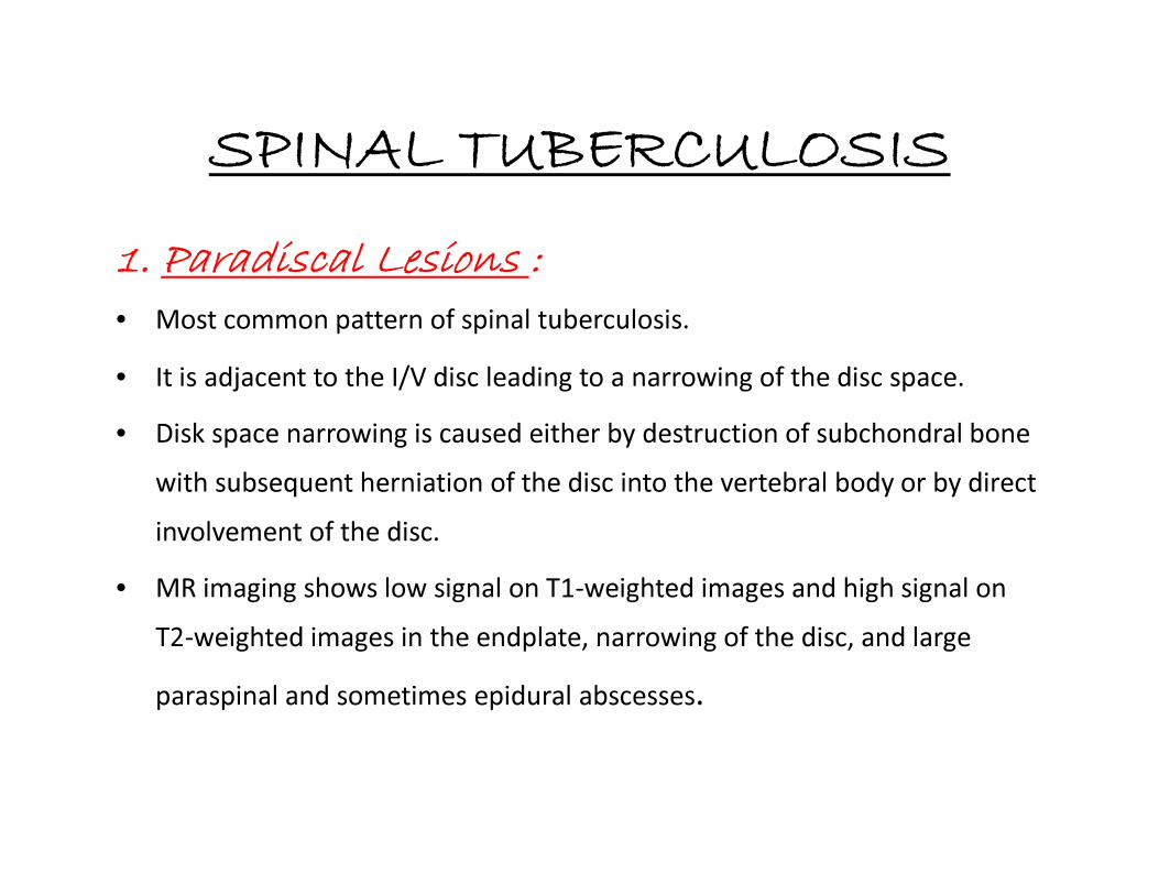
1. Paradiscal Lesions :
• Most common pattern of spinal tuberculosis.
• It is adjacent to the I/V disc leading to a narrowing of the disc space.
• Disk space narrowing is caused either by destruction of subchondral bone • Disk space narrowing is caused either by destruction of subchondral bone
with subsequent herniation of the disc into the vertebral body or by direct
involvement of the disc.
• MR imaging shows low signal on T1-weighted images and high signal on
T2-weighted images in the endplate, narrowing of the disc, and large
paraspinal and sometimes epidural abscesses.

SPINAL TUBERCULOSIS
Paradiscal
Lesions

SPINAL TUBERCULOSIS
2. Anterior Lesions :
• The anterior type is a subperiosteal lesion under the ALL.
• Pus spreads over multiple vertebral segments, stripping the periosteum
and ALL from the anterior surface of the vertebral bodies.
• The periosteal stripping renders the vertebrae avascular and susceptible
to infection.
• Both pressure and ischemia combine to produce anterior scalloping.

SPINAL TUBERCULOSIS
• Collapse of the VB & diminution of the disc space is
usually minimal & occurs late.
• This lesion is relatively more common in thoracic spine in
children.children.
• MR imaging shows the subligamentous abscess,
preservation of the discs, and abnormal signal involving
multiple vertebral segments representing vertebral
tuberculous osteomyelitis.

SPINAL TUBERCULOSIS
Anterior
Lesions
SPINAL TUBERCULOSIS
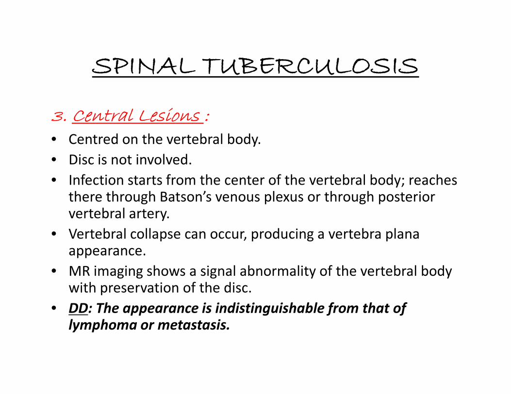
3. Central Lesions :
• Centred on the vertebral body.
• Disc is not involved.
• Infection starts from the center of the vertebral body; reaches there through Batson’s venous plexus or through posterior there through Batson’s venous plexus or through posterior vertebral artery.
• Vertebral collapse can occur, producing a vertebra plana appearance.
• MR imaging shows a signal abnormality of the vertebral body with preservation of the disc.
• DD: The appearance is indistinguishable from that of lymphoma or metastasis.

SPINAL TUBERCULOSIS
Central
Lesions Lesions
SPINAL TUBERCULOSIS
4. Appendicial type :
• Isolated infection of the pedicles & laminae (neural arch),
transverse processes, & spinous process.
• Uncommon lesion (< 5%).
• Occur in isolation or in conjunction with the typical paradiscal
variant
• Radiographically, they appear as erosive lesions, paravertebral
shadows with intact disc space.
• Rarely, present as synovitis of facet joints.

SPINAL TUBERCULOSIS
Appendicial lesion

Paravertebral abscess :
• With collapse of the vertebral body, tuberculous granulation tissue, caseous matter,
and necrotic bone accumulate beneath the anterior longitudinal ligament.
• Gravitate along the fascial planes and present externally at some distance from the
site of the original lesion.
• In lumbar region, along the psoas fascial sheath points into the groin just below the
inguinal ligament.
• In thoracic region, the longitudinal ligaments limit the abscess, which is seen in the
radiogram as a fusiform radiopaque shadow at or just below the level of the
involved vertebra or may reach the anterior chest wall in the parasternal area by
tracking via the intercostal vessels.

LOCATION OF PARAVERTEBRAL ABSCESS
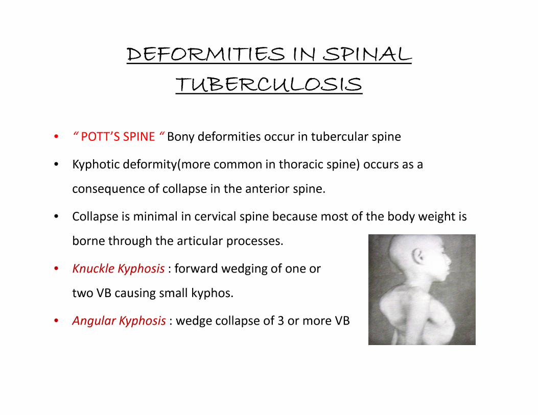
DEFORMITIES IN SPINAL TUBERCULOSIS
• “ POTT’S SPINE “ Bony deformities occur in tubercular spine
• Kyphotic deformity(more common in thoracic spine) occurs as a
consequence of collapse in the anterior spine.
Collapse is minimal in cervical spine because most of the body weight is • Collapse is minimal in cervical spine because most of the body weight is
borne through the articular processes.
• Knuckle Kyphosis : forward wedging of one or
two VB causing small kyphos.
• Angular Kyphosis : wedge collapse of 3 or more VB

Angle of Kyphosis

INTRADURAL EXTRAMEDULLARY INVOLVEMENT
1. Intradural extramedullary tuberculoma • Uncommon form of tuberculosis with only 18 cases have been reported in
literature till date.
• Intradural extramedullary tuberculomas can further be separated into two groups:
1. The first type comprises of hard rounded lesions generally 2 to 3 cm in diameter, having relatively thin membranes often containing granular calcified material, and are generally attached to dura but are relatively easily separated from the underlying cord.
2. The second form is diffuse involvement of subdural space by masses of relatively avascular grayish tubercular granulation tissue.
•
2. Spinal Arachnoiditis
• Frequently involves the spinal cord, meninges and the nerve roots and is more
appropriately referred to as radiculomyelitis (TBRM) .
Should be suspected when patient develops spinal cord symptoms.
• PATHOGENESIS :
1. Hematogenous spread from extra CNS source.
2. Secondary extension of intracranial disease.2. Secondary extension of intracranial disease.
3. Secondary intraspinal extension from tuberculous spondylitis.
Gross granulomatous exudates fill the subarachnoid space.
With time exudates get organized and fibrin coated nerve roots adhere to each
other.
Vasculitis of spinal arteries may cause spinal cord ischemia.

• The MR imaging features of spinal tuberculous arachnoiditis consist of :
1. CSF loculation and obliteration of the spinal subarachnoid space.
2. loss of outline of the spinal cord against CSF in spinal canal on T1.
3. matting of the nerve roots in the lumbar region .
4. Contrast-enhanced imaging reveals nodular, thick, linear intradural4. Contrast-enhanced imaging reveals nodular, thick, linear intradural
enhancement which can completely fill the subarachnoid space,
sometimes giving the appearance of a normal unenhanced MR image .
Key point : when T1 contrast enhanced images look like T2 images .
.


Syringomyelia can occur as a complication of arachnoiditis .
• Early syrinx formation is due to spinal cord ischemia
• Late onset syrinx in chronic arachanoiditis is due to focal scarring of the
subarachnoid space by adhesions which impedes free circulation of CSF
thus forcing CSF into the central canal of the spinal cord via VR spaces
• The differential diagnosis of nodular or diffuse thickening in
spinal canal on MRI :
1. meningeal carcinomatosis.
2. lymphoma.

Intradural intramedullary involvement
• Intramedullary tuberculomas is due to hematogenous spread from otherprimary site in the body.
• Predominantly in the young patients.
• Thoraco-lumbar region is the most common site of involvement.
• MRI shows low or intermediate signal intensity on T1W images and low• MRI shows low or intermediate signal intensity on T1W images and lowsignal on T2W images (Low signal on T2W images is due to caseousnecrosis in the tuberculoma, which has high protein content).
• Post Gadolinium study shows ring enhancement.
• Sometimes associated with changes of myelitis with altered cord signalintensity.


CLINICO-RADIOLOGICAL CLASSIFICATION OF
SPINAL TUBERCULOSIS
STAGE CLINICO- RADIOLOGICAL FEATURE DURATION
1) Pre-destructive straightening of curvatures < 2 months
Spasm of perivertebral muscles
MRI: marrow edema
2) Early destructive decreased disc space+ paradiscal erosion 2-4 months
MRI: marrow edema; break of osseous marginMRI: marrow edema; break of osseous margin
CT: marginal erosion or cavitations
3) Mild angular kyphosis 2-3 vertebrae involvement 4-9 months
(K: 10-30 degree)
4) Moderate angular kyphosis > 3 vertebral involvement 6-24month
(K: 30-60 degree)
5) Severe kyphosis > 3 vertebrae involvement > 2years
(K: > 60 degree)
(K: angle of Kyphosis)

BONE SCAN (Technitium (Tc) – 99 m )
• Increased uptake in up to 60 per cent patients with active
tuberculosis.
• >= 5mm lesion size can be detected.
• Avascular segments and abscesses show a cold spot due to• Avascular segments and abscesses show a cold spot due to
decreased uptake.
• Highly sensitive but nonspecific.
• Aid to localise the site of active disease and to detect
multilevel involvement.

DIFFERENTIAL DIAGNOSIS
The differential diagnosis of the tuberculous spine
includes:
1. SPINAL INFECTIONS- pyogenic, brucella & fungal.2.NEUROPATHIC spine
3.NEOPLASTIC commonly lymphoma/ metastasis
4.DEGENERATIVE
No pathognomonic imaging signs allow tuberculosis to
be readily distinguished from other conditions. Biopsy
is definitive.

DD: PYOGENIC SPONDYLITIS
TUBERCULAR
• Long standing history of months to years
• Presence of active pulmonary tuberculosis -60%
• Most common location thoracic spine followed by thoraco-lumbar region.
PYOGENIC
• History of days to months.
• Not present.
• Most common location lumbar spine.
followed by thoraco-lumbar region.
• > 3 contiguous vertebral body involvement common- 42%.
• Vertebral collapse -67%
• Bone destruction : 73%
• Posterior elements involvement common
• Skip lesions common
• 19% only. Mostly involves 1 spinal
segment – 2vertebrae & intervening
disc.
• 21% only.
• 48%
• Rare
• Rare

TUBERCULAR
• Disc is involved with less frequency
and severity. Disc spared in central
type TB.
• Paraspinal and epidural abscesses-
60%
1. large involving many contiguous
vertebral bodies level.
PYOGENIC
• Disc destruction is most often seen in
pyogenic osteomyelitis.
• 30%
1. Rarevertebral bodies level.
2. calcification if present is pathognomic.
3. Smooth rim enhancement -74%
• TO SUMMARISE: atypical
features + abscess character.
2. Not seen.
3. Heterogenous enhancement. Thick
irregular Rim enhancement only 9%
cases.

• A well-defined paraspinal lesion with abnormal signal intensity; a
thin, smooth ring enhancing abscesswall; subligamentous spread to
three or more vertebral levels;and multiple/ skip vertebral bodies
or entire-body involvement are findings more suggestive of
tuberculous spondylitis than of pyogenic spondylitis.
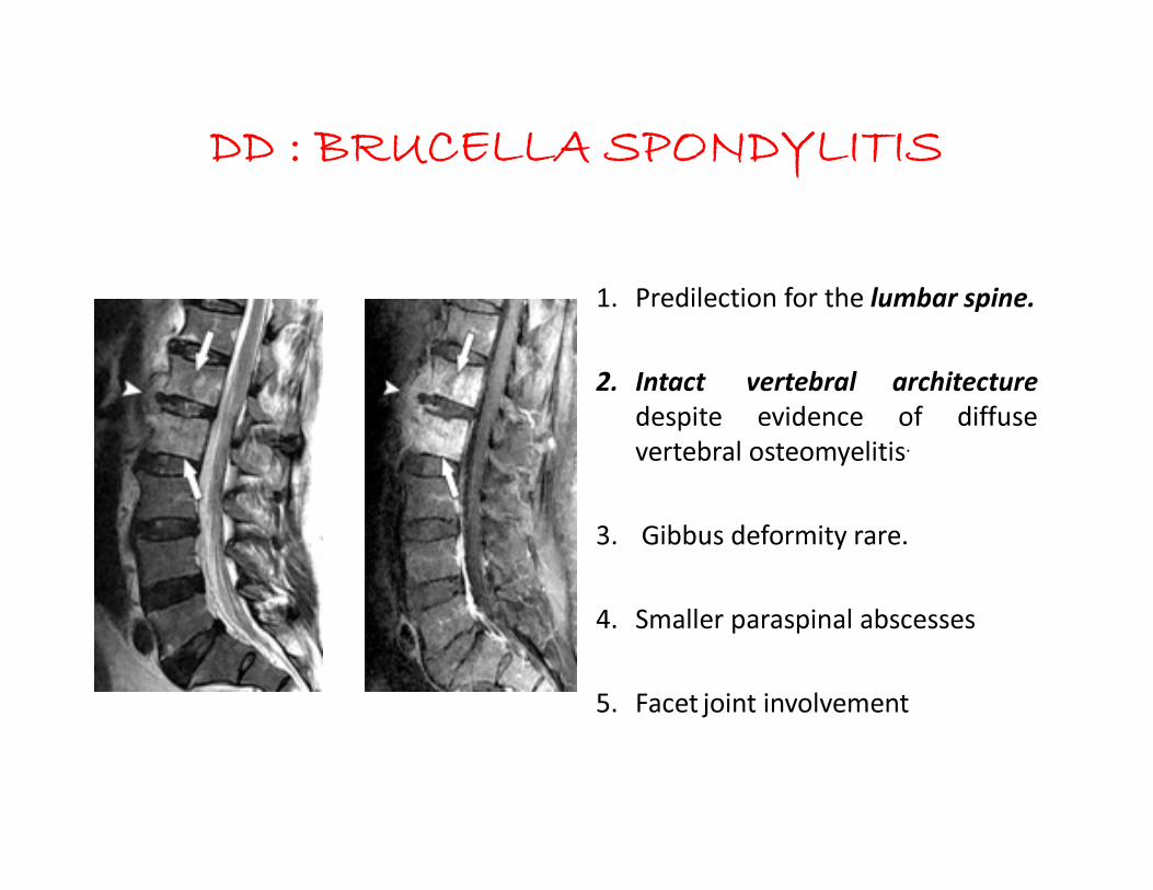
DD : BRUCELLA SPONDYLITIS
1. Predilection for the lumbar spine.
2. Intact vertebral architecture
despite evidence of diffuse
vertebral osteomyelitis.vertebral osteomyelitis.
3. Gibbus deformity rare.
4. Smaller paraspinal abscesses
5. Facet joint involvement
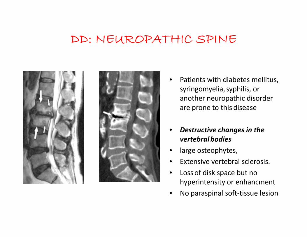
DD: NEUROPATHIC SPINE
• Patients with diabetes mellitus,
syringomyelia, syphilis, or
another neuropathic disorder
are prone to this disease
• Destructive changes in the
vertebral bodies
• large osteophytes,
• Extensive vertebral sclerosis.
• Loss of disk space but no
hyperintensity or enhancment
• No paraspinal soft-tissue lesion

DD: NEOPLASTIC
when 2 contiguous vertebral bodies are involved without intervening disc,
it is difficult to differentiate tubercular spondylitis (central type) from
neoplastic condition.
TUBERCULAR
• A destructive bone lesion
associated with a poorly defined
LYMPHOMA/ METASTASIS
• The saying "good disk, bad news;
bad disk, good news" describesassociated with a poorly defined
vertebral body endplate, with or
without a loss of disk height,
suggests an infection, which has a
better prognosis.
bad disk, good news" describes
the idea that a destructive bone
lesion associated with a well-
preserved disk space with sharp
endplates suggests neoplastic
infiltration.

Most important d/d for this central type tuberculosis is
a neoplasm commonly lymphoma/ metastasis.

COMPLICATIONS OF SPINAL TUBERCULOSIS
• Paraplegia
• Cold abscess
• Spinal deformity• Spinal deformity
• Sinuses
• Secondary infection
• Amyloid disease
• Fatality

TUBERCULOUS SPINE WITH PARAPLEGIA
• Incidence : 10 – 30 %
• Dorsal spine most common
• Motor functions affected before / greater than
sensory.
• Sense of position & vibration last to disappear.

PATHOLOGY OF TUBERCULOUS PARAPLEGIA
• Inflammatory Edema :
Vascular stasis , Toxins.
• Extradural Mass :
Tuberculous osteitis of VB & Abscess.Tuberculous osteitis of VB & Abscess.
• Meningeal Changes : “Dura as a rule not involved”.
Extradural granulation → Contraction / Cicatrization →Peridural fibrosis →
Recurrent Paraplegia

PATHOLOGY OF TUBERCULOUS PARAPLEGIA
• Bony disorders :
Sequestra , Internal Gibbus
• Infarction of Spinal Cord :• Infarction of Spinal Cord :
Endarteritis, Periarteritis or thrombosis of tributary to ASA.
• Changes in Spinal Cord :
Thinning (Atrophy), Myelomalacia & Syrinx.

SEDDON’S CLASSIFICATION OF TUBERCULOUS PARAPLEGIA:
GROUP A (EARLY ONSET PARAPLEGIA) a/k/a Paraplegia associated with activedisease :
• During the active phase of the disease within first 2 years of onset.
• Pathology can be inflammatory edema, granulation tissue, abscess, caseous
material or ischemia of cord.
GROUP B (LATE ONSET PARAPLEGIA) a/k/a Paraplegia associated with healeddisease :
• Usually after 2 years of onset of disease.
• Can be due to recrudescence of the disease or due to mechanical pressure
on the cord.
• Pathology can be sequestra, debris, internal gibbus or stenosis of the canal.

KUMAR’S CLASSIFICATION OF TUBERCULOUS PARA/TETRAPLEGIA (Predominantly based on motor
weakness)
Stage Clinical features
I Negligible Patient unaware of neural deficit,
Plantar extensor and / or ankle clonus
II Mild Patient aware of deficit but manages to walk with
support (Spastic paresis)
III Moderate Nonambulatory because of paralysis (in extension),
sensory deficit less than 50%
IV Severe III + Flexor spasms / paralysis in flexion/sensory deficit
more than 50% / sphincters involved

BASIC PRINCIPLES OF MANAGEMENT
• Early diagnosis
• Expeditious medical treatment
Aggressive surgical approach• Aggressive surgical approach
• Prevent deformity
• Expect good outcome

WHAT IS MIDDLE PATH REGIME ?
• Rest in hard bed
• Chemotherapy
• X-ray & ESR once in 3 months
• MRI/ CT at 6 months interval for 2 years• MRI/ CT at 6 months interval for 2 years
• Gradual mobilization is encouraged in absence of neural
deficits with spinal braces & back extension exercises at 3 – 9
weeks.
• Abscesses – aspirate when near surface & instil 1gm
Streptomycin +/- INH in solution

• Sinus heals 6-12 weeks after treatment.
• Neural complications if showing progressive recovery on ATT
b/w 3-4 weeks :- surgery unnecessary
• Excisional surgery for posterior spinal disease associated with
abscess / sinus formation +/- neural involvement.
• Operative debridement–if no arrest after 3-6 months of ATT /
with recurrence of disease .
• Post op spinal brace → 18 months-2 years

DRUGS IN MIDDLE PATH REGIME
Phase Duration Drugs
Intensive
(for replicating mycobacteria)
5 – 6 months INH + Rifampicin &
ofloxacillin
Continuation 7 – 8 months INH + Pyrazinamide x 3-4 Continuation
(for persisters, slow growing
or dormant or intracellular
mycobacteria)
7 – 8 months INH + Pyrazinamide x 3-4
months f/b
Rifampicin x 4-5
months
Prophylactic 4 – 5 months INH + Ethambutol

VARIOUS REGIMES
• 3(HRZE) / 3(HRZS) + 3(HRZ) + 12(HR)
Pediatric age group, streptomycin (for two
months) replaces ethambutol to avoid optic months) replaces ethambutol to avoid optic
neuropathy .
• 4(HRZE) + 14(HR)

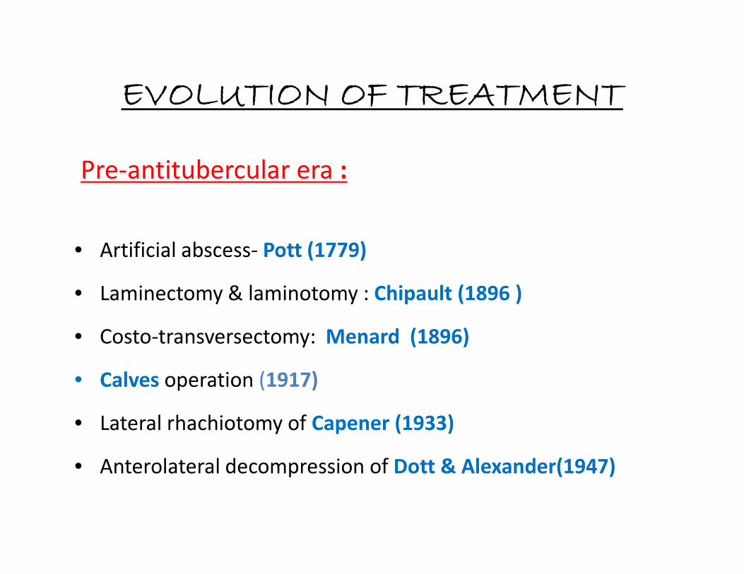
EVOLUTION OF TREATMENT
Pre-antitubercular era :
• Artificial abscess- Pott (1779)
• Laminectomy & laminotomy : Chipault (1896 )• Laminectomy & laminotomy : Chipault (1896 )
• Costo-transversectomy: Menard (1896)
• Calves operation (1917)
• Lateral rhachiotomy of Capener (1933)
• Anterolateral decompression of Dott & Alexander(1947)

SURGICAL INDICATIONS :
• No sign of neurological recovery after trial of 3-4 weeks therapy
• Neurological complications develop during conservative treatment
• Neuro deficit becoming worse on drugs & bed rest
• Recurrence of neurological complication
• Prevertebral cervical abscess with difficulty in deglutition &
respiration
• Advanced cases- Sphincter involvement, flaccid paralysis or severe
flexor spasms

OTHER INDICATIONS :
• Recurrent paraplegia
• Painful paraplegia– d/t root compression, etc
• Posterior spinal disease
• Spinal tumor syndrome resulting in cord compression
• Rapid onset paraplegia (due to thrombosis, etc)
• Doubtful diagnosis & for mechanical instability after healing, &
• Cauda equina paralysis

SURGERY INDICATIONS
1 Decompression (+/- fusion) Too advanced ds, failure to respond to
conservative therapy
2 Debridement + /- decompression +/-
fusion
Recurrence of disease or of neural
complication
3 Anterior transposition of cord
(Extrapleural anterolateral approach)
Severe kyphosis (>60⁰) + neural deficits
4 Laminectomy Extradural granuloma / tuberculoma (STS), Old
healed disease presenting as secondary canal
stenosis / posterior spinal disease
SURGICAL APPROACHES :WORKERS C1-C2 CERVICAL C7-D1 DORSAL DORSO-
LUMBAR
LUMBAR L5-S1
Kirkaldy-Willis
(1965)
- Anterior Transpleural
through bed of
3rd rib
Anterolateral or
transpleural
Anterolateral Retroperitoneal
sympathectomy
or ureter
approach
Transperitoneal,
paramedian
incision in
Trendelenburg
position
Hodgson
(1969)
Transoral /
transthyroi
d
Through
anterior or
posterior∆
Transpleural via
bed of 3rd rib
/split sternal for
extensive lesion
Anterior
transpleural
decompression
Bed of 11th rib
extrapleural
extraperitone
al / left
transpleural
Renal approach Transperitoneal in
Trendelenburg
position. Lower
midline incision
transpleural
via bed of 9th
rib
Kemp et al
(1973)
- Anterior Anterior cervical Trans-sternal for
D3-D4.Anterior
transpleural for
D5-D12
Bed of 12th rib Retroperitoneal
approach
Retroperitoneal
through oblique
renal incision
Smith &
Robinson
(1985)
Anterior Anterior - - - - -
Mc Afee et al
(1987)
Retrophary
ngeal extra-
mucosal
- - - - - -
Tuli et al
(1988)
Transoral
for
drainage
Anterior Low anterior
cervical
Anterolateral or
transpleural
Anterolateral Retroperitoneal
approach
Retroperitoneal or
Retropsoas
transverse
vertebrotomy

Tuli’s recommended approach
• Cervical spine –T1
Anterior approach
• Dorsal spine –DL junction• Dorsal spine –DL junction
Anterolateral approach
• Lumbar spine &Lumbosacral junction
Extraperitoneal Transverse Vertebrotomy

ANTERIOR APPROACH TO THE CERVICAL SPINE (C2 to D1)
Smith & Robinson
• Oblique / transverse incision.
• Plane b/w SCM & carotid sheath laterally & T-O medially.
• Longitudinal incision in ALL open a perivertebral abscess, or
the diseased vertebrae may be exposed by reflecting the ALL
& the longus colli muscles.
Hodgson approach via posterior triangle by retracting SCM,
Carotid sheath, T & O anteriorly & to the opposite side.

SURGICAL APPROACHES TO DORSAL SPINE
• Anterior transpleural transthoracic approach (Hodgson &
Stock, 1956)
• Anterolateral extrapleural approach (Griffiths, Seddon & Roaf,
1956)
• Posterolateral approach (Martin,1970)
{Dura is exposed by hemilaminectomy first & then
extended laterally to remove the posterior ends of 2 – 4
ribs, corresponding transverse processes & the pedicles}.

TRANSTHORACIC TRANSPLEURAL
• Left sided incision preferable
• Incision is made along the rib which in the mid-axillary line, lies
opposite the centre of the lesion (i.e. usually 2 ribs higher than the
centre of the vertebral lesion).
• For severe kyphosis, a rib along the incision line should be removed.
• A J-shaped parascapular incision for C7 – D8 lesions, scapula uplift
& rib resection.
• After cutting the muscles & periosteum, rib is resected
subperiosteally.

• Parietal pleural incision applied & lung should be freed from
the parieties & retracted anteriorly.
• A plane developed b/w the descending aorta & the
paravertebral abscess / diseased vertebral bodies by ligating
the intercostal vessels & branches of hemiazygos veins.the intercostal vessels & branches of hemiazygos veins.
• T-shaped incision over the paravertebral abscess.
• Debridement / decompression with or without bone grafting.

ANTEROLATERAL DECOMPRESSION• Griffith et al -- prone position
• Tuli --- Right lateral position
• Advantage:-
1. avoid venous congestion
2. avoid excessive bleeding
3. permits free respiration3. permits free respiration
4. Lung & mediastinal contents fall anteriorly
• Parts to remove :
• Posterior part of rib (~8cm from the TP)
• Transverse process (TP)
• Pedicle
• Part of the vertebral body
• Semicircular incision
• For severe kyphosis, additional 3-4 transverse processes and
ribs have to be removed.
• Intercostal nerves serve as guide to the intervertebral • Intercostal nerves serve as guide to the intervertebral
foramina & the pedicles.

ANTERO-LATERAL APPROACH TO LUMBAR SPINE ( LUMBO-
VERTEBROTOMY)
• Left side approach
• Semicircular incision
• Expose and remove transverse process
subperiosteally.
• Preserve lumbar nerves

EXTRA PERITONEAL ANTERIOR APPROACH TO LUMBAR SPINE
• 45 ⁰ right lateral position with a bridge centered over the area to beexposed.
• Similar incision as nephroureterectomy or sympathectomy
• Strip peritoneum off posterior abdominal wall and kidney,• Strip peritoneum off posterior abdominal wall and kidney,preserving ureter.
• Longitudinal incision along psoas fibers for abscess drainage
• Retract the sympathetic chain
• Double ligation of lumbar vessels.

EXTRA PERITONEAL APPROACH TO LUMBO-SACRAL REGION
• Left side preferred ( left Common iliac vessels longer
& retracted easily).
• Lazy “S” incision• Lazy “S” incision
• Strip & reflect the parietal peritoneum along with
ureter & spermatic vessels towards right side.

TRANS PERITONEAL HYPOGASTRIC/ SUPRAPUBIC ANTERIOR APPROACH TO
LUMBO-SACRAL REGION
• Supine position
• Midline incision from umbilicus to pubis.
• Lumbo-sacral region identified distal to aortic bifurcation and left• Lumbo-sacral region identified distal to aortic bifurcation and leftcommon iliac vein.
• Longitudinal incision on parietal peritoneum over lumbo-sacralregion in midline.
• Avoid injury to sacral nerve & artery and sympathetic ganglion.

POSTERIOR SPINAL ARTHRODESIS
• By– Albee & Hibbs
• Albee– Tibial graft inserted longitudinally in to the split
spinous processes across the diseased site.
• Hibbs– overlapping numerous small osseous flaps from
contiguous laminae , spinous processes & articular facetscontiguous laminae , spinous processes & articular facets
• Indications–
1. Mechanical instability of spine in otherwise healed disease.
2. To stabilize the craniovertebral region (in certain cases of T.B.)
3. As a part of panvertebral operation

SURGERY IN SEVERE KYPHOSIS
• HIGH RISK PATIENTS:
• Patients < 10 years
• Dorsal lesions
• Involvement of >= 3 vertebrae
• Severe deformity in presence of active disease, especially in children is• Severe deformity in presence of active disease, especially in children isan absolute indication for decompression , correction and stabilization.
• Staged operations-
1. Anteriorly at the site of disease,
2. Osteotomy of the posterior elements at the deformity &
3. Halopelvic or halofemoral tractions post-operatively.

TREATMENT OF PARAPLEGIA IN SEVERE KHYPHOSIS
• Griffiths et al (1956) :anterior transposition of cord through
laminectomy
• Rajasekaran (2002): posterior stabilization f/b anterior • Rajasekaran (2002): posterior stabilization f/b anterior
debridement and bone grafting ( titanium cages) in active
stage of disease and vice versa for healed disease.
• Antero-lateral (Preferred approach) .

SURGICAL CORRECTION OF SEVERE KYPHOTIC DEFORMITY
• Fundamentals of correction:
1. to perform an osteotomy on the concave side of the curve and wedge it open ( secured with strong autogenous iliac grafts) .grafts) .
2. to remove a wedge on the convex side and close this wedge ( Harrington compression rods and hooks)

CLINICAL FACTORS INFLUENCING PROGNOSIS IN CORD INVOLVEMENT
CORD INVOLVEMENT BETTER PROGNOSIS RELATIVELY POOR PROGNOSIS
Degree Partial Complete (stage IV)
Duration Shorter Longer (>12 months)
Speed of onset Slow Rapid
Age Younger OlderAge Younger Older
General condition Good Poor
Vertebral disease Active Healed
Kyphotic deformity <60⁰ >60⁰
Cord on MRI Normal Myelomalacia / Syrinx
Peroperative Wet lesion Dry lesion

CVJ TUBERCULOSIS
• Less than 1% of all spinal tuberculosis.
• Young patients (14 to 65 years), F: M = 2:1.
• Infection from primary sites (paranasal sinuses, nasopharyngeal or
retropharyngeal lymph nodes) spreads retrograde via lymphatic route.
PRESENTATION:
2 months to 2 years to produce symptoms
– Cervico-medullary compression,
– Cranial nerve deficits
– Atlanto-axial instability,
– Abscess formation

? Tuberculosis of retropharyngeal space
Infiltration
Atlanto-axial ligaments +/-
Hyperemia
Bony architecture AAD Tuberculous
granulation tissuegranulation tissue
Compression Cold Abscess
Gross bony destruction
& angulation

DIAGNOSIS:
X-ray / CT Scan / MRI :
• Destruction of lateral masses,
• Secondary atlanto-axial subluxation
• Basilar invagination,
• Adjacent bony destruction,
• Increase in the pre-vertebral shadow/ prevertebral enhancing soft tissue• Increase in the pre-vertebral shadow/ prevertebral enhancing soft tissue
mass.
• Spinal cord signal changes / compression.

STAGES OF CVJ TUBERCULOSIS (Lifeso et al)
STAGES LIGAMENTS BONY
DESTRUCTION
C1-C2 INVOLVEMENT TREATMENT
I Intact Minimal No e/o anterior displacement
of C1 on C2
Cervico-thoracic
orthosis until stability
II Disruption Minimal Anterior displacement of C1
on C2 +/- proximal
translocation of odontoid
Halo traction for
normal alignment, f/b
posterior fusion &
Halo vest
immobilization
III Marked
disruption
Marked Complete obliteration of
anterior arch of C1 with
complete loss of odontoid
process, marked AAD / A-O
instability
Halo traction for
alignment f/b
posterior fusion , halo
vest immobilization
until stability

TREATMENT
CONSERVATIVE :– Absolute bed rest
– Cervical traction for unstable spine
– Prolonged external immobilization
– ATT X 18 months
SURGERY :
� Gross bony destruction with instability
� Abscess formation
� Severe or progressive neurological deficit
� Unstable spine following conservative therapy(failed
therapy)
� Doubtful diagnosis (esp. with neoplasm)