spine surgery in new york
DESCRIPTION
Executive Spine Surgery in Manhattan NY offers cost effective treatment for back pain and spinal injuries.TRANSCRIPT
SPINE SURGERY A Patient’s Guide
-An illustrated guide to spine
diseases and treatments
By Carl J. Spivak, MD
Illustrated By Ginger
Weatherford

Library of Congress Control Number:
ISBN
First printing : 1,000 copies : January 2007
Published by Carl Spivak
Copyright © 2007 Carl J. Spivak
All Rights Reserved.
All rights reserved. No part of this book may be reproduced in any form, except for the inclusion of brief quotations in a review, without permission in writing from the author or publisher.. Important Note: Medical knowledge is ever-changing. As new research and clinical experience broaden our knowledge, changes in treatment and drug therapy may be required. The author of the material herein have consulted sources believed to be reliable in their efforts to provide information that is complete and in accord with the standards accepted at the time of publica-tion. However, in the view of the possibility of human error by the author, of the work herein, or changes in medical knowledge, the author, or any other party that has been involved in the preparation of this work, warrants that the information contained herein is in every respect accu-rate or complete, and they are not responsible for any errors or omissions or for the results ob-tained from use of such information. Readers are encouraged to confirmed herein with other sources. For example, readers are advised to check the product information sheet included in the package of each drug they plan to administer or use to be certain that the information contained in this publication is accurate and that changes have not been made in the recommended dose or in the contraindications for administration. Some of the product names, patents and registered designs referred to in this book are in fact registered trademarks or proprietary names even though specific reference to this fact is not always made in the text. Therefore, the appearance of a name without designation as proprietary is not to be construed as a representation by the author that it is in the public domain.

This book is dedicated to my wife Bridgette and three children Chanelle, Luke and Nicholas, who are a constant source of blessing, inspiration, encouragement and joy to my life.
Dedication
INTRODUCTION
1.Preface……………………………………………….i. 2.Introduction…………………………………………..ii
NECK DISEASE
3.Cervical disk herniation. (slipped disk neck)..………1 4.Cervical spine stenosis (tightness)……..…………….14
MIIDLE BACK DISEASE
5.Spinal compression fractures (broken back bone)……28
LOW BACK DISEASE
6. Lumbar disk herniation (slipped disk) ……………….40 7. Lumbar spinal stenosis (tightness)…………………....52 8..Lumbar spondylolithesis (slipped spine)……..……….64 9. Glossary……………………………………………….79
TABLE OF CONTENTS

Today there is an enormous amount of medical information available in textbooks, medical journals and on the internet. This information ranges from simple to very complex. Most information is written for medical specialists and may be wordy, full of medical jargon, difficult to understand and ex-pensive to obtain. These obstacles are difficult for patients to overcome when trying to understand their disease and treatment options. This book was written to help fill that gap. This book contains valuable medical information, diagrams, X-rays, CT and MRI images to help understand neck and back problems. It is comprehensive, yet easy to understand. It re-views anatomy (the structure of the spine), pathology (spine disease), treatments and outcomes of treatments. This book is best used to educate yourself about a medical disease. It will give you greater breadth of understanding of the problem and will help you make an informed decision. The more information you have the better decision you will make!
PREFACE
i

INTRODUCTION
Neck and back problems are very common medical problems in North American society. Most people will be troubled by neck and back pain during their lifetime. The pain may range from mild to severe. Many times this pain may resolve with rest, activity modification or anti-inflammatory medications and the passage of time. Neck and back problems may become more severe and be associated with dysfunction of the nervous system. This nerv-ous system dysfunction may present itself with pain, numbness, tingling or weakness of the arms or legs. It may also present with problems controlling bowel and bladder function with in-continence or severe constipation. In the following chapters I provide detailed, but easy to un-derstand information about spine disease. Each chapter de-scribes the relevant spinal anatomy, disease process, clinical and radiological investigations, treatments, outcomes and com-plications. I have also included many drawings, X-Rays, CT and MRI scans to help increase your understanding of this complex subject. I hope you find this book interesting and useful to you.
ii

Chapter 1: Cervical Disk Herniation and Anterior Cervical Diskectomy, Fusion and Plating What is the spine? The neck is made up of seven bones called cervical vertebrae (figure 1). These vertebrae surround and protect the spinal cord from damage. The front of the vertebrae is made up of a square shaped vertebral body and the back of the vertebrae is made up of pedicles, facet joints, lateral mass and lamina, called the pos-terior elements. The vertebrae are held together by a spongy disk in the front and strong ligaments in the front and back. The disk helps cushion the neck bones.
Figure 1. Normal cervical spine. A) Side view of cervical ver-tebrae C3-C7. B) Top view of a vertebrae on the right.
Disk
Nerve Root
Spinal Cord
C3
C4
C5
C6
C7 Spinal Cord
A. B.
Vertebrae
Disk
1

What is a cervical disk? Cervical disks are located in between the spinal vertebrae and are made up of a tough outer shell (annulus fibrosis) and a soft gel-like center (nucleus pulposus). Their structure is similar to a jelly donut.
Why do cervical disks pinch the spinal cord and nerves? As people age the spine slowly wears out through a process called degeneration. Degeneration is first seen in the nucleus pulposus (“jelly center”) and annulus fibrosis (“donut”) in adults. The annulus may weaken and bulge outward or tear, al-lowing the nucleus pulposus to squeeze (herniate) out of the annulus into the spinal canal (figure 2)1. This disease is referred to by many names including slipped disk, bulging disk, ruptured disk, pinched nerve, herniated nu-cleus pulposus and disk herniation. The herniated disk may compress or “pinch off” the spinal nerves resulting in neck and arm pain, numbness, tingling and weakness. This pain may be worsened or maintained by inflam-mation around the nerve roots2,3. Compression of the spinal cord may cause myelopathy (spinal cord injury) producing elec-trical shocks down the spine, weakness, numbness and bladder incontinence. Possible risk factors for ruptured disks are hereditary, smok-ing, heavy work, injury to head or neck, heavy lifting or opera-tion of vehicles4,5,6. Sometimes the process begins after a memorable accident.
2
Chapter 1: Cervical Disk Herniation

Figure 2. Herniated cervical disk compressing the spinal cord with evidence of spinal cord compression and swelling. A) Side view of the herniated disk highlighted in black. B) top view of the herniated disk (large black arrow) compressing the spinal cord.
How do I know I have a ruptured disk? The ruptured disk usually begins with neck and shoulder pain followed later with the development of arm pain1. This arm pain may be associated with numbness (loss of feeling in the arm or hand), tingling, burning and weakness. The neck and arm pain may be worsened by neck movements and relieved by placing their hand on their head. Rarely, a herniated disk may cause electrical shocks running down the back or clumsiness, weakness or paralysis in the arms or legs or loss of bowel and-bladder control. These changes may come on quickly or gradu-ally over time, with slow transition to using cane, walker and finally wheelchair. If any of these symptoms occur the patient should seek emergency medical care.
A. B.
Spinal cord swelling
3

What should I do? If you are experiencing neck pain associated with pain, numbness or weakness in your arm, unsteadiness on your feet or have bowel and bladder difficulties you should see your doc-tor. You will require a history and physical examination, which may include an examination of you neck, gait, strength, sensa-tions and reflexes. After an initial assessment, you may require radiological in-vestigations, including x-rays and magnetic resonance imaging (MRI) of the spine. MRI is the best test for looking for herni-
Figure 3. MRI (T2WI) of the neck showing a C4/5 herniated disk compressing the spinal cord and exiting nerve root. A) The side view. The white fluid (cerebral spinal fluid) at the level of the disk. B) top view of the herniated disk.
Spinal cord
Herniated disk
Spinal cord
Herni-ated disk
A. B.
4
Chapter 1: Cervical Disk Herniation

Patients with pacemakers, spinal cord stimulators or other metal within their body are unable to have an MRI. These pa-tients should undergo a computer tomogram (CT) with or with-out a myelogram. The CT myelogram produces better images then the CT alone. A myelogram is the injection of contrast medium (dye) into spinal canal to improve the visibility of the nerves on the CT (figure 4).
Figure 4. CT myelogram of the neck showing a herniated disk (arrows) top view on the right. Compliments Dr. A. Eisenberg.
Should I have surgery? Many patients with neck and arm pain secondary to a rup-tured disk may improve with non-operative treatments includ-ing bed rest, physical therapy, head traction, neck collar, non steroidal anti-inflammatory drugs (NSAIDS), steroids (Medrol dose pack), muscle relaxants, antidepressants and steroid injec-tions7,8.
Disk herniation
Contrast (Dye)
Spinal cord
5
Patients should consider surgery if they fail to improve with conservative therapy, have severe pain or significant neurologi-cal dysfunction (table 1). Additionally, patients with significant spinal cord compression and swelling on MRI scan may require surgery.
Table 1 Indications for Surgery 1. Failure of conservative treatment
2. Severe pain
3. Weakness
4. Loss of bowel & bladder control
5. Compression and swelling of spinal cord
What are the surgical treatment op-tions? Neck and arm pain are a result of the herniated disk com-pressing the spinal nerve. To relieve the pain surgery must re-move the herniated disk and take pressure off the nerve. The removal of disk is called a discectomy. Today most cervical herniated disks are removed through the front of the neck by an operation called an anterior cervical discectomy, fusion and plating (ACDFP). Cervical herniated disks are less commonly removed through the back of the neck by a posterior lami-notomy (parial removal of the lamina which is part of the back of the spine) and diskectomy.
6
Chapter 1: Cervical Disk Herniation

How is a discectomy done? The patient is given antibiotics prior to surgery. They are then taken to the operating room and are put to sleep under a general anesthetic. A tube is placed down their throat to help them breath. They lie down on their back looking up at the ceil-ing. Their neck is washed and sterile drapes are placed around the operative site. An incision is made just off midline, usually on the right side. The skin is separated, the esophagus (food “pipe”), larynx and trachea (voice box and breathing “tube”) and carotid artery (supplies the brain with blood) are retracted to the side. This opens up a tunnel to the front of the spine. The level of the her-niated disk is found with the x-ray machine. The operating room microscope is used to magnify and light the disk space (figure 5). The disk is incised with a knife and removed with a variety of biting and scraping instruments. Af-ter the disk is removed, the posterior longitudinal ligament is opened up. This ligament separates the disk from the spinal canal. Removal of this ligament provides direct visualization of the thecal sac (with contains the spinal cord), the exiting nerve roots and herniated disk. The disk is then carefully removed re-lieving pressure off of the spinal cord and/or nerve roots. After the disk is removed, the ends of the vertebrae are clean and prepared for the bone fusion. A bone fusion is when two bone heal solidly together. A graft is placed into the empty disk space (figure 5). The graft holds the vertebrae apart and the ver-tebrae eventually fuse together through the graft. This de-creases movement across the abnormal vertebrae and helps re-duce pain. This graft may be made from the patient’s bone (autograft) and is usually taken from the hip, or may be taken from the bone bank (allograft). Recently cages made out of plastic (polyetheretherkeytone, PEEK), metal (titanium) and
7

Figure 5. Anterior cervical diskectomy, fusion and plate sur-gery (ACDF). A&B) The disk is removed through the front of the spine relieving pressure off the spinal cord. C) After the disk is removed, the vertebrae are prepared for the fusion and the bone graft is inserted. D) The bone graft is held into position by the metal plate and screws.
A. B.
C. D.
8
Chapter 1: Cervical Disk Herniation

carbon fiber have become available. These cages are presently filled with autograft or allograft but bone morphogenic protein (BMP) , a bone hormone which promotes bone growth in the body, will soon be available. After the graft or cage has been placed into the disk space, the spine is stabilized with a metal plate and screws (figure 5&6). The size of the plate and screws depends on the number of disks removed and the size of the patient’s vertebrae. The metal plate is made of titanium which produces minimal interference on MRI. The titanium plate does not trigger airport metal detectors. Plastic plated may soon be available. After the plate is secured, bleeding is stopped and the muscle and skin are brought to-gether with sutures. Sometimes a drain may be temporary placed to remove blood over night. Patients are woke up and
Figure 6. An x-ray of the cervical spine with plate and screws placed after an anterior cervical discectomy and fusion of C5/6 disk in (A) and C4-5-6-7 in (B). Cervical plates are labeled by white arrows.
A. B.
9

Does surgery work? Surgery is very effective for treatment of neck and arm pain9,10,11. Approximately 75-90% of patients will have good pain relief after surgery. The resolution of numbness, weak-ness and bowel and bladder function is less consistent. After surgery there is a 3% chance of developing another disk herni-ation per year12. There is a low risk of complications (2%)11 but complications may potentially include: death, stroke, heart at-tack, weakness/paralysis, loss of bowel and bladder function, infection, clots in legs (deep venous thrombosis), clots in the lungs (pulmonary embolus), blood vessel injury, failure of fu-sion, breakage of screws and plates, movement of cage/graft, difficulty swallowing, hoarse voice, but not limited to these complications. Neck pain, numbness and difficulty swallowing or speaking are the most common complaints after surgery and they usually resolve. When can I go home? Most people are discharged home after surgery. Some patients are kept over night. Dissolvable stitches are used to close the wound and do not require removal. Sometimes non-dissolvable stitches or staples are used and must be removed. Please ask your doctor prior to discharge. What if I have neck pain or arm pain af-ter surgery? It is normal to have neck pain and soreness from the operation for few weeks. It is also normal to have pain, numbness and tingling that comes and goes after surgery. You should contact your doctor right away if you develop difficulty breathing, neck
10
Chapter 1: Cervical Disk Herniation
What are my limitations after surgery? Please remove the dressing over your incision the day after surgery and wash your incision in the shower. You may use soap and water. Do not rub your incision. Please do not sub-merge your incision in the bath tub for 2 weeks after surgery. Soaking in dirty bath water may increase your risk of infection. Get plenty of rest after surgery. Avoid driving, bending, ex-tending and twisting of your neck. Most people can return to work 3 to 12 weeks after surgery. Please discuss your specific limitations with your doctor. Should I use a bone stimulator? Bone stimulators have been found to improve bone healing rates by stimulation of bone cells by electrical fields. They are especially useful in patients who are at high risk of malunion (the bones not healing together). Patients who should consider a bone stimulator include multi-level fusion surgery, revision sur-gery for failed bone fusion, smokers and patients with osteopo-rosis, diabetes and metabolic bone disease. If the fusion does not heal this may result in multiple neurological and medial problems and may require further surgery. The Orthofix, Inc. bone stimulator has been found to improve fusion rates by 15%, from 69% to 84% in high risk patients13.
11

Discharge Instructions 1. Strict control of sugar levels in patients with diabetes. Poorly
controlled sugar levels may increase risk of infection.
2. Do not smoke or use non-steroidal anti-inflammatory drugs.
They may interfere with bone fusion.
3. Keep wound clean and dry. Please shower the day after sur-
gery. Do not submerge your wound in the bath for 2 weeks.
4. Use neck collar or bone stimulator as directed by your doc-
tor.
5. No driving, twisting, bending neck for up to 1 month after
surgery.
6. Watch for the development neck swelling, difficult breath-
ing, problems swallowing, change in your voice, fever, red-
ness or drainage from the wound.
7. Pain, numbness and weakness often require days to months
to resolve.
8. Call your doctor if you have any concerns.
12
Chapter 1: Cervical Disk Herniation

References 1. Connell Md, Wiesel SW. Natural history and pathogenesis of cervical
disk disease. Orthop Clin North Am. 1992 Aug;13(4):345-9. 2. Omarker K, Meyers RR. Pathogenesis of sciatic pain: role of herniated
nucleus pulposus and deformation of spinal nerve root and dorsal root ganglion. Pain 1998 Nov; 78(2):99-105.
3. Hou SX, Tang JG, Chen HS, Chen J. Chronic inflammation and com-pression of the dorsal root contributing to sciatica induced by the in-tervertebral disc herniation in rats. Pain 2003 Sep;105(1-2):255-64.
4. Sambrook PN, MacGregor AJ, Spector TD. Genetic influences on cervi-cal and lumber disk degeneration: a magnetic resonance study in twins. Arthritis Rheum. 1999; 42(2):366-72.
5. Irvine DH, Foster JB, Newel DJ, et al. Prevalence of cervical spondylo-sis in a general practice. Lancet 1965; 1: 1089-1092.
6. Kelsey JL, Githens PB, Walter SD et al. An epidemiological study of acute prolapsed cervical intervertebral disc. J Bone Joint Surg AM. 1984 Jul; 66(6):907-14.
7. Tan JC, Nordin M: Role of physical therapy in the treatment of cervical disk disease. Orthop Clin N AM 23:435-449, 1992.
8. Rexhtine GR: Nonsurgical treatment of cervical degenerative disease. Inst Course Lect. 1999; 48:433-5.
9. Whitecloud TS, Werner J. Cervical spondylosis and disk herniation: The anterior approach. In Frymoyer J, (ed): The Adult Spine: Principles and Practice, 2nd ed. Philadelphia, Lippincott-Raven, 1997, pp 1357-1379.
10. Sampath P, Bendebba M, Davis JD, Ducker T: Outcome in patients with cervical radiculopathy: Prospective, multicenter study with independent clinical review. Spine 24:591-597, 1999.
11. Bruneau M, Nisolle JF, Gillard C, Gustin T. Anterior cervical interbody fusion with hyroxyapatite graft and plate system. Neurosurg Focus 10 (4): Article 8, 2001.
12. Hillibrand AS, Carlson GD, Palumbo MA, et al: Radiculopathy and myelopathy at segments adjacent to the site of a previous anterior cervi-cal arthrodesis. J Bone Joint Surg Am 81:519-528, 1999.
13. Orthofix, Inc. PMA Randomized, Prospective Clinical Trial of Pulsed Electromagnetic Field Stimulation for Cervical Fusion, 2004 (unpublished).
13
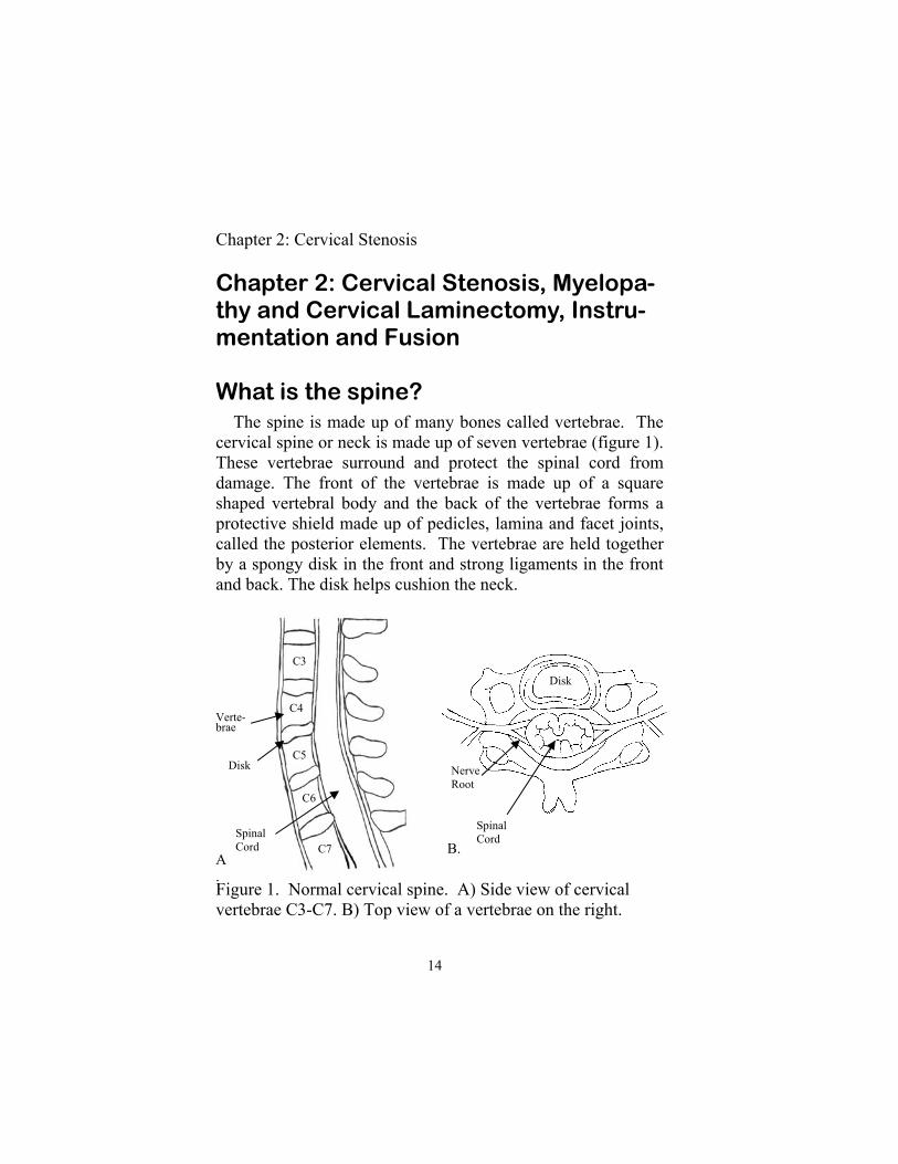
Chapter 2: Cervical Stenosis, Myelopa-thy and Cervical Laminectomy, Instru-mentation and Fusion What is the spine? The spine is made up of many bones called vertebrae. The cervical spine or neck is made up of seven vertebrae (figure 1). These vertebrae surround and protect the spinal cord from damage. The front of the vertebrae is made up of a square shaped vertebral body and the back of the vertebrae forms a protective shield made up of pedicles, lamina and facet joints, called the posterior elements. The vertebrae are held together by a spongy disk in the front and strong ligaments in the front and back. The disk helps cushion the neck.
Figure 1. Normal cervical spine. A) Side view of cervical vertebrae C3-C7. B) Top view of a vertebrae on the right.
Disk
Nerve Root
Spinal Cord
C3
C4
C5
C6
C7 Spinal Cord
A.
B.
Verte-brae
Disk
14
Chapter 2: Cervical Stenosis

What is cervical stenosis? The spine contains a central cavity called the spinal canal. The spinal cord and spinal nerves are found inside this canal. Normally the spinal canal is wide open and does not impinge upon the cord. Degenerative (“wear and tear arthritis”) may cause narrowing or tightening of the canal. This small canal is called cervical stenosis. This often affects people who were al-ready born with a small canal (congenital spinal stenosis). How does cervical stenosis form? As people age, the neck begins to “wear out”. This begins with the drying out and collapse of the cervical disks1,2. This collapsed disk changes the forces across the spine and results in abnormal motion. To stop this motion the body strengthens the neck by thickening the spinal ligaments and stabilizing the mo-bile joints with bone spurs. This is especially seen behind the vertebral bodies, around the facet joints located at the side of the spine and in the ligamentum flavum at the back of the spinal canal. These changes lead to decrease in the size of the spinal canal and may result in spinal cord compression (figure 2). Spinal stenosis may result in spinal cord injury and dysfunc-tion. This may be due to compression from the thickened liga-ments and bone spurs, abnormal spinal motion or from interrup-tion of the blood supply1,3,4. Spinal cord dysfunction (myelopathy) commonly presents in middle-age or elderly peo-ple with clumsy hands and difficulty walking.
15

Figure 2. Cervical stenosis is caused by bone spurs and thick-ened ligmaments. It may cause nerve root and spinal cord com-pression. A) Side view and B) top view.
Do I have spinal cord dysfunction? Spinal cord dysfunction secondary to spinal stenosis in the neck is called cervical spondylotic myelopathy. This is the most common cause of spinal cord dysfunction in people over 55 years old in North America5. Spinal cord dysfunction usually begins in middle-age to elderly people. It usually progresses slowly over many years, but may have a rapid progression with disabling neurological dysfunction.. Spinal cord compression (cervical myelopathy) may be diffi-cult to diagnose because of the variable symptomatology. It may range from mild dysfunction with numbness in the hands to complete paralysis of the arms and legs. The symptoms de-pend upon the level of spinal cord affected, the location in the spinal cord and involvement of spinal nerves. The spinal cord begins at the brain and runs down to the middle of the back to
Thickened ligaments
Bone spurs
Bone spur & disk bulge
Thickened ligaments
A. B.
16
Chapter 2: Cervical Stenosis

approximately the first lumbar vertebrae (L1). Each part of the spinal cord has specific functions. Injury to the spinal cord af-fects the function of the spinal cord below it. Injury to the spinal cord in the neck may cause weakness or paralysis of the arms and legs. While injury in the upper back only affects the legs. Similarly to the vertical arrangement of function, there is also right to left and front to back arrangement of function. Injury to the right side of the spinal cord may cause weakness on the right side of the body and numbness of the left side of the body (called the Brown Secord Syndrome). Lastly, the involvement of spinal nerves cause characteristic syndromes of pain, numb-ness and weakness and are clinically distinct from spinal cord compression6. Cervical spinal cord compression most commonly presents with poor hand coordination and a stiff, unsteady gait. Patients may or may not have neck pain. The spinal cord compression may increase with head movements (particularly bending the head forward) resulting in electrical sensations shooting down the back (Lhermite’s sign) or weakness or paralysis of an ex-tremity. The patient may notice numbness, tingling, weakness in their arms and occasionally pain. It maybe difficult to per-form activities of daily living like buttoning a shirts latching a bra or using fork and knife or chopsticks6. Walking may be-come difficult secondary to weakness, numbness or stiffness in the legs. At times people may decline in a step wise course from walking independently to using a cane, walker and finally require a wheelchair. Bowel and bladder function are less often affected. Bladder urgency is the most common presentation of bladder dysfunction. Patients who experience bladder urgency develop a sudden need to empty their bladder. If they can not immediately void they may wet their pants.
17

What should I do? If you are experiencing neck or arm pain, numbness, weak-ness or bowel and bladder dysfunction you should see your doc-tor. You will require a thorough history and physical examina-tion, which may include examination of your neck, gait, strength, sensations and reflexes. After an initial assessment, you may require radiological in-vestigations, including x-rays and magnetic resonance imaging (MRI) of the cervical spine. MRI is the best test for looking for spinal cord compression (figure 3). Patients with pacemakers, spinal cord stimulators or other metal within their body are unable to have an MRI. These pa-tients
Figure 3. MRI scan of the neck demonstrating severe cervical stenosis (tightness), spinal cord compression and spinal cord swelling (arrow). Spinal cord swelling is white on T2WI MRI. A) Side view and B) top view.
A. B.
18
Chapter 2: Cervical Stenosis

Should I have surgery? Most patients with spinal cord dysfunction should consider surgery, especially if it is new or progressively worsening. A small amount of people may improve without surgery7,8. This is presently an area of research. Other indications for surgery may include pain or severe spinal stenosis with evidence of spi-nal cord injury on MRI.
should undergo a computer tomogram (CT) with or without a myelogram. The CT myelogram produces better images then the CT alone. A myelogram is the injection of contrast medium (dye) into spinal canal to improve the visibility of the nerves on the CT (figure 4).
Figure 4. CT of the neck showing severe spinal stenosis, abnor-mal bending (kyphosis) and slipping of the C4 on C5 vertebral bodies. A) Side view and B) top view.
Spinal stenosis
Kyphoisis & slipping of spine
A B.
Small spinal canal
19

What are the surgical treatment op-tions? Surgery for spinal stenosis may be done through the front of the neck (anterior) or the back of the neck (posterior) or a com-bination of both (anterior-posterior or 360 degree operation). There are many factors considered when deciding if surgery is the best treatment and what type of surgery would best treat the patient. These include the location of the disease in the spine (front, back or both), the stability of the spine (the ability of the spine to keep the spine aligned and prevent abnormal move-ments) and the extent of spinal disease. The patient’s age, medical health and neurological disability are also taken into consideration. Anterior surgery is done through an incision in the front of the neck. It usually involves removal of one or more cervical disks to take pressure off of the spinal cord or nerves. After the disk is removed the disk space is filled with a bone graft. The spine is then held together with a plate and screws as described in chapter 1. More extensive spinal cord compres-sion may require the removal of the spinal vertebra in addition to disks (figure 5). This removes bony pressure from the verte-bral bodies. This area is then reconstructed with a plastic or metal cage filled with bone graft and is stabilized with a plate and screws. Posterior decompressive surgery is done through the back of the neck. There are many ways to remove pressure from the back of the neck. The treatment options include laminoplasty, laminectomy and laminectomy and lateral mass instrumenta-tion. Laminoplasty is more commonly done in children then adults. The lamina at the back of the spine is removed in one large
20
Chapter 2: Cervical Stenosis

piece, the spinal cord is decompressed and the lamina is re-placed with small metal plates and screws. Laminectomy is the removal of the lamina to take pressure off of the nervous structures. This is usually done in adults and is similar to a laminoplasty except the lamina are not replaced. This is similar to a lumbar laminectomy discussed in chapter 5. Laminectomy and lateral mass screw-rod instrumentation is needed in patients with posterior spinal cord compression and instability of the spine. This procedure combines decompres-sion with stabilization similar to lumbar spondylolithesis in chapter 6. The lamina are first removed to decompress the spi-nal cord and then small screws and rods are inserted into the lateral mass (the part of the spine beside the lamina which makes up part of the facet joints) of the spine to hold it to-gether. Bone graft is also laid onto the lateral mass to promote long term stabilization through bone fusion. Front and back (anterior and posterior surgery) is sometimes needed for severe spinal cord compression or instability. Since similar topics are covered in other areas of the book, I will discuss the surgery for laminectomy and lateral mass instrumentation in detail in this chapter. How is a laminectomy and lateral mass instrumentation done? The patient is given antibiotics prior to surgery. They are then taken to the operating room and are put to sleep under a general anesthetic. A tube is placed down their throat to help them breath. The patient’s head is secured by a clamp. The patient is positioned face down on the operating table. Her
21

Figure 5. Before (A) and after (B) CT scan of a patient with se-vere cervical stenosis and angulation treated with removal of cervical vertebral body compression, correction of abnormal angle and placement of plastic cage filled with bone graft and stabilization with plate and screws.
head and clamp are secured to the table. The back of her neck is shaved, washed and draped around the operative site. An x-ray machine is used to find the level of the spinal steno-sis. After the correct levels are found, a midline incision is made. The skin and muscle are retracted from the spine and the spinous process and lamina are exposed (figure 6). The spinous processes, lamina and thickened ligaments are removed with a drill and bone biting punches instruments. This removes the pressure off of the spinal cord and spinal nerves. After the neu-ral elements have been decompressed, efforts are then directed at strengthening the spine through the placement of instrumen-tation and fusion. This is done by using short screws placed into the lateral mass (facet joints). These are usually placed with the aid of an X-ray machine. After the screws have been suc-cessfully placed they are connected together with rods. This same processes is then repeated on the other side of the spine. The bone surfaces are then prepared for fusion by removal of all
A. B.
Cage
22
Chapter 2: Cervical Stenosis
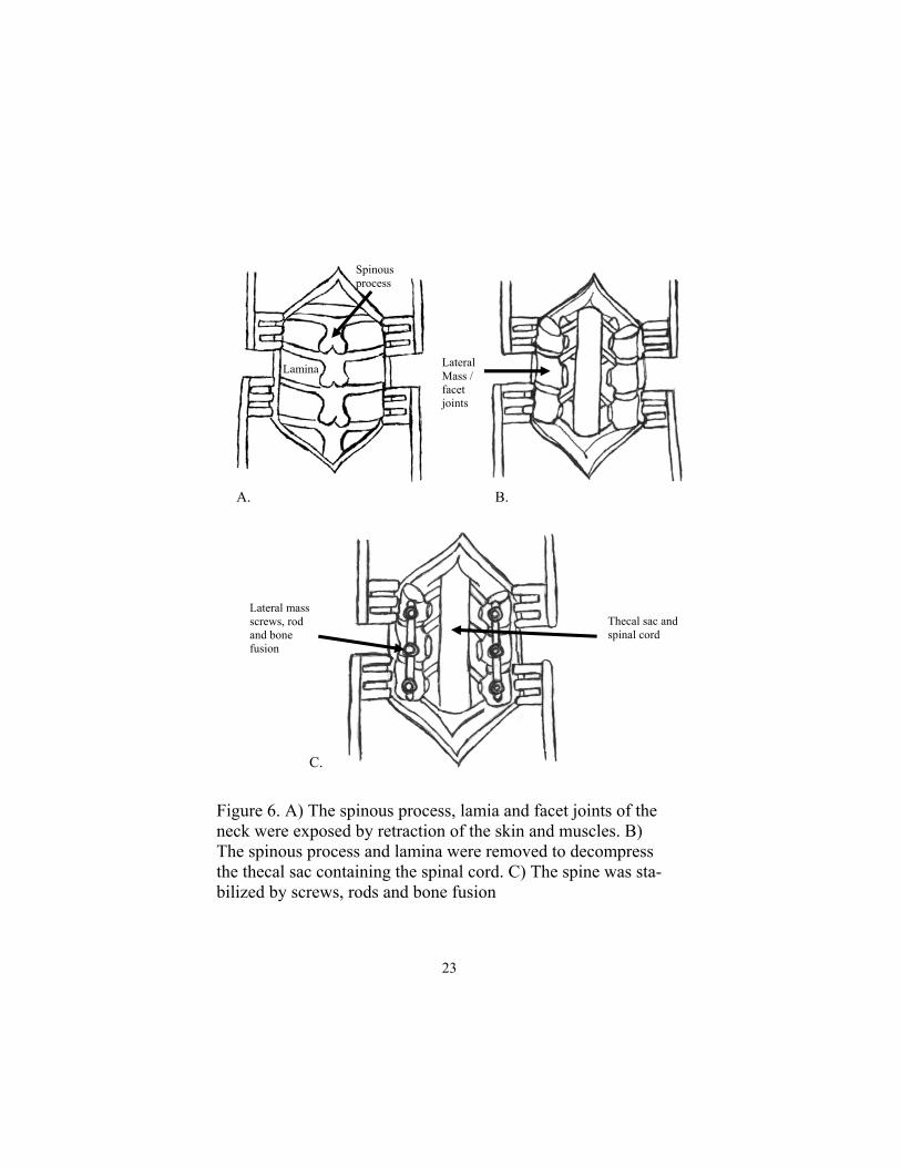
Figure 6. A) The spinous process, lamia and facet joints of the neck were exposed by retraction of the skin and muscles. B) The spinous process and lamina were removed to decompress the thecal sac containing the spinal cord. C) The spine was sta-bilized by screws, rods and bone fusion
Spinous process
Lamina Lateral Mass / facet joints
Thecal sac and spinal cord
Lateral mass screws, rod and bone fusion
A. B.
C.
23

soft tissues and outer bony surfaces from the facet joints/lateral mass with the high speed drill. This promotes strong bony heal-ing at the surgery site. Bone previously removed from the laminectomy and Infuse ™ sponges (Medtronic, Inc, Memphis, TN) are placed over top of the prepared bone surfaces for fu-sion. Infuse™ is a bone morphogenic protein (BMP) which greatly increases bone healing. The bleeding is stopped and the muscle and skin are brought together with su-tures. Patients are then taken to recovery room.
Does surgery work? Surgery is effective treatment for cervical myelopathy. The ma-jority of people see some improvements in their symptoms, but this maybe small. Patients are rarely cured. Anterior (corpectomy and fusion), posterior (laminectomy, instrumenta-tion and fusion) or combination produced some improvement in approximately 65-100% of people after surgery depending upon which study is reviewed9,10,11,12,13,14. Rates of complications vary between studies but they may be as high as 8-38%9,10,11,12,14. Complications vary between studies but may include: death, stroke, heart attack, weakness/paralysis, loss of bowel and blad-der function, infection, clots in legs (deep venous thrombosis), clots in the lungs (pulmonary embolus), blood vessel injury, failure of fusion, breakage instrumentation, movement of cage/graft, difficulty swallowing, hoarse voice, but not limited to these complications. When can I go home? Most people are discharged home 1 to 3 days after surgery. Sta-ples or stitches are removed 10 to 14 days after surgery. Please ask your doctor prior to discharge.
24
Chapter 2: Cervical Stenosis
What if I have pain, numbness or weak-ness after surgery? These symptoms may require several weeks or months to im-prove. It is normal for the symptoms to vary over time. You should contact your doctor right away if you develop fever, dif-ficulty breathing, neck swelling, horse voice, severe pain or weakness. What are my limitations after neck sur-gery? You may remove your neck dressing 1 to 2 days after sur-gery. You may shower but do not scrub or submerge your inci-sion in the bath tub for 2 weeks to decrease risk of infection. Get plenty of rest after surgery. Avoid driving, bending, ex-tending and twisting of your neck. Most people can return to work 3 to 12 weeks after surgery. Please discuss your specific limitations with your doctor. Should I use a bone stimulator? Bone stimulators have been found to improve bone healing rates by stimulation of bone cells by electrical fields. They are especially useful in patients who are at high risk of the bone not healing. Patients who are typically suggested to wear stimu-lators include multi-level fusion surgery, smokers, osteoporosis, diabetes, revision (second) surgery and patients with metabolic bone disease. If the cervical fusion does not heal you may re-quire further surgery. The Orthofix, Inc. bone stimulator has been found to improve fusion rates by 15%, from 69% to 84% in high risk patients15. This data has not yet been peer re-viewed.
25

Discharge Instructions 1. Strict control of sugar levels in patients with diabetes.
Poorly controlled sugar levels may increase risk of infec-
tion.
2. Do not smoke or use non-steroidal anti-inflammatory drugs.
They may interfere with bone fusion.
3. Keep wound clean and dry. Please shower the day after sur-
gery. Do not submerge your wound in the bath for 2 weeks.
4. Use neck collar or bone stimulator as directed by your doc-
tor.
5. No driving, twisting, bending neck for up to 1 month after
surgery.
6. Watch for the development neck swelling, difficult breath-
ing, problems swallowing, change in your voice, fever,
redness or drainage from the wound.
7. Pain, numbness and weakness often require days to months
to resolve.
8. Call your doctor if you have any concerns.
26
Chapter 2: Cervical Stenosis

References 1. Parke WW: Correlative anatomy of cervical spondylotic myelopathy. Spine
1988; 13:831-837. 2. Connell Md, Wiesel SW. Natural history and pathogenesis of cervical disk
disease. Orthop Clin North Am. 1992 Aug;13(4):345-9. 3. Nurick S: The pathogenesis of the spinal cord disorder associated with cer-
vical spondylosis. Brain 1972; 95:87-100. 4. Panjabi MM, White AA: Biomechanics of nonacute cervical spinal cord
trauma. Spine 1988; 13:838-842. 5. Cooper P R: Cervical Spondylotic Myelopathy. Contemp Neurosurge 1997;
19 (25): 1-7. 6. Kumar VGR, Madden C, Rea GL: Cervical spondylotic myelopathy. In
Winn HR (ed): Youmans Neurological Surgery 5th Ed. USA, Saunders, 2004, p 4448.
7. Epstein N, Epstein J, Carras R, et al. Coexisting cervical and lumbar steno-sis: Diagnosis and management. Neurosurgery 1984; 15: 489-496.
8. Kadanka Z, Mares M, Bednarik J et al.: Approaches to spondylotic cervical myelopathy conservative versus surgical results in a 3-year follow-up study. Spine 2002; 20:2205-2211.
9. Rajshekhar V, Kumar GS: Functional outcome after central corpectomy in poor-grade patients with cervical spondulotic myelopathy or ossified poste-rior longitudinal ligament. Neurosurgery 2005 Jun; 56(6):1279-84.
10. Chagas H, Domingues F, Aversa A, Vidal Fonseca Al, de Souza JM. Cervi-cal spondylotic myelopathy: 10 years of prospective outcome analysis of anterior decompression and fusion. Surg Neurol 2005; 64 Suppl 1:S1:30-5.
11. Kumar VG, Rea GL, Mervis LJ, McGregor JM. Cervical spondylotic mye-lopathy: functional and radiographic long-term outcome after laminectomy and posterior fusion. Neurosurgery 1999 Apr; 44(4):771-7.
12. Houten JK, Cooper PR. Laminectomy and posterior cervical plating for multilevel cervical spondylotic myelopathy and ossification of the posterior longitudinal ligament: effects on cervical alignment, spinal cord compres-sion and neurological outcome. Neurosurgery 2003 May; 52(5): 1081-7.
13. Chibbo S, Benvenuti L, Carnesecchi S et al. Anterior cervical corpectomy for cervical spondylotic myelopathy: experience and surgical results in a series of 70 consecutive patients. J Clin Neurosci. 2006 Feb; 13(2):233-8.
14. Kabok S, Mehmet T, Ufuk T et al. Results of surgical treatment for degen-erative cervical myelopathy. Spine 2004; 29:2493-2500.
15. Orthofix, Inc. PMA Randomized, Prospective Clinical Trial of Pulsed Elec-tromagnetic Field Stimulation for Cervical Fusion, 2004 (unpublished)..
27

What is the spine? The spine or backbone is made up of many bones called vertebrae (figure 1). There are 7 cervical ver-tebrae in the neck, 12 thoracic ver-tebrae in the upper back (thorax) and 5 lumbar vertebrae in the lower back. The front of the vertebrae is made up of the vertebral body. The normal vertebral bodies have a square to rectangular shape. The back of the spine is made up of ped-icles, lamina and facet joints, named the posterior elements. The vertebrae are held together by a spongy disk in the front and strong ligaments in the front and back. The disk helps cushion the spine and support the body’s weight when upright. Figure 1. The side view of the spine.
Figure 1. A side view of the spine.
T1
T6
T12
L5
28
Chapter 3: Spinal Compression Fractures
Chapter 3: Spinal Compression Frac-tures

Why does the spine break? In young, healthy individuals the verterae are very strong and require substantial forces to break them, such as a fall from a high height or car accident. Spinal bones may weaken from osteoporosis, infection or cancer and break from very little trauma, such as sitting down on a chair, heavy lifting, rolling over in bed, swinging a golf club and falls (figure 2). Osteoporosis is a disease that affects the whole body and is characterized by thinning and weakening of the bones. Osteo-porosis affects approximately 10 million Americans and occurs in as much as 30% of woman older then the age of 651. Risk factors for osteoporosis include female sex, increased age, white race, family history of osteoporosis, prior fracture, low estrogen and low body weight. Other secondary causes include low estrogen, alcoholism, overactive thyroid or parathyroid glands, poor absorption of nutrients from the gut, anorexia, glu-cocorticoid (“steroid”) and seizure medications1. In the United States, 700, 000 vertebral compression fractures occur each year, more than the number of hip and wrist fractures com-bined2,3. Infection and cancer is a less common, but important cause of spinal fractures.
How do I know my spine is broken? Spinal compression fractures may present with severe pain, weakness, loss of bowel and bladder control or initially may have no symptoms at all. Pain is the most common symptom of vertebral body com-pression fractures; it may range from mild to severe. It may be improved with lying down and worsened by moving, especially walking. Approximately 150,000 people per year are hospital-ized in the United States for compression fractures. Fortu-nately, the pain often improves the first month after the frac-ture4.
29

Figure 2. A) Normal square shaped vertebral body on the left. B) Broken squished wedge shaped vertebral body on right (arrow).
Fractures resulting in compression of the spinal nerves or spi-nal cord may result in mild to extreme weakness in the legs, and sometimes paralysis (Figure 3). It may also compress the nerves to the bowel and bladder producing urinary retention or inconti-nence.
What should I do? If you are experiencing back pain, weakness or bowel and bladder difficulties you should urgently see your doctor. You will require a thorough history and physical examination, which may include feeling your back for tenderness, checking your strength, sensations and reflexes in your legs. After an initial assessment, you may require radiological in-vestigations, including x-ray and magnetic resonance imaging (MRI) of the spine. MRI is best for finding spinal compression
A. B.
30
Chapter 3: Spinal Compression Fractures
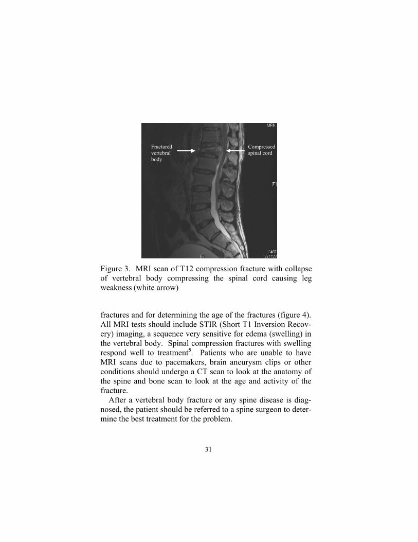
Figure 3. MRI scan of T12 compression fracture with collapse of vertebral body compressing the spinal cord causing leg weakness (white arrow)
fractures and for determining the age of the fractures (figure 4). All MRI tests should include STIR (Short T1 Inversion Recov-ery) imaging, a sequence very sensitive for edema (swelling) in the vertebral body. Spinal compression fractures with swelling respond well to treatment5. Patients who are unable to have MRI scans due to pacemakers, brain aneurysm clips or other conditions should undergo a CT scan to look at the anatomy of the spine and bone scan to look at the age and activity of the fracture. After a vertebral body fracture or any spine disease is diag-nosed, the patient should be referred to a spine surgeon to deter-mine the best treatment for the problem.
Fractured vertebral body
Compressed spinal cord
31

Figure 4. A) MRI of subacute T12 compression fracture. B) Side and C) Front X-rays of the fracture fixed with percutane-ous balloon kyphoplasty.
A. B. C.
What may happen without treatment? Vertebral compression fractures may result in pain, physical and psychological dysfunction sometimes leading to death4. The pain may interfere with mobility and make everyday tasks more difficult or impossible to complete. People often require assistance at home, hospitalization or placement in a nursing home. Without treatment the acute pain may improve, with a loss of swelling on the MRI scan. Sometimes this acute pain may be replaced by chronic long term pain due to changes in spinal shape and mechanics or from pressure of the rib cage on the pelvis4. Unfortunately, this chronic pain is not relieved by vertebroplasty or kyphoplasty. The pain and loss of independence may cause psychological distress leading to sleep disorders, anxiety and depression.
32
Chapter 3: Spinal Compression Fractures
The hunched back deformity from multiple thoracic (upper back) fractures may compress the lungs preventing the lungs from properly expanding leading to shortness of breath. The hunched back may also compress the abdomen causing a loss of appetite, weight loss and malnutrition. Vertebral body compression fractures have been associated with increased risk of death.. Woman over 65 years old appear to have a 23% higher death rate after a fracture6. These patients have two to three times more likely to die of lung disease, espe-cially chronic obstructive pulmonary disease (COPD) and pneu-monia.
What are the non-surgical treatment op-tions? There are many treatments for compression fractures ranging from bed rest to large spinal instrumentations and fusions4. Traditionally compression fractures have been treated with bed rest with progressive mobilization, spinal bracing (a brace is like a cast for the body), physical therapy, medications (muscle relaxants, anti-inflammatory drugs and narcotics) and surgery4. Immobilization of the broken vertebrae may relieve the pain. This can be accomplished by bed rest, bracing and physical therapy. Bed rest decreases movement across the broken spine. Compression fractures are aggravated by standing and walking and relieved by lying down. Prolonged bed rest is poorly toler-ated and is associated with significant medical risks. Spinal bracing restricts spine movements by squeezing the abdomen and back, but some people find a brace to be uncomfortable. Physical therapy strengthens the back and abdominal muscles to reduce back movements and pain.
33

What are the surgical treatment options? Vertebral body compression fractures may collapse, angle forward (kyphosis) or expand into the spinal canal and com-press the spinal cord or nerves. Compression of the spinal cord and nerves requires urgent decompression to prevent permanent damage (figure 5). Unfor-tunately damage may already be irreversible. Pressure is re-lieved through surgery from the front and/or back of the spine. Afterwards the spine is reconstructed with metal cage filled with bone and held together with screws, rods and plates. Sur-gery is associated with many risks including death and is rarely required for osteoporosis compression fractures. Most osteoporosis fractures result in the collapse of the bone without compression of the nervous tissue. These fractures can be fixed by the injection of bone cement through a needle. There are two common surgical procedures for treating verte-bral body compression fractures, vertebroplasty and ky-phoplasty4. Vertebroplasty involves the placement of a needle through the back into the broken vertebral body. After the needle is in the correct position liquid plastic (polymethylmethacrylate) is injected into the fractured bone. The plastic hardens and stabi-lizes the bone preventing painful movements of the vertebrae. Kyphoplasty ™ is similar to vertebroplasty except a balloon is initially used to expand the vertebral body. Plastic is then in-jected into the cavity after the balloon is removed. Vertebro-plasty and kyphoplasty are very effective at relieving back pain.
34
Chapter 3: Spinal Compression Fractures

Figure 5: Spinal fracture with spinal cord compression on MRI scan (figure 3) treated with removal of broken vertebral body. The spine reconstructed with metal cage and strengthened by metal plates, screws and rods. A) Side view and B) front view.
How is kyphoplasty done? The patient is sedated or put to sleep in the operating room. They are positioned face down on the operating table. Their back is cleaned and draped for the operation. Under x-ray guid-ance two needles are inserted through the back into the spine (figure 6). Next a balloon is placed through the needle and is inflated to make a cavity and expand the flattened vertebral body. The balloon is then removed and the cavity if filled with liquid plastic.
A. B.
35
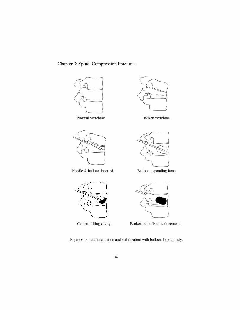
Figure 6: Fracture reduction and stabilization with balloon kyphoplasty.
Normal vertebrae. Broken vertebrae.
Needle & balloon inserted. Balloon expanding bone.
Cement filling cavity. Broken bone fixed with cement.
36
Chapter 3: Spinal Compression Fractures

How do people feel after surgery? Most people have significant improvement in back pain. Studies report greater than 80% significant pain relief 4. The pain relief is usually immediate, but may be delayed due to lo-cal soreness from the procedure. What are the possible complications? The risk of significant complications with vertebroplasty and kyphoplasty are low, probably less than 1% per fracture treat-ed4. Complications may include death, stroke, heart attack, pa-ralysis, bowel and bladder dysfunction, infection, bleeding, al-lergic reaction and pulmonary embolus, but are not limited to these complications7. Please discuss these risks with your doc-tor. When can I go home? Most people are discharge home after vertebroplasty. Pa-tients are often kept overnight after Kyphoplasty. Patients may start showering the next day after surgery. Sometimes stitches in the wounds may need to be removed 1 week after surgery.
What are my restrictions? No heavy lifting, twisting or bending or strenuous activity the first month after surgery.
How do I prevent future fractures? Fractures of the wrist, hip or spine increases the risk of future bone fractures8. Women who developed vertebral body com-pression fractures are at least 4 times higher risk of developing subsequent vertebral compression fractures8. Elderly patients with bone fractures should undergo investigation and treatment for osteoporosis.
37

Discharge Instructions 1. Keep wound clean and dry.
2. No lifting greater than 10 pounds, strenuous activity, crawl-
ing, stooping, bending or twisting for 1 months after sur-
gery.
3. Watch for the development of fever or redness and drainage
from the wound.
4. Pain may require many days to resolve. Please alert your
doctor of sudden onset of new pain.
5. Call your doctor if you have any concerns.
6. Evaluation and treatment for osteoporosis.
38
Chapter 3: Spinal Compression Fractures

Reference 1. Wei GS, Jackson JL, Hatzigeorgiou C et al. Osteoporosis management in the new millennium. Prim Care Clin Office Pract 30 (2003): 711-741. 2. Wasnich U. Vertebral fracture epidemiology. Bone 1996; 18:1791-6.
3. Melton LJ. Epidemiology of vertebral fractures in women. Am J Epide-miol 1989; 129:1000-11. 4. Truumees E, Hilibrand A, Vaccaro AR. Percutaneous vertebral augmen-tation. Spine J 2004; 4(2):218-229. 5. Tanigawa N, Komemushi A, Kariya S et al. Percutaneous vertebro-plasty: relationship between vertebral body bone marrow edema pattern on MR images and initial clinical response. Radiology 2006 Apr; 239(1):195-200. 6. Kado DM, Browner WS, Palermo L et al. Vertebral fractures and mortality in older women: a prosepective study. Arch Intern Med 1999; 159:1215-20. 7. Nussbaum D, Gailloud P, Murphey K. A review of complications asso-ciated with vertebroplasty and kyphoplasty as reported to the Food and Drug Administration medical device related web site. J Vasc Interv Radiol 2004; 15:1185-1192.. 8. Klotzbuecher CM, Ross PD, Landsmann PB et al. Patients with prior fractures have an increased risk of future fractures: a summary of the literature and statistical synthesis. J Bone Miner Res, 2000; Apr 15(4):721-39.
39
Chapter 4: Lumbar Disk Disease and Lumbar Diskectomy
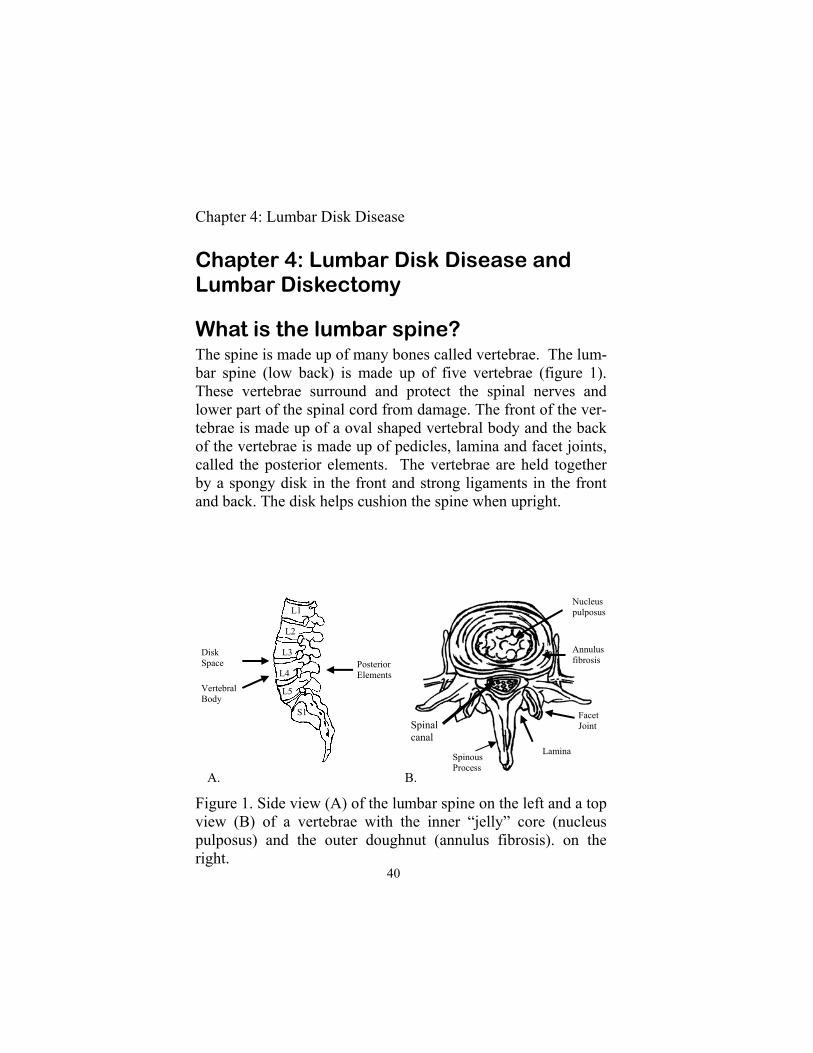
What is the lumbar spine? The spine is made up of many bones called vertebrae. The lum-bar spine (low back) is made up of five vertebrae (figure 1). These vertebrae surround and protect the spinal nerves and lower part of the spinal cord from damage. The front of the ver-tebrae is made up of a oval shaped vertebral body and the back of the vertebrae is made up of pedicles, lamina and facet joints, called the posterior elements. The vertebrae are held together by a spongy disk in the front and strong ligaments in the front and back. The disk helps cushion the spine when upright.
Figure 1. Side view (A) of the lumbar spine on the left and a top view (B) of a vertebrae with the inner “jelly” core (nucleus pulposus) and the outer doughnut (annulus fibrosis). on the right.
L1
L2
L3
L4
L5
S1
Vertebral Body
Disk Space Posterior
Elements
Nucleus pulposus
Annulus fibrosis
Facet Joint
Lamina Spinous Process
A. B.
Spinal canal
40
Chapter 4: Lumbar Disk Disease

What is a lumbar disk? Lumbar disks are located in between the spinal vertebrae and are made up of a tough outer shell (annulus fibrosis) and a soft gel-like center (nucleus pulposus). Their structure is similar to a jelly donut (figure 1). Why do lumbar disks pinch nerves? As people age the spine slowly wears out through a process called degeneration. Degeneration is first seen in the nucleus pulposus (“jelly center”) and annulus fibrosis (“donut”) in adults in their thirties to fifties1. With time the annulus may weaken and allow the nucleus to bulge outward into the spinal canal forming a bulging disk. If the annulus tears, the nucleus can squeeze out through the tear into the canal and form a herni-ated disk (figure 2). This disease is referred to by many names including slipped disk, bulging disk, ruptured disk, pinched nerve, herniated nu-cleus pulposus, disk herniation, disk protrusion, disk extrusion or disk sequestration. Even though these terms have specific meanings they are used interchangeably by most health care providers. The herniated disk may compress or “pinch off” spinal nerves resulting in back and leg pain, numbness, tingling and weakness. This is commonly referred to as sciatica. This pain may be worsened or maintained by inflammation around the nerve roots. Possible risk factors for ruptured disks are smok-ing, pregnancy, jobs with heavy lifting, repetitive lifting and twisting or operation of vehicles2. Sometimes the process be-gins after a memorable accident.
41
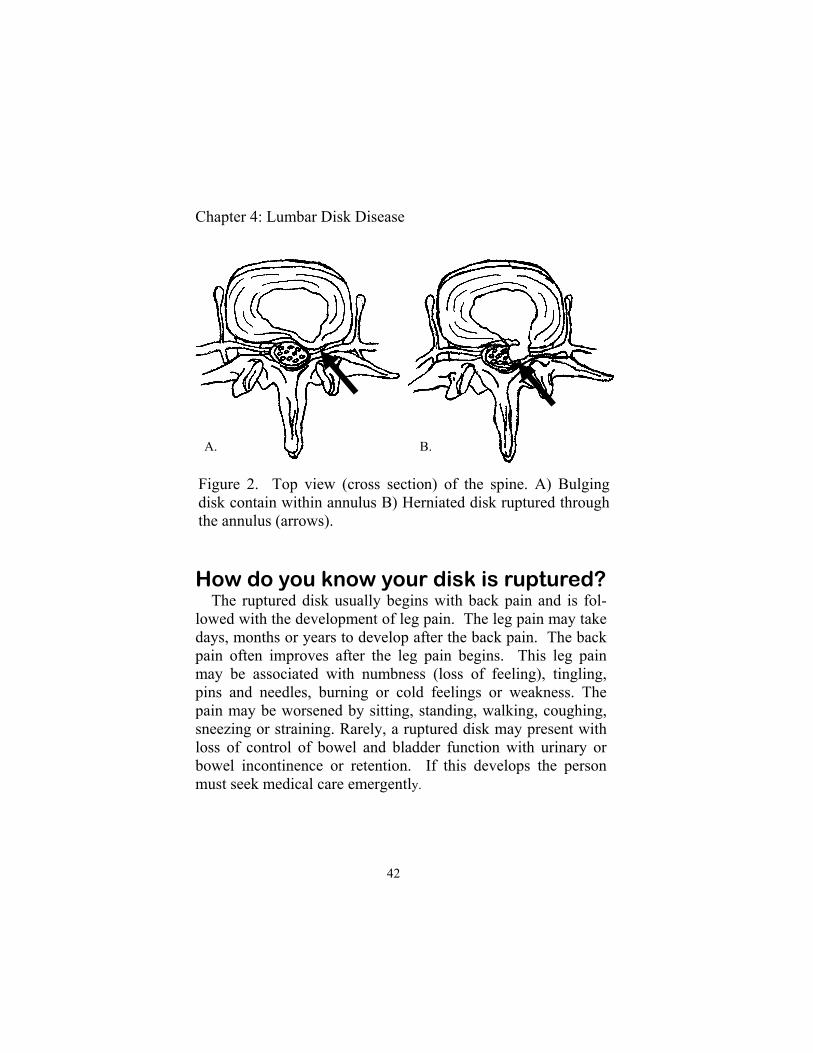
Figure 2. Top view (cross section) of the spine. A) Bulging disk contain within annulus B) Herniated disk ruptured through the annulus (arrows).
Chapter 4: Lumbar Disk Disease
How do you know your disk is ruptured? The ruptured disk usually begins with back pain and is fol-lowed with the development of leg pain. The leg pain may take days, months or years to develop after the back pain. The back pain often improves after the leg pain begins. This leg pain may be associated with numbness (loss of feeling), tingling, pins and needles, burning or cold feelings or weakness. The pain may be worsened by sitting, standing, walking, coughing, sneezing or straining. Rarely, a ruptured disk may present with loss of control of bowel and bladder function with urinary or bowel incontinence or retention. If this develops the person must seek medical care emergently.
A. B.
42

What should you do? If you are experiencing back pain, leg pain, numbness or weakness or bowel and bladder difficulties you should urgently see your doctor. You will require a thorough physical examina-tion, which may include feeling your back, testing flexibility of low back and legs, walking and careful testing of strength, sen-sations and reflexes in your legs. After an initial assessment, you may require radiological in-vestigations, including x-rays and magnetic resonance imaging (MRI) of the spine. MRI is the best test for looking for herni-ated disk and nerve root compression (figure 3). Patients with pacemakers, spinal cord stimulators or other metal within their body are unable to have an MRI. These pa-tients should undergo a computer tomogram (CT) with or with-out a myelogram. The CT myelogram produces better images then the CT alone. A myelogram is the injection of contrast medium (dye) into spinal canal to improve the visibility of the nerves on the CT (figure 4).
Figure 3. MRI scan of L4/5 herniated disk on the left side com-pressing the nerve (arrows). A) Side view and B) top view.
A. B.
43

Figure 4. A) Saggital (side view) CT myelogram and B) axial (top view) CT myelogram showing a left herniated disk at L5/1 compressing the exiting nerve root (arrows).
Who should have surgery? Many patients with back and leg pain secondary to a ruptured disk may improve with non-operative treatments including physical therapy, bed rest, non steroidal anti-inflammatory drugs (NSAIDS), steroids (Medrol dose pack) and epidural ster-oid injections. Patients who should consider surgery include patients who fail to improve with 4 to 8 weeks of conservative therapy, pa-tients with severe pain requiring narcotic medications, like mor-phine, demoral, codeine, or hydrocodone or who require admis-sion to the hospital, patients with weakness, or bowel and blad-der dysfunction (table 1)3.
A. B.
44
Chapter 4: Lumbar Disk Disease
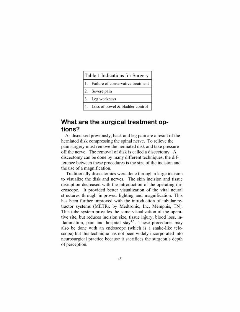
Table 1 Indications for Surgery 1. Failure of conservative treatment
2. Severe pain
3. Leg weakness
4. Loss of bowel & bladder control
What are the surgical treatment op-tions? As discussed previously, back and leg pain are a result of the herniated disk compressing the spinal nerve. To relieve the pain surgery must remove the herniated disk and take pressure off the nerve. The removal of disk is called a discectomy. A discectomy can be done by many different techniques, the dif-ference between these procedures is the size of the incision and the use of a magnification. Traditionally discectomies were done through a large incision to visualize the disk and nerves. The skin incision and tissue disruption decreased with the introduction of the operating mi-croscope. It provided better visualization of the vital neural structures through improved lighting and magnification. This has been further improved with the introduction of tubular re-tractor systems (METRx by Medtronic, Inc, Memphis, TN). This tube system provides the same visualization of the opera-tive site, but reduces incision size, tissue injury, blood loss, in-flammation, pain and hospital stay4,5 . These procedures may also be done with an endoscope (which is a snake-like tele-scope) but this technique has not been widely incorporated into neurosurgical practice because it sacrifices the surgeon’s depth of perception.
45

How is a discectomy done? The patient is given antibiotics prior to surgery. They are then taken to the operating room and are put to sleep under a general anesthetic. A tube is placed down their throat to help them breath. They are positioned face down on the operating table. Their back is washed and sterile drapes are placed around the operative site. This procedure can be down open or through a tube. The tube procedure is done similar to a standard micro-discectomy, except the skin incision is made just off midline, and the muscle is spread apart instead of stripped off the bone. The tube provides similar visualization of the bone, ligaments, nerves and ruptured disk (figure 5).
Figure 5. (A) X-ray machine used to locate the level of the her-niated disk (B) METRx tube docked on the spine over the her-niated disk. Compliments of Medtronics, Inc, Memphis, TN.
METRx
Disk
X-RAY A. B.
46
Chapter 4: Lumbar Disk Disease

An x-ray machine is used to find the level of the herniated disk. After the correct level is found, a small midline incision is made. The skin and muscle is retracted from the spine and the back (lamina) of the spine is exposed. A window into the spinal canal is made by removing a small amount of bone and liga-ment (figure 6). The nerve sac (thecal sac) and exiting spinal nerve are identified under the microscope . The sac and nerves are retracted and the herniated disk is identified and carefully removed to take pressure off the nerve. After the disk is re-moved, bleeding is stopped and the muscle and skin are brought together with sutures. Patients are then taken to recov-ery room.
Figure 6. (A) A window is cut into the bone and ligament illus-trating the herniated disk compressing the nerve. (B) The nerve is retracted and the disk is pulled out from underneath.
Disk
Nerve
Retractor
Disk remover
A. B.
47

Does surgery work? Surgery is very effective for relieving back and leg pain from a ruptured lumbar disk6,7,8. Approximately 90% of patients will have good pain relief after surgery (table 2). This appears to be long lasting9. The resolution of numbness, weakness and bowel and bladder problems is less consistent. There is approximately 6 –10% risk of a recurrent herniated disk7,10 and 4-11% of com-plications. These complications are usually minor but may in-clude: death, stroke, heart attack, weakness/paralysis, loss of bowel and bladder function, infection, clots in legs (deep ve-nous thrombosis), clots in the lungs (pulmonary embolus), large blood vessel injury, scar tissue formation (arachnoditis) and in-stability6,7,8.
Chapter 4: Lumbar Disk Disease
Table 2: Outcome after disk surgery
Pain Relief 90%
Recurrent disk 6-10%
Complications 4-11%
When can I go home? Most people are discharged home the same day after surgery. The wound is closed with deep sutures and do not need to be removed. Occasionally external sutures may require removal 1-2 weeks after surgery. The patients are seen 2 weeks after sur-gery and are released to return to work if they are doing well.
48

What if I have back pain or leg pain af-ter surgery? It is normal to have back pain for few weeks after surgery. Leg pain, numbness and tingling may come and go after surgery as the inflammation in the nerve settle. You should contact your doctor if you develop new pain, weakness or bowel and bladder problems. What are my limitations after back sur-gery? You may remove your back dressing the next day after sur-gery and begin to shower. Please return to work, house duties and recreational activities as soon as you feel able to. Most pa-tients return to work two weeks after surgery and can return to full duty by 8 weeks11.
49

Discharge Instructions 1. Strict control of sugar diabetes.
2. STOP SMOKING!
3. Keep wound clean and dry.
4. You may remove your dressing and shower the day after
surgery.
5. Return to work, housework and recreational activities as
soon as you feel your are able to.
6. Watch for the development of fever and redness or drainage
from the wound. Call your doctor if you have any concerns.
7. Pain, numbness and weakness often require days to months
to resolve. Call your doctor if you worsen.
50
Chapter 4: Lumbar Disk Disease

References 1. Kramer J. Intervertebral disk diseases: causes, diagnosis, treatment and
prophylaxis, 2nd ed. New York, Thieme, Medical, 1990. 2. Hardy R. Extradural cauda equine and nerve root compression from
benign lesions of the lumbar spine. In Youman’s Neurological Surgery. Philadelphia, WB Saunders Company, 1996, pp.2357-74.
3. Erico TJ, Fardon DF, Lowell TD. Open discectomy as treatment for herniated nucleus pulposus of the lumbar spine. Spine 20, 16: pp 1829-1833, 1995.
4. Sasaoka R, Nakamura H et al. Objective assessment of reduced inva-siveness in MED compared with conventional one-level laminotomy. Eur Spine J. May 31, 2005.
5. Foley KT, Smith MM. Microendoscopic discectomy. Techn Neurosurg 3:301-307, 1997.
6. Pappas, CT, Harrington T, Sonntag VK.Outcome analysis in 654 surgi-cal treated lumbar disk herniations. Neurosurgery 30: 862-866, 1992.
7. Davis, RA. Long-term outcome analysis of 984 surgically treated herni-ated lumbar disks. J Neurosurg 80:415-421, 1994.
8. Sylvain Palmer. Use of a tubular retractor system in microscopic lumbar discectomy: 1 year prospective results in 135 patients. Neurosurg Focus 13 (2): Article 5, 2002.
9. Findlay GF, Hall BI, Musa S, Oliveira MD, Fear SC. A 10-year follow-up of the outcome of lumbar microdiscectomy. Spine 23;10:pp 1168-1171.
10. Connolly ES. Surgery for recurrent lumbar disk herniation. Clin Neuro-surgery 39:211-216, 1992.
11. Carragee EJ, Han MY, Yang B et al. Activity restrictions after posterior lumbar discectomy A prospective study of outcomes in 152 cases with no postoperative restrictions. Spine 24; 22:pp 2346-2351.
51

Chapter 5: Lumbar Spinal Stenosis
Figure 1. The normal lumbar spine. A) Side view and B) top view (axial).
Chapter 5: Lumbar Spinal Stenosis & Lumbar Laminectomy
What is the lumbar spine? The spine is made up of many bones called vertebrae. The lumbar spine or low back is made up of five vertebrae (figure 1). These vertebrae surround and protect the spinal nerves and lower part of the spinal cord from damage. The front of the ver-tebrae is made up of a oval shaped vertebral body and the back of the vertebrae is made up of pedicles, lamina and facet joints, called the posterior elements. The vertebrae are held together by a spongy disk in the front and strong ligaments in the front and back. The disks help cushion the spine and prevent the ver-tebral bodies from rubbing together.
Nerves to legs, bowel and bladder
L1
L2
L3
L4
L5
S1
Vertebral Body
Disk Space Posterior Elements
Nucleus pulposus
Annulus fibrosis
Lamina Spinous process A. B.
52

The spinal vertebrae surround and protect the spinal canal. The spinal canal contains the spinal nerves (figure 1). These nerves connect the spinal cord to the legs, bowel and bladder. They control leg movements and emptying of the bowel and bladder and receive sensory information about touch, pain, joint position and bladder fullness. What is spinal stenosis? Spinal stenosis is the narrowing of the spinal canal. As people age, the lumbar disks dry out and collapse. The body stiffens the spine by thickening the spinal ligaments and hardening the disk and facet joints with bone spurs1. Unfortunately, these changes result in the narrowing of the spine canal and compres-sion of the nerves and blood vessels (figure 2). This decreases the blood supply and oxygen to the nerves producing pain, numbness, tingling and weakness in the legs2. The brain thinks the legs are the cause of the pain when it is actually the pressure in the back. Surgery relieves pain by removal of mechanical irritation to the nerves and improving blood supply and drain-age.
Figure 2. Spinal stenosis (narrowed spinal canal) from bony spurs and thickened ligaments. A) Top view and B) Side view.
A. B.
53

Chapter 5: Lumbar Spinal Stenosis
How do I know I have spinal stenosis? Spinal stenosis usually develops in patients between 50 and 80 years old. It is characterized by slowly worsening back and leg pain, numbness, tingling and weakness2,3. The pain may be constant but is usually brought on by walking or certain posi-tions. It is relieved with sitting, lying down or using a shopping cart. People sometimes feel like they are walking on a cloud, cotton wool or that their legs do not belong to them. Rarely patients may develop urinary and bowel incontinence with wetting or bowel movement in their pants or retention with the inability to pee or have bowel movements.
What should I do? If you are experiencing back pain, leg pain, numbness or weakness or bowel and bladder difficulties you should urgently see your doctor. You will require a thorough physical exami-nation, which may include feeling your back, testing flexibility of low back and legs, walking and careful testing of strength, sensations and reflexes in your legs. After an initial assessment, you may require radiological investigations including X-rays and Magnetic Resonance Im-aging (MRI) of the spine. MRI is the best test for looking for spinal stenosis and nerve root compression (figure 3). Patients with pacemakers, spinal cord stimulators or other metal within their body are unable to have an MRI. These pa-tients should undergo a computer tomogram (CT) with or with-out a myelogram. The CT myelogram produces better images then the CT alone. A myelogram is the injection of contrast medium (dye) into spinal canal to improve the visibility of the nerves on the CT (figure 4).
54

Figure 3: MRI scan of the lumbar spine with severe spinal stenosis with loss of white spinal fluid signal (arrows) side view (A) and top view (B).
A. B.
Figure 4: CT myelogram of the lumbar spine with severe spinal stenosis with loss of white spinal contrast dye (arrows) side view (A) and top view (B).
A. B.
55

Chapter 5: Lumbar Spinal Stenosis
Who should have surgery? Patients with spinal stenosis may improve, stay the same or worsen over time. If the spinal stenosis is found to be moderate to severe the pain usually continues to worsen without surgery. On the bright side, most people will not become paralysed or loose control of their bowel and bladder function. If you can tolerate the pain you can continue with normal activities. Many patients with back and leg pain secondary to spinal stenosis may improve with non-operative treatments including physical therapy, bed rest, back brace, non steroidal anti-inflammatory drugs (NSAIDS), steroids (Medrol dose pack) and epidural steroid injections. Unfortunately, like surgery the pain relief from conservative treatment maybe incomplete and temporary. Patients who fail to improve with conservative treatment may consider decompression surgery, especially patients with leg pain or weakness. Patients with loss of bowel and bladder con-trol may require urgent surgical treatment (table 1). Patients with multiple back surgeries, spondylolithesis (slipping of the spine), scoliosis (abnormal curvature of the spine) may require realignment and stabilization of their spine with metal screws, rods and bony fusion. This is called a spinal fusion (please see chapter 6). A spinal fusion holds the weak-ened spine together to prevent abnormal movements which may cause back and leg pain.
Table 1 Indications for Surgery 1. Severe pain
2. Leg weakness
3. Loss of bowel & bladder control
56

What are the surgical treatment op-tions? Bone spurs and thickened ligaments compress spinal nerves producing back and leg pain. Surgery removes the pressure off the nerves, improves blood supply and relieves the pain. This surgery is called a laminectomy since the lamina is removed (figure 5).
Thickened spinous proc-ess and lamina
Figure 5. The lamina and spinous process (striped area) are re-moved to decompress the nerves in the spinal canal.
Narrowed spinal canal
Spinal stenosis may be treated with a laminectomy through a large midline skin incision or through a tube (METRx MD by Medtronics Sofamor Danek). Surgery done through a tube re-quires one or more small skin incisions, sometimes on both sides of the back. This tube system provides the same visualiza-tion of the operative site but reduces incision size and tissue in-jury.
57
How is a laminectomy done?
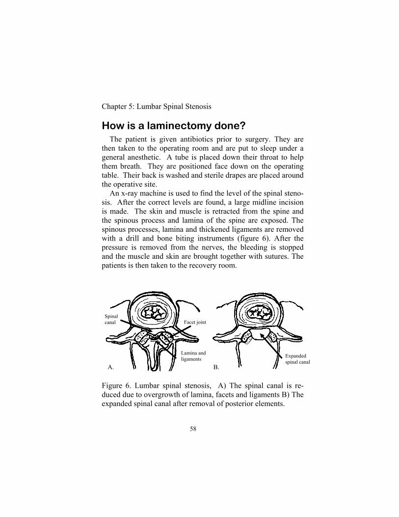
The patient is given antibiotics prior to surgery. They are then taken to the operating room and are put to sleep under a general anesthetic. A tube is placed down their throat to help them breath. They are positioned face down on the operating table. Their back is washed and sterile drapes are placed around the operative site. An x-ray machine is used to find the level of the spinal steno-sis. After the correct levels are found, a large midline incision is made. The skin and muscle is retracted from the spine and the spinous process and lamina of the spine are exposed. The spinous processes, lamina and thickened ligaments are removed with a drill and bone biting instruments (figure 6). After the pressure is removed from the nerves, the bleeding is stopped and the muscle and skin are brought together with sutures. The patients is then taken to the recovery room.
A. B.
Figure 6. Lumbar spinal stenosis, A) The spinal canal is re-duced due to overgrowth of lamina, facets and ligaments B) The expanded spinal canal after removal of posterior elements.
Facet joint
Lamina and ligaments
Spinal canal
Expanded spinal canal
58
Chapter 5: Lumbar Spinal Stenosis

This procedure can be down through a tube. Procedures done through a tube have smaller incision size and are associated with less blood loss, pain and shorter hospital stay. They pro-duce less tissue injury and inflammatory response by the body4,5. The tubular decompression is done similar to a stan-dard laminectomy once the bony lamina of the spine is reached. The main difference is the approach to the spine. In a standard laminectomy the muscle is stripped off the spine and then re-tracted under high pressure. This may cause permanent muscle damage and back pain. In a tubular laminectomy the muscle is dilated with progressively larger tubes. There is no muscle stripping and probably less muscle retraction pressure since the pathway to the spine is smaller. The tube provides good visuali-zation of the bone, ligaments and nerves (figure 7).
Figure 7. Bilateral spinal decompression through the METRx MD tube (Compliments of Medtronics Sofamor Danek).
59

Does surgery work? Surgery is effective for improving back and leg pain3,6,7,8,9,10 Approximately 55 to 82% of people have good pain relief after surgery (see table 1). There is approximately 10-18% risk of requiring further surgery in the future8,9,11. Surgery for recurrent spinal stenosis is less successful (usually less than 50%) and are associated with higher complication rates8. Surgery can be safely done in people older the 75 years old12. There is approximately 12% chance of complication for lum-bar laminectomy surgery7. Most complications are minor, but possible complications may include: death, stroke, heart attack, weakness/paralysis, loss of bowel and bladder function, infec-tion, clots in legs (deep venous thrombosis), clots in the lungs (pulmonary embolus), large blood vessel injury, scar tissue for-mation (arachnoditis) and instability, but not limited to these complications.
Chapter 5: Lumbar Spinal Stenosis
Table 2: Outcome after laminectomy
Pain Relief 60-70%
Recurrent (new) stenosis 10%
Complications 10%
When can I go home? Most people are discharged home the day of surgery, or one to two days afterwards. Sometimes stitches in the wound may need to be removed 1-2 weeks after surgery.
60
What if I have back pain or leg pain af-ter surgery? It is normal to have back pain related to the surgery for a few weeks after surgery. Back pain which was present prior to sur-gery may or may not resolve after surgery. It is also normal to have pain, numbness and tingling that comes and goes after surgery as inflammation settles in the nerves. You should con-tact your doctor if you develop severe leg pain or develop new weakness or bowel and bladder problems, especially inconti-nence or inability to urinate. What are my limitations after back sur-gery? You may remove your back dressing the next day after sur-gery and wash your incision in the shower. Please do not bathe for 2 weeks after surgery because bathing may increase your risk of infection. Please rest after discharge from the hospital. Remember you have had recent surgery and do not overdo it! Do not lift greater than 10 pounds or do any strenuous activities like run-ning, jumping, stooping, crawling, bending and twisting for 4- 6 weeks after surgery. Most people can return to work after this time. Please discuss your limitations with your doctor since every person is a little different.
61

Discharge Instructions 1. Strict control of sugar levels in patients with diabetes.
Poorly controlled sugar levels may increase risk of infec-
tion.
2. STOP SMOKING!
3. Keep wound clean and dry. Please shower the next day after
surgery, but no baths for 2 weeks.
4. No lifting greater than 10 pounds, strenuous activity, crawl-
ing, stooping, bending or twisting for 4-6 weeks.
5. Watch for the development of fever and redness or drainage
from the wound. Call your doctor if you have any concerns.
6. Pain, numbness and weakness often require days to months
62
Chapter 5: Lumbar Spinal Stenosis
References 1. Yong-Hing K, Kirkaldy-Willis WH. The pathophysiology of degenera-
tive disease of the lumbar spine. Orthop Clin North Am 14:491-504, 1983.
2. Watanabe R, Park WW: Vascular and neural pathology of lumbosacral , spinal stenosis: J Neurosurg 64:64-70, 1986.
3. Lemaire JJ, Sa5r2utreaux JL, Chabannes J, et al: Lumbar canal stenosis: Retrospective study of 158 operated cases. Neurochirurgie 41:89-97, 1995.
4. Sasaoka R, Nakamura H et al. Objective assessment of reduced inva-siveness in MED compared with conventional one-level laminotomy. Eur Spine J. May 31, 2005.
5. Foley KT, Smith MM. Microendoscopic discectomy. Techn Neurosurg 3:301-307, 1997.
6. Herron LD, Mangelsdorf C: Lumbar spinal stenosis: Results of surgical treatment. J Spinal Disord 4:26-33, 1991.
7. Atlas SJ, Deyo RA, Keller RB, et al: The Main lumbar spine study, Part III. 1 year outcomes of surgical and non-surgical management of lumbar spinal stenosis. Spine 21(15): 1787-1794, 1996.
8. Jonsson B, Annertz M, Sjoberg C, et al. A prospective and consecutive study of surgically treated lumbar spinal stenosis. Part II. Five year fol-low-up by an independent observer. Spine 22:2938-2944, 1997.
9. Katz JN, Lipson SJ, Larson MG, et al. The outcome of decompressive laminectomy for degenerative lumbar spinal stenosis. J bone Joint Surg Am 73:809-813, 1991.
10. Turner JA, Ersek M, Herron L, et al. Surgery for lumbar spinal stenosis: Attempted meta-analysis of the literature. Spine 17:1-8, 1992.
11. Herno A, Airaksinen O, Saari T: Long-term results of surgical treatment of lumbar spinal stenosis. Spine 18: 1471-1474, 1993.
12. Vitaz TW, Raque GH, Shields CB, Glassman SD: Surgical treatment of lumbar spinal stenosis in patients older than 75 years old of age. J Neu-rosurg 91(2 Suppl): 181-5, 1999.
63

Chapter 6: Lumbar Spondylolisthesis & Lumbar Fusion
What is the lumbar spine? The spine is made up of many bones called vertebrae. The lumbar spine or low back is made up of five vertebrae (figure 1). These vertebrae surround and protect the spinal nerves and lower part of the spinal cord from damage. The front of the ver-tebrae is made up of a square shaped vertebral body and the back of the vertebrae is made up of pedicles, lamina and facet joints, called the posterior elements. The vertebrae are held to-gether by a spongy disk in the front and strong ligaments in the back. The disk helps cushion the spine.
Nerves to legs, bowel and bladder
L1
L2
L3
L4
L5
S1
Vertebral Body
Disk Space Posterior Elements
Nucleus pulposus
Annulus fibrosis
Figure 1. The normal lumbar spine. A) the side view and B) the top view (axial).
A. B.
64
Chapter 5: Lumbar Spondylolisthesis

What is lumbar spondylolisthesis? Lumbar spondylolisthesis is the slipping of the spine. The vertebral body slips forward over the lower vertebral body (figure 2). There are many possible causes of spondylolisthesis. The spine may slip because the spine was made abnormally from birth (congenital spondylolithesis), broke in early childhood (isthmic spondylolithesis), from an injury (traumatic spondylo-lithesis), infection or tumor (pathologic spondylolithesis) or “wearing out” from aging (degenerative spondylolithesis). The spine may also weaken and slip after back surgery. The most common causes of spondylolithesis are isthmic (the pars of the posterior elements break in early childhood) and degenerative (the spine wears out and can no longer hold the bones to-geather). Isthmic spondylolithesis usually presents in early adulthood. It may be caused by repeated injuries to the posterior elements of the spine by walking, gymnastics or football1. The posterior elements act as a hook to hold the spine together. If part of this hook breaks (that is the pars interarticularis) then the spine can not hold itself together and may result in spinal instability and the spine slipping apart (figure 2). This instability and slip may cause back pain. The body forms scar tissue around the broken pars in attempt to heal the break, unfortunately this scar tissue may compress spinal nerves and cause leg pain, numbness and weakness. Degenerative spondylolithesis presents in later adult hood and is more common in black females2. It is usually associated with spinal stenosis (chapter 5) and presents with severe back and leg pain. The spine is held together by the disk in the front and the posterior elements in the back. The posterior elements form a hook (made up of the pars and facet joint) which keeps it
65

Figure 2: Isthmic spondylolithesis. The posterior hook holds the spine together. If the pars interarticularis (arrows) breaks the spine can no longer hold the vertebrae together and they may slip apart. Side view (A) and rotated view (B).
attached to the vertebrae above and below. As the spine ages and becomes worn out the disk and facet joints weaken and al-low the spine to slip apart (figure 3). This “wear and tear” re-sults in thickening of ligaments, scar tissue and formation of bone spurs which may compress nerves causing pain, numbness and weakness in the legs. How do you know your back is slipping? Patients with spondylolithesis present with back pain and leg pain, numbness and weakness. The back pain may be constant but usually varies with positon. The pain is worsened by stand-ing or walking and improved by lying down. Patients with lytic spondylolithesis may develop disabling deformities of their body including stiffness, “square” buttock, scoliosis, waddling gait3. It is uncommon to have problems with bowel and bladder function with spondylolithesis.
A. B.
66
Chapter 5: Lumbar Spondylolisthesis

What should you do? If you are experiencing back pain, leg pain, numbness or weakness or bowel and bladder difficulties you should urgently see your doctor. You will require a thorough history and physi-cal examination, which may include feeling your back, testing flexibility of low back and legs, walking and careful testing of strength, sensations and reflexes in your legs. After an initial assessment, you may require radiological in-vestigations including X-rays and Magnetic Resonance Imaging (MRI) of the spine. MRI is the best test for looking for spinal stenosis and nerve root compression (figure 4). Patients with pacemakers, spinal cord stimulators or other metal within their body are unable to have an MRI. These pa-tients should undergo a computer tomogram (CT) with or with-out a myelogram. The CT myelogram produces better images
Figure 4. MRI scan of lumbar spondylolithesis of L5 on S1 ver-tebrae (see arrows). A) Side view and B) top view on the right.
A. B.
67
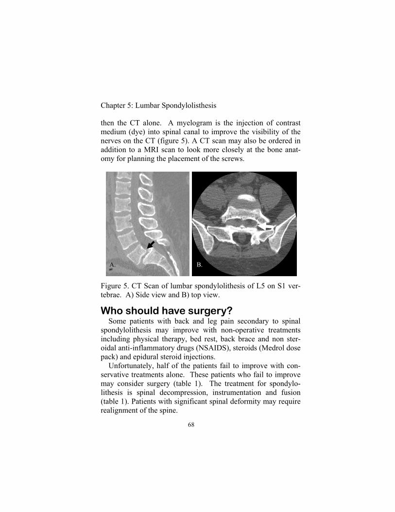
Who should have surgery? Some patients with back and leg pain secondary to spinal spondylolithesis may improve with non-operative treatments including physical therapy, bed rest, back brace and non ster-oidal anti-inflammatory drugs (NSAIDS), steroids (Medrol dose pack) and epidural steroid injections. Unfortunately, half of the patients fail to improve with con-servative treatments alone. These patients who fail to improve may consider surgery (table 1). The treatment for spondylo-lithesis is spinal decompression, instrumentation and fusion (table 1). Patients with significant spinal deformity may require realignment of the spine.
Figure 5. CT Scan of lumbar spondylolithesis of L5 on S1 ver-tebrae. A) Side view and B) top view.
then the CT alone. A myelogram is the injection of contrast medium (dye) into spinal canal to improve the visibility of the nerves on the CT (figure 5). A CT scan may also be ordered in addition to a MRI scan to look more closely at the bone anat-omy for planning the placement of the screws.
A. B.
68
Chapter 5: Lumbar Spondylolisthesis

Table 1 Indications for Surgery
1. Severe pain
2. Leg weakness
3. Loss of bowel & bladder control (rare)
4. Slip worsening over time
5. Severe slip
What are the surgical treatment op-tions? Surgery for spinal spondylolithesis have significantly ad-vanced over the years. Initial surgery consisted of a decompres-sive laminectomy with removal of the back of the spine (see chapter 5) to decompress the spinal nerves and relieve leg pain. Unfortunately this did not treat the back pain and resulted in further slipping of the spine. This lead to development of bony fusions to make the spine solid and prevent slips. Unfortu-nately, it takes a long time for the body to fuse the spine and sometimes fusion did not occur. Most people believe the intro-duction of metal screws and rods fixed these problems. The spine is now held rigidly together by screws and rods until the bones heal (figure 6). The newest advancement in spinal fu-sions are the discovery of bone morphogenic protein (BMP), a bone “hormone” which improves fusion rates and minimal inva-sive surgery which decreases tissue injury and increases patient level of satisfaction4.
69

Figure 6. Post operative X-rays of the lumbar spine after de-compression, pedicle screw instrumentation and fusion. A) Side view and B) front view.
How is a fusion done?
The patient is given antibiotics prior to surgery. They are then taken to the operating room and are put to sleep under a general anesthetic. A tube is placed down their throat to help them breath. They are positioned face down on the operating table. Their back is washed and sterile drapes are placed around the operative site. An x-ray machine is used to find the level of the spinal spondylolithesis. After the correct level is found, a midline in-cision is made. The skin and muscle is retracted from the spine and the back of the spine (spinous process and lamina) are ex-posed. The spinous processes, lamina and thickened ligaments are removed with a drill and bone biting instruments. After the laminectomies are completed and the pressure has been
A. B.
70
Chapter 5: Lumbar Spondylolisthesis
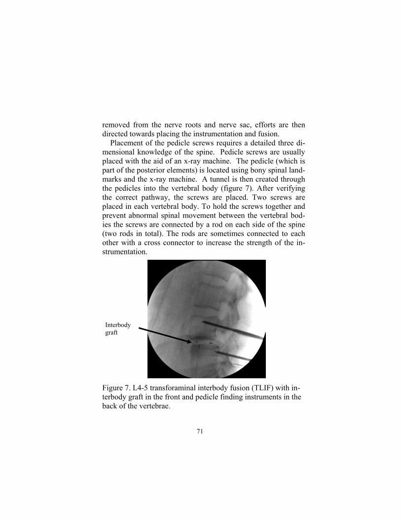
removed from the nerve roots and nerve sac, efforts are then directed towards placing the instrumentation and fusion. Placement of the pedicle screws requires a detailed three di-mensional knowledge of the spine. Pedicle screws are usually placed with the aid of an x-ray machine. The pedicle (which is part of the posterior elements) is located using bony spinal land-marks and the x-ray machine. A tunnel is then created through the pedicles into the vertebral body (figure 7). After verifying the correct pathway, the screws are placed. Two screws are placed in each vertebral body. To hold the screws together and prevent abnormal spinal movement between the vertebral bod-ies the screws are connected by a rod on each side of the spine (two rods in total). The rods are sometimes connected to each other with a cross connector to increase the strength of the in-strumentation.
Figure 7. L4-5 transforaminal interbody fusion (TLIF) with in-terbody graft in the front and pedicle finding instruments in the back of the vertebrae.
Interbody graft
71

After the placement of the instrumentation, the spine is pre-pared for fusion and placement of bone graft. There are many types of fusion procedures (figure 8). The spine may be fused with placement of bone on the back of the spine beside the screws and rods (posterolateral fusion, PLF), in the facets (facet fusion), or between the vertebral bodies (interbody fusion). There is a variety of interbody fusions, including posterior lum-bar interbody fusion (PLIF), transforaminal lumbar interbody fusion (TLIF) and anterior lumbar interbody fusion (ALIF) . Interbody fusion procedures are all done through the back ex-cept an ALIF which is done through the abdomen. Each fusion procedure has its own risks and benefits. These operations can now be down with minimally invasive techniques. The grafts, screws and rods can be placed through small incisions. After completion of the fusion, the bleeding is stopped and a drain is placed. The muscle and skin are brought together with sutures. Patients are then taken to recovery room.
72
Chapter 5: Lumbar Spondylolisthesis

A. Posterolateral fusion
Posterior lumbar interbody fusion B..
Figure 8. There are many types of spinal fusions, including posterorlateral fusion (PLF, A), posterior lumbar interbody fu-sion (PLIF, B), transforaminal interbody fusion (TLIF, C) and anterior lumbar interbody fusion (ALIF, D). The area of the spinal fusion are represented by black blocks or ovals.
73

C. Transforaminal lumbar interbody fusion
D. Anterior lumbar interbody fusion
74
Chapter 5: Lumbar Spondylolisthesis

Does surgery work? Surgery is effective for improving back and leg pain from lumbar spondylolithesis5,6,7,8,9,10,11. Approximately 70-95% of people improve with surgery (see Table 1). The amount of im-provement varies but is usually incomplete. Patients with high grade (large slips) have been found to do better then patients with low grade (small slips) and degenerative disk disease6. There is a 5-18% risk of development of new spinal disease above and below the surgical site12 . The spine moves during normal activities. This movement is prevented by a spinal fu-sion. The adjacent spine must make up for this loss of move-ment. This produces increased “wear and tear” degeneration at these levels which may progress to symptomatic spinal disease. This is called adjacent level disease. There are approximately 5 -10% chance of complications from surgery, studies in the lit-erature have reported a range of 2 to 33%5,6,8,10. Complications are uncommon and usually are insignificant. Potential complications may include: death, stroke, heart at-tack, leg weakness/paralysis, loss of bowel and bladder func-tion, infection, clots in legs (deep venous thrombosis), clots in the lungs (pulmonary embolus), large blood vessel injury pro-ducing a blood clot, hemorrhage or death, scar tissue formation (arachnoditis) and instability, but not limited to these complica-tions.
Table 1: Outcome after lumbar fusion Decreased pain 70-95%
Adjacent level degenerative disease 5-18%
Complications 5-10%
75

When can I go home? Most people are discharged home a few days after surgery, elderly patients may require up to 1 week in the hospital. Stitches or staples need to be removed within 2 weeks after surgery. Patients will have pain at the incision site for weeks after the surgery.
What if I have back pain or leg pain after sur-gery? It is normal to have back pain and soreness from the operation for few weeks. It is also normal to have pain, numbness and tingling that comes and goes after surgery as inflammation resolves. You should contact your doctor right away if you develop new leg pain or weak-ness or bowel and bladder problems, especially urinary incontinence or inability to urinate.
What are my limitations after back surgery? You may remove your back dressing the day after surgery and wash your incision in the shower. Please keep your incision clean and dry. Please do not bathe for 2 weeks after surgery because bathing may in-crease your risk of infection. Please relax after discharge from the hospital. Slowly return to nor-mal activities. Most patients can return to work after 2-4 weeks. Do not lift greater than 10 pounds or do any strenuous activities like running, jumping, stooping, crawling, bending and twisting for 2 to 3 months after surgery. Smoking and non-steroidal anti-inflammatory drugs (NSAIDS) interfere with bone fusion. If the fusion does not heal you may require further surgery. All patients should stop smoking and us-ing NSAIDS after surgery. Please discuss your personal limitations with your doctor.
76
Chapter 5: Lumbar Spondylolisthesis
Discharge Summary 1. Strict control of sugar levels in patients with diabetes.
Poorly controlled sugar levels may increase risk of infec-
tion.
2. Do not smoke or use non-steroidal anti-inflammatory drugs.
They may interfere with bone fusion.
3. Keep wound clean and dry. Please shower the day after sur-
gery. Do not submerge your wound in the bath for 2 weeks.
4. Use back brace or bone stimulator as directed by your doc-
tor.
5. No lifting greater than 10 pounds, strenuous activity, crawl-
ing, stooping, bending or twisting for 2-3 months.
6. Watch for the development of fever and redness or drainage
from the wound
7. Pain, numbness and weakness often require days to months
to resolve.
8. Call your doctor if you have any concerns.
77

References 1. Weir MR and Smith DS. Stress reaction of the pars interarticularis leading
to spondylolysis. A cause of adolescent low back pain. J Adolesc Health Care. 1989 Nov; 10(6):573-7.
2. Vogt MT, Rubin DA, Palermo L et al. Lumbar spine listhesis in older Afri-can American women. Spine J. 2003 Jul-Aug; 3(4):255-61.
3. Newman PH. A clinical syndrome associated with severe lumbosacral sub-luxation. J bone Joint Surg [Br]. 1965: 47:472-481.
4. Villavicencio AT, Burneikiense S, Nelson EL et al. Safety of transforaminal lumbar interbody fusion and intervertebral recombinant human bone morphogenetic protein-2. J Neurosurg Spine. 2005 Dec; 3(6):436-43.
5. Thomsen K, Christensen FB, Eiskiaer SP et al. 1997 Volvo Award winner in clinical studies. The effect of pedicle screw instrumentation on functional outcome and fusion rates in posterolateral lumbar spinal fusion: a prospec-tive, randomized clinical study. Spine. 1997 Dec 15;22(24):2813-22.
6. Buttermann GR, Garvey TA, Hunt AF et al. Lumbar fusion results related to diagnosis. Spine. 1998 Jan 1;23(1):116-27.
7. Biarke Christensen F, Stender Hansen E, Laursen M et al. Long-functional outcome of pedicle screw instrumentation as a support for posterolateral spinal fusion: randomized clinical study with a 5-year follow-up. Spine. 2002 Jun 15; 27(12):1269-77.
8. Ekman P, Moller H, Hedlund R. The long-term effect of posterolateral fu-sion in adult isthmus spondylolisthesis: a randomized controlled study. Spine J. 2005 Jan-Feb; 5(1):36-44.
9. Fischgrund JS, Mackay M, Herkowitz HN, et al: 1997 Volvo Award winner in clinical studies. Degenerative lumbar spondylolisthesis with spinal steno-sis: A prospective randomized study comparing decompression laminec-tomy and arthrodesis with and without spinal instrumentation. Spine 22:2807-2812, 1997.
10. Zdeblick TA: A prospective, randomized clinical study of lumbar fusion. Preliminary report. Spine Jun 15;18(8):983-91, 1997.
11. Mardjetko SM, Connolly PJ, Shott S. Degerative lumbar spondylolithesis: A meta-analysis of the literature 1970-1993. Spine 1994; 10L2256S-2265S.
12. Park P, Garton HJ, Gala VC et al. Adjacent segment disease after lumbar or lumbosacral fusion: review of the literature. Spine. 2004 Sep 2;29(17):1938-44.
78
Chapter 5: Lumbar Spondylolisthesis

Glossary Anterior Cervical Discectomy and Fusion (ACDF) - the slipped disk is removed and the spine is reconstructed with a bone graft in the disk space and plate and screws. The bone allows for solid fusion of the vertebrae. Anterior Lumbar Interbody Fusion (ALIF) - the disk is removed through the abdomen and the vertebral bodies are fused together. Bone Morphogenic Protein (BMP) - a synthesized “hormone” that promotes bone growth and healing. Bone Stimulator— a device that produces electrical fields to promote bone growth and healing. Computer Tomography (CT) - a complex x-ray machine that produce detailed internal body images, especially of bones. Degeneration—the “wear and tear” changes in the spine as a result of aging, which may include thickening and breaking of ligaments, formation of bone spurs and disk herniation. Disk— supports and cushions the vertebral body. It is composed of an inner gelatinous nucleus pulposus and outer tough annulus fibrosis. The disk is located between the vertebral bodies. Disk herniation— the disk slips into the spinal canal. Facet joints— they connect to the lamina and pedicles, and form part of posterior hook prevent-ing the spine from slipping apart. Inflammation— chemical and blood cell response to injury or irritation in the body. Kyphoplasty—the reduction and stabilization of a vertebral body compression fracture with a balloon and liquid plastic. It is similar to vertebroplasty. Lamina—form the roof of the spinal canal and protect the spinal cord and nerves. They are connected to the back of the pedicle. Laminotomy—partial removal of the lamina. Laminectomy– complete removal of the lamina. Magnetic Resonance Imaging (MRI) - a machine which produces detailed images of internal body structures with the use of strong magnets.
79

METRx MD— Tubular retractor system that dilates soft tissue to make a pathway to the spine. METRx system is made by Medtronic Softmore Danek. Myelopathy— spinal cord dysfunction that may present as numbness, weakness, limb stiffness, bowel and/or bladder dysfunction as a result of spinal cord compression. Pars interarticularis—the bone that connects the superior articular process with the inferior articular process. It is part of the bony hook which prevents spondylolithesis. Pedicles—bone cylinders extending backwards from the vertebral body. Screws are placed through them into the vertebral body to stabilize the spine. Posterior Lateral Fusion (PLF) - the facet joints, pars interarticularis and transverse processes are roughed up (decorticated) and covered with bone. With time these structures grow together and form a solid bone fusion. Posterior Lumbar Interbody Fusion (PLIF)—the disk is removed through the lamina from the back and the vertebral bodies are fused together. Radiculopathy—limb pain, numbness and weakness resulting form nerve compression. Sciatica— leg pain resulting from nerve compression in the back. Spine— (back bone) provides shape and structure to the body. Spinal cord— is part of the central nervous system and is connected to the lower part of the brain (brain stem), it contains nerve tracts sending information to and from the brain. Spondylolithesis—the slipping of one vertebral body over another vertebral body. Stenosis—is a small hole, for example spinal stenosis is when the spinal canal becomes smaller. Tranforaminal Lumbar Interbody Fusion (TLIF)—the disk is removed through the nerve fora-men from the back and the vertebral bodies are fused together. Transverse process— extend out laterally from the pedicle and is an important site of bony fusion in posterior lateral fusions. Vertebrae—the individual spine bones, composed of a body and posterior elements (made up of the pedicles, lamina and facet joints, transverse process and spinal process). Vertebral body compression fracture—squished broken vertebral body. Vertebroplasty—the repair of the vertebral body compression fracture with injected liquid plas-tic. Vertebroplasty usually produces significant back pain relief.
80