sporozoon tincae infection in tench. an ultrastructural · pdf filesporozoon tincae infection...
TRANSCRIPT

Vol. 20: 143-151,1994 DISEASES OF AQUATIC ORGANISMS
Dis. aquat. Org. Published November 10
Sporozoon tincae infection in tench. An ultrastructural investigation
'Institute of Pathology, ' ~ i s h Diseases Research Unit, School of Veterinary Medicine, Biinteweg 17, D-30559 Hannover, Germany
ABSTRACT: For several years now, pathological skin changes have been observed in tench Tinca tinca L. from northern Germany. Using light microscopy, numerous instances of the causative agent have been detected, cell-bound in macrophages and/or between altered tissue structures. The infection shows systemic spread, as the microorganisms, Sporozoon tincae, also appear in the spleen. Using elec- tron microscopy, it was possible to depict numerous different features of the microorganism associated with the intracellular development and multiplication of the agent. The structures lacked a true cell nucleus, so the previous assignment of the organism to the Protozoa is no longer maintainable. Our findings demonstrate that S. tincae is not a eukaryotic organism. We suggest that S. tincae be classified as bacteria.
KEY WORDS: Sporozoon tincae . Tench . Tinca tinca . Electron microscopy
INTRODUCTION
Tench Tinca tinca L. belong to the group of prized, edible fish that prefer standing waters with a muddy bottom. Frequently they are bred together with carp in earth ponds. Since 1980, tench collected from northern Germany have shown multiple ulcers at the base of the fins, on the side of the body and in the re- gion of the tail (Korting et al. 1987). The alterations were associated with parasites which were first de- scribed by Volf & Dvoiak (1928). The disease ascribed to Sporozoon tincae occurred in southern Bohemia (present Czech Republic). The symptoms and the light microscopy of the causative agent were redescribed by Jirovec et al. (1947). In contrast to the disease in Bohemia which, with a mortality rate of up to 90%, reflects a widespread condition, those instances re- ported from northern Germany were only individual cases, which occurred regularly in the autopsy mater-
'Address for correspondence: Experimental Pathology, German Primate Centre (DPZ), Kellnerweg 4. D-37077 Gottingen. Germany
ial. From an electron microscopy study, it became clear that an unequivocal assignment to the Protozoa or to other eukaryotic organisms was not possible (Korting et al. 1987). The present study describes the morphology of the disease pattern and documents ultrastructural characteristics of its agent, information which until now has not been available in the pub- lished literature.
MATERIAL AND METHODS
The present study results from the investigation of 5 adult tench (3 to 4 yr old) collected in the autumn from a carp farm in northern Germany. Following macroscopic inspection, samples were obtained from affected skin regions and from spleen immediately after sacrifice. The material was fixed with 10% buffered neutral formaldehyde for light microscopy and conventionally embedded in paraplast. The fol- lowing staining techniques were carried out on paraffin sections: H&E, PAS, Grocott, Giemsa, Ladewig, Azan. For electron microscopy, tissue sam- ples were cut into blocks of approximately 1 mm3
O Inter-Research 1994

144 Dis. aquat. Org. 20: 143-151. 1994
Fig. l Sporozoon tincae infecting Tinca tinca. Light micrographs demonstrating (a) severe hyaline degeneration of muscle cells (M), which is occasionally associated with (bl inflammatory cell infiltrates. Semithin section, toluidine blue, scale bars = 20 pm
and fixed for 24 h in 2 .5% cacodylate buffered glu- Toluidine-blue-stained semithin sections were used taraldehyde. Blocks were postfixed in 1% osmium for light microscopic orientation. Ultrathin sections tetroxide buffered in 0.15 M sodlum cacodylate, re- were automatically (Leica ultrostainer) contrasted peatedly rinsed in cacodylate buffer, dehydrated in with uranyl acetate (30 min. 40°C) and lead citrate increasing concentrations of alcohol, a n d embedded (72 S, 20°C) , and examined with a Zeiss EM 10C at in Epon 812 with propylenoxide as intermedium. 60 kV.
Flg. 2. Sporozoon t~ncae infecting Tinca tinca. Numerous microorganisms as punctiform structures are observed either extra- cellularly [(a) intraepithelial, (b) subepithelial] or (c) intracellularly in macrophages. E: epithelium of the sk~n . Semithin section,
toluidine blue, scale bars = (a. b) 20 pm, (c) 15 pm

4 *
b.. .
146 Dis. aquat. Org. 20: 143-151, 1994
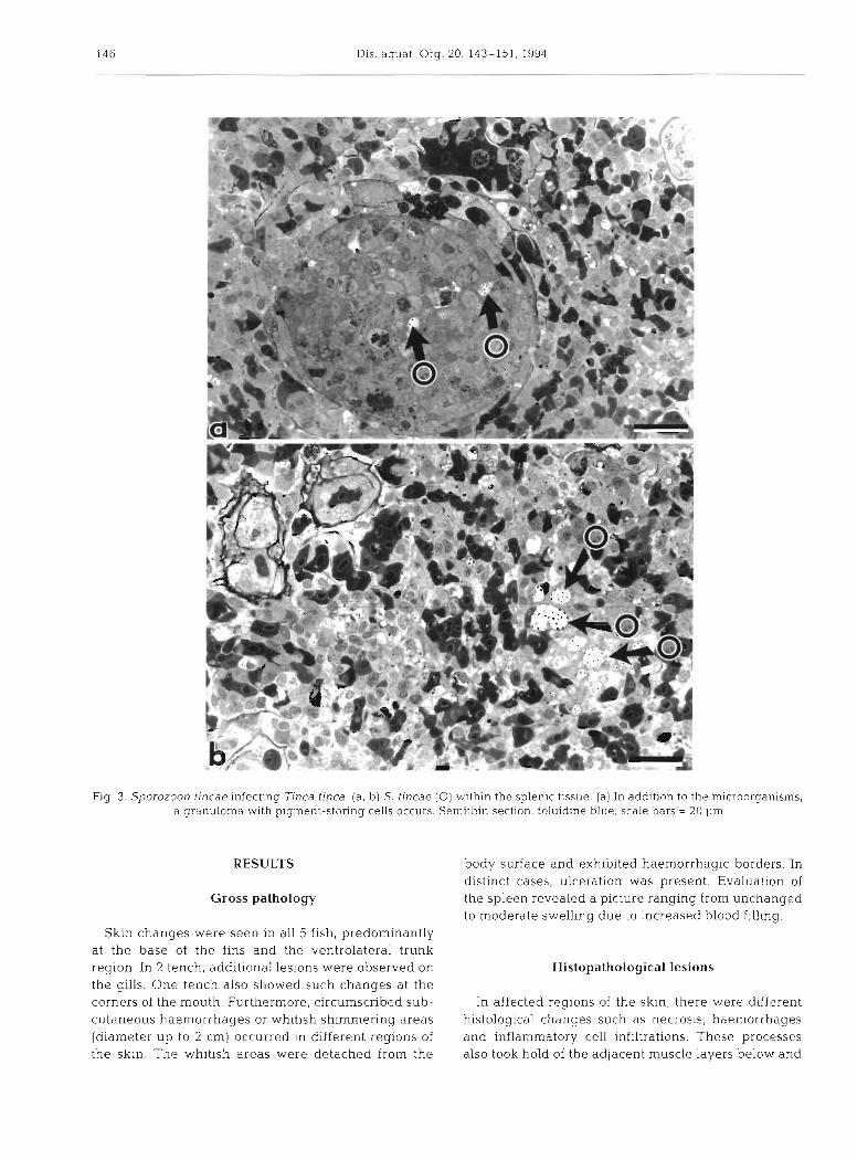
Fig. 3. Sporozoon tincae infecting Tinca tinca. (a, b) S. tincae (0) within the splenic tissue. (a) In addition to the microorganisms, a granuloma with pigment-storing cells occurs. Semithin section, toluidine blue, scale bars = 20 pm
RESULTS
Gross pathology
Skln changes were seen in all 5 f ~ s h , predominantly at the base of the fins and the ventrol.atera1 trunk region. In 2 tench, additional lesions were observed on the gills. One tench also showed such changes at the corners of the mouth. Furthermore, circumscribed sub- cutaneous haemorrhages or whitish sh~mm.ering areas (diameter up to 2 cm) occurred in different regions of the skin. The whitish areas were detached from the
body surface and exhibited haemorrhagic borders. In distinct cases, ulceration was present. Evaluation of the spleen revealed a picture ranging from unchanged to moderate swelling due to increased blood filling.
Histopathological lesions
In affected regions of the skin, there were different histological changes such as necrosis, haemorrhages and inflammatory cell infiltrations. These processes also took hold of the adjacent muscle layers below and

Kaup R Kortlng. Sporozoon tlncae In tench 147
Fig. 4 . Sporozoon tincae infecting Tinca tlnca. Intracellular accumulation of S tincae in a degenerating cell (N. nucleus) resem- bling the ultrastructure of a macrophage Transmssion electron nxcroscopy, scale bar = 1 pm
caused varying degrees of hypercontraction and hya- causative agent (Fig. 2c). The affected cells lay either line degeneration (Fig, l a , b). In 2 of the affected fish, closely packed in clusters or were located lndlv~dually the changes -particularly in the region of the base of in the tissues. The cell-bound microorganisms were the fins and on the tail fin - were accompanied by the observed in vacuoles that showed signs of confluence. appearance of numerous macrophages. Intracellularly, In addition to the intracytoplasmic parasites, numerous these macrophages revealed vacuoles containing the ones also lay free in the tissue (Fig. 2a, b) This was

148 Dis aquat. Org. 20: 143-151, 1994
Fig. 5. Sporozoon tincae infecting Tinca tinca. Electron micrographs demonstrating various stages of proposed early develop- ment: the central structure consists of an electron-lucent centre and a peripheral dense membrane-bound border. The ultrastruc-
ture is similar to bacterial morphology. Signs of division are shown in (b). Scale bars = (a) 0.8 pm, (b, C) 0.5 pm, (d) 0.4 pm
observed particularly in areas with severe cell necro- sis. Light microscopy revealed 2 features of the microorganims in semithin sections: in addition to dark points with a light halo, there were point-like struc- tures with dark margins, which resembled fried eggs. Using Giemsa staining, the microorganisms stained strongly red-violet. PAS reaction was weakly positive, Grocott staining revealed negative results.
In the spleen of both fish showing cutaneous para- sitic infection, the microorganisms could likewise be observed. In contrast to the skin lesions, the para- sites in smaller numbers were only located intracellu- larly (Fig. 3a, b). Furthermore, accumulations of pig- ment-storing (probably hemosiderin) cells occurred (Fig. 3a) .
Transmission electron microscopy
Ultrastructurally, there were different images of the parasites that lay in macrophages (Fig. 4). The micro- organisms showed a central rod-like structure which was circular in cross section. The dimensions of the central structure ranged up to 1 X 0.3 pm. The central structure presented an outer membrane, a peripheral electron-dense border and an electron-lucent, granu- lar centre (Fig. 5a to d). The intracellular microorgan- isms were observed as single structures in the unchanged cytoplasm or in cytoplasmic vacuoles of different size and shape. Due to the confluence of these vacuoles, large intracytoplasmic cyst-like struc- tures resulted which contained numerous microorgan-

2 2 0
0
h*
F-
E
II C
5 C
O
V) m
m
-a-
a 0
ll -I
a, z 2 2
V)a
al
S E; 5!
12
L
0 P
2
wo
o
2 2 2
g g F 2
-2
W
m-
0
O
al
B c u
30
c
+
.m a
22
3
5 -
2 $8
c 5 5
G
8
,cc
m2
a
>-d
Z
2 5 l1
$1 2
--a
a c
0
al
l
5z
z
m q
v)
S
b a,
Eg
2
5
5 U
S"
$ lv
,
m B
2
4 2
5 II z
z22
2 V) m
.- Q
.l$%
'O
3c
2 3 .S
'V
- 9
U
g +Lc % 8
6:
3: g
d S
U
E 9


Kaup & Kijrting: Spor -ozoon tincae in tench 151
isms. The membranes of the vacuoles or cysts were fre- quently only fragmentary. The affected macrophages revealed various signs of degeneration. Cell necrosis was accompanied by the extracellular release of micro- organisms.
In addition to the central structure, the vacuoles contained a fine, granular material or presented an electron-lucent space which was permeated by radial filaments drawing towards the centre (Fig. 6a to d) . In cross section, these filaments had a star-shaped appearance and were inserted in the membranes of the central structure (Fig. 6c). In tangential sections, one could recognize that these attachment points were arranged in rows on the long side of the central, rod- like agent (Fig. 6b). Occasionally, it was possible to detect a division of the central rods (Fig. 5b).
In several affected cells, in addition to the described structures, intracytoplasmic formations were found which were round to ovoid, with a diameter of about 0.5 pm, and partly extruded (Fig. ?a to c). These struc- tures possessed an electron-dense shell and a compart- mentalised centre.
DISCUSSION
Following the first report by Volf & Dvoiak (1928), Sporozoon tincae was described in detail by Jirovec et al. (1947). The comparison of these descriptions with our results give us reason to believe that the same causative microorganisms are involved. In contrast to our cases, the previously mentioned authors observed a mortality of about 90 % or more, while the infection and mortality rates in our cases were negligible (less than l %) (Korting et al. 1987). To our knowledge, there are no further reports concerning the occurrence of S, tincae. Whether all observed microscopic changes are due to S. tincae is not unequivocal as, despite pathohistological alterations, evidence of the causative agent was seen in only 2 cases. The contribution of other causes to the pathogenesis of the skin lesions is therefore possible (Korting et al. 1994). The occurrence of infected macrophages in the spleen indicates the systemic character of the infection and the way of colonisation of other organs via macrophages. In other cases, we also observed the microorganisms in the liver (Korting et al. 1994).
Editorial Responsibility: Managing Editor
According to their light microscopical observation of schizogony and sporogony, Jirovec et al. (1947) assigned the microorganisms to the Haplosporidia. We suggest relating the microorganisms to the bacteria since no nucleus appeared ultrastructurally. Further- more, the central structure of the agent seems to be similar to that of a bacterium. However, all attempts to cultivate these organisms have until now remained without success (Korting et al. 1994). Whether the for- mation of intracytoplasmic vacuoles is a reaction of the host cell to the agent or a part of this mysterious organ- ism cannot be answered with the available electron micrographs. The strange, radiated surface coat of the organisms may also be a reaction of the host cells. An influence of fixation (e.g. shrinkage) on the formation of these structures during EM preparation cannot be excluded. Furthermore, the meaning of the unusual compartmentalised structures, which are partly ex- truded, remains unclear.
The origin and transn~ission of the organisms are enigmatic. The preferred lifestyle of tench, i.e. dwelling in standing water, possibly plays a role in the uptake of the microorganisms, which perhaps origi- nate from the muddy bottoms of the ponds. Mechanical damage to the skin due to dense fish population may be another factor in the route of transmission. The microorganism is evidently a bacterium, however strange and uncultivable it may be, and should no more be designated as a parasite. The name of the bacterium could remain as Sporozoon tincae.
Acknowledgements. We thank Mrs K. Rohn and Mrs E. Nicksch for their technical assistance
LITERATURE CITED
Jirovec, O., Schaferna. K., Skorpil, F. (1947). Sporozoon tin- cae, a pathogenic parasite of the tench. Parasitology 38: 145-149
Korting, W., Kaup, F.-J., Hermanns, W. (1987). Sporozoon tin- cae. In: Deutsche Veterinarmedizinische Gesellschaft (ed.) Tagung der Fachgruppe Fischkrankheiten, 28-29 0kto: ber 1987, Miinchen. DVG, GieBen, p. 77-83
Korting, W. , Bernoth, E. M. , Kaup, F -J. (1994). Krankheiten der Schleie. In: Deutsche Veterinarmedizinische Gesell- schaft (ed.) Tagung der Fachgruppe Fischkrankheiten, 22-23 September 1993, Hannover. DVG, CieBen (in press)
Volf, F., Dvoiak, B. (1928). Protozoalni nakaza link Sbor. Vyzkum. ~ s t a v u ZemBdEilskych. 43: 3-23
Manuscript first received: June 7, 1994 Revised version accepted: September 20, 1994