sports medicine 15 unit i: anatomy part 4 anatomies of the lower limbs the knee, thigh, hip and...
TRANSCRIPT
Sports Medicine 15
Unit I: AnatomyPart 4 Anatomies of the Lower
Limbs
The knee, Thigh, Hip and Groin
Anatomy of the lower limbs In Part 3 of this section we focused upon
11 of the 12 extrinsic muscles of the foot.
During the final part of this four part series, we focus on 7 more muscles that make up front and rear parts of the thigh: the ‘hamstrings’ and ‘quadriceps’ groups. Plus we throw in the Gluteus Maximus just for good measure.
We also take a brief look at the anatomy of the knee.
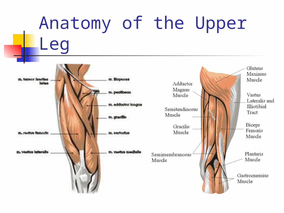
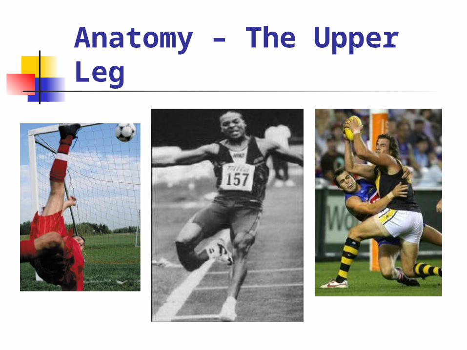
Anatomy of the Upper Leg
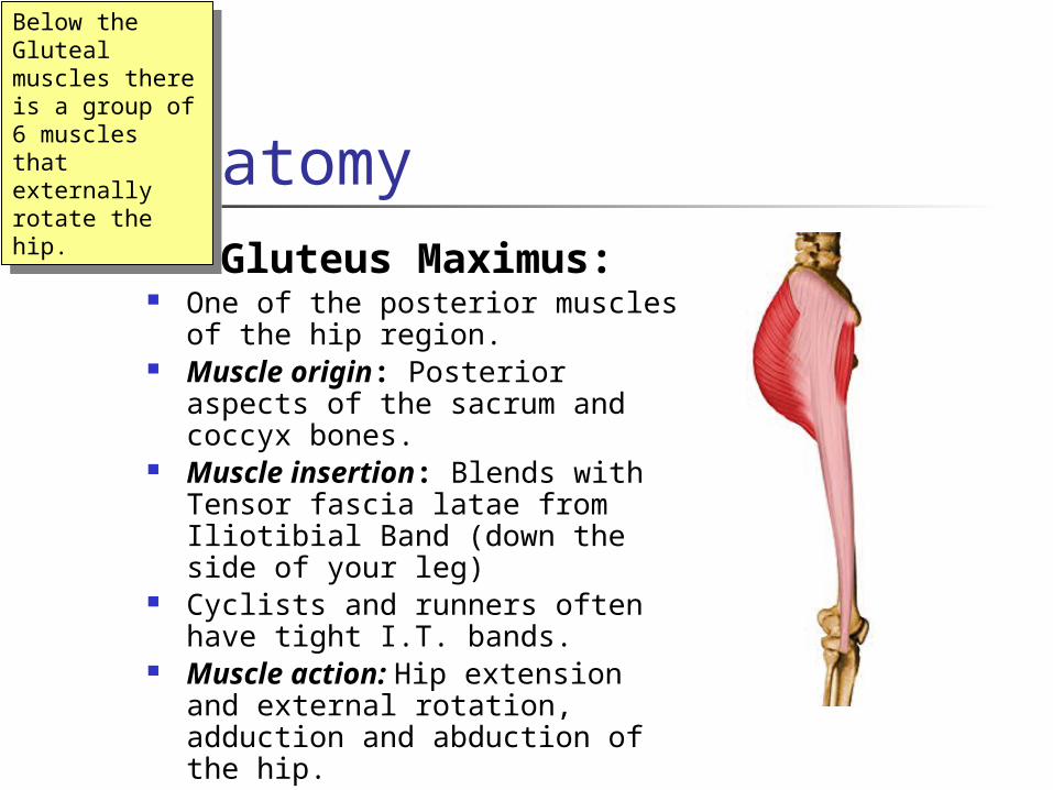
Anatomy1. Gluteus Maximus: One of the posterior muscles of
the hip region. Muscle origin: Posterior aspects
of the sacrum and coccyx bones. Muscle insertion: Blends with
Tensor fascia latae from Iliotibial Band (down the side of your leg)
Cyclists and runners often have tight I.T. bands.
Muscle action: Hip extension and external rotation, adduction and abduction of the hip.
Below the Gluteal muscles there is a group of 6 muscles that externally rotate the hip.
Below the Gluteal muscles there is a group of 6 muscles that externally rotate the hip.

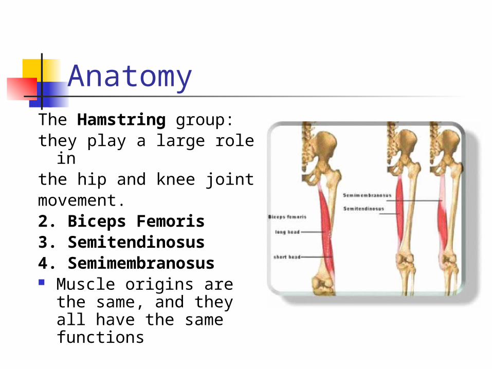
AnatomyThe Hamstring group:they play a large role inthe hip and knee jointmovement.2. Biceps Femoris3. Semitendinosus4. Semimembranosus Muscle origins are the
same, and they all have the same functions
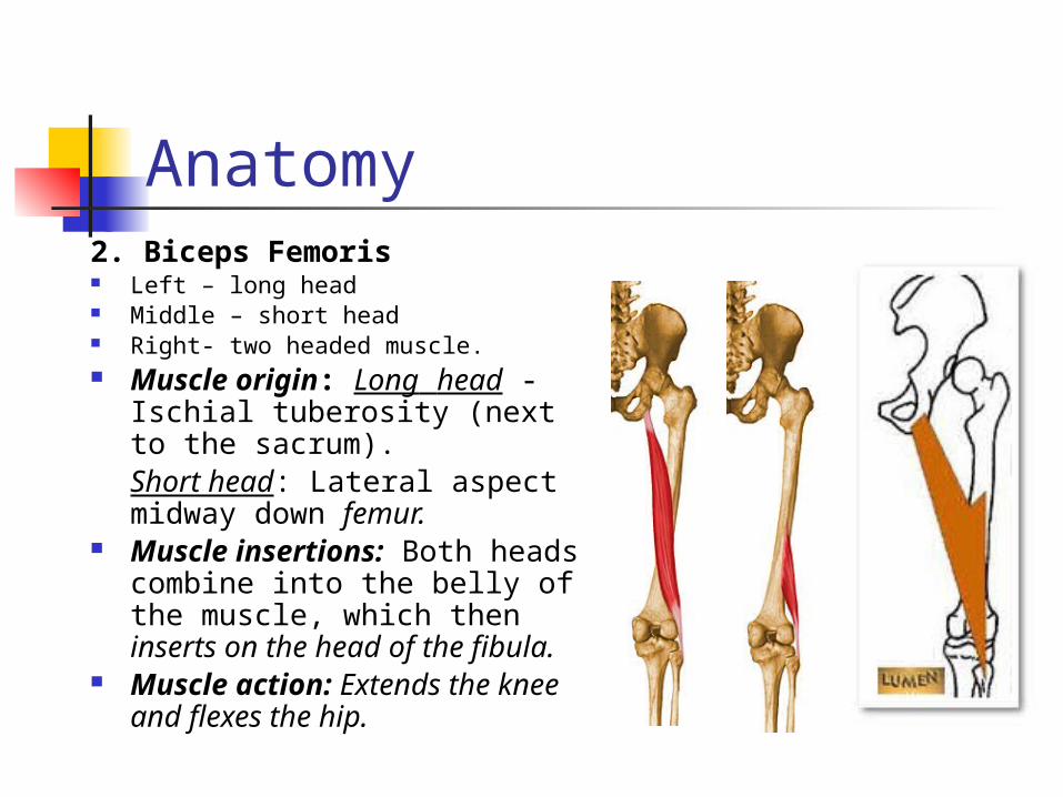
Anatomy2. Biceps Femoris Left – long head Middle – short head Right- two headed muscle. Muscle origin: Long head -
Ischial tuberosity (next to the sacrum).Short head: Lateral aspect midway down femur.
Muscle insertions: Both heads combine into the belly of the muscle, which then inserts on the head of the fibula.
Muscle action: Extends the knee and flexes the hip.
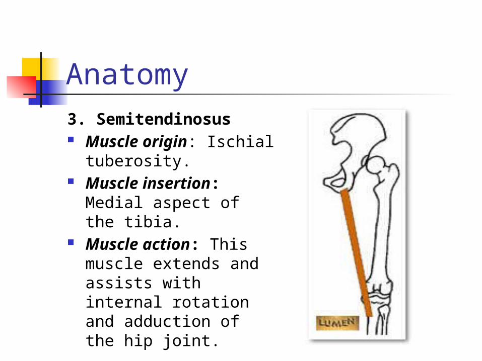
Anatomy3. Semitendinosus Muscle origin: Ischial
tuberosity. Muscle insertion:
Medial aspect of the tibia.
Muscle action: This muscle extends and assists with internal rotation and adduction of the hip joint.
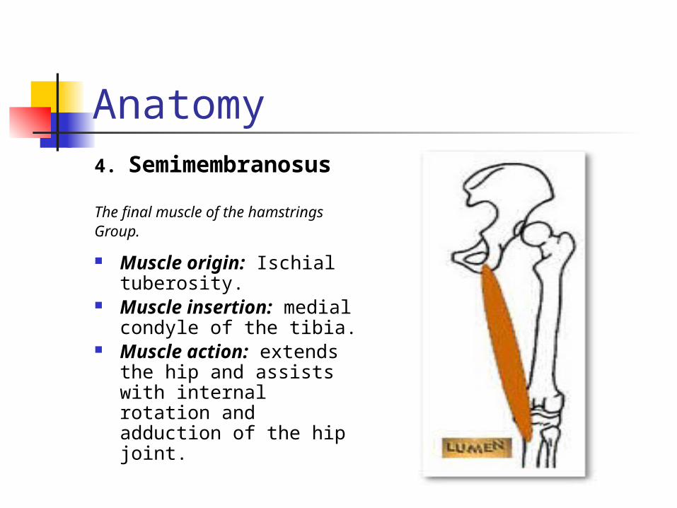
Anatomy4. Semimembranosus
The final muscle of the hamstringsGroup.
Muscle origin: Ischial tuberosity.
Muscle insertion: medial condyle of the tibia.
Muscle action: extends the hip and assists with internal rotation and adduction of the hip joint.
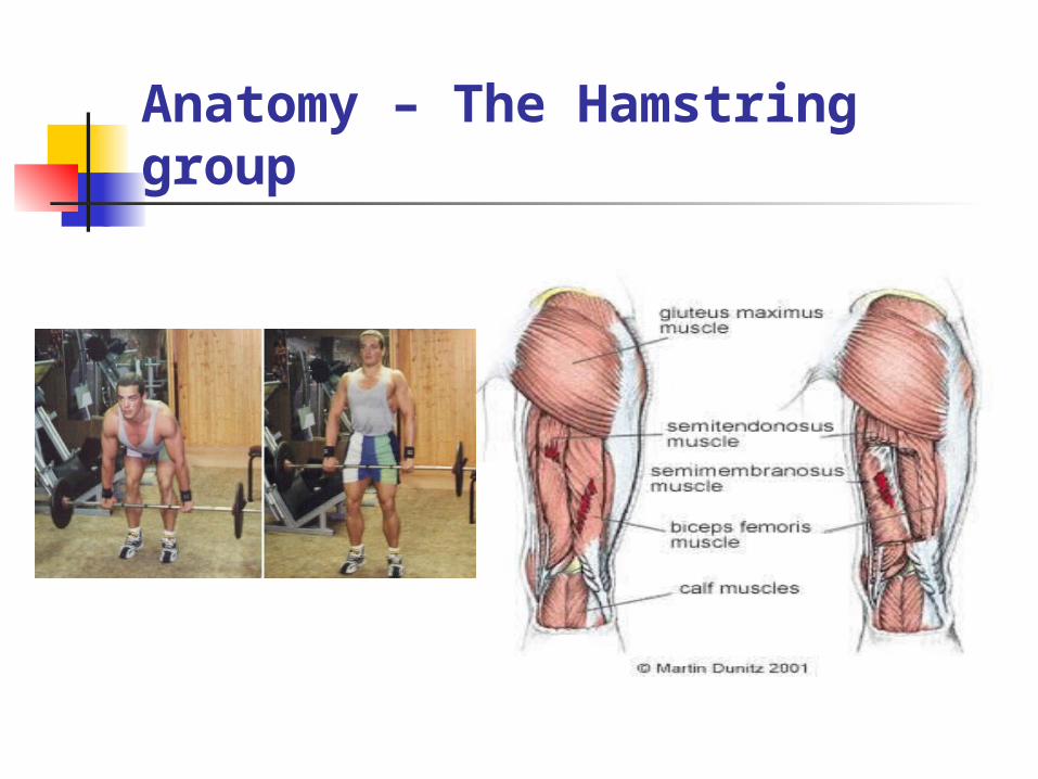
Anatomy – The Hamstring group

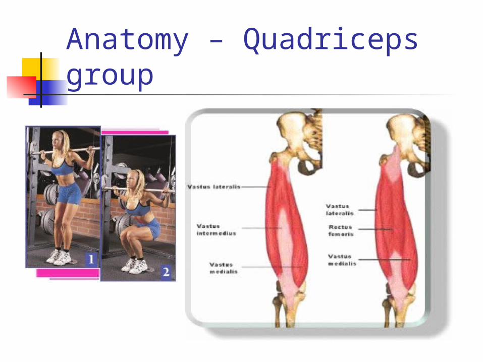
AnatomyThe Quadriceps Femoris,frequently referred to as the‘quads’ is a group of fourmuscles. One of these
musclesthe Rectus Femoris,
crossesboth the knee and the hip
joint.The other three muscles –
theVastus Intermedius, theVastus Medius and
theVastusLateralis cross only the
kneejoint and have only onefunction: extension of the
knee.
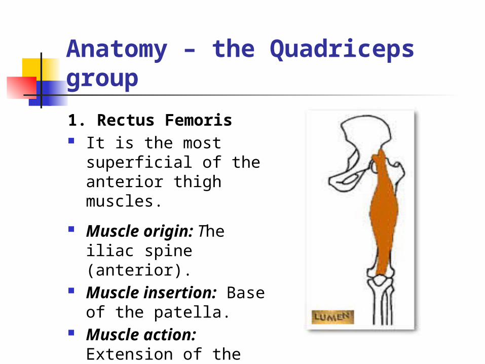
Anatomy – the Quadriceps group
1. Rectus Femoris It is the most
superficial of the anterior thigh muscles.
Muscle origin: The iliac spine (anterior).
Muscle insertion: Base of the patella.
Muscle action: Extension of the knee joint
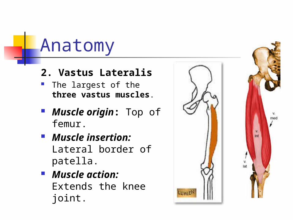
Anatomy2. Vastus Lateralis The largest of the three
vastus muscles.
Muscle origin: Top of femur.
Muscle insertion: Lateral border of patella.
Muscle action: Extends the knee joint.
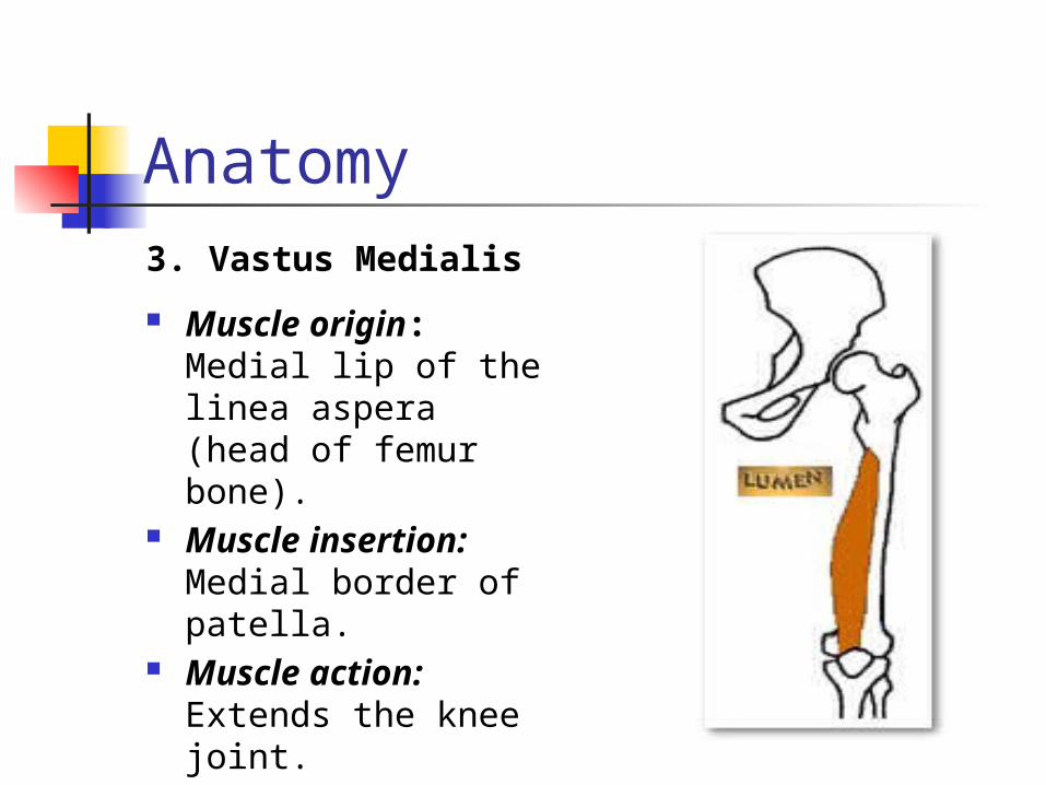
Anatomy3. Vastus Medialis
Muscle origin: Medial lip of the linea aspera (head of femur bone).
Muscle insertion: Medial border of patella.
Muscle action: Extends the knee joint.
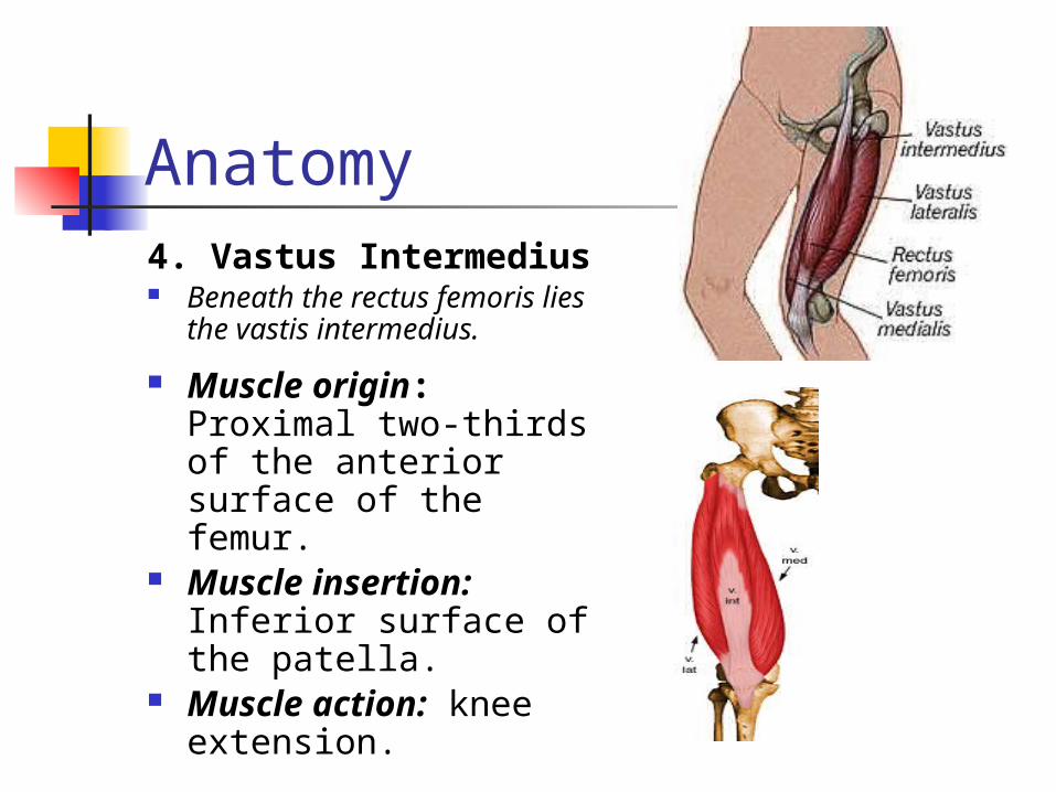
Anatomy4. Vastus Intermedius Beneath the rectus femoris
lies the vastis intermedius.
Muscle origin: Proximal two-thirds of the anterior surface of the femur.
Muscle insertion: Inferior surface of the patella.
Muscle action: knee extension.
Anatomy – Quadriceps group
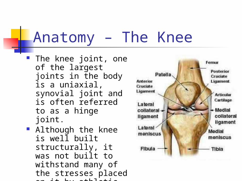
Anatomy – The Knee The knee joint, one of
the largest joints in the body is a uniaxial, synovial joint and is often referred to as a hinge joint.
Although the knee is well built structurally, it was not built to withstand many of the stresses placed on it by athletic activities.
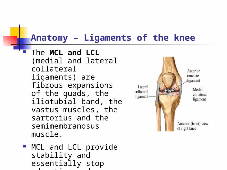
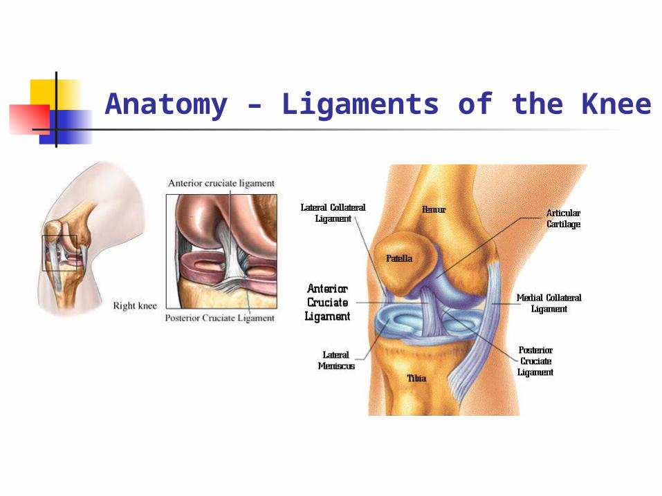
Anatomy – Ligaments of the knee The MCL and LCL
(medial and lateral collateral ligaments) are fibrous expansions of the quads, the iliotubial band, the vastus muscles, the sartorius and the semimembranosus muscle.
MCL and LCL provide stability and essentially stop adduction and abduction of the knee.
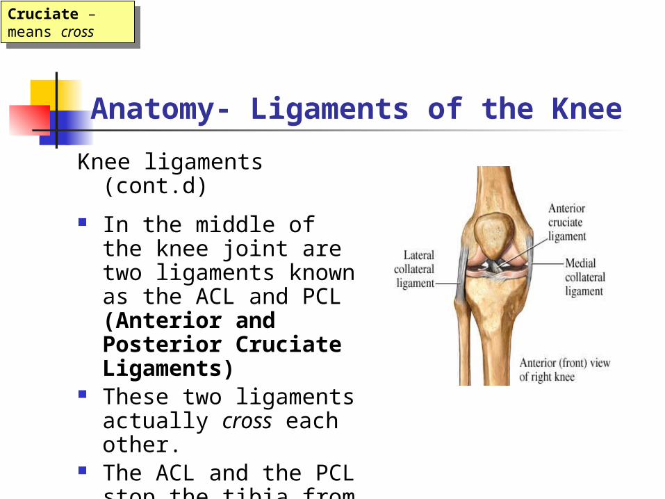
Anatomy- Ligaments of the Knee
Knee ligaments (cont.d)
In the middle of the knee joint are two ligaments known as the ACL and PCL (Anterior and Posterior Cruciate Ligaments)
These two ligaments actually cross each other.
The ACL and the PCL stop the tibia from displacing from the femur
Cruciate – means cross
Cruciate – means cross
Anatomy – Ligaments of the Knee
Anatomy
Movements of the knee
Flexion and Extension: When the knee flexes and extends the Tibia and Fibula rotate. When the knee flexes the leg internally rotates, and when it extends the knee externally rotates.
Anatomy – The Upper Leg
Anatomy – The Upper Leg