sps y2-2013
DESCRIPTION
SPS Y2-2013. What is different. 3 Weeks No Assignments! Combined assessment [MFS]. Schedule. Nitty Gritty. SDL Can be converted to an interactive discussion Email: ([email protected]) Content MUST be decided! 10ish C2, all working days (almost). - PowerPoint PPT PresentationTRANSCRIPT
SPS Y2-2013

What is different
• 3 Weeks• No Assignments! • Combined assessment [MFS]
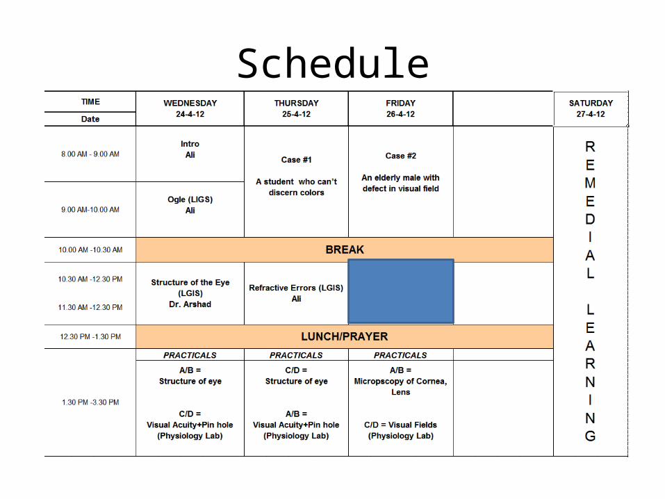
Schedule

Nitty Gritty
• SDL• Can be converted to an interactive discussion• Email: ([email protected])• Content MUST be decided!• 10ish C2, all working days (almost)
THE EYE BALL …A clinicians Perspective

OBJECTIVESIdentify the Multi-layered structure of the eye
Associate structure of each layer with its function
Infer the loss of integrity to loss of function of each layer of the eye
Advocate workplace safety in reference to protection of ocular structures


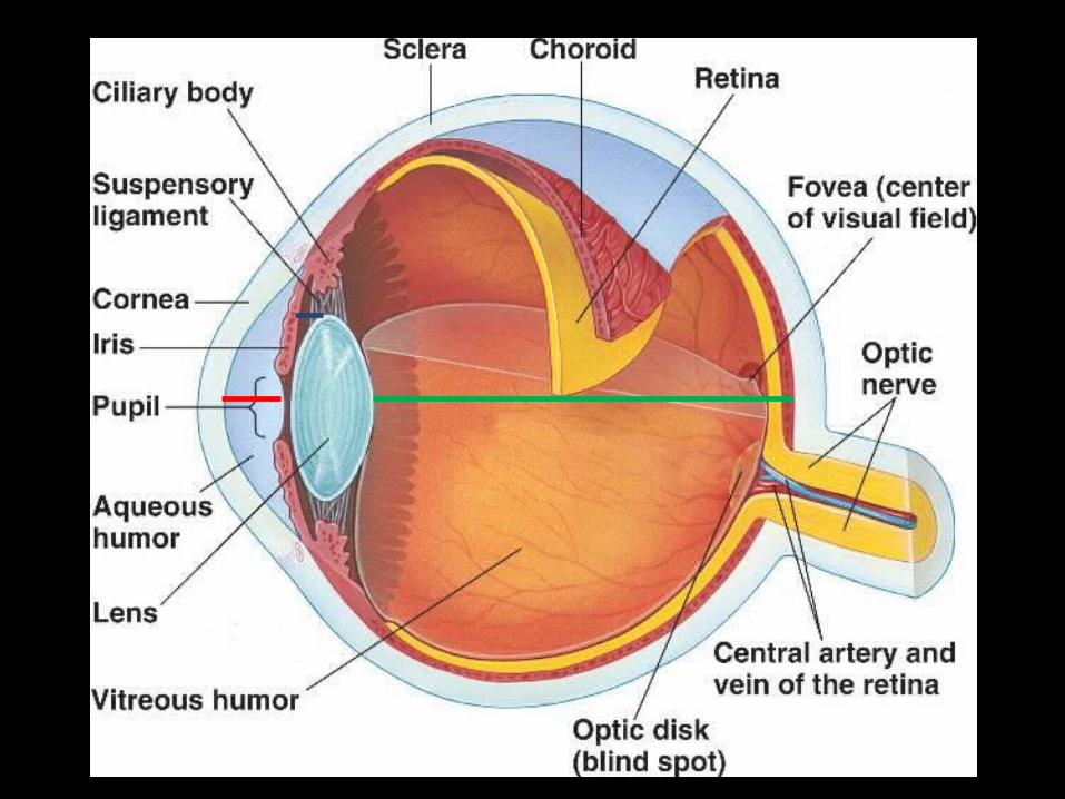
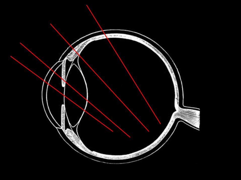
Cornea
Sclera*
Limbus
Pupil Iris
*Covered by a transparent vascular ‘skin’ -Conjunctiva

• 3 Skins/ Coverings• 3 Walls/ Layers/ Coats/ Tunic

THE SKINS/ COVERINGS
• Conjunctiva• Tenon’s Capsule• Episclera

SKINS/ COVERINGS
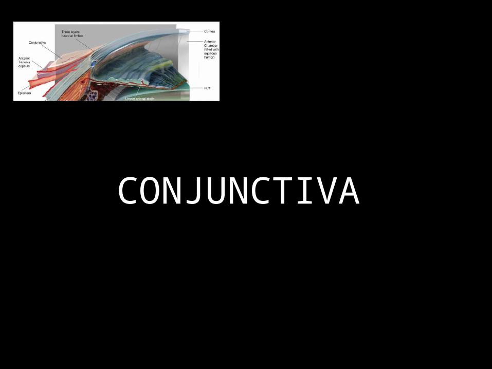
CONJUNCTIVA

Conjunctiva
1. Bulbar2. Palpebral

Conjunctiva
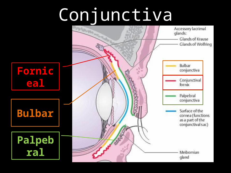
Bulbar; Forniceal; Palpebral
Conjunctiva
Bulbar
Forniceal
Palpebral

Conjunctiva
1. Bulbar2. Palpebral [hidden]3. Forniceal [hidden]
Conjunctiva
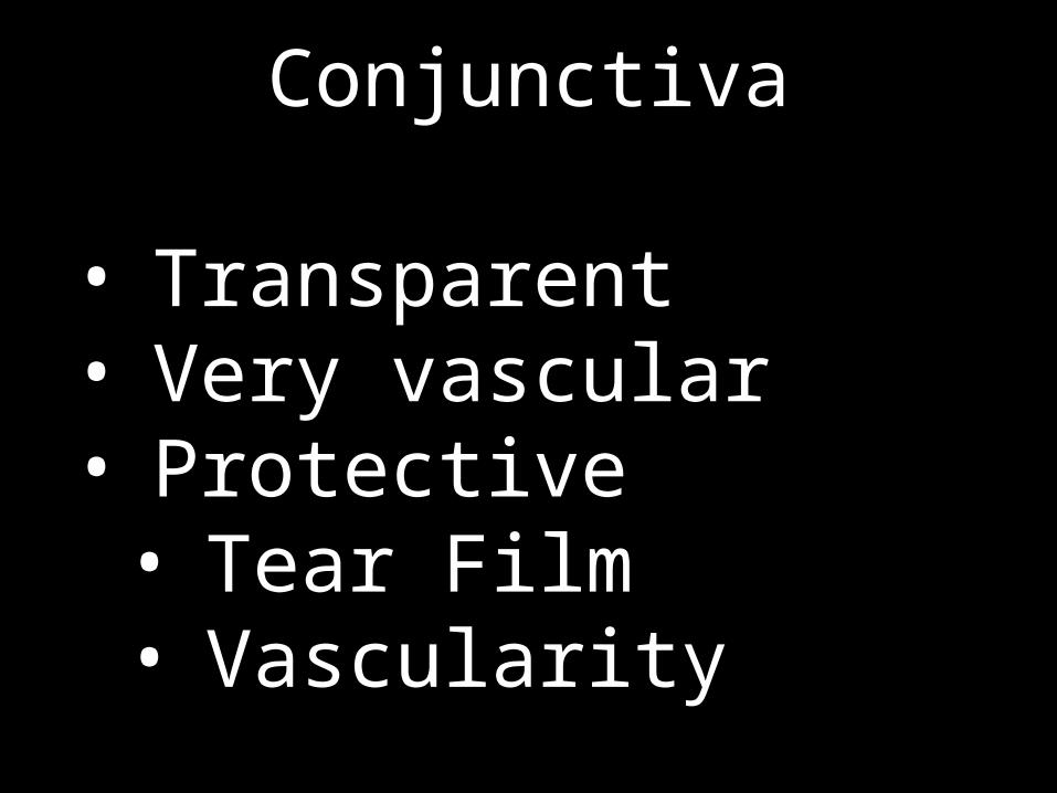
• Transparent• Very vascular• Protective• Tear Film• Vascularity
Conjunctiva
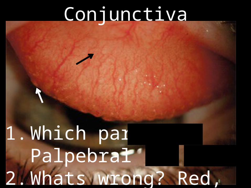
1. Which part? Palpebral2. Whats wrong? Red, Papillae3. Allergic Conjunctivitis
Conjunctiva
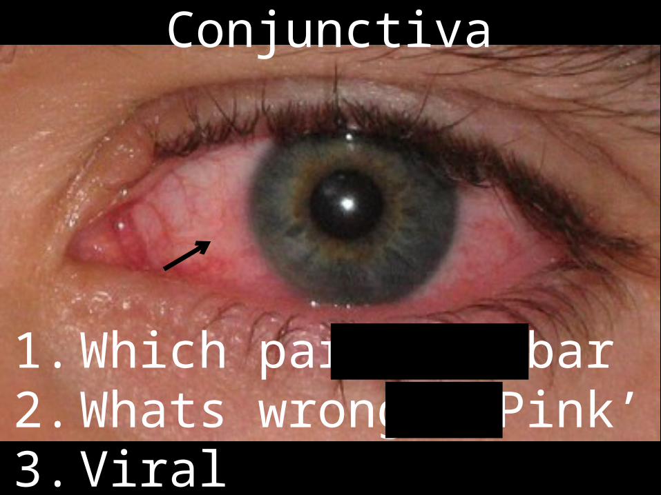
1. Which part? Bulbar2. Whats wrong? ‘Pink’ 3. Viral Conjunctivitis
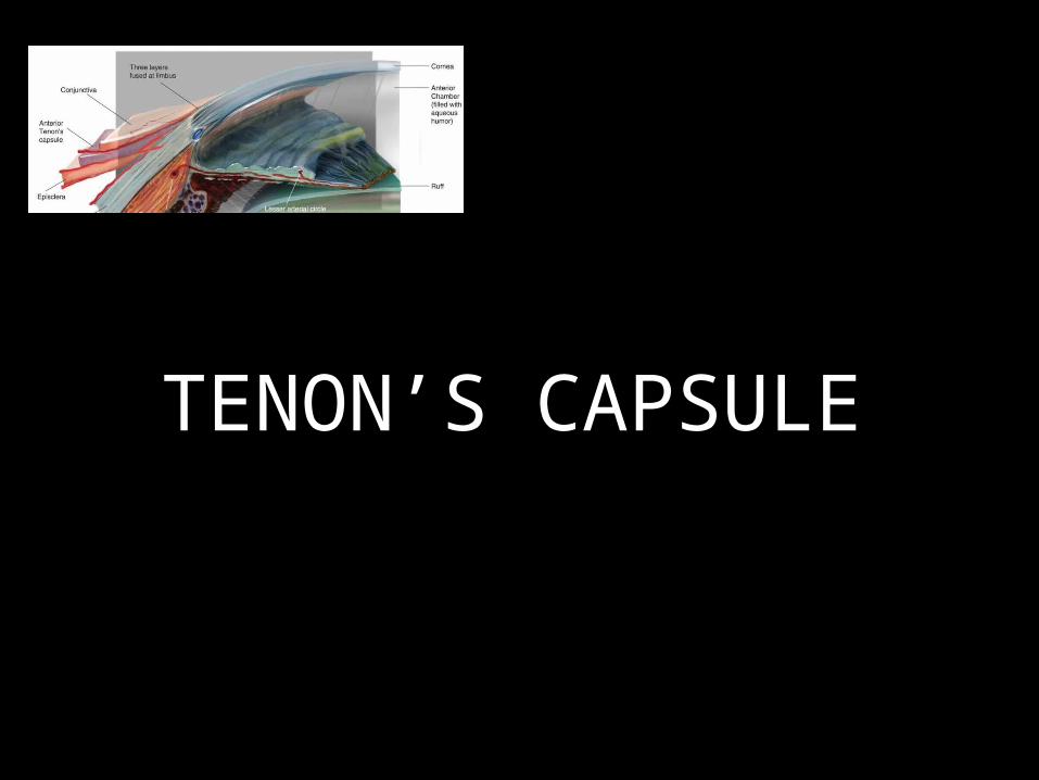
TENON’S CAPSULE
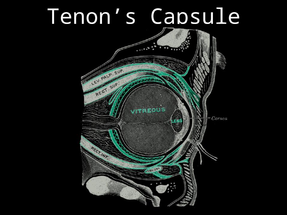
Tenon’s Capsule

Tenon’s Capsule

Tenon’s Capsule
• ‘Barrier’ Protection

EPISCLERA

Episclera

Episclera
• Inflammation of Episclera
Episcleritis
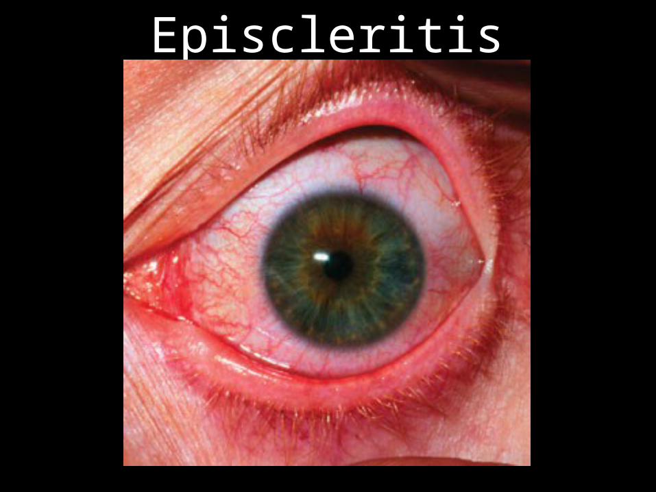
Episcleritis
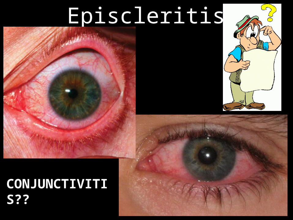
Episcleritis
CONJUNCTIVITIS??

• Blanching test…
Episcleritis Vs. Conjunctivitis

• Transparent• Vascular• Nutritive
Episclera

SKINS/ COVERINGS
THE WALLS/ COATS/ LAYERS

Protection
NutritionCrucial
Protection
NutritionCrucial

Protection
Nutrition
Crucial
WALLS/ COATS/ LAYERS

WALLS/ COATS/ LAYERS

CHAMBERS & SEGMENTS
Anterior Chamber
PosteriorChamber
AnteriorSegment
PosteriorSegment
AC ANGLE
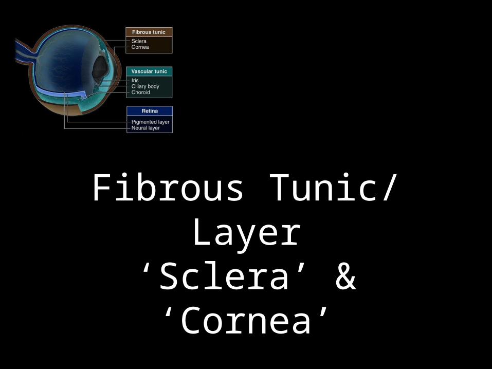
Fibrous Tunic/ Layer‘Sclera’ & ‘Cornea’
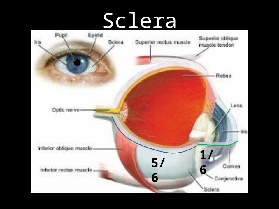
Sclera
5/61/6

Sclera

Sclera
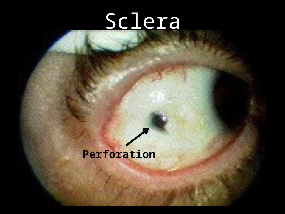
Sclera
Perforation

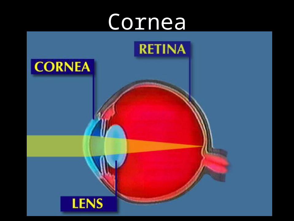
Cornea

Cornea
1. Epithelium2. Bowman’s3. Stroma4. Basement
membrane5. Endothelium
5. ENDOTHELIUM:Na-K PumpKeeps Cornea ‘Dry’
3. STROMA:Heals by scarring
1. EPITHELIUMBarrierRichly innervated
Cornea

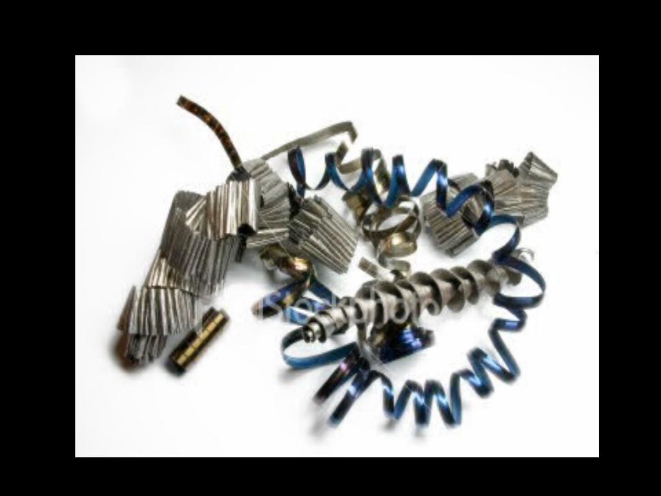
Cornea –Epithelial Injury
Metallic foreign body

Cornea –Stromal Injury
Opacity (Inflammation)

Cornea –Endothelial Injury

Cornea
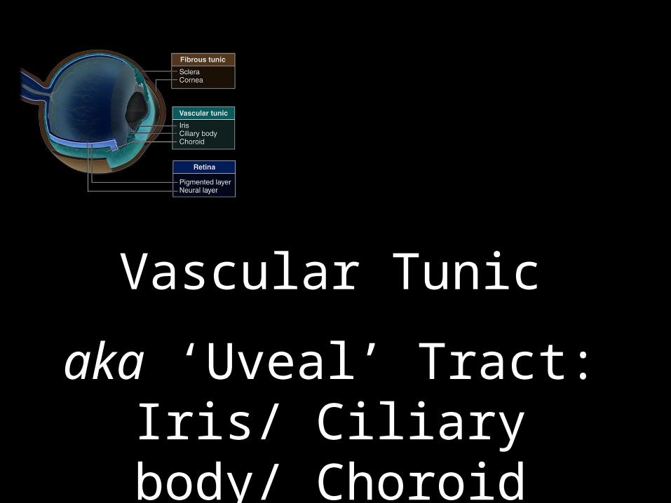
Vascular Tunic
aka ‘Uveal’ Tract:Iris/ Ciliary body/ Choroid


Choroid/ Ciliary Body/ Iris

Choroid
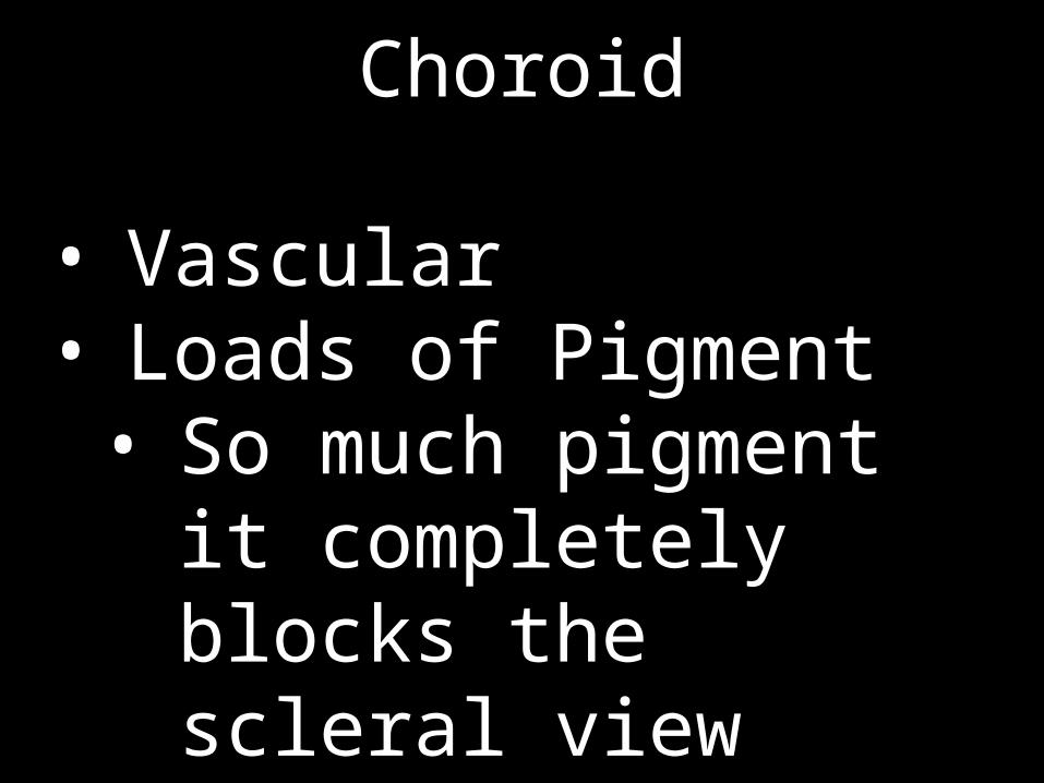
Choroid
• Vascular• Loads of Pigment• So much pigment it
completely blocks the scleral view

Ciliary Body
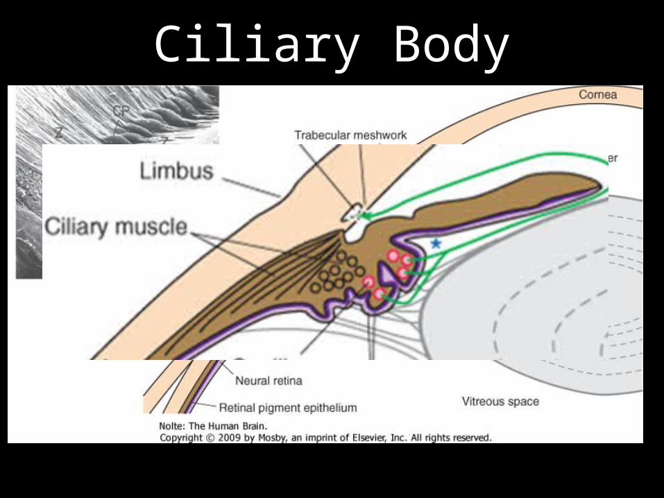
Ciliary Body
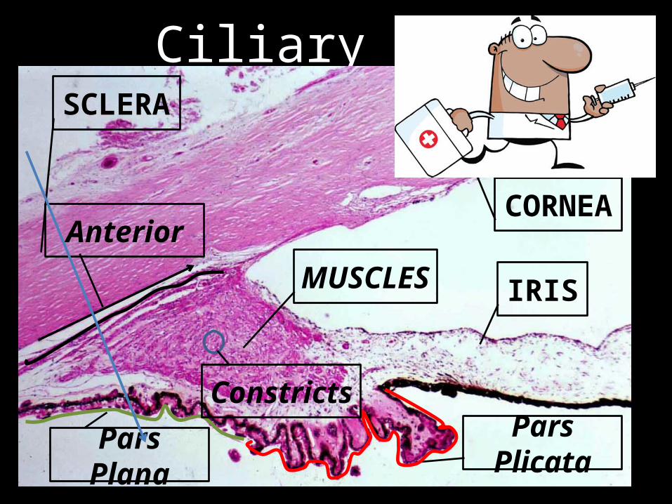
Ciliary Body
IRIS
CORNEA
SCLERA
MUSCLES
Pars PlicataPars Plana
Anterior
Constricts

Ciliary Body

Ciliary Body
• Supports Lens• Aids Accommodation• Produces Aqueous Humor
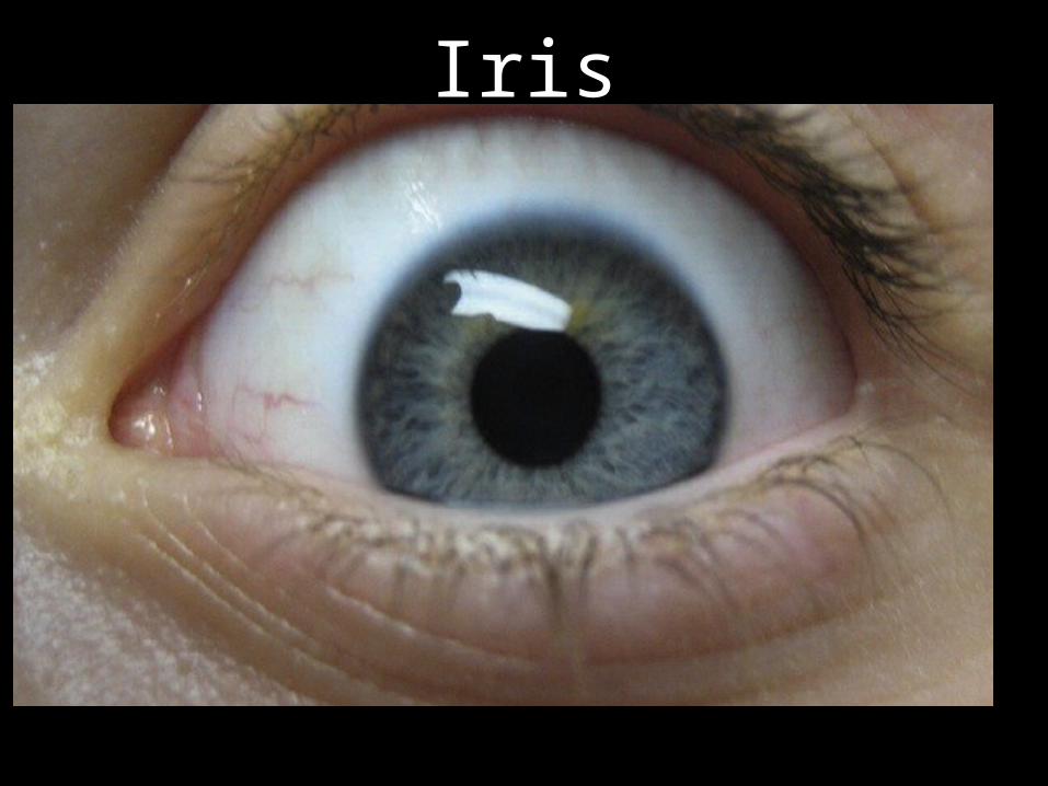
Iris
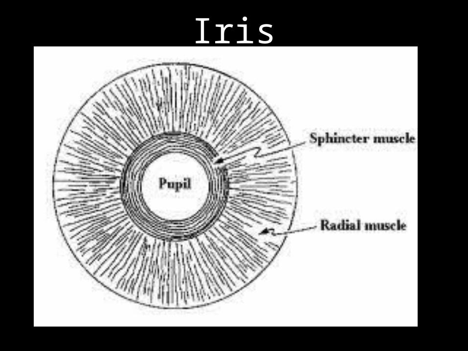
Iris
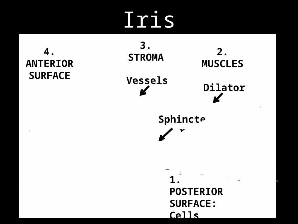
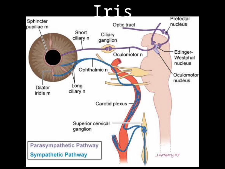
Iris2. MUSCLES
Dilator
Sphincter
3. STROMA4.
ANTERIOR SURFACE Vessels
1. POSTERIOR SURFACE:Cells

Iris

IrisIRIS
CORNEA
SCLERA
Ciliary Body

Iris

Iris
Iris

Iris
• Controls light entry • Attraction• Color is more than just
pigment!• Crypts!

• LENS• ANTERIOR CHAMBER
ANGLE
AREAS OF INTEREST

Lens

Lens
Lens
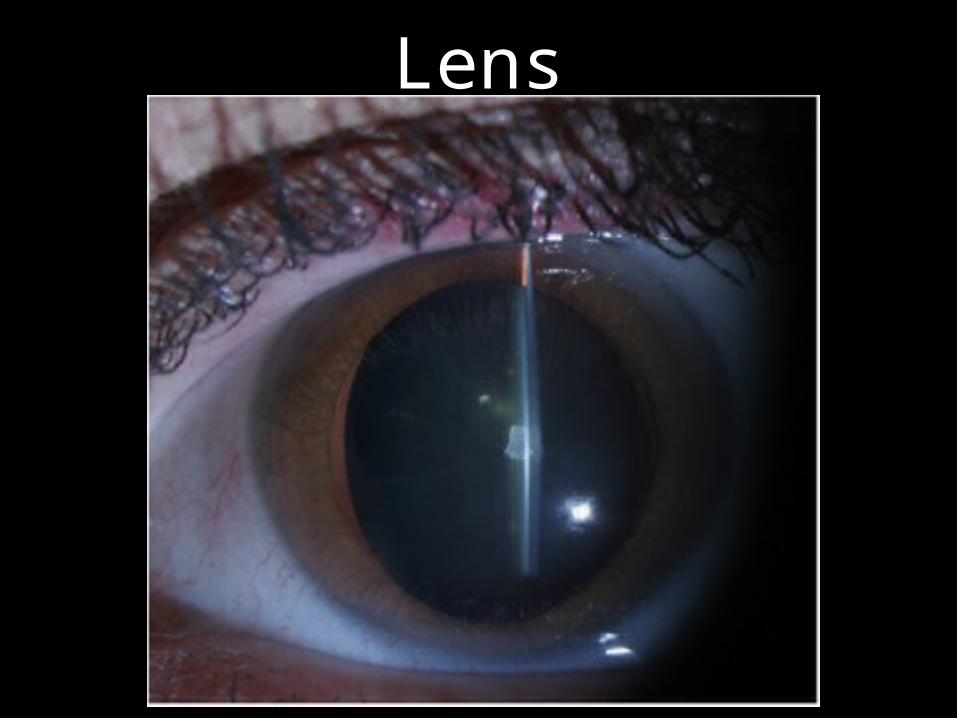
Lens

Lens
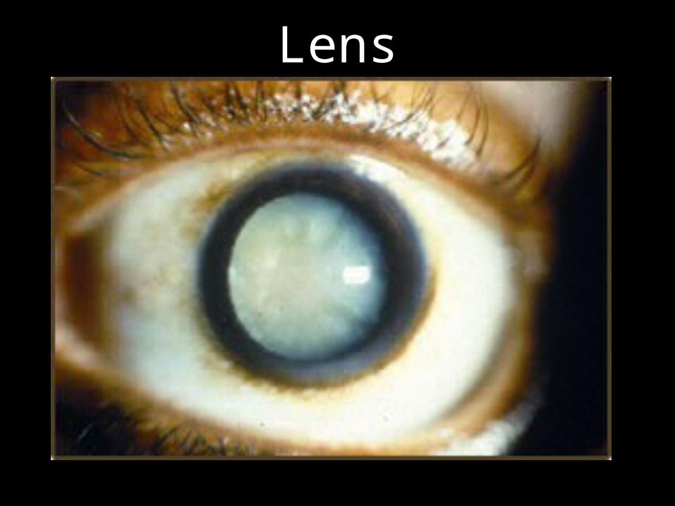
Cataract
Lens

AGE
Lens
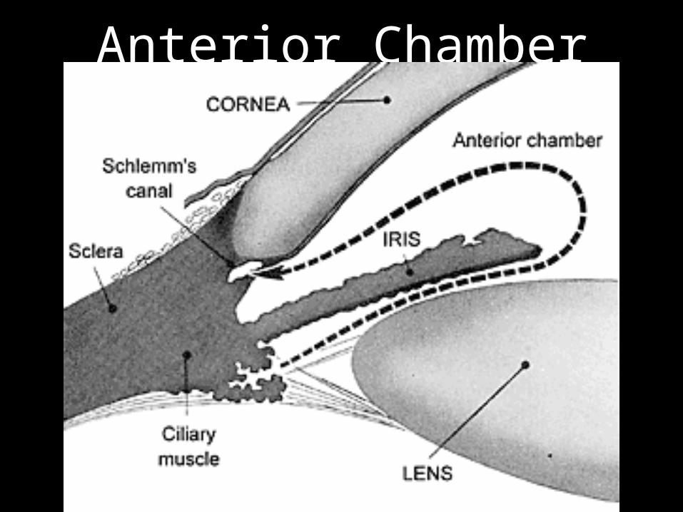
Anterior Chamber Angle
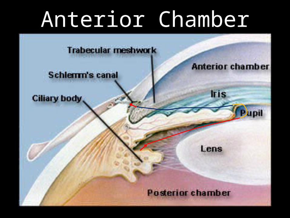
Anterior Chamber Angle
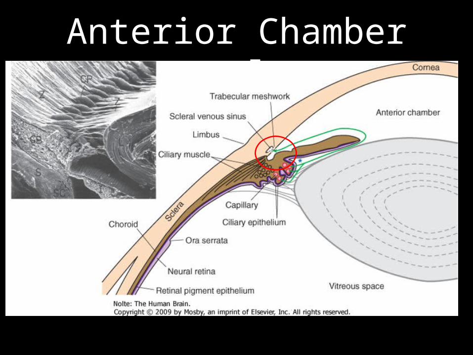
Anterior Chamber Angle
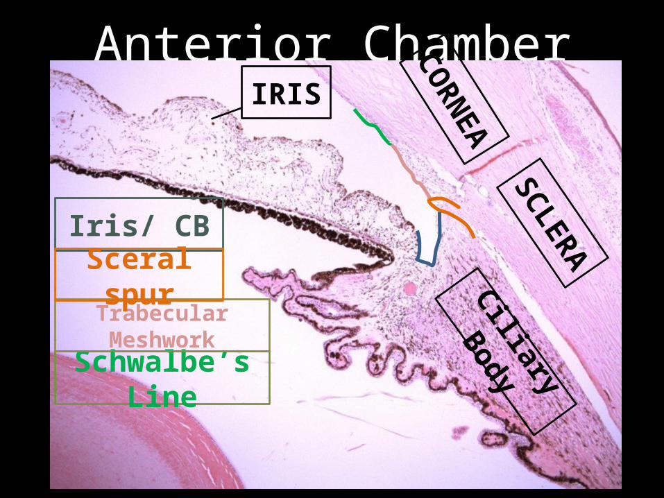
Anterior Chamber AngleIRIS
CORNEA
SCLERA
Ciliary Body
Iris/ CB
Schwalbe’s Line
Trabecular Meshwork
Sceral spur

Anterior Chamber Angle
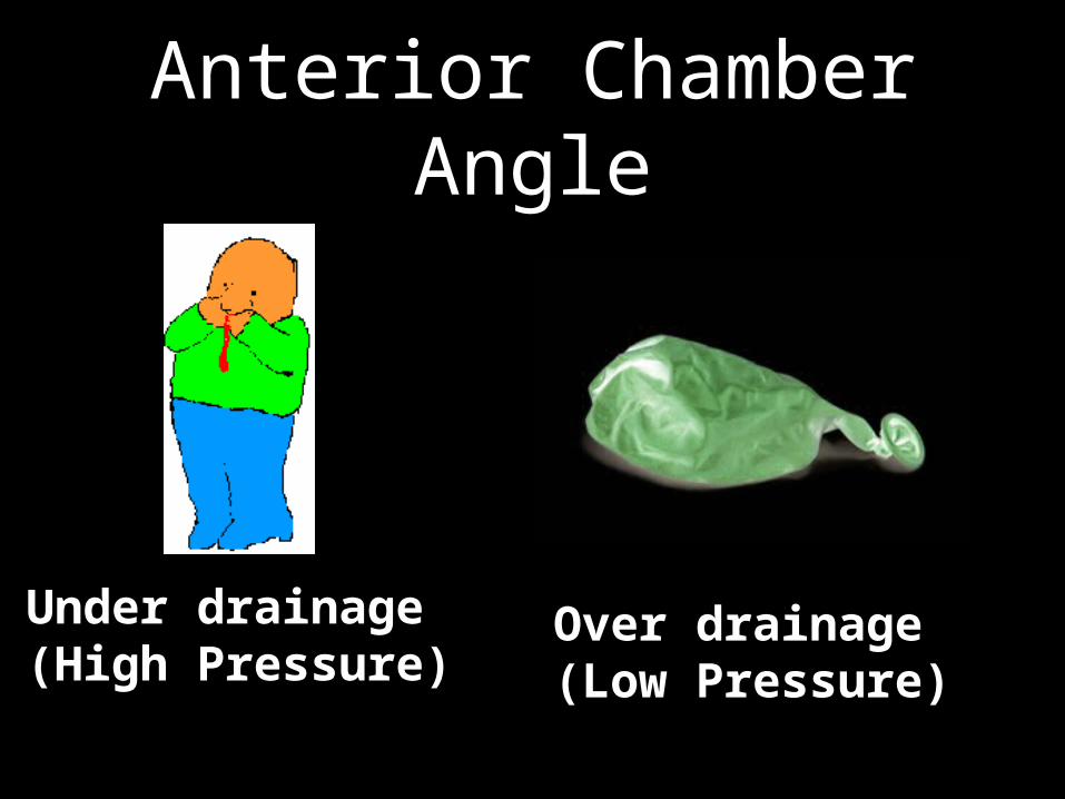
Anterior Chamber Angle
Under drainage (High Pressure)
Over drainage (Low Pressure)

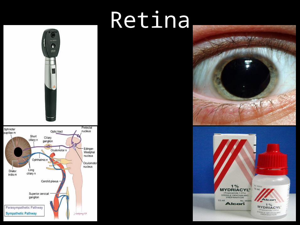
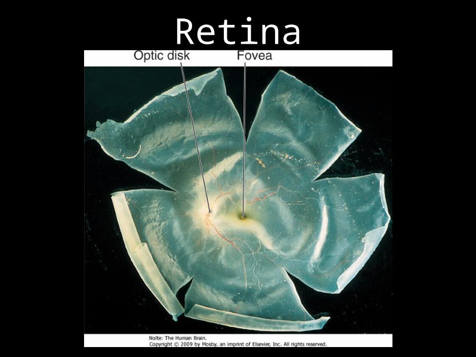
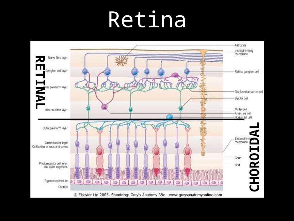
Retina

Retina

Retina –Ora Serrata

Retina –Ora Serrata
Retina
Retina

Retina
…pigment it completely blocks the scleral view

Retina
View obtained via an ophthalmoscope = fundus

Retina

Retina

Retina
Retina
CHO
ROID
ALRETIN
AL

Arterial Occlusion
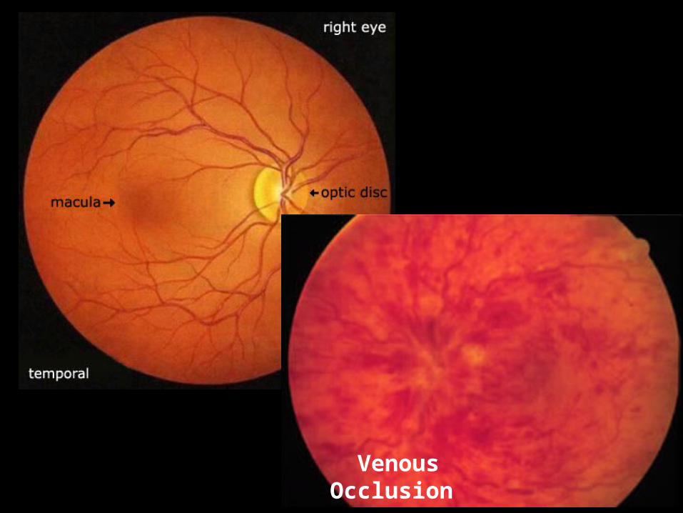
Venous Occlusion

Arterial Venous


Retina

Retina

Retina

Retina
• Light to electrical signals• All sorts of visual info
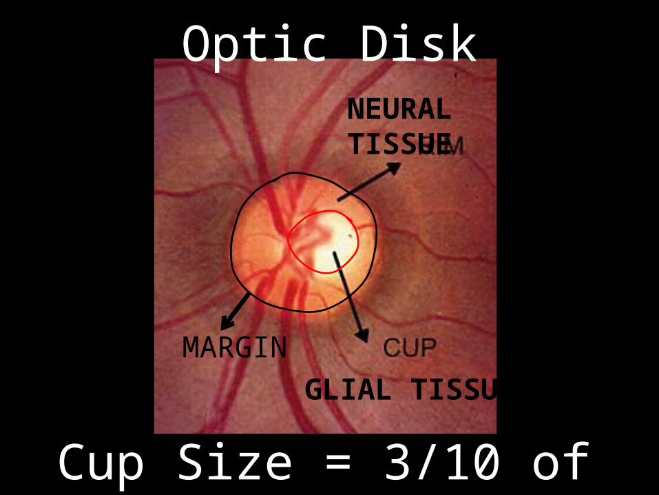
Optic Disk
GLIAL TISSUE
NEURAL TISSUE
MARGIN
Cup Size = 3/10 of disk Size

Optic Disk
• Visible portion of CN II• aka optic nerve head• Afferent for visual
information to the brain

Optic Disk
Cup Size = 8/10 of disk Size

Optic Disk
GLAUCOMA
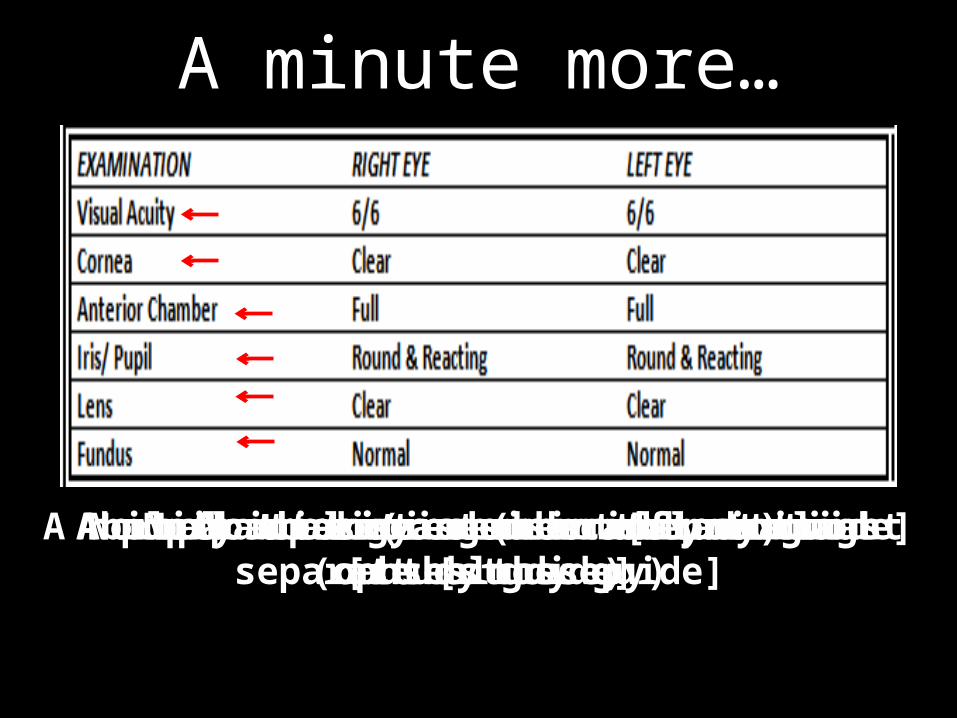
A minute more…
Ability to distinguish two points as separate [study guide]No opacities (remember inflammation causes these)A normal anterior chamber [study guide]A pupil that reacts normally to light [study guide]No pathology seen in the retina (ophthalmoscopy)No opacities (no cataract)
