srgs: critical care of surgical patients, part ii
TRANSCRIPT
® Vol 43 | 3 | 2017
SE L EC T E D R E A DI NGS in GENER A L SU RGERY
Critical Care of Surgical Patients, Part II
AMERICAN COLLEGE OF SURGEONS | DIVISION OF EDUCATIONBlended Surgical Education and Training for Life®
Cover: Printed on paper manufactured from 10% post-consumer waste and Green-e certified renewable energy.
Interior: Printed on paper manufactured from 100% post-consumer waste, Green Seal certified and processed chlorine free.
American College of SurgeonsDivision of Education633 N. Saint Clair St.Chicago, IL 60611-3211
[email protected]/srgs
®
SE L E C T E D R E A DI NG S in G E N E R A L SU RG E RY
®Vo
l 43 | 3 | 2017A
ME
RIC
AN
CO
LL
EG
E O
F S
UR
GE
ON
SC
ritical Care of Surgical Patients, Part II
Contemporary Management of Hemorrhagic Shockpage 10
Transfusion in Surgical Patientspage 2
Cardiogenic Shock Managementpage 44

iAmerican College of Surgeons facs.org/srgs SRGS Vol 43 | 3 | 2017
| CRITICAL CARE OF SURGICAL PATIENTS, PART II
Editor in chief Lewis Flint, MD, FACS
ACS steering committeeL. D. Britt, MD, MPH, FACS, chair
Ajit K. Sachdeva, MD, FACS, FRCSC
Patrice Gabler Blair, MPH
Editorial board Nita Ahuja, MD, FACS, The Johns Hopkins Medical Institutions, Baltimore, MD
L. D. Britt, MD, MPH, FACS, Eastern Virginia Medical School, Norfolk, VA
Ara Darzi, MD, FACS, FRCS(Eng), KBE, FMedSci, Imperial College of London, London, UK
Karen Deveney, MD, FACS, Oregon Health and Science University, Portland, OR
Michael B. Edye, MD, FACS, University of Western Sydney, Seven Hills, Australia
Jean C. Emond, MD, FACS, Columbia University Medical Center/New York-Presbyterian Hospital, New York, NY
John Ferrara, MD, FACS, Virginia Tech Carilion School of Medicine, Roanoke, VA
Donald E. Fry, MD, FACS, Michael Pine & Associates, Chicago, IL
Amy L. Halverson, MD, FACS, Northwestern Memorial Hospital, Chicago, IL
Tyler G. Hughes, MD, FACS, Memorial Hospital, McPherson, KS
Roger Keith, MD, FACS, University of Saskatchewan, Saskatoon, Canada
Solly Mizrahi, MD, FACS, Soroka Medical Center, Beer Sheva, Israel
Chandrajit Premanand Raut, MD, MSc, FACS, Brigham and Women’s Hospital, Boston, MA
Raul J. Rosenthal, MD, FACS, Cleveland Clinic Florida, Weston, FL
Ajit K. Sachdeva, MD, FACS, FRCSC, American College of Surgeons, Chicago, IL
Eduardo de Santibañes, MD, PhD, FACS, Instituto Universitario del Hospital Italiano de Buenos Aires, Buenos Aires, Argentina
Nathaniel J. Soper, MD, FACS, Northwestern Memorial Hospital, Chicago, IL
Steven Steinberg, MD, FACS, The Ohio State University Hospitals, Columbus, OH
Girma Tefera, MD, FACS, University of Wisconsin, Madison, WI
Christopher B. Weldon, MD, PhD, FACS, Children’s Hospital Boston, Boston, MA
Steven D. Wexner, MD, PhD(Hon), FACS, FRCS, FRCS(Ed), Cleveland Clinic Florida, Weston, FL
Editorial & business officesACS-SRGS 633 N. Saint Clair St. Chicago, IL 60611-3211P 800-631-0033 or 312-202-5227 F 312-202-5009 [email protected] | facs.org/srgs
Managing editorWhitney Greer, [email protected]
Project assistant Claire Sydow, [email protected]
The American College of Surgeons is a scientific and educational organization of surgeons that was founded in 1913 to raise the standards of surgical practice and improve the quality of care for surgical patients. The College is dedicated to the ethical and competent practice of surgery. Its achievements have significantly influenced the course of scientific surgery in America and have established it as an important advocate for all surgical patients. The College has more than 80,000 members and is the largest organization of surgeons in the world.
ACS disclosure policyIn accordance with ACCME accreditation criteria, ACS must ensure that anyone in a position to control the content of SRGS has disclosed all relevant financial relationships with any commercial interest. Members of the SRGS editorial board and those providing editorial assistance are required to disclose all financial relationships. All reported conflicts are managed by a designated official to ensure bias-free content. However, if you perceive a bias, please contact us at [email protected]. The following relationships were disclosed in 2017:
Ara Darzi, MD, FACS, FRCS(Eng), KBE, FMedSci has disclosed a commercial interest in G.E. Healthymagination; Donald E. Fry, MD, FACS, had disclosed a commercial interest in Becton Dickinson, IrriMax Corporation, and Prescient Surgical Co.; Raul J. Rosenthal, MD, FACS, has disclosed a commercial interest in Ethicon, Medtronics, and STORZ; Nathaniel Soper, MD, FACS has disclosed a commercial interest in Flex Dex, Inc., MD Insider, and Miret Surgical, Inc.; Steven D. Wexner, MD, PhD(Hon), FACS, FRCS, FRCS(Ed), has disclosed a commercial interest in Actamax, Asana Medical, Axonics Modulation Technologies, Brace Pharmaceuticals, CRH Medical, Covidien, Intuitive Surgical, KARL STORZ Endoscopy-America, Inc., LifeBond, Mederi Therapeutics, Medtronic, NeatStitch, Novadaq, novoGI, Pragma, Renew Medical, and Unique Surgical Innovations, LLC.
Subscription informationVisit facs.org/srgs for order information. Prepayment in U.S. dollars is required to activate a subscription.
Back issues:Current subscribers can purchase back issues (print only) for $50/issue; nonsubscribers, $85/issue.
Payment should be sent to:ACS-SRGS 633 N. Saint Clair St. Chicago, IL 60611-3211
Renew online at facs.org/ publications/srgs/subscriptions/renew.
To place an order over the telephone:
Call 800-631-0033 or 312-202-5227. Please have your ACS-SRGS ID number and credit card information available.
Address changes:Please notify us of any address changes six weeks prior to a move.
Missing issues:Lost or missing issues must be reported within eight weeks after the issue has been mailed. Consult facs.org/srgs for mailing dates. Two missing issues per year per subscription can be replaced.
To change your address or to report a missing issue: Call 800-631-0033 or 312-202-5227 Fax 312-202-5009 E-mail [email protected]
Postmaster:Send address changes to: ACS-SRGS 633 N. Saint Clair St. Chicago, IL 60611-3211

ii American College of Surgeons facs.org/srgs SRGS Vol 43 | 3 | 2017
| CRITICAL CARE OF SURGICAL PATIENTS, PART II
Learning objectivesThis activity is designed for general surgeons, surgical residents, and allied professionals. Regular reading of SRGS should enable learners to:
• Maintain an excellent knowledge base in all areas of general surgery
• Develop comparative and critical literature reading skills
• Apply newly acquired knowledge to surgical practice
• Prepare effectively for recertification exams
Additional information at facs.org/publications/srgs/cme
Maintenance of certification The American Board of Surgery (ABS) recognizes SRGS as a resource for surgeons enrolled in its Maintenance of Certification (MOC) program. Successful completion of the SRGS program fulfills MOC Part 2 requirements that focus on lifelong learning and self-assessment.
ACS in cooperation with ABS has created a process wherein ACS members can directly submit their ACS CME transcript to the ABS for MOC purposes. For more information, go to facs.org, click Member Login and enter your ACS user name and password. Then, go to My Profile, My CME, and click on “Send Credit to ABS.”
For information on ABS’s MOC requirements, go to http://absurgery.org and click on “Maintenance of Certification (MOC)” or e-mail [email protected].
Questions about ACS CME can be e-mailed to [email protected] or call 866-918-4799.
Statement of purposeSelected Readings in General Surgery (SRGS) is a topic oriented, in-depth review of the field of general surgery presented eight times annually as an educational offering of the Division of Education of the American College of Surgeons. The mission of the Division of Education is to improve the quality of surgical care through lifelong learning, based on educational programs and products designed to enhance the competence or performance of practicing surgeons, surgery residents, and members of the surgical team. The intent of the publication is to analyze relevant
medical literature to give the surgeon the knowledge necessary to practice state-of-the-art surgery. To accomplish this goal, the editor selects 100–125 pertinent articles from the literature for each issue. Each article is reviewed and an overview is written that places the content of these articles in the perspective of the best, day-to-day, clinical practice. In addition to the overview, 12–18 full-text articles are reprinted in each issue.
The literature review is compiled with the assistance of an 18-member, international board of editors who are experts in the various focus areas that comprise the specialty of surgery. In addition, the editorial board has representation and expertise in such important fields as
medical evidence evaluation, surgical education, outcomes research, standard setting, and performance improvement. SRGS is a unique resource because the overview and selected full-text articles provide the reader with the most valuable and pertinent content illuminated with informed opinion and critique. Unnecessary material is eliminated. SRGS does not present itself as infallible and the editor-in-chief takes responsibility for the content that appears in each issue. The editor-in-chief and the editorial board recognize that there is no such thing as the “average” surgical patient, and that the information in the literature must be interpreted in the light of the clinical presentation of each individual patient.
CopyrightMaterial printed in SRGS is covered by copyright law. The overview and CME tests are copyrights of the American College of Surgeons. Permission has been obtained from individual journal publishers to reprint articles that appear in SRGS. Copying all or portions of this journal for distribution to a group practice, residency program, university, hospital, or colleague is strictly prohibited.
© 2017 American College of Surgeons All rights reserved
CONTINUING MEDICAL EDUCATION CREDIT INFORMATION
Accreditation The American College of Surgeons is accredited by the Accreditation Council for Continuing Medical Education (ACCME) to provide continuing medical education for physicians.
CME credit The American College of Surgeons designates this enduring material for a maximum of 10 AMA PRA Category 1 Credits.™* Physicians should claim only the credit commensurate with the extent of their participation in the activity.
*Of the AMA PRA Category 1 Credits™ listed above, a maximum of 10 credits meet the requirements for Self-Assessment.
AMERICAN COLLEGE OF SURGEONSDIVISION OF EDUCATION

| CRITICAL CARE OF SURGICAL PATIENTS, PART II
AMERICAN COLLEGE OF SURGEONS | DIVISION OF EDUCATIONBlended Surgical Education and Training for Life®
®
SE L EC T E D R E A DI NGS in GE NE R A L SU RGE RY
Spend your time learning, not searching • Explore critical, evidence-based surgical research • Earn up to 80 CME Self-Assessment Credits • Access SRGS in print, online, or on any mobile device
Subscribe today!facs.org/srgs
Title Volume/Issue Publication Date
Vascular Surgery, Part III V43N1 Published
Critical Care of Surgical Patients, Part I V43N2 Published
Critical Care of Surgical Patients, Part II V43N3 Published
Trauma, Part I V43N4 Summer
Trauma, Part II V43N5 Summer
Surgical Infection V43N6 Fall
Nutrition and Metabolic Disease V43N7 Fall
Wound Healing and Burn Injuries V43N8 Winter
2017 SRGS Publishing Schedule
Visit facs.org/publications/srgs/issues/upcoming for a list of previously published topics and next year’s topics.
Are you taking advantage of all the
American College of Surgeons has to offer?
Membership has its benefits.• ACS advocacy efforts that work for you and
your patients at both the federal and state levels
• Educational programs to help you stay up to date and ahead of the curve while meeting your continuing medical education requirements
• The Journal of the American College of Surgeons, the Bulletin of the American College of Surgeons, and other publications that bring you cutting-edge research and news from the College and surgical community across the globe
• Robust research data and programs that focus on outcomes and other quality issues
• Scholarships and fellowships
• A major visibility program to enhance your public image, underscoring why surgeons are an essential and integral part of this country’s health care delivery system
• Committees that offer opportunities for engagement
• Insurance products with special ACS rates
• A career center and resume posting opportunities
• A coding consultation hotline
• Access to local ACS chapters
“ As the largest and most robust surgical organization in the world, we have so much to offer surgeons of all specialties, at any point in their careers. From transition to practice support for those just starting out, to ongoing training and education, to advocacy and leadership, we help surgeons advance their careers and elevate the profession in a way no other organization can.”
— Patricia L. Turner, MD, FACS, Director, ACS Division of Member Services
T H E A M E R I C A N C O L L E G E O F S U R G E O N S ’ T O P P R I O R I T Y I S P R O V I D I N G VA L U E F O R I T S M E M B E R S .
B E C O M E A M E M B E R T O DAY.
facs.org/member-services/join

vAmerican College of Surgeons facs.org/srgs SRGS Vol 43 | 3 | 2017
Introduction ..................................................1
Transfusion in Surgical Patients ............2Risks of Transfusion
Transfusion-Related Disease Transmission
Current Transfusion Practices
Techniques of Perioperative Blood Salvage
Management of Patients Who Refuse Blood & Blood Products
Shock Due to Hemorrhage & Trauma ......................................................10Monitoring Patients in Shock
Hemodynamic Monitoring
Monitoring Oxygenation & Perfusion
Monitoring at the Tissue Level
Shock & Resuscitation-Related Coagulopathy
Diagnosis of Trauma-Related Coagulopathy
Resuscitation Strategies
Outcomes of Hemostatic Resuscitation Protocols
Potential Complications of Hemostatic Resuscitation
Hypertonic Saline Resuscitation
Vasopressor Agents
Use of Trauma Resuscitation Strategies in Nontrauma Patient Care
Recombinant Factor VIIa
Sepsis & Septic Shock: Pathophysiology & Management ......................................... 33Critical Care of the Patient with Sepsis & Septic Shock
Goal-Directed Management of Suspected Sepsis & Septic Shock
Vasopressor Use
Corticosteroid Use
Recombinant Activated Protein C Use
Antimicrobial Therapy
Cardiogenic Shock Management ............................................. 44
Conclusion .................................................. 48
References .................................................. 49
Posttest........................................................ 55
Recommended Reading ....................... 60
Literature Review Editor in Chief: Lewis Flint, MD, FACS Associate Editor: Nicholas Namias, MD, FACS
VOLUME 43 | 3 | 2017
Table of Contents CRITICAL CARE OF SURGICAL PATIENTS, PART II
THE AMERICAN COLLEGE OF SURGEONS is a leader in initiatives to improve quality of care for surgical patients in the areas of trauma, cancer, bariatric surgery,
breast care, general surgery, and surgeon-specific outcomes.
Visit facs.org/quality-programs to learn more.
1American College of Surgeons facs.org/srgs SRGS Vol 43 | 3 | 2017
W elcome to Selected Readings in General Surgery (SRGS). In the second part of our two-issue series focusing on critical care of the surgical patient, we will begin by reviewing articles relevant to the use of blood and
blood products in surgical practice. Emphasis on safe transfusion practices has increased over the past decade and the articles reviewed will provide information on recognizing and managing transfusion reactions, evidence-based guidance for choosing the optimum hemoglobin trigger for transfusion, and the controversy regarding the potential risks of transfusing blood that is approaching the end of its storage cycle vs. using fresh blood.
Following the transfusion discussion, we will review articles on managing shock in surgical patients. We will also explore the mechanisms and management of hemorrhage-associated coagulopathy as well as the management of sepsis, septic shock, and cardiogenic shock.
Nicholas Namias, MD, FACS, provided valuable editorial assistance for this critical care series and I am grateful for his help.
VOLUME 43 | 3 | 2017
Introduction CRITICAL CARE OF SURGICAL PATIENTS, PART II

2 American College of Surgeons facs.org/srgs SRGS Vol 43 | 3 | 2017
Transfusion in Surgical PatientsRed blood cell transfusion is a valuable clinical tool that can be a lifesaving intervention. To transfuse effectively, an understanding of the risks associated with transfusion is important; common risks include transfusion reactions and disease transmission. Similarly, knowledge of data supporting current transfusion practices is necessary to use blood and blood products as safely and effectively as possible.
Risks of Transfusion
Transfusion reactions are the most common type of com-plication of blood product administration. Delaney and coauthors1 discussed this topic in The Lancet, 2016. The authors noted that transfusions are an essential element of health care but are associated with significant risks and costs. Recognition of the feasibility and safety of restricted transfusion protocols has helped reduce the frequency of transfusions and transfusion reactions as well as reduce associated costs. Transfusion reactions occur in up to one
Symptoms and suggested interventions based on type of transfusion reaction, part one. Reproduced from Delaney and coauthors1 with permission.Figure 1
Transfusion in Surgical Patients | CRITICAL CARE OF SURGICAL PATIENTS, PART II
3American College of Surgeons facs.org/srgs SRGS Vol 43 | 3 | 2017
out of every 100 transfusions; fatal transfusion reactions are rare, occurring in one out of every 200,000−400,000 units transfused.
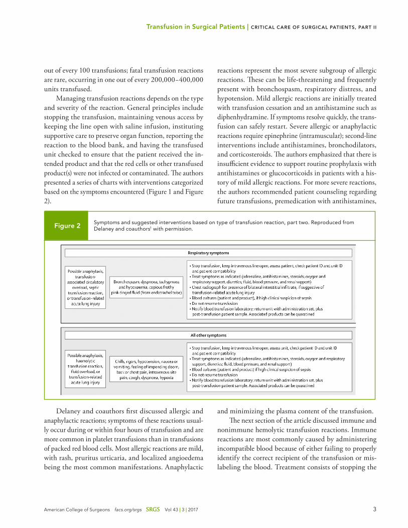
Managing transfusion reactions depends on the type and severity of the reaction. General principles include stopping the transfusion, maintaining venous access by keeping the line open with saline infusion, instituting supportive care to preserve organ function, reporting the reaction to the blood bank, and having the transfused unit checked to ensure that the patient received the in-tended product and that the red cells or other transfused product(s) were not infected or contaminated. The authors presented a series of charts with interventions categorized based on the symptoms encountered (Figure 1 and Figure 2).
Delaney and coauthors first discussed allergic and anaphylactic reactions; symptoms of these reactions usual-ly occur during or within four hours of transfusion and are more common in platelet transfusions than in transfusions of packed red blood cells. Most allergic reactions are mild, with rash, pruritus urticaria, and localized angioedema being the most common manifestations. Anaphylactic
reactions represent the most severe subgroup of allergic reactions. These can be life-threatening and frequently present with bronchospasm, respiratory distress, and hypotension. Mild allergic reactions are initially treated with transfusion cessation and an antihistamine such as diphenhydramine. If symptoms resolve quickly, the trans-fusion can safely restart. Severe allergic or anaphylactic reactions require epinephrine (intramuscular); second-line interventions include antihistamines, bronchodilators, and corticosteroids. The authors emphasized that there is insufficient evidence to support routine prophylaxis with antihistamines or glucocorticoids in patients with a his-tory of mild allergic reactions. For more severe reactions, the authors recommended patient counseling regarding future transfusions, premedication with antihistamines,
and minimizing the plasma content of the transfusion. The next section of the article discussed immune and
nonimmune hemolytic transfusion reactions. Immune reactions are most commonly caused by administering incompatible blood because of either failing to properly identify the correct recipient of the transfusion or mis-labeling the blood. Treatment consists of stopping the
Symptoms and suggested interventions based on type of transfusion reaction, part two. Reproduced from Delaney and coauthors1 with permission.Figure 2
Transfusion in Surgical Patients | CRITICAL CARE OF SURGICAL PATIENTS, PART II

4 American College of Surgeons facs.org/srgs SRGS Vol 43 | 3 | 2017
transfusion and using supportive care as necessary. The authors stated that evidence is insufficient to recommend interventions such as red cell or plasma exchange, intrave-nous immunoglobulin, or complement-inhibiting drugs. The authors added that delayed hemolytic transfusion reactions are most common in patients with sickle cell disease.
Acute hypotensive reactions are encountered mostly in patients who are taking angiotensin-converting enzyme inhibitors for hypertension. The mechanism of these reac-tions is through the production of bradykinin caused by activation of the coagulation cascade. Hypotensive reac-tions have occurred during cardiopulmonary bypass and radical prostatectomy procedures; transfusion cessation and supportive care are the mainstays of management.
Prolonged storage of blood may cause febrile acute transfusion reactions, which may be mediated by leu-cocytes and platelets. Prolonged storage is also associ-ated with changes such as increased plasma iron levels, increased cell-free DNA, and increased cellular vesicles. The duration of storage is related to the degree of these changes. The topic of possible harm due to transfusing older blood was the focus of an article by Heddle and coauthors2 in the New England Journal of Medicine, 2016. This article is supplied as a full-text reprint accompanying some formats of SRGS. The authors indicated that the transfusion of older units of banked red blood cells in patients with few risk factors for infection has been as-sociated, in observational studies, with an increased risk of infection and mortality. They reported a randomized, prospective trial conducted in six hospitals. Outcomes data were available from more than 20,000 patients. Over-all, in-hospital mortality was 9% and did not differ in patients transfused with older blood compared with those who received blood stored for short intervals (13 days of less). The authors concluded that in-hospital postopera-tive mortality was not influenced by the age of transfused blood. In an editorial that accompanied this article, To-bian and Ness3 pointed out that current available data do not indicate an association between the age of transfused blood and an increased in-hospital mortality risk. They stressed, however, that the available data do not provide guidance on the possible harm of blood transfusions using blood products during the last week of storage. Recent
clinical practice guidelines promulgated by the AABB4 state that blood at any point within the licensed storage interval can be utilized for transfusions.
The immunomodulatory actions of stored blood are thought to affect postoperative outcomes. Aside from the influences of the underlying disease (malignancy, cirrhosis) that create the need for operation, both the technical difficulty of the operation that influences the need for and volume of transfusion and the specific ef-fects of transfusion have been challenging to researchers seeking to quantify the immunomodulatory effects and possible harms of perioperative transfusions. Associations of transfusion and adverse outcomes have been published frequently in the surgical literature but establishing causa-tion has not been achieved.
Smilowitz and coauthors5 evaluated the association of transfusion with adverse postoperative outcomes in the American Journal of Medicine, 2016. They obtained long-term follow-up data (5–7 years) on more than 3,000 patients who underwent orthopedic procedures. They found that transfusion was associated with an increased risk of long-term mortality, but this risk was attenuated when adjustments for preoperative anemia and volume of blood loss (perhaps indicative of the operation’s dif-ficulty) were made. The authors concluded that reducing perioperative transfusion is indicated, but measures to reduce perioperative blood loss and preoperative anemia are equally or perhaps more important.
The association of blood transfusion and operative outcomes for one major surgical procedure, liver trans-plantation, was the focus of an article by Rana and coau-thors6 in the Journal of the American College of Surgeons, 2013. The authors conducted a single-center retrospective review of liver transplantation performed for a variety of disease processes. Disease severity varied widely in the patients reviewed. The analysis included outcomes data on 233 patients seen over an interval of three years. All operations were performed by a single surgical team. The authors identified the preoperative diagnosis of hepato-cellular carcinoma and intraoperative blood transfusion volumes as discrete risk factors for increased mortality. Hepatocellular carcinoma was a risk factor for medium- and long-term mortality because of recurrence of disease during follow-up. Blood transfusion was a risk factor for short-term mortality and the mortality risk increased by
Transfusion in Surgical Patients | CRITICAL CARE OF SURGICAL PATIENTS, PART II

5American College of Surgeons facs.org/srgs SRGS Vol 43 | 3 | 2017
1% for each unit of blood administered. Interestingly, an elevated serum bilirubin, a history of prior operation, and prolonged recipient hepatectomy time were significant risk factors for the need for intraoperative transfusion. This observation suggests that the severity of the underlying liver disease and the technical difficulty of the operation contribute significantly to the need for transfusion. It is possible, therefore, that the worse outcomes observed in patients who received larger transfusion volumes are markers of disease severity.
Blood transfusions’ immunomodulatory effects have been thought to contribute to early recurrence and/or the development of metastatic disease after cancer resections. Harlaar and coauthors7 reported long-term outcomes data on patients who received a banked blood transfusion or an autologous blood transfusion during operations for colorectal cancer resection in Annals of Surgery, 2012. The authors reported 20-year outcomes data on 423 patients from a randomized, prospective trial comparing outcomes after curative colorectal cancer resection in patients who received autologous blood transfusion. Follow-up data were available for 94% of the patients originally enrolled in the prospective trial. The analysis showed that outcomes at four years after operation were worse for patients who received any type of blood transfusion compared with pa-tients who received no transfusion. At 20 years of follow-up, patients with donated autologous blood transfusion had reduced overall and cancer-specific survival compared with patients who received no transfusions or banked blood transfusions. The authors hypothesized that the anemia produced by autologous blood donation may have contributed to tissue hypoxia that could have produced accelerated growth of residual tumor cells, resulting in metastatic disease occurring more in the anemic patients. While the reported data are insufficient to settle the ques-tion of the impact of blood transfusion on the risk of adverse outcomes, the authors suggested an effect of blood loss and blood replacement on short-term outcomes. The reason for the increased mortality risk for patients who donated autologous blood was not completely elucidated.
Transfusion-Related Disease Transmission
Delaney and coauthors1 cited data suggesting that up to 75,000 septic blood transfusion reactions occur, world-wide, every year. Presenting symptoms included fever, rigors, and hypotension. They recommended evaluating all recently transfused units if a patient develops signs of sepsis. After obtaining cultures of blood and intravenous lines, broad-spectrum antimicrobials with efficacy against Pseudomonas sp. are indicated.
An older article that presented data on disease trans-mission from transfusion was by Goodnough8 in Critical Care Medicine, 2003. The author began by explaining that the rarity of serious transfusion reactions and trans-fusion-related disease transmission has made it difficult to estimate transfusion risks. Mathematical models have been developed to predict transfusion risks and these models assume that disease transmission is most likely to occur when blood from infected donors is collected in the “window period” when the donor is infectious, but donor-screening tests are not positive. This assumption may lead to underestimating disease transmission risk. Another potentially hazardous assumption is related to the fact that patients who receive blood have 1- and 10-year mortality rates ranging from 24% to 52%; these patients may not survive long enough for the disease transmission to be diagnosed. Despite these limitations, Goodnough emphasized that the risk of transmitting viral diseases (mainly hepatitis B and C and human immunodeficiency virus) are lower than at any time in the history of transfu-sion. The author cited data indicating a risk of hepatitis transmission of 1 out of 220,000 transfusions to 1 out of 800,000 transfusions; the risk of human immunodeficien-cy virus transmission is 1 out of 1.4 million transfusions. Available data show that these risks continue to decline with improved donor screening practices, particularly the recently implemented practice of nucleic acid testing.
Goodnough next discussed specific aspects of viral disease transmission. He cited the first documented in-stance of human immunodeficiency virus transmission through banked blood as occurring in 1982, before screen-ing blood tests were available. Following documentation of disease transmission caused by infected blood products, blood banks began screening donors for specific charac-teristics and allowing donors to self-exclude their blood
Transfusion in Surgical Patients | CRITICAL CARE OF SURGICAL PATIENTS, PART II

6 American College of Surgeons facs.org/srgs SRGS Vol 43 | 3 | 2017
after donation. This practice resulted in an immediate decline in disease transmission. With the implementa-tion of antibody testing in March 1985, the rate of re-ported transmissions of human immunodeficiency virus decreased from more than 700 per year to five per year. Additional small increments of progress occurred after the introduction of screening for p24 antigen.
Hepatitis B virus transmission declined markedly after the introduction of third-generation tests for the hepatitis B surface antigen. Hepatitis B currently accounts for only 10% of the instances of hepatitis transmission in transfused blood products.
Hepatitis C is an important viral disease that can be transmitted via transfusion. Transfusion-related hepatitis C leads to chronic disease in more than 80% of cases and 20% of infections progress to cirrhosis; 1%–5% of infec-tions lead to hepatocellular carcinoma. The mortality risk for hepatitis C over 25–30 years after transfusion-related infection is nearly 15%.
Screening for human immunodeficiency and hepa-titis viral disease is very effective. Goodnough cited data indicating that worldwide, since 1999, only two cases of human immune deficiency virus infection and one case of hepatitis C have been reported to be caused by transfusion-related transmission.
Cytomegalovirus infection, related to transfusion of blood products, is an important cause of mortality and morbidity in patients immunocompromised from treat-ment for malignant disease. The largest risk is observed in patients undergoing stem cell or bone marrow transplanta-tion. The prevalence of cytomegalovirus infection in these patient groups is 60% and overt viral disease develops in up to one-half of the infected patients. Risk for disease can be reduced, but not eliminated, by using transfusions of blood cells from donors with negative serum tests of cytomegalovirus infection. The author cited data to show that granulocyte transfusions from serum-negative donors resulted in seroconversion in 1%–4% of patients.
Other data cited by the author indicated that the risk of infection is not different with cells originating from donors who are positive for cytomegalovirus infection compared with negative; however, the risk of clinically overt viral disease seems to be less when negative donor cells are used. Cytomegalovirus infection is much less common in patients who undergo autologous stem cell or
bone marrow transplantation. This protection is observed despite equivalent rates of serum assay conversion and urinary excretion of viral products in patients undergoing allogeneic or autologous bone marrow transplantation. Goodnough disclosed that 10%–50% of blood donors test negative for cytomegalovirus infection. Leuco-reduction also seems to reduce the risk of cytomegalovirus infection. Current recommendations state that patients receiving al-logeneic or autologous bone marrow transplants should re-ceive blood products donated by serum-negative patients; these products should also undergo leuco-reduction.
Bacterial infection of banked blood products can cause rare but often fatal sepsis in patients who receive a transfusion. Yersinia enterocolitica is the most com-mon pathogen infecting red blood cell units. The con-tamination rate is less than 1 per million donated units, but clinical infection may become manifest during the transfusion and the mortality rate for clinical infection is 60%. Platelets carry the greatest risk for bacterial infec-tion because these units are stored, for up to five days, at temperatures of 20–24°C. This means that stored platelets are an excellent culture medium for bacteria. Goodnough cited surveillance data indicating that bacterial contami-nation occurs in 1 of every 1,000–2,000 units. Because 4 million platelet units are transmitted annually in the United States (25% of these are apheresis platelets), es-timates are that 300–1,000 cases of bacterial sepsis may be expected each year. Pooled platelets carry a higher risk than apheresis platelet transfusions. Liquid medium culturing of platelets has been implemented in an effort to reduce the risk of infection.
Other infectious diseases that can potentially be transmitted by blood products include leukemia caused by human T-lymphotropic viruses, type I (HTLV-I) or type II (HTLV-II). Only a single case had been reported at the time of publication of Goodnough’s article. Blood products can potentially transmit Epstein-Barr virus dis-ease, leishmaniasis, babesiosis, toxoplasmosis, brucellosis, malaria, Chagas disease, West Nile virus, and prion dis-ease, but such instances are extremely rare.
Transfusion in Surgical Patients | CRITICAL CARE OF SURGICAL PATIENTS, PART II

7American College of Surgeons facs.org/srgs SRGS Vol 43 | 3 | 2017
Current Transfusion Practices
The most recent practice guidelines for the use of blood and blood products in the perioperative period were de-veloped and promulgated by the American Society of Anesthesiologists in 2006. A summary of the practice guidelines was presented in a report published in An-esthesiology in 2006.9 The guidelines reviewed practical measures for detecting patients who might require blood or blood products. The initial evaluation should include, at a minimum, a review of medical records, a detailed history focusing on historical evidence of congenital disorders of coagulation, hemoglobinopathy, and other blood disor-ders. Additional findings in the history should provide information on the risk of ischemic cardiac disease, history of prior transfusion, and any history of use of prescrip-tion drugs, over-the-counter drugs, or herbal remedies that alter coagulation. The history should also include evidence of prior exposure to drugs (aprotinin) that could precipitate an allergic reaction on repeat administration. Recommended laboratory assessments include hemoglo-bin and hematocrit determinations and a coagulation profile. Both the patient and their family’s willingness, or lack of willingness, to accept blood transfusions should be documented. Managing patients who refuse blood products will be discussed in a later section of this issue.
The guidelines state that specific evidence to support use of drugs such as aprotinin, epsilon-amino caproic acid, and tranexamic acid (which is a synthetic modifica-tion of the amino acid lysine that blocks the interaction of plasminogen and fibrin and, therefore, prevents clot lysis) is lacking except for certain cardiac or orthopaedic procedures associated with a significant risk of bleeding, such as repeat open cardiac procedures and reoperation for joint replacement. According to the guidelines, anti-fibrinolytic drugs have been implicated in vascular graft thrombosis and severe allergic reactions have resulted from aprotinin use. Later in this review, we will discuss the use of tranexamic acid as a component of resuscitation strate-gies for patients at risk for shock-associated coagulopathy.
Documentation of the value of preoperative eryth-ropoietin treatment of anemia is not available, although certain patient groups, such as patients with renal disease, anemia of chronic disease, and patients who refuse trans-fusion, may benefit. Several weeks of treatment are re-quired for correction of anemia using erythropoietin. The
guidelines state that preoperative collection of autologous blood has been associated with reduced transfusion risks. The guidelines document emphasizes, however, that pro-grams using preoperative collection of autologous blood have reported severe transfusion reactions (associated with misidentification of patients) and bacterial infections.
The guidelines recommend cessation of drugs such as warfarin, aspirin, and clopidogrel before elective op-erations. Intervals of one to three weeks are required to reverse the effects of clopidogrel and aspirin on platelets. The method chosen for preoperative restoration of clot-ting function depends on a careful assessment of the type, urgency, and risk of the surgical procedure chosen. The guidelines also assess the thromboembolic complication risk that might occur soon after the chronic warfarin dose is decreased or eliminated. Modules that deal with the perioperative management of patients who are taking drugs that interfere with coagulation and patients with indwelling coronary stents are available for review on the American College of Surgeons Evidence-based Decisions in Surgery website at ebds.facs.org.
The decision to transfuse before, during, and after an operation is based on clinical assessments of the vol-ume of blood loss and the physiologic effects of blood loss. Monitoring of ongoing blood loss requires clear and frequent communication between the surgical team and the anesthesiology team. The use of assessments of blood pressure, heart rate, arterial oxygen saturation, central venous pressure, pulmonary artery occlusion pressure, central venous (or mixed venous) oxygen saturation, and lactate levels depend on the type of operation and the patient risk. For patients with a history of cardiac dysfunc-tion, intraoperative echocardiography may be indicated.
The practice guidelines state that assessing blood pres-sure, heart rate, and arterial oxygen saturation is routine during surgical procedures, but the literature is unclear about the specific contributions these assessments make regarding the need for transfusion. A synthesis of various information points will usually be required to support clinical decisions. Intraoperative measurement of hemo-globin and hematocrit is common, but these values may not reflect end-organ perfusion status. The guidelines state that transfusion is not needed in stable patients who do not have cardiac disease until the hemoglobin level is below 6 g/dL. Transfusion is definitely not needed when the hemoglobin level is above 10 g/dL.
Transfusion in Surgical Patients | CRITICAL CARE OF SURGICAL PATIENTS, PART II
8 American College of Surgeons facs.org/srgs SRGS Vol 43 | 3 | 2017
The decision to transfuse patients whose hemoglobin levels are between 6 g/dL and 10 g/dL is made based on the assessment of each patient’s cardiovascular reserve, the risk of ongoing blood loss, and the status of end-organ per-fusion. Hebert and coauthors10 documented the safety of lower hemoglobin levels in stable, critically ill patients in a 1999 report. An appreciation of the risks of transfusion and an understanding of the limitations of the blood sup-ply has stimulated caution and overall conservatism about using banked blood and blood products. The benefits of a conservative approach to transfusion are most obvi-ous in patients under the age of 55 who have no clinical evidence of cardiovascular disease. The clinical practice guidelines promulgated by the AABB4 recommend care-ful clinical evaluation of the patient and determination of the appropriate hemoglobin level for transfusion based on the evaluation of each individual patient. For stable patients, a hemoglobin level of 7 g/dL is recommended and a level of 8 g/dL is recommended for patients with comorbid conditions or patients undergoing procedures associated with significant blood loss.
We will highlight coagulopathy from massive transfu-sion later in this review. The practice guidelines supported the use of blood, platelets, and fresh frozen plasma for this condition as well as cryoprecipitate for the support of fibrinogen levels. Recombinant activated factor VII may be useful as “rescue therapy” for patients with microvas-cular bleeding. The American Society of Anesthesiologists guidelines9 stated that measures such as normovolemic hemodilution and deliberately lowered venous pressures may be helpful in selected patients undergoing cardiac, hepatic, and orthopedic procedures.
Techniques of Perioperative Blood Salvage
Blood recovery and reinfusion during and/or after an op-eration is potentially a means of reducing the use of blood and blood products in surgical patients. Collection and reinfusion of blood after tube thoracostomy for traumatic hemothorax has been shown to be safe, but blood salvaged from thoracostomy tube drainage has been shown to be deficient in platelets and clotting factors.
The recovery of postoperative blood drainage from the operative site is possible using closed drainage systems that collect blood through a filtration system into a blood ad-
ministration bag. This approach has been used in Europe and is especially well suited for patients undergoing ortho-pedic procedures. An article that described the potential value of postoperative blood salvage was by Mirza and coauthors11 in the Annals of the Royal College of Surgeons, 2007. The authors reported results in 109 patients under-going total hip replacement. The amount of perioperative blood loss (as indicated by the change in hemoglobin level) in these patients was compared with a group of historical control patients. Patients who had blood salvage had a smaller perioperative hemoglobin drop. Nine percent of the salvage patients required perioperative transfusion of banked blood compared with 30 percent of the historical control group. The authors concluded that this approach may reduce the use of banked blood in patients undergo-ing operations associated with predictable postoperative drainage of uncontaminated blood.
Management of Patients Who Refuse Blood & Blood Products
Surgeons inevitably will encounter patients who decline all or some parts of proposed strategies for care of surgical conditions. The use of blood and blood products is unac-ceptable to some patients. Typically, this patient group includes, but is not limited to, Jehovah’s Witnesses. A significant body of knowledge has accumulated that has permitted safe surgical care for Jehovah’s Witnesses and this knowledge can be used to optimize the care of any patient who declines the use of blood and blood products. In this section, we will discuss two articles that focus on the elements of care for Jehovah’s Witnesses.
Hughes and coauthors12 discussed this topic in the Journal of Trauma, 2008. The authors began by pro-viding background on the origins and development of Jehovah’s Witnesses. Hughes and coauthors explained that the group evolved from a Bible study group formed by Charles Taze Russell in Alleghany, Pennsylvania, in 1869. Based on extensive study, the group identified what they perceived as fundamental errors in interpretations of biblical text. This group’s interpretation led to the belief that there were philosophical errors in the practice of Christian doctrine. The literal interpretations of scripture by the group evolved into commands. Obedience to these commands, in the eyes of the adherents to this doctrine,
Transfusion in Surgical Patients | CRITICAL CARE OF SURGICAL PATIENTS, PART II

9American College of Surgeons facs.org/srgs SRGS Vol 43 | 3 | 2017
was required for any chance of life after death. In 1879, the group was known as “Bible Students” and the group produced a religious magazine later called The Watchtower. In 1931, the group adopted the name “Jehovah’s Wit-nesses.” Currently, this is the fastest growing religion in the Western world, with nearly 7 million members; in the United States, more than 1 million members are enrolled. The organization is based on a person-to-person minis-try and is recognized for their work promoting literacy and disaster relief. Each member must commit a certain amount of time each month to the ministry. Fundamental to their beliefs is the recognition that God’s kingdom is the only legitimate government. Thus, members do not join service organizations or serve in the military; they remain politically neutral.
The rejection of blood transfusion was codified in the belief system of Jehovah’s Witnesses in 1945. The belief is based on the literal interpretation of several Old Testament passages that prohibit the eating of food that contains blood. In 1951, an article in The Watchtower cited the use of intravenous fluids in hospitals as “intravenous feeding” and declared that the intravenous administration of blood was equivalent to the “feeding” of blood and was, therefore, unacceptable based on the interpretation of Old Testament text. Hughes and coauthors noted that this prohibition has been interpreted to extend to the use of banked blood, blood cells, plasma, and platelets. Pre-operative autologous blood donation is prohibited because the blood is separated from the body. This interpretation permits the use of intraoperative blood salvage as long as the collection circuit is continuous with the administra-tion device so that the separation of the blood from the patient’s body does not occur.
In 2000, the Watch Tower Society issued a statement that Jehovah’s Witnesses would no longer excommunicate members who accepted transfusion. Instead, the member would be judged to have voluntarily given up membership based on having accepted a blood transfusion. Hughes and colleagues also emphasized that there is variability among Jehovah’s Witnesses about the acceptability of blood derivatives. Some adherents will accept albumin, immunoglobulins, and factor concentrates. Recombinant erythropoietin may also be accepted. In addition, some Jehovah’s Witnesses do not believe they have committed
an act requiring them to leave the organization if the agreement to accept a transfusion is made privately with the treating physician or surgeon. In such circumstances, patient confidentiality is obviously critical. Against this approach is the frequent physical presence of a Jehovah’s Witnesses member, who is in the care area to ensure that the treated member does not receive a transfusion or any unacceptable blood product.
It is important that surgeons decide, ahead of time, whether they will be able to accept the fact that the patient may not accept a transfusion. The surgeon will need to reconcile the patient’s wishes with the possibility that a “preventable” death may occur if a transfusion is withheld. Acceptable arrangements for transferring the patient’s care to an alternate surgeon may become necessary. Hospital resources are available for both patients and treating sur-geons: ethics committees, risk management groups, and transfusion medicine specialists are potentially valuable. In many cities, Jehovah’s Witnesses groups offer consulta-tive advice for patients and treating surgeons.
Legal aspects of the care of Jehovah’s Witnesses are grounded in the 1914 legal decision that established the right of patients to refuse treatment. The patient should confirm, in writing, their decision to exercise this right, particularly a Jehovah’s Witnesses member. Many Je-hovah’s Witnesses carry an advance directive document with them at all times and Hughes and associates stressed the importance of making this knowledge known to all caregivers; thus, the medical record should display, pre-dominantly, the wishes of the patient. It is worth men-tioning that many court decisions have determined that parents do not have the right to refuse transfusions for their children. The ability of adolescents, or mature mi-nors, to refuse a transfusion is less clear and rulings have varied depending on the jurisdiction. Surgeons will need to consult hospital risk management and counsel for as-sistance in dealing with these issues.
Where time permits a full discussion of the risks and benefits of an operation, formal informed consent will be possible. The decision to proceed with treatment when the informed consent discussion has not occurred is challenging and evidence-based guidelines to support the decision process are not available. The presence of an advance directive card does not preclude the necessities of obtaining informed consent or proceeding with needed
Transfusion in Surgical Patients | CRITICAL CARE OF SURGICAL PATIENTS, PART II

10 American College of Surgeons facs.org/srgs SRGS Vol 43 | 3 | 2017
treatment in emergencies where informed consent cannot be obtained. This means that the decision to transfuse, or to withhold a transfusion for a Jehovah’s Witnesses patient, in an emergency may be viewed, in retrospect, as a basis for legal action against the surgeon and/or hospital. To manage Jehovah’s Witnesses patients in emergencies, early knowledge of the patient’s religious beliefs is nec-essary. This information should be actively sought and made available to all caregivers; Hughes and colleagues cited data from a Level I trauma center indicating that this necessary knowledge was frequently not obtained. In this analysis, 5% of the Jehovah’s Witnesses patients received transfusion, but in only one case was this reason-ably preventable.
Data about outcomes of injured Jehovah’s Witnesses patients have not shown an increased risk of death from injury in this group. Earlier decisions for an operation have been consistently observed in studies of the management of injured Jehovah’s Witnesses patients. In elective surgery patients, measures to prevent bleeding and evidence-based operative approaches have resulted in improved outcomes for Jehovah’s Witnesses as indicated in data cited by these authors. Additional data have shown that postoperative anemia (hemoglobin level of 6 g/dL) did not predict mor-tality or morbidity. While data indicating an increased risk of mortality have been reported for patients with chronic severe anemia, anemia, in this setting, has been interpreted as evidence of increased overall risk.
Hughes and colleagues explained that the surgical management of Jehovah’s Witnesses should include mea-sures to minimize iatrogenic blood loss (phlebotomy), minimize intraoperative blood loss (hemodilution, in-traoperative blood salvage), enhance red blood cell pro-duction, ensure hemostasis, and maintain blood volume with electrolyte and/or colloid solutions. Protocols for the elective management of Jehovah’s Witnesses have avoided transfusion without increasing operative risk. In Archives of Surgery, 2006, Jabbour and coauthors13 reported on the effectiveness of a transfusion-free program for the conduct of orthotopic liver transplantation in Jehovah’s Witnesses. The authors compared patients treated by a formal protocol approach to transfusion avoidance to a group of historic controls. The protocol stressed the maintenance of low venous pressure, use of acute normo-
volemic hemodilution, and intraoperative blood salvage. These authors acknowledged that their protocol group was sicker, as judged by MELD scores, than the nonproto-col historic controls. Despite this, there was no operative mortality increase in the transfusion-free patients. These data indicate that avoidance of transfusion in complex elective surgery is safe when a protocol approach is used.
Shock Due to Hemorrhage & TraumaHemorrhagic shock is encountered in most types of sur-gical practice. This section will focus on the pathophysi-ology and management of this condition as well as de-rangements in homeostasis that are unique to traumatic/hemorrhagic shock.
The basic features of the pathophysiology of shock were described in an article by Cestero and Dent14 in Sur-gical Clinics of North America, 2015. The authors stressed that the physiologic state that defines all forms of shock is a failure of oxygen supply to meet tissue demand. This leads to a series of compensatory changes that vary de-pending on the etiology of the shock state (hemorrhage/trauma, sepsis, cardiogenic). Failure of compensatory mechanisms will predictably occur if necessary inter-ventions to remedy the abnormalities that are causing the shock state are not available or are inadequate; decompen-sated shock follows with loss of vascular tone and cardiac contractility, increasing severity of acidosis, development of hypothermia, and death.
Traditionally, shock from hemorrhage and trauma has been thought to proceed sequentially through stages of compensation that include a phase of catecholamine-mediated vasoconstriction and redistribution of blood volume toward the brain and heart and away from skeletal muscle, kidneys, and splanchnic circulation. During this phase, movement of proteins, water, and ions from the extracellular fluid space into the intravascular space pro-vides partial refilling of the volume lost to bleeding. This response, coupled with catecholamine-mediated vasocon-striction, serves to support blood pressure. Vasoconstric-
Shock Due to Hemorrhage & Trauma | CRITICAL CARE OF SURGICAL PATIENTS, PART II
11American College of Surgeons facs.org/srgs SRGS Vol 43 | 3 | 2017
tion in small- and medium-sized vessels may limit blood loss. If there is a large area of retained nonviable tissue (mangled extremity) or if large arteries and/or veins are injured, bleeding will continue, albeit at a slower rate.
The patient will then enter a subsequent phase of partial compensation where they are mildly to moderately hypotensive; this is followed, within a variable interval, by a final decompensatory phase characterized by sys-temic vasodilation, bradycardia, and death. The duration of these two phases is determined mainly by the rate and volume of continued bleeding. The phase of partial compensation has been envisioned as a phase wherein autoregulation of vascular beds fails and tissue blood flow becomes pressure dependent. During this phase, cellular hypoxia is thought to lead to disordered mito-chondrial function and a shift to anaerobic metabolism. Accumulation of acid metabolites and lactate are evidence of these phenomena. As partial compensation proceeds to decompensation, cell membrane function fails with movement of water and ions from the extracellular space into the intracellular space. During these final two phases, consumption of clotting factors at sites of vascular injury, dilution of clotting factors as a result of resuscitation with fluids deficient in clotting factors, activation of the inflam-matory cascade, and the creation of diffuse microvascular thrombosis contribute to the development of shock-related coagulopathy. This state is characterized by emergence of the “lethal triad” of acidosis, hypothermia, and diffuse microvascular bleeding.
Beginning in the Korean War and continuing into the current conflicts in Iraq and Afghanistan, lessons learned from combat casualty care experiences have stimulated research into injury care. An early understanding emerged from these experiences: unless bleeding was controlled and oxygen delivery to tissues restored, decompensation could not be reversed. Spinella and coauthors15 summa-rized the recent lessons learned in combat casualty care that have led to efforts to control bleeding and reverse elements of the decompensatory phases of shock (mainly hypothermia and coagulopathy) in the prehospital phase of care; this article was published in the United States Army Medical Department Journal, 2016. Data cited by the authors show that 90% of deaths from combat inju-ries occur in the prehospital phase of care and that 25%
of these deaths are potentially preventable. Hemorrhage is the most common cause of death in the preventable death group. These observations have stimulated efforts to apply interventions to stop bleeding and to begin re-suscitation with blood, plasma, and platelets during the prehospital phase of care in order to avoid decompensation and shock-related coagulopathy. The authors observed that hypotensive resuscitation (see later discussion) can potentially avoid adverse effects that occur when infusion of large volumes of crystalloid is used to support “nor-mal” arterial pressure. Systolic arterial pressure targets of 80–90 mm Hg were used and data cited by the authors showed that patients with penetrating torso trauma and evacuation times of less than 30 minutes had improved survival with hypotensive resuscitation compared with patient managed with resuscitation protocols that used normalized arterial pressure as a treatment target. The authors emphasized, however, that data to support the use of hypotensive resuscitation when evacuation times are longer are not available. Potential risks of hypotensive resuscitation when evacuation times are longer include re-duced cardiac output and decreased blood oxygen content. The authors recommended using this approach cautiously when longer evacuation times are unavoidable. Prehospital use of hemostatic resuscitation (whole blood, platelets, and plasma) is possible in combat injury care due to the development of universal donor (low anti-A, anti-B Group O) blood stored at 4°C, freeze-dried plasma, and platelets obtained by apheresis and stored at 4°C.
The concept of “damage control” for patients with severe injuries associated with massive blood loss was de-veloped to manage patients with torso injuries and severe hemorrhaging. As combat casualty care has developed, this concept has been extended to include patients with severe lower extremity, pelvic, and perineal injuries that result from encounters with improvised explosive devices (IED). The objectives of damage control approaches are to achieve hemostasis and control contamination using preoperative interventions such as tourniquet application and hemostatic dressing application. In the in-hospital phase of care, resuscitation is combined with an abbre-viated operation to avoid the “lethal triad” and improve the chances of patient survival. The physiologic construct described above has, over an extended period of time be-
Shock Due to Hemorrhage & Trauma | CRITICAL CARE OF SURGICAL PATIENTS, PART II

12 American College of Surgeons facs.org/srgs SRGS Vol 43 | 3 | 2017
ginning with the Vietnam War, driven the development of treatment protocols for shock based on early identification of bleeding sites and control of hemorrhage, replacement of red blood cells, and replacement of volumes of elec-trolyte solution calculated based on expected losses from the extracellular fluid. Among the major features of the current understanding of shock and resuscitation that have recently undergone modification include the concept that shock and trauma-related coagulopathy begins early after injury, therefore, early resuscitation may need to include both red blood cell replacement and replacement of clot-ting factors. Based on research conducted during the care of combat casualties, use of “hemostatic” resuscitation and massive transfusion protocols has progressed and these approaches are currently used to care for civilian trauma center patients who present with severe injuries, shock, and ongoing bleeding.
In addition, basic and clinical research has led to an understanding that patients with compensated and partially compensated shock have expected losses of ex-tracellular fluid smaller than those calculated on the basis of experimental evidence from models of severe, decom-pensated shock. It is also now clear that restoring effective tissue perfusion pressure and oxygen delivery are the most important resuscitation targets— and that restoration of arterial pressure to “normal” is not necessary.
Finally, abundant clinical and experimental research has confirmed that severe injury and hemorrhagic shock may predispose susceptible patients to multiple organ fail-ure. The traditional understanding of postshock multiple organ failure was that the original injury/shock insult set the stage for multiple organ failure and a “second hit,” usually an infection, precipitated organ failure. Advanced prehospital care, trauma systems, and advanced resuscita-tion techniques have led to new understandings of the sequence of events leading to multiple organ failure. This topic was discussed in an article by Minei and coauthors16 in Critical Care Medicine, 2012. The authors used patient data from the prospectively collected multi-institutional database associated with the Inflammation and the Host Response to Injury collaborative research program. Strict definitions of organ failure and infection were used and standard care protocols were employed in all participating centers. The authors used standard multiple organ failure scoring systems to detect the onset and course of organ
failure. The analysis showed that multiple organ failure became evident within the first 48 hours after injury. Organ failure was more likely to occur in older patients, patients with severe injury, and patients with comorbid conditions, especially liver disease. The evidence of or-gan failure declined over the first week after admission. If evidence of multiple organ failure persisted, as deter-mined by cumulative organ failure scores, mortality risk increased. The authors hypothesized that stricter defini-tions of infections such as pneumonia and adherence to standard diagnostic and treatment protocols contributed to the changed pattern of multiple organ failure observed in their patients. They also emphasized the influence of newer approaches to resuscitation that aim to minimize the risk of coagulopathy and restrict the volume of elec-trolyte solutions used during resuscitation.
Additional research on cellular mechanisms that con-tribute to the pathophysiology of shock has focused on the role of the endothelium in the development of shock-related coagulopathy. Chang and coauthors17 reviewed this topic in Blood, 2016. Early observations of the relationship between prolonged uncontrolled bleeding and coagulopa-thy were recorded during the Vietnam War. Subsequent studies have shown that most traumatic hemorrhage can be controlled with compression, angioembolization, and/or vascular repair. When early control of bleeding is not accomplished, coagulopathy becomes a major cause of continued hemorrhage and clinical deterioration.
Activated protein C is a contributor to hemorrhage-related coagulopathy. Protein C is activated by binding to endothelial receptors; the activated form inactivates factors Va and VIIIa in the coagulation cascade. This fac-tor also contributes to fibrinolysis. Activated protein C is cytoprotective by virtue of activation of anti-inflammatory and antiapoptotic cell signaling. Data cited in the article show that patients with prolonged hypoperfusion and high injury severity scores are most likely to develop coagulopa-thy due to activated protein C. The authors emphasized that mechanisms of coagulopathy that develop after injury are complex. Certain forms of injury (traumatic brain injury, pulmonary contusion) can result in coagulopathy in the absence of hypoperfusion. Recent research has dis-closed evidence of a major role of the endothelium in the development of injury-related coagulopathy. Activation of endothelial cells can be caused by the cells’ exposure
Shock Due to Hemorrhage & Trauma | CRITICAL CARE OF SURGICAL PATIENTS, PART II

13American College of Surgeons facs.org/srgs SRGS Vol 43 | 3 | 2017
to catecholamines and inflammatory mediators such as tumor necrosis factor. Activation of the endothelial cell glycocalyx layer contributes to a procoagulant state. One possible hypothesis is that coagulopathy develops as mech-anisms are activated to counter the procoagulant state in an effort to preserve microvessel patency and maintain tissue perfusion. The authors found that “shedding” of components of the glycocalyx layer results in increases in two anticoagulant factors, chondroitin sulfate and hepa-ran sulfate. As these components increase due to shedding, a coagulopathic environment is created.
Frith and coauthors18 presented additional data sug-gesting that coagulopathic changes can be detected early after injury before prolonged bleeding leads to hypoperfu-sion in Current Opinion in Anesthesiology, 2012. Data cited by the authors indicate that abnormalities of coagulation can be detected in the prehospital phase of care and that these are more common in severely injured patients and in patients with hypotension and increased lactate levels. The authors hypothesized that early onset coagulopathy contributes to ongoing bleeding in injured patients. They reviewed a number of studies that have shown improved outcomes with early administration of red blood cells, plasma, and platelets. Data from these studies provide evidence of hyperfibrinolysis and endothelial disruption as major mechanisms of trauma-related coagulopathy. The authors indicated that data to support a critical deficiency of platelets and plasma factors are not available. They proposed the interesting hypothesis that the effective-ness of platelet and plasma infusion may not be due to replacement of depleted factors, but factors that have been deactivated by the coagulopathic process. Data presented in the article confirm the value of point-of-care throm-boelastography for the early diagnosis of coagulopathy. These tests can detect coagulation abnormalities that arise from hyperfibrinolysis as well as dysfunction of plasma and platelets. Early diagnosis can guide the implementa-tion of massive transfusion protocols and the use of anti-fibrinolytic agents such as tranexamic acid.
The contribution of genomic changes to outcomes in patients with injury and shock was the focus of an article by Cuenca and coauthors19 in Critical Care Medi-cine, 2013. The authors performed rapid genomic analysis on blood samples of 167 injured patients. The genomic patterns were used to construct a score to predict a com-
plicated vs. a noncomplicated clinical course. A com-plicated clinical course was defined as a multiple organ failure score >6 and a recovery time of >14 days. The analysis showed that the genomic score was a moderately strong predictor of a complicated course (area under the receiver operating characteristic [ROC] curve of 0.811). The authors concluded than genomic scoring may be use-ful for predicting outcomes in severely injured patients. The authors hypothesized that these data could be used to define populations that would be candidates for pro-spective clinical trials of therapies for injury and multiple organ failure. In an editorial that accompanied this article, Abraham20 stressed the potential value of this approach but urged caution, since the scoring system was devel-oped and tested in the same group of patients. He added that the altered genomic patterns could be a reflection of either altered genetic patterns or altered cell populations in the samples used. Further research will be needed to refine these observations and develop clinically useful approaches based on genomic patterns.
Current understandings hold that splanchnic isch-emia leads to the movement of substances into intestinal lymph and that these substances contribute to the creation of a proinflammatory state and progression to multiple organ failure in patients who are genetically susceptible to the development of dysfunctional systemic inflam-matory responses. In this section, we will review some of the research and clinical data that have contributed to the current understanding of the mechanisms of the proinflammatory state following shock from hemorrhage and trauma.
Peltz and coauthors21 used a murine shock model and examined the pattern of proteins present in lymph draining from the intestine in Surgery, 2009. There were significant alterations in the patterns of proteins present in lymph from shocked animals. Low levels of haptoglobin recovered in lymph from shocked animals suggest the activation of a hemolytic process. There was an increase in the number of intracellular proteins, suggesting that sig-nificant cell damage occurred in the intestines of shocked animals; the increase in alpha-enolase and actin possibly indicated the activation of the coagulation mechanism and simultaneous endothelial damage. Of interest is that several androgen-related proteins were also increased. The authors concluded that evaluating the protein pattern
Shock Due to Hemorrhage & Trauma | CRITICAL CARE OF SURGICAL PATIENTS, PART II

14 American College of Surgeons facs.org/srgs SRGS Vol 43 | 3 | 2017
of intestinal lymph may illuminate the mechanisms of remote organ damage and identify possible therapeutic targets.
A potential means of reducing shock-induced dam-age to the intestine is with direct peritoneal resuscitation using peritoneal dialysis fluid. Matheson and coauthors22 presented research on the flow and composition of mes-enteric lymph in a study of rodent hemorrhagic shock in Archives of Surgery, 2009. Earlier work by these au-thors had demonstrated endothelial swelling leading to reductions in intestinal microvascular flow. Conventional resuscitation did not prevent this phenomenon, but re-ductions of cell swelling with direct peritoneal resuscita-tion combined with conventional resuscitation preserved intestinal microcirculatory flow. In this experiment, the authors hypothesized that preservation of intestinal mi-crovascular flow would alter intestinal lymph flow and composition. They were able to demonstrate increased mesenteric lymph flow with shock and resuscitation. In-creased levels of lymph hyaluronic acid and proinflam-matory cytokines were observed, with shock treated by conventional resuscitation. Direct peritoneal resuscitation added to conventional resuscitation preserved intestinal microvascular flow and function. Mesenteric lymph flow was restored to normal and cytokine levels were returned to control levels by treating rodent hemorrhagic shock with the combination of conventional and direct perito-neal resuscitation.
Matheson and coauthors documented a logical mechanism for cellular dysfunction and reduced intes-tinal blood flow; they have also explained the mecha-nisms of cellular swelling, reduced tissue blood flow, and elaboration of proinflammatory cytokines from the gut. In other experiments, the authors showed that skeletal muscle cell swelling and fluid sequestration occur. These experiments confirm the existence and the magnitude of fluid shifts in profound shock and the authors proposed a plausible link between mesenteric ischemia and postshock inflammation. This same group of investigators showed that direct peritoneal resuscitation improves hepatic in-flammation produced by hemorrhagic shock in a rodent model.23 Matheson and coauthors also extended this line of research into the clinical arena and showed that direct peritoneal resuscitation shortens the interval required for complete abdominal wall closure after damage control
laparotomy.24 In another report,25 they confirmed that direct peritoneal resuscitation improves hepatic dysfunc-tion in obese injured patients. A recent clinical study from this group showed that direct peritoneal resuscita-tion improved function in livers obtained from deceased organ donors.26
The question of whether lymph draining from other tissues rendered ischemic during shock would also cause cellular changes consistent with multiple organ failure was the topic of several reports by Diebel and coauthors.27-29 These authors showed that lymph gathered from a skeletal muscle bed two hours after shock in a canine model led to acute lung cell injury and neutrophil priming. Taken together with the research described earlier, it seems likely that lymph from tissue rendered ischemic during shock can cause transfer of cytotoxic substances into the general circulation. These toxic molecules are capable of producing tissue damage in remote sites that could lead to multiple organ failure. Whether these mechanisms are active in human shock requires further clarification.
Additional data from a study by Morishita and coau-thors30 in the Journal of Trauma and Acute Care Surgery, 2012, indicated that toxic lipid substances are also pres-ent in lymph draining the intestines in a rodent model of hemorrhagic shock. The results of this study showed that phosphatidylcholine, phosphatidylethanolamine, and metabolites of these lipids, as well as sphingomyelin, were increased in intestinal lymph. The authors cited data indicating that these substances can cause damage to the pulmonary endothelium and produce changes in lung tissue that mimic those seen in acute respiratory distress syndrome (ARDS).
Microvascular flow abnormalities associated with shock and injury may be produced by alterations in cir-culating blood cells as well as anatomic and functional abnormalities in the small vessels of the microcirculation. This topic was addressed in an article by Machiedo and coauthors31 in the Journal of Trauma, 2009. The authors evaluated the effects of blood infusion from animals subjected to trauma and shock on organ blood flow in a rodent model. Organ blood flow was measured using radioactive microspheres. The authors were able to docu-ment increased sequestration of red blood cells from ani-mals subjected to trauma and shock and subsequently infused into normal animals in organs such as the liver
Shock Due to Hemorrhage & Trauma | CRITICAL CARE OF SURGICAL PATIENTS, PART II

15American College of Surgeons facs.org/srgs SRGS Vol 43 | 3 | 2017
and spleen. Organ blood flows were reduced. The authors concluded that red blood cell changes induced by trauma and shock predispose these cells to lodge in organ mi-crocirculations, obstructing blood flow. These data and the studies discussed earlier suggest a diverse group of changes in organ microcirculation resulting from trauma and shock. Detailed studies of the human microcircula-tion are needed to provide clinical evidence of outcome improvement measures.
Protection against cellular death is another avenue that could improve outcomes in severely injured patients suffering from shock. One measure leading to improved cell survival is the inhibition of histone deacetylation. Inhibiting this process was the topic of an older but still relevant report by Gonzales and coauthors32 in the Journal of Trauma, 2008. According to the authors, hemorrhagic shock-induced changes in the liver produce aberrations in gene-regulatory programs; one important pathway for these changes is the recruitment of histone deacetylases in liver tissue. Valproic acid, a regularly prescribed antiepilep-tic drug, was recently shown to produce neuroprotective effects through the inhibition of histone deacetylation. Pretreatment with valproic acid in a rodent model of severe hemorrhagic shock produced favorable changes leading to liver cell survival and these changes were directly related to hyper-acetylation of hepatic histones.
Alam and coauthors33 evaluated the cell protective activity of valproic acid in a porcine model of severe hem-orrhage, trauma, and shock in Surgery, 2009. The authors treated animals with valproic acid after hemorrhage and liver injury in contrast to the pretreatment model exam-ined in the article by Gonzales.32 Alam and coauthors33 examined outcomes in animals untreated, animals treated with fresh whole blood, and animals treated with intra-venous valproic acid. Survival was 100% and 86% in animals treated with fresh whole blood and valproic acid, respectively, compared with 25% in the control group. The authors examined liver tissue and found that survival was associated with the preservation of the AKT cell survival pathway. Normal function of this pathway depends on prevention of histone deacetylation. These two studies in-dicate that interventions at the cellular level hold promise as therapeutic pathways to manage trauma and shock.
Monitoring Patients in Shock
The clinical hallmark of shock in the critically ill surgical patient is arterial hypotension. The American College of Surgeons (ACS) Committee on Trauma’s (COT) Ad-vanced Trauma Life Support® (ATLS®) course defines shock as an arterial blood pressure <90 mm Hg confirmed before patient arrival at the hospital and/or present dur-ing initial assessment. The review article by Cestero and Dent14 indicated that measurements that help determine the adequate resuscitation of a patient in shock can be divided into two categories, hemodynamic measurements and perfusion measurements. The latter category contains measurements that provide insight into oxygen availability at the cellular level. The following sections of this issue will provide relevant information on various monitoring approaches.
Hemodynamic Monitoring
Basic features of hemodynamic monitoring include inter-mittent or continuous measurements of arterial pressure, heart rate, arterial oxygen saturation and, in selected pa-tients, central venous oxygen saturation. Arterial pressure can be measured with blood pressure cuffs (automatic or manual) and with indwelling arterial catheters attached to continuous monitoring transducers. Automatic blood pressure cuffs produce blood pressure values consistently higher than actual pressures. This feature is particularly noticeable in lower pressure ranges. Manual blood pres-sure cuffs require personnel and the measurement process is cumbersome in critically ill patients. Placement of arte-rial catheters in peripheral arteries may be challenging in vasoconstricted patients. Arterial pressure measurements can be supplemented with serial measurements of central venous pressure, but utilization of venous pressure has limitations. In the intensive care unit or the operating room, additional variables such as central venous oxygen saturation, right heart pressures, mixed venous oxygen saturation, and cardiac output can be measured. Transtho-racic or transesophageal echocardiography can be used to determine the presence and degree of cardiac dysfunction.
Cestero and Dent confirmed that mean arterial pres-sure (MAP) is a frequently cited measurement that clini-cians utilize to determine endpoints of resuscitation. The
Shock Due to Hemorrhage & Trauma | CRITICAL CARE OF SURGICAL PATIENTS, PART II

16 American College of Surgeons facs.org/srgs SRGS Vol 43 | 3 | 2017
formula for calculating MAP is MAP = diastolic pressure + 1/3(systolic pressure - diastolic pressure). Despite the fact that this assessment is widely used, there is no universally accepted value of MAP that can be used as a target for defining shock or adequacy of resuscitation. Additional data cited in the article support the use of hypotensive resuscitation based on a systolic pressure goal of 80–90 mm Hg or a MAP goal of 65 mm Hg as a means of im-proving outcomes in injured patients. One classic study has shown improved outcomes in patients sustaining torso trauma when resuscitation was delayed until arrival in the operating room— as long as patients maintained a systolic pressure in the 80–90 mm Hg range. The authors also asserted that hypotensive resuscitation is recommended for combat casualty care as long as there is no evidence of traumatic brain injury; hypotension in traumatic brain injury patients is associated with a greater risk of adverse outcomes. Guidelines for sepsis management recommend resuscitation of patients diagnosed with septic shock us-ing a MAP target of 65 mm Hg. Guidelines updated in 2016 by the Surviving Sepsis Campaign34 continue to use this target.
Additional hemodynamic variables that can be as-sessed include measurements of central venous pressure, mixed venous oxygen saturation, and central venous oxy-gen saturation. Cestero and Dent14 stated that central venous pressure measurements have been shown to cor-relate poorly with other markers of fluid volume status and assessments of tissue perfusion. Administration of blood and/or intravenous fluid to achieve a certain central venous pressure target has been used to determine “fluid responsiveness”; when the patient is fluid responsive, ad-ditional administration of volume is indicated to achieve normalization of hemodynamics and tissue perfusion. A meta-analysis by Marik and Cavallazzi35 in Critical Care Medicine, 2013, challenged the effectiveness of central venous pressure monitoring during resuscitation. Using standard analysis methods, the authors identified 43 ac-ceptable studies. Use of central venous pressure to guide fluid therapy in the intensive care unit and in the operat-ing room was analyzed. The study outcomes showed that no evidence supported the use of static central venous pressure measurements to assess fluid volume status and to provide guidance when resuscitating patients in shock.
Marik36 evaluated the various methods of determin-ing fluid responsiveness in critically ill patients in Critical Care Medicine, 2016. The author emphasized that hemo-dynamic measurements need to be available, relatively easy to use, and accurate. These measurements should also be sensitive to the changes that patient characteristics (e.g., obesity and cardiac arrhythmias) and therapeutic measures (e.g., ventilator therapy) impose on the assess-ments chosen by caregivers. Based on an extensive review of the literature, Marik recommended using the passive leg-raising maneuver or administering a 200–500 mL fluid bolus over 15 minutes, with the response determined by direct measurement of stroke volume using a non-invasive cardiac output monitor. Marik37 also reviewed the capabilities of the various noninvasive cardiac out-put devices in the Journal of Cardiothoracic and Vascular Anesthesia, 2013. Readers are encouraged to review this article as needed.
Cestero and Dent14 cited additional data that show a good correlation of stroke volume and stroke volume variation (variation of stroke volume at different phases of the ventilation cycle) measured with a noninvasive cardiac output monitor with fluid responsiveness. The main limitation of this form of measurement is the inac-curacies that occur in patients with cardiac arrhythmias, aortic valvular disease, and peripheral vascular disease. Because some of the recommended measurements can only be performed when the patient is on a ventilator, the passive leg-raising test or the fluid bolus response test recommended in the article by Marik36 may be more suited for use in nonventilated patients.
Cestero and Dent14 stated that echocardiographic evaluation of stroke volume and volume status using indi-ces such as the inferior vena cava diameter can accurately determine fluid volume status and fluid responsiveness. They pointed out that the main limitation is the need for specialized training to assess some of the desirable variables. The use of echocardiographic monitoring in the initial assessment and resuscitation of injured patients was the focus of an article by Tchorz and coauthors38 in the Journal of Trauma and Acute Care Surgery, 2012. The authors measured sequential cardiac output and stroke volume using standard pulmonary artery catheter mea-surements and compared these to the same variables mea-
Shock Due to Hemorrhage & Trauma | CRITICAL CARE OF SURGICAL PATIENTS, PART II

17American College of Surgeons facs.org/srgs SRGS Vol 43 | 3 | 2017
sured with transthoracic echocardiography. They evalu-ated 29 patients for 48 hours (at 12-hour intervals) after admission to the intensive care unit. Twenty-five of the 29 patients were critically ill trauma patients. The overall mortality for the group was 22%. The analysis showed that measurements of cardiac output were strongly cor-related using the two techniques. Stroke volume showed a weaker but still significant correlation. Of interest is that echocardiography showed tricuspid regurgitation in 80% of patients and mitral regurgitation in 50% of patients. The significance of these mitral and tricuspid valve regurgitation findings is unknown; however, it sug-gests that variability in the response of patients to fluid volume resuscitation using central venous pressure as a guide may be due to unrecognized tricuspid regurgitation. Tricuspid regurgitation in these patients tended to cluster in the early phases of resuscitation and, theoretically, this could explain some of the variability of responses to fluid therapy based on central venous pressure measurements in hemodynamically unstable patients. The authors con-cluded that transthoracic echocardiography has promise as a means of monitoring hemodynamic status in injured patients. This paper was presented at a plenary session of the annual meeting of the American Association for the Surgery of Trauma in September 2009. The discus-sion that occurred after the presentation is included with the article. Discussants noted the limitations to using transthoracic echocardiography in obese patients and in patients with subcutaneous emphysema. Despite these concerns, echocardiography probably has value for sur-geons providing care for critically ill patients.
Murthi and coauthors39 provided additional data on the use of echocardiography in critically ill and injured patients in the Journal of Trauma and Acute Care Sur-gery, 2012. The authors compared surgeon-performed echocardiography with measurements of cardiac function using the pulmonary artery catheter in 115 critically ill patients. The analysis showed a strong concurrence be-tween echocardiography and pulmonary artery catheter measurements. This association was especially strong for patients with depressed cardiac function. Of interest is that depressed cardiac function detected by echocardiog-raphy was confirmed by the presence of reduced cardiac index on pulmonary artery catheter measurements in only
27% of patients. This suggests that echocardiography has a significantly better ability to detect cardiac dys-function. The authors concluded that surgeon-performed echocardiography has significant potential value when caring for critically ill surgical patients. A recent article from this group40 used transthoracic echocardiography to determine fluid responsiveness in a mixed population of injured and acute care surgery patients in a single-institution intensive care unit. This article is supplied as a full-text reprint accompanying some formats of SRGS. Similar to the findings in the article by Marik,36 stroke volume change, as determined by the combination of the velocity time integral and the change in diameter of the internal jugular vein with a positional change were the most dependable measures of fluid responsiveness.
Additional data on the use of ultrasound to define the hemodynamic status of critically ill trauma patients were in an article by Prekker and coauthors41 in Critical Care Medicine, 2013. These authors used ultrasonography to measure the dimensions of the internal jugular vein and inferior vena cava. The collapsibility of the inferior vena cava during inspiration was assessed. These measurements were then compared to venous pressure measurements obtained with an indwelling central venous catheter. The ability of inferior vena cava diameter to predict changes in central venous pressure was very strong (area under the ROC curve of 0.91). The authors concluded that point-of-care ultrasound has potential value as a means of de-termining central venous pressure in critically ill patients. These data are limited by the fact that the measurements were made on critically ill medical patients rather than surgical patients and sequential measurements in patients undergoing acute fluid resuscitation during episodes of clinically significant bleeding were not reported.
Monitoring Oxygenation & Perfusion
Cestero and Dent14 reviewed data on the use of mixed venous oxygen saturation as a means of diagnosing shock and as a measure of resuscitation effectiveness. Normal values for mixed venous oxygen saturation are 65%–75%, with values below 50% indicating inadequate tissue ox-ygenation. The authors stated that determining mixed venous oxygen saturation requires pulmonary artery
Shock Due to Hemorrhage & Trauma | CRITICAL CARE OF SURGICAL PATIENTS, PART II
18 American College of Surgeons facs.org/srgs SRGS Vol 43 | 3 | 2017
catheter placement. They cited data from several studies that showed no improvement in outcomes of critically ill patients when mixed venous oxygen saturation was used to determine the adequacy of resuscitation. The authors emphasized that failure to achieve the target goal of mixed venous oxygen saturation was associated with in-hospital mortality and this suggests that the value of mixed venous oxygen saturation might be more accurate as a predictor of mortality than as a goal for resuscitation. According to Cestero and Dent, a central venous oxygen saturation determination does not require pulmonary artery catheter placement. They reviewed data that showed a good cor-relation of central venous oxygen saturation and mixed venous oxygen saturation if the central venous catheter tip was located near the entrance to the right atrium.
Giraud and coauthors42 presented data on the useful-ness of central venous oxygen saturation as an indicator of fluid responsiveness in the Journal of Trauma, 2011. The authors measured central venous oxygen saturation and a variety of pulmonary artery catheter variables in 30 critically ill surgical patients who required acute volume expansion. The authors found that an increase in central venous oxygen saturation in response to volume expansion was moderately predictive of a positive response to fluid therapy (with a sensitivity and specificity of 80%–84%). Giraud and coauthors recommended that this variable be included as one of several indices of fluid responsive-ness. As a general statement, combining arterial pressure and heart rate with central venous oxygen saturation will provide adequate estimates of hemodynamic function to guide the clinician dealing with most forms of shock encountered in surgical patients.
A fundamental concept of shock is that circulation inadequacy leads to a cellular shift from aerobic to an-aerobic metabolism. Anaerobic metabolism leads to the accumulation of acid metabolites in tissue. The main form of acidic metabolic product measured in critically ill pa-tients has been serum lactate and this has been used to diagnose the presence of lactic acidosis. A review article focusing on the pathophysiology of lactic acidosis was by Kraut and Madias43 in the New England Journal of Medicine, 2014. The authors stated that lactate accumu-lates because of the generation of pyruvate by anaerobic glycolysis and the subsequent conversion of pyruvate to lactate in the cytosol through a reaction catalyzed by lac-
tate dehydrogenase. When tissue hypoxia occurs during a shock state, lactate is overproduced and underutilized. In the hyperdynamic phase of sepsis, stimulation of beta receptors by epinephrine augments glycolytic flux and contributes to lactate accumulation.
Cestero and Dent14 confirmed that measurements of lactate indicate the balance between lactate produc-tion and clearance. Clearance of lactate occurs mostly in the liver, with a small amount occurring in the kidneys. Because of this, patients with liver or kidney disease may have elevated lactate levels in the absence of hypoperfu-sion. Elevated lactate levels and abnormal base deficit (cal-culated from pH and pCO2) are associated with mortality in patients diagnosed with hemorrhagic or septic shock. Data cited in the article show that the times to lactate normalization or base deficit correction are accurate mark-ers of the degree of physiologic derangement caused by the shock state. The main limitations of these markers in determining resuscitation adequacy are elevations of the markers that may be encountered in patients with liver disease, kidney disease, and in patients with seizures. Ad-ditional data indicated that lactate levels may be falsely low in patients with mesenteric ischemia.
Cestero and Dent cited data that outlined the limita-tions of base deficit calculations in diagnosing the severity of shock and determining the adequacy of resuscitation. They emphasized that hypothermia, administration of sodium bicarbonate, and alcohol intoxication can affect base deficit calculations. Diabetic ketoacidosis and ad-ministration of saline solution can also alter base deficit. For these reasons, base deficit should be used cautiously as an indicator of the shock state and response to therapy.
Traditionally, assessments of acid base status have been routine in the management of shock. Assessment of global acid base status has usually been achieved by cal-culating base deficit from values for pH, bicarbonate, and PCO2 obtained from arterial blood gas analysis or from assessments of bicarbonate levels obtained from venous blood samples. The potential usefulness of the base deficit as a guide to resuscitation of injured patients was analyzed in a study by Mutschler and coauthors44 in Critical Care, 2013. The authors queried a national trauma database used in Europe and recovered data on more than 16,000 patients seen over eight years. The authors classified four categories of patient injury severity based on the admission
Shock Due to Hemorrhage & Trauman | CRITICAL CARE OF SURGICAL PATIENTS, PART II
19American College of Surgeons facs.org/srgs SRGS Vol 43 | 3 | 2017
base deficit measurement. The categories progressed from an admission base deficit of <2 mmol/L (class 1) to >10 mmol/L (class 4). The authors found a linear correlation when base deficit class was related to mortality, need for transfusion, and risk of coagulopathy. The use of base deficit class predicted the presence of hypovolemic shock more accurately than the shock classification system used by the ACS COT ATLS course. The authors concluded that admission base deficit has value for establishing a diagnosis of hypovolemic shock.
For surgeons managing critically ill patients, mea-suring lactate is most useful when the elevated lactate level occurs in the setting of metabolic acidosis; acidosis is independently confirmed by measurements of serum bicarbonate and/or base deficit. The presence of elevated lactate with metabolic acidosis is predictive of mortal-ity and resolution of hyperlactatemia suggests successful reversal of anaerobic metabolism.
Monitoring at the Tissue Level
Global markers of inadequate oxygen delivery such as central venous oxygen saturation, base deficit, and lactate levels represent the resultant vector of all vascular beds. There has been wide recognition that some vascular beds (especially the splanchnic bed) are persistently under per-fused and hypoxic, even though global indices of oxygen supply and consumption have been corrected to normal levels. Studies in humans and experimental animals have demonstrated significant variability in blood flow and recovery of normal cellular metabolism in resuscitated hemorrhagic shock. Accessible microvascular beds evalu-ated in clinical studies include skin, skeletal muscle, the sublingual bed, and the gastric mucosa. Mucosal pH and PCO2, tissue oxygen saturation, and tissue oxygen tension have been studied as possible indices of successful resus-citation. Data from these evaluations will be discussed in this section.
Values for pH and PCO2 can be measured in the gastric and sublingual mucosa using saline-filled latex chambers applied to the mucosal surface. Because the chamber is permeable to CO2, the carbon dioxide ten-sion of the mucosa is equivalent to the carbon dioxide tension of the saline within the chamber after equilibra-tion, and this can be measured in a standard blood gas
analyzer. Simultaneous measures of blood bicarbonate permit calculation of the mucosal pH. An important as-sumption included in these measurements is that mucosal and blood bicarbonate levels are equivalent. This assump-tion is controversial.
A classic clinical study by Ivatury and coauthors45 in the Journal of the American College of Surgeons, 1996, was a randomized, prospective trial in which 57 severely injured patients were resuscitated from shock using global indicators of oxygen delivery and consumption. Consis-tent with the practice at the time, resuscitation to “su-pranormal” levels of oxygen delivery and consumption was performed. Gastric mucosal pH was assessed using the tonometry technique described above. Elevation of the gastric mucosal pH to a level >7.3 was defined as adequate tissue-level resuscitation. Forty-four patients reached target levels of gastric mucosal pH and three of these developed multiple organ failure; fifty-four percent of the patients who did not reach target levels for gastric mucosal pH developed multiple organ failure. Low levels of gastric mucosal pH were observed in the hours before a serious complication, such as abdominal compartment syndrome, was diagnosed. The authors concluded that patients who reach target levels of gastric mucosal pH are less likely to develop organ failure and low gastric mucosal pH may be a warning signal of an impending serious complication. Unfortunately, this study did not show that resuscitation to a target level of gastric mucosal pH was superior to resuscitation using global indicators of oxygen delivery and consumption. In fact, the frequency of complications such as organ failure and abdominal compartment syndrome were equivalent in the group resuscitated to a target mucosal pH compared with the group resuscitated using global markers.
Per Cestero and Dent,14 the initial enthusiasm for monitoring gastric mucosal pH has waned because of the recognition that multiple confounders (enteral feeding, buffering of gastric acid, and inaccurate measurement of gastric PCO2) render the measurements undependable.
The sublingual mucosa has a microcirculatory anat-omy and physiology similar to the gastric mucosa. An available probe will rapidly measure sublingual mucosal carbon dioxide tension and this might be helpful in es-tablishing prognosis and in defining adequate resuscita-
Shock Due to Hemorrhage & Trauma | CRITICAL CARE OF SURGICAL PATIENTS, PART II

20 American College of Surgeons facs.org/srgs SRGS Vol 43 | 3 | 2017
tion. Cestero and Dent stated that studies of sublingual capnometry have not shown better predictive value than measures such as serum lactate and base deficit.
Editorial Comment
Additional investigation should clarify the com-
plex interrelationships between vascular beds and
permit the development of more precise approach-
es to resuscitation in the future. Routine use of
tissue oxygen measurements, though theoretically
attractive, is not supported by sufficient data at this
time. The difficulty in using tissue tonometry as a
continuous monitoring methodology has limited
its use. Global indices of oxygen delivery and con-
sumption such as central venous oxygen saturation,
base deficit, and lactate levels are valuable indices
when used as supplements to sequential clinical
assessments during resuscitation. It is likely that
practical, easy-to-use instruments for assessing
microcirculation (see earlier discussion) will soon
be available and these assessments will provide
potentially useful information for guiding therapy
of shock as well as identifying new avenues for
research.
Shock & Resuscitation-Related Coagulopathy
In earlier sections of this issue, the general principles and effective application of “damage control” techniques for managing critically injured patients were discussed. Rec-ognition of the lethal triad of acidosis, hypothermia, and coagulopathy leading to death in patients with prolonged bleeding and hypovolemic shock motivated the develop-ment of these techniques. The critically important contri-bution of “damage control” leading to improved survival in this patient group was to document methods for rapid control of hemorrhage, followed by restoration of tissue perfusion, body temperature, and coagulation system function in the controlled environment of the intensive care unit. The concept of hemostatic resuscitation grew out of the application of damage control techniques used to manage combat injuries and civilian trauma.
Experience with combat casualties in Iraq and Af-ghanistan has shown that persistent microvascular hemor-rhage from coagulopathy is a component of severe injury that may have its onset early in the management of the trauma victim. These observations have led to the devel-opment of “massive transfusion” protocols that stress the replacement of clotting factors along with blood. Com-bat resuscitation protocols using fresh whole blood are an outgrowth of the understanding that clotting factor replacement along with restoration of volume and oxygen-carrying capacity ideally should occur simultaneously with hemorrhage control in the severely injured patient. As research into the mechanisms of trauma-related co-agulopathy has progressed, understanding of the roles of various parts of the coagulation cascade in the develop-ment of coagulopathy has increased.
Historically, trauma-related coagulopathy has been thought to be the result of loss of coagulation factors due to hemorrhage, dilution of clotting factors with electrolyte fluid resuscitation, and consumption of clotting factors as the intrinsic defense mechanisms seek to achieve hemo-stasis at the site(s) of injury. Evidence is increasing that the cellular and microcirculatory events (discussed earlier) that damage the endothelium contribute to the proinflam-matory environment that is characteristic of the early response to injury and shock. Endothelial damage and the proinflammatory responses activate coagulation within the small vessels of the microcirculation and contribute to the consumption of coagulation factors. Hyperfibrinoly-sis may occur in response to microvascular thrombosis. This section will cover the diagnosis, management, and outcomes of treatment for coagulopathy associated with severe injury.
Although trauma-related coagulopathy is frequently a complication of hemorrhagic shock and tissue destruc-tion, it can occur without evidence of shock. Research into the pathophysiology of traumatic brain injury (TBI) has disclosed that coagulopathy can be diagnosed in pa-tients with this type of trauma. The article by Chang and coauthors17 indicated that mechanisms other than endothelial damage and activation of the coagulation cascade contribute to coagulopathy associated with TBI. Possible mechanisms might include elaboration of tissue factor from the injured brain and interactions of plasma
Shock Due to Hemorrhage & Trauma | CRITICAL CARE OF SURGICAL PATIENTS, PART II

21American College of Surgeons facs.org/srgs SRGS Vol 43 | 3 | 2017
proteins with brain tissue that is normally isolated from contact with circulating blood. Data cited in the article show that microparticles released from the injured brain tissue increase in the circulation after TBI and these may play a role in the development of coagulopathy. Other research referred to in the article has identified micropar-ticles in cerebrospinal fluid following TBI. Animal studies have confirmed a hypercoagulable state after TBI that is associated with increased levels of brain-derived mi-croparticles. The final studies described by Chang and colleagues showed that TBI produced in animals was associated with a hyperfibrinolytic state.
Another article that presented useful information regarding TBI-associated coagulopathy was by Laroche and coauthors46 in Neurosurgery, 2012. This article is sup-plied as a full-text reprint accompanying some formats of SRGS. The authors explained that thromboelastography can confirm dysfunction of the enzymatic coagulation cascade, platelet function abnormalities, and activation of the fibrinolytic system in patients with TBI. Additional data cited by the authors associated coagulopathy with an increased risk of secondary brain injury due to delayed intracranial hemorrhage and an increased mortality risk. The data cited documented the presence of coagulopathy in 23% of patients with significant TBI. When coagulopa-thy was present, mortality was 50.4% compared to 17.3% in patients without coagulopathy. Risk factors for coagu-lopathy were Glasgow Coma Score <8, age >75 years, and hypotension prior to initial in-hospital evaluation.
Diagnosis of Trauma-Related Coagulopathy
Early trauma-related coagulopathy has typically been identified by documenting abnormalities of prothrom-bin time (expressed as INR [international normalized ratio]) and activated partial thromboplastin time. The availability of these two assays and the fact that results can generally be obtained during the initial assessment and resuscitation phase of trauma management has been the main driver of this practice. The facts that these two tests do not sufficiently characterize the coagulopathy to predict, accurately, the risk of persistent microvascular bleeding or to guide therapy have been widely recog-
nized; thromboelastography has emerged as a useful test for predicting coagulopathy. A rapid version of this assay has significantly reduced the time required to obtain the test results.
An article describing the value of early thromboelas-tography in diagnosing postinjury coagulopathy was by Cotton and coauthors47 in the Journal of Trauma, 2011. These authors reported the results of early rapid thrombo-elastography in 272 patients from a single trauma center. Rapid thromboelastography uses different activator sub-stances than conventional thromboelastography and test results are available within minutes and can be displayed graphically in the trauma resuscitation area so that deci-sions regarding implementation of a massive transfusion protocol can be made. In this study, the authors compared results of thromboelastography to conventional clotting tests such as INR and partial thromboplastin time. The analysis showed that results of rapid thromboelastogra-phy were available with 15 minutes of patient arrival. Conventional clotting tests were available, on average, at 48 minutes after patient arrival. A comparison of rapid thromboelastography results with conventional clotting test results showed that rapid thromboelastography pro-vided accurate results, with no changes in management made after reviewing the results of standard clotting stud-ies. The most important component of rapid thrombo-elastography for coagulopathy prediction and the need for massive transfusion was the activated clotting time.
Holcomb and coauthors48 evaluated using rapid thromboelastography results to activate a massive trans-fusion protocol in Annals of Surgery, 2012. The authors reviewed data from 1,974 admissions to a single trauma center. Rapid thromboelastography was performed at admission on all patients and the accuracy of this test in predicting coagulopathy and the need for massive transfusion was compared to the predictive accuracy of conventional clotting studies. The analysis showed that rapid thromboelastography was very accurate in predict-ing coagulopathy and the need for massive transfusion. Massive transfusion protocol implementation based on the results of rapid thromboelastography was also accurate and permitted the development of a protocol for red blood cell transfusion and clotting factor replacement based on the rapid thromboelastography results. The data in this
Shock Due to Hemorrhage & Trauma | CRITICAL CARE OF SURGICAL PATIENTS, PART II

22 American College of Surgeons facs.org/srgs SRGS Vol 43 | 3 | 2017
article were presented to the plenary session of the 2012 annual meeting of the American Surgical Association. Presentation discussants pointed out that rapid throm-boelastography does not provide information to support the use of fibrinogen. Discussants also affirmed that rapid thromboelastography results can be used to detect the ef-fects of the newer antithrombotic drugs that may be used on a chronic basis by patients who are admitted for injury.
Resuscitation Strategies
The objective of resuscitation from shock is to restore tissue blood flow and oxygen delivery by manipulating systemic arterial pressure, cardiac preload, intravascular volume, and blood-oxygen-carrying capacity. Tradition-ally, resuscitation from shock because of injury and shock due to hemorrhage, septic shock, and cardiogenic shock have shared several common features, namely, an initial fluid bolus using electrolyte solutions followed by red blood cell transfusion to improve oxygen carrying capac-ity. If cardiac dysfunction is present, vasopressor and/or inotropic drug therapies are added. It is clear that factors other than fluid and blood therapy contribute significantly to the success of resuscitation.
For shock from hemorrhage, the central factors that lead to successful resuscitation are identifying the bleeding sites and rapidly controlling the bleeding. Septic shock patients require identification of the source of infection and control of that source, if possible, to ensure successful resuscitation. Patients with cardiogenic shock may benefit from early percutaneous coronary artery stenting. Patients with “obstructive” shock (pulmonary embolus, tension hemothorax or pneumothorax, pericardial tamponade) require relief of the obstruction to circulation for resus-citation to be successful.
The success of trauma-related coagulopathy manage-ment requires an accurate early diagnosis, timely use of operation or angioembolization to control bleeding, and early use of “hemostatic resuscitation” strategies. “Dam-age control” resuscitation and operative management in this patient group (with early termination of operation before the development of acidosis and hypothermia and continued supportive management in the intensive care unit) is a major contributing factor to the success of he-mostatic resuscitation. Hemostatic resuscitation strategies
emphasize red blood cell transfusion along with clotting factor replacement, usually with thawed or never-frozen plasma and platelets. Recently, hemostatic resuscitation has been guided by point-of-care, rapid thromboelas-tography. There is an increasing recognition that some patients with injury and shock have hyperfibrinolysis (confirmed by rapid thromboelastography); because of this, antifibrinolytic agents such as tranexamic acid have been investigated.
Recent research has focused on massive transfusion protocols that aim to replace clotting factors and red blood cells while reducing the volume of electrolyte solution administered. Because it is now known that large vol-umes of plasma can contribute to complications such as ARDS, current protocols emphasize the need to avoid an aggressive administration of clotting factors in patients who are not at risk for hemorrhage-related coagulopathy. While massive transfusion protocols and hypotensive re-suscitation strategies were originally developed for trauma patient care, these strategies have been shown to be useful in elective and emergency surgical procedures as well.
Regarding massive transfusions, current management protocols recommend that red blood cells, plasma, and platelets be administered in a 1:1:1 ratio. The effectiveness of this approach was evaluated in a randomized control trial (the PROPPR trial). Holcomb and coauthors49 pub-lished the results in JAMA, 2015. The study randomized patients to receive the components in a 1:1:1 ratio (N=338) or in a 1:1:2 ratio (N=342). Results of the trial showed that overall mortality at 24 hours and 30 days was equivalent in the two groups. Risk of death from exsanguination was significantly reduced in the 1:1:1 group and the rate of achieving hemostasis was significantly higher in this group. The authors observed that potential complica-tions of plasma infusion, such as ARDS, multiple organ failure, and venous thromboembolism, did not increase in the 1:1:1 group. The authors concluded that massive transfusion protocols using the 1:1:1 approach are safe and effective.
Chang and Holcomb50 presented data to guide the use of hemostatic resuscitation in Critical Care Clinics, 2017. The authors found that available data support the effectiveness of the 1:1:1 ratio of red cells, plasma, and platelets. They stressed that reductions in the volume of crystalloid administered to injured patients has been as-
Shock Due to Hemorrhage & Trauma | CRITICAL CARE OF SURGICAL PATIENTS, PART II

23American College of Surgeons facs.org/srgs SRGS Vol 43 | 3 | 2017
sociated with lower mortality and coagu-lopathic bleeding risks. Data cited in the article support early thromboelastography to identify patients at risk of coagulopathy. The authors also recommended hypoten-sive resuscitation for this high-risk patient group if TBI is not present and the hypo-tensive interval is relatively short. In the concluding sections of their article, the authors emphasized that fibrinogen and cryoprecipitate use cannot be strongly recommended until data from ongoing prospective randomized trials become available. The authors also recommended the current ACS COT guidelines (use of cryoprecipitate to maintain fibrinogen lev-els of 180 mg/dL or greater).
Available data have supported adjust-ing components of hemostatic resuscitation protocols based on findings of early throm-boelastography; Bogert and coauthors51 re-viewed this topic in the Journal of Intensive Care Medicine, 2016. An illustration in the article revealed how the measuring device shows thromboelastography results and of-fered suggestions for component use based on the report (Figure 3).
Gruen and coauthors52 reviewed vari-ous hemorrhage control approaches in in-jured patients in Lancet, 2012. The authors stated that early (coagulopathy) and late (multiple organ failure) complications from hemorrhage are mediated by a pattern of genomic alterations that affect up to 75% of the human genome. These abnormalities contribute to a “genomic storm” characterized by the simultaneous activa-tion and suppression of the innate and adaptive immune systems and the production of proinflammatory mediators from damaged portions of the microcirculation. Current resuscitation approaches that aim to replace red blood cells and coagulation factors while reducing electrolyte solution volumes are thought to facilitate recovery from the genomic abnormalities if bleeding is controlled with early operative approaches. The authors explained that procoagulant dressings and topical hemostatic agents could improve bleeding control outcomes; the authors
provided a table listing available agents (Figure 4). The authors also confirmed the effectiveness of angioembo-lization techniques for patients with acute or persistent bleeding from solid organ injuries and pelvic fractures.
Our earlier discussion of the mechanisms of trauma-related coagulopathy confirmed that some patients have a component of hyperfibrinolysis that contributes to the coagulopathic state. Evidence from a very large random-ized prospective trial conducted in European trauma centers documented a significant, albeit small (14.5% vs. 16%), improvement in mortality in patients injured primarily by blunt mechanisms when tranexamic acid was added to a hemostatic resuscitation protocol.53 A later study analyzing the same data in a time-dependent fashion confirmed a reduction in deaths from bleeding with the use of tranexamic acid during the first hour after injury.54
Adjustment of hemostatic resuscitation components based on results of early thromboelastography. Reproduced from Bogert and coauthors51 with permission.
Figure 3
Shock Due to Hemorrhage & Trauma | CRITICAL CARE OF SURGICAL PATIENTS, PART II

24 American College of Surgeons facs.org/srgs SRGS Vol 43 | 3 | 2017
Vascular occlusive events (a potential complication of the use of tranexamic acid) were observed in less than 0.5% of patients and the risk of a vascular occlusive event (myocardial infarction, stroke, etc.) was the same in both the treatment and control groups.
Experience from care of patients injured in combat operations in Afghanistan reported by Morrison and co-authors55 in JAMA-Surgery, 2013, suggested that mortality risk is lower with the use of supplemental fibrinogen (in the form of cryoprecipitate) combined with a specific an-tifibrinolytic agent, tranexamic acid. The authors queried the trauma registries of U.S. and British combat injury care facilities in Afghanistan and reviewed outcomes data
on 1,332 patients. The authors identified groups of pa-tients resuscitated with approaches using cryoprecipitate, tranexamic acid, a combination of cryoprecipitate and tranexamic acid, and neither agent. The mortality rates for patients who received cryoprecipitate or neither agent exceeded 21%, while the mortality for patients receiving both agents was 11%; for patients receiving tranexamic acid alone, the mortality was 18%. The authors concluded that a hemostatic resuscitation protocol that includes fi-brinogen replacement and a specific antifibrinolytic agent is associated with improved outcomes.
Available hemostatic adjuncts for use during the primary operative procedure. Reproduced from Gruen and coauthors52 with permission.Figure 4
Shock Due to Hemorrhage & Trauma | CRITICAL CARE OF SURGICAL PATIENTS, PART II

25American College of Surgeons facs.org/srgs SRGS Vol 43 | 3 | 2017
Cotton and coauthors56 assessed the association of hyperfibrinolysis with trauma mortality in the Journal of Trauma and Acute Care Surgery, 2012. The authors tested 1,996 consecutive patients for evidence of hyperfibrinoly-sis on rapid thromboelastography performed immediately upon arrival at a single trauma center over a one-year in-terval. Hyperfibrinolysis was diagnosed in 2% of patients. This group was older, had higher injury severity scores, was more often in shock, and had received larger volumes of crystalloid fluid in the prehospital phase of care. The authors observed a significantly higher mortality (76% vs. 10%) in patients with evidence of hyperfibrinolysis. Surgeons from this same trauma center also analyzed their trauma registry over a four-year interval and reported a mortality comparison of patients who had hyperfibrinoly-sis and received tranexamic acid compared with patients who did not receive the drug. Hyperfibrinolysis was diag-nosed based on findings from early thromboelastography. The authors recorded outcomes from 1,032 patients with evidence of hyperfibrinolysis. Ten percent of the patients received tranexamic acid. After multiple logistic regres-sions, no mortality benefit from tranexamic acid could be identified. This study was presented to a plenary session of the annual meeting of the American Association for the Surgery of Trauma in 2014. During the discussion portion of this meeting, it was revealed that making comparisons from a retrospective review of trauma registry data can result in selection bias because of factors not captured in the registry data and because precise causes of mortality may not be accurately recorded. Despite these limita-tions, discussants agreed that these outcomes challenged the conclusions of the European study referred to earlier.
An article by Napolitano and coauthors57 in the Jour-nal of Trauma and Acute Care Surgery, 2013, reviewed data on the use of tranexamic acid and made recommendations for the use of this agent in injured patients. The authors conducted a comprehensive review of available literature dealing with tranexamic acid. They found that several studies have documented the presence of hyperfibrinolysis in 10%–20% of severely injured patients. Evidence of hyperfibrinolysis may be present in up to 35% of patients who have massive bleeding due to injury and coagulopa-thy. The authors then examined the data supporting the use of tranexamic acid in trauma patients and acknowl-
edged a significant risk of selection bias in the large ran-domized trial referred to earlier53 because surgeons were given significant latitude in selecting patients for enroll-ment. Nonetheless, tranexamic acid seemed to provide a small mortality benefit, especially when given within the first hour of injury to patients with an admitting systolic blood pressure of 70 mm Hg or less. The authors stressed that tranexamic acid given more than three hours after injury was actually associated with an increased mortality risk, with a significant increase in deaths from bleeding.
Napolitano and colleagues57 confirmed the observa-tion of a small risk increase of pulmonary embolus and deep venous thrombosis in several studies of tranexamic acid treatment and an increased risk of postoperative sei-zures in cardiac surgery patients receiving tranexamic acid. Based on this comprehensive literature review, the authors concluded that tranexamic acid has not been ad-equately studied in patient populations who are treated with contemporary hemostatic resuscitation protocols. Based on the present state of knowledge, the authors recommended consideration of tranexamic acid use in patients with an admitting systolic blood pressure of 75 mm Hg or less if the drug can be given within the first hour after injury. Early use (within the first hour) of the drug may also be considered in patients with early evi-dence of hyperfibrinolysis based on results of point-of-care thromboelastography.
Resuscitative endovascular balloon occlusion of the aorta (REBOA) is an intervention designed to occlude the aorta just above the bleeding source, thus helping to achieve hemostasis in patients bleeding from sites in the thorax, abdomen, and pelvis. Morrison and coauthors58 conducted a systematic review of the literature to assess evidence of this device’s ability to achieve early control of life-threatening bleeding in the Journal of Trauma and Acute Care Surgery, 2016. The authors found 41 acceptable studies that reported outcomes data on 857 patients. The studies focused on bleeding from obstetric complications, pelvic oncologic surgery, gastrointestinal bleeding, rup-tured abdominal aortic aneurysm, and torso injuries. The device was placed, in most instances, via the femoral ar-tery. The authors noted that fluoroscopy guided placement in more than half of the reports. Fluoroscopy-free place-ment was successful overall, with only one placement-
Shock Due to Hemorrhage & Trauma | CRITICAL CARE OF SURGICAL PATIENTS, PART II

26 American College of Surgeons facs.org/srgs SRGS Vol 43 | 3 | 2017
related complication reported (aortic injury). The studies disclosed a rise in systolic blood pressure averaging 53 mm Hg following inflation of the occluding balloon. Only two studies compared outcomes in a REBOA group with outcomes in a group of similar patients not treated with REBOA. There was no report of improved mortality risk. The authors concluded that the device may possibly be beneficial, but additional prospective studies are needed.
Hypotensive resuscitation is potentially useful in re-ducing the rate of blood loss during the surgical control of bleeding sites. Morrison and coauthors59 presented re-sults of this approach in injured patients in the Journal of Trauma, 2011. These authors presented the results of a randomized prospective trial comparing hypotensive resuscitation (target mean arterial pressure of 50 mm Hg) with standard intraoperative management (target mean arterial pressure of 65 mm Hg). All patients had at least one documented preoperative hypotensive episode and all patients underwent either laparotomy or thoracotomy for ongoing bleeding. The authors emphasized that patients with spontaneous blood pressures above the target range did not receive drugs to reduce blood pressure.
The results showed that early (seven-day) mortality was lower in the hypotensive resuscitation group. At 30 days, there was no mortality difference. The authors con-cluded that hypotensive resuscitation is a safe method of facilitating intraoperative bleeding control. The authors observed that evidence of early coagulopathy was signifi-cantly lower in the hypotensive resuscitation group. This group received significantly lower volumes of blood, clot-ting factors, and electrolyte fluids. The authors emphasized that this report was an interim analysis of 90 patients and the target enrollment of patients was 271. They concluded from this early analysis that hypotensive resuscitation is safe and a potentially beneficial means of facilitating operative hemostasis and preventing hemorrhage-related coagulopathy.
Outcomes of Hemostatic Resuscitation Protocols
According to retrospective studies, patients treated with massive transfusion protocols have a survival advantage. The increased identification of coagulopathy early after trauma has resulted in an increased availability of red blood cells and clotting factors in the trauma resuscitation area. Other changes in trauma center protocols include hypotensive resuscitation and damage control operative techniques. Whether massive transfusion protocols alone improve results remains controversial.
Cap and coauthors60 documented improved survival in severely injured patients treated with the early imple-mentation of a massive transfusion protocol in the Journal of Trauma and Acute Care Surgery, 2012. These authors reviewed outcomes in more than 8,000 patients injured during combat operations and treated in a single hospital in Iraq. A massive transfusion protocol targeting a blood to plasma ratio of 1:1, along with platelet infusion, was employed within the first six hours of injury in 274 pa-tients. Twenty-four-hour mortality was 10% in patients who received the massive transfusion protocol compared with 21% in patients with massive bleeding who did not receive the protocol. Thirty-day mortality rates were the same in both groups. The authors concluded that rapidly identifying patients at risk for coagulopathy and imple-menting a protocol to replace lost red blood cells along with clotting factors and platelets leads to improved out-comes at 24 hours after injury.
The success of massive transfusion protocols is depen-dent on recognizing patients at risk for massive bleeding as early as possible and immediately administering blood and clotting factors. Cotton and coauthors61 discussed a predictive score for identifying candidates for massive transfusion protocols in the Journal of Trauma, 2010. The authors described a multicenter prospective use of a mas-sive transfusion predictive score. The score was developed in a cohort of 571 patients at a single trauma center and validated in a prospective cohort of nearly 1,100 patients seen at three other centers. The score awarded one point for penetrating trauma mechanism, presence of fluid on abdominal ultrasound, one or more episodes of hypo-tension (systolic pressure <90 mm Hg), and a heart rate
Shock Due to Hemorrhage & Trauma | CRITICAL CARE OF SURGICAL PATIENTS, PART II

27American College of Surgeons facs.org/srgs SRGS Vol 43 | 3 | 2017
of 120 beats per minute or more on admission to the resuscitation area. A score of 2 or more was the trigger for instituting the massive transfusion protocol. The analysis showed that the score was a moderately strong (area un-der the ROC curve of 0.73−0.81) predictor of the need for massive transfusion. The authors concluded that the predictive score could facilitate the massive transfusion decision-making process.
Callcut and coauthors62 presented additional data on predicting the need for massive transfusion in the Journal of Trauma and Acute Care Surgery, 2013. The authors evaluated the massive transfusion predictive score reported by Cotton and coauthors61 and added an assessment of INR levels from blood samples obtained soon after arrival in the trauma resuscitation area. The data were gathered from records of patients enrolled in a prospective multicenter trial of trauma resuscitation. The analysis showed that abnormally high INR levels in the resuscitation area and a massive transfusion score of 2 or higher were predictive of massive transfusion. A massive transfusion score of less than 2 had a negative predictive value of 89% for massive transfusion. The prediction sen-sitivity of a massive transfusion score of 2 or greater was 85%. The authors concluded that a massive transfusion score of less than 2 is an accurate indicator that massive transfusion will not be needed. The predictive accuracy of early point-of-care thromboelastography in patients at risk for massive bleeding will be presented in a later section of this issue.
Refining the definition of a massive transfusion pro-tocol has been challenging. Retrospective data suggest a survival benefit for patients at risk for massive bleeding and coagulopathy with a ratio of red blood cells to fresh frozen plasma to platelets of 1:1:1. During resuscitation using this approach, electrolyte fluid infusion is mini-mized and adjusted to a target of 100−200 mL per hour. Prompt surgical and angiographic approaches to hemor-rhage control are applied, as appropriate. De Biasi and coauthors63 attempted to clearly define the optimum ratio of transfusion components in Transfusion, 2011. These authors retrospectively reviewed outcomes data in 438 patients from a single center. Patients were included in the study if they received a red blood cell transfusion of three or more units in the first 24 hours after injury. The
authors analyzed mortality on an hourly basis following admission. Mortality was then related to red blood cell to plasma ratio and plasma deficit (defined as the volume of red blood cells transfused minus the volume of plasma ad-ministered) using linear regression statistical techniques. The analysis related mortality to the volume of plasma deficit—but not to the ratio of red blood cells to plasma administered. The benefits of a massive transfusion pro-tocol were realized within the first six hours after injury and resuscitation was complete in more than half of the patients by the end of the third hour and in nearly 80% of patients by the end of the sixth hour. Early recognition and implementation of the massive transfusion protocol was very efficient at the authors’ center and patients whose plasma deficit was corrected within the first 6−12 hours had a survival advantage. The authors emphasized the need for an approach that seeks to implement a massive transfusion protocol early that prevents a plasma deficit, improves outcomes, and reduces the volumes of blood and clotting factors. Protocol effectiveness is probably due to more efficient early hemorrhage control using the combined effects of prompt operative or nonoperative control of bleeding and clotting factor replacement.
As mentioned earlier, quickly recognizing at-risk patients and administering blood and clotting factors are key to the success of a massive transfusion protocol; therefore, the protocol is dependent on having blood and clotting factors immediately available. Many trauma cen-ters now keep universal donor red blood cells and thawed fresh plasma in the trauma resuscitation area. It is well known that red blood cells undergo changes in physical function (deformability) and oxygen-carrying capacity during storage. In aggregate, these changes are known as the “storage lesion.” An approach to optimum blood availability would be to preserve red blood cells in a solu-tion that led to a longer “shelf life” for the blood while maintaining oxygen-carrying capacity and transfusion safety. Fabricant and coauthors64 described one approach to this in the Journal of Trauma and Acute Care Surgery, 2013. These authors conducted a randomized prospective study comparing the administration of deglycerolized cryopreserved red blood cells to conventional refrigerated red blood cells. Tissue oxygenation was monitored during and after transfusion. The analysis showed that the risk of transfusion reaction and changes in hematocrit and
Shock Due to Hemorrhage & Trauma | CRITICAL CARE OF SURGICAL PATIENTS, PART II

28 American College of Surgeons facs.org/srgs SRGS Vol 43 | 3 | 2017
thromboelastography were equivalent in both groups. Tissue oxygenation was preserved at a significantly higher level in the patients receiving deglycerolized cryopreserved red blood cells.
Accumulating data have confirmed that documenting the benefits of massive transfusion protocols depends on time-dependent analysis. This is because the benefits of clotting factor replacement are realized only when admin-istered early and bleeding cessation efforts are successful. In the Journal of Trauma, 2011, Brakenridge and coau-thors65 confirmed that it is important to institute clotting factor replacement early in order to prevent mortality and postinjury complications. These authors reported data on the risk of postinjury multiple organ failure in a group of 1,366 patients drawn from a prospective study database. The authors found that the risk of multiple organ failure was related to the volume of blood transfused in the first 12 hours after injury. Total volumes of blood, electrolyte solutions, and clotting factors administered over the first three days after injury were not related to multiple organ failure.
Savage and coauthors66 also confirmed the impor-tance of the time-dependent administration of clotting factors in the Journal of Trauma and Acute Care Surgery, 2013. The authors retrospectively analyzed outcomes data on 167 patients from a single trauma center. The definition of massive transfusion as arbitrary numbers of units transfused over the first 24 hours after injury may provide misleading data because the analyses are subject to survivor bias—only patients surviving long enough to receive the massive transfusion protocol fluids are in-cluded in the outcomes analysis. The authors proposed a “critical administration threshold” (CAT) of three units of blood transfusion per hour. The authors analyzed the outcomes in this patient group in a time-dependent man-ner, beginning with the trauma center admission time. The analysis showed the CAT was reached once in 21% of patients, twice in 14% of patients, and three times in 11% of patients. The documentation of a CAT predicted 77% of deaths from massive bleeding. The conventional definition of massive transfusion identified only 33% of the deaths. The authors concluded that identifying early massive bleeding using the CAT is an effective means of choosing patients who may benefit from a massive transfusion protocol.
A later study by Moren and coauthors67 in the Journal of Trauma and Acute Care Surgery, 2015, utilized a refined statistical analysis (recursive partitioning) to evaluate pa-tient data from a prospective trial (PROMMTT trial) that assayed patients based on rates of transfusion to de-fine massive transfusion. These investigators reanalyzed the data, adjusting for other features such as Glasgow Coma Score, findings of fluid on abdominal ultrasound, hemoglobin less than 11, and base deficit greater than five. When they adjusted the data, the authors confirmed that a very accurate definition of massive transfusion is the receipt of four units of blood per hour or more. The authors also verified the ability to use this definition with thromboelastography to initiate a massive transfusion protocol.
Holcomb and coauthors48 also supported the impor-tance of a time-dependent analysis of massive bleeding and the use of massive transfusion protocols; in Archives of Surgery, 2012, the authors reported data from a multi-institutional prospective trial of trauma resuscitation in-volving 10 trauma centers. The authors reported outcomes data from 905 patients. The data related mortality to the volume of blood transfusion and clotting factor replace-ment in the first six hours after injury. Patients receiving ratios of blood and clotting factors of 1:1 or 1:2 had lower mortality from hemorrhaging in this early time interval. The authors concluded that early recognition and treat-ment of trauma-related bleeding and coagulopathy is critical for reducing mortality from bleeding.
It is important to consider the relative risks of using massive transfusion protocols when patients do not meet the criteria for the protocol’s implementation. An article that evaluated outcomes of injured patients who received plasma transfusions despite not fulfilling the criteria for massive transfusion was by Inaba and coauthors68 in the Journal of the American College of Surgeons, 2010. Using the propensity score technique, 284 patients who received plasma were matched to a similar number of patients who did not. The authors documented a significantly higher overall complication rate in patients who received plasma. The most common complication was ARDS. Although articles reviewed earlier have documented no increased risk of complications such as ARDS in massively trans-
Shock Due to Hemorrhage & Trauma | CRITICAL CARE OF SURGICAL PATIENTS, PART II
29American College of Surgeons facs.org/srgs SRGS Vol 43 | 3 | 2017
fused patients who receive plasma, caution is obviously required to minimize plasma use in patients who do not meet the requirements for it.
Potential Complications of Hemostatic Resuscitation
Trauma patients are known to be at an increased risk for thromboembolic complications. As hemostatic re-suscitation protocols increase in usage, it is important for surgeons to acknowledge the hypercoagulable state that accompanies injury and hemostatic resuscitation’s poten-tial contribution to this problem. Park and coauthors69 described the value of admission thromboelastography to predict a hypercoagulable state in injured patients in the Journal of Trauma, 2009. Fifty-eight patients injured by burns or other mechanisms were admitted to the intensive care unit of a single trauma center. Thromboelastography was performed on admission and then daily for seven days. Conventional coagulation studies were also performed and all patients received recommended doses of enoxaparin for thromboembolism prevention. Regardless of throm-boembolism prevention attempts, pulmonary embolus occurred in 6% of patients. Thromboelastography results, particularly maximal clot strength and rate of clot forma-tion, were diagnostic of a hypercoagulable state—despite the use of recommended doses of enoxaparin. The authors recommended that sequential thromboelastography be used to detect a persistent hypercoagulable state and pro-vide data for enoxaparin dosage adjustment.
In the Journal of Trauma and Acute Care Surgery, 2012, Cotton and coauthors70 confirmed the value of ad-mission thromboelastography in predicting thromboem-bolic complications. The authors reported a single-center retrospective medical record review of 2,070 patients who had rapid thromboelastography done on admission. Pul-monary embolism was diagnosed in 2.5% of the patients. Review of thromboelastography results showed evidence of a hypercoagulable state reflected by elevated maxi-mum clot strength in these patients. The hypercoagulable patients were injured by blunt mechanisms, were older, and had additional comorbid conditions. Patients with pulmonary embolism were more likely to be Caucasian. The authors concluded that patients found to be hyperco-
agulable based on admission thromboelastography might be candidates for adjusted-dose thromboembolism pre-vention protocols.
Effective thromboembolism prevention with enoxapa-rin is associated with elevated trough levels of anti-factor Xa >0.1 IU/mL. Malinoski and coauthors71 determined the relationship of depressed levels of anti-factor Xa and venous thromboembolism in the Journal of Trauma, 2010. The authors obtained anti-factor Xa levels on 54 patients admitted to a single surgical intensive care unit. Most of the patients were admitted for traumatic injury. Anti-factor Xa levels were drawn after the third dose of thromboembolism prevention anticoagulant. Fifty percent of patients had low anti-factor Xa levels. Deep venous thrombosis developed in 37% of patients with low anti-factor Xa levels vs. 11% of those with normal levels. The authors concluded that the standard dosing of preventive anticoagulants is inadequate in 50% of critically ill surgi-cal patients. This same group of investigators examined the use of adjusted enoxaparin doses in patients with low anti-factor Xa levels. Costantini and coauthors72 reported the results of this study in the Journal of Trauma and Acute Care Surgery, 2013. The authors detected low levels of anti-factor Xa in 70.5% of 61 trauma patients evaluated. These patients had their enoxaparin dosage adjusted upward by 10 mg twice daily until adequate anti-factor Xa levels were achieved. All patients underwent screening Doppler ultrasound examinations of the lower extremities. Deep venous thrombosis occurred in less than 5% of this patient group. There were no bleeding complications observed in the patients who had an upward adjustment of their enoxaparin dosage. The authors recommended prospective study evaluations of dose adjustment protocols.
Hypertonic Saline Resuscitation
Hypertonic saline resuscitation has been proposed as a means of maintaining cerebral perfusion in patients with traumatic brain injury. Bulger and coauthors73 conducted a prospective randomized study of prehospital hypertonic saline administration to patients with traumatic brain injury and reported their findings in JAMA, 2010. The authors compared mortality and six-month neurologic outcomes in 1,087 patients randomized to receive 250 mL
Shock Due to Hemorrhage & Trauma | CRITICAL CARE OF SURGICAL PATIENTS, PART II

30 American College of Surgeons facs.org/srgs SRGS Vol 43 | 3 | 2017
of 7.5% saline plus dextran, 7.5% saline, or 0.9% saline during ambulance transport. Patients were enrolled if they were not hypotensive on initial evaluation by ambulance personnel and if they had a Glasgow coma score or 8 or less. The analysis showed no benefits from hypertonic saline use.
The authors revealed that the data monitoring board terminated their study early because the definition of futility had been met. The authors also acknowledged that data interpretation was limited because postadmis-sion management of brain injury was not standardized and that the data on six-month outcomes was incomplete in a significant proportion of patients. Nonetheless, the prospective randomized design of the study is strong and the data suggest that prehospital use of hypertonic saline does not improve outcomes in patients with traumatic brain injury.
Vasopressor Agents
There is a focus on early and aggressive maintenance of cerebral perfusion pressure in patients with traumatic brain injury. This fact, combined with concern over pos-sible adverse consequences of aggressive electrolyte fluid resuscitation, has led to increasing use of vasopressor drugs in early shock resuscitation. This approach was the topic of a report by Sperry and coauthors74 in the Journal of Trauma, 2008. The authors reviewed data from a large multicenter trial of treatment for injured patients in hemorrhagic shock. They performed a Cox proportional hazards analysis to determine the influence on patient outcomes from using norepinephrine, phenylephrine, dopamine, and/or vasopressin within the first 24 hours after injury.
The authors’ analysis disclosed an almost 80% in-crease in the risk of death for patients who were adminis-tered these drugs. After adjustment for several confound-ing factors, the authors showed that early vasopressor use was an independent driver of mortality risk. Additional data reported in an article by Plurad and coauthors75 in the Journal of Trauma, 2011, assessed outcomes in patients entered into a prospective database at a single trauma center. The authors excluded patients with spinal cord injuries and severe brain injuries. Patients who received any vasopressor drug within the first 24 hours after admis-
sion were included. The analysis associated vasopressor use with a mortality rate of 43% compared to a mortal-ity rate of less than 5% in patients who did not receive vasopressors. The authors concluded that vasopressor use in patients at risk for hypovolemia is associated with an increased mortality risk.
Editorial Comment
Available data disclose an association between
vasopressor use and an increased mortality risk
in injured patients. Data confirming a causal rela-
tionship is not yet available. Given that available
information supports the safety of “hypotensive
resuscitation” strategies, it is likely that a reexami-
nation of blood pressure targets for vasopressor
use in trauma patients needs to occur.
Use of Trauma Resuscitation Strategies in Nontrauma Patient Care
Several data sources suggest the benefit of resuscitation protocols that stress the early use of blood products and clotting factors combined with reduced volumes of elec-trolyte solution. There are also reports of similar approach-es in elective surgery patients. An article by McArdle and coauthors76 in Annals of Surgery, 2009, reported data from a preliminary analysis of a randomized prospective trial comparing a standard fluid administration protocol to a restricted volume protocol in patients undergoing elective open repair of abdominal aortic aneurysms.
Major complications after abdominal aortic aneurysm repair are often associated with signs of pulmonary edema and the onset of a systemic inflammatory response. Data cited in the article related these clinical features to vigor-ous perioperative fluid replacement. The authors random-ized 21 patients to receive a standard fluid replacement protocol (12 mL/kg preload with 12 mL/kg/hour intraop-eratively) vs. a restricted volume protocol (no preload and 4 mL/kg/hr intraoperatively). Postoperatively, standard therapy patients received 3 liters a day of electrolyte solu-tion, whereas restricted volume patients received 2 liters a day. A comparison of the groups revealed that fluid balance was significantly higher in the standard therapy group and the frequency of major postoperative com-plications was higher in this group as well. The authors
Shock Due to Hemorrhage & Trauma | CRITICAL CARE OF SURGICAL PATIENTS, PART II

31American College of Surgeons facs.org/srgs SRGS Vol 43 | 3 | 2017
concluded that a restrictive perioperative fluid protocol is associated with a decreased risk of major complications following open abdominal aortic aneurysm repair.
Stewart and coauthors77reported additional data on the relationship between fluid volume management and complication risks in the Journal of the American College of Surgeons, 2009. These authors conducted a post hoc analysis of surgical patients entered into a prospective randomized trial sponsored by the ARDS Clinical Tri-als Network. The trial investigated a conservative fluid management protocol. Fluid management was determined according to central venous pressure or pulmonary artery pressure. Liberal fluid management targeted a central venous pressure of >18 mm Hg, while restricted man-agement targeted a central venous pressure of 13 mm Hg. Surgical patients were defined as patients admitted to surgical, cardiac surgical, burn, or trauma intensive care units. The analysis showed that major complications such as ARDS and multiple organ failure were observed significantly less often in patients randomized to the con-servative fluid management group.
Recombinant Factor VIIa
Although a number of agents are available to assist in hemorrhage control during the initial resuscitation and operative care of bleeding patients, few of these are effec-tive in managing life-threatening diffuse microvascular bleeding. Agents such as fibrin glue and recombinant thrombin-saturated gelatin sponges are helpful with small areas of bleeding. Data supporting the use of desmopressin and aprotinin are limited; these agents are most successful when given before the bleeding event. Recent reports have emphasized the potential benefit of recombinant factor VIIa as an adjunctive measure to control persistent, dif-fuse, microvascular bleeding.
Dutton and coauthors78 discussed recombinant fac-tor VIIa use in the Journal of Trauma, 2004. The authors began by pointing out that this agent was originally devel-oped to manage hemophilia patients who had developed neutralizing antibodies to factor VIII. Recombinant factor VIIa acts by binding tissue factor at the sight of vascu-lar disruption. Coagulation is initiated and a “thrombin burst” is produced on the surface of platelets. According to the authors, prospective trials have documented the
usefulness of this agent in reducing blood loss during major elective surgery and in managing coagulopathy resulting from warfarin. The authors stated that no stan-dard dose exists for using this agent in injured patients and that they chose to use a dose of 100 mcg/kg in injured patients with bleeding; a dose of 50 mcg/kg was chosen for patients who were coagulopathic secondary to warfarin use. Patients received repeat dosing as needed, with up to three total doses applied. The authors reported the data regarding efficacy separately for patients with acute trau-matic injury, congenital or pharmacologic coagulopathy, and traumatic brain injury. Patients with bleeding due to acute injuries were more likely to be treated within the first 24 hours of injury. The authors observed reduc-tions in bleeding in most instances. Thirteen of the 46 patients with acute traumatic bleeding died within the first 24 hours after admission; all of these patients received relatively large doses of recombinant factor VIIa, raising the possibility that these patients represented failures of treatment. Overall, the authors felt the treatment suc-cessfully achieved hemostasis in 75% of patients treated. Most of the treatment failures were deemed unsalvageable because of nonsurvivable injuries or severe brain dam-age. Dutton and coauthors noted the main advantage of recombinant factor VIIa is the immediate nature of the drug effect. The authors stressed the gaps in knowledge regarding safety. For example, it is not clear whether the agent will accelerate microvascular thrombosis in injured tissue such as the brain. It is also not known whether or not this agent enhances the hypercoagulable state seen in patients surviving shock.
Additional data from case series of combat injuries have supported the use of early recombinant factor VIIa in patients with diffuse microvascular bleeding from injury and shock.79, 80 Patients identified as “massive transfusion” patients were treated with doses of recombinant factor VIIa equivalent to 120 mcg/kg, with the dose repeated usually up to three times, as needed. These series have cited reductions in overall mortality with no notable in-crease in venous thromboembolism episodes; however, readers should heed the fact that a causal relationship between use of recombinant factor VIIa and improved survival cannot be established based on data provided. Similarly, with regard to thromboembolic complications, these articles did not provide the approach to thrombo-embolism detection and protocols for managing throm-
Shock Due to Hemorrhage & Trauma | CRITICAL CARE OF SURGICAL PATIENTS, PART II

32 American College of Surgeons facs.org/srgs SRGS Vol 43 | 3 | 2017
boembolism prophylaxis. Despite these limitations, the evidence suggests that recombinant factor VIIa is useful as an adjunctive means of hemorrhage control and that the drug has a satisfactory safety profile.
Horton and coauthors81 presented data about patterns of use of recombinant factor VIIa among trauma surgeons in The American Surgeon, 2008. These authors surveyed surgeons, trauma program managers, and pharmacists who served in Level I or Level II trauma centers. The survey had a relatively low 36% response rate. According to the responses, nearly 90% of Level 1 trauma centers and just over half of the Level 2 centers used recombinant fac-tor VIIa. There was an association between patient volume and usage, with increasing usage reported in high-volume centers. Wide variability of dosing was observed. Few centers used the 200 mcg/kg dose that was reported by Boffard and coauthors82—the only reported prospective evaluation of the drug in trauma patients. Most centers used doses at or near the 90 mcg/kg dose recommended for hemophiliac patients.
Thrombotic complications are associated with the use of recombinant factor VIIa. Thomas and coauthors83 addressed this topic in the Journal of Trauma, 2007. They reviewed data from 285 patients treated with this agent during a five-year interval, beginning with data on throm-botic complications associated with the use of recombi-nant factor VIIa in patients without traumatic bleeding. For example, in a reported trial consisting of patients with acute hemorrhagic stroke, 7% of the treated patients had documented thrombotic complications, compared with a frequency of 2% in patients receiving placebo. Reported trials and retrospective series dealing with in-jured patients have not observed an increase in thrombotic complications; Thomas and coauthors observed an overall incidence of thrombotic complications in 9.4% of the 285 patients analyzed. The most serious complications recorded included eight instances of mesenteric infarc-tion, three instances of myocardial infarction, and five instances of cerebral infarction. The severity of injury in this patient group prevented definite attribution of the thrombotic complications to the use of recombinant factor VIIa, but the association of these events to the use of the drug suggests that additional surveillance to
document the actual risk is warranted. When planning to use recombinant factor VIIa in a patient with persistent microvascular bleeding, several clinical considerations are appropriate.
In their systematic review, Ranucci and coauthors84 discussed using recombinant factor VIIa to assist hemo-stasis in major elective surgical procedures in Archives of Surgery, 2008. The authors reported data gleaned from seven randomized controlled trials. A consistent finding in all studies was a reduction in the number of allogeneic red cell transfusions in groups where recombinant fac-tor VIIa was used as an adjunct to achieve hemostasis. Ranucci and coauthors reported an apparent variability of effectiveness depending on the dose of the agent used in the selected trials. The authors stressed the need for additional research so that the appropriate dose for varying indications can be determined. The cost of the drug is an important factor: where cost/benefit analyses have been performed, cost-effectiveness is only obtained in patients requiring massive transfusion (more than 10 units of red blood cells in 24 hours).
In an editorial comment by Dr. Margaret Knudson, data is cited supporting the use of recombinant factor VIIa in situations where major blood loss can be anticipated. Knudson stated that maximum benefit is obtained when the drug is used relatively early and is not relied on as a last resort. Another important point emphasized in the edito-rial is the need for additional assessments of the risk of thrombotic complications. According to Knudson, most reports have documented thrombotic events in 2%–4% of patients; this number is, however, smaller than the frequency reported by Thomas and coauthors.83
Shock Due to Hemorrhage & Trauma | CRITICAL CARE OF SURGICAL PATIENTS, PART II

33American College of Surgeons facs.org/srgs SRGS Vol 43 | 3 | 2017
Sepsis & Septic Shock: Pathophysiology & ManagementDefinitions of sepsis and septic shock were revised in 2016 because of a detailed meta-analysis conducted by an expert consensus panel convened by the Society of Critical Care Medicine and the European Society of Intensive Care Medicine. Singer and coauthors85 published the revised definitions in JAMA, 2016. The authors explained that prior definitions had been limited by an excessive focus on inflammation and a misleading model that proposed a continuum of sepsis to septic shock. In addition, there was insufficient sensitivity and specificity in the criteria to diagnose systemic inflammatory response syndrome (SIRS). Based on the meta-analysis and the expert opinion
of the panel, a new definition of sepsis was proposed: a life-threatening organ dysfunction caused by a dysregulated host response to infection. The panel recommended using the Sequential Organ Failure Assessment (SOFA) score to assist clinicians in defining sepsis in a patient admitted with signs of infection. The SOFA score assigns points to variables such as the PaO2/FIO2 ratio, Glasgow Coma Scale, MAP, type and rate of vasopressor administration, serum creatinine, bilirubin level, and platelet count (Fig-ure 5). An increase in the SOFA score of 2 points or more supports a sepsis diagnosis; a score at this level is associated with an in-hospital mortality risk of 10%.
The panel recommended that septic shock be defined as patients with clinical signs of hypoperfusion who re-quire vasopressors to maintain a MAP >65 mm Hg and who have serum lactate levels >2 mmol/L and no hypo-volemia. Shankar-Hari and coauthors86 confirmed this definition’s validity in JAMA, 2016.
The SOFA scoring system for estimating risk of mortality from sepsis and septic shock. Reproduced from Vincent and coauthors131 with permission, as modified on the ASPR TRACIE website of the Department of Health and Human Services (HHS.gov).
Figure 5
Sepsis & Septic Shock: Pathophysiology & Management | CRITICAL CARE OF SURGICAL PATIENTS, PART II
34 American College of Surgeons facs.org/srgs SRGS Vol 43 | 3 | 2017
The panel stressed that sepsis is shaped by factors related to the infecting agent and patient age, comorbid conditions, gender, race, and other genetic determinants. The panel indicated that sepsis-related organ dysfunction could be occult and that patients presenting with organ dysfunction should be evaluated for infection. The panel suggested that the quick SOFA score (qSOFA) could be used to document sepsis in patients being evaluated in the emergency department. The variables considered in the qSOFA score include respiratory rate, mental status, and systolic blood pressure.
Seymour and coauthors87 evaluated the validity of the SOFA score in a study in JAMA, 2016. The authors compared the performance of the SOFA score, the qSOFA score, the Logistic Organ Dysfunction System (LODS), and the SIRS criteria in an analysis of nearly 150,000 patients admitted to the hospital with suspected infec-tion. The analysis showed that the SOFA score and the LODS score both accurately predicted mortality risk, but the SOFA score was easier to use; both scores were superior to the SIRS criteria and the qSOFA score. The authors concluded that the SOFA score should be used for inpatients suspected of having sepsis.
Freund and coauthors88 assessed the value of the qSOFA score as a valuable adjunctive measure with which to diagnosis sepsis in patients presenting to the emergency department with suspected infection; this pro-spective study of more than 1,000 patients was published in JAMA, 2017. The analysis showed that the qSOFA score accurately predicted risk in emergency department patients suspected of having infection and sepsis. The authors recommended using this score to improve risk calculation in these patients.
Lagu and coauthors89 provided information on the epidemiology of sepsis and septic shock in Critical Care Medicine, 2012. The authors presented data on the health burden of severe sepsis (defined as end-organ dysfunc-tion in a patient with a known infection). Data presented in the report confirmed that severe sepsis causes more than 700,000 hospital admissions annually in the United States. Hospital costs for these patients total more than $24 billion. The analysis showed that all-cause hospital mortality for patients admitted with a diagnosis of severe
sepsis has declined at a rate of 2% per year over the interval from 2003 to 2007. The authors hypothesized that this reduction could be the result of improved care outcomes or the result of improved accuracy in diagnosis coding.
Moore and coauthors90 provided data relevant to the epidemiology of sepsis in surgical patients in the Journal of Trauma, 2011. These authors used the definitions of sepsis, severe sepsis, and septic shock promulgated by the American College of Chest Physicians and the Society of Critical Care Medicine in 1992.91 The authors classified patients admitted to a surgical intensive care unit using the following definitions:
Sepsis: Symptoms of SIRS with a focus of infection requiring operative intervention
Severe sepsis: Sepsis with evidence of organ dysfunc-tion such as hypoxemia, central nervous system depres-sion, renal insufficiency, or coagulopathy
Septic shock: Severe sepsis with hypotension and evidence of myocardial depression
The analysis showed that nearly 70% of surgical pa-tients with sepsis had an abdominal source of infection. The data presented confirmed that evidence of coagu-lopathy (abnormal INR), evidence of renal insufficiency (elevated creatinine), evidence of myocardial damage (elevated beta natriuretic peptide), and evidence of liver dysfunction (elevated bilirubin and/or liver enzymes) were associated with increased mortality.
Turner and coauthors92 reported data on the possible value of monitoring levels of brain natriuretic peptide (BNP) in septic patients in the Journal of the American College of Surgeons, 2011. The analysis showed that BNP elevations were associated with sepsis severity and mor-tality. Patients with elevated BNP levels who had echo-cardiographic studies early in their course demonstrated depressed left ventricular ejection fraction. The degree of ventricular dysfunction was a strong predictor of mor-tality from sepsis. The authors concluded that it might be valuable to measure BNP levels early in the course of management when assessing cardiac dysfunction in septic patients and suggesting a need for additional diagnostic investigations such as echocardiography.
Sepsis & Septic Shock: Pathophysiology & Management | CRITICAL CARE OF SURGICAL PATIENTS, PART II
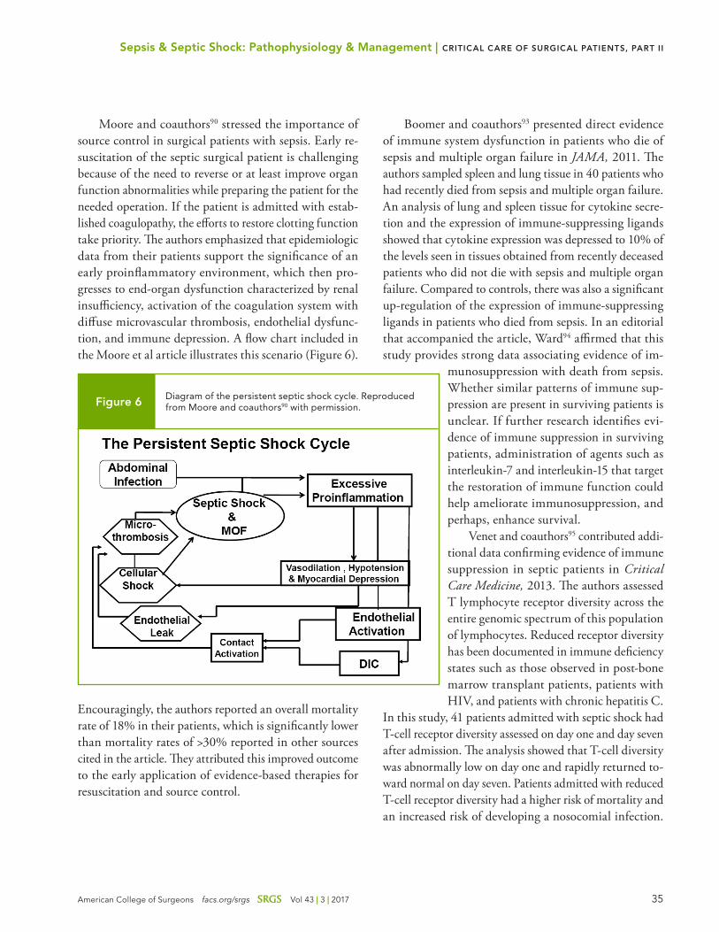
35American College of Surgeons facs.org/srgs SRGS Vol 43 | 3 | 2017
Moore and coauthors90 stressed the importance of source control in surgical patients with sepsis. Early re-suscitation of the septic surgical patient is challenging because of the need to reverse or at least improve organ function abnormalities while preparing the patient for the needed operation. If the patient is admitted with estab-lished coagulopathy, the efforts to restore clotting function take priority. The authors emphasized that epidemiologic data from their patients support the significance of an early proinflammatory environment, which then pro-gresses to end-organ dysfunction characterized by renal insufficiency, activation of the coagulation system with diffuse microvascular thrombosis, endothelial dysfunc-tion, and immune depression. A flow chart included in the Moore et al article illustrates this scenario (Figure 6).
Encouragingly, the authors reported an overall mortality rate of 18% in their patients, which is significantly lower than mortality rates of >30% reported in other sources cited in the article. They attributed this improved outcome to the early application of evidence-based therapies for resuscitation and source control.
Boomer and coauthors93 presented direct evidence of immune system dysfunction in patients who die of sepsis and multiple organ failure in JAMA, 2011. The authors sampled spleen and lung tissue in 40 patients who had recently died from sepsis and multiple organ failure. An analysis of lung and spleen tissue for cytokine secre-tion and the expression of immune-suppressing ligands showed that cytokine expression was depressed to 10% of the levels seen in tissues obtained from recently deceased patients who did not die with sepsis and multiple organ failure. Compared to controls, there was also a significant up-regulation of the expression of immune-suppressing ligands in patients who died from sepsis. In an editorial that accompanied the article, Ward94 affirmed that this study provides strong data associating evidence of im-
munosuppression with death from sepsis. Whether similar patterns of immune sup-pression are present in surviving patients is unclear. If further research identifies evi-dence of immune suppression in surviving patients, administration of agents such as interleukin-7 and interleukin-15 that target the restoration of immune function could help ameliorate immunosuppression, and perhaps, enhance survival.
Venet and coauthors95 contributed addi-tional data confirming evidence of immune suppression in septic patients in Critical Care Medicine, 2013. The authors assessed T lymphocyte receptor diversity across the entire genomic spectrum of this population of lymphocytes. Reduced receptor diversity has been documented in immune deficiency states such as those observed in post-bone marrow transplant patients, patients with HIV, and patients with chronic hepatitis C.
In this study, 41 patients admitted with septic shock had T-cell receptor diversity assessed on day one and day seven after admission. The analysis showed that T-cell diversity was abnormally low on day one and rapidly returned to-ward normal on day seven. Patients admitted with reduced T-cell receptor diversity had a higher risk of mortality and an increased risk of developing a nosocomial infection.
Diagram of the persistent septic shock cycle. Reproduced from Moore and coauthors90 with permission. Figure 6
Sepsis & Septic Shock: Pathophysiology & Management | CRITICAL CARE OF SURGICAL PATIENTS, PART II
36 American College of Surgeons facs.org/srgs SRGS Vol 43 | 3 | 2017
The authors concluded that analyses of lymphocyte func-tion may contribute significantly to our understanding of the pathophysiology of sepsis.
Surgical patients with sepsis present with a variety of hemodynamic patterns. Signs of end-organ perfusion problems are frequently discovered early in the course of management. One possible mechanism of end-organ hypoperfusion is thought to be diffuse occlusion of the vessels in the microcirculation. De Backer and coauthors96 provided data to support this interpretation in Critical Care Medicine, 2013. The authors assessed the sublingual microcirculation in 252 patients with severe sepsis admit-ted to a single-institution intensive care unit. The data reported confirmed an early reduction in the proportion of perfused microvessels, with a return toward normal by day seven. Interestingly, the proportion of nonperfused microvessels was predictive of mortality and the level of perfusion recovery was associated with an improved chance of survival. As sepsis persisted, the reduction in perfused microvessels continued to be a moderately strong (area under the ROC curve of 0.818) predictor of mortal-ity. The authors concluded that an early and persistent depression of microvessel perfusion is a clinical indica-tor of sepsis severity and, if confirmed in other patient groups, could be used to determine both the adequacy of therapy and the risk of adverse outcomes. In an editorial accompanying the De Backer et al article, van Griensven97 emphasized the discrepancy between most measures of global hemodynamics and tests that assess the adequacy of microcirculation and tissue oxygen delivery. Cited data indicated that the single global hemodynamic variable most closely associated with tissue oxygen delivery and microvessel perfusion adequacy is central venous oxygen saturation. Measures that would increase the recruitment of perfused microvessels may improve sepsis outcomes and these are worthy targets of future research.
The epidemiology and risk factors of sepsis in multiple trauma patients was the focus of an article by Wafaisade and coauthors98 in Critical Care Medicine, 2011. The au-thors conducted a retrospective analysis of outcomes in patients entered into a national trauma registry in Ger-many between 1993 and 2008; nearly 30,000 patients with complete data were identified. The analysis showed that the incidence of sepsis declined over the study interval
but had become stable in recent years. Overall mortality declined from 17% to 12% over the study interval, but the mortality of patients who developed sepsis was stable (in the range of 20%). Risk factors for sepsis in this patient group were age, male gender, Glasgow Coma Score <8 in the prehospital phase of care, severity of injury, number of transfusions needed, number of operative procedures needed, and need for laparotomy. The authors concluded that knowledge of risk factors for sepsis could assist in developing sepsis prevention protocols for injured patients.
Critical Care of the Patient with Sepsis & Septic Shock
The earlier discussion of the pathophysiology of sepsis and septic shock emphasized the diversity of physiologic responses to inflammation and infection. Generalized vasodilation and a hyperdynamic circulation usually char-acterize the septic state. Arterial pressure may be within the low-normal range and cardiac index may be increased. Despite these observations, signs of end-organ hypoperfu-sion, manifested by elevated blood lactate levels, may be present. In patients with severe sepsis, evidence of end-organ hypoperfusion is clear-cut. Septic shock is present when patients exhibit hypotension resistant to intravascu-lar and extracellular volume expansion. Faced with these combinations of clinical manifestations, surgeons caring for septic patients need to be familiar with the available means of assessing circulatory status (see the earlier sec-tion on monitoring patients in shock) and with the use of volume expanders, vasopressors, inotropic agents, and adjunctive drugs such as corticosteroids.
Goal-Directed Management of Suspected Sepsis & Septic Shock
Because sepsis and septic shock are associated with in-fection-induced inflammation, fluid exudation frequently occurs into the inflammation sites. In postoperative pa-tients who become septic, recent or ongoing blood loss may also contribute to hypovolemia. Only when volume status is optimized will clear evidence of myocardial dysfunction become evident. Fluid therapy that is too aggressive contributes to sequestration of edema fluid in
Sepsis & Septic Shock: Pathophysiology & Management | CRITICAL CARE OF SURGICAL PATIENTS, PART II
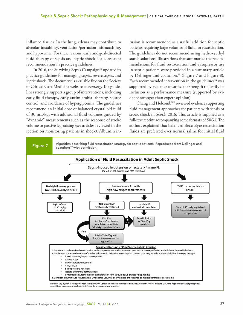
37American College of Surgeons facs.org/srgs SRGS Vol 43 | 3 | 2017
inflamed tissues. In the lung, edema may contribute to alveolar instability, ventilation/perfusion mismatching, and hypoxemia. For these reasons, early and goal-directed fluid therapy of sepsis and septic shock is a consistent recommendation in practice guidelines.
In 2016, the Surviving Sepsis Campaign34 updated its practice guidelines for managing sepsis, severe sepsis, and septic shock. The document is available free on the Society of Critical Care Medicine website at sccm.org. The guide-lines strongly support a group of interventions, including early fluid therapy, early antimicrobial therapy, source control, and avoidance of hyperglycemia. The guidelines recommend an initial dose of balanced crystalloid fluid of 30 mL/kg, with additional fluid volumes guided by “dynamic” measurements such as the response of stroke volume to passive leg-raising (see articles reviewed in the section on monitoring patients in shock). Albumin in-
fusion is recommended as a useful addition for septic patients requiring large volumes of fluid for resuscitation. The guidelines do not recommend using hydroxyethyl starch solutions. Illustrations that summarize the recom-mendations for fluid resuscitation and vasopressor use in septic patients were provided in a summary article by Dellinger and coauthors99 (Figure 7 and Figure 8). Each recommended intervention in the guidelines34 was supported by evidence of sufficient strength to justify its inclusion as a performance measure (supported by evi-dence stronger than expert opinion).
Chang and Holcomb100 reviewed evidence supporting fluid management approaches for patients with sepsis or septic shock in Shock, 2016. This article is supplied as a full-text reprint accompanying some formats of SRGS. The authors explained that balanced electrolyte resuscitation fluids are preferred over normal saline for initial fluid
Algorithm describing fluid resuscitation strategy for septic patients. Reproduced from Dellinger and coauthors99 with permission. Figure 7
Sepsis & Septic Shock: Pathophysiology & Management | CRITICAL CARE OF SURGICAL PATIENTS, PART II
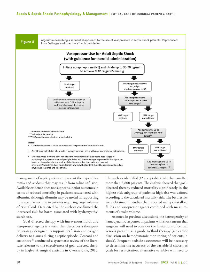
38 American College of Surgeons facs.org/srgs SRGS Vol 43 | 3 | 2017
management of septic patients to prevent the hyperchlo-remia and acidosis that may result from saline infusion. Available evidence does not support superior outcomes in terms of reduced mortality in patients resuscitated with albumin, although albumin may be useful in supporting intravascular volume in patients requiring large volumes of crystalloid. Data cited by the authors confirmed the increased risk for harm associated with hydroxyethyl starch use.
Goal-directed therapy with intravenous fluids and vasopressor agents is a term that describes a therapeu-tic strategy designed to support perfusion and oxygen delivery to tissues during a septic episode. Cecconi and coauthors101 conducted a systematic review of the litera-ture relevant to the effectiveness of goal-directed thera-py in high-risk surgical patients in Critical Care, 2013.
The authors identified 32 acceptable trials that enrolled more than 2,800 patients. The analysis showed that goal-directed therapy reduced mortality significantly in the highest-risk subgroup of patients; high-risk was defined according to the calculated mortality risk. The best results were obtained in studies that reported using crystalloid fluids and vasopressor agents combined with measure-ments of stroke volume.
As noted in previous discussions, the heterogeneity of hemodynamic responses in patients with shock means that surgeons will need to consider the limitations of central venous pressure as a guide to fluid therapy (see earlier discussion on hemodynamic monitoring of patients in shock). Frequent bedside assessments will be necessary to determine the accuracy of the variable(s) chosen as goals for resuscitation; alternative variables will need to
Algorithm describing a sequential approach to the use of vasopressors in septic shock patients. Reproduced from Dellinger and coauthors99 with permission.Figure 8
Sepsis & Septic Shock: Pathophysiology & Management | CRITICAL CARE OF SURGICAL PATIENTS, PART II

39American College of Surgeons facs.org/srgs SRGS Vol 43 | 3 | 2017
be considered if resuscitation is not adequate. Jones and coauthors102 reported a randomized prospective trial of two approaches to goal-directed resuscitation of patients with sepsis and septic shock in JAMA, 2010. In this trial, the authors compared resuscitation outcomes when fluid volumes were guided by central venous oxygen saturation vs. lactate clearance. Per the authors, the generally accept-ed approaches to resuscitating patients with severe sepsis or septic shock include assessing cardiac preload using central venous pressure and estimating perfusion pressure using mean arterial pressure. More controversial are the various approaches to estimating the adequacy of tissue oxygen delivery. The 2013 Surviving Sepsis Campaign practice guidelines103 recommend the use of central venous oxygen saturation of 70% or greater as a target indicat-ing adequate tissue oxygen delivery. There are significant barriers to the use of central venous oxygen saturation, including the problems of appropriate sampling and tech-nical challenges related to the measurement process. Jones and coauthors hypothesized that lactate clearance of at least 10% (based on two separate blood samples where lactate was measured) would be as accurate an estimate of tissue oxygen delivery as central venous oxygen saturation. Three hundred patients from several institutions were randomized to one of two comparison groups. The goals of resuscitation for both groups included normalization of systemic arterial pressure and central venous pressure of 8–10 mm Hg. Adequate tissue oxygen delivery was as-sumed present if lactate clearance was at least 10% when two peripheral blood samples for lactate were compared or if lactate was normal when two samples were drawn at least two hours apart. The analysis showed that short-term mortality rates were equivalent in the two groups, with the lactate clearance group having a 17% mortality compared with 23% in the central venous oxygen satura-tion group. The authors concluded that lactate clearance can be substituted for central venous oxygen saturation as a surrogate for tissue oxygen delivery.
In an editorial accompanying this article, Lewis104 reviewed the data supporting the use of goal-directed fluid therapy and remarked that the data supporting this approach are not as strong as would be desired. Also noted was that randomized trials to determine the effectiveness of goal-directed therapy have been flawed. Nonetheless, the accumulated data suggest that improved survival is
associated with goal-directed therapy. The difficulty in obtaining accurate measurements of some variables in-cluded in goal-directed strategies (e.g., central venous oxygen saturation) has resulted in lower levels of usage of goal-directed strategies than envisioned by the guidelines-sponsoring organizations. Because of this, it is desirable to substitute an easier measurement technique. The edi-torialist pointed out a significant limitation to the study reported by Jones and colleagues: advanced resuscitation interventions, such as the use of vasopressors and red blood cell transfusions, were employed in fewer than 10% of the enrolled patients. This suggests that the enrolled patients were a sample drawn from a relatively good-risk cohort. Despite this limitation, the study does support the use of a more easily measured variable as a valid re-suscitation goal.
Trof and coauthors105 offered additional data on the use of monitored hemodynamic variables for goal-directed resuscitation in Critical Care Medicine, 2012. The authors conducted a randomized prospective multi-institutional trial comparing transpulmonary thermal dilution (assess-ment of extravascular lung water) with pulmonary artery catheter-guided resuscitation of patients in shock due to septic and nonseptic etiologies. The analysis equated the two strategies in septic shock patients in terms of hospital length of stay and ventilator-free days. The authors chose these endpoints as surrogates for lung damage risk during resuscitation. For patients with nonseptic shock, the ther-mal dilution strategy resulted in longer lengths of stay and more time on the ventilator. The authors concluded that the thermal dilution monitoring methodology is useful in patients with septic shock, but these results need to be interpreted with caution. As pointed out by Takala106 in an editorial that accompanied the article, the authors failed to demonstrate a clear benefit of thermal dilution-guided fluid resuscitation in septic patients. This probably was the result of a significantly higher fluid volume delivery to this patient group when monitored by thermal dilution.
Boyd and coauthors107 sought to confirm the potential harm of fluid overload during goal-directed resuscitation of patients with sepsis in Critical Care Medicine, 2011. The authors reported a retrospective review of data gathered during a randomized prospective trial of vasopressin as an adjunct to resuscitation of septic and septic shock pa-tients. The reported data included 12-hour and four-day
Sepsis & Septic Shock: Pathophysiology & Management | CRITICAL CARE OF SURGICAL PATIENTS, PART II

40 American College of Surgeons facs.org/srgs SRGS Vol 43 | 3 | 2017
fluid balance and central venous pressure measurements. The principle end point of the study was 28-day all-cause mortality. The authors found that patients with the high-est quartile of positive fluid balance (17–24 liters positive balance by day four) had a significantly increased risk of death. The lowest risk of death was observed in patients who had a positive fluid balance of <3 liters after 12 hours of therapy. The authors also discovered that central venous pressure was reflective of fluid balance during the first 12 hours of resuscitation, but that after that time point, there was no correlation between central venous pressure and fluid balance. Patients with sustained central venous pressures of 12 mm Hg or higher were at increased risk of positive fluid balances in the higher mortality range. The authors concluded that there is a direct relationship between high positive fluid balance and mortality and that central venous pressure is an undependable guide for resuscitation after 12 hours of treatment. In an editorial, Ytrebo108 observed that these data strongly confirm the lack of consistent benefit associated with using central venous pressure as a guide for fluid resuscitation. Instead, the editorialist recommended using a continuous measure of variables such as stroke volume and cardiac index com-bined with assessments of oxygen delivery and frequent bedside clinical examinations to optimize tissue oxygen delivery and avoid fluid overload.
The Surviving Sepsis Campaign practice guidelines34 recommend red blood cell transfusions during the initial resuscitation of patients with sepsis and septic shock if the patient is anemic. In hemodynamically stable patients, transfusion is recommended for hemoglobin levels <7 g/dL. Parsons and coauthors109 focused on the association of red blood cell transfusion and mortality at 28 and 90 days in patients with sepsis and septic shock in an article in Critical Care, 2011. The authors performed a secondary analysis of outcomes data in patients originally enrolled in a randomized prospective trial. The analysis showed that red blood cell transfusion was not a significant pre-dictor of mortality at 28 and 90 days, even when septic patients met standard criteria for transfusion (hypoten-sion, hemoglobin level <10 g/dL, and depressed central venous oxygen saturation). The authors emphasized that these data should be interpreted with caution because the included patients received transfusions more than 12
hours after a sepsis diagnosis. The authors also argued the possible benefit of red blood cell transfusions early in the management of sepsis patients.
Experts have suggested that critical care “bundles” may improve outcomes by increasing compliance with protocol-based fluid administration, antimicrobials, and monitoring approaches. Whether these techniques actu-ally improve outcomes is unclear. Thompson and coau-thors110 compared outcomes of protocol-based care with usual care in septic patients in Critical Care Medicine, 2016. This article is supplied as a full-text reprint accom-panying some formats of SRGS. The authors compared outcomes from administrative data in nearly 50,000 pa-tients treated in hospitals in a single state. The analysis showed that compliance with using the care bundle did not improve mortality risk compared with usual care in the entire patient group. The authors acknowledged vari-ability in adherence to the protocol and that hospitals with very high adherence had improved outcomes compared to hospitals with low adherence. Their conclusion was that additional research is needed to determine the reasons for variability and the factors that might contribute to improved outcomes in high-adherence hospitals.
McKinley and coauthors111 explored the effectiveness of evidence-based care using computer-generated protocols in a surgical intensive care unit in the Journal of Trauma, 2011. The authors assessed compliance with accepted practice guidelines using computer-generated protocols compared with a paper protocol in a group of historical control patients. The computer-generated protocol showed significant increases in compliance. Antimicrobial therapy was instituted sooner with the computer protocol and the volume of fluids given during resuscitation was not different. Of interest was that mortality decreased from 34% to 14% when the two intervals were compared. The authors concluded that care bundles applied with com-puter-generated protocols are associated with a significant improvement in patient outcomes.
Sepsis & Septic Shock: Pathophysiology & Management | CRITICAL CARE OF SURGICAL PATIENTS, PART II
41American College of Surgeons facs.org/srgs SRGS Vol 43 | 3 | 2017
Vasopressor Use
A considerable proportion of septic patients have mild arterial hypotension. By definition, patients with septic shock are hypotensive. The Surviving Sepsis Campaign 34 practice guidelines recommend a target MAP of 65 mm Hg for septic patients. If volume expansion fails to achieve this recommended pressure, the practice guidelines rec-ommend using norepinephrine as the first-line vasopres-sor of choice. For patients failing to achieve the pressure target, the guidelines also recommend adding arginine vasopressin to the vasopressor therapy. Current practice guidelines recommend against the use of dopamine as either a low-dose therapy or standard-dose approach for sepsis-associated hypotension. Patients with persistent hypoperfusion may benefit from escalation of vasopressor therapy using epinephrine and phenylephrine in sequence.
De Backer and coauthors112 confirmed the potential harm of dopamine use in the New England Journal of Medicine, 2010. The authors’ randomized prospective trial compared norepinephrine to dopamine in 1,000 patients with septic shock. The trial results showed no significant difference between the dopamine and the norepinephrine groups in terms of mortality. A 95% confidence limit for the mortality difference was wide and the upper bound was a 42% mortality increase for patients treated with do-pamine. The frequency of cardiac arrhythmia was signifi-cantly higher in patients randomized to receive dopamine. The authors concluded that patients with cardiogenic shock should not be treated with dopamine because of an increased risk of death. They also emphasized that the occurrence of cardiac arrhythmias in patients treated with dopamine suggests that this drug should not be used to manage hypotension in patients with shock.
A study by Jhanji and coauthors113 in Critical Care Medicine reported increases in cutaneous oxygen tension and microvascular flow with increasing norepinephrine doses in 16 patients with septic shock. These authors used transcutaneous oxygen measurements and Doppler laser flowmetry to assess microvascular oxygen availability and blood flow. Sublingual microvascular flow was assessed with Sidestream Dark Field imaging. The authors adjusted the dosing of norepinephrine to achieve mean arterial pressures of 60, 70, 80, and 90 mm Hg, sequentially. Global oxygen delivery as well as cutaneous oxygen ten-
sion and cutaneous microvascular flow increased in keep-ing with each MAP increase. Sublingual microcirculatory status did not change. The authors did not obtain outcome assessments in these patients. The authors concluded that additional research is desirable to refine definitions of optimum endpoints for vasopressor therapy in patients with septic shock.
An older randomized prospective double-blind trial compared norepinephrine treatment alone with norepi-nephrine combined with arginine vasopressin infusion.114 The outcomes of interest were 28-day and 90-day mortal-ity. The authors did not confirm a mortality benefit in the group treated with norepinephrine plus vasopressin. In the subset of patients with less severe septic shock, there was a trend toward lowered mortality in the group treated with vasopressin, but the authors did not design the study to analyze patients based on shock severity. Overall mortality rates for both groups were low com-pared with prior experience and this may reflect an overall improvement in outcomes for patients with sepsis treated with modern protocol approaches. Because vasopressin, in the low doses recommended, has few, if any, adverse side effects, combining norepinephrine and vasopressin in selected patients may be beneficial. There is a need for additional data regarding this association.
There are provocative data that suggest a beneficial effect of heart rate control in patients with sepsis and septic shock to improve microcirculation and overall outcome. Morelli115 evaluated the potential benefits of heart rate control with the b-blocking agent esmolol in a prospective observational study in Critical Care Medicine, 2013. The author recorded changes in stroke volume and microcirculatory flow measured with sublingual capnom-etry. The study enrolled 25 patients and target heart rate range was 80–94 beats per minute. All patients achieved target heart rates, according to the analysis. Stroke volume was maintained at baseline levels during esmolol infusion and a significant increase in microcirculatory flow was observed. Norepinephrine requirements decreased during esmolol administration.
An article by Macchia and coauthors116 in Critical Care Medicine, 2012, examined outcomes recorded for patients with sepsis in a national database. They com-pared mortality rates in patients taking b-blocking agents
Sepsis & Septic Shock: Pathophysiology & Management | CRITICAL CARE OF SURGICAL PATIENTS, PART II

42 American College of Surgeons facs.org/srgs SRGS Vol 43 | 3 | 2017
prior to admission with patients not taking these drugs. Chronic use of b-blocking agents was associated with a significantly reduced mortality rate (17% vs. 22%). Mo-relli and coauthors115 reported hemodynamic data ob-tained in patients with septic shock who were receiving corticosteroids (200 mg per day) and were being treated with norepinephrine to support arterial pressure (target MAP of 65 mm Hg). The authors administered esmo-lol intravenously to achieve a target heart rate of 80–94 beats per minute. Patients who achieved the target heart rates showed reduced cardiac index, but stroke volume remained unchanged. In treated patients, the study also reported:
� Improved microcirculatory flow, as measured in the sublingual vessels
� Reduced norepinephrine levels
� Preserved recommended target level of MAP and central venous oxygen saturation
The authors did not report overall mortality rates. Confirmation of these observations in larger prospective randomized trials may lead to heart rate control becoming a component of hemodynamic management for patients with sepsis and septic shock.
Corticosteroid Use
The Surviving Sepsis Campaign practice guidelines34 do not recommend the routine use of corticosteroids when resuscitating patients with sepsis and septic shock. In pa-tients with septic shock, the guidelines suggest 200 mg per day of cortisone if patients remain hypotensive with signs of end-organ hypoperfusion after maximal support with intravenous fluids and vasopressors. The guidelines also do not recommend using the ACTH stimulation test to detect relative adrenal insufficiency.
Recombinant Activated Protein C Use
Sepsis and septic shock trigger an inflammatory and procoagulant cascade that predisposes to microvascular thrombosis; this may be the etiology of organ failure lead-ing to death in septic patients. A number of interventions designed to modulate these cascades have been evaluated
to target the underlying pathophysiology of sepsis. Suc-cess has been limited in these trials. An agent targeting the coagulation system is recombinant activated protein C. A randomized clinical trial comparing recombinant activated protein C to placebo in patients with sepsis and septic shock was by Ranieri and coauthors117 in the New England Journal of Medicine, 2012. The authors re-ported a trial that enrolled 1,697 patients. According to the authors, an earlier trial reported by Bernard and co-authors118 in the New England Journal of Medicine, 2001, documented a 6% absolute mortality reduction in patients with severe sepsis who were treated with recombinant activated protein C. This survival advantage was observed in patients who were severely ill only (APACHE II score >25). The main adverse effect of treatment with recombi-nant activated protein C was bleeding; there are reports of fatal intracranial hemorrhage in patients with use of the drug. Ranieri and colleagues117 found no improvement in 28-day or 90-day mortality in patients treated with recombinant activated protein C. There was no significant benefit of treatment, even with protein C level depression. The authors concluded that there is no evidence of benefit of recombinant activated protein C use in patients with sepsis or septic shock.
Antimicrobial Therapy
The Surviving Sepsis Campaign guidelines34 recommend the early use of intravenous antimicrobial therapy de-signed to cover all expected organisms, with a subsequent de-escalation of therapy based on culture results. It is important that clinicians be aware of the frequencies of infecting organisms in the hospital environment. As men-tioned previously, most episodes of sepsis in surgical pa-tients are the result of intraabdominal infections. Enteric organisms will be the most frequent etiologic agents in these situations. For patients who develop intraabdominal sepsis while hospitalized, antimicrobial choices will need to include the multidrug-resistant organisms. Antimi-crobial therapy is important but not the sole component of source control. Source control refers to eradicating the infection that is the cause of the episode of sepsis and/or septic shock. Over time, published research has suggested that sepsis and septic shock are the result of a
Sepsis & Septic Shock: Pathophysiology & Management | CRITICAL CARE OF SURGICAL PATIENTS, PART II

43American College of Surgeons facs.org/srgs SRGS Vol 43 | 3 | 2017
dysfunctional set of inflammatory, microcirculatory, and immune responses that proceed regardless of the outcome of the infection that stimulated the response.
An article that focused on infection as the main driver of the septic response was by Kumar119 in Virulence, 2014. This article is supplied as a full-text reprint accompanying some formats of SRGS. The author cited data supporting the concept that the microbial load drives the inflam-matory response and that reducing the microbial load as quickly as possible following patient presentation would be the best method of limiting the systemic inflammatory, microcirculatory, and immunologic changes that produce the clinical and laboratory features of sepsis and septic shock. The natural conclusion from the data cited is that controlling the infection is the best way to reduce the mortality risk for patients with sepsis and septic shock. The data presented in the article support early antimicro-bial therapy and surgical or percutaneous interventions to treat localized foci of infection. The author recommends choosing antimicrobials based on an understanding of the microbiologic patterns of infections occurring in the treating hospital, the location of the infection, and patient risk factors. Optimization of antimicrobial therapy using bactericidal agents, combination therapies, and monitor-ing of antimicrobial blood levels are measures supported by the data reviewed in the article.
Martinez and coauthors120 analyzed the effects of source control on outcomes of care for patients with sep-sis and septic shock in a study in Critical Care Medicine, 2017. This article is supplied as a full-text reprint accom-panying some formats of SRGS. The authors performed a prospective observational study that included more than 3,600 patients with sepsis cared for in 99 intensive care units in a single country. Source control interventions for urinary, abdominal, and soft tissue infections were performed in 1,173 patients. The patient group under-going source control were older, had a higher propor-tion of organ dysfunction, higher levels of serum lactate, and lower compliance with recommended resuscitation. Despite these factors, mortality was significantly lower in patients who underwent source control. The authors observed that time from admission to source control did not influence mortality. The authors proposed that de-lays might occur because of small foci of infection that might not be detected, because caregivers want to avoid
emergency intervention, or because of a desire by surgeons to wait until an area of necrosis had completely defined itself (in soft tissue infections, for example). The authors concluded that source control, whenever possible, has significant benefit for septic patients—supported by the finding of no adverse outcomes related to the time from source detection to the source control intervention. The authors confirmed that hospital stay was longer for pa-tients undergoing source control. They hypothesized that this occurred because the majority of patients needing source control had abdominal infections and required one or more operative procedures.
An article that provided evidence confirming the importance of the infection source as the driver of the pathophysiologic patterns and outcomes of sepsis and septic shock was by Kalil and coauthors121 in Critical Care Medicine, 2017. The authors conducted a meta-analysis of prospective randomized trials and observational studies that focused on the effectiveness of early goal-directed fluid therapy for patients with sepsis and septic shock. The included studies reported on nearly 20,000 patients. The authors’ analysis showed evidence of benefit for early goal-directed therapy in observational studies but not in randomized prospective trials. The consistent difference in the two types of studies was that early, appropriate anti-microbial therapy was used in the randomized prospective trials but was inconsistently used in the observational studies. The authors concluded that early control of the infection causing sepsis or septic shock was the main factor associated with successful outcomes of treatment of sepsis and septic shock.
Data supporting the use of early antimicrobial therapy were presented in an article by Ferrer and coauthors122 in Critical Care Medicine, 2014. The authors performed a retrospective study of prospectively gathered data used in the Surviving Sepsis Campaign; they reviewed outcomes from nearly 18,000 septic patients. Overall mortality in the group was 29.7%, reflecting the severity of illness in the included patients. The authors identified a statisti-cally significant increase in risk of death with increasing time to administration of antimicrobial agents. This trend persisted after adjusting for risk factors such as intensive care unit admission.
Sepsis & Septic Shock: Pathophysiology & Management | CRITICAL CARE OF SURGICAL PATIENTS, PART II

44 American College of Surgeons facs.org/srgs SRGS Vol 43 | 3 | 2017
Labelle and coauthors123 reported outcomes of sep-tic patients treated initially with appropriate antibiotic therapy in Critical Care Medicine, 2012. These authors analyzed medical record data on 436 consecutive, mixed medical and surgical patients with sepsis and septic shock treated in a single institution. Record review of antibiotic use and culture results identified patients who received appropriate initial antimicrobial therapy. More than half of the patients reviewed died during the hospitalization where sepsis treatment occurred. The analysis showed that determinants of mortality in patients treated with appro-priate initial antimicrobial therapy were illness severity, as reflected in the APACHE II scores, and acquisition of infection in the intensive care unit. Of interest was that MRSA infection was a predictor of lower mortality risk. The authors concluded that future improvements in out-comes for this patient group could result from measures designed to prevent and aggressively treat intensive care unit-acquired infections.
Cardiogenic Shock ManagementThe clinical features of cardiogenic shock comprise arterial hypotension (systolic blood pressure <90 mm Hg) com-bined with signs of end-organ hypoperfusion (acidosis, elevated serum creatinine, abnormal sensorium), depressed cardiac index, and signs of ventricular dysfunction. Car-diogenic shock occurs in 7% of patients after myocardial infarction and this condition is the most common cause of early death after myocardial infarction. Other con-ditions that complicate myocardial infarction, such as papillary muscle rupture with acute mitral insufficiency and ventricular wall rupture with pericardial tamponade, are surgical emergencies that also cause cardiogenic shock.
Clinical practice guidelines for cardiogenic shock management were included in guidelines for managing ST-segment elevation myocardial infarction promulgated in 2013 by the American College of Cardiology and the American Heart Association.124 These guidelines state that
cardiogenic shock following myocardial infarction occurs most often during the first 24 hours after the ischemic event.
Topalian and coauthors125 discussed the epidemiol-ogy of cardiogenic shock in Critical Care Medicine, 2008. These authors cited three reports indicating that cardio-genic shock complicating myocardial infarction (defined as ST-segment elevation and/or new left bundle branch block on electrocardiogram) occurred in 7.1%–8.6% of patients after myocardial infarction. Once cardiogenic shock was diagnosed, subsequent mortality rates reported were 60%–80%. Mortality rates were higher in patients older than 75. Topalian and colleagues stressed recent data showing that aggressive attempts to provide complete re-vascularization of critical coronary stenoses and occlusions in patients with cardiogenic shock have resulted in lower mortality rates approaching 48%. The clinical practice guidelines124 referred to earlier recommend revasculariza-tion using percutaneous interventions or coronary artery bypass as soon as possible after the shock diagnosis and delineation of the vascular anatomy. Fibrinolytic therapy is recommended for patients when revascularization is not feasible.
Patients at an increased risk of cardiogenic shock after myocardial infarction include older patients and patients with a history of hypertension, dyslipidemia, and previous coronary angioplasty. Cardiogenic shock has an average time of onset, after the appearance of clinical evidence of myocardial infarction, of seven hours. According to the Topalian et al review,125 cardiogenic shock can occur after myocardial infarctions that result in ST-segment eleva-tion and in those patients without ST-segment elevation accompanying the infarction. The etiology of infarction and cardiogenic shock is usually multivessel coronary atherosclerosis. In one report cited by the authors, more than half the patients with cardiogenic shock after myo-cardial infarction had triple vessel disease and more than 15% of patients had significant left main coronary artery stenosis. Anterior infarction is common, but multiple sites of infarction are diagnosed in more than half the patients who develop cardiogenic shock. Nearly 80% of patients who develop cardiogenic shock have left ventricular failure as the main pathogenesis of the shock event. Acute mitral insufficiency, septal rupture, right ventricular infarction, and ventricular wall rupture with pericardial tamponade
Cardiogenic Shock Management | CRITICAL CARE OF SURGICAL PATIENTS, PART II
45American College of Surgeons facs.org/srgs SRGS Vol 43 | 3 | 2017
account for the remaining patients. Ventricular wall rup-ture is the least frequent etiology of cardiogenic shock, accounting for less than 1.5% of instances. Acute mitral insufficiency accounts for 7% of patients developing car-diogenic shock.
The pathophysiology of cardiogenic shock was the subject of an older review by Aymong and coauthors126 in Medical Clinics of North America, 2007. The authors emphasized the critical importance of reduced coronary blood flow that results in a state of myocardial oxygen de-privation and disordered myocardial energy metabolism. They noted that heart muscle is very reliant on aerobic energy metabolism. Oxygen utilization in contracting heart muscle ranges from 8 mL to 15 mL of oxygen per 100 g tissue per minute. The noncontracting muscle uses only 1.5 mL of oxygen per 100 g tissue per minute. The heart, even in periods of low activity, extracts two to three times more oxygen than any other single organ. Myocardial oxygen demand is determined by ventricular tension, heart rate, and contractility. Oxygen delivery to the myocardium via the coronary arteries is primarily a diastolic phenomenon. Coronary perfusion pressure can be estimated from the difference in aortic diastolic pres-sure and mean left ventricular pressure. Coronary vascular resistance interacts with coronary perfusion pressure to determine net coronary flow.
Complicated interactions among various local and systemic mediators determine coronary vascular resis-tance. Endothelial dysfunction occurs because of coronary atherosclerosis and produces abnormal coronary responses to endogenous mediators of vasoconstriction and vasodi-lation. The net result is disordered autoregulation, with coronary flow becoming progressively more dependent on coronary perfusion pressure. Diastolic coronary perfusion can be reduced during conditions where cardiac output is decreased and when the diastolic interval is reduced (tachycardia). Myocardial infarction resulting from coro-nary thrombosis directly reduces coronary flow because of obstruction. With extensive involvement of the ventricular wall, cardiac output is reduced. Tachycardia results from the catecholamine response to reduced cardiac output and these factors combine to produce a self-perpetuating cycle of myocardial ischemia and infarction. Topalian and associates125 emphasized that “ischemia begets ischemia” during this cycle.
Ventricular dysfunction leads to elevated end-diastolic pressures and pulmonary congestion. Elevated ventricular filling pressures increase myocardial wall stress. The au-thors of a series cited by Topalian et al observed restricted left ventricular filling in more than 60% of patients evalu-ated by echocardiography. This pathophysiologic process is complicated by the elaboration of inflammatory media-tors. A report the authors referred to cited clinical evidence of systemic inflammatory response syndrome in 20% of patients. Aymong and colleagues126 cited data indicating that elevated levels of interleukin-6 have been documented in patients with cardiogenic shock and that these levels are equivalent to the levels recorded in patients with sepsis and septic shock. Topalian and associates125 and Aymong and colleagues126 also found that the proinflammatory state observed in some patients with cardiogenic shock was associated with the stimulation of inducible nitric oxide. Unfortunately, a blockade of inducible nitric oxide synthe-sis did not produce a benefit in patients with cardiogenic shock. Aymong and coauthors stated that myocardial ischemia can result in myocyte necrosis. Other cells die because of apoptosis (programmed cell death). In addi-tion, myocardial cells may be “stunned” from ischemia. Stunned cells do not recover full contractile function with early restoration of perfusion from revascularization procedures. Thus, normal contractile function might not be restored for several weeks after coronary artery throm-bosis followed by revascularization.
The extensive multivessel coronary atherosclerosis seen in autopsy reports of patients dying of myocardial in-farction and cardiogenic shock usually produces multiple myocardial infarctions of varying age rather than a single massive infarction. Low coronary flow and increased co-agulation activity because of unstable coronary plaque can produce progressive coronary thromboses that extend to involve branches of the artery where the thrombosis originated. The authors explained that declining myocyte contractility can occur in areas remote from the infarc-tion and this may be a consequence of hypoxia-inducible factor-1 production, which is stimulated when cardiac cell mitochondria sense hypoxia. This factor produces reduced cell metabolism and contractility. The resulting low metabolism state produced in areas remote from the infarction is termed “myocardial hibernation.” One of
Cardiogenic Shock Management | CRITICAL CARE OF SURGICAL PATIENTS, PART II

46 American College of Surgeons facs.org/srgs SRGS Vol 43 | 3 | 2017
the benefits of treating cardiogenic shock with intraaortic balloon counterpulsation may be restoration of coronary perfusion and myocyte oxygenation that serves to reverse myocyte hibernation and recover contractility.
Managing patients with suspected cardiogenic shock begins with a rapid assessment to determine adequacy of the airway, oxygenation, and circulation. Patients suspect-ed to be hypovolemic may benefit from a fluid challenge and/or blood transfusion. Physical examination assisted by plain chest radiograph can disclose clinical evidence of pulmonary congestion. Electrocardiography and echo-cardiography are useful, early in patient management, to quantify the extent of ischemia and left ventricular dysfunction. Similarly, pulmonary artery catheter place-ment can supply sequential data on pulmonary artery pressures and cardiac index. Echocardiographic assess-ments of preload and ejection fraction (discussed in the section on diagnosing and monitoring patients in shock) may supply valuable data to guide the use of volume ex-pansion strategies.
Topalian and colleagues125 stressed that signs of hy-poperfusion can occur without profound hypotension. For patients with arterial pressures consistently above 100 mm Hg, intravenous nitroglycerine in doses of 10–20 mcg per minute will produce coronary vasodilation and improve coronary flow. Dobutamine is used for hypoten-sive patients without signs of end-organ hypoperfusion to elevate arterial pressure. Doses of 2–20 mcg/kg/minute are indicated initially. If signs of hypoperfusion are present, the initial dose can be increased to 5–15 mcg/kg/minute. Failure to achieve improved blood pressure is an indication for using intravenous norepinephrine. Norepinephrine doses are titrated in the 0.5–30 mcg per minute range as a continuous intravenous infusion.
Epinephrine is another vasoactive agent that has been used to improve blood pressure and end-organ perfu-sion in patients with cardiogenic shock. A randomized prospective trial comparing the combination of dobu-tamine-norepinephrine to epinephrine in patients with cardiogenic shock not due to an acute ischemic event was by Levy and coauthors127 in Critical Care Medicine, 2011. These authors randomized 30 patients to receive either epinephrine infusion or dobutamine-norepinephrine in-fusion. Patients were enrolled if standard definitions of
cardiogenic shock were fulfilled, a trial of dopamine had not improved blood pressure, and an acute myocardial ischemic event had been ruled out. The analysis showed that both agents raised MAP and improved end-organ perfusion, as reflected by improved urinary output. The epinephrine group had a higher frequency of new cardiac arrhythmias diagnosed and measurements of splanch-nic perfusion (gastric mucosal perfusion) showed worse splanchnic perfusion in the epinephrine group.
The authors concluded that the combination of do-butamine-norepinephrine is preferable for vasopressor support in patients with cardiogenic shock who have not responded to dopamine infusion. In an editorial that ac-companied the article, Krenn and Delle Karth128 stressed that the increased rate-pressure product that resulted from epinephrine infusion may have reduced oxygen delivery to the myocardium and caused decreased splanchnic per-fusion and a tendency toward new arrhythmia develop-ment in the patients receiving epinephrine. Despite the relatively small number of patients included in the trial, the editorialists agreed with the authors’ conclusion that dobutamine-norepinephrine is preferable for managing cardiogenic shock that is not the result of an acute isch-emic event.
Topalian and coauthors125 also discussed intraaor-tic balloon counterpulsation. These devices, which can now be introduced percutaneously, can improve coronary flow and reduce left ventricular afterload. Currently, these devices are mainly used as “bridges” to revasculariza-tion interventions. Nearly one-third of the insertions of intraaortic balloon devices, in a report cited by these au-thors, were for cardiogenic shock. Practice guidelines list intraaortic balloon pump support as a useful intervention for managing patients with cardiogenic shock complicat-ing a myocardial ischemic event.124
Other data have called the perceived benefit of in-traaortic balloon counterpulsation into question. A ran-domized, prospective trial by Thiele and coauthors129 in the New England Journal of Medicine, 2012, reported data on 600 patients with cardiogenic shock (systolic blood pressure <90 mm Hg, need for vasopressor support, and/or signs of end-organ hypoperfusion) complicating a myocardial ischemic event. Patients were eligible for randomization if a myocardial revascularization procedure
Cardiogenic Shock Management | CRITICAL CARE OF SURGICAL PATIENTS, PART II

47American College of Surgeons facs.org/srgs SRGS Vol 43 | 3 | 2017
was planned. The data analysis showed that intraaortic balloon counterpulsation did not reduce 30-day all-cause mortality (39% vs. 41% for control patients). Although there was a brief early improvement in laboratory mark-ers of end-organ hypoperfusion, this benefit was not ob-served after day four of counterpulsation. The authors concluded that intraaortic balloon counterpulsation has no measurable benefit in terms of reduced mortality. In an editorial that accompanied the article, O’Connor and Rogers130 pointed out that the patients included in this trial were moderate-risk patients and the results may not be generalizable to the highest-risk patient groups. They agreed that the data support the discontinuation of any routine use of intraaortic balloon counterpulsation for the majority of patients with cardiogenic shock complicating a myocardial ischemic event.
Topalian and coauthors125 concluded their review by noting that death from cardiogenic shock occurs more often when there is a delay in diagnosing myocardial infarction; therefore, the authors encourage public educa-tion efforts to inform patients of the importance of early medical care at the first sign of acute myocardial ischemia.
Cardiogenic Shock Management | CRITICAL CARE OF SURGICAL PATIENTS, PART II

48 American College of Surgeons facs.org/srgs SRGS Vol 43 | 3 | 2017
I hope our surgical critical care series has been informative and helpful to you and your practice. The next issue of SRGS will begin our two-part Trauma series.
Thanks for reading SRGS!
Lewis Flint, MD, FACS Editor in Chief
VOLUME 43 | 3 | 2017
Conclusion CRITICAL CARE OF SURGICAL PATIENTS, PART II

49American College of Surgeons facs.org/srgs SRGS Vol 43 | 3 | 2017
1. Delaney M, Wendel S, Bercovitz RS, et al. Transfusion reactions: prevention, diagnosis, and treatment. Lancet. 2016;388(10061):2825-2836.
2. Heddle NM, Cook RJ, Arnold DM, et al. Effect of Short-Term vs. Long-Term Blood Storage on Mortality after Trans-fusion. N Engl J Med. 2016;375(20):1937-1945.
3. Tobian AA, Ness PM. Red Cells - Aging Gracefully in the Blood Bank. N Engl J Med. 2016;375(20):1995-1997.
4. Carson JL, Guyatt G, Heddle NM, et al. Clinical Practice Guidelines From the AABB: Red Blood Cell Transfusion Thresholds and Storage. JAMA. 2016;316(19):2025-2035.
5. Smilowitz NR, Oberweis BS, Nukala S, et al. Association Between Anemia, Bleeding, and Transfusion with Long-term Mortality Following Noncardiac Surgery. Am J Med. 2016;129(3):315-323 e312.
6. Rana A, Petrowsky H, Hong JC, et al. Blood transfusion requirement during liver transplantation is an important risk factor for mortality. J Am Coll Surg. 2013;216(5):902-907.
7. Harlaar JJ, Gosselink MP, Hop WC, Lange JF, Busch OR, Jeekel H. Blood transfusions and prognosis in colorectal can-cer: long-term results of a randomized controlled trial. Ann Surg. 2012;256(5):681-686; discussion 686-687.
8. Goodnough LT. Risks of blood transfusion. Crit Care Med. 2003;31(12 Suppl):S678-686.
9. Practice guidelines for perioperative blood transfusion and adjuvant therapies: an updated report by the American Society of Anesthesiologists Task Force on Perioperative Blood Transfusion and Adjuvant Therapies. Anesthesiology. 2006;105(1):198-208.
10. Hebert PC, Wells G, Blajchman MA, et al. A multicenter, randomized, controlled clinical trial of transfusion require-ments in critical care. Transfusion Requirements in Critical Care Investigators, Canadian Critical Care Trials Group. N Engl J Med. 1999;340(6):409-417.
11. Mirza SB, Campion J, Dixon JH, Panesar SS. Efficacy and economics of postoperative blood salvage in patients under-going elective total hip replacement. Ann R Coll Surg Engl. 2007;89(8):777-784.
12. Hughes DB, Ullery BW, Barie PS. The contemporary approach to the care of Jehovah’s witnesses. J Trauma. 2008;65(1):237-247.
13. Jabbour N, Gagandeep S, Shah H, et al. Impact of a transfu-sion-free program on non-Jehovah’s Witness patients under-going liver transplantation. Arch Surg. 2006;141(9):913-917.
14. Cestero RF, Dent DL. Endpoints of resuscitation. Surg Clin North Am. 2015;95(2):319-336.
15. Spinella PC, Perkins JG, Cap AP. Lessons Learned for the Resuscitation of Traumatic Hemorrhagic Shock. US Army Med Dep J. 2016(2-16):37-42.
16. Minei JP, Cuschieri J, Sperry J, et al. The changing pattern and implications of multiple organ failure after blunt injury with hemorrhagic shock. Crit Care Med. 2012;40(4):1129-1135.
17. Chang R, Cardenas JC, Wade CE, Holcomb JB. Advances in the understanding of trauma-induced coagulopathy. Blood. 2016;128(8):1043-1049.
18. Frith D, Davenport R, Brohi K. Acute traumatic coagulopa-thy. Curr Opin Anaesthesiol. 2012;25(2):229-234.
19. Cuenca AG, Gentile LF, Lopez MC, et al. Development of a genomic metric that can be rapidly used to predict clinical outcome in severely injured trauma patients. Crit Care Med. 2013;41(5):1175-1185.
20. Abraham E. It’s all in the genes: moving toward precision medicine in critical illness. Crit Care Med. 2013;41(5):1363-1364.
21. Peltz ED, Moore EE, Zurawel AA, et al. Proteome and system ontology of hemorrhagic shock: exploring early con-stitutive changes in postshock mesenteric lymph. Surgery. 2009;146(2):347-357.
22. Matheson PJ, Mays CJ, Hurt RT, Zakaria ER, Richardson JD, Garrison RN. Modulation of mesenteric lymph flow and composition by direct peritoneal resuscitation from hemor-rhagic shock. Arch Surg. 2009;144(7):625-634.
23. Hurt RT, Matheson PJ, Smith JW, et al. Preservation of hepatic blood flow by direct peritoneal resuscitation im-proves survival and prevents hepatic inflammation following hemorrhagic shock. Am J Physiol Gastrointest Liver Physiol. 2012;303(10):G1144-1152.
VOLUME 43 | 3 | 2017
References CRITICAL CARE OF SURGICAL PATIENTS, PART II
50 American College of Surgeons facs.org/srgs SRGS Vol 43 | 3 | 2017
References | CRITICAL CARE OF SURGICAL PATIENTS, PART II
24. Smith JW, Garrison RN, Matheson PJ, Franklin GA, Har-brecht BG, Richardson JD. Direct peritoneal resuscitation accelerates primary abdominal wall closure after damage con-trol surgery. J Am Coll Surg. 2010;210(5):658-664, 664-657.
25. Matheson PJ, Franklin GA, Hurt RT, Downard CD, Smith JW, Garrison RN. Direct peritoneal resuscitation improves obesity-induced hepatic dysfunction after trauma. J Am Coll Surg. 2012;214(4):517-528; discussion 528-530.
26. Smith JW, Matheson PJ, Morgan G, et al. Addition of direct peritoneal lavage to human cadaver organ donor re-suscitation improves organ procurement. J Am Coll Surg. 2015;220(4):539-547.
27. Diebel LN, Liberati DM, Ledgerwood AM, Lucas CE. Sys-temic not just mesenteric lymph causes acute lung injury following hemorrhagic shock. Surgery. 2008;144(4):686-693; discussion 693-684.
28. Diebel LN, Liberati DM, Lucas CE, Ledgerwood AM. Sys-temic not just mesenteric lymph causes neutrophil priming after hemorrhagic shock. J Trauma. 2009;66(6):1625-1631.
29. Diebel LN, Liberati DM, Ledgerwood AM, Lucas CE. Changes in lymph proteome induced by hemorrhagic shock: the appearance of damage-associated molecular patterns. J Trauma Acute Care Surg. 2012;73(1):41-50; discussion 51.
30. Morishita K, Aiboshi J, Kobayashi T, Mikami S, Yokoyama Y, Ogawa K, Yokota H, Otomo Y. Lipidomics analysis of mesenteric lymph after trauma and hemorrhagic shock. J Trauma Acute Care Surg. 2012;72(6):1541-1547.
31. Machiedo GW, Zaets SB, Berezina TL, et al. Trauma-hemorrhagic shock-induced red blood cell damage leads to decreased microcirculatory blood flow. Crit Care Med. 2009;37(3):1000-1010.
32. Gonzales ER, Chen H, Munuve RM, Mehrani T, Nadel A, Koustova E. Hepatoprotection and lethality rescue by histone deacetylase inhibitor valproic acid in fatal hemorrhagic shock. J Trauma. 2008;65(3):554-565.
33. Alam HB, Shuja F, Butt MU, et al. Surviving blood loss with-out blood transfusion in a swine poly-trauma model. Surgery. 2009;146(2):325-333.
34. Rhodes A, Evans LE, Alhazzani W, et al. Surviving Sepsis Campaign: International Guidelines for Management of Sep-sis and Septic Shock: 2016. Crit Care Med. 2017.
35. Marik PE, Cavallazzi R. Does the central venous pres-sure predict fluid responsiveness? An updated meta-anal-ysis and a plea for some common sense. Crit Care Med. 2013;41(7):1774-1781.
36. Marik PE. Fluid Responsiveness and the Six Guiding Princi-ples of Fluid Resuscitation. Crit Care Med. 2016;44(10):1920-1922.
37. Marik PE. Noninvasive cardiac output monitors: a state-of the-art review. J Cardiothorac Vasc Anesth. 2013;27(1):121-134.
38. Tchorz KM, Chandra MS, Markert RJ, et al. Comparison of hemodynamic measurements from invasive and noninvasive monitoring during early resuscitation. J Trauma Acute Care Surg. 2012;72(4):852-860.
39. Murthi SB, Hess JR, Hess A, Stansbury LG, Scalea TM. Focused rapid echocardiographic evaluation versus vascu-lar cather-based assessment of cardiac output and function in critically ill trauma patients. J Trauma Acute Care Surg. 2012;72(5):1158-1164.
40. Murthi SB, Fatima S, Menne AR, et al. Ultrasound as-sessment of volume responsiveness in critically ill surgical patients: Two measurements are better than one. J Trauma Acute Care Surg. 2017;82(3):505-511.
41. Prekker ME, Scott NL, Hart D, Sprenkle MD, Leather-man JW. Point-of-care ultrasound to estimate central venous pressure: a comparison of three techniques. Crit Care Med. 2013;41(3):833-841.
42. Giraud R, Siegenthaler N, Gayet-Ageron A, Combescure C, Romand JA, Bendjelid K. ScvO(2) as a marker to define fluid responsiveness. J Trauma. 2011;70(4):802-807.
43. Kraut JA, Madias NE. Lactic acidosis. N Engl J Med. 2014;371(24):2309-2319.
44. Mutschler M, Nienaber U, Brockamp T, et al. Renaissance of base deficit for the initial assessment of trauma patients: a base deficit-based classification for hypovolemic shock de-veloped on data from 16,305 patients derived from the Trau-maRegister DGU(R). Crit Care. 2013;17(2):R42.
45. Ivatury RR, Simon RJ, Islam S, Fueg A, Rohman M, Stahl WM. A prospective randomized study of end points of resus-citation after major trauma: global oxygen transport indices versus organ-specific gastric mucosal pH. J Am Coll Surg. 1996;183(2):145-154.
46. Laroche M, Kutcher ME, Huang MC, Cohen MJ, Manley GT. Coagulopathy after traumatic brain injury. Neurosurgery. 2012;70(6):1334-1345.
47. Cotton BA, Faz G, Hatch QM, et al. Rapid thrombelastogra-phy delivers real-time results that predict transfusion within 1 hour of admission. J Trauma. 2011;71(2):407-414; discussion 414-407.
51American College of Surgeons facs.org/srgs SRGS Vol 43 | 3 | 2017
References | CRITICAL CARE OF SURGICAL PATIENTS, PART IIReferences | CRITICAL CARE OF SURGICAL PATIENTS, PART II
48. Holcomb JB, Minei KM, Scerbo ML, et al. Admission rapid thrombelastography can replace conventional coagulation tests in the emergency department: experience with 1974 con-secutive trauma patients. Ann Surg. 2012;256(3):476-486.
49. Holcomb JB, Tilley BC, Baraniuk S, et al. Transfusion of plasma, platelets, and red blood cells in a 1:1:1 vs a 1:1:2 ratio and mortality in patients with severe trauma: the PROPPR randomized clinical trial. JAMA. 2015;313(5):471-482.
50. Chang R, Holcomb JB. Optimal Fluid Therapy for Traumatic Hemorrhagic Shock. Crit Care Clin. 2017;33(1):15-36.
51. Bogert JN, Harvin JA, Cotton BA. Damage Control Resusci-tation. J Intensive Care Med. 2016;31(3):177-186.
52. Gruen RL, Brohi K, Schreiber M, et al. Haemorrhage control in severely injured patients. Lancet. 2012;380(9847):1099-1108.
53. CRASH-2 trial collaborators C-t, Shakur H, Roberts I,et al. Effects of tranexamic acid on death, vascular occlusive events, and blood transfusion in trauma patients with significant haemorrhage (CRASH-2): a randomised, placebo-controlled trial. Lancet. 2010;376(9734):23-32.
54. CRASH-2 collaborators C-, Roberts I, Shakur H, et al. The importance of early treatment with tranexamic acid in bleed-ing trauma patients: an exploratory analysis of the CRASH-2 randomised controlled trial. Lancet. 2011;377(9771):1096-1101, 1101 e1091-1092.
55. Morrison JJ, Ross JD, Dubose JJ, Jansen JO, Midwinter MJ, Rasmussen TE. Association of cryoprecipitate and tranexamic acid with improved survival following wartime injury: findings from the MATTERs II Study. JAMA Surg. 2013;148(3):218-225.
56. Cotton BA, Harvin JA, Kostousouv V, et al. Hyperfibri-nolysis at admission is an uncommon but highly lethal event associated with shock and prehospital fluid administration. J Trauma Acute Care Surg. 2012;73(2):365-370; discussion 370.
57. Napolitano LM, Cohen MJ, Cotton BA, Schreiber MA, Moore EE. Tranexamic acid in trauma: How should we use it? J Trauma Acute Care Surg. 2013;74(6):1575-1586.
58. Morrison JJ, Galgon RE, Jansen JO, Cannon JW, Rasmussen TE, Eliason JL. A systematic review of the use of resuscita-tive endovascular balloon occlusion of the aorta in the man-agement of hemorrhagic shock. J Trauma Acute Care Surg. 2016;80(2):324-334.
59. Morrison CA, Carrick MM, Norman MA, et al. Hypotensive resuscitation strategy reduces transfusion requirements and severe postoperative coagulopathy in trauma patients with hemorrhagic shock: preliminary results of a randomized con-trolled trial. J Trauma. 2011;70(3):652-663.
60. Cap AP, Spinella PC, Borgman MA, Blackbourne LH, Per-kins JG. Timing and location of blood product transfusion and outcomes in massively transfused combat casualties. J Trauma Acute Care Surg. 2012;73(2 Suppl 1):S89-94.
61. Cotton BA, Dossett LA, Haut ER, et al. Multicenter valida-tion of a simplified score to predict massive transfusion in trauma. J Trauma. 2010;69 Suppl 1:S33-39.
62. Callcut RA, Cotton BA, Muskat P, et al. Defining when to initiate massive transfusion: a validation study of individual massive transfusion triggers in PROMMTT patients. J Trauma Acute Care Surg. 2013;74(1):59-65, 67-58; discussion 66-57.
63. de Biasi AR, Stansbury LG, Dutton RP, Stein DM, Scalea TM, Hess JR. Blood product use in trauma resuscitation: plasma deficit versus plasma ratio as predictors of mortality in trauma (CME). Transfusion. 2011;51(9):1925-1932.
64. Fabricant L, Kiraly L, Wiles C, et al. Cryopreserved de-glycerolized blood is safe and achieves superior tissue oxy-genation compared with refrigerated red blood cells: a pro-spective randomized pilot study. J Trauma Acute Care Surg. 2013;74(2):371-376; discussion 376-377.
65. Brakenridge SC, Phelan HA, Henley SS, et al. Early blood product and crystalloid volume resuscitation: risk association with multiple organ dysfunction after severe blunt traumatic injury. J Trauma. 2011;71(2):299-305.
66. Savage SA, Zarzaur BL, Croce MA, Fabian TC. Redefining massive transfusion when every second counts. J Trauma Acute Care Surg. 2013;74(2):396-400; discussion 400-392.
67. Moren AM, Hamptom D, Diggs B, et al. Recursive partition-ing identifies greater than 4 U of packed red blood cells per hour as an improved massive transfusion definition. J Trauma Acute Care Surg. 2015;79(6):920-924.
68. Inaba K, Branco BC, Rhee P, et al. Impact of plasma transfu-sion in trauma patients who do not require massive transfu-sion. J Am Coll Surg. 2010;210(6):957-965.
69. Park MS, Martini WZ, Dubick MA, et al. Thromboelastog-raphy as a better indicator of hypercoagulable state after inju-ry than prothrombin time or activated partial thromboplastin time. J Trauma. 2009;67(2):266-275; discussion 275-266.

52 American College of Surgeons facs.org/srgs SRGS Vol 43 | 3 | 2017
References | CRITICAL CARE OF SURGICAL PATIENTS, PART II
70. Cotton BA, Minei KM, Radwan ZA, et al. Admission rapid thrombelastography predicts development of pulmonary embolism in trauma patients. J Trauma Acute Care Surg. 2012;72(6):1470-1475; discussion 1475-1477.
71. Malinoski D, Jafari F, Ewing T, et al. Standard prophylactic enoxaparin dosing leads to inadequate anti-Xa levels and in-creased deep venous thrombosis rates in critically ill trauma and surgical patients. J Trauma. 2010;68(4):874-880.
72. Costantini TW, Min E, Box K, et al. Dose adjusting enoxa-parin is necessary to achieve adequate venous thromboem-bolism prophylaxis in trauma patients. J Trauma Acute Care Surg. 2013;74(1):128-133; discussion 134-125.
73. Bulger EM, May S, Brasel KJ, et al. Out-of-hospital hyper-tonic resuscitation following severe traumatic brain injury: a randomized controlled trial. JAMA. 2010;304(13):1455-1464.
74. Sperry JL, Minei JP, Frankel HL, et al. Early use of vaso-pressors after injury: caution before constriction. J Trauma. 2008;64(1):9-14.
75. Plurad DS, Talving P, Lam L, Inaba K, Green D, Demetria-des D. Early vasopressor use in critical injury is associated with mortality independent from volume status. J Trauma. 2011;71(3):565-570; discussion 570-562.
76. McArdle GT, McAuley DF, McKinley A, Blair P, Hoper M, Harkin DW. Preliminary results of a prospective ran-domized trial of restrictive versus standard fluid regime in elective open abdominal aortic aneurysm repair. Ann Surg. 2009;250(1):28-34.
77. Stewart RM, Park PK, Hunt JP, et al. Less is more: improved outcomes in surgical patients with conservative fluid admin-istration and central venous catheter monitoring. J Am Coll Surg. 2009;208(5):725-735; discussion 735-727.
78. Dutton RP, McCunn M, Hyder M, et al. Factor VIIa for cor-rection of traumatic coagulopathy. J Trauma. 2004;57(4):709-718; discussion 718-709.
79. Spinella PC, Perkins JG, McLaughlin DF, et al. The effect of recombinant activated factor VII on mortality in combat-related casualties with severe trauma and massive transfusion. J Trauma. 2008;64(2):286-293; discussion 293-284.
80. Perkins JG, Schreiber MA, Wade CE, Holcomb JB. Early ver-sus late recombinant factor VIIa in combat trauma patients requiring massive transfusion. J Trauma. 2007;62(5):1095-1099; discussion 1099-1101.
81. Horton JD, DeZee KJ, Wagner M. Use of rFVIIa in the trau-ma setting--practice patterns in United States trauma centers. Am Surg. 2008;74(5):413-417.
82. Boffard KD, Riou B, Warren B, et al. Recombinant factor VIIa as adjunctive therapy for bleeding control in severely in-jured trauma patients: two parallel randomized, placebo-con-trolled, double-blind clinical trials. J Trauma. 2005;59(1):8-15; discussion 15-18.
83. Thomas GO, Dutton RP, Hemlock B, et al. Thromboembolic complications associated with factor VIIa administration. J Trauma. 2007;62(3):564-569.
84. Ranucci M, Isgro G, Soro G, Conti D, De Toffol B. Effi-cacy and safety of recombinant activated factor vii in major surgical procedures: systematic review and meta-analysis of randomized clinical trials. Arch Surg. 2008;143(3):296-304; discussion 304.
85. Singer M, Deutschman CS, Seymour CW, et al. The Third International Consensus Definitions for Sepsis and Septic Shock (Sepsis-3). JAMA. 2016;315(8):801-810.
86. Shankar-Hari M, Phillips GS, Levy ML, et al. Develop-ing a New Definition and Assessing New Clinical Criteria for Septic Shock: For the Third International Consensus Definitions for Sepsis and Septic Shock (Sepsis-3). JAMA. 2016;315(8):775-787.
87. Seymour CW, Liu VX, Iwashyna TJ, et al. Assessment of Clinical Criteria for Sepsis: For the Third International Con-sensus Definitions for Sepsis and Septic Shock (Sepsis-3). JAMA. 2016;315(8):762-774.
88. Freund Y, Lemachatti N, Krastinova E, et al. Prognostic Ac-curacy of Sepsis-3 Criteria for In-Hospital Mortality Among Patients With Suspected Infection Presenting to the Emer-gency Department. JAMA. 2017;317(3):301-308.
89. Lagu T, Rothberg MB, Shieh MS, Pekow PS, Steingrub JS, Lindenauer PK. Hospitalizations, costs, and outcomes of se-vere sepsis in the United States 2003 to 2007. Crit Care Med. 2012;40(3):754-761.
90. Moore LJ, McKinley BA, Turner KL, et al. The epide-miology of sepsis in general surgery patients. J Trauma. 2011;70(3):672-680.
91. American College of Chest Physicians/Society of Critical Care Medicine Consensus Conference: definitions for sepsis and organ failure and guidelines for the use of innovative therapies in sepsis. Crit Care Med. 1992;20(6):864-874.
92. Turner KL, Moore LJ, Todd SR, et al. Identification of car-diac dysfunction in sepsis with B-type natriuretic peptide. J Am Coll Surg. 2011;213(1):139-146; discussion 146-137.

53American College of Surgeons facs.org/srgs SRGS Vol 43 | 3 | 2017
References | CRITICAL CARE OF SURGICAL PATIENTS, PART II References | CRITICAL CARE OF SURGICAL PATIENTS, PART II
93. Boomer JS, To K, Chang KC, et al. Immunosuppression in patients who die of sepsis and multiple organ failure. JAMA. 2011;306(23):2594-2605.
94. Ward PA. Immunosuppression in sepsis. JAMA. 2011;306(23):2618-2619.
95. Venet F, Filipe-Santos O, Lepape A, et al. Decreased T-cell repertoire diversity in sepsis: a preliminary study. Crit Care Med. 2013;41(1):111-119.
96. De Backer D, Donadello K, Sakr Y, et al. Microcirculatory alterations in patients with severe sepsis: impact of time of assessment and relationship with outcome. Crit Care Med. 2013;41(3):791-799.
97. van Griensven M. Has the cat got your tongue? Evalua-tion of circulation by microcirculation. Crit Care Med. 2013;41(3):920-921.
98. Wafaisade A, Lefering R, Bouillon B, et al. Epidemiology and risk factors of sepsis after multiple trauma: an analysis of 29,829 patients from the Trauma Registry of the German Society for Trauma Surgery. Crit Care Med. 2011;39(4):621-628.
99. Dellinger RP, Schorr CA, Levy MM. A Users’ Guide to the 2016 Surviving Sepsis Guidelines. Crit Care Med. 2017;45(3):381-385.
100. Chang R, Holcomb JB. Choice of Fluid Therapy in the Ini-tial Management of Sepsis, Severe Sepsis, and Septic Shock. Shock. 2016;46(1):17-26.
101. Cecconi M, Corredor C, Arulkumaran N, et al. Clinical review: Goal-directed therapy-what is the evidence in surgi-cal patients? The effect on different risk groups. Crit Care. 2013;17(2):209.
102. Jones AE, Shapiro NI, Trzeciak S, et al. Lactate clearance vs central venous oxygen saturation as goals of early sepsis thera-py: a randomized clinical trial. JAMA. 2010;303(8):739-746.
103. Dellinger RP, Levy MM, Rhodes A, et al. Surviving Sep-sis Campaign: international guidelines for management of severe sepsis and septic shock, 2012. Intensive Care Med. 2013;39(2):165-228.
104. Lewis RJ. Disassembling goal-directed therapy for sepsis: a first step. JAMA. 2010;303(8):777-779.
105. Trof RJ, Beishuizen A, Cornet AD, de Wit RJ, Girbes AR, Groeneveld AB. Volume-limited versus pressure-limited he-modynamic management in septic and nonseptic shock. Crit Care Med. 2012;40(4):1177-1185.
106. Takala J. Hemodynamic support: focus on monitoring tools or treatments? Crit Care Med. 2012;40(4):1359-1360.
107. Boyd JH, Forbes J, Nakada TA, Walley KR, Russell JA. Fluid resuscitation in septic shock: a positive fluid balance and el-evated central venous pressure are associated with increased mortality. Crit Care Med. 2011;39(2):259-265.
108. Ytrebo LM. Stop filling patients against central venous pres-sure, please! Crit Care Med. 2011;39(2):396-397.
109. Parsons EC, Hough CL, Seymour CW, et al. Red blood cell transfusion and outcomes in patients with acute lung injury, sepsis and shock. Crit Care. 2011;15(5):R221.
110. Thompson MP, Reeves MJ, Bogan BL, DiGiovine B, Posa PJ, Watson SR. Protocol-Based Resuscitation Bundle to Improve Outcomes in Septic Shock Patients: Evaluation of the Michi-gan Health and Hospital Association Keystone Sepsis Collab-orative. Crit Care Med. 2016;44(12):2123-2130.
111. McKinley BA, Moore LJ, Sucher JF, et al. Computer protocol facilitates evidence-based care of sepsis in the surgical inten-sive care unit. J Trauma. 2011;70(5):1153-1166; discussion 1166-1157.
112. De Backer D, Biston P, Devriendt J, et alI. Comparison of dopamine and norepinephrine in the treatment of shock. N Engl J Med. 2010;362(9):779-789.
113. Jhanji S, Stirling S, Patel N, Hinds CJ, Pearse RM. The effect of increasing doses of norepinephrine on tissue oxygenation and microvascular flow in patients with septic shock. Crit Care Med. 2009;37(6):1961-1966.
114. Russell JA, Walley KR, Singer J, et al. Vasopressin versus nor-epinephrine infusion in patients with septic shock. N Engl J Med. 2008;358(9):877-887.
115. Morelli A, Donati A, Ertmer C, et al. Microvascular effects of heart rate control with esmolol in patients with septic shock: a pilot study. Crit Care Med. 2013;41(9):2162-2168.
116. Macchia A, Romero M, Comignani PD, et al. Previous pre-scription of beta-blockers is associated with reduced mortality among patients hospitalized in intensive care units for sepsis. Crit Care Med. 2012;40(10):2768-2772.
117. Ranieri VM, Thompson BT, Barie PS, et al. Drotrecogin alfa (activated) in adults with septic shock. N Engl J Med. 2012;366(22):2055-2064.
118. Bernard GR, Vincent JL, Laterre PF, et al. Efficacy and safety of recombinant human activated protein C for severe sepsis. N Engl J Med. 2001;344(10):699-709.
119. Kumar A. An alternate pathophysiologic paradigm of sepsis and septic shock: implications for optimizing antimicrobial therapy. Virulence. 2014;5(1):80-97.
54 American College of Surgeons facs.org/srgs SRGS Vol 43 | 3 | 2017
References | CRITICAL CARE OF SURGICAL PATIENTS, PART II
120. Martinez ML, Ferrer R, Torrents E, et al. Impact of Source Control in Patients With Severe Sepsis and Septic Shock. Crit Care Med. 2017;45(1):11-19.
121. Kalil AC, Johnson DW, Lisco SJ, Sun J. Early Goal-Directed Therapy for Sepsis: A Novel Solution for Discordant Survival Outcomes in Clinical Trials. Crit Care Med. 2017;45(4):607-614.
122. Ferrer R, Martin-Loeches I, Phillips G, et al. Empiric an-tibiotic treatment reduces mortality in severe sepsis and septic shock from the first hour: results from a guideline-based performance improvement program. Crit Care Med. 2014;42(8):1749-1755.
123. Labelle A, Juang P, Reichley R, et al. The determinants of hospital mortality among patients with septic shock receiv-ing appropriate initial antibiotic treatment*. Crit Care Med. 2012;40(7):2016-2021.
124. American College of Emergency P, Society for Cardiovas-cular A, Interventions, O’Gara PT, et al. 2013 ACCF/AHA guideline for the management of ST-elevation myocardial infarction: a report of the American College of Cardiology Foundation/American Heart Association Task Force on Prac-tice Guidelines. J Am Coll Cardiol. 2013;61(4):e78-140.
125. Topalian S, Ginsberg F, Parrillo JE. Cardiogenic shock. Crit Care Med. 2008;36(1 Suppl):S66-74.
126. Aymong ED, Ramanathan K, Buller CE. Pathophysiology of cardiogenic shock complicating acute myocardial infarction. Med Clin North Am. 2007;91(4):701-712; xii.
127. Levy B, Perez P, Perny J, Thivilier C, Gerard A. Comparison of norepinephrine-dobutamine to epinephrine for hemody-namics, lactate metabolism, and organ function variables in cardiogenic shock. A prospective, randomized pilot study. Crit Care Med. 2011;39(3):450-455.
128. Krenn L, Delle Karth G. Essential lessons in cardiogenic shock: epinephrine versus norepinephrine/dobutamine. Crit Care Med. 2011;39(3):583-584.
129. Thiele H, Zeymer U, Neumann FJ, et al. Intraaortic balloon support for myocardial infarction with cardiogenic shock. N Engl J Med. 2012;367(14):1287-1296.
130. O’Connor CM, Rogers JG. Evidence for overturn-ing the guidelines in cardiogenic shock. N Engl J Med. 2012;367(14):1349-1350.
131. Vincent, J.L., Moreno, R., Takala, J. et al. Intensive Care Med (1996) 22: 707.

55American College of Surgeons facs.org/srgs SRGS Vol 43 | 3 | 2017
1. Which of the following is the expected frequency of fatal transfusion reactions?
a) 1/100 transfusions
b) 1/650 transfusions
c) 1/200,000–400,000 units transfused
d) 1/1,250 units transfused
e) 1/7,500 units transfused
2. Which of the following is true regarding viral disease transmission by blood transfusion?
a) Since 1999, only two instances of HIV transmission by blood and blood product transfusion have been reported
b) Hepatitis B transmission occurs once in every 2,000–4,000 transfusions
c) Hepatitis C transmission is the most common cause of death resulting from transfusion
d) HIV transmission by blood transfusion is gradually increasing after several years of declining incidence
e) Hepatitis C transmission is associated with the lowest risk of chronic liver disease
3. Which of the following patient groups is most vulnerable to transfusion-related cytomegalovirus disease?
a) Bone marrow transplant patients
b) Patients with chronic renal disease
c) Diabetic patients
d) Patients with a history of myocardial infarction
e) Liver transplant patients
4. Current clinical practice guidelines promulgated by the AABB suggest that blood transfusion should be considered at which of the following hemoglobin levels?
a) Hemoglobin of 10 g/dL
b) Hemoglobin of 8 g/dL
c) Hemoglobin of 12.5 g/dL in patients with heart disease
d) Hemoglobin of 7 g/dL
e) Hemoglobin of 4.5 g/dL
5. Which of the following led to the practice of refusal of blood transfusions by Jehovah’s Witnesses?
a) The belief that banked blood was contaminated with animal blood
b) The definition of intravenous fluid administration as “intravenous feeding”
c) The belief that donation of blood was a sin leading to excommunication from the church
d) The belief that receiving bank blood increased the risk of contracting syphilis
e) The belief that New Testament teachings forbid blood transfusion
6. A Jehovah’s Witness patient informs you that he will accept a blood transfusion but that his permission to administer blood is a private agreement between the two of you. During the subsequent operation, bleeding is encountered requiring transfusion of two units of packed red blood cells. Postoperatively, the surgeon should do which of the following?
a) Inform the representative of the local Jehovah’s Witness congregation that blood was necessary and was given consistent with the patient’s wishes
b) Do not inform the patient that blood was given
c) Inform the patient, but no one else, that blood was given
d) Inform the hospital ethics committee that blood was given
e) Obtain written assurance from the patient that no malpractice action will be pursued for any reason
7. The “damage control” approach to managing patients with severe injury, shock, and bleeding includes which of the following?
a) Prehospital electrocardiographic monitoring
b) Intravenous fluid resuscitation based on serial serum lactate determinations
c) Early tetanus immunization
d) Abbreviated operation to control bleeding and contamination
e) Prehospital administration of prophylactic antibiotics
To earn self-assessment CME credit, the posttest MUST be completed online AFTER reading the literature review: facs.org/publications/srgs/cme. For more CME information, please see page ii.
VOLUME 43 | 3 | 2017
Posttest CRITICAL CARE OF SURGICAL PATIENTS, PART II

56 American College of Surgeons facs.org/srgs SRGS Vol 43 | 3 | 2017
Posttest | CRITICAL CARE OF SURGICAL PATIENTS, PART II
8. Persistent splanchnic ischemia following resuscitation of experimental hemorrhagic shock is caused by?
a) Splanchnic microemboli
b) Occult ischemic pancreatitis
c) Exaggerated production of lactate from the lungs
d) Reduced cardiac output due to coronary artery spasm
e) Swelling of endothelial cells in intestinal microvessels
9. Valproic acid protects liver cells from cell death after hemorrhagic shock by what mechanism?
a) Preservation of hepatic blood flow
b) Improved hepatic tissue oxygen tension
c) Neutralization of acid metabolites
d) Stabilization of hepatic cell membranes
e) Prevention of histone deacetylation
10. Which of the following statements is true regarding hemodynamic monitoring of patients in shock?
a) Percutaneous arterial catheters are easily placed in shock patients
b) Automated blood pressure cuff readings are falsely high in patients with low blood pressure
c) Echocardiography is not accurate in diagnosing cardiac dysfunction in hypotensive patients
d) Patients in shock after injury are always hypotensive
e) Heart rate below 100 bpm accurately excludes the diagnosis of shock
11. Which of the following is a relative contraindication to the use of hypotensive resuscitation?
a) Age >55 years
b) Severe pelvic fracture
c) Suspected solid organ injury
d) High mangled extremity score
e) Presence of traumatic brain injury
12. According to data reported by Murthi and coauthors, evidence of myocardial dysfunction observed with the use of transthoracic echocardiography was diagnosed by simultaneous findings of reduced cardiac index recorded by pulmonary artery catheter measurements in what percentage of patients?
a) 27%
b) 50%
c) 10%
d) 74%
e) 95%
13. According to data reported by Giraud and coauthors, a rise in venous oxygen saturation in response to an intravenous fluid bolus predicts a favorable outcome from intravenous fluid therapy with a sensitivity range of which of the following?
a) 98%–100%
b) 22%–35%
c) 50%–55%
d) 80%–85%
e) 70%–75%
14. Cytosolic production of lactate results from conversion of pyruvate to lactate in a reaction catalyzed by which of the following?
a) Insulin
b) Albumin
c) Lactic dehydrogenase
d) Tumor necrosis factor
e) Tyrosine kinase
15. Which of the following can produce trauma-related coagulopathy without hemorrhagic shock?
a) Multiple long-bone fractures
b) Liver injury
c) Bladder injury with urinary extravasation
d) Traumatic brain injury
e) Trauma-associated adrenal hemorrhage

57American College of Surgeons facs.org/srgs SRGS Vol 43 | 3 | 2017
Posttest | CRITICAL CARE OF SURGICAL PATIENTS, PART II
16. Improved mortality from coagulopathy has been associated with all of the following except which one?
a) Early recognition of massive bleeding
b) Early use of aprotinin
c) Administration of fresh frozen plasma in a ratio of 1:1 with red blood cells and platelets
d) Damage control approaches to stop bleeding, control contamination, and shorten duration of initial operation
e) Early administration of platelets
17. Each of the following is acceptable for goal-directed therapy of patients in septic shock except which one?
a) Red blood cell transfusion
b) Albumin
c) Balanced electrolyte solutions
d) Norepinephrine
e) Hydroxyethyl starch
18. The 2013 Surviving Sepsis Campaign practice guidelines recommendations for the hemodynamic management of septic patients include all of the following except which one?
a) The target mean arterial pressure is 65 mm Hg to 70 mm Hg
b) The target central venous pressure is 8 mm Hg to 12 mm Hg
c) Elevated serum lactate should return to normal within 24 hours of admission
d) Target urine output is 0.5 mL/kg
e) Target venous oxygen saturation is 90%
19. All of the following are true regarding cardiogenic shock except which one?
a) Most patients who develop cardiogenic shock have triple-vessel coronary artery disease
b) Patients older than 75 years have lower risk for cardiogenic shock and lower mortality if it occurs
c) Ventricular wall rupture is the cause of cardiogenic shock in 1.5% of patients
d) Left ventricular failure is the main physiologic abnormality leading to cardiogenic shock
e) The myocardial cell depends mainly on aerobic metabolism for energy
20. Recent data indicate that which of the following interventions does not improve mortality risk in cardiogenic shock?
a) Percutaneous coronary revascularization
b) Intraaortic balloon pump counterpulsation
c) Norepinephrine
d) Dobutamine
e) Intravenous fluid therapy and/or blood transfusion in hypovolemic patients
The following four questions are required by the American College of Surgeons for accreditation purposes. You must complete these four questions before submitting your answers.
21. This issue met the stated learning objectives.
a) Strongly agree
b) Agree
c) Neutral
d) Disagree
e) Strongly disagree
22. The content was relevant to my educational needs and practice environment.
a) Strongly agree
b) Agree
c) Neutral
d) Disagree
e) Strongly disagree
23. There are potential barriers to incorporating what I have learned from this issue into my practice.
a) Strongly agree
b) Agree
c) Neutral
d) Disagree
e) Strongly disagree
24. The content was fair, objective, and unbiased.
a) Strongly agree
b) Agree
c) Neutral
d) Disagree
e) Strongly disagree
© 2017 American College of Surgeons
58 American College of Surgeons facs.org/srgs SRGS Vol 43 | 3 | 2017
Purchase a previously published issueBack issues of SRGS, published in 2016, 2015, 2014, and 2013, are available for sale at a discount to current subscribers.
For pricing and an order form:facs.org/publications/srgs/subscriptions
2017 topic list is available:facs.org/publications/srgs/issues/upcoming
Manage your subscriptionsIf you are an ACS member, go to facs.org, log in, and click on the My Profile tab to manage your SRGS subscription.
• Check SRGS CME credit earned (My CME)• Update your e-mail address (My Profile)• Change your address–please note that this change
will affect your official ACS record (My Profile)
Send us comments and suggestionsFor answers to frequently asked questions:
facs.org/publications/srgs/subscriptions/faq
For editorial questions or concerns:Lewis Flint, MD, FACS, Editor in ChiefPhone: 312-202-5224 E-mail: [email protected]
For questions about CME credit:Whitney Greer, Managing Editor Phone: 312-202-5698 E-mail: [email protected]
For questions about login, subscriptions, renewal, or to place an order:
Claire Sydow, Project AssistantPhone: 312-202-5227 E-mail: [email protected]

Mail form with payment in U.S. dollars to: ACS-SRGS | 633 N. Saint Clair St. | Chicago, IL 60611-3211 E-mail: [email protected] • Customer Service: 800-631-0033
If you would like to receive SRGS at the ACS member rate, please consider joining. Membership details are available at facs.org/member-services.
GENERAL INFORMATION• Prices include postage and are
subject to change without notice.
• Print subscriptions begin with the next issue, which can take up to six weeks to arrive; online subscriptions begin immediately with the current issue.
• Group discounts are available to individuals in a group practice of two or more. Inquire before placing an order: 800-631-0033.
* Institutional rates apply to libraries, hospitals,
and corporations. We do not provide IP
address access. A single login is provided
and can be used simultaneously by multiple
users. Institutional subscriptions do not
include the ability to earn CME credit.
** Non-ACS resident members must
complete a verification letter template at
facs.org/publications/srgs/subscriptions/resident
to qualify for discounted pricing.
P R I N T
ACS Member NonmemberU.S./Canada International U.S./Canada International
WITH CME PROGRAMIndividual $490 $615 $570 $680
WITHOUT CME PROGRAMIndividual $380 $505 $460 $570
Institution* Not available $865 $965
Resident** $300 $430 $375 $485
PLEASE CHECK THE ITEM YOU WISH TO PURCHASE
Order online at facs.org/srgsORDER FORM
American College of Surgeons
Member ID# _________________________________
O N L I N E
• Includes the CME program at no additional charge.• Practicing Surgeon subscriptions DO NOT include the full-text reprints
of selected articles cited in the review.• Premium DOES include the full-text reprints of selected articles cited in the review.
ACS Member NonmemberPracticing Surgeon Premium Practicing
Surgeon Premium
Individual $355 $425 $415 $505
Institution* Not available $715 $840
Resident or fellow in training** $175 Not available $200 Not available
P R I N T P L U S O N L I N E
• Includes a print issue mailed eight times a year plus online access.• Includes the CME program at no additional charge.• Practicing Surgeon DOES NOT include online access to the full-text reprints;
however, full-text reprints are available in the print issue.
ACS Member NonmemberPracticing Surgeon Premium Practicing
Surgeon Premium
Individual $525 $595 $625 $710
Institution* Not available $1,115 $1,240
AMERICAN COLLEGE OF SURGEONS | DIVISION OF EDUCATION
Blended Surgical Education and Training for Life®
CAMPAIGN CODE (IF APPLICABLE)
P L E A S E P R I N T L E G I B LY
SHIP TO:
NAME
ADDRESS 1
ADDRESS 2
CITY/STATE/ZIP
PHONE
FAX
E-MAIL (REQUIRED)
BILL TO:
NAME
ADDRESS 1
ADDRESS 2
CITY/STATE/ZIP
PHONE
FAX
E-MAIL (REQUIRED)
Check if same as “Ship to” address WE ACCEPT: Check No: ______________________________________
(make checks payable to ACS-SRGS)
MasterCard
VISA
American Express
ACCT. NO.
EXP. DATE CVV
PRINT CARDHOLDER'S NAME
SIGNATURE
SEP
2019

60 American College of Surgeons facs.org/srgs SRGS Vol 43 | 3 | 2017
1. Effect of Short-Term vs. Long-Term Blood Storage on Mortality after Transfusion.…61-69
Heddle NM, Cook RJ, Arnold DM, et al.
This article provides valuable data on the level of risk associated with using older stored blood for transfu-sion.
2. Ultrasound assessment of volume responsiveness in critically ill surgical patients: Two measurements are better than one…70-76
Murthi SB, Fatima S, Menne AR, et al.
The best approach for documenting volume respon-siveness as a guide for adjusting fluid therapy for septic patients is the subject of ongoing debate. This report offers data supporting the use of complimen-tary measurements to document fluid responsiveness.
3. Coagulopathy after traumatic brain injury…77-88
Laroche M, Kutcher ME, Huang MC, Cohen MJ, Manley GT
The authors review the mechanisms of coagulopathy following traumatic brain injury.
4. Choice of Fluid Therapy in the Initial Management of Sepsis, Severe Sepsis, and Septic Shock…89-98
Chang R, Holcomb JB
This is a well written, valuable review of fluid therapy approaches for the septic patient.
5. An alternate pathophysiologic paradigm of sepsis and septic shock: implications for op-timizing antimicrobial therapy…99-116
Kumar A
Kumar provides data supporting the importance of the infection as a driver of the pathophysiology and outcomes of sepsis and septic shock.
6. Impact of Source Control in Patients With Severe Sepsis and Septic Shock…117-125
Martinez ML, Ferrer R, Torrents E, et al.
This report supports the importance of early source control in patients with sepsis and septic shock.
The SRGS Recommended Reading List is a carefully selected summary of current, classic, and seminal articles for further study. All of the articles below are cited in the order they appear in the literature review; they also appear in the reference list (49-54).
Full-text reprints of these articles are included in certain formats of SRGS.
SRGS has obtained permission from journal publishers to reprint these articles. Copying and distributing these reprints is a violation of our licensing agreement with these publishers and is strictly prohibited.
VOLUME 43 | 3 | 2017
Recommended Reading CRITICAL CARE OF SURGICAL PATIENTS, PART II

facs.org/ttp | [email protected] | 312-202-5653
30 PARTICIPATING INSTITUTIONS:
AMERICAN COLLEGE OF SURGEONS
TRANSITION TO PRACTICEFROM RESIDENT TO GENERAL SURGEON
Gain INDEPENDENCE through a hands-on learning experience tailored to your specific needs. Contact the ACS Division of Education today!
AMERICAN COLLEGE OF SURGEONS | DIVISION OF EDUCATIONBlended Surgical Education and Training for Life®
HonorHealth Scottsdale Osborn Medical Center, Scottsdale, AZSurgery Group of Los Angeles, Los Angeles, CAKaiser Permanente, San Diego, San Diego, CA University of Florida/St. Vincent’s Health System, Gainesville/Jacksonville, FLMercer University School of Medicine, Macon, GA Metro Surgical Savannah, Savannah, GA Surgery South, Stockbridge, GA Loyola University Medical Center, Maywood, IL
University of Louisville School of Medicine, Louisville, KY Louisiana State University Health Sciences Center at Shreveport School of Medicine, Shreveport, LA Anne Arundel Medical Center, Annapolis, MD Carolinas Medical Center, Charlotte, NC Wake Forest University School of Medicine, Winston-Salem, NCNorthern Navajo Medical Center–IHS, Shiprock, NM Montefiore Medical Center, Bronx, NY
The Ohio State University College of Medicine, Columbus, OH Oregon Health & Science University, Portland, OR Geisinger Health System, Danville, PA Alpert Medical School of Brown University, Providence, RIMedical University of South Carolina, Charleston, SC Greenville Health System, Greenville, SCThe University of Tennessee College of Medicine, Chattanooga, Chattanooga, TN University of Texas Medical Branch, Crockett, TX
University of Texas Health Science Center at San Antonio, University of Texas School of Medicine at San Antonio, San Antonio, TX Waco Surgical Group, Waco, TX Kaiser Permanente, Mid-Atlantic Permanente Medical Group, McLean, VA Eastern Virginia Medical School, Norfolk, VA University of Vermont Medical Center, Burlington, VTGundersen Health System, La Crosse, WI Camden Clark Medical Center, Parkersburg, WV
Program participants will receive:
• Hands-on learning tailored to individual needs
• Independence and autonomy in clinical decision-making
• Practical general surgery experience under the guidance of notable practicing surgeons
• One-year, paid staff appointments at institutions accredited by the American College of Surgeons (ACS)
• Exposure to important elements of practice management
THE FIRST YEAR AFTER RESIDENCY IS CRITICAL.