sri lanka prescriber - spc.lk · sri lanka prescriber, vol. 26, no.3, 2018 1 management of glaucoma...
TRANSCRIPT
Editors
Professor Gita Fernando MBBS, FRCP, FCCP
Professor Colvin Goonaratna MBBS, FRCP, FRCPE, FCCP, PhD, DSc
Professor Laal Jayakody MBBS, MRCP, PhD
Editorial BoardChinta Abayawardana Diploma in Pharmacy
Professor Anuja Abayadeera MBBS, FRCA, MD
Dr Nanda Amarasekara MBBS, MD, FRCP, FCCP, FRACP
Professor Kusum de Abrew MBBS, MD, FCCP, MRCP
Professor Shamya de Silva MBBS, DCH, MD
Professor Priyadarshani Galappatthy MBBS, MD, FRCP, DMT
Dr A M O Peiris BDS, FDSRCPS, FFDRCS
Professor Hemamali Perera MBBS, MRCPsych, MD
Dr Priyanga Ranasinghe MBBS
Professor Harshalal Seneviratne MBBS, FRCOG, DM
Dr Chamari Weeraratne MBBS, MD, FCCP
Professor Anura Weerasinghe MBBS, MD, FRCP, DCH, DTM&H, PhD, FCCP
Copies of the Sri Lanka Prescriber and inquiries from Ms SuranganiePerera, Deputy General Manager, Marketing, and Ms Anusha
Gunatilleke, Manager Promotions and Publicity (Telephone
2338102), State Pharmaceuticals Corporation, P. O. Box 1757, 75,Sir Baron Jayathilake Mawatha, Colombo 1.
Price per copy Rs 50.00 (students Rs 25.00). Personal callers may
also obtain copies from the Departments of Pharmacology atthe Medical Faculties in Colombo, Galle and Sri Jayewardenepura.
Published by
Department of PharmacologyFaculty of Medicine271, Kynsey Road, Colombo 8, Sri Lanka.Telephone: + 94 11 2695300 Ext 315E-mail: [email protected] Pharmaceuticals Corporation75, Sir Baron Jayathilake Mawatha, Colombo 1.Telephones + 94 11 2320356-9Fax: + 94 11 447118E-mail: [email protected] Web site: www.spc.lk
Printed byAnanda Press82/5, Sir Ratnajothi Saravanamuttu Mawatha,Colombo 13.Telephone: + 94 11 2774793E-mail: [email protected]
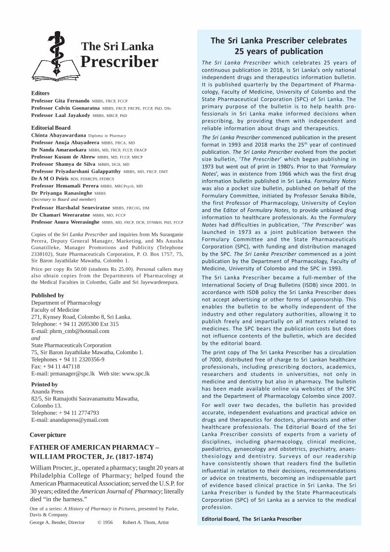
Cover picture
FATHER OF AMERICAN PHARMACY –
WILLIAM PROCTER, Jr. (1817-1874)
William Procter, jr., operated a pharmacy; taught 20 years atPhiladelphia College of Pharmacy; helped found theAmerican Pharmaceutical Association; served the U.S.P. for30 years; edited the American Journal of Pharmacy; literallydied “in the harness.”
The Sri Lanka
Prescriber
(Secretary to Board and member)
One of a series: A History of Pharmacy in Pictures, presented by Parke,Davis & Company.
George A. Bender, Director © 1956 Robert A. Thom, Artist
The Sri Lanka Prescriber celebrates25 years of publication
The Sri Lanka Prescriber which celebrates 25 years ofcontinuous publication in 2018, is Sri Lanka’s only nationalindependent drugs and therapeutics information bulletin.It is published quarterly by the Department of Pharma-cology, Faculty of Medicine, University of Colombo and theState Pharmaceutical Corporation (SPC) of Sri Lanka. Theprimary purpose of the bulletin is to help health pro-fessionals in Sri Lanka make informed decisions whenprescribing, by providing them with independent andreliable information about drugs and therapeutics.
The Sri Lanka Prescriber commenced publication in the presentformat in 1993 and 2018 marks the 25th year of continuedpublication. The Sri Lanka Prescriber evolved from the pocketsize bulletin, ‘The Prescriber’ which began publishing in1973 but went out of print in 1980’s. Prior to that ‘FormularyNotes’, was in existence from 1966 which was the first druginformation bulletin published in Sri Lanka. Formulary Noteswas also a pocket size bulletin, published on behalf of theFormulary Committee, initiated by Professor Senaka Bibile,the first Professor of Pharmacology, University of Ceylonand the Editor of Formulary Notes, to provide unbiased druginformation to healthcare professionals. As the FormularyNotes had difficulties in publication, ‘The Prescriber’ waslaunched in 1973 as a joint publication between theFormulary Committee and the State PharmaceuticalsCorporation (SPC), with funding and distribution managedby the SPC. The Sri Lanka Prescriber commenced as a jointpublication by the Department of Pharmacology, Faculty ofMedicine, University of Colombo and the SPC in 1993.
The Sri Lanka Prescriber became a full-member of theInternational Society of Drug Bulletins (ISDB) since 2001. Inaccordance with ISDB policy the Sri Lanka Prescriber doesnot accept advertising or other forms of sponsorship. Thisenables the bulletin to be wholly independent of theindustry and other regulatory authorities, allowing it topublish freely and impartially on all matters related tomedicines. The SPC bears the publication costs but doesnot influence contents of the bulletin, which are decidedby the editorial board.
The print copy of The Sri Lanka Prescriber has a circulationof 7000, distributed free of charge to Sri Lankan healthcareprofessionals, including prescribing doctors, academics,researchers and students in universities, not only inmedicine and dentistry but also in pharmacy. The bulletinhas been made available online via websites of the SPCand the Department of Pharmacology Colombo since 2007.
For well over two decades, the bulletin has providedaccurate, independent evaluations and practical advice ondrugs and therapeutics for doctors, pharmacists and otherhealthcare professionals. The Editorial Board of the SriLanka Prescriber consists of experts from a variety ofdisciplines, including pharmacology, clinical medicine,paediatrics, gynaecology and obstetrics, psychiatry, anaes-thesiology and dent is try. Surveys of our readershiphave consistently shown that readers find the bulletininfluential in relation to their decisions, recommendationsor advice on treatments, becoming an indispensable partof evidence based clinical practice in Sri Lanka. The SriLanka Prescriber is funded by the State PharmaceuticalsCorporation (SPC) of Sri Lanka as a service to the medicalprofession.
Editorial Board, The Sri Lanka Prescriber

1Sri Lanka Prescriber, Vol. 26, No.3, 2018
Management of glaucoma
IntroductionGlaucoma is a progressive optic neuropathy with charac-teristic changes in the optic nerve head and correspondingloss of visual field, that is associated frequently but notinvariably with a raised intraocular pressure (IOP). It is thesecond commonest cause of blindness in the world.
The global prevalence of glaucoma for a population aged40-80 years is 3.54%; ie. an estimate of 64.3 million withglaucoma in 2013, increasing to 76.0 million in 2020 and111.8 million in 2040 worldwide [1]. Glaucoma is the silentthief of sight where undetected and untreated patientsend up in blindness.
Glaucoma encompasses many different sub-types whichhave varied symptoms, pathophysiology and treatmentoptions. Knowledge of aqueous humour dynamics is ofparamount importance in understanding and managingglaucoma. The aqueous humour secreted by ciliaryprocesses into the posterior chamber, flows into theanterior chamber through the pupil. Aqueous humour exitsfrom the eye via two main pathways. Conventionally mostof the aqueous is thought to leave through the trabecularmeshwork at the irido-corneal angle via the Schlemm canalto drain into the episcleral venous plexus. Any non-trabecular outflow involving passage of aqueous throughciliary muscle and sclera is termed uveo-scleral outflow.The balance of synthesis and drainage maintains theintraocular pressure within the normal range (12-21 mmHg). In most individuals with glaucoma the optic nerveand visual field changes seen are determined by both thelevel of IOP and the resistance of the optic nerve to damage[2].
As glaucoma is a heterogeneous group of disorderssymptoms depend on the sub-type of glaucoma. In acuteglaucoma the main symptom is ocular pain. In chronicglaucoma e.g. primary open angle glaucoma, most of thepatients are symptomless.
In primary open angle glaucoma (POAG) the resistance toaqueous outflow is thought to be in the trabecular mesh-work. Conversely, in acute angle closure glaucoma theprimary pathology is proximal to the meshwork. In acuteangle closure there is apposition of peripheral iris to thetrabecular meshwork causing the irido-corneal angle to“close”. Pupillary block is the most frequent cause of angleclosure where flow of aqueous from posterior chamber toanterior chamber through the pupil is impeded at the lensiris interface. This results in a pressure gradient betweenthe two chambers causing peripheral iris to bow forwardagainst the trabecular meshwork [3] obstructing thedrainage of aqueous. Traditionally, secondary glaucoma
denotes an external mechanism such as trauma, tumours,cataractous lens, inflammatory cells, membranes ordevelopmental abnormality etc. playing a part in impedingthe outflow.
Symptoms and signs of GlaucomaPrimary open angle glaucoma is a sub-type that occursmore commonly in the elderly. It is characterized by increasein intraocular pressure (IOP > 21mmHg), an open drainageangle, glaucomatous optic nerve head damage and visualfield loss. The risk factors include increasing age, positivefamily history and myopia.
Another sub-type seen in Sri Lanka is the normotensive(normal tension) glaucoma (NTG) where the eye pressureis within normal limits but with progressive optic nervedamage and visual field defects. Ocular hypertension(OHT) is the sub-type where the IOP increases in theabsence of identifiable optic nerve damage or visual fieldloss. POAG and elevated NTG are asymptomatic until asignificant part of the visual field is lost and the presen-tation is often late.
In contrast acute angle closure glaucoma mostly occurringin elderly females has a dramatic presentation with acutesigns and symptoms.
Starting with ocular pain, headache, blurring of vision andsometimes with visualization of halos it may rapidlyprogress to severe ocular pain, marked redness, photo-phobia and loss of vision. Sometimes the patient has severeheadache, abdominal pain and vomiting so that the patientmay end up being admitted to a medical or surgical ward.
This condition, if not detected and treated immediately,the acute IOP rise will lead to permanent optic nervedamage and irreversible blindness.
Diagnosis of GlaucomaSince glaucoma is mostly a symptomless disease in theinitial stages, eye examination and screening is importantfor early detection. In the assessment of the patient severalinvestigations are utilized. Measuring the intraocularpressure (IOP), evaluating the irido-corneal angle (Gonio-scopy), visual field assessment (HVF), and assessing theoptic nerve head (OCT) are some of them.
Predisposing causes of GlaucomaGenetic factors, congenital defects, positive family history,previous eye trauma, ocular inflammation, cataract, long
2 Sri Lanka Prescriber, Vol. 26, No. 3, 2018
term uncontrolled diabetes and the use of steroid medi-cations may be associated with glaucoma. For these patientsregular screening for glaucoma is important.
Treatment options for GlaucomaIn current practice the main aim of treatment is to minimizethe progressive optic nerve damage by reducing the intra-ocular pressure. Treatment depends on the sub-type andthe options include topical, oral, and intravenous medi-cations, laser treatment and surgery. In the management ofpatients with a diagnosis of glaucoma, factors such astype and stage of the disease, rate of progression and lifeexpectancy of the patient are important considerations.Many clinicians would consider a target pressure based
on above factors to prevent further damage to the opticnerve. However this objective should be achieved withthe least disruption to patients’ daily life. Hence not onlyadverse effects, interactions and dosage regimes but alsothe cost of the medications should be taken into conside-ration in selecting optimum treatment options.
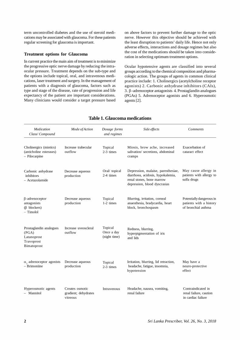
Ocular hypotensive agents are classified into severalgroups according to the chemical composition and pharma-cologic action. The groups of agents in common clinicalpractice include: 1. Cholinergics (acetylcholine receptoragonists) 2. Carbonic anhydrase inhibitors (CAIs),3. -adrenoceptor antagonists 4. Prostaglandin analogues(PGAs) 5. Adrenoceptor agonists and 6. Hyperosmoticagents [2].
Table 1. Glaucoma medications
Medication Mode of Action Dosage forms Side effects CommentsClass/ Compound and regimes
Cholinergics (miotics)(anticholine esterases)– Pilocarpine
Increase trabecularoutflow
Topical2-3 times
Miosis, brow ache, increasedsalivation/ secretions, abdominalcramps
Exacerbation ofcataract effect
Carbonic anhydrase inhibitors– Acetazolamide
Decrease aqueousproduction
Oral/ topical2-4 times
Depression, malaise, paresthesiae,diarrhoea, acidosis, hypokalemia,renal stones, bone marrowdepression, blood dyscrasias
May cause allergy inpatients with allergy tosulfa drugs
-adrenoceptorantagonists(blockers)– Timolol
Decrease aqueousproduction
Topical1-2 times
Blurring, irritation, cornealanaesthesia, bradycardia, heartblock, bronchospasm
Potentially dangerous inpatients with a historyof bronchial asthma
Prostaglandin analogues(PGA)LatanoprostTravoprostBimatoprost
Increase uveoscleraloutflow
Topical2-3 times
TopicalOnce a day(night time)
Redness, blurring,hyperpigmentation of irisand lids
2 adrenoceptor agonists– Brimoniine
Decrease aqueousproduction
Irritation, blurring, lid retraction, headache, fatigue, insomnia,hypotension
May have aneuro-protectiveeffect
Hyperosmotic agents– Mannitol
Creates osmoticgradient; dehydratesvitreous
Intravenous Headache, nausea, vomiting,renal failure
Contraindicated inrenal failure, cautionin cardiac failure

3Sri Lanka Prescriber, Vol. 26, No.3, 2018
Treatment typically begins with the selection of an agentfor IOP reduction. Although -adrenoceptor antagonistsare still commonly used by many clinicians, especially inthe low resource settings, the PGAs are playing anincreasingly important role in the first-line therapy ofglaucoma.
Adjunctive agents, such as -adrenoceptor agonists andCAIs are often effective at providing additional reductionin IOP for patients not controlled on monotherapy. As withany chronic disease, effective treatment depends onminimizing the adverse effects of therapy and maximizingpatient compliance.
The introduction of a variety of well tolerated and potentmedications over the past few years has allowed theclinician to choose a treatment regimen on an individualpatient basis resulting in safe and efficacious therapy inglaucoma. Many of the known systemic adverse effectsare rarely encountered in topical therapy, and long termtreatment is thus possible as in the case of introduction oftopical carbonic anhydrase inhibitors. Redness in the eyeand darkening around the eyes are known effects ofprostaglandin analogues. blockers (eg.timolol) shouldnot be prescribed to patients with a history of bronchialasthma. The side-effects of blockers can be minimizedby using selective blockers such as Levobunolol 0.25%.However, 2 adrenoceptor agonists should be used withcaution in the elderly due to its potential systemic hypo-tensive effect.
Combined therapy as a single vial has enhanced compliancewhile reducing the toxic effects of preservatives at thesame time. There are different regimes of administrationof glaucoma medications ranging from once a dayadministration to 4 times a day. Use of gel form eg; timololcan reduce the frequency of administration. As glaucomamedications should be continued long-term the patientshould fully understand the importance of the continuoustreatment.
As the patient does not feel improvement or worsening, itis important to monitor the disease with regular eyepressure checking and visual field assessment. The patientshould be involved in understanding and interpreting theinvestigations. Informed decisions should be takeninvolving the patient in continuing treatment. Othertreatment options in glaucoma management include lasertreatment and surgery in selected cases. Alternativestrategies that include neuroprotectants are also beinginvestigated at present [3].
Dr. M. Madhuwanthi Dissanayake, MBBS, MD, Senior Lecturer, Faculty of Medicine, University of Colombo;Specialist Eye Surgeon.
Before concluding that a medication is ineffective it isimportant to check for the correct technique of instillationof eye drops. The correct way of instillation of eye dropsis to place one drop in the lower fornix of each eye.Instilling more than one drop is a waste as the conjunctivalsac is only capable of holding up to 30 microlitres [4].Leaking out drops may cause unwanted side-effectssuch as pigmentation of peri-orbital skin in the case ofprostaglandin analogues. When there is more than onetype of topical medications, a gap of at least 10 minutesshould be allowed between the two types for absorptionof the first and to avoid washing off of the first medicationby the one that follows. Applying pressure with the indexfinger over the naso-lacrimal duct at the medial angle ofthe eye is known to reduce systemic absorption therebyreducing systemic side-effects.
Glaucoma and progressive optic neuropathy withcharacteristic changes in the optic nerve head andcorresponding visual field defects is the second commonestcause of blindness in the world. It is composed of aheterogeneous group of disorders. In current practice themain aim of treatment is to minimize the progressive opticnerve damage by reducing the intraocular pressure.Adverse effects, interactions, dosage regimes and thetreatment cost of the medications should also beconsidered in selecting the optimum treatment option.However, this objective should be achieved with the leastdisruption to patients’daily life. In current clinical practicemany patients achieve good control with advanced medicaltherapy. When IOP cannot be reduced sufficiently withpharmacological treatment to prevent the risk of pro-gression further treatment options should be considered [5].
References1. Tham S, Yih-Chung, et al. Global prevalence of glaucoma
and projections of glaucoma burden through 2040.Ophthalmology 2014; 121: 2081 2090.
2. Cioffi GA, Durcan FJ, Girkin CA, et al. Basic and clinicalscience course – glaucoma, American Academy ofOphthalmology 2012-2013; 161-73.
3. Varma R, Peeples P, Walt JG, Bramley TJ. Diseaseprogression and the need for neuroprotection in glaucomamanagement. American Journal of Managed Care 2008;14(1 Suppl): S15-9.
4. Bye L, Modi N, Stanford M. Basic Sciences for Ophthal-mology (Oxford Specialty Training) 1st Edition. Oxford:Oxford University Press 2013: 232.
5. http://pathways.nice.org.uk/pathways/glaucoma NICEPathway last updated: 12 October 2018.
4 Sri Lanka Prescriber, Vol. 26, No. 3, 2018
Management of acute ST-segment elevationmyocardial infarction
IntroductionAcute ST-segment Elevation Myocardial Infarction[STEMI] is a leading cause of morbidity and mortalityworldwide with an annual incidence of approximately 50per 100,000 in Europe and America [1, 2]. In these countriesthe in-hospital mortality rates have decreased due toimprovements in treatment, and presently vary around4-12% [1, 2]. Over the years preferred treatment strategyfor STEMI has shifted from thrombolysis to primarypercutaneous coronary intervention (PCI). In England98% of approximately 20,000 new STEMI patientsreceived primary PCI in 2014 [3].
In Sri Lanka (SL) the first cross sectional clinical audit ofacute coronary syndrome patients in government hospitalscovering the entire country done in 2015 showed a countrywide in-hospital mortality of 4% in the STEMI group[4, 5]. Only 5.7 % STEMI patients received primary PCIwhile 63% received fibrinolysis [4, 5]. Extrapolated fromthe Ministry of Health annual fibrinolytic requirementof approximately 13000 vials, in Sri Lanka an estimated20,000 acute STEMI patients may present to governmenthospitals annually. This article describes the current trendsin therapeutic management of acute STEMI according torecent evidence based guidelines [1, 2, 6, 7].
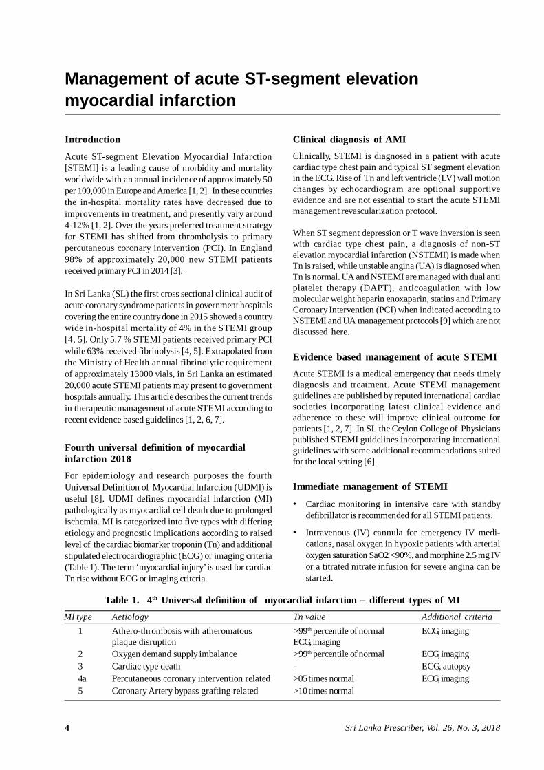
Fourth universal definition of myocardialinfarction 2018For epidemiology and research purposes the fourthUniversal Definition of Myocardial Infarction (UDMI) isuseful [8]. UDMI defines myocardial infarction (MI)pathologically as myocardial cell death due to prolongedischemia. MI is categorized into five types with differingetiology and prognostic implications according to raisedlevel of the cardiac biomarker troponin (Tn) and additionalstipulated electrocardiographic (ECG) or imaging criteria(Table 1). The term ‘myocardial injury’ is used for cardiacTn rise without ECG or imaging criteria.
Clinical diagnosis of AMIClinically, STEMI is diagnosed in a patient with acutecardiac type chest pain and typical ST segment elevationin the ECG. Rise of Tn and left ventricle (LV) wall motionchanges by echocardiogram are optional supportiveevidence and are not essential to start the acute STEMImanagement revascularization protocol.
When ST segment depression or T wave inversion is seenwith cardiac type chest pain, a diagnosis of non-STelevation myocardial infarction (NSTEMI) is made whenTn is raised, while unstable angina (UA) is diagnosed whenTn is normal. UA and NSTEMI are managed with dual antiplatelet therapy (DAPT), anticoagulation with lowmolecular weight heparin enoxaparin, statins and PrimaryCoronary Intervention (PCI) when indicated according toNSTEMI and UA management protocols [9] which are notdiscussed here.
Evidence based management of acute STEMIAcute STEMI is a medical emergency that needs timelydiagnosis and treatment. Acute STEMI managementguidelines are published by reputed international cardiacsocieties incorporating latest clinical evidence andadherence to these will improve clinical outcome forpatients [1, 2, 7]. In SL the Ceylon College of Physicianspublished STEMI guidelines incorporating internationalguidelines with some additional recommendations suitedfor the local setting [6].
Immediate management of STEMI
• Cardiac monitoring in intensive care with standbydefibrillator is recommended for all STEMI patients.
• Intravenous (IV) cannula for emergency IV medi-cations, nasal oxygen in hypoxic patients with arterialoxygen saturation SaO2 <90%, and morphine 2.5 mg IVor a titrated nitrate infusion for severe angina can bestarted.
MI type Aetiology Tn value Additional criteria1 Athero-thrombosis with atheromatous >99th percentile of normal ECG, imaging
plaque disruption ECG, imaging2 Oxygen demand supply imbalance >99th percentile of normal ECG, imaging3 Cardiac type death - ECG, autopsy4a Percutaneous coronary intervention related >05 times normal ECG, imaging5 Coronary Artery bypass grafting related >10 times normal
Table 1. 4th Universal definition of myocardial infarction – different types of MI

5Sri Lanka Prescriber, Vol. 26, No.3, 2018
• As soon as possible and in the absence of contra-indications following drugs are recommended Loading dose of non-enteric coated aspirin 300 mg,
preferably chewed and swallowed. Loading dose of a platelet P2Y12 inhibitor
clopidogrel or ticagrelor depending on thereperfusion strategy planned as described below.
High intensity statin therapy such as atorvastatin40-80 mg.
A proton pump inhibitor (PPI) such as panto-prazole 40 mg orally to prevent gastric irritationdue to DAP T is reasonable.
• A reperfusion strategy is initiated depending on clinicalcircumstances and availability of facilities.
A. Reperfusion strategy
The primary aim in acute STEMI is early reperfusion of theinfarct related coronary artery (IRA) occluded by theathero-thrombus. A delay in reperfusion leads to loss ofmyocardium resulting in worsening LV function and poorprognosis.
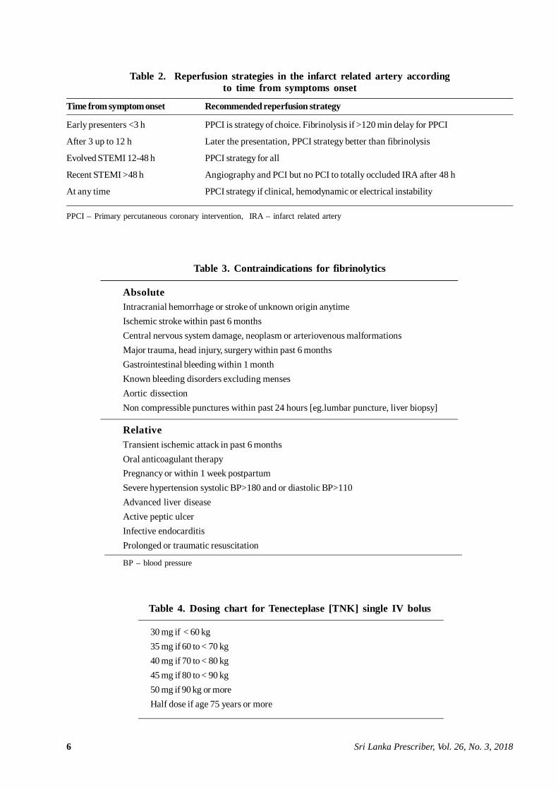
The best strategy for any given patient is decided on factorssuch as the time since onset of chest pain, time delay fromSTEMI diagnosis to primary PCI, clinical condition ofpatient and bleeding risk. Recommendations for reperfusionstrategy in the IRA are given in Table 2.
1. Primary PCIPrimary PCI (PPCI) is the revascularization method of choiceunless the anticipated time from STEMI diagnosis to PCImediated reperfusion is >120 minutes. STEMI patientspresenting within 12 hours from start of chest pain, andeven those admitted after12-48 hours with clinical or ECGevidence of ongoing ischemia or in cardiogenic shock arealso candidates for PPCI. Patients admitted to hospitalswithout PCI facilities should be transferred urgently in anambulance with cardiac monitoring, defibrillator facility andan attending doctor, if PCI can be done in <120 minutes,after contacting the emergency PCI team at the hospital.For such transfers a door-in door-out time < 30 minutes atthe transferring hospital should be achieved. A patientadmitted to a PPCI capable hospital should undergo PPCIwithin 90 minutes.
Adjunctive treatment for PPCI
• The loading dose of clopidogrel is 600 mg orally isadded to initially given aspirin 300 mg. Ticagrelor hasshown superiority in clinical outcomes to clopidogrelin the setting of PPCI and is the preferred P2Y12inhibitor. If available and affordable, loading dose ofticagrelor 180 mg orally is given instead of clopidogrelfor PPCI patients.
• At the beginning of PCI an IV bolus of unfractionatedheparin (UFH) 70-100 units /kg is given to maintain anactivated clotting time of approximately 300 seconds.It is reasonable to give part of this dose (Eg. UFH 4000units IV bolus) earlier at the ETU or at the transferringhospital in the SL setting if advised by the PPCI teamespecially when radial access for PPCI is planned.
• Platelet glycoprotein IIb/IIIa receptor inhibitors suchas abciximab are used intracoronary at the time of PCIfor a large thrombus burden. IV heparin dose at timeof PCI needs to be reduced if abciximab is planned.
Routine radial access and drug-eluting stent implant is thestandard of care. PPCI to the IRA usually results inimmediate reperfusion.
2. FibrinolyticsWhen delay for PPCI exceeds 120 minutes from STEMIdiagnosis, there is no survival advantage of primary PCIover fibrinolysis. When PPCI is not an option and in theabsence of contraindications, fibrinolysis should beinitiated immediately ideally <10 minutes of STEMIdiagnosis and with a door to needle time <30 minutes.STEMI patients are considered for thrombolysis ifpresenting within12 hours of initial chest pain and rarelyeven after 12-24 hours if clinical or ECG evidence of ongoingischemia is present. Contraindications for fibrinolyticsgiven in Table 3 needs exclusion.
Fibrinolytics available in SL are Tenecteplase (TNK-tPA),Streptokinase (SK) and Alteplase (tPA).• Tenecteplase (TNK-tPA) is the preferred fibrin specific
thrombolytic with better reperfusion rates than SK.TNK is now available in most Teaching and Provincialhospitals in SL. It is given as a single IV bolus accor-ding to body weight calculated from a dosing chart(Table 4). In some developed countries TNK IV bolusis given by the ambulance team as pre-hospital fibrino-lysis, minimizing delays when decided by the hospitalemergency cardiac team after telemetric ECG review.
• Streptokinase (SK) 1.5 million units IV infusion over60 minutes is a less expensive alternative but withlesser reperfusion efficacy as it is not fibrin specific. Itneeds to be started in-hospital preferably in theintensive care unit (ICU) setting with resuscitationfacilities while monitoring for allergic reactions,hypotension and arrhythmias. Hydrocortisone 100 mgIV and chlorpheniramine 10 mg IV are usually givenprior to starting SK infusion in the ICU setting in mostSL hospitals. Previous treatment with SK is a contra-indication for its use.
• Alteplase (tPA) 15 mg IV bolus followed by IV infusiongiven over 90 minutes according to body weight isanother alternative thrombolytic. It is more expensivethan TNK and carries higher reperfusion rates as wellas higher bleeding risks.
6 Sri Lanka Prescriber, Vol. 26, No. 3, 2018
Time from symptom onset Recommended reperfusion strategy
Early presenters <3 h PPCI is strategy of choice. Fibrinolysis if >120 min delay for PPCI
After 3 up to 12 h Later the presentation, PPCI strategy better than fibrinolysis
Evolved STEMI 12-48 h PPCI strategy for all
Recent STEMI >48 h Angiography and PCI but no PCI to totally occluded IRA after 48 h
At any time PPCI strategy if clinical, hemodynamic or electrical instability
PPCI – Primary percutaneous coronary intervention, IRA – infarct related artery
AbsoluteIntracranial hemorrhage or stroke of unknown origin anytimeIschemic stroke within past 6 monthsCentral nervous system damage, neoplasm or arteriovenous malformationsMajor trauma, head injury, surgery within past 6 monthsGastrointestinal bleeding within 1 monthKnown bleeding disorders excluding mensesAortic dissectionNon compressible punctures within past 24 hours [eg.lumbar puncture, liver biopsy]
RelativeTransient ischemic attack in past 6 monthsOral anticoagulant therapyPregnancy or within 1 week postpartumSevere hypertension systolic BP>180 and or diastolic BP>110Advanced liver diseaseActive peptic ulcerInfective endocarditisProlonged or traumatic resuscitation
BP – blood pressure
Table 2. Reperfusion strategies in the infarct related artery accordingto time from symptoms onset
Table 4. Dosing chart for Tenecteplase [TNK] single IV bolus
30 mg if < 60 kg35 mg if 60 to < 70 kg40 mg if 70 to < 80 kg45 mg if 80 to < 90 kg50 mg if 90 kg or moreHalf dose if age 75 years or more
Table 3. Contraindications for fibrinolytics

7Sri Lanka Prescriber, Vol. 26, No.3, 2018
Adjunctive treatment for fibrinolytics
• The loading dose of clopidogrel is 300 mg orally isadded to initial dose of aspirin 300 mg.Ticagrelor has not been studied as adjunctivetreatment for fibrinolytics.
• If age < 75 years, enoxaparin 30 mg IV bolus is givenfollowed by subcutaneous (SC) injection 15 min laterat 1 mg per kg up to a maximum of 100 mg per injectionevery 12 hours. Enoxaparin is continued until PCI orminimum 48 hours to a maximum of 8 days or untilhospital discharge.
• If > 75 years, enoxaparin IV is not given. Enoxaparin isstarted with SC dose of 0.75 mg per kg, up to maximumof 75 mg per injection given 12 hourly. SC dose isgiven once daily in renal failure.
3. Pharmaco-invasive strategyFor this option, following initial fibrinolysis and successfulreperfusion, patient is routinely transferred for PCI to beperformed within 3-10 hours. This strategy is especiallyuseful in the SL setting for hospitals with transport delays> 120 min to reach a PPCI capable hospital. This strategy isespecially successful for low bleeding risk patients whopresent early within < 2 to 3 hours of chest pain onset.Routine PCI after fibrinolysis may improve outcomes bypreventing re-infarction associated deaths.
In failed reperfusion indicated by clinical deterioration andST segment resolution < 50% at 90 min from fibrinolyticinitiation, the patient should be immediately transferredfor rescue PCI. Rescue PCI has shown better outcome thanrepeat fibrinolysis or conservative management in thiscategory of patients.
Adjunctive treatment for pharmaco-invasive strategy
• Adjunctive treatment is similar to that given withfibrinolytics.
• At PCI, Heparin IV dose may need reduction due toprior fibrinolytic and enoxaparin use and GPIIb/IIIainhibitors are best avoided to minimize bleedingcomplications.
B. Late (>12 hours) presentation of STEMI
Supportive medications are similar to adjunctive treatmentfor fibrinolysis. Fibrinolysis is not indicated in this group.
• Regardless of the time from symptoms onset, ongoingischemia, hemodynamic instability, or life threateningarrhythmia is an indication for a primary PCI strategy[1].
• Evolved STEMI (12-48 hours) and recent STEMI
(>48 hours) are also recommended a Primary PCIstrategy but a completely occluded IRA is notopened after 48 hours [1].
• After 48 hours in stable patients, non-invasive testsfor residual ischemia or viability are done to decidewhether a late invasive strategy or elective coronaryangiography should be done.
C. Secondary prevention medications
Following drugs have proven survival benefit in AMI. Inthe absence of contraindications these are started within24 hours and continued at hospital discharge and longterm.
• Low dose aspirin 75-150 mg nocte is recommended forlife.
• P2Y12 inhibitor
Clopidogrel – As part of DAPT with aspirin,clopidogrel 75 mg orally daily is recommended forminimum 1 year and can be extended at thediscretion of the treating physician.
Ticagrelor – For PPCI patients who received aloading dose, maintenance dose of ticagrelor is 90mg orally twice daily [BD] for one year and 60 mgBD if continued after one year up to 3 years.Aspirin dose should be <100 mg when ticagrelor isused.Ticagrelor is not suitable if body weight <60 kgand for triple therapy with warfarin.
• Statin – high intensity atorvastatin 40-80 mg to achievefasting low density cholesterol (LDL) <70 mg/dl.Statins act in plaque stabilization.
• Beta blockers (BB) – carvedilol, bisoprolol,metoprolol or nebivolol are useful in all patients andparticularly if left ventricular ejection fraction (LVEF)<40%. Atenolol can be given if no LV dysfunction. BBreduce risk of ventricular fibrillation after AMI.
• Angiotensin converting enzyme inhibitor (ACEI) orangiotensin receptor blocker [ARB] should be startedwithin 24 hours in all AMI patients and particularly inpatients with LVEF < 40%, diabetes, anterior STEMI.
An ACEI such as enalapril, ramipril or lisinopril or ifnot tolerated due to ACEI related cough, an ARB suchas valsartan, candesartan or losartan is recommended.ACEI and ARB act in preventing cardiac remodelingespecially with LVEF <40%.
• Mineralocorticoid receptor antagonist (MRA) –spironolactone is given as add on therapy to ACEI orARB if LVEF < 40%. Eplerenone can be given if trouble-some antiandrogenic effects occur with spirono-lactone.

8 Sri Lanka Prescriber, Vol. 26, No. 3, 2018
LV function should be measured in all patients with STEMIwith a 2D echocardiogram. In post AMI patients with LVEF< 40% treatment with ACEI or ARB, MRA, specific BB andivabradine reduce mortality. Amiodarone is useful to treatatrial fibrillation and ventricular tachycardia.
A PPI such as pantoprazole 40 mg nocte is recommendedlong term along with DAPT to prevent peptic ulcer.Ezetimibe may be added to a statin to achieve lipid goals.Medications to be given at discharge to all STEMIpatients in the absence of contraindications are listed inTable 5.
Calcium channel blockers, nitrates, frusemide, vitamin E,nicorandil, and fish oil have no proven survival benefit inSTEMI patients but may be used for specific indications.Non-steroidal anti-inflammatory drugs (NSAID) are harmfulin AMI patients and should not be prescribed for analgesia.Coronary artery bypass graft surgery (CABG) should beconsidered for patients with a patent IRA but withunsuitable anatomy for PCI. P2Y12 inhibitor needs to bestopped 3-7 days prior to CABG.
D. Risk factors and life style modification
The major cardiovascular risk factors need identifying andcorrection. ACS Sri Lanka audit project showed a highprevalence of risk factors in AMI patients in SL withdiabetes in 28%, hypertension in 45%, and current smokingin 35%. Goals of therapy include
• Diabetes – Goal of treatment is HBa1C <7.0
• Hypertension – Goal of treatment is BP < 130/ 80
• Hyperlipidemia – Goal of treatment is LDL < 70 mg/dl
• Smoking cessation advice needs to be given to allsmokers.
• Obesity, diet and sedentary lifestyle need addressing.
• Enrolment in a cardiac rehabilitation program withdietary and lifestyle advice, exercise prescribing andmonitoring recovery and resumption of activities ofdaily living.
E. Management of complications
Management of complications of AMI are not discussedin detail here. Complications such as cardiogenic shock,acute pulmonary edema and arrhythmias need intensivecare management. Mechanical complications such asventricular septal rupture or papillary muscle rupture needidentification and early corrective surgery.
F. Quality indicators and clinical audit in STEMI
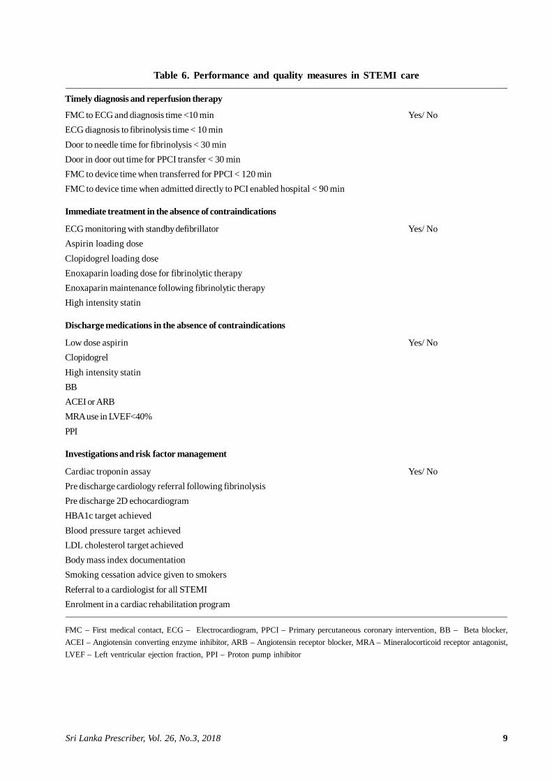
A gap between optimal guideline-based treatment andactual care of STEMI patients could exist in clinical practice.In order to reduce this gap, validated quality indicatorsneed to be measured to improve STEMI care and patientsurvival [1, 2,10]. Suggested performance measures to berecorded for each STEMI patient and clinically audited byall physicians managing STEMI patients in SL are given inTable 6.
Low dose aspirin 75-100 mg
P2Y12 inhibitor such as clopidogre 175 mg or ticagrelor 90 mg twice daily
High intensity statin such as atorvastatin 40-80 mg
BB-carvedilol, metoprolol, bisoprolol, nebivolol, dose titrated
ACEI or ARB [not both together] dose titrated
MRA when LVEF <40% spironolactone or eplerenone dose titrated
PPI such as pantoprazole 40 mg
BB – Beta blocker, ACEI – Angiotensin converting enzyme inhibitor, ARB – Angiotensin receptor blocker,MRA – Mineralocorticoid receptor antagonist, LVEF – Left ventricular ejection fraction,
PPI – Proton pump inhibitor
Table 5. Medications at discharge for STEMI patients
9Sri Lanka Prescriber, Vol. 26, No.3, 2018
Table 6. Performance and quality measures in STEMI care
Timely diagnosis and reperfusion therapy
FMC to ECG and diagnosis time <10 min Yes/ NoECG diagnosis to fibrinolysis time < 10 minDoor to needle time for fibrinolysis < 30 minDoor in door out time for PPCI transfer < 30 minFMC to device time when transferred for PPCI < 120 minFMC to device time when admitted directly to PCI enabled hospital < 90 min
Immediate treatment in the absence of contraindications
ECG monitoring with standby defibrillator Yes/ NoAspirin loading doseClopidogrel loading doseEnoxaparin loading dose for fibrinolytic therapyEnoxaparin maintenance following fibrinolytic therapyHigh intensity statin
Discharge medications in the absence of contraindications
Low dose aspirin Yes/ NoClopidogrelHigh intensity statinBBACEI or ARBMRA use in LVEF<40%PPI
Investigations and risk factor management
Cardiac troponin assay Yes/ NoPre discharge cardiology referral following fibrinolysisPre discharge 2D echocardiogramHBA1c target achievedBlood pressure target achievedLDL cholesterol target achievedBody mass index documentationSmoking cessation advice given to smokersReferral to a cardiologist for all STEMIEnrolment in a cardiac rehabilitation program
FMC – First medical contact, ECG – Electrocardiogram, PPCI – Primary percutaneous coronary intervention, BB – Beta blocker,ACEI – Angiotensin converting enzyme inhibitor, ARB – Angiotensin receptor blocker, MRA – Mineralocorticoid receptor antagonist,LVEF – Left ventricular ejection fraction, PPI – Proton pump inhibitor

10 Sri Lanka Prescriber, Vol. 26, No. 3, 2018
References1. Ibanez B, James S, Agewall S, Antunes MJ, Bucciarelli-
Ducci C, Bueno H, Caforio AL, Crea F, Goudevenos JA,Halvorsen S, Hindricks G. 2017 ESC Guidelines for themanagement of acute myocardial infarction in patientspresenting with ST-segment elevation: The Task Force forthe management of acute myocardial infarction in patientspresenting with ST-segment elevation of the EuropeanSociety of Cardiology (ESC). European Heart Journal 2017;39(2): 119-77.
2. O'Gara PT, Kushner FG, Ascheim DD, et al. 2013 ACCF/AHA guideline for the management of ST-elevationmyocardial infarction: a report of the American College ofCardiology Foundation/American Heart Association TaskForce on Practice Guidelines. J Am Coll Cardiol. 2013; 61:e78-140.
3. Weston C, Gavalova L, Whittaker T, Van Leeven R.Myocardial Ischaemia National Audit Project (MINAP)How the NHS cares for patients with heart attack. 6th AnnualPublic Report. April 2013-March 2014. downloaded fromhttp://www.ucl.ac.uk/nicor/audits/minap/publicreports
4. Galappatthy P, Bataduwaarachchi VR, Ranasinghe P,Galappatthy GKS, Wijayabandara M, Warapitiya DS,Sivapathasundaram M, Wickramarathna T, Senarath U,Sridharan S, Wijeyaratne CN. Management, characteristicsand outcomes of patients with acute coronary syndrome inSri Lanka. Heart 2018; 104: 1424-31.
5. Lodi-Junqueira L, Ribeiro AL Tackling acute coronary
syndrome in low-income and middle-income countries. Heart2018; 104: 1390-911.
6. Sri Lanka Heart Association, Sri Lanka STEMI Forum.STEMI Management guidelines. Journal of the CeylonCollege of Physicians 2015; 45: 1-20.
7. NICE National Institute for Health and Care Excellence.Myocardial Infarction with ST segment elevation: acutemanagement, Clinical Guideline 167, 10 July 2013,nice.oeg.uk/guidance/cg167 accessed November 2018.
8. Thygesen K, Alpert JS, Jaffe AS, Chaitman BR, Bax JJ,Morrow DA, White HD. Fourth Universal Definition ofMyocardial Infarction (2018). Journal of the AmericanCollege of Cardiology 2018; 72(18): 2231-64.
9. Roffi M, Patrono C, Collet JP, et al. 2015 ESC guidelinesfor the management of acute coronary syndromes in patientspresenting without persistent ST-segment elevation: TaskForce for the management of acute coronary syndromes inpatients presenting without persistent st-segment elevationof the European Society of Cardiology (ESC). Eur Heart J2016; 37: 267-315.
10. Jneid H, Addison D, Bhatt DL, Fonarow GC, Gokak S,Grady KL, Green LA, Heidenreich PA, Ho PM, JurgensCY, King ML, Kumbhani DJ, Pancholy S. 2017 AHA/ACCclinical performance and quality measures for adults withST-elevation and non-ST-elevation myocardial infarction: areport of the American College of Cardiology/American HeartAssociation Task Force on Performance Measures. J AmColl Cardiol. 2017; 70: 2048-90.
Dr. Gamini Galappatthy, MBBS, MD, MRCP (UK), FRCP (LOND), FCCP, Consultant Cardiologist, Institute of Cardiology,National Hospital of Sri Lanka.E-mail: [email protected]
Conflicts of interest: none declared

11Sri Lanka Prescriber, Vol. 26, No. 3, 2018
Neuropathic pain: current definition and review ofdrug treatment
IntroductionNeuropathic pain is associated with impaired quality oflife, and is often poorly managed. Around 7-8% of adultshave pain with neuropathic characteristics. A quarter ofpeople with diabetes and 35% of people with HIV haveneuropathic pain.1
The management of neuropathic pain can be challengingand, as with all pain, should be approached with a bio-psychosocial framework. There are several options fordrug treatment as part of an overall approach to improvepatients’ quality of life and function.2
International guidelines have clarified the definition ofneuropathic pain and updated their recommendations fordrug treatment based on evidence from a systematic reviewand meta-analysis.3,4 Being aware of these changes isimportant in the clinical assessment and treatment.
A new definition for neuropathic painNeuropathic pain is now defined by the InternationalAssociation for the Study of Pain (IASP) as ‘pain causedby a lesion or disease of the somatosensory nervous
SummaryNeuropathic pain is relatively common and oftenpoorly treated.
Management options include tricyclic antidep-ressants or serotonin and noradrenaline reuptakeinhibitors in the first instance, followed by pregabalinor gabapentin.
Tramadol or topical lidocaine (lignocaine) could beconsidered as second line. Stronger opioids havebeen relegated to third line.
It is important to remember that opioids and gaba-pentinoids have abuse potential.
Fibromyalgia and chronic low back pain withoutradiculopathy do not meet the current criteria for thedefinition of neuropathic pain.
Keywords: anticonvulsants, antidepressants,cannabinoids, neuropathic pain, opioids
(Aust Prescr 2018; 41: 60-3)
system’.3 This replaces the older definition of ‘pain initiatedor caused by a primary lesion, dysfunction or transitoryperturbation of the peripheral or central nervous system’.The definition was reviewed and updated because theterm dysfunction in the old definition was thought to beover-inclusive and did not reflect the pathophysiology.Additionally, neuropathic pain is not one disease entitybut a number of diseases or lesions with a cluster ofsymptoms and signs, where understanding of patho-physiology is evolving.5
Proponents of the change believe it has greater scientificrigour. It removes confusion around pain arising as a resultof disease within the nervous system but outside thesomatosensory system, for example pain from musclespasticity. It now excludes syndromes where patho-physiology is unclear, such as fibromyalgia or complexregional pain syndrome, which is controversial and hasbeen perceived by some to be overly restrictive.6
Primary disease managementThe primary disease management of neuropathic painneeds to consider the individual as a whole. For instance,in patients with diabetic neuropathy, erratic glycaemiccontrol worsens symptoms and improving glycaemiccontrol may reduce progression of neuropathy. However,there is increased mortality with intensive insulin regimensin patients with established diabetic neuropathy comparedto patients without neuropathy.7 HIV associated neuro-pathy presents an even more complex picture – startingantiretrovirals may initially improve symptoms althoughnerve damage may progress. Some antiretrovirals can causeneuropathy, and neurotoxicity may be a feature of con-comitant medicines such as isoniazid for tuberculosis.8,9
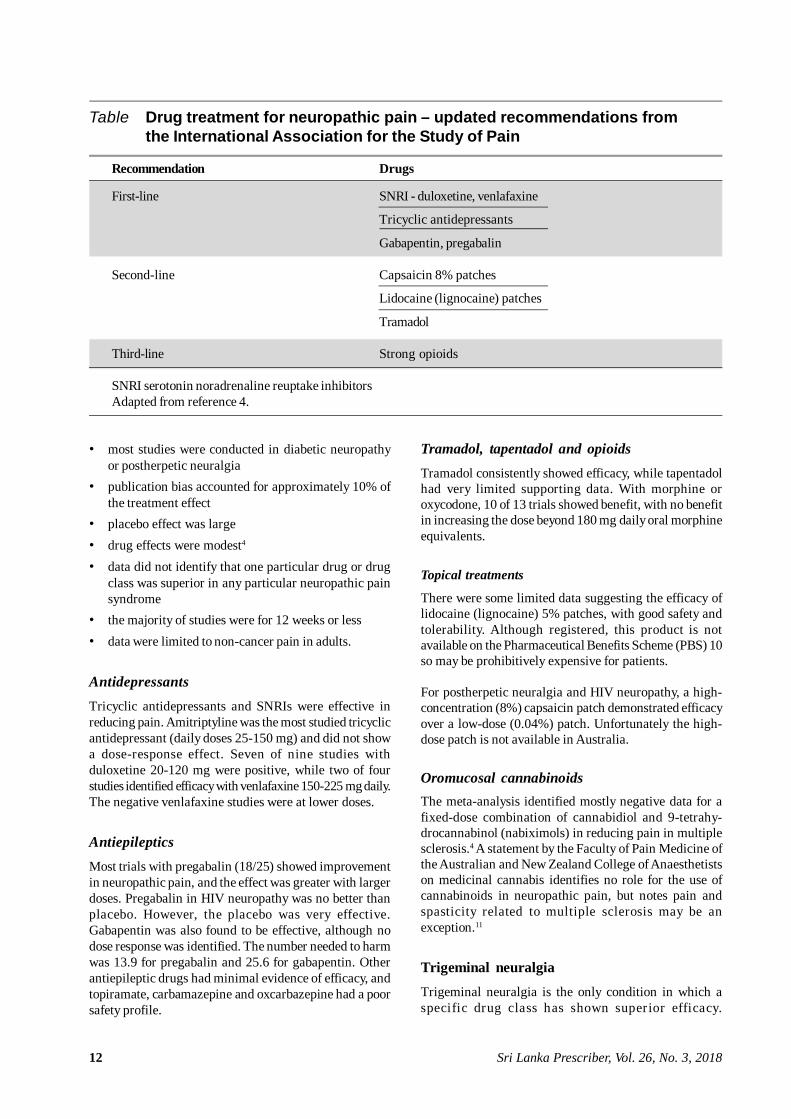
Drugs for neuropathic painThe IASP’s Neuropathic Pain Special Interest Group(NeuPSIG) has recently undertaken a systematic review ofmedicines for neuropathic pain (Table).4 Fibromyalgia,atypical facial pain, complex regional pain syndrome andchronic low back pain without radiculopathy were notincluded in the review as they do not meet the currentcriteria for the definition of neuropathic pain.
The review included tricyclic antidepressants, serotoninand noradrenaline reuptake inhibitors (SNRIs), antiepilepticdrugs, opioids, topical lidocaine (lignocaine), capsaicinhigh-concentration patches and oromucosal cannabinoids.A number of overarching themes were identified:
12 Sri Lanka Prescriber, Vol. 26, No. 3, 2018
• most studies were conducted in diabetic neuropathyor postherpetic neuralgia
• publication bias accounted for approximately 10% ofthe treatment effect
• placebo effect was large• drug effects were modest4
• data did not identify that one particular drug or drugclass was superior in any particular neuropathic painsyndrome
• the majority of studies were for 12 weeks or less• data were limited to non-cancer pain in adults.
AntidepressantsTricyclic antidepressants and SNRIs were effective inreducing pain. Amitriptyline was the most studied tricyclicantidepressant (daily doses 25-150 mg) and did not showa dose-response effect. Seven of nine studies withduloxetine 20-120 mg were positive, while two of fourstudies identified efficacy with venlafaxine 150-225 mg daily.The negative venlafaxine studies were at lower doses.
AntiepilepticsMost trials with pregabalin (18/25) showed improvementin neuropathic pain, and the effect was greater with largerdoses. Pregabalin in HIV neuropathy was no better thanplacebo. However, the placebo was very effective.Gabapentin was also found to be effective, although nodose response was identified. The number needed to harmwas 13.9 for pregabalin and 25.6 for gabapentin. Otherantiepileptic drugs had minimal evidence of efficacy, andtopiramate, carbamazepine and oxcarbazepine had a poorsafety profile.
Tramadol, tapentadol and opioidsTramadol consistently showed efficacy, while tapentadolhad very limited supporting data. With morphine oroxycodone, 10 of 13 trials showed benefit, with no benefitin increasing the dose beyond 180 mg daily oral morphineequivalents.
Topical treatments
There were some limited data suggesting the efficacy oflidocaine (lignocaine) 5% patches, with good safety andtolerability. Although registered, this product is notavailable on the Pharmaceutical Benefits Scheme (PBS) 10so may be prohibitively expensive for patients.
For postherpetic neuralgia and HIV neuropathy, a high-concentration (8%) capsaicin patch demonstrated efficacyover a low-dose (0.04%) patch. Unfortunately the high-dose patch is not available in Australia.
Oromucosal cannabinoidsThe meta-analysis identified mostly negative data for afixed-dose combination of cannabidiol and 9-tetrahy-drocannabinol (nabiximols) in reducing pain in multiplesclerosis.4 A statement by the Faculty of Pain Medicine ofthe Australian and New Zealand College of Anaesthetistson medicinal cannabis identifies no role for the use ofcannabinoids in neuropathic pain, but notes pain andspasticity related to multiple sclerosis may be anexception.11
Trigeminal neuralgiaTrigeminal neuralgia is the only condition in which aspecific drug class has shown superior efficacy.
Table Drug treatment for neuropathic pain – updated recommendations fromthe International Association for the Study of Pain
Recommendation Drugs
First-line SNRI - duloxetine, venlafaxine
Tricyclic antidepressants
Gabapentin, pregabalin
Second-line Capsaicin 8% patches
Lidocaine (lignocaine) patches
Tramadol
Third-line Strong opioids
SNRI serotonin noradrenaline reuptake inhibitorsAdapted from reference 4.
13Sri Lanka Prescriber, Vol. 26, No. 3, 2018
Carbamazepine and oxcarbazepine are first line forpharmacological pain management.12
It is currently recommended that Asian people of non-Japanese origin are tested for the HLA-B*1502 allele asthis confers an increased risk of cutaneous drug reactionswith carbamazepine.13
Interventional modalitiesLocal nerve blocks, spinal or epidural medicines, andneuro-ablative, neuromodulatory and neurosurgicalprocedures are also used for neuropathic pain.14
Updated recommendations for treatmentAs a result of the meta-analysis, NeuPSIG has updated itsrecommendations for the treatment of non-cancerassociated neuropathic pain in adults. With the exceptionof trigeminal neuralgia, there were no data identifying thatany particular drug was superior to another in anyparticular disease state.4
The guidelines recommend tricyclic antidepressants,gabapentin or pregabalin, and the SNRIs venlafaxine orduloxetine as first line.4
Second-line treatments include tramadol. Topical lidocaine(lignocaine) or high-concentration capsaicin may beconsidered for neuropathic pain when there is a presumedlocal generator.4
The consensus is that opioids can no longer be recom-mended as first-line treatment, and there is generalagreement that they should only be considered as thirdline, with appropriate monitoring for safety and efficacy.4
It is increasingly recognised that the harms of opioids, inparticular addiction, cannot be adequately identified inshort-term studies. Also, these short-term studies couldnot identify if any benefit persists or is lost as tolerancedevelops.
A pragmatic approach to drug therapyChoose a tricyclic antidepressant or SNRI with conside-ration of the patient’s comorbidities, potential druginteractions and adverse effects, and consider pregabalinor gabapentin next before tramadol. There is a paucity ofguidance on duration of treatment. Again, a pragmaticapproach may be to try a therapy for 12 weeks as this is themaximum duration of most of the trials. Monitor for efficacy(using multidimensional tools for pain intensity, quality oflife and patient function) and safety, and stop if thetreatment is not working.
The PBS listing for pregabalin in neuropathic pain is that‘the condition must be refractory to treatment with other
drugs’. Cost of treatment is significant. In 2016-17, morethan 3.5 million PBS scripts for pregabalin were issued at acost of over $190 million.15 Gabapentinoids haveneurocognitive adverse effects, can cause weight gain andare associated with an increased risk of falls. They areanxiolytic, and there is emerging evidence of significantpregabalin abuse.16
Any consideration of psychotropic drugs includinggabapentinoids or opioids (tramadol or stronger opioids)should involve:• assessing the risk of abuse, including history of
psychiatric, personality or substance use disorder• ongoing monitoring for development of abuse• multidimensional assessment of efficacy.
A plan to stop therapy should be discussed with the patientbefore treatment starts, and daily opioid doses should notexceed 60 mg oral morphine equivalents without specialistreview.17
Conclusion
A well-conducted meta-analysis reviewing drug treatmentof neuropathic pain provides clear recommendations.Tricyclic antidepressants and SNRIs should be trialled first.If they are ineffective, consider a trial of a gabapentinoidthen tramadol. This should be accompanied by multi-dimensional assessment of efficacy, review for harmsassociated with treatment and a plan for stopping treatmentif there is no benefit.
Conflict of interest: none declared
References1. International Association for the Study of Pain. Epidemiology
of neuropathic pain: how common is neuropathic pain, andwhat is its impact? Washington: IASP; 2014. [cited 2018May 1]
2. Pain Management Network [Internet]. Sydney: Agency forClinical Innovation; 2018 [cited 2018 May 1]
3. International Association for the Study of Pain. IASPTaxonomy. Pain terms. Neuropathic pain. Updated 2017Dec 14. [cited 2018 May 1]
4. Finnerup NB, Attal N, Haroutounian S, McNicol E, BaronR, Dworkin RH, et al. Pharmacotherapy for neuropathicpain in adults: a systematic review and meta-analysis. LancetNeurol 2015; 14: 162-73.
5. Jensen TS, Baron R, Haanpää M, Kalso E, Loeser JD, RiceAS, et al. A new definition of neuropathic pain. Pain 2011;152: 2204-5.
6. Oaklander AL, Wilson PR, Moskovitz PA, Manning DC,Lubenow T, Levine JD, et al. Response to “A new definitionof neuropathic pain”. Pain 2012; 153: 934-5.
14 Sri Lanka Prescriber, Vol. 26, No. 3, 2018
Bridin P Murnion, Senior Clinical Lecturer, University of Sydney; Senior Staff Specialist, Drug Health Services,Concord Repatriation General Hospital, Sydney.
ISSN 1391-0736Printed by Ananda Press, Colombo.
This article is reproduced from the Australian Prescriber 2018; 41: 60-3 by prior arrangement, courtesy of Australian Prescriber.
7. Javed S, Alam U, Malik RA. Treating diabetic neuropathy:present strategies and emerging solutions. Rev Diabet Stud2015; 12: 63-83.
8. Centner CM, Little F, Van Der Watt JJ, Vermaak JR, DaveJA, Levitt NS, et al. Evolution of sensory neuropathy afterinitiation of antiretroviral therapy. Muscle Nerve 2018; 57:371-9.
9. Centner CM, Bateman KJ, Heckmann JM. Manifestationsof HIV infection in the peripheral nervous system. LancetNeurol 2013;12:295-309.
10. Pharmaceutical Benefits Scheme. PBAC Public SummaryDocuments - March 2015. Lignocaine; 5% patch: dermal,30; Versatis. [cited 2018 May 1]
11. Faculty of Pain Medicine, Australian and New ZealandCollege of Anaesthetists. Statement on "medicinal cannabis"with particular reference to its use in the management ofpatients with chronic non-cancer pain. PM10. Melbourne:ANZCA; 2015. [cited 2018 May 1]
12. Cruccu G, Gronseth G, Alksne J, Argoff C, Brainin M,
Burchiel K, et al.; American Academy of Neurology Society;European Federation of Neurological Society. AAN EFNSguidelines on trigeminal neuralgia management. Eur J Neurol2008; 15: 1013-28.
13. eTG complete [Internet]. Neurology. Trigeminal neuralgiaand other cranial neuralgias. [cited 2018 May 1]
14. Dworkin RH, O'Connor AB, Kent J, Mackey SC, Raja SN,Stacey BR, et al. Interventional management of neuropathicpain: NeuPSIG recommendations. Pain 2013; 154: 2249-61.
15. Pharmaceutical Benefits Scheme. Expenditure andprescriptions twelve months to 30 June 2017. Canberra:Commonwealth of Australia; 2017. [cited 2018 May 1]
16. Schifano F. Misuse and abuse of pregabalin and gabapentin:cause for concern? CNS Drugs 2014; 28: 491-6.
17. Faculty of Pain Medicine, Australian and New ZealandCollege of Anaesthetists. Recommendations regarding theuse of opioid analgesics in patients with chronic non-cancer pain. PM01. Melbourne: ANZCA; 2015. [cited 2018May 1]