standard operating procedure: volume quantification · sop volume quantification 1 of 2 version 1.1...
TRANSCRIPT
SOP Volume Quantification 1 of 2 Version 1.1
Standard Operating Procedure: Volume Quantification Author: Kanishka Sharma
Anna Caroli Andrea Remuzzi
Date: 24.04.2015
Version: V 1.0.
Approval: Andreas Serra, MD, MPH Norberto Perico, MD
Date: 24.04.2015
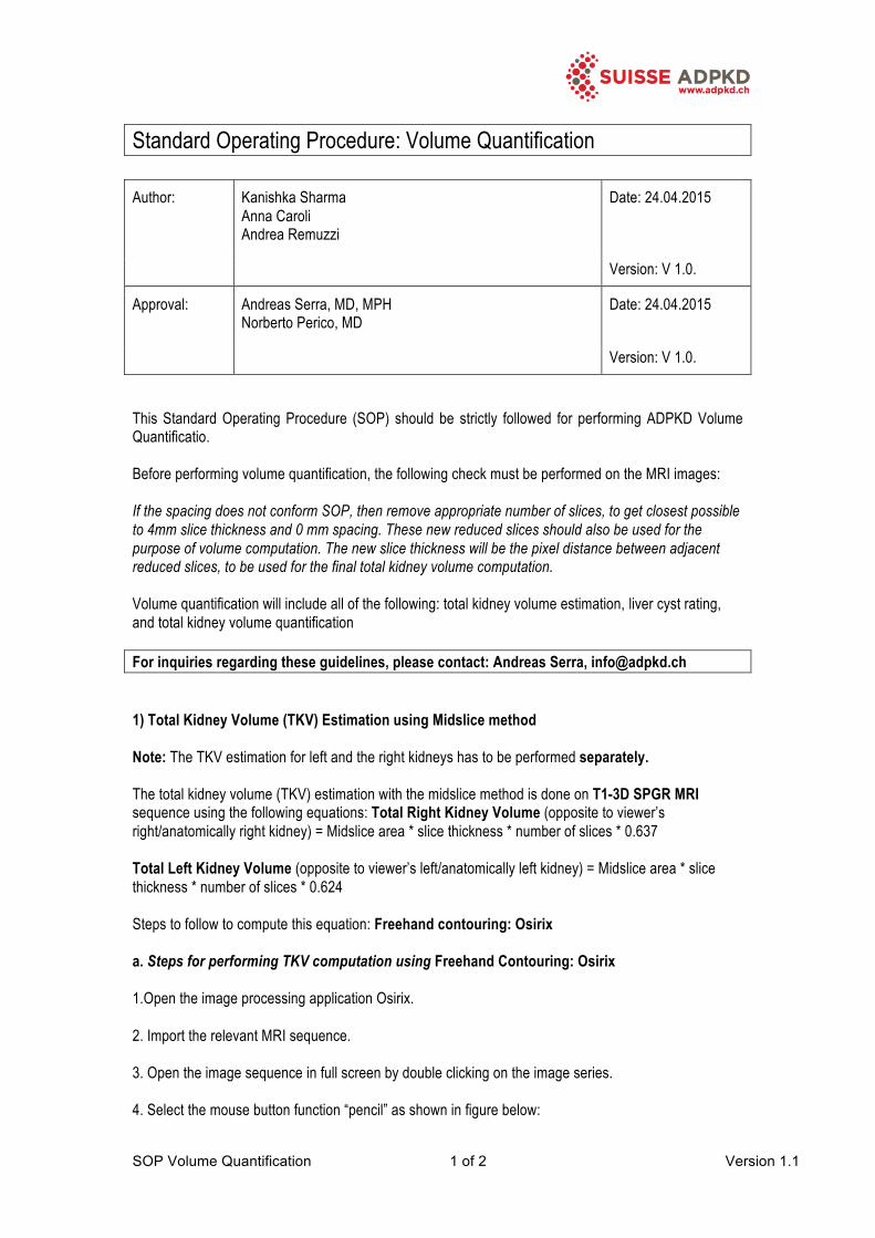
Version: V 1.0. This Standard Operating Procedure (SOP) should be strictly followed for performing ADPKD Volume Quantificatio. Before performing volume quantification, the following check must be performed on the MRI images: If the spacing does not conform SOP, then remove appropriate number of slices, to get closest possible to 4mm slice thickness and 0 mm spacing. These new reduced slices should also be used for the purpose of volume computation. The new slice thickness will be the pixel distance between adjacent reduced slices, to be used for the final total kidney volume computation. Volume quantification will include all of the following: total kidney volume estimation, liver cyst rating, and total kidney volume quantification For inquiries regarding these guidelines, please contact: Andreas Serra, [email protected] 1) Total Kidney Volume (TKV) Estimation using Midslice method Note: The TKV estimation for left and the right kidneys has to be performed separately. The total kidney volume (TKV) estimation with the midslice method is done on T1-3D SPGR MRI sequence using the following equations: Total Right Kidney Volume (opposite to viewer’s right/anatomically right kidney) = Midslice area * slice thickness * number of slices * 0.637 Total Left Kidney Volume (opposite to viewer’s left/anatomically left kidney) = Midslice area * slice thickness * number of slices * 0.624 Steps to follow to compute this equation: Freehand contouring: Osirix a. Steps for performing TKV computation using Freehand Contouring: Osirix 1.Open the image processing application Osirix. 2. Import the relevant MRI sequence. 3. Open the image sequence in full screen by double clicking on the image series. 4. Select the mouse button function “pencil” as shown in figure below:
SOP Volume Quantification 2 of 2 Version 1.1
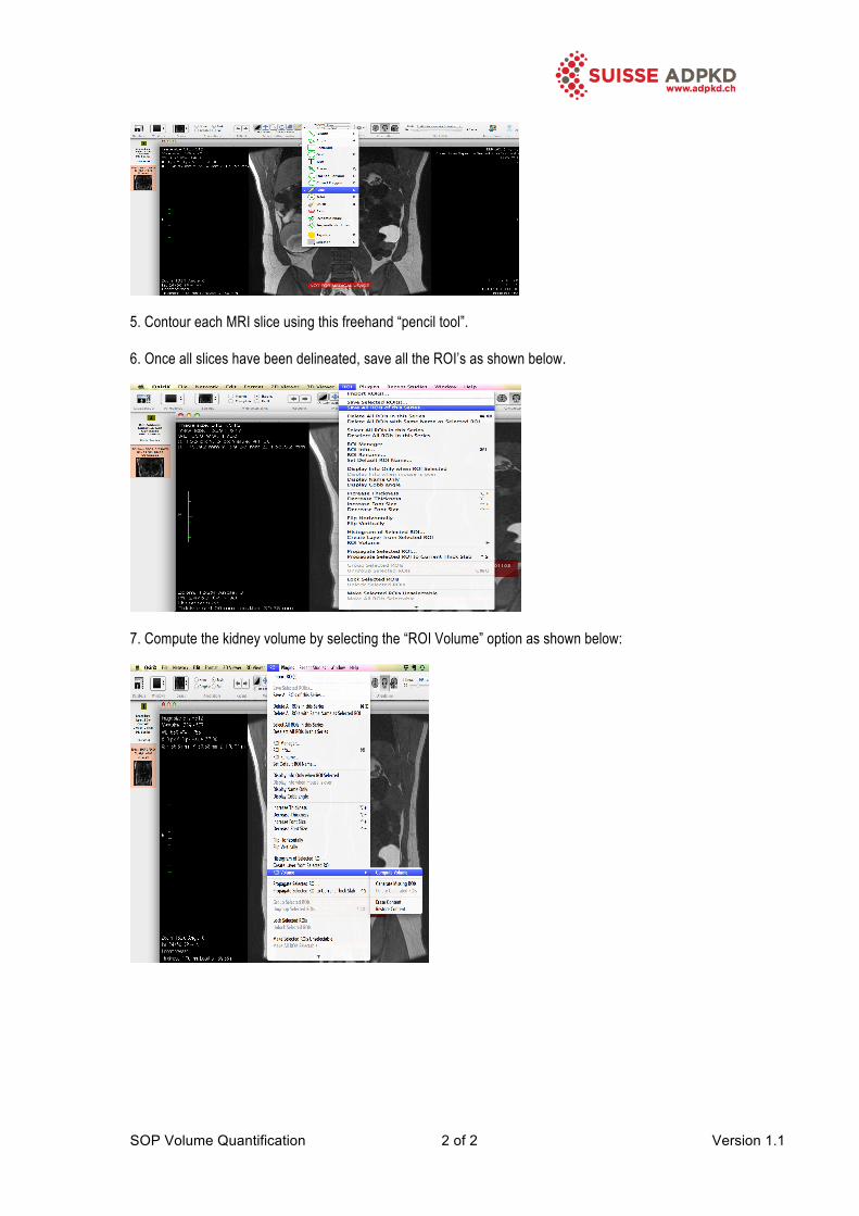
5. Contour each MRI slice using this freehand “pencil tool”. 6. Once all slices have been delineated, save all the ROI’s as shown below.
7. Compute the kidney volume by selecting the “ROI Volume” option as shown below: