standard treatment guidelines endocrinologyclinicalestablishments.gov.in/writereaddata/3601.pdf ·...
TRANSCRIPT

1
STANDARD TREATMENT GUIDELINES ENDOCRINOLOGY
Ministry of Health & Family Welfare
Govt. of India
2
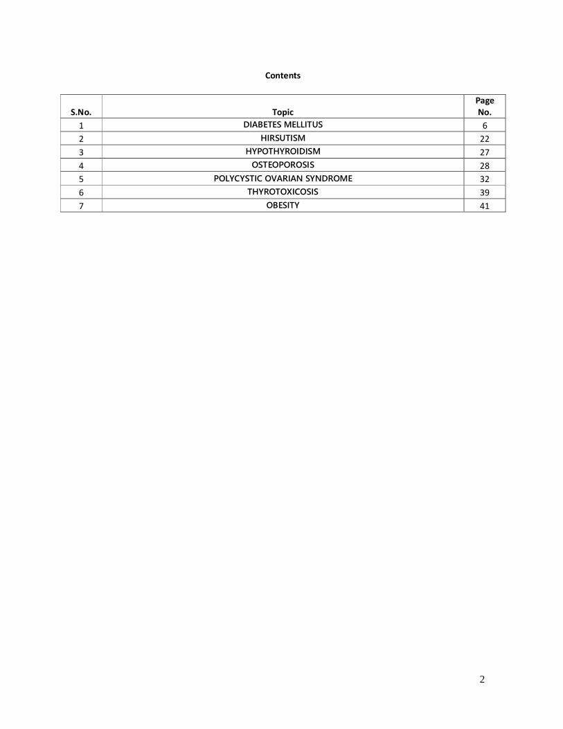
Contents
S.No. Topic Page No.
1 DIABETES MELLITUS 6 2 HIRSUTISM 22 3 HYPOTHYROIDISM 27 4 OSTEOPOROSIS 28 5 POLYCYSTIC OVARIAN SYNDROME 32 6 THYROTOXICOSIS 39 7 OBESITY 41

3
Group Head Coordinator of Development Team
Dr Sailesh Lodha Fortis Escorts Jaipur

4
Contributors-
1. Dr Anil Bhansali Head ,Department of Endocrinology PGI Chandigarh
2. Dr.Anil Kumar Singh Consulting Senior Endocrinologist Patna
3. Dr.K.D.Modi Consulting Senior Endocrinologist Hyderabad
4. Dr.Nikhil Tandon Professor,Department of Endocrinology AIIMS,New Delhi
5. Dr Pramod Gandhi Consulting senior Endocrinologist Nagpur
6. Dr Sailesh lodha Consulting senior Endocrinologist Jaipur
Reviewers
1. Col M K Garg Head ,Department of Endocrinology R & R New Delhi
2. Professor M.S.Sheshadri Head,Department of Endocrinology CMC Vellore
3. Dr.Rajesh Khadgawat Asso.Professor ,Department of Endocrinology AIIMS ,New Delhi
4. Dr Rakesh Sahay Professor ,Department of Endocrinology Osmania Medical college Hyderabad
5. Dr Rama Walia Asst Professor ,Department of Endocrinology PGI,Chandigrah
6. Dr Sanjay Bhadada Asso.Professor ,Department of Endocrinology

5
PGI ,Chandigarh 7.Dr Tushar Asso.Professor,Department of Endocrinology Seth GS Medical College Mumbai
8. Department of Endocrinology, AIIMS, New Delhi
9. Department of Endocrinology, I.P. Apollo Hospital, New Delhi

6
DIABETES MELLITUS
Introduction
Diabetes Mellitus (DM) is a global epidemic. Type -2 diabetes which forms almost 95 % of the total diabetes in India is largely a preventable disorder. India harbours more than 50 million diabetics. DM is associated with development of specific long-term organ damage (chronic complications) including retinopathy with potential blindness, nephropathy with a risk of progression to renal failure requiring lifelong dialysis or renal transplantation, neuropathy with risk for foot ulcers, amputation, and Charcot joints and autonomic dysfunction such as sexual, bowel and bladder impairment. Patients with diabetes are at a particularly high risk for vascular complications like cardiovascular, cerebrovascular, and peripheral artery disease. A recent rise in the number of adolescent and young Type -2 diabetics in India is an alarming trend. This is largely due to lifestyle factors.Recent Indian data suggest prevelence rates of 15 to 20 % in urban areas and about half of it in rural areas.
DM poses a great threat to the health of our nation, it costs dearly the exchequer as the treatment of acute and chronic complications of diabetes are expensive. Beside this is the cost of day to day medication, tests, monitoring and loss of productive work due to illness.
Hence it is important to recognise this disorder as early as possible. Attempt should be made to detect hyperglycemia in prediabetes stage to prevent progression to diabetes. And finally the aim is to treat this disorder effectively to prevent the devastation of the chronic complications.
Definition
DM is a chronic metabolic disorder of carbohydrate, fat, and protein metabolism characterized by hyperglycaemia,resulting from defects of insulin secretion, insulin action, or a combination of both 1 . Type 1 diabetes is due to an absolute lack of endogenous pancreatic insulin production, whereas in Type 2 diabetes, insulin resistance is the basic defect which is contributed by a combination of genetic predisposition, physical inactivity, and obesity.
Classification
Old terms like IDDM (insulin dependant DM), NIDDM (non insulin dependant DM)adult onset diabetes or juvenile diabetes have been abondoned.
ADA classification of 1997 which was later accepted by WHO is the most widely accepted classification of DM .
Classification of Diabetes Mellitus
1. Type 1 -(beta -cell destruction, usually leading to absolute insulin deficiency)
A. Autoimmune B. Idiopathic

7
2. Type 2 -(may range from predominantly insulin resistance with relative insulin deficiency to a predominantly secretory defect with or without insulin resistance)
3. Other specific types (see Table ) Genetic defects of b-cell function -MODY 1 to 9, mitochondrial DNA. Genetic defects in insulin action Diseases of the exocrine pancreas (pancreatitis trauma, pancreatectomy, neoplasm cystic-fibrosis etc) Endocrinopathies (acromegaly, Cushing's syndrome, gucagonoma, pheochromocytoma, hyperthyroidism etc) Drug- or chemical-induced Infections Uncommon forms of immune-mediated diabetes Other genetic syndromes sometimes associated with diabetes, e.g.: Down’s syndrome, Friedreich’s ataxia, Klinefelter’s syndrome, Wolfram’s syndrome
4. Gestational diabetes
Characteristics of Diabetes In India
Onset is about one decade earlier than the west
Occurs at a lower BMI
Central obesity is more common
Type 1- less than 5% of the total diabetes
Cardiovascular complications occur early
PVD is less common than the west
Foot problems more common
No structured treatment protocol
Lack of trained manpower like diabetes educators,endocrinologists
Cost of the treatment is a big hinderance
Social and religious factors play an important role
Diagnosis
Any one of the following:
Symptoms of diabetes + casual plasma glucose concentration > 200mg/dl (Casual - any time of the day without regard to time since last meal)
Fasting plasma glucose > 126mg/dl (Fasting - no caloric intake for atleast 8 hours)
2 Hour plasma glucose during OGTT > 200mg/dl (OGTT according to WHO criteria)
Note-HbA1c > 6.5% indicates diabetes, however it cannot be considered as a diagnostic tool in isolation. Impaired glucose metabolism
Impaired Fasting Glucose (IFG):

8
FPG > 100 and 125 mg per dL
Impaired Glucose Tolerance (IGT):
2hr PGPG > 140 and 199 mg per dL FPG > 100mg/dl and > 125mg/dl be classified as having IFG.
Prevalence of diabetes is increasing rapidly in developing countries like India
Occurs due to relative insulin deficiency in an individual with insulin resistance
Usually asymptomatic and detected during evaluation for unrelated indications, but may present with hyperglycemic symptoms-
Frequent urination, excessive thirst, excessive hunger
Weight loss, delayed healing of wounds, pain in calves, burning feet
Tiredness, Itching -especially in genital area
Tingling and numbness, blurring of vision, lethargy, somnolence,
Sometimes the symptoms are due to complications of diabetes when patient presents to a clinician for the first time with a complication like erectile dysfunction.
Type 1 DM:
Absolute insulin deficiency due to autoimmune destruction of pancreatic beta cells.
Onset is mostly in childhood or in young adults before 35 years, about may rarely occur in elderly.
Symptoms
Abrupt onset of severe hyperglycemic symptoms. Sometimes the child may present with just weakness, lethargy, bedwetting.
May present as diabetic ketoacidosis .
MODY-Maturity onset diabetes of the young:
Monogenic form of diabetes associated with mutation in a single gene
Diagnostic Criteria
• Aged less than 25 years at onset
• Autosomal dominant transmission of diabetes with three generations involved
• Absence of ketosis at any time
• Controllable without insulin at least in the early stages of the disease
9
• Clinical criteria are no longer acceptable and genetic analysis is needed to confirm the diagnosis.
Clinical Features
• Patients usually non-obese.Respond better to sulphonylureas in initial few years of diabetes.
• Do not need insulin initially- later may become insulin-requiring (MODY 1, 3 and 5)
• Isolated Mild fasting hyperglycemia- MODY 2- no treatment required
• Diabetes with renal cysts/ renal agenesis RCAD (renal cysts and diabetes syndrome)- MODY 5
Fibrocalculous pancreatic diabetes-(FCPD):
Severe hyperglycemia in a lean patient from a tropical country like India with recurrent pain abdomen, evidence of chronic pancreatitis after excluding other causes of chronic pancreatitis like alcoholism ,should be considered for FCPD.Absence of ketosis and presence of pancreatic calculi is the characteristic feature.
Latent Autoimmune Diabetes Of Adults (LADA): Type 1 Diabetes Which Presents Late GAD(Glutamic acid decarboxylase) autoantibody positive
Age at onset > 35 years
Lean patient
Insulin treatment required within one year of onset
Many patients with Type 2 diabeteswho are lean, have no family history of diabetes and who require insulin early in the course of the disease actually have LADA
Screening for Diabetes:
Ethnically every Indian falls into a high risk category however it may not be feasible to screen everyone. The following population group need screening.
Family history of diabetes Cardiovascular disease, Hypertension Overweight or obesity, males with a waist >90cm females with a waist >80cm Sedentary lifestyle Patients who are on steroids Previously H/O IFG ,IGT Dyslipidemia History of gestational diabetes People who were born with a birth weight >9 pounds or LBW Polycystic ovary syndrome

10
If normal, repeat screening in 3 yrs for low risk subjects and every year for high risk subjects. If IGT or IFG repeat screening in 1 year.
All pregnant ladies should be screened at -24-28 weeks if the risk is low, or at first ante natal visit if she belongs to a high risk group.
Risk factors for GDM:
Risk factors for GDM- Overweight or obesity Family history of diabetes mellitus History of IGT,IFG Poor obstetric history
History of delivery of an infant with a birth weight >9 pounds History of polycystic ovary syndrome Fasting plasma glucose concentration >90 mg/dL or 2-hour postprandial glucose >140 mg/dL
Physical Examination :
• Height, weight, BMI
• Blood pressure measurement, including postural variation if indicated
• Thyroid examination
• Skin examination of insulin injection site and for acanthosis nigricans.
• Xanthelesma
Comprehensive foot examination
• Inspection-see for redness or cracks or dryness of skin and shape of the foot
• Palpation of dorsalis pedis and posterior tibial pulses
• Ankle and knee jerks
• Proprioception, vibration and monofilament sensation
• Dilated retinal examination
• Dental examination
Investigations at the time of diagnosis:
11
• Fasting and postprandial plasma glucose
• HbA1c
• Fasting lipid profile
• AST/ALT
• Urine complete exam, including assessment of microalbuminuria
• Serum creatinine, and estimated GFR
• TSH if there is dyslipidemia and in women >40
• ECG
Markers of glycemic status
• Plasma glucose
• Whole blood glucose (glucose meters)
• Urine glucose
• Glycosylated hemoglobin (HbA1c) .HbA1c is an index of long term Glucose Control,
performed on venous blood
Test results are not affected by
- Time of the day
- Meal intake
- Exercise
- Just administered diabetes drugs
- Emotional stress
- Patient cooperation
In certain situations HbA1c can be falsely high(uremia,alcoholism etc) or low(blood loss iron
defeciency anemia,pregnancy,hemoglobinopathies ,reduced RBC survival).
eptide
• It connects the A and B chain of insulin in the proinsulin molecule ,
• by-product of insulin biosynthesis
• For every molecule of insulin in the blood, there is one molecule of C-Peptide.

12
• Very low levels:
- Type 1 diabetes
- Complete surgical removal of pancreas
• Clinical utility of C-peptide measurement is very limited .Its measurement and interpretation of the results should be done by an Endocrinologist.
Microalbuminuria
• Test for presence of microalbuminuria annually in all Type 2 diabetics starting at diagnosis and Type 1 diabetic with DM more than 5 years
Methods Measurement of the albumin to creatinine ratio in a random spot collection or 24 hour urine collection for creatinine and albumin with simultaneous measurement of serum creatinine.
Normoalbuminuria Microalbuminuria Macroalbumiuria
Albumin/day <30 mg 30-300 >300
Albumin/creatinine ratio*
<30 30-300 >300
Albumin excretion rate <20(mcg/min) 20-200 >200
* mcg of albumin/mg creatinine Albumin excretion rate (AER)- It is based on the albumin concentration ,the duration of the urine collection and the urine volume, and has the advantage of giving results which are independent of the ingested fluid quantity.AER is the gold standard for the diagnosis of microalbuminuria.
Foot examination- this should include examination of the shape of feet, bony prominences, calluses, deformities of the toes. Skin examination for texture ,color and loss of sweating, loss of hair. Nail examination for thickening, ingrowings, fungal infections. Inspection of interdigital webspaces for fungal infections and minor traumas Sensory examination of foot. Palpation of dorsalis pedis and posterior tibial arteries. Joint movements for checking the power of the muscles (muscle wasting) and joint integrity. Vibration test (128Hz), nylon monofilament test, ankle brachial index, peripheral doppler may provide very important information.
Continuous glucose monitoring(CGM) is indicated in pregnancy,brittle diabetes,insulin pump users.For Type -1 diabetics and elderly it can be used periodically to study hypoglycemia more closely.
Monitoring
Glucose Frequency depends on the the kind of treatment and the severity of
illness.It may be as low as twice a month to as high as 6-7 times in a day.#
A1C Once in 3-4 months and six monthly if glycemic control is good
Lipids Quarterly if uncontrolled, otherwiseOnce in 12 months*
Creatinine Once*

13
Urine complete exam Once*
Urine microalbumin Once in a year*
ECG Once in a year*
Fundoscopy Once in a year for all Type -2 diabetics from the time of diagnosis*.In Type -1 once every year from 4-5 years after diagnosis or at the age of 10 years.More frequent exams are needed in advanced retinopathy
*Frequency of these tests can be more whereever indicated
# monitoring should include fasting,preprandial and 2-hour postprandial and bed time glucose levels .Occasionally between 2:00 -3:00AM glucose measurement is indicated to detect nocturnal hypoglycemia.Self monitoring of glucose by Glucose measuring devices at home is desirable for all diabetics.Patients on insulin need to monitor more frequently as compared to those on OADs.
Nutrition therapy: Diet therapy has to be individualised based on the likes and dislikes, calorie requirement (overweight or underweight), lifestyle and comorbidities. Carbohydrates should form 50-65 % of the total calories, proteins should form 15-20% (like non diabetics) and fats -15-25%. Average Indian diets usually contain the desired fiber content (15-25 gm/1000 cal). The saturated fat consumption should be minimised to < 10% of the total calories. If the LDL is >100 than it should be further reduced to <7%.Trans fats should preferably be eliminated completely from the diet. Vegetable oils should be used interchangeably so that the desired omega 3:6 ratio is achieved .No one kind of oil can be recommended. Coconut and palm oils should be avoided. There is no role of any additional multivitamins, trace elements ,antioxidants or any kind of neutraceuticals except Vit D. Consumption of fresh fruits ,salads and green vegetables should be encouraged which would supply all the multivitamins, trace elements, antioxidants. Absolute restriction of sucrose is not essential however it should be minimised. Restricted protein intake of 0.8 to 1.0 g/kg per day is required in patients who are in the earlier stages of chronic kidney disease and to
g/kg per day in patients who are in the later stages of chronic kidney disease .
Artificial sweeteners: Sucralose,Aspartame,Saccharin,Acesulfamec and a few others are non caloric sweeteners.Their consumption should be minimised .The scientific data favouring their safety is scanty hence they can be used in smaller quantities with a caution although the accepted daily intake for Aspartame is 50 mg/kg/day and for Sucralose and saccharin is 5 mg/kg/day.
Vaccination- Pneumococcal vaccine may be recommended for all diabetics.
Life style interventions – The Diabetes Prevention Program found that people at risk of developing type 2 diabetes were able to cut their risk by 58% (it was 74% in subgroup of asians) with moderate physical activity (30 minutes a day) and weight loss (5 to 7% of body weight, or about 15 lb). For people over age 60, the risk was cut by almost 71% .Adopting healthy lifestyle with increasing physical activity and weight reduction is key for achieving success in diabetes management. The effect of physical activity and other life style interventions on glucose and BP is comparable to pharmacological treatment. The goals of the life style interventions should be to achieve at least 5% reduction in body weight in obese. The individuals with high risk should be systematically targeted with lifestyle interventions regardless of their glucose status. Regular daily physical activity of 45 minutes or more is recommended for all diabetics in general. Besides leisure time physical activity, other forms of activity like walking ,occupational or daily commuting on foot or
14
bicycle, household chores ,activity at work place is also helpful and should be encouraged. Later the intensity and duration of exercise can be increased according to one's general health and age. To begin with it could be just brisk walk. The aim is to raise and sustain the heart rate for 15-20 minutes to 60-85% of the maximal heart rate for that age. Maximal heart rate is 220 minus age. Exercise should be preceded by an additional 05 minute warmup and followed by a 05 minute cool down. Thorough examination before starting physical activity is mandatory in view of associated comorbidities like CAD, proliferative retinopathy, autonomic neuropathy, arthritis, feet problems etc. Cessation of smoking, tobacco and moderating alcohol intake(only for those who are already taking alcohol, it cannot be recommended in any form for those who are not taking alcohol) should be reemphasised on every clinic visit.
Stress management: Relieving stress by adopting healthy lifestyle or by any other method including the traditional Indian methods is helpful.
Oral drugs: Oral anti diabetic drugs (OADs) have no or very little role in Type -1 diabetes. They can be initiated at the time of detection of diabetes or after a trial of lifestyle measures for 6-8 weeks. Choice of an oral agent depend on many factors like duration of the diabetes, age and weight of the patient, presence of other comorbidities ,lifestyle of the patient ,degree and type of hyperglycemia(fasting /postprandial)and susceptibility to hypoglycemia. Metformin(MF) and Sulphonylureas (SU) alone or in combination should form the base or the first line therapy in most of the OAD requiring diabetics.Sensitisers (like MF , Pioglitazone )and Alpha glucosidase inhibitors (AGI) are useful throughout the lifespan of a diabetic. Secretagogues like SU and Glinides are not very useful in long duration of diabetes and progressively lose their efficacy as their action is dependant on residual beta cell mass which progressively decline in all Type -2 diabetics. Glinides are useful as monotherapy in elderly where a potent long acting SU is relatively contrindicated. DPP-4 inhibitors are not suitable for burnt out or long standing diabetes. As cost is often an issue for choosing an OAD ,DPP-4 inhibitors can be added if mono or dual therapy fails. At present they cannot be recommended as first line therapy. Pioglitazone can be used as mono,dual or tripple therapy.Alpha glucosidase inhibitors are useful adjunct with any of the above mentioned drugs if postprandial hyperglycemia is the main target, though by themselves they are weaker drugs if used alone. Two OADs from a same group should not be used together. If HbA1c is mildly elevated (around 7%) monotherapy may be sufficient. For moderately elevated HbA1c (around 8%)dual drug therapy as seperate or in fixed combination can be initiated. However MF or SU as monotherapy in higher doses may be sufficient even for these patients. For HbA1c of more than 9%, combination therapy is initiated or intensified as dual or tripple therapy or insulin can be added as single bed time dose if fasting hyperglycemia is the main target .Insulin as multiple doses(premixed or split mixed )can be added to the OADs. In some situations where other measures fail, insulin can be used along with a combination of OADs and GLP-1 analogues.
IGT- MF and Acarbose can be used to delay the onset of diabetes.
Side effects of OADs
Sulphonyureas: Hypoglycemia, nausea, constipation, pain abdomen, weight gain, hematological abnormalities, headache, skin disorders. Not very useful with higher doses of insulin,avoided in advanced renal failure.
Glinides: Hypoglycemia,nausea,constipation,pain abdomen,diarrhoea.

15
Metformin: Pain abdomen, nausea, vomiting, diarrhoea, anorexia, weight loss, flatulence, B-12 defeciency. Contraindicated if serum creatinine is >1.4 in women and >1.5 in males, also contraindicated in CHF, advanced hepatic and respiratory failure.Avoided in periop period.
Alpha glucosidase inhibitors- Flatulence, constipation, pain abdomen, diarrhoea, vomiting. Avoided in serious GI disorders.
DPP-4 inhibitors- constipation, pain abdomen, diarrhoea, swelling ,headache, URI, arthralgia, anorexia. Not very useful in long standing diabetes.
Pioglitazone: fluid retension, anaemia, weight gain, sinusitis, worsening of retinopathy, Fractures in postmenopausal women. Contraindicated in CHF, fluid overload states and in advanced hepatorenal dysfunction. Recent data impicates it in bladder cancer. It has been banned in Germany. A great caution is required in patient selection for its use .
Mechanism of action of OADs
Insulin secretagogues Sulphonylureas,Glinides
Insulin sensitisers Pioglitazone,Metformin
Drugs acting locally on gut Alpha glucosidase inhibitors
Potentiating Incretin axis,glucagon suppression
DPP-4 Inhibitors
When HbA1c is high ,fasting glucose should be the target first and when Its relatively better or towards normal side the postprandial glucose should be targeted first to achieve better glycemic control.
Insulin – Insulin is a potent anabolic hormone .The effects other than glucose lowering are important in many clinical situations where anabolic activity is desired.
Indications
Type -1 DM, pregnancy, severe infections, severe catabolic states, OAD failure,acute stressful situations like acute coronary syndrome, stroke, acute renal or hepatic failure, periop period. Secondary diabetes, post transplant diabetes ,congestive heart failure are also indications for insulin usage. Insulin can be added to existing OADs for short durations (e.g. steroid use in a diabetic on OADs) or long durations.
Insulin preparations and their pharmakokinetics-
Insulin Onset Peak Effective Duration
Rapid-actingAnalogue Aspart ,Lispro 5-15 min 30-90 min <5 h Glulisine 5-15 min 30-90 min <4 h Short-acting Regular 30-60 min 2-3 h 5-8 h Intermediate
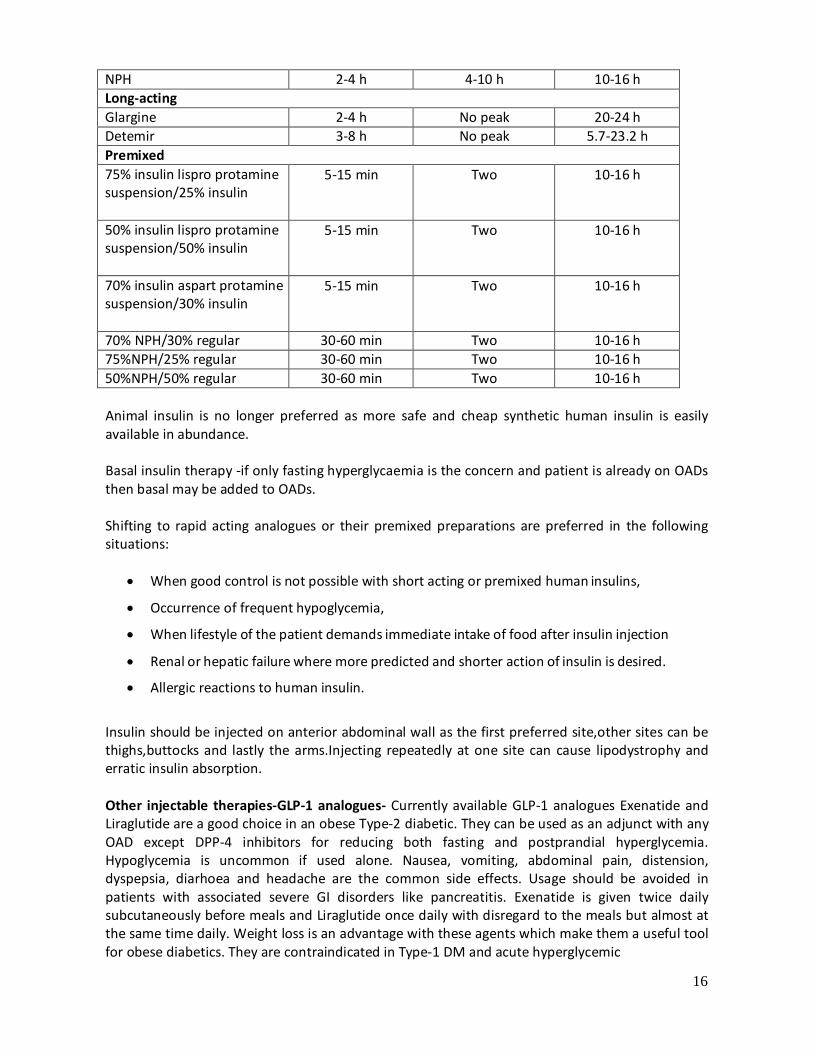
16
NPH 2-4 h 4-10 h 10-16 h Long-acting Glargine 2-4 h No peak 20-24 h Detemir 3-8 h No peak 5.7-23.2 h Premixed 75% insulin lispro protamine suspension/25% insulin
5-15 min Two 10-16 h
50% insulin lispro protamine suspension/50% insulin
5-15 min Two 10-16 h
70% insulin aspart protamine suspension/30% insulin
5-15 min Two 10-16 h
70% NPH/30% regular 30-60 min Two 10-16 h 75%NPH/25% regular 30-60 min Two 10-16 h 50%NPH/50% regular 30-60 min Two 10-16 h
Animal insulin is no longer preferred as more safe and cheap synthetic human insulin is easily available in abundance.
Basal insulin therapy -if only fasting hyperglycaemia is the concern and patient is already on OADs then basal may be added to OADs.
Shifting to rapid acting analogues or their premixed preparations are preferred in the following situations:
• When good control is not possible with short acting or premixed human insulins,
• Occurrence of frequent hypoglycemia,
• When lifestyle of the patient demands immediate intake of food after insulin injection
• Renal or hepatic failure where more predicted and shorter action of insulin is desired.
• Allergic reactions to human insulin.
Insulin should be injected on anterior abdominal wall as the first preferred site,other sites can be thighs,buttocks and lastly the arms.Injecting repeatedly at one site can cause lipodystrophy and erratic insulin absorption.
Other injectable therapies-GLP-1 analogues- Currently available GLP-1 analogues Exenatide and Liraglutide are a good choice in an obese Type-2 diabetic. They can be used as an adjunct with any OAD except DPP-4 inhibitors for reducing both fasting and postprandial hyperglycemia. Hypoglycemia is uncommon if used alone. Nausea, vomiting, abdominal pain, distension, dyspepsia, diarhoea and headache are the common side effects. Usage should be avoided in patients with associated severe GI disorders like pancreatitis. Exenatide is given twice daily subcutaneously before meals and Liraglutide once daily with disregard to the meals but almost at the same time daily. Weight loss is an advantage with these agents which make them a useful tool for obese diabetics. They are contraindicated in Type-1 DM and acute hyperglycemic

17
complications.
Insulin pump: At present due to its prohibitive cost its usage is very limited. All patients who qualify for insulin pump therapy should be referred to a centre where expertise for a pump is available. Pump therapy requires a lot of motivation and time from both - the patient and the clinician.
Indications • All Type -1 diabetics who are poorly controlled with basal-bolus therapy. • Pregnancy • Frequent hypoglycemia or hypoglycaemic unawareness • Persistently elevated fasting glucose (Dawn phenomenon) • Multiple chronic complications with labile glucose requiring fine tuning of glycemic control • Management of labile diabetes after renal or hepatic transplant • In situations where insulin requirement is very high a trial of pump therapy can be given.
Hypertesion in diabetics:
Lifestyle measures form the cornerstone of hypertension management. Salt intake should be restricted to less than 6 gm per day. Restriction of salt intake should however be individualised .Care has to observed in elderly and in people who sweat much particularly in summers. The target BP is 130/80 for all diabetics and 120/70 in established nephropathy (creatinine >1.5 or proteinuria >1 gm per day) .BP should be measured at each visit .Measuring postural variation in BP is useful in autonomic dysfunction. Therapy should be individualised based on the associated comorbidities and side effects of drugs. Lifestyle measures and dietry adjustments are essential to attain the desired goals.
ACE (Angiotensin Converting Enzyme )Inhibitors and ARBs (Angiotensin Receptor Blockers) can be used interchangeably as a first line antihypertensive agent in all diabetics. Second line agent could be calcium channel blockers or cardioselective betablockers. Diuretics and alpha blockers can also be used whereever needed. Glucose intolerence due to any of these agents is not clinically significant.
Diabetic dyslipidemia- Classically, DM induces elevation in triglyceride and LDL, and low HDL levels.It should be treated aggressively with a LDLc target of <100 mg/ dl without CAD and <70 mg/ dl with CAD.Goal for triglycerides is <150 mg/dl and for HDLc is > 45 mg/dl. Statins form the cornerstone of lipid lowering therapy.Statins are also useful if microvascular complications like nephropathy and retinopathy are present. Addition of fibrate or ezitimibe is indicated when desired goals are not met.Triglycerides of >400 is an indication for the use of fibrate as a primary therapy. Combination of niacin can be useful if HDL is persistently low. Usually anti lipid therapy is lifelong and doses need adjustments based on periodic lipid profile. Dietary modifications and lifestyle measures can alone be sufficient sometimes. Statins can produce myalgias and rarely myopathy .
Aspirin(ASA) usage in diabetes- Low dose(75-150mg) ASA prophylaxis should be given to all

18
diabetics over 50 years of age with one additional cardivascular risk factor. Watch for adverse events -GI bleed and hemorrhagic stroke .
Management of diabetes in pregnancy- prepregnancy counseling for the desired goals for glucose and blood pressure,lifestyle measures ,nutrition.Shifting to insulin and withdrawl of ACE inhibitors and lipid lowering drugs is required in prepregnancy period.Folic acid 10 mg daily should be started to prevent neural tube defects.
Screening for GDM
Glycemic targets in pregnancy- fasting- 95 mg/dl, 2hours post prandial-<120 and one hour <140.
Diabetic Foot: Even a small ulcer should be given full attention .Neglecting small ulcers or trauma can finally become the reason for limb loss.
Diabetic foot results predominantly due to neuropathy ,associated with infection and ischemia.X- ray of the foot may expose foreign body or gas and must be done .Shifting these patients to insulin helps in better glycemic control and early healing. Infections of the foot are polymicrobial and require wide spectrum coverage. Limb and life threatening infections must be treated with parenteral antibiotics. Pus collection of any magnitude should be drained out and explored widely. Early surgery is always desired .Iodinated solutions , H2O2 and other strong antiseptics should be avoided for cleaning the wound as they prevent angiogenesis. Planter lesions will heal only with offloading the affected foot.
Referral to a higher center necrotising fascitis, any chronic non healing ulcer, lesion associated with systemic involvement, presence of significant ischemia charcot's joint, requirement of advanced imaging and customised footwear or orthotic support.
Targets
BMI 21-25
Fasting glucose 80-120
2 hours postprandial glucose 130-160
HbA1c 6.5-7.0
BP 140 / 80
Total Cholesterol <150
LDLc <100
Triglycerides <150
HDLc >40 for males, > 50 for females
Hospitalised patient -periop period
Targets-Preprandial glucose- <110 2 hours Postprandial glucose-<180 Critically ill patients- 140-180

19
Most of these patients will be on insulin which preferably should be given as bolus -basal therapy i.e.3-4 boluses of short acting insulin and a bedtime dose of NPH or long acting insulin. Stable patients who are eating well can be managed with 2-3 time insulin (premixed or split mixed)In all hemodynamically compromised situations, acute myocardial infarction, stroke, high dose steroid therapy, immediate post op period of major cardiac surgery, severe sepsis, very high glucose levels ,patients on ventilator. IV infusion of short acting insulin with glucose monitoring every 1-2 hours is preferred. Basal insulin requirement must be assessed for all patients to prevent fasting and inter prandial glucose peaks. Patients who are nil by mouth may also need some basal insulin. Overlap between subcutaneous and IV insulin for 30-60 minutes should be done to prevent sudden hyperglycemia relapse when shifting from IV to subcutaneous insulin.
Hypoglycemia: more common in following situations
Patients who are older, have renal or liver disease
Have a long duration of diabetes, autonomic neuropathy, hypopituitarism
Regularly miss meals, erratic meal pattern
unusual exercise
Take greater than the prescribed dose of their medication
Concomitant medication – Levofloxacin*, beta blockers,
* hyperglycemia is more common with Levofloxacin though rarely hypoglycemia can occur.
Diabetes Education: Educating the patient for lifestyle modifications, hypoglycemia, insulin therapy, foot care, benefits of quitting smoking and tobacco, reducing alcohol helps in achieving the targets of therapy.
Sick day guidelines: The objective of these guidelines is to avoid hospitalisation during situations like acute diarrhoea, vomiting febrile illness. And if at all it is required the patient is able to prevent DKA or other diabetes related acute complications. All diabetics should be taught these simple rules or a handout may be given mentioning- In any febrile illness or whenever there is poor intake of food and water due to any illness dehydration and ketosis must be avoided .Never omit your medication -insulin or OAD. Infact sometimes a higher dose is required when we are sick even when we are not eating. Blood glucose and urine ketones must be checked frequently -every 4-6 hourly. Type 1 diabetics may require monitoring 2-4 hourly. Try to eat small amounts of carbohydrates every 2-4 hourly ,soft food or liquids are easier to take .!00-120ml of fluid every hour (carbohydrate free if glucose is >250 and with carbohydrates if glucose is less than 250. Fluids and carbohydrates are required to maintain hydration and calorie requirement and preventing hypoglycemia. Dehydration and worsening ketosis are ominous signs . Physician must be consulted if nausea or vomiting persists with ketosis .If blood glucose is persisting more than 300 on two consecutive times .If breathing becomes rapid or laboured, fever >100 degree F persisting more than 24 hours, persisting diarrhoea or vomiting or pain abdomen. Inability to take fluids for more than 4 hours due to vomiting drowsiness or altered sensorium or anyother unexplained symptoms. Drug treatment adjustments during sickdays- stop metformin, and preferably other OADs except sulphonylureas, increase or decrease sulphonylureas according to the glucose readings. Addition of

20
short acting insulin may be required to OADs.In situations where patient is already on insulin, long acting or intermediate insulin should be continued in the same doses. Dose of short acting insulin can be adjusted by measuring sugars 4-6 hourly.Premixed insulins should be supplimented by short acting insulins usually 20-25 %extra or more dose of insulin is required in addition to the usual dose of insulin.However if vomiting or diarrhoea persists hypoglycemia can occur.
Prevention of diabetes: Identify the high risk population .Fasting glucose and OGTT should be used for identifying IFG and IGT.No drug is recommended for the prevention of diabetes.MF and Acarbose can be used in IGT and IFG.Lifestyle measures mentioned in section 2.3 should be persued for prevention of diabetes. Referral to a tertiary center /Endocrinologist: In the following situations a referral to a tertiary center where endocrinologist is available should be done at least once or more in a year. Diabetes in a neonate or an infant, all Type -1 diabetics with polyendocrinopathy, Coeliac disease or with growth hormone defeciency. Diabetes with complications, brittle diabetes, pregnancy with diabetes, diabetes with endocrine tumors like pheochromocytoma, pituitary tumor, rare varieties of diabetes like diabetes with lipodystrophies or rare syndromes, requirement of CGM or insulin pump. Diabetes after liver or renal transplant. In any other situation where glycemic control is not achieved despite of all efforts.
Referral to a nephrologist: serum creatinine >1.5 ,proteinuria more than >1 gm/day,nephropathy in absence of retinopathy ,suspicion of a nondiabetic renal disease.
Referral to a retinal surgeon: when early NPDR is progressing ,CSME,advanced retinopathy.
Referral to a neurologist: severe neuropathic symptoms not responding to the standard treatment, suspicion of non diabetic neuropathy, amyotrophy, stroke, mononeuropathy.
Referral to a cardiologist: angina or its equivalent ,diabetic cardiomyopathy,peripheral vascular disease requiring further workup
Who does what
Doctor- history taking, physical examination, ordering investigations, prescrbing treatment.
Nurse- patient education -like explaining the basics of diabetes and its complications, foot care, exercises, hypoglycemia, self monitoring of glucose, insulin injection devices and techniques.
Dietician- calorie calculation, explaining the concept of carbohydrate counting, food exchanges, glycemic index, modifications in diet during special situations like pregnancy, renal impairment.
Technician- drawing blood ,doing ECG, installing CGMS, insulin pump installation and its management except the insulin dose calculation and its modifications . Biothesiometry and use of other gadgets for foot care.
Suggested reading /references
1. Epidemiology of Type -2 Diabetes : Indian scenario.Indian Journal of medical research 2007:
125(3)
2. Joslin's Diabetes Mellitus,14th edition.Edited by C.Ronald kahn et al.Publisher -Lippincott Williams & Wilkins.

21
3. American Association of clinical Endocrinologists -medical guidelines for clinical practice for the management of Diabetes Mellitus 2007
4. Summary and Recommendations of the Fifth International Workshop-Conference on Gestational Diabetes Mellitus.Boyd E. et al.Diabetes care, 2007: 30, supplement 2
5. American Diabetes Association .Standards of medical care in diabetes. Diabetes care 2010; 33: suppliment.
6. The Diabetes Prevention Program Research Group. Reduction in the incidence of type 2 diabetes with lifestyle intervention or metformin. New Engl Med J. 2002;346:393-403.
22
HIRSUTISM
I. WHEN TO SUSPECT /RECOGNISE
a) Introduction:
Hirsutism is the presence of excessive growth of terminal hair that appears in a male pattern in a woman. Though the presence of hirsutism may be a purely cosmetic and social concern to the affected woman, medically it can be indicative of conditions like Polycystic Ovarian Syndrome (PCOS), non classic Congenital Adrenal Hyperplasia (CAH) or virilizing tumours of the ovaries or adrenals. Androgens are essential for the conversion of vellus (small, straight and fair) hairs into terminal (larger,thicker,coarser, curlier and darker) hairs. This conversion is dependent on the level of the circulating androgens, their metabolism locally at the pilo-sebaceous unit by the enzyme 5 alpha reductase into dihydrotestosterone, and the subsequent binding of the latter to its receptor. This local metabolism and androgen-receptor interaction is highly variable. As a result pilosebaceous units vary in their sensitivity to androgen. Therefore, the degree of hirsutism may be very different between individuals with similar levels of circulating androgens.
b) Case definition: Hirsutism is a clinical diagnosis. Hirsutism should be distinguished from hypertrichosis which is generalised excessive vellus hair growth in a non sexual pattern. Hypertrichosis is androgen- independent. Its origin is hereditary or secondary to systemic conditions and certain drugs. Typically, hirsutism is defined clinically by judging the severity of hirsutism (graded subjectively from 1 to 4) in each of nine defined body areas. A total score of more than 8 out of a maximum of 36 is considered significant. This is the Ferriman-Gallwey hirsutism scoring system. Apart from its subjective nature and other drawbacks, the total score does not reflect the significance of higher degrees of hirsutism in one area alone (“focal hirsutism”) if the other areas are unaffected. Thus focal hirsutism which is causing distress to a woman may not lead to a “significant” total FG score of 8. This has led to the concept of “patient-important hirsutism” – i.e. hirsutism that is significant for the individual seeking medical attention. II. INCIDENCE OF THE CONDITION IN OUR COUNTRY: About 5 % of women in the west have hirsutism. Data for our country are lacking. It is encountered commonly in practice. III. ETIOLOGY: Polycystic ovary syndrome Idiopathic (hirsutism without hyperandrogenemia) Late-onset or non-classic congenital adrenal hyperplasia Androgen-secreting tumours
Ovarian tumours Adrenal tumours
Cushing's syndrome Hyperprolactinaemia Acromegaly Thyroid dysfunction Iatrogenic
Androgen therapy (testosterone, anabolic steroids) Danazol Androgenic progestins (levonorgestrel, norethindrone and norgestrel)

23
Glucocorticoids Polycystic ovaries, idiopathic hirsutism and drug-induced hirsutism constitute the largest proportion of cases. Hypertrichosis can occur in the following conditions or with some drugs as indicated below Porphyria Phenytoin Cyclosporine IV. COUNSELLING Patients have to be informed of treatment options and time frame of efficacy of various modalities – especially the delayed onset of pharmacologic therapy and that laser and intense pulsed light (IPL) modalities are methods of long term hair reduction and not “permanent” removal. The need for continued therapy for sustained control should also be explained. V. OPTIMAL DIAGNOSTIC CRITERIA, INVESTIGATIONS, TREATMENT AND REFERRAL CRITERIA: 1) Diagnosis: Hirsutism is a clinical diagnosis. The presence of excess terminal hairs in a male pattern has to be documented. The Ferriman Gallewey score of > 8 is helpful if the scoring is feasible. A baseline FG score is also useful for assessing treatment response. Strict requirement of FG score >8 for diagnosing and treating hirsutism is not applicable. Some patients have already removed their hair or have been treated before their first medical encounter precluding any scoring attempt. One also has to bear in mind the situation of “focal hirsutism” and “patient-important hirsutism”.
The Ferriman Gallwey Scoring System Hirsutism is scored from 0 (none) to 4 (severe) in each of nine defined areas, and the total score added.

24
Clinical evaluation is also directed at discerning clues to specific etiologies. ----Drug use ----Androgen secreting neoplasm: Sudden onset, rapid course, progression, virilisation (clitoromegaly, temporal hair recession, muscularity, voice change) or abdominal/pelvic mass suggest the possibility of androgen secreting neoplasm. ----PCOS : Perimenarcheal onset of menstrual irregularity, infertility, acne, male-pattern alopecia, obesity, acanthosis nigricans. ---Non-classic CAH: history of consanguinity in parents, family history of infertility, hirsutism, menstrual irregularity or both ---Cushing’s syndrome: Hypertension, striae, easy bruising ---Hyperprolactinemia: Galactorrhoea ---Primary hypothyroidism: Goitre, other clinical features ---Acromegaly: Acral enlargement, coarse features, prognathism 2) Investigations: Patients with isolated, mild hirsutism need not have any tests done. Patients with the following need to be investigated: Moderate or severe hirsutism, i.e. FG score >15 Hirsutism of any degree when it is
sudden in onset and/or rapidly progressive progressing despite therapy associated with menstrual irregularity/infertility/obesity/acanthosis nigricans associated with features of virilization
1. Serum Testosterone: Serum testosterone should be checked early morning. In women who are having regular cycles it should be checked between days 4-10. In others, either a random sample or one obtained after progesterone induced bleeding should be used. Testosterone levels in the upper normal range or mildly elevated (70 -150 ng/dl) is usual for PCOS. Serum testosterone values in the adult normal male ranges (200 – 1100 ng /dl) are seen in virilizing neoplasms. If serum total testosterone is normal in the situation where elevated levels are expected, serum albumin and SHBG should be checked in addition and the free androgen index calculated. Commercially available free testosterone assays may be unreliable. 2. Other tests are to be chosen according to the suspected etiology i) Ultrasound : PCOS, adrenal/ovarian neoplasm ii) Serum basal 17 hydroxy progesterone and if this is between 3 -10 ng/ml a Synacthen stimulation test : non classic CAH iii) Serum DHEAS : Adrenal neoplasm iv) Additional tests for hypothyroidism, hyperprolactinemia, acromegaly, Cushing ’s syndrome, when suspected
c) Treatment : Treatment has to be prolonged and often continuous for control of hirsutism. When cosmetic measures alone are insufficient treatment, it has to be complemented with (a) pharmacologic therapy, or (b) direct long term hair removal methods. The choice between pharmacological therapy and hair removal methods depends on patient preference, cost, and the area needed to be treated (wider areas require pharmacologic therapy). In those with hyperandrogenemia, pharmacologic therapy needs to be combined with hair removal methods to achieve better control of hirsutism. 1. Pharmacologic therapy : Any chosen pharmacologic therapy needs to be given a trial of at least six month before
25
augmenting dosage, combining, or changing drugs. i) Oral contraceptives : OCPs containing progestin with less (norgestimate, desogestrel) or no (drospirenone) androgenic potential are better than those containing levonorgestrel. ii) Antiandrogens : Because of their teratogenic potential, antiandrogens should not be used alone in all those who can conceive and are not on effective contraception. Spironolactone is the most commonly used antiandrogen at 100-200 mg/day in divided doses. Finasteride at 2.5 to 5 mg per day is less effective. It is a type 2 alpha reductase inhibitor and inhibits conversion of only one of the isoenzymes responsible for formation of dihydrotestosterone from testosterone. Flutamide (250-500 mg per day) is rarely used because of its hepatotoxic potential but can be used with periodic monitoring of LFT in severe refractory cases. Topical eflornithine cream is used for focal hirsutism (e.g. facial hirsutism). It inhibits ornitihine decarboxylase which catalyses the rate limiting step for polyamine synthesis which is necessary for hair growth. It does not remove hair but reduces the growth and appearance of facial hair. 2. Hair removal/reduction methods :
• Temporary
Depilation - Removal of hair shaft from the skin surface e.g. shaving and chemical depilatory creams
Epilation – Removal of hair shaft from above the bulb – waxing and plucking
• “Permanent” methods of hair reduction
Electrolysis – destruction of hair follicles by insertion of a needle and use of electric current can be used for limited focal hairs as it is time consuming. The cost is less than photoepilation.
Photoepilation (laser or intense pulsed light) is a costly modality, but can be used for treating relatively wider areas. The energy absorbed by the melanin destroys the hair follicles. There is a reduction of 30 % or more in the number of terminal hair after a given setting. More than one sitting is therefore required. Vellus hair follicles remain and can exhibit conversion into terminal hairs when androgen excess is not taken care of.
Standard Operating Procedure : 1. Confirm hirsutism. Distinguish from hypertrichosis.
2. Ascertain history of development and progression of hirsutism.
3. Check for other features of hyperandrogenism (acne, male-pattern alopecia), virilisation
4. Rule out exposure to androgens, and features of PCOS, Cushing’s syndrome, acromegaly,
hyperprolactinemia and hypothryoidism.
26
5. Isolated mild hirsutism in women with normal cyclical menstruation, does not require further testing unless there is inadequate response to or progression despite empirical treatment.
6. Total testosterone should be checked in all others. Free testosterone index in case the latter is unexpectedly normal.
7. Other tests would depend on the endocrinopathy clinically suspected.
8. Treatment is offered to all women with patient-important hirsutism despite cosmetic measures.
9. Pharmacotherapy and electrolysis/photoepilation are the available options – choice for latter measures being made on patient preference, availability, affordability and extent of area affected. Pharmacotherapy often needs to be combined with hair reduction methods in women with hyperandrogenemia to achieve better and sustained control.
10. Pharmacotherapy is precluded in all those trying to or likely to conceive
11. OCPs alone are an option for most. Antiandrogens alone can be used only in those who follow effective contraception. Antiandrogens and OCPs are therefore often used in combination.
12. Any pharmacotherapy should be given a trial of at least 6-9 months before modification.
d) Referral criteria
1. When endocrinological conditions like acromegaly, Cushing’s syndrome, CAH are suspected
2. When facilities for photoepilation or electrolysis is not available.
VI. FURTHER READING / REFERENCES
Evaluation and treatment of hirsutism in premenopausal women. J Clin Endocrinol Metab : 93(4),1105-20, 2008 Rosenfeld RL. Clinical Practice. Hirsutism. N Eng J Med: 353, 2578-88, 2005
27
HYPOTHYROIDISM
Hypothyroidism is the second most common endocrine disorder after diabetes and affects individuals of all ages from new born to elderly population. The prevalence of congenital hypothyroidism varies from 1 in 2500 to 1 in 3000 newborns and the prevalence of primary hypothyroidism in adults is around 5-40%. However, the incidence of subclinical thyroid dysfunctions is on rise possibly because of increasing awareness about the disease among masses ,easy and wide spread availability of thyroid hormone assays and possibly universal salt iodization programme. Screening is recommended for the following high risk groups: -All new born infants -Pregnant women -Strong family history of thyroid disorders/or autoimmune disorders -Having an autoimmune disease like T1DM -Patients with depression, dyslipidemia and infertility Primary hypothyroidism constitutes around 95% of the patients with hypothyroidism and in rest 5% it is due to secondary hypothyroidism and drugs. The majority of patients with hypothyroidism are due to Hashimoto’s thyroiditis and postablative therapies particularly 131I radioablation and thyroid surgery in patients with Graves’ disease. Hypothyroidism in few patients is related to thyroiditis and drugs like lithium and amiodarone. Iodine deficiency is a rare cause of hypothyroidism . The symptoms and signs include weakness (99%), dry coarse skin (76%), slow speech (34%), periorbital puffiness (60%), constipation (40%) and others like pallor and cold skin(48%). Presence of goitre (40%) and delayed deep tendon reflexes (77%) are usual finding on examination. Diagnosis of primary hypothyroidism is easily established by low T4, low T3 and high TSH (≥10µU/ml). Serum T3 levels are usually normal as increased 5’ monodeiodinase activity and preferential secretion of T3 by the thyroid gland maintains it until the disease advances. Subclinical hypothyroidism is diagnosed by normal T3 and T4 levels and serum TSH is above the reference range. Secondary hypothyroidism is characterized by low T3, T4 and low TSH which may be accompanied with other hormone deficiencies as well. Management Management of adult hypothyroidism requires treatment with L-thyroxine which is built-up gradually over a period of weeks from 25 µg and increasing to 100-125 µg daily. The tablet is to be taken in the morning fasting state 45 min, prior to intake of food. The doses may be required to built up more gradual in elderly patients and patients with coronary artery disease. The concurrent administration of iron, calcium and antacids interfere with L-thyroxine absorption and therefore these medications should be administered 6-8h later after L-thyroxine administration. The L-thyroxine replacement is unambiguous in pregnant women with subclinical hypothyroidism (SCH), while in other patient with SCH, presence of goitre, TMA positivity, high LDL-Ch or signs/symptoms related to hypothyroidism warrants L-thyroxine replacement. If there is no benefit in symptoms with L-thyroxine over a period of 3-6 months, then treatment can be withdrawn. Clinical improvement with therapy with L-thyroxine is marked by dieresis, weight loss, increase in heart rate, appetite and feeling of well being. However, change in voice and improvement in myopathy may take a longer time to recover. Periodic monitoring of thyroid function is required to assess the adequacy of therapy. The target TSH to be maintained is between 0.5 to 2.5 µU/ml. Initially at 6 weeks and later once in 6 months estimating TSH is usually sufficient to assess the adequacy of replacement therapy.
28
OSTEOPOROSIS
I. WHEN TO SUSPECT/ RECOGNIZE?
a) Introduction: Osteoporosis is a skeletal disease characterized by low bone mass and microarchitectural deterioration of bone tissue with a consequent increase in bone fragility and susceptibility to fracture.
Osteoporosis is a silent disease until it is complicated by fractures—fractures that can occur following minimal trauma. These fractures are common and place an enormous medical and personal burden on aging individuals and a major economic toll on the nation. Osteoporosis can be prevented and can be diagnosed and treated before any fracture occurs. Importantly, even after the first fracture has occurred, there are effective treatments to decrease the risk of further fractures.
b) Case definition: According to the World Health Organization (WHO) diagnostic classification, osteoporosis is defined by bone mineral density (BMD) at the hip, spine or forearm, that is less than or equal to 2.5 standard deviations below the young normal mean reference population (1).
The categories in WHO definition are:
1. Normal: BMD higher than 1 SD below the young adult mean 2. Osteopenia, or low bone mass: BMD between 1 and 2.5 SD below the young adult
mean 3. Osteoporosis: BMD lower than 2.5 SD below the young adult mean 4. Established (or severe) osteoporosis: BMD lower than 2.5 SD below the young adult
mean and the presence of one or more fragility fractures
Thus, osteoporosis is defined in practice by a surrogate marker (i.e., BMD), not a health outcome (fracture), even if other factors can influence the likelihood of fractures. The relationship between BMD and fractures is very similar to that between hypertension and stroke and stronger than that between serum cholesterol and coronary heart disease.
II. INCIDENCE OF THE CONDITION IN OUR COUNTRY Osteoporosis affects an enormous number of people, of both sexes and all races, and its prevalence will increase as the population ages. It is estimated that with this 2.5 SD threshold, 30% of white women older than 50 years have osteoporosis (2), a fraction that is similar to the lifetime risk of fracture at the hip, spine, and forearm for a 50-year-old woman (3). By this definition, about 0.6% of young adult women have osteoporosis, and 16% have low bone mass. Though there is paucity of large population based data that define the prevalence of osteoporosis and fracture from India, recent reviews estimate there are over 25 million people in India with osteoporosis, which is expected to reach about 36 million by 2013 (4, 5). Osteoporosis was attributed to widespread vitamin D deficiency and reduced dietary calcium intake. Vitamin D deficiency is very common in India.
III. DIFFERENTIAL DIAGNOSIS Evaluation of a patient suspected of having osteoporosis includes an adequate history and physical examination and assessment of the potential causes of secondary osteoporosis and diseases
29
mimicking osteoporosis like osteomalcia. Occasionally metastasis to spine can mimic osteoporotic fractures. Risk factors for secondary osteoporosis Endocrine disorders - Hyperparathyroidism, hypogonadism, hyperthyroidism, diabetes mellitus, Cushing disease, prolactinoma, acromegaly, adrenal insufficiency Gastrointestinal/nutritional conditions - Inflammatory bowel disease, celiac disease, malnutrition, gastric bypass surgery, chronic liver disease, anorexia nervosa, vitamin D or calcium deficiency Renal disease - Chronic kidney disease, idiopathic hypercalciuria Rheumatologic diseases - Rheumatoid arthritis, ankylosing spondylitis, systemic lupus erythematosus
• Hematologic disease – Multiple myeloma, thalassemia, haemophilia, systemic mastocytosis, lymphoma, leukemia, sickle cell disease,
Genetic disorders - Cystic fibrosis, osteogenesis imperfecta, homocystinuria, Ehlers-Danlos syndrome, Marfan syndrome, hemochromatosis, hypophosphatasia Others - Porphyria, sarcoid, immobilization, pregnancy/lactation, chronic obstructive pulmonary disease (COPD), parenteral nutrition, HIV/AIDS Medications known to cause or accelerate bone loss • Corticosteroids - Prednisone (≥ 5 mg/d for ≥ 3 mo) Anticonvulsants - Phenytoin, barbiturates, carbamazepine (These agents are associated with treatment-induced vitamin D deficiency.) Heparin (long-term) Chemotherapeutic/transplant drugs - Cyclosporine, tacrolimus, platinum compounds, cyclophosphamide, ifosfamide, methotrexate Hormonal/endocrine therapies - Gonadotropin-releasing hormone (GnRH) agonists, luteinizing hormone-releasing hormone (LHRH) analogs, depomedroxyprogesterone, excessive thyroid supplementation Lithium Aromatase inhibitors - Exemestane, anastrozole
IV. PREVENTION AND COUNSELING Several interventions to reduce fracture risk can be recommended to the general population. These include an adequate intake of calcium and vitamin D, lifelong participation in regular weight- bearing and muscle-strengthening exercise, avoidance of tobacco use, identification and treatment of alcoholism, and treatment of other risk factors for fracture such as impaired vision.
V. OPTIMAL DIAGNOSTIC CRITERIA, INVESTIGATIONS, TREATMENT & REFERRAL CRITERIA Target group: Postmenopausal women and men age 50 and older. • Counsel on the risk of osteoporosis and related fractures. • Check for secondary causes. • Advise on adequate amounts of calcium (at least 1,200 mg per day) and vitamin D (1000-2,000 IU per day) including supplements if necessary for individuals age 50 and older. Serum vitamin D level of > 30 ng is required to prevent vitamin D deficiency • Recommend regular weight-bearing and muscle-strengthening exercise to reduce the risk of falls and fractures. • Advise avoidance of tobacco smoking and excessive alcohol intake. • In women age 65 and older and men age 70 and older, recommend BMD testing. • In postmenopausal women and men age 50-69, recommend BMD testing when there is concern based on their risk factor profile.
30
• Recommend BMD testing to those who have had a fracture, to determine degree of disease severity. • Initiate treatment in those with hip or vertebral (clinical or morphometric) fractures. • Initiate therapy in those with BMD T-scores ≤ -2.5 at the femoral neck or spine by dual-energy x- ray absorptiometry (DXA), after appropriate evaluation. • Initiate treatment in postmenopausal women and men age 50 and older with low bone mass (T- score between -1.0 and -2.5, osteopenia) at the femoral neck or spine and a 10-year hip fracture probability ≥ 3% or a 10-year major osteoporosis-related fracture probability ≥ 20% based on WHO absolute fracture risk model (FRAX®).FRAX tool computes the 10 year probability of hip fracture or a major osteoporotic fracture in a specific population.Till It is validated for Indian population data from other asian countries can be used. • Current pharmacologic options for osteoporosis prevention and/or treatment are bisphosphonates (alendronate, ibandronate, risedronate and zoledronic acid), calcitonin, estrogens and/or hormone therapy, parathyroid hormone (teriparatide) and estrogen agonist/antagonist (raloxifene). Adequate vitamin D is also required for maintaining bone health. The daily requirement of vitamin D is 1000-2000 IU. Mega doses of vitamin D (sachets containing 60,000 IU ,Injection of cholicalceferol as 3.0 lacs or 6.0 lacs IU) is appropriate in most situations.Toxicity due to over dose of vitamin D is rare.
• BMD testing performed in DXA centers using accepted quality assurance measures is appropriate for monitoring bone loss. For patients on pharmacotherapy, it is typically performed two years after initiating therapy and every two years thereafter; however, more frequent testing may be warranted in certain clinical situations.Ideally DXA scan of three sites (hip, spine and forearm)should be done .Results of DXA should be interpreted with caution in situations like spondylosis,and other degenerative diseases of hip and spine .Other conditions like fluorosis,metallic implants in bone can also lead to falsely elevated DXA score.DXA will not differentiate between low calcium content in the bone being caused by osteoporosis (quantitative abnormality)and osteomalacia which is a qualitative abnormality.
*Situation 1: At Secondary Hospital/ Non-Metro situation: Optimal Standards of Treatment in Situations where technology and resources are limited By definition, diagnosis of osteoporosis requires assessment of BMD by DXA. Formal diagnosis of osteoporosis thus can only be made in the centers where DXA is available. Monitoring of therapy for osteoporosis also requires serial analysis of BMD by DXA preferably by the same machine. a) Clinical Diagnosis: Osteoporosis is to be suspected in situations like occurrence of fractures following trivial trauma and presence of risk factors for osteoporosis. b) Investigations: DXA is an essential modality for establishing the diagnosis and monitoring therapy for osteoporosis. X-ray of spine can show an osteoportic vertebral fracture. Biochemical parameters like serum calcium, phosphate, alkaline phosphatase are within normal range in osteoporotic patients. Serum intact parathyroid level is also in the normal range. c) Treatment: Bisphosphonate or teriperatide along with adequate calcium and vitamin D supplementation can be instituted in all settings. Oral Bisphosphonates because of their efficacy,safety and ease of administration are the first line therapy for osteoporosis.GI toxicity with oral Bisphosphonates can be avoided with appropriate precautions.The cost of treatment with teriperatide can be a constraint.Teripartide should be given under supervision of an endocrinologist.It is important to remember and recognise atrial fibrillation and osteonecrosis of jaw ,which are rare side effects of Zolidronic acid therapy. Standard Operating procedure: Orthopedic interventions can be required for management of

31
osteoporotic fractures. a. In Patient - NA b. Out Patient – Treatment can be instituted in outpatient settings. c. Day Care – Injection of zolidronate once every year
d) Referral criteria: For DXA For biochemical and hormonal investigations
*Situation 2: At Super Specialty Facility in Metro location where higher-end technology is available DXA facilities are usually available in speciality centers. Otherwise, the basic treatment remains the same. a) Clinical Diagnosis: same for situation 1 b) Investigations: do c) Treatment: do Standard Operating procedure: do a. In Patient b. Out Patient c. Day Care d) Referral criteria: NA
VI. WHO DOES WHAT? a. Doctor: Endocrinologist, orthopedician or a physician who can assess the risk factors for
osteoporosis and assess the need for ordering a DXA scan, interpret the BMD report and institute appropriate therapy.
b. Nurse: Educate the patient about injecting teriperatide and administration of bisphosphonates
c. Technician: DXA technician, laboratory staff for biochemical and hormonal assay.
VII. FURTHER READING / REFERENCES 1. World Health Organization: Assessment of fracture risk and its application to screening for postmenopausal osteoporosis. World Health Organ Tech Rep Ser 1994. 2. Kanis JA, Melton III LJ, Christiansen C, et al: The diagnosis of osteoporosis. J Bone Miner Res 1994; 9:1137-1141. 3. Melton III LJ: How many women have osteoporosis now?. J Bone Miner Res 1995; 10:175-177 4. Handa, R., Management of osteoporosis: the Indian perspective. Clin. Calcium 2004, 14, 100– 105. 5. Malhotra, N., Mithal, A., Osteoporosis in Indians. Indian J. Med. Res. 2008, 127, 263–268.
RESOURCES REQUIRED FOR ONE PATIENT / PROCEDURE (PATIENT WEIGHT 60 KGS) (Units to be specified for human resources, investigations, drugs and consumables and equipment. Quantity to also be specified) Situation HUMAN RESOURCES INVESTIGATIONS DRUGS & CONSUMABLE EQUIPMENT 1 2
32
POLYCYSTIC OVARIAN SYNDROME
I. WHEN TO SUSPECT/ RECOGNIZE?
a) Introduction:
Polycystic Ovarian Syndrome (PCOS) is the commonest endocrine disorder of the reproductive age group females. Though the structural term “polycystic” is an integral part of its name, the presence of polycystic ovaries is not an essential component of the condition. In fact, PCOS is a functional disorder of the ovaries characterised by complete or incomplete triad of chronic oligo-/anovulation, hyperandrogenism and, sonologically detectable “polycystic” ovaries.
PCOS is known to have significant association with some very important medical conditions like metabolic syndrome, diabetes, cardiovascular disease, endometrial cancer and, obstructive sleep apnoea.
The syndrome typically begins around menarche but may appear later in life especially in women who experience weight gain.
PCOS is to be suspected in women with one or more of the following :
Oligomenorrhoea/amennorhoea
Subfertility/infertility
Hirsutism/acne/androgenic alopecia
Incidental sonologic detection of “polycystic” ovaries
b) Case definition:
A universally accepted definition has been lacking. It is currently diagnosed by applying the diagnostic criteria proposed at the 2003 Rotterdam international consensus conference. As PCOS is a syndrome, it is defined by a combination of features rather than any one single manifestation.

33
It is also a diagnosis of exclusion. Other conditions which can mimic PCOS by causing menstrual irregularity and/or hyperandrogenism must be excluded.
II. INCIDENCE OF THE CONDITION IN OUR COUNTRY
The prevalence in the west is between 5-7% and increasing with the trend of increasing obesity. The prevalence in our country is undetermined but is a common condition encountered in practice specially by endocrinologists, gynaecologists and dermatologists.
III. DIFFERENTIAL DIAGNOSIS Non classic congenital adrenal hyperplasia Cushing’s
syndrome Primary hypothyroidism
Hyperprolactinemia Acromegaly Virilising adrenal and ovarian tumour
Drugs
IV. PREVENTION AND COUNSELING
The only mode of prevention is prevention of obesity. Women with PCOS need to be counseled about the condition itself along with its associations, specially metabolic and cardiovascular. A properly informed patient would be better equipped to choose between different modalities of treatment, especially in case of hirsutism.
V. OPTIMAL DIAGNOSTIC CRITERIA, INVESTIGATIONS, TREATMENT & REFERRAL CRITERIA
1) Clinical Diagnosis:
The current definition of PCOS is the one that was evolved in 2003 at the Rotterdam ESHRE/ASRM-sponsored PCOS consensus workshop. According to this definition PCOS can be diagnosed if ANY two out of the three features mentioned below are present, and other
34
mimicking conditions (e.g. congenital adrenal hyperplasia, androgen-secreting tumours, Cushing's syndrome) have been excluded:
1. Oligo- and/or anovulation 2. Clinical and/or biochemical signs of hyperandrogenism 3. Polycystic ovaries
It is a condition arising out of functional abnormalities of the ovaries and the documentation of polycystic ovaries is not an essential criterion. By the same token, the presence of polycystic ovaries alone does not suffice to make the diagnosis.
History and physical examination is important to determine the presence of features of the syndrome, their temporal profile, and relevant family history. Clinical clues to ascertain likelihood of other mimicking medical conditions are to be looked for. Similarly clinical evaluation should also focus on the possibilities of other known associations of PCOS.
Clinical Features of PCOS :
i) Oligo- and/or anovulation
(i.e.fewer than 9 menstruations in a year) amenorrhoea dysfunctional uterine bleeding subfertilty /infertility.
ii) Hyperandrogenism
hirsutism acne male pattern alopecia
Associations of PCOS::
i) Metabolic syndrome and insulin resistance
obesity hypertension acanthosis nigricans
ii) Sleep apnoea
excessive snoring and daytime sleepiness
Differential diagnosis of PCOS :
i) Non-classic CAH: family history of infertility, hirsutism or both ii)Cushing’s syndrome: Hypertension, striae, easy bruising
oligomenorrhoea

35
iii) Hyperprolactinemia : Galactorrhoea iv) Primary hypothyroidism : Goitre, other clinical features v)Acromegaly : Acral enlargement, coarse features, prognathism vi) Virilising adrenal/or ovarian neoplasm : clitorimegaly, extreme hirsutism and male- pattern baldness, abdominal/pelvic mass vii) Drug history for exposure to androgens, anabolic steroids, valproic acid, cyclosporine
2) Investigations:
To confirm diagnosis :
i) Ultrasound of ovaries.
Regularly menstruating women should be scanned in the early follicular phase (cycle days 3-5). Oligo-/amenorrhoeic women should be scanned either at random or between days 3 and 5 after a progestin-induced withdrawal bleeding. Presence of 12 or more follicles, measuring 2-9 mm in diameter in either ovary, and/or increased ovarian volume (>10ml) is consistent with the diagnosis. Whenever feasible, transvaginal approach should be used specially in obese individuals.
ii) Serum testosterone.
It should be measured in the morning preferably in the follicular phase. A level in the upper normal range or slightly elevated is generally seen in PCOS. A level in the range of normal adult males is suggestive of virilising disorders. If total testosterone is normal, a reliable free testosterone level if available should be asked for.
For differential diagnosis :
i) TSH
TSH should be measured to rule out primary hypothyroidism. Hypothyroidism can cause ovulatory dysfunction but not significant hyperandrogenism. Therefore the role of measuring TSH in women with significant hyperandrogenism is to rule out a chance co- existence of a common disorder responsible for ovarian dysfunction and subfertility.
ii) Prolactin
Prolactin levels should be measured in the fasting state in the morning to rule out hyperprolactinemia resulting from prolactionomas. Like hypothyroidism, hyperprolactinemia can cause ovulatory dysfunction but not significant hyperandrogenism. Importantly, many patients with PCOS may have prolactin levels in the upper normal range or slightly elevated and, therefore, this should not be considered indicative of prolactinomas.
iii) Gonadotropins and Estradiol
36
Measurement of FSH and LH levels is not required for diagnosis of PCOS These are useful for differential diagnosis in the group of females with oligo- or anovulation without features of hyperandrogenism, namely : a) Pre mature ovarian failure (high FSH/LH) b) Hypogonadotropic hypogonadism (normal/low FSH/LH and low estradiol).
PCOS, in contrast, has normal FSH, with or without elevated LH, and normal estradiol
iv) Serum 17-hydroxyprogesterone
This is to be measured in the follicular phase of menstrual cycle in the morning only in those in whom non-classic CAH is considered to be a possibility.
v) Serum DHEAS
In those whom virilising adrenal neoplasms need to be ruled out.
vi) Tests for acromegaly and Cushing’s syndrome need to be done only in those with
suspicious clinical features.
Ancillary Studies to look for associations of PCOS :
i) Oral Glucose Tolerance Test ii) Fasting Lipid Profile iii) Polysomnography
Only when there is a suspicion of sleep apnoea
3) Treatment:
Treatment choice depends on the component of the condition which is desired to be addressed in a given clinical situation.
Applicable to all :
i) Weight loss in those overweight.
All overweight/obese individuals with PCOS, weight reduction is recommended. A 5-7 % weight loss can help improve Insulin resistance and thereby improve ovulatory dysfunction, hyperandrogenism, metabolic abnormalities
ii) Metformin

37
By reducing hepatic glucose output reduces insulin levels and can have a wide effect like weight loss. Effect of metformin is evident regardless of baseline body weight.
Applicable to those with menstrual irregularity and desire to conceive:
i) Metformin
ii) ovulation induction with antiestrogens (clomiphene) or aromatase inhibitor (lerotrozole), or gonadotropins
Metformin can be used in combination with other ovulation inducing agents.
Applicable to those needing treatment for menstrual irregularity alone :
i) cyclical progesterone ii) OCPs
Applicable to those needing treatment for hirsutism :
i) Cosmetic measures (bleaching, waxing, shaving, chemical depilation) ii) Permanent hair reduction (electrolysis, photoepilation) iii) Pharmacologic therapy (not for those desiring/likely to conceive)
OCPs
iv) Antiandrogens (spironolactone) and 5 alpha reductase inhibitor (finasteride)
OCPs are commonly combined with antiandrogens
Standard operating procedure
1. Consider PCOS in women with hirsutism and/or other cutaneous markers of hyperandrogenism, oligo- or amenorrhoea, subfertility.
1. Rule out pregnancy in amenorrhoeic women.
38
2. Check testosterone level 3. Pelvic sonography 4. Exclude other conditions mimicking PCOS to confirm diagnosis of PCOS. Choice of test
would depend on the clinical situation. TSH and prolactin should be checked in all those with menstrual irregularity and/ or infertility.
5. Check for associated features of metabolic syndrome – including glucose intolerance and dyslipidemia.
6. Consider possibility of sleep apnoea 7. Insulin resistance reduction with weight loss (in those overweight) and metformin can
help ameliorate all features 8. For oligo- or amenorrhoea : cyclic OCPs or progestin alone 9. For hirsutism : cosmetic measures along with pharmacologic therapy and/or
electrolysis/photoepilation 10. For infertility : metoformin and/or other ovulation induction measures
4) Referral criteria:
i) When endocrinological conditions like acromegaly, Cushing’s syndrome, virilising conditions and non-classic CAH are suspected or diagnosed and appropriate endocrinologic and surgical services are not available.
ii) Infertile women needing ovulation induction with complex regimen if experience with the same is not available.
iii) Assisted reproduction services.
VI. FURTHER READING / REFERENCES
Revised 2003 consensus on diagnostic criteria and long-term health risks related to polycystic ovary syndrome(PCOS). Human Reproduction Vol.19, No.1; 41-47, 2004
Ehrmann DS. Polycystic Ovary Syndrome. N Eng J Med 352 :1333-36, 2005
39
HYROTOXICOSIS
Thyrotoxicosis refer to clinical syndrome of toxicosis associated with excess circulating thyroid hormones which may or may not be associated with increased gland activity while hyperthyroidism is associated with increased gland activity and increased levels of circulating thyroid hormones. Therefore, all patients with hyperthyroidism are thyrotoxic while vice versa is not true.
The common cause of thyrotoxicosis include Graves’ disease, toxic multinodular goitre (MNG), toxic adenoma and thyroidits. The symptoms associated with thyrotoxicosis include weight loss, despite increased appetite, palpitation, heat intolerance, fatigue, weakness, frequent motions and trembling. The signs include tachycardia, tremor, warm moist skin, goitre (96%), hyperreflexia eye signs and dermopathy®. Grades of goiter grading as per WHO classification include a) Grade 0: Thyroid neither palpable nor visible. b). Grade 1: Thyroid palpable but not visible with neck in normal position c). Grade 2:Thyroid palpable and visible with neck in normal position The monosymptomatic manifestation of thyrotoxicosis include malabsorption syndrome, loan atrial fibrillation, attention deficiency disorder, tall stature & hypokalemic/hyperkalemic periodic paralysis.
The diagnosis of hyperthyroidism is based on increased T3, T4 and suppressed TSH (<0.05 µU/ml) with clinically evident toxicosis. The usefulness of 99mTc scan is only in a situation when there is a clinical suspicious of thyroiditis particularly when there is history of fever, neck pain, rapid weight loss and tender goitre. 99mTc scan is also useful in establishing a diagnosis of toxic adenoma. Estimation of TPO antibody is not required, however it is positive in significant titres in 70-80% of patients with Graves’ disease and its presence is a marker of autoimmune thyroid disease. Ultrasonography is useful for establishing a diagnosis of multinodular goitre as sometimes a single large nodule is only present clinically.
Treatment of thyrotoxicosis include drugs, (thionamides) 131radioablation after failure of drug treatment and rarely surgery if goitre is very large (III/IV). The drug treatment include the use of neomercazole (NMZ) in doses of 30-40 mg per day usually in a single daily dose preferably in the morning fasting state. The doses higher than this does not yield any additional benefit. However, the NMZ should be administered in three divided doses if patient is severely toxic. Use of non iodized salt is currently recommended along with NMZ to avoid iodine as a fuel to hyperthyroid gland. The NMZ doses are gradually tapered once the patient becomes euthyroid that usually spans over 3 months and continued for 24 months usually in doses of 5mg as even this minimum dose possesses immunomodulatory action. There is no extra-benefit of extending the treatment beyond 24 months in preventing the recurrence of disease. The disease is said to be in remission

40
when the patients remains euthyroid at least for a period of one year after stoppage of treatment. However, remission rate is around 40-50%, therefore majority of the patients required reintroduction of drug or ablative treatment. Management of associated thyroid orbitopathy include methyl prednisolone in low dose pulse therapy (4.5 gm over 8 weeks) and if failed to response addition of azathropine is recommended. Recently rituximab has been used for orbitopathy with success.
41
OBESITY
Obesity is an increasingly important health problem worldwide including the developing countries. In India, obesity is emerging as an important health problem particularly in urban areas. Almost 30- 65% of adult urban Indians are either overweight or obese or have abdominal obesity. The rising prevalence overweight and obesity in India has a direct correlation with the increasing prevalence of obesity-related co-morbidities; hypertension, the metabolic syndrome, Dyslipidemia, Type 2 diabetes mellitus (T2DM), and cardiovascular disease (CVD). "obesitygenic" lifestyle of excess caloric intake and decreased physical activity, these same genes contribute to obesity and poor health. Obesity is commonly considered to be one of the most important preventable causes of premature death, second only to smoking. Therefore, this marked increase in obesity has enormous public health implications
Definitions and measurement of overweight and obesity
Obesity is generally defined conceptually as the condition of excess body fat. However, There is no precise definition of excess. The degree of adiposity is a continuous trait not marked by any clear division into normal and abnormal. In addition, direct measurement of body fat is difficult. Thus, for practical purposes, obesity is often defined as excess body weight rather than as excess fat. In epidemiologic studies, for both children and adults, body mass index (BMI) is used to express the degree of obesity. This index is calculated as the body weight (kg) divided by the stature ([height in Meter]2 squared) (wt/M2), BMI correlates well with body fat and is relatively unaffected by height.
BMI = Weight in Kg
(height in meter)2
Waist Circumference (WC) and Waist Hip Ratio (WHR) Cut-offs for Obesity
Abdominal obesity is increasingly being recognized as an important cardiovascular risk factor. The cardiovascular risk associated with abdominal obesity can be attributed to excess abdominal adipose tissue (visceral Fat), both intra-abdominal adipose tissue (IAT) and subcutaneous adipose tissue (SCAT). Common surrogate measures of abdominal obesity are Waist Circumference (WC) and Waist Hip Ratio (WHR). Waist circumference is a simple, easily obtainable anthropometric parameter, which can be assessed in the outpatient setting. Measurement of WHR is more difficult as accurate measurement of hip circumference may not always be possible since it requires difficult task especially in women in India. Further, changes in WHR may not accurately reflect the extent of obesity or changes in weight. As with BMI, the relationship of central fat to risk factors for health varies among populations as well as within them. Current classifications of obesity are based on BMI and waist circumference. The one recommended by the World Health Organization and the NHLBI is shown in Table-1. BMI has a curvilinear relationship to risk. Several levels of risk can be identified using the BMI.
Table – 1. Definitions for adults
Classification of Overweight & Obesity and risk of disease as Recommended by the WHO & NHLBI.
Risk of disease with waist circumference Waist Circumference < 35 in (<88 cm)(women) >35 in (>88 cm) (women)
Weight category BMI < 40 in (<102 cm) (men) > 40 in (>102 cm) (men) Underweight <18.5
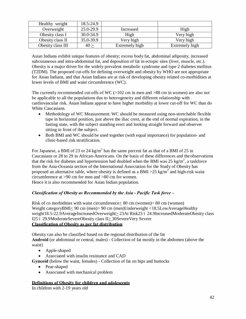
42
Healthy weight 18.5-24.9 Overweight 25.0-29.9 Increased High
Obesity class I 30.0-34.9 High Very high Obesity class II 35.0-39.9 Very high Very high Obesity class III 40 > Extremely high Extremely high
Asian Indians exhibit unique features of obesity; excess body fat, abdominal adiposity, increased subcutaneous and intra-abdominal fat, and deposition of fat in ectopic sites (liver, muscle, etc.). Obesity is a major driver for the widely prevalent metabolic syndrome and type 2 diabetes mellitus (T2DM). The proposed cut-offs for defining overweight and obesity by WHO are not appropriate for Asian Indians, and that Asian Indians are at risk of developing obesity related co-morbidities at lower levels of BMI and waist circumference (WC).
The currently recommended cut-offs of WC (>102 cm in men and >88 cm in women) are also not be applicable to all the populations due to heterogeneity and different relationship with cardiovascular risk. Asian Indians appear to have higher morbidity at lower cut-off for WC than do White Caucasians.
• Methodology of WC Measurement: WC should be measured using non-stretchable flexible tape in horizontal position, just above the iliac crest, at the end of normal expiration, in the fasting state, with the subject standing erect and looking straight forward and observer sitting in front of the subject.
• Both BMI and WC should be used together (with equal importance) for population- and clinic-based risk stratification.
For Japanese, a BMI of 23 or 24 kg/m2 has the same percent fat as that of a BMI of 25 in Caucasians or 28 to 29 in African-Americans. On the basis of these differences and the observations that the risk for diabetes and hypertension had doubled when the BMI was 25 kg/m2, a taskforce from the Asia-Oceania section of the International Association for the Study of Obesity has proposed an alternative table, where obesity is defined as a BMI >25 kg/m2 and high-risk waist circumference at >90 cm for men and >80 cm for women. Hence it is also recommended for Asian Indian population.
Classification of Obesity as Recommended by the Asia - Pacific Task force –
Risk of co morbidities with waist circumference< 80 cm (women)> 80 cm (women) Weight categoryBMI< 90 cm (men)> 90 cm (men)Underweight <18.5LowAverageHealthy weight18.5-22.9AverageIncreasedOverweight> 23At Risk23 – 24.9IncreasedModerateObesity class I25 – 29.9ModerateSevereObesity class II> 30SevereVery Severe Classification of Obesity as per fat distribution
Obesity can also be classified based on the regional distribution of the fat Android (or abdominal or central, males) - Collection of fat mostly in the abdomen (above the waist)
• Apple-shaped • Associated with insulin resistance and CAD
Gyneoid (below the waist, females) - Collection of fat on hips and buttocks • Pear-shaped • Associated with mechanical problem
Definitions of Obesity for children and adolescents In children with 2-19 years old

43
BMI : > 85th to 95th percentile for that age – Possible risk of overweight and should undergo further evaluation. BMI : > 95th percentile for that age – Overweight In the United States, the 2000 CDC BMI for age growth charts are recommended for screening of individuals who are overweight and those who are at risk for overweight. No BMI for age reference exist for children younger than 2 years. Nor are there any consistent recommendations for the definition of overweight in this age group.
• Overweight • BMI between 85th to 94th percentile for age and sex
• Obese • BMI ≥95th percentile for age and sex -or- BMI ≥30 kg/m²
• Severe obesity • ≥99th percentile (equivalent to BMI of 30-32 kg/m² for 10-12y.o. -or- ≥34 kg/m² for
14-16y.o.) • Weight for length is used in the under 2-yr age group
For the evaluation of obesity in Indian children IAP growth charts are used. At present, IAP
suggests to use WHO growth charts and Growth charts by Agrawal DK. IAP expect to come out with its own new growth charts. Till the time IAP comes out with IAP Charts, it is suggested to use WHO growth charts, which can be accessed at http://www.who.int/childgrowth/standards/chart_catalogue/en/index.htmlHowever with secular trends growth charts often change & periodic revision of Growth charts done should be accepted.
Obesity is associated with various organ System Dysfunction & they are
Cardiovascular Hypertension, Coronary artery disease, Congestive heart failure, Cor pulmonale, Varicose veins, Pulmonary embolism. Musculoskeletal Hyperuricemia and gout, Immobility-Osteoarthritis (knees and hips), Low back pain Psychologic Depression, low self-esteem, Body image disturbance, Social stigmatization Respiratory Dyspnea, Obstructive sleep apnea, Hypoventilation syndrome, Pickwickian syndrome, Asthma Gastrointestinal Gastroesophageal reflux disease (GERD), Nonalcoholic fatty liver disease (NAFLD), Cholelithiasis, Hernia, Colon cancer Genitourinary Urinary stress incontinence, Obesity-related glomerulopathy, Hypogonadism (male), Breast and uterine cancer, Pregnancy complications Neurological Stroke, Idiopathic intracranial hypertension, Meralgia paresthetica Integument Striae (stretch marks), Stasis pigmentation of legs Lymphedema, Cellulitis, Intertrigo, carbuncles, Acanthosis nigricans, skin tags, Hirsutism in patients with PCOS. Endocrine Metabolic syndrome, Type 2 diabetes, Dyslipidemia, PCOS, Hyperandrogenemia, menstrual disorders, Amenorrhea, infertility. There two ways by which endocrine system is affected in Obesity.

44
Endocrine Diseases associated with Obesity
Hormonal abnormalities due to Obesity
• Hypothyroidism • Cushing’s Syndrome • Growth Hormone Deficiency • Hypogonadism • Polycystic Syndrome (PCOS) • Insulinoma • Traumatic Hypothalamic Obesity. • Monogenic Mutations
• Û cortisol production • ÛInsulin resistance • Ü SHBG in women • Ü progesterone levels in women • Ü testosterone levels in men • Ü growth hormone production
• Other Genetic Syndromes – Prader Willi Syndrome, Lawerence Moon Bield Syndrome, Bardett Bield Syndrome, Alstroms Syndrome, etc
Note: Hypothyroidism is not the cause of significant obesity. Weight gain in hypothyroidism is due to fluid retention & is usually not more than 2-5 kg.
Obesity and Obstructive Sleep Apnea Obesity is the most powerful risk factor for obstructive sleep apnea (OSA). Several pathophysiologic mechanisms relates obesity to the development of OSA These mechanisms include anatomic and functional obstruction of the pharyngeal airway. Sleep deprivation, daytime somnolence and metabolic dysregulation may also contribute to obesity in the setting of OSA. Although history and physical examination may help predict OSA in obese individuals, polysomnography is the gold standard for making the diagnosis of OSA and assessing effects of therapy. Weight loss is an important strategy for treating OSA. However, the cornerstone of current management is continuous positive airway pressure.
Clinical Evaluation Basic evaluation in clinics is measurement of Height, Weight, BMI, Waist Circumference. Detection of acanthosis nigricans or skin tags should suggest significant insulin resistance. A number of physical features of an obese individual may help identify a specific cause for the individual's problem. Features of the hypothalamic syndrome, Cushing's syndrome, Polycystic ovarian disease (Which is a common in younger women) should be looked for. Among the various genetic diseases that produce obesity, Prader-Willi is the most common. It includes hypotonia, mental retardation, and sexual immaturity, and can usually be recognized clinically. Bardet-Biedl syndrome, with its polydactyly and retinal disease, is distinctive. Obesity and red hair in a child might suggest a defect in the processing of POMe.
Laboratory Evaluation Common laboratory tests which should be done as a part of the obesity evaluation are - Plasma Glucose (Fasting & Post glucose or postprandial), Plasma Lipids, Thyroid Functions, serum uric acid, Liver function tests, Prostate-Specific Antigen in Males, Ultrasound of the Gall Bladder. Other tests like overnight dexamethasone suppression test, serum gonadotropins, etc may be done depending on the patients clinical profile. (Serum insulin has no role in the evaluation of obesity in day today practice except in suspected cases of Insulinoma which is very rare.)
Management of Obesity Obesity prevalence has increased markedly over the past few decades. The obesity pandemic has huge implications for public health and our society. There are many Health benefits of moderate weight loss (5-1 0% of present body weight) Reduction in Death - 20% overall, 30% diabetes-related & 40% cancer- related
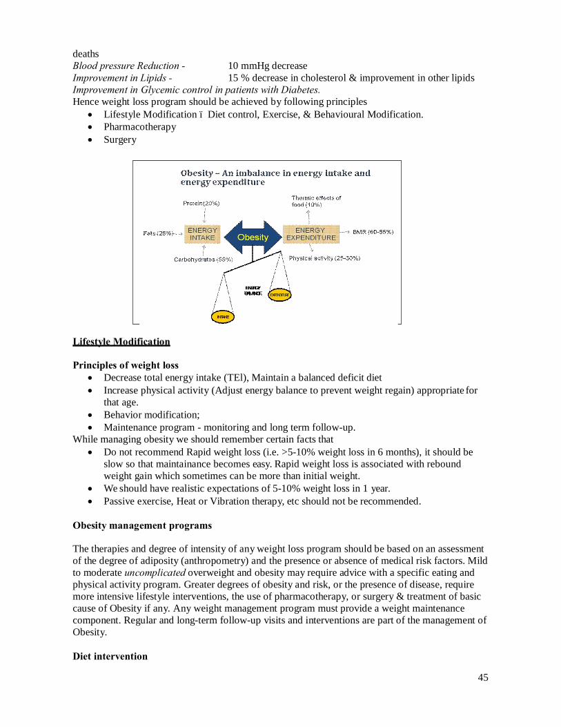
45
deaths Blood pressure Reduction - 10 mmHg decrease Improvement in Lipids - 15 % decrease in cholesterol & improvement in other lipids Improvement in Glycemic control in patients with Diabetes. Hence weight loss program should be achieved by following principles
• Lifestyle Modification – Diet control, Exercise, & Behavioural Modification. • Pharmacotherapy • Surgery
Lifestyle Modification
Principles of weight loss • Decrease total energy intake (TEl), Maintain a balanced deficit diet • Increase physical activity (Adjust energy balance to prevent weight regain) appropriate for
that age. • Behavior modification; • Maintenance program - monitoring and long term follow-up.
While managing obesity we should remember certain facts that • Do not recommend Rapid weight loss (i.e. >5-10% weight loss in 6 months), it should be
slow so that maintainance becomes easy. Rapid weight loss is associated with rebound weight gain which sometimes can be more than initial weight.
• We should have realistic expectations of 5-10% weight loss in 1 year. • Passive exercise, Heat or Vibration therapy, etc should not be recommended.
Obesity management programs
The therapies and degree of intensity of any weight loss program should be based on an assessment of the degree of adiposity (anthropometry) and the presence or absence of medical risk factors. Mild to moderate uncomplicated overweight and obesity may require advice with a specific eating and physical activity program. Greater degrees of obesity and risk, or the presence of disease, require more intensive lifestyle interventions, the use of pharmacotherapy, or surgery & treatment of basic cause of Obesity if any. Any weight management program must provide a weight maintenance component. Regular and long-term follow-up visits and interventions are part of the management of Obesity.
Diet intervention

46
Although energy (calorie) restriction produces good weight loss, some individuals may find this irksome and impossible to maintain. It is the total calorie intake which decides patients weight rather than the type of food. However for some patients, a low-fat eating plan may prove effective. Such plans may be maintained for several years. Greater losses may be obtained by additionally reducing energy intake. Men may also need to reduce energy intake by limiting their alcohol intake. There is no long term difference between low calorie diet (LCD) & Very low calorie Diet (VLCD).
Physical activity /exercise program
Emphasis should be placed on increasing total daily activity to between 60 and 90 min each day. Although exercise and fitness are important, the initial emphasis should be on increasing the activities of daily living, in particular more walking. A formal written exercise prescription has been shown to increase effectiveness in general practice. Giving patients the opportunity to use or purchase a pedometer and then setting the number of steps they need do in a day may prove an effective method of increasing activity. For patients with arthritis or other disabilities, hydrotherapy (exercising in water) may be a way of initiating movement and this type of activity can assist weight loss. Patient can do any type of exercise which is suitable for his/her age & associated comorbid condition.
Behavior modification
This therapy is important and central to any weight loss program. There are many components to behavior modification, but one simple is the use of a food and exercise diary. This allows habit recognition and changes accordingly. All subjects, but most particularly those who are overweight or obese, under-report food intake and over report the activity they undertake. A diary provides an important starting point for discussion and suggests possible interventions. Additional behavioral therapies include discussion about habits, change and alternative ways of approaching situations. Other techniques involve stress management, improving self-esteem and, occasionally, more spe- cific counseling or psychiatric intervention.
Such a lifestyle program should involve a team of health professionals (dietitians, physiotherapists, nurses, psychologists). Good results and satisfaction are usually obtained by the involvement of multidisciplinary teams. Some commercial groups use conventional therapy (e.g. Weight Watchers or programs used in some gyms), whereas others rely on alternative or natural therapies and still others use magic treatments that sound plausible but which really have neither have any scientific basis nor effectiveness. Examples of the magic type of therapy would be total-body wrapping, some herbal and bulking agents. Few have been tested rigorously and very are costly, but the very existence of such program and therapies shows that many individuals desire to lose weight but need to be guided into the correct approach and provided with effective therapy.
Maintenance program, Monitoring and longer term follow-up This is an essential part of any weight loss program but the most neglected part. Follow-up programe are essential and very effective. Patients involved in an obesity treatment program require the following monitoring of weight (ideally monthly, no greater than bimonthly); monitoring of pulse rate and blood pressure; monitoring of obesity-related risks and diseases (e.g. dyslipidemia, type 2 diabetes).
Drug therapy
The decision to give pharmacotherapy for obesity is made from clinical judgment-based on the

47
requirement of the each individual patient in terms of potential benefits balanced against potential risks. BMI between 25 and 30 with comorbid conditions is the definite indication to start pharmacotherapy along with lifestyle intervention. The various drugs which have been used for obesity are :
1. Appetite suppressants a. Adrenergic agents (e.g. amphetamine, methamphetamine phenylpropanolamine,
Phentermin) 2. Serotonergic agents (e.g. fenfluramine desfenfluramine, SSRIs like setoraline, fluoxetine) 3. Thermogenc agents - Ephedrine, caffeine 4. New ones
a. Serotonin and Noradrenaline reuptake Inhibitor - Sibutramine; b. Intestinal Lipase inhibitors - Orlistat c. The CB1 blocker - Rimonabant,
Among all these drugs currently only Orlistat is approved drug for the medical management of obesity. All other drugs are either not approved or banned for their use in obesity. Orlistat is an intestinal lipase inhibitor. It irreversibly inhibits the action of gastric and pancreatic lipases which are required to hydrolyse dietary fat before absorption. Unabsorbed triglycerides and cholesterol are excreted in faeces. It can cause Weight loss of 5-8.5 % at the end of 1 yr.
Side effects include steatorrhoea, flatus, fecal incontinence and oily spotting. Absorption of fat soluble vitamins may be lowered with orlistat. Hence vitamin supplementation is recommended. The maximum effect of orlistat is achieved at a dose of 120 mg thrice daily with every fatty meal. In patients with Fat restricted diet this drug is of no use.
There is no good scientific evidence for the alternative therapies.
Surgical Treatment of Obesity – Overview
Gastrointestinal surgery is the most effective approach for inducing major weight loss in extremely obese patients. Bariatric surgery has evolved over the last half century as a treatment option for patients suffering from morbid obesity. It involves modification of the digestive system by either decreasing the gastric volume (restrictive) or altering the path of the food bolus causing an element of malabsorption (Malabsorptive). These alterations effect appropriate changes in eating behavior and aid lifestyle modifications to help weight loss.
Indications for Bariatric Surgery -
• Patients with a BMI 40 kg/m2 or more without any comorbid condition. • Patients with a BMI of 35.0 to 39.9 kg/m2 (with comorbidiry) and one or more severe
medical complications of obesity (e.g., hypertension, heart failure, Type 2 Diabetes Mellitus, sleep apnea).
• Additional eligibility criteria are the inability to maintain weight loss with conventional therapy, acceptable operative risks,
The Surgical Options for Weight loss Surgery Restrictive Procedures
• Laparoscopic adjustable gastric banding (LAGB) • Sleeve gastrectomy
Combined Procedures • Roux-en-Y gastric bypass (RYGBP)
Malabsorptive Procedures
48
• Bilio-pancreatic diversions (BPD) • Bilio-pancrearic diversion with duodenal switch (BPD-DS)
Experimental Procedures Duodeno-jejunal bypass Ileal interposition Implantable pulse generators
Over the years, minimally invasive surgery has evolved from general laparoscopic surgery to advanced laparoscopic surgery. Given the advantage of laparoscopy with other general surgery procedures, it was a logical step to attempt these complex bariatric procedures using minimally invasive approach. Laparoscopic BPD-DS has higher complication rate compared to gastric bypass or banding and a mortality rate as high as 5%.
Bariatric Surgery Outcomes - Weight Loss Bariatric surgery is currently the most effective method to treat severe obesity. The amount of excess weight loss (EWL) varies according to procedure. Long term data is now emerging that shows maintenance of weight loss 5-15 years after modern bariatric procedures. This sustained weight loss has a major impact on individual patient health and longevity.
Approach to Obesity management
Prevention:
With Limited options of drug treatment prevention becomes an important step. Prevention should start from Childhood so that we can prevent future health hazards. Balanced diet & regular Aerobic physical activity is must for everyone & should be educated since childhood.
References:
• A Misra et al Consensus Statement for Diagnosis of Obesity, Abdominal Obesity and the Metabolic
Syndrome for Asian Indians and Recommendations for Physical Activity, Medical and Surgical Management. JAPI Vol. 57 • February 2009
Misra A, Khurana L. Obesity and the metabolic syndrome in developing countries. J Clin Endocrinol Metab 2008;93(11 Suppl 1):S9-30.
World Health Organization. Obesity: preventing and managing the global epidemic. Report of a WHO consultation presented at: the World Health Organization; June 3-5, 1997;Geneva, Switzerland. Publication WHO/NUT/NCD/98.1.
NHLBI Expert Panel. The Practical Guide: Identification, Evaluation, and Treatment of Overweight and Obesity in Adults. Bethesda, MD: National Institutes of Health. NIH publication 00-4084. 2000.
R.F. Kushner, J.L. Roth / Endocrinol Metab Clin N Am 32 (2003) 9/5-933 National Heart Lung, and Blood Institute. Clinical guidelines on the identification
evaluation, and treatment of overweight and obesity in adults. The evidence report. Ob Res 1998;6(Suppl 2):51 S-21 OS.
American Association of Clinical Endocrinologists, American College of Endocrinology AACE/ACE position statement on the prevention, diagnosis and treatment of obesity (1998 revision). Endocr Pract 1998;4:299-350. Available at: http://www.aace.com/clin/guides/obesityguide.pdf. Accessed October 19, 2003.
WHO Expert Consultation. Appropriate body-mass index for Asian populations and its implications for policy and intervention strategies. Lancet. 2004;363(9403):157-63.

49
World Health Organization. Obesity: preventing and managing the global epidemic. Report of a WHO consultation presented at: the World Health Organization; June 3-5, 1997;Geneva, Switzerland. Publication WHO/NUT/NCD/98.1.
• George A Bray, Claude Bouchard – Handbook of Obesity Clinical Applications third edition: Classification & evaluation of Overweight or Obese Patient. 2008: 1-28
Agarwal DK et al. Physical and sexual growth of affluent Indian children from 5 to 10 years of age. Indian Pediatr 1992; 29: 1203-1282.
Agarwal DK and Agarwal KN. Physical growth in affluent Indian children (birth to 6years). Indian Pediatr 1994; 31: 377-413.