standard treatment guidelines medicine
TRANSCRIPT

1
STANDARD TREATMENT
GUIDELINES
MEDICINE (RESPIRATORY DISEASES)
Ministry of Health & Family Welfare
Govt. of India

2
Group Head Coordinator of Development Team
Dr S.K. SHARMA
AIIMS
3
ACUTE RESPIRATORY DISTRESS SYNDROME
I. WHEN TO SUSPECT/ RECOGNIZE? a) Introduction: Acute respiratory distress syndrome (ARDS) is the most severe form of acute lung injury resulting in extensive bilateral pulmonary infiltrates, severe refractory arterial hypoxemia and stiff lungs. Of the numerous clinical conditions identified to cause ARDS, sepsis syndrome, polytrauma, obstetric complications, and surgery, among others appear to be the most common causes. The site of injury may be focused on either the vascular endothelium (eg. sepsis) or the alveolar epithelium (eg. aspiration of gastric contents). Injury to the endothelium results in increased capillary permeability and the influx of protein-rich fluid into the alveolar space. Injury to the alveolar lining cells also leads to pulmonary edema. In tropical countries, malaria, leptospirosis, tuberculosis (including miliary), enteric fever, and dengue haemorrhagic fever, organophosporus and paraquat poisoning; scorpion sting; inhalation of toxic fumes (e.g.chlorine); and heat stroke are important causes of ARDS. Other known risk factors are bacteremia, sepsis, trauma with or without pulmonary contusion, fractures, particularly multiple fractures and long bone fractures, burns, massive transfusion, pneumonia, aspiration, drowning, postperfusion injury after cardiopulmonary bypass and acute pancreatitis.
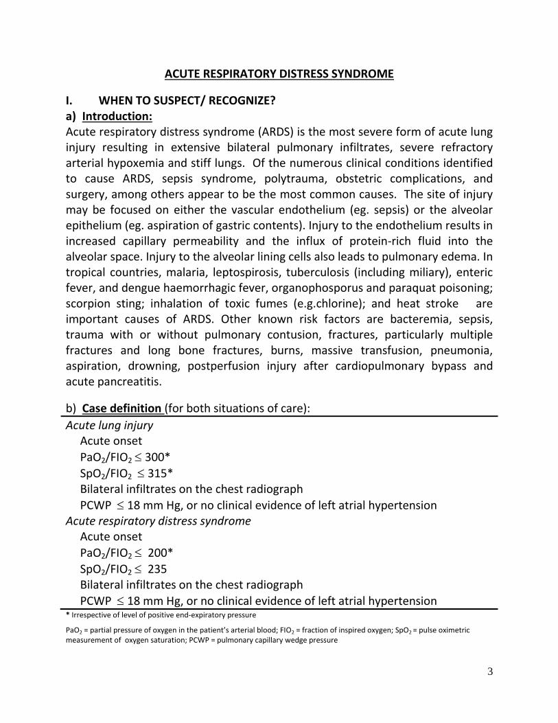
b) Case definition (for both situations of care):
Acute lung injury Acute onset
PaO2/FIO2 300*
SpO2/FIO2 315* Bilateral infiltrates on the chest radiograph
PCWP 18 mm Hg, or no clinical evidence of left atrial hypertension Acute respiratory distress syndrome Acute onset
PaO2/FIO2 200*
SpO2/FIO2 235 Bilateral infiltrates on the chest radiograph
PCWP 18 mm Hg, or no clinical evidence of left atrial hypertension * Irrespective of level of positive end-expiratory pressure
PaO2 = partial pressure of oxygen in the patient’s arterial blood; FIO2 = fraction of inspired oxygen; SpO2 = pulse oximetric measurement of oxygen saturation; PCWP = pulmonary capillary wedge pressure
4
The severity of hypoxemia is necessary to make the diagnosis of ARDS. The cardiogenic pulmonary edema must be excluded either by clinical criteria or by a pulmonary capillary wedge pressure (PCWP) lower than 18 mmHg in patients with a Swan-Ganz catheter in place. II. INCIDENCE OF THE CONDITION IN OUR COUNTRY Global estimates suggest that ARDS occurs in 1.5 to 75 cases / 100,000 population. No systematic studies have been carried out till date to define the incidence of ARDS in India and reliable epidemiological data are not available. III. DIFFERENTIAL DIAGNOSIS 1. Cardiogenic pulmonary oedema 2. Other considerations
2.1 Pneumonia 2.2 Diffuse alveolar haemorrhage 2.3 Idiopathic acute eosinophilic pneumonia 2.4 Cryptogenic organizing pneumonia 2.5 Acute interstitial pneumonia (Hamman-Rich syndrome) 2.6 Metastatic malignancy
2.7 Near drowning 2.8 Drug reaction 2.9 Noncardiogenic pulmonary edema
Acute hypersensitivity pneumonitis Transfusion-related acute lung injury (TRALI) Leukemic infiltration Fat embolism syndrome
IV. PREVENTION AND COUNSELING No form of therapy is known to prevent the occurrence of ARDS.

5
OPTIMAL DIAGNOSTIC CRITERIA, INVESTIGATIONS, TREATMENT & REFERRAL CRITERIA
*Situation 1: At Secondary Hospital: Optimal Standards of Treatment in Situations where technology and resources are limited
a) Clinical Diagnosis Symptoms The onset of ARDS is acute. The acute, or exudative, phase is characterised by rapid onset of dyspnoea, dry cough, respiratory failure, disorientation and agitation that usually develop 24 to 72 hours after the inciting event. The patient may be febrile or hypothermic. Signs
Tachypnoea, tachycardia, cyanosis, crepitations and rhonchi may be present. Arterial hypoxaemia that is refractory to high concentrations of supplemental oxygen therapy is a hall mark of ARDS. Chest auscultation may reveal bilateral rales and rales may be absent despite diffuse involvement. In intubated and mechanically ventilated patient decreased breath sounds over one lung may indicate a pneumothorax or endotracheal tube down the opposite main bronchus. Careful physical examination to look for potential causes of sepsis including signs of lung consolidation or findings consistent with an acute abdomen, sites of intravascular lines, surgical wounds, drain sites, and decubitus ulcers for evidence of infection are needed. The cardiogenic pulmonary edema should be differentiated from ARDS, by carefully looking for signs of congestive heart failure or intravascular volume overload, including jugular venous distention, cardiac murmurs and gallops, hepatomegaly, and edema.

6
b) Investigations 1. Pulse oximetry: Pulse oximetry reveals SpO2/FIO2 ratio less than or equal to 235. Severe refractory hypoxaemia in spite of supplemental oxygen therapy is evident. 2. Chest radiograph: The radiographic changes become evident by about 12 hours after the clinical onset of respiratory failure. Initially, patchy, ill-defined opacities may become apparent throughout the lungs. 3. Echocardiography: Echocardiography is useful in differentiating ARDS from cardiogenic pulmonary oedema. 4. Arterial blood gas (ABG) analysis: ABG analysis usually reveals severe refractory hypoxemia, hypocapnia and alkalosis if the patient is breathing spontaneously. Hypercapnia usually does not occur unless chronic lung disease is co-existent.
c) Treatment:
Standard Operating procedure a. In Patient
1. General therapeutic measures
1. SpO2 monitoring by pulse oximetry 2. Ensure adequate circulation and blood pressure using volume infusion
and/or vasopressors 3. Treatment of the aetiologcal cause of ARDS (e.g., appropriate antibiotic therapy for patients with sepsis syndrome)
2. Supplemental oxygen therapy Initially, spontaneous ventilation using a face mask with a high flow gas delivery system can be used to deliver a FIO2 of up to 0.5 to 0.6.
b. Out Patient Not applicable. ARDS is managed in an intensive care (ICU) unit setting
d) Referral criteria:
1. Diagnosis not clear
7
2. Response to therapy not optimal. Hypoxaemia (SpO2 ≤ 90) persists in spite of supplemental oxygen therapy
3. Haemodynamic instability
*Situation 2: At Tertiary hospital where higher-end technology is available a) Clinical Diagnosis
As in situation V.1 a) above b) Investigations
1. As in situation V.1b) above PLUS
2. Computed tomography of the chest: Early in the exudative phase, computed tomography (CT) of the chest reveals diffusely distributed non-uniform ground glass opacification or consolidation which may not conform to the gravity distribution. Later in the exudative phase, the consolidation becomes more homogeneous and gravity dependent. In the proliferative and fibrotic stages, there is a decrease in the overall lung density and the appearance of an interstitial reticular pattern. CT also facilitates identification of underlying pulmonary causes of ARDS (e.g., pneumonia and lung abscess) and complications of ARDS, such as, pneumothorax, pneumomediastinum and interstitial emphysema may also be evident.
3. Swan-Ganz catheterization: The pulmonary capillary wedge pressure (PCWP) is less than 18 mm Hg and the cardiac index is more than 2.1 L/min.
4. Bronchoscopic procedures: Bronchoscopic procedures are helpful to exclude infectious causes of ARDS.
c) Treatment:
Standard Operating procedure
a. In Patient
1. As in situation V.1c)a.1 above PLUS
8
2. General therapeutic measures 2.1. Haemodynamic stabilization. Pulmonary and systemic arterial lines are
inserted for hemodynamic monitoring and rational fluid replacement therapy. Adequate circulation and blood pressure is ensured using volume infusion (crystalloids) and/or vasopressors, taking the CVP/PCWP as the guideline. The intravascular volume is maintained as low as possible while maintaining an adequate cardiac index, mean arterial pressure, at the same time ensuring adequate organ perfusion. In anaemic patients, transfusion of packed red cells can help improving oxygenation.
2.2. Adequate nutrition should be ensured through the use of enteral feeding. 2.3. Efforts should also be directed to prevent gastrointestinal bleeding and
pulmonary thromboembolism 3. Supplemental oxygen therapy
3.1 Spontaneous breathing using a face mask with a high flow gas delivery system can be used to deliver a FIO2 of up to 0.5 to 0.6. Continuous positive airway pressure (CPAP) may be added to improve PaO2 without increasing FIO2. If a FIO2 of more than 0.6 and CPAP of more than 10 cm H2O are needed to achieve PaO2 of more than 60 mm Hg, tracheal intubation and assisted mechanical ventilation are required.
4. Ventilatory support 4.1 Aim of mechanical ventilation is to maintain gas exchange with minimal complications. Tidal volume should be set in the region of 6 ml/kg (“lung protective ventilation”) rather than the conventional 10 to 12 ml/kg and the plateau pressure should be limited to 30 to 35 cm H2O to prevent alveolar overdistension. PEEP and FIO2 settings are set as per ARDSnet protocol.
4.2 Other alternative approaches, such as, prone positioning of the patient, high frequency jet ventilation (HFJV), high frequency oscillatory ventilation (HFOV), and liquid ventilation among others can also been tried in selected patients. The relative merits of these alternative methods of mechanical ventilation must be critically weighed against the potential side effects in every setting.

9
5. Corticosteroids and ARDS
5.1 It is now conclusively established that corticosteroids have no useful role before the onset of ARDS or early in its course. When patients with ARDS have severe disease, and, do not show signs of improvement 7 to 14 days after the onset of ARDS (late ARDS), a one to two week trial with corticosteroids (prednisolone 2-4 mg/kg/day or equivalent) can be tried.
6. ARDS in the tropics
6.1 Infectious causes such as pulmonary and miliary tuberculosis, falciparum malaria, enteric fever, and leptospirosis are rare but treatable causes of ARDS. When recognized to be the aetiological cause of ARDS, appropriate specific treatment must be instituted for primary conditions.
b. Out Patient
Not applicable. ARDS is managed in an intensive care (ICU) unit setting
V. WHO DOES WHAT? and TIMELINES
a. Doctor Diagnosis and Management including counseling b. Nurse Implementation of orders, monitoring of patients and counseling c. Technician Investigations
Respiratory physiotherapist For administering supportive care to patients admitted in ICU
VI. FURTHER READING / REFERENCES
1. Bernard GR, Artigas A, Brigham KL, Carlet J, Falke K, Hudson L, et al. The American-European consensus conference on ARDS: definitions, mechanisms, relevant outcomes, and clinical trial coordination. Am J Respir Crit Care Med 1994; 149:818-24. 2. Rice TW, Wheeler AP, Bernard GR, Hayden DL, Schoenfeld DA, Ware LB. for the National Institutes of Health, National Heart, Lung, and Blood Institute ARDS Network. Comparison of the SpO2/FIO2 ratio and the PaO2/ FIO2 ratio in patients with acute lung injury or ARDS. Chest 2007;132:410-7. Epub 2007 Jun 15. 3. Wiedemann HP, Wheeler AP, Bernard GR, Thompson BT, Hayden D, deBoisblanc B, et al. National Heart, Lung, and Blood Institute acute respiratory

10
distress syndrome (ARDS) clinical trials network. Comparison of two fluid management strategies in acute lung injury. N Engl J Med 2006;354: 2564-75. 4. Wheeler AP, Bernard GR, Thompson BT, Schoenfeld D, Wiedemann HP, deBoisblanc B, et al. National Heart, Lung, and Blood Institute acute respiratory distress syndrome (ARDS) clinical trials network. Pulmonary-artery versus central venous catheter to guide treatment of acute lung injury. N Engl J Med 2006;354: 2213-24.
RESOURCES REQUIRED FOR ONE PATIENT
Situation
HUMAN RESOURCES
INVESTIGATIONS DRUGS & CONSUMABLES
EQUIPMENT
1 1. Physician with
training in
echocardiography
2. Nurse
3. Radiographer
1.Pulse oximetry
2.Chest X-ray
3.Echocardiography
4.ABG analysis
1.Oxygen
2. Antibiotics
3. Inhaled
bronchodilators
1.Oxygen cylinder
2.Nasal prongs
3.MC mask
4.X-ray machine
5.Echocardiography
machine
6.ABG analyzer
2 Above plus
1.Intensivist
2.ICU staff with
pulmonary training
3.Respiratory
physiotherapists
4.Radiologist
5.Microbiologist
Above plus
1.Computed
tomography including
CT pulmonary
angiography
2.Bronchoscopy
Above plus
1.Parenteral and oral
bronchodilators
(theophylline,
terbutaline)
2.Parenteral and oral
steroids
(hydrocortisone,
prednisolone)
3.Central venous
catheter
4.Swanz Ganz
catheter
Above plus
1.CT machine
2.Microbiology
laboratory service
for infectious
aetiology work-up
3.ICU
4.Noninvasive and
invasive ventilators
5. HFOV (high
frequency
oscillatory
ventilation) [if
available]
6. Bronchoscope
11
BRONCHIAL ASTHMA
I. WHEN TO SUSPECT/ RECOGNIZE?
a) Introduction: Asthma is a common clinical problem encountered at all levels of health care. Asthma can be defined as a chronic inflammatory disorder of the airways. Different terms such as allergic or asthmatic bronchitis, wheezy bronchitis, intrinsic and extrinsic asthma are frequently employed in clinical practice. Asthma commonly begins in childhood and early youth, but may also start later in life at any age. Contrary to common belief, children do not necessarily ‘grow out of asthma’. Almost two third of the asthmatic children continue to have symptoms in puberty/adulthood. About 5-10% children with ‘mild’ asthma may go on to develop severe asthma later in life. b) Case definition (for both situations of care): Asthma is characterized by recurrent episodes of cough, wheezing, breathlessness, and chest tightness that are often reversible, either spontaneously or with treatment.
II. INCIDENCE OF THE CONDITION IN OUR COUNTRY The prevalence rates are variable depending upon the definition and methodology employed. As per the results of the Indian Study on Epidemiology of Asthma, Respiratory symptoms and Chronic bronchitis (INSEARCH), the population prevalence in adults is about 2 percent or more. In children, the prevalence is likely to be higher, exceeding 5 percent. Both men and women are about equally affected.
III. DIFFERENTIAL DIAGNOSIS Diagnosis of asthma is a two-step approach. The first important step is to suspect the diagnosis while the second step involves exclusion of other diagnoses and laboratory assessment to confirm the diagnosis and assess the severity stage of asthma. Documentation of reversibility and/or variability of forced expiratory flow, 1st second (FEV1) or peak expiratory flow (PEF) is important to diagnose asthma and differentiate it from chronic obstructive pulmonary disease.
12
IV. PREVENTION AND COUNSELING Exact cause of asthma is not known. Both environmental and genetic factors are important. A family history of asthma or atopy (allergy), presence of other atopic manifestations (e.g. allergic rhinitis, skin allergies) and airway hyper-responsiveness predispose an individual to develop asthma. However, asthma can develop in the absence of a family history. Asthma attacks are generally triggered by one or more of exposures/ risk-factors. Most patients may have more than one trigger. Besides complying with the therapy for treatment and control of asthma, the common triggers (as below) should be identified and avoided: Respiratory infections – usually viral Allergens (Indoor/Outdoor) Air pollution (Indoor/Outdoor) including smoke and fumes (biomass fuel) Tobacco smoke (both active and passive) Drugs – beta-blockers and non-steroidal anti-inflammatory drugs (NSAIDs) Food additives and preservatives- food is normally not a trigger unless it is specifically proved to be so in an individual.
Since asthma is a lifelong problem, it is crucial that the patient and the family are educated about the disease and its management for a normal and healthy life. V. OPTIMAL DIAGNOSTIC CRITERIA, INVESTIGATIONS,
TREATMENT & REFERRAL CRITERIA *Situation 1: At Secondary Hospital: Optimal Standards of Treatment in Situations where technology and resources are limited Diagnosis
e) The diagnosis is essentially based on history and physical findings of wheezing.
FEV1/FVC ratio less than 70% and reversibility more than 12% and 200 ml establishes the diagnosis.This should also be categorized in to mild, moderate and severe depending upon the symptoms and treatment requirement.

13
Mild Moderate Severe Symptoms disturbing sleep < once per week > once per week Daily
Daytime symptoms < Daily Daily Daily Limitation of accustomed activities
Nil Some
limitation Severe
limitation Use of rescue medication * <1 unit per day 1-2 units per day >2 units per day
FEV1 or peak expiratory flow Normal 60-80% <60%
f) Treatment: Basically in asthma four components are important in management; i) diagnosis, assessment and monitoring of severity ii) education for partnership of patient and doctor iii) identification and control of trigger factors and iv) pharmacologic management. All these components of care are possible even at a place where resources are limited. Therefore primarily management of asthma does not vary due to resource constraint or lack of technology. It is important to effectively manage asthma to help an individual to live a normal life, and avoid acute exacerbations as well as long-term complications. Currently available important anti-asthma drugs can be classified as controllers (required for maintenance treatment) and relievers (required for quick relief, rescue drugs). Inhaled corticosteroids constitute the cornerstone of maintenance therapy.
• Controllers (Prophylactic, Preventive, Maintenance) • Taken daily to keep asthma under control • Steroids, long-acting beta-2 agonists, sustained-release theophyllines, leukotriene receptor antagonists, and cromones
• Relievers (Quick relief, Rescue) • Rapid acting drugs that relieve bronchoconstriction • Short acting beta-2 agonists, anticholinergics, theophyllines, short-course oral steroids
The recommendations for use of drugs vary depending upon the stage of asthma. Mild asthma can be further divided into intermittent (symptoms for less than two days per week) and persistent (symptoms for more than two days per week) categories, and treatment given accordingly. Low dose inhaled steroids are

14
recommended. Alternatively, oral theophyllines can be used. Moderate asthma is treated with medium dose ICS + long acting beta-agonists (LABA) and/or leukotriene antagonists (LTRA). Alternate choices are: - medium dose ICS + LTRA / theophylline. Severe asthma is managed with high-dose ICS and/or oral steroids at the lowest dose + LABA + theophylline + LTRA. Systemic corticosteroids on long-term basis must be avoided. A short-course of up to two weeks (0.5 mg/kg/day) is, however, often valuable for managing acute severe asthma. In addition to daily controller therapy, reliever medications on as-needed basis may be taken in all stages. Asthma control requires frequent stepping up or down of therapy. Patients with intermittent or seasonal symptoms can be managed with only reliever medications taken on an as-needed basis.
Management of acute severe asthma Hour 1: 4 doses of inhaled salbutamol ± ipratropium, 100 mg hydrocortisone
(IV) or oral prednisolone 60 mg, oxygen, adequate hydration Hour 2: 4 more doses of inhaled salbutamol with ipratropium, IV
aminophylline, subcutaneous terbutaline/adrenaline 0.3-0.5 mg (0.01 mg/kg for children) for 3 doses
Acute severe asthma not responding within 2 hours of treatment, or deteriorating:
REFER IMMEDIATELY Expectorants and mucokinetic drugs do not have any significant role.
General principles of pharmacotherapy in patients with bronchial asthma • Inhaled drugs should preferably be given using metered dose inhaler with spacer
• Education about proper inhalation technique is most essential for optimal results
• Long-acting beta-agonists (LABA) should always be combined with ICS • Short-acting beta-agonists (SABA) should be used only as reliever medication • Methylxanthines can be used as an alternative to inhaled steroids only in mild disease, or in acute severe asthma when standard treatment is not effective
• Anticholinergic drugs provide additive effect to SABA aerosol during exacerbations
Systemic glucocorticoids are important only in the treatment of mild to moderate

15
exacerbations of asthma.
g) Referral criteria:
Diagnosis unclear or in doubt
Atypical signs or symptoms
Failure to respond to treatment over one month
Other conditions complicating asthma or its diagnosis, necessitating additional work-up
Severe persistent asthma
Life-threatening asthma (cyanosis, mental obtundation)
Patients requiring additional tests such allergy testing, induced sputum eosinophil, FeNO etc
Patients requiring special treatment such as immunotherapy.
*Situation 2: At Super Specialty Facility in Metro location where higher-end technology is available
d) Clinical Diagnosis:
Same as in Situation 1 (a)
e) Investigations: exhaled nitric oxide (FeNO), induced sputum eosinophils f) Treatment:
Standard Operating procedure a. In Patient: ventilator care b. Out Patient: same c. Day Care: same
VI. WHO DOES WHAT? and TIMELINES a. Doctor: diagnosis, assessment of severity and prescription of medicines
for the patient. Explaining action plan to deal with exacerbation.
b. Nurse: Educating patients about inhaler techniques and controlling
trigger factors.
c. Technician: Proper spirometry and measurement of peak flow

16
VII. Further reading/ references 1. Global Initiative for Asthma (GINA). Global strategy for the diagnosis and management
of asthma in children 5 years and younger. Bethesda (MD): Global Initiative for Asthma
(GINA); 2009. 21 p.
2. Asher MI, Montefort S, Bjorksten B, Lai CKW, Strachan D, Weiland SK, Williams H
and the ISAAC Phase Three Study Group. Worldwide time trends in the prevalence of
symptoms of asthma, allergic rhinoconjunctivitis and eczema in childhood-ISAAC Phase
Three. Lancet 2006; 368:733-43
RESOURCES REQUIRED FOR ONE PATIENT
Situation HUMAN
RESOURCES
INVESTIGATIONS DRUGS &
CONSUMABLES
EQUIPMENT
1 4. Physician
5. Nurse
6. Pulmonary
function test
technician
7. Radiographer
1. Chest radiograph
2. Pulmonary function
test
3. Arterial blood gases
1. Inhaled
bronchodilators
(salbutamol,
ipratropium)
2. Inhaled
steroids
(budesonide,
beclamethasone)
3. ICS
(fluticasone/budeson
-ide)+ LABA
(formoterol/salmete-
rol)
4. Parenteral and oral
bronchodilators
(theophylline,
terbutaline)
5. Parenteral and oral
steroids
(hydrocortisone,
prednisolone)
6. Parenteral and oral
antibiotics
preferably
macrolides
1. Oxygen
cylinder
2. Nebulizers
3. Spirometer
4. Handheld
spirometer
(PEFR meter)
5. X-ray machine
6. ABG analyzer
2 Above plus
1. ICU staff with
pulmonary training
Above plus
1. FeNO
Above plus
1. Higher generation
antibiotics
Above plus
1. ICU set-up
2. Noninvasive
and invasive
ventilators
3. FeNO analyzer

17
CHRONIC OBSTRUCTIVE PULMONARY DISEASE (COPD)
I. WHEN TO SUSPECT/ RECOGNIZE?
c) Introduction:
COPD should be suspected in any person, particularly in middle life, with history of tobacco smoking and/or other risk factors, and presenting with:
Cough, more during morning hours
Frequent sputum production
Breathlessness, mainly on exertion On clinical examination features of hyperinflation, diminished vesicular breath sounds with prolonged expiration and rhonchi are found.
d) Case definition: For both situations of care (mentioned below*)
COPD is a preventable and treatable disease condition characterised by airflow limitation, which is not fully reversible. The airflow limitation is usually progressive and associated with an abnormal inflammatory response of the lung to noxious particles or gases, primarily caused by tobacco smoking. Chronic cough and sputum production often precede the development of airflow limitation by many years. Although COPD affects lung, it also produces significant systemic consequences. The diagnosis must be substantiated by quality spirometry showing post bronchodilator FEV1/FVC < 0.7 and FEV1 < 80% of predicted.
II. INCIDENCE OF THE CONDITION IN OUR COUNTRY
COPD is primarily a disease of the adults. The prevalence of COPD reported in different population based studies from India is highly variable (Table 1). The prevalence rates in male subjects of 2.12% to 9.4% in studies reported from North are generally higher than 1.4% to 4.08% reported from South

18
India. The respective range for female subjects varies from 1.33% to 4.9% from North and from 2.55% to 2.7% from South India. For epidemiological assessment, the rounded-off median prevalence rates were assessed as 5 percent for male and 2.7 percent for female subjects of over 30 years of age. The disease is distinctly more common in males. The male to female ratio varies from 1.32:1 to 2.6:1 in different studies with a median ratio of 1.6:1.COPD results from chronic inhalational exposure to various smokes, noxious particles and gases.
Table 1. Prevalence of COPD and its smoking association in various population studies from India.
Population COPD prevalence (%) Smoker: Non-smoker Ratio
Men Women M:F Ratio
Wig (1964) Rural Delhi 3.36 2.54 1.3 2.0
Sikand (1966) Delhi 7.0 4.3 1.6 2.5
Viswanathan (1966) Patna 2.12 1.33 1.6
Bhattacharya (1975) Rural U.P 6.67 4.48 1.6
Radha (1977) New Delhi 8.1 4.6 1.8 1.8
Thiruvengadam (1977) Madras 1.9 1.2 1.6 10.2
Viswanathan (1977) Delhi Rural 4.7 3.5 1.3 9.6
Urban 8.0 4.3 1.9 4.0
Charan (1977) Rural Punjab 2.28 1.63 1.4
Malik (1986) N.India Rural 9.4 4.9 1.9 5.5
Urban 3.7 1.6 2.3 7.0
Jindal (1993) N.India Rural 6.2 3.9 1.6
Urban 4.2 1.6 2.6 9.6
Ray (1995) South India 4.08 2.55 1.6 1.6
III. DIFFERENTIAL DIAGNOSIS
Chronic bronchial asthma, bronchiectasis, tuberculosis, obliterative bronchiolitis, congestive cardiac failure, dilated cardiomyopathy, diffuse panbronchiolitis.
19
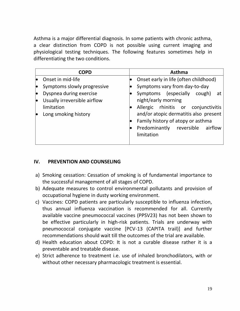
Asthma is a major differential diagnosis. In some patients with chronic asthma, a clear distinction from COPD is not possible using current imaging and physiological testing techniques. The following features sometimes help in differentiating the two conditions.
COPD Asthma
Onset in mid-life
Symptoms slowly progressive
Dyspnea during exercise
Usually irreversible airflow limitation
Long smoking history
Onset early in life (often childhood)
Symptoms vary from day-to-day
Symptoms (especially cough) at night/early morning
Allergic rhinitis or conjunctivitis and/or atopic dermatitis also present
Family history of atopy or asthma
Predominantly reversible airflow limitation
IV. PREVENTION AND COUNSELING
a) Smoking cessation: Cessation of smoking is of fundamental importance to the successful management of all stages of COPD.
b) Adequate measures to control environmental pollutants and provision of occupational hygiene in dusty working environment.
c) Vaccines: COPD patients are particularly susceptible to influenza infection, thus annual influenza vaccination is recommended for all. Currently available vaccine pneumococcal vaccines (PPSV23) has not been shown to be effective particularly in high-risk patients. Trials are underway with pneumococcal conjugate vaccine [PCV-13 (CAPITA trail)] and further recommendations should wait till the outcomes of the trial are available.
d) Health education about COPD: It is not a curable disease rather it is a preventable and treatable disease.
e) Strict adherence to treatment i.e. use of inhaled bronchodilators, with or without other necessary pharmacologic treatment is essential.

20
f) Prompt recognition of features of an exacerbation is of paramount
importance, therefore, patients should be educated about warning signs and symptoms.
g) Lifestyle modification and treatment of extrapulmonary manifestations such as coronary artery disease, osteoporosis, sarcopenia and anxiety/depression, and co-morbidities like type2 diabetes mellitus and metabolic syndrome.
V. OPTIMAL DIAGNOSTIC CRITERIA, INVESTIGATIONS, TREATMENT & REFERRAL CRITERIA
*Situation 1: At Secondary Hospital/ Non-Metro situation: Optimal Standards of Treatment in Situations where technology and resources are limited
h) Clinical Diagnosis: Patients usually present with:
History of tobacco use.
Cough with or without sputum production. Cough is particularly worse in the morning and sputum production is a frequent complaint in the early stages of COPD.
Dyspnoea on exertion: progressive breathlessness in COPD is a persistent symptom with waxing and waning but there is never any time when patients are entirely asymptomatic. Onset of breathlessness is insidious with progressive course, and often a significant amount of ventilatory capacity has been lost before the patient presents to the physician.
Many patients complain of intermittent wheezing. However, nocturnal wheezing is uncommon in COPD and suggests diagnosis of asthma or heart failure. On clinical examination: patients are frequently seen sitting leaning forward with their arms resting on a stationary object. Some patients also develop pursed lip breathing. Percussion of chest is not very helpful in COPD (might reveal features of hyperinflation of lungs). Auscultation

21
reveals diminished vesicular breath sounds with or without crackles and wheeze. Measurement of FET (forced expiratory time) is a valuable bedside technique to detect and assess airflow obstruction. An FET <5 seconds suggests that severe airflow limitation does not exist, whereas FET > 6 seconds indicates considerable airflow limitation. Many a times patients get disoriented, somnolent, or may complain of headache, which indirectly suggests CO2 retention. Such a situation will require the application of non-invasive ventilation (NIV), if possible at the treating centre. Peripheral edema, raised JVP, and hepatomegaly are signs indicative of pulmonary hypertension and right ventricular failure (chronic cor pulmonale).
i) Investigations:
Spirometry is essential to firmly establish the diagnosis. Post bronchodilator FEV1/FVC <0.7 and post bronchodilator FEV1<80%, 50% or 30% predicted are used for purpose of spirometric classification of COPD according to GOLD.
Stage FEV1/FVC FVC
Mild COPD 0.7 80%
Moderate COPD 0.7 50% - 80%
Severe COPD 0.7 30% - 50%
Very severe COPD 0.7 <30% or <50% plus chronic respiratory failure or pulmonary hypertension
Blood tests like haematocrit, chest radiograph (postero-anterior and lateral views) may be helpful in resource-poor settings.
ECG and echocardiography (if available).
6-minute walk test.

22
j) Treatment: The goals of COPD management include: 1. Relief of symptoms 2. Prevent disease progression 3. Improve exercise tolerance 4. Improve health status 5. Prevent and treat extrapulmonary manifestations 6. Prevent and treat complications 7. Prevent and treat exacerbations 8. Prevent or minimise side effects from treatment 9. Reduce mortality
Standard Operating procedure
a. In Patient Patients would present to the hospital in an exacerbation which is defined as an event in the natural course of the disease characterised by a change in the patient’s baseline dyspnoea, cough, and/or sputum that is beyond normal day-to-day variations, is acute in onset, and may warrant a change in regular medication. Signs of severity of an exacerbation that would require hospitalisation include: use of accessory muscles of respiration, paradoxical chest wall movements, cyanosis, peripheral oedema, haemodynamic instability, and decreased mental alertness. A chest X-ray would help in identifying alternative diagnoses that could mimic an exacerbation, while an ECG would aid in the diagnosis of right heart hypertrophy, arrhythmias and ischaemic episodes. Blood tests including haemogram, sugar and renal functions should be performed. Blood sample for ABG measurement should be drawn, if facility exists. Management of exacerbations:
Administer controlled oxygen therapy

23
Repeated administration of bronchodilators (β2 agonists and anticholinergics). Increased doses may be required in some patients.
Add oral or intravenous glucocorticoids
Consider adding an i.v. methylxanthine, if needed
Consider antibiotics when signs of bacterial infection
Monitor fluid balance and nutrition
b. Out Patient Stable COPD can be managed with therapy at each stage of COPD as follows:
Mild COPD Moderate COPD Severe COPD Very Severe COPD
FEV1/FVC<0.7 FEV1>= 80% PREDICTED
FEV1/FVC<0.7 50%<FEV1<80%
FEV1/FVC<0.7 30%<FEV1<50%
FEV1/FVC<0.7 FEV1<30% PREDICTED or FEV1<50% plus chronic respiratory failure
Active reduction of risk factor(s): Influenza vaccination Add short acting bronchodilator(when needed)
Add regular treatment with one or more long acting bronchodilator(when needed); add rehabilitation
Add inhalational corticosteroids (ICS) if repeated exacerbations
Refer the patients to the specialist
Antibiotics:
Antibiotics may be initiated in patients with altered sputum characteristics (increased volume and increased purulence) and increased dyspnoea. The choice should be based on local bacteria resistance patterns.
c) Referral criteria:

24
i. Marked increase in intensity of symptoms
ii. Severe COPD iii. Appearance of cyanosis, edema iv. Signs of respiratory failure v. Significant co-morbidities
vi. Recurrent exacerbations vii. New onset arrhythmias viii. Diagnostic uncertainty
ix. Old age x. Failure to control exacerbation
*Situation 2: At Super Specialty Facility in Metro location where higher-end technology is available
g) Clinical Diagnosis:
Same as in Situation 1 (a)
h) Investigations:
Same as in Situation 1 (b)
Assessment of pulmonary haemodynamics
CT-thorax and ventilation-perfusion scanning (especially for those being planned for lung surgery)
Polysomnographic studies (if indicated)
Cardiopulmonary exercise testing
DEXA scan for osteoporosis [bone mineral density (BMD) and sarcopenia [fat free mass index (FFMI)]
Psychiatric evaluation for anxiety/depression
Alpha-1 antitrypsin deficiency screening (for those who develop COPD at a younger age and those with a strong family history of COPD).

25
i) Treatment: Standard Operating procedure
a. In Patient
Same as in Situation 1 (c)
Noninvasive ventilation using BiPAP
Mechanical (invasive) ventilation
Surgical options including bullectomy and LVRS Indications for mechanical ventilation: 1. Severe dyspnea, use of accessory muscles of respiration and
paradoxical abdominal motion 2. Severe acidosis pH< 7.25, and/or hypercapnia PaCO2>
60mmHg which does not respond to NIV 3. Somnolence, impaired mental status 4. Unable to tolerate NIV 5. Life-threatening hypoxemia 6. Respiratory rate > 35/min 7. Impending respiratory arrest
Diagnosis and management of cor pulmonale The patient develops chronic cor pulmonale when there are alterations in the structure and function of the right ventricle caused by a primary disorder of the respiratory system. Invariably this would manifest as pulmonary hypertension on echocardiography. Symptoms would include tachypnoea and increase in exertional dyspnoea. Patients with chronic cor pulmonale and right heart failure will present with distended neck veins with prominent ‘V-y’ pattern suggestive of tricuspid regurgitation, cyanosis, peripheral oedema, splitting of second heart sound with accentuation of pulmonary component, left parasternal heave, long systolic or pansystolic murmur with accentuation during inspiration in the lower left sterna border, occasionally presence of right ventricular third heart sound, right upper quadrant abdominal discomfort and pulsatile hepatomegaly.
26
Medical therapy for chronic cor pulmonale is generally focused on treatment of the underlying pulmonary disease and improving oxygenation and right ventricular (RV) function by increasing RV contractility and decreasing pulmonary vasoconstriction. In general, in patients with COPD, long-term oxygen therapy is recommended when the PaO2 is less than 55 mm Hg or the O2 saturation is less than 88%. However, in the presence of cor pulmonale or impaired mental or cognitive function, long-term oxygen therapy can be considered even if the PaO2 is greater than 55 mm Hg or the O2 saturation is greater than 88%. This must be documented in at least two consecutive ABGs done 3 weeks apart in stable state. Pharmacotherapy includes salt restriction, diuretics, calcium channel blockers (indicated only if cardiac catheterization facilities available and vasoreactivity can be demonstrated), prostacyclin analogues (epoprostenol, treprostinil, and iloprost), endothelin-receptor antagonists (bosentan), PDE5 inhibitors (sildenafil and tadalafil). The algorithms for correction of hypoxia and long term oxygen therapy are given below.

27
Algorithm to correct hypoxaemia in an acutely ill COPD patient
For conversion of kPa into mmHg: 1 mmHg=0.133 kPa

28
Algorithm for long term oxygen therapy

29
b. Out Patient
Same as in Situation 1 b)
c) Referral criteria: Patients with very severe COPD who are not controlled even on maximal medical therapy and LTOT and who fulfill the selection criteria for lung transplantation should be referred to a specialist centre where expertise of lung transplantation is available.
VI. Who does what? and timeliness
a. Doctor: Diagnosis and management including counseling b. Nurse : Implementation of orders and monitoring of patients c. Technician: Carrying out investigations
VII. FURTHER READING / REFERENCES
Global Initiative for Chronic Obstructive Lung Disease. Global strategy for the diagnosis, management and prevention of chronic obstructive lung disease. NHLBI/WHO workshop report. Bethesda, National Heart, Lung and Blood Institute. Updated 2010; pp 1-92.
Jindal SK, Gupta D, Aggarwal AN. Guidelines for the management of COPD in India: a guide for physicians (2003). Indian J Chest Dis Allied Sci 2004; 46: 137-153.

30
RESOURCES REQUIRED FOR ONE PATIENT Situati
on HUMAN
RESOURCES INVESTIGATIONS DRUGS &
CONSUMABLES
EQUIPMENT
1 8. Physician 9. Nurse 10. Laboratory
technician 11. Pulmonary
function test technician
12. Radiographer 13. Physician trained
in ECHO/ cardiologist
4. Chest radiograph 5. Hemogram 6. Pulmonary function
tests 7. ABG 8. Eletrocardiogram 9. 2D-ECHO
7. Inhaled bronchodilators (salbutamol, ipratropium, tiotropium)
8. ICS
(fluticasone/bude
-sonide)+ LABA
(formoterol/salm-
eterol)
9. Parenteral and oral bronchodilators (theophylline, terbutaline)
10. Parenteral and oral steroids (hydrocortisone, prednisolone)
11. Parenteral and oral antibiotics (Amoxycillin and clavulanate combination, cefadroxil, cefotaxime, Piperacillin and tazobactum combination)
7. Oxygen cylinder/concentrator
8. Nebulizers (both electric and electronic)
9. Spirometer along with 6-MW facility
10. X-ray machine 11. ABG analyzer 12. EKG machine 13. Echocardiography
machine
2 Above plus 2. ICU staff with
pulmonary training
3. Physiotherapist 4. Sleep laboratory
technician 5. Thoracic surgeon 6. Psychiatrist 7. Cardiologist 8. Radiologist 9. Anesthetist
Above plus 1. CT Scan 2.Polysomnography 3.DEXA scan
Above plus 2. Higher
generation antibiotics
Above plus 1. CT machine 2. ICU set up 3. Noninvasive and
invasive ventilators 4. Polysomnography
machine 5. DEXA machine 6. Operation theatre
with trained staff

31
INTERSTITIAL LUNG DISEASE
I. WHEN TO SUSPECT/ RECOGNIZE?
a. Introduction:
Interstitial lung disease (ILD) is a heterogeneous group of disorders with a
common clinico-radiological presentation, hence grouped together. These diseases
are characterized by predominant involvement of lung ‘interstitium’ i.e. the space
between alveolar basement membrane and pulmonary vascular endothelial
membrane. As the disease progresses it encroaches alveolar spaces, terminal
bronchioles and perivascular spaces, thereby ILDs are referred to as diffuse
parenchymal lung disease (DPLD). ILDs due to systemic diseases such as
connective tissue diseases, sarcoidosis, hypersensitivity pneumonitis, drugs,
pneumoconioses and other miscellaneous conditions are termed as secondary
ILDs. The primary pulmonary involvement is generally the Idiopathic pulmonary
fibrosis (IPF/fibrosing alveolitis) or usual interstitial pneumonitis.
b. Case definition:
For both situations of care:
ILD is suspected in patients with onset of respiratory symptoms of relatively acute/
sub-acute duration of weeks to months. The predominant symptoms consist of
breathlessness and dry cough. Weakness and weight loss are usually present. Chest
pain/ heaviness and haemoptysis can occur in a few conditions. Elicitation of
occupational history is mandatory. The chest X-ray film shows distinctly reticular,
reticulo-nodular or nodular opacities which are patchy /basilar or diffuse in
distribution. Chest X-ray can occasionally be normal. High resolution CT scan of
the chest is usually characteristic. Investigations for the underlying disease are
important to rule out a secondary cause especially in those with atypical HRCT
findings. Pathological diagnosis with bronchoscopic/ thoracoscopic and other
surgical modalities is required for confirmation in only a few cases.
II. INCIDENCE OF THE CONDITION IN INDIA
The exact incidence and prevalence of the disease is not known. The common
causes of ILD in India include IPF, connective tissue related ILD (systemic
sclerosis and rheumatoid arthritis) and sarcoidosis. In general, at most secondary-

32
level hospitals, 1-3 cases per month and at super-specialty hospitals, 1-3 cases per
week of idiopathic ILD are likely.
III. DIFFERENTIAL DIAGNOSIS
Many disorders can present with symptoms similar to that of ILD and interstitial
opacities on chest radiograph. The following are the common disorders that should
be ruled out before making a diagnosis of ILD:
a) Tuberculosis
b) Atypical pneumonias (Mycoplasma and legionella)
c) Fungal pneumonias (Aspergillosis, crytococcosis)
d) Chronic bronchitis
Exclusion of a secondary causes such as a connective tissue disease,
hypersensitivity pneumonia, pneumoconiosis, drug-toxicity is important before
making the diagnosis of primary idiopathic ILD (such as IPF).
IV. PREVENTION AND COUNSELLING
Occupational and environment related interstitial lung disease may benefit from
avoidance or decrease in intensity and duration of exposure. High index of
suspicion and early recognition of interstitial lung disease can be helpful as it can
prevent further deterioration of lung function by avoidance of exposure. Smoking
cessation is another important caution which must be exercised.
The following are the few diseases in which avoidance of exposure can be
advocated if ILD is detected early.
1) Silicosis (occupations-miners, sandblasting, workers in abrasive industries,
such as stone, clay, glass, cement manufacturing and granite quarrying)
2) Asbestosis(asbestos manufacturing, construction trade, mining, milling and
pipefitters)
3) Coal worker’s pneumoconiosis
4) Chronic hypersensitivity pneumonitis (pigeon breeders, farm dust and grain
husk containing thermophilic actinomyces)
V. OPTIMAL DIAGNOSTIC CRITERIA, INVESTIGATIONS,
TREATMENT & REFERRAL CRITERIA
While ILD can be suspected at any secondary-level hospital, the confirmation,
exclusion of secondary causes and initiation of management lies essentially in the

33
domain of the specialty hospitals. Follow-up management afterwards can,
however, continue at the secondary –level, referring hospital.
Diagnosis:
The diagnosis of ILD is based on the combination of clinical, radiographic, and
histopathology criteria.
a. Clinical: i. Progressive breathlessness and/ or dry cough,
ii. Physical findings: basal end-inspiratory crackles- superficial, dry
and
velcro-like. Finger clubbing may be present in IPF.
b. Radiographic findings: A good quality postero-anterior chest radiograph should
be initial investigation of choice followed by HRCT chest for confirmation of
presence of ILD.
i. Chest radiograph- presence of reticular, nodular or
reticulonodular shadows, shrunken lung fields and obscuration of
cardiac and diaphragmatic margins.
ii. HRCT Chest- presence of reticular shadows (inter and
intralobular septal thickening), patchy ground-glass
opacities/consolidation (predominantly subpleural and peripheral) and
honeycombing (indicates end-stage ILD)
c. Pulmonary function tests:
i. Spirometry: A restrictive abnormality is classical of ILD with
reduced total lung capacity (TLC), functional residual capacity, and
residual volume. Forced expiratory volume in one second (FEV1)
and forced vital capacity (FVC) are reduced. FEV1/FVC ratio is
normal or increased. However, mixed (restrictive-
obstructive)/obstructive pattern can be seen in patients with
lymphangioleiomyomatosis, sarcoidosis, hypersensitivity
pneumonitis due to involvement of small airways.
ii. Diffusing capacity for carbon monoxide (DLCO): reduced in all
patients with ILD except in alveolar hemorrhage syndromes in
which it is increased.
iii. Significant oxygen desaturation on exercise (>3-4%).
iv. ECG and 2D ECHO for pulmonary hypertension/cor pulmonale.
d. Histopathology: The histopathology is required only in the presence of an early
disease when the differential diagnosis is important from a treatable condition.
The pattern varies according to the disease. Idiopathic pulmonary fibrosis is

34
characterized UIP pattern. Sarcoidosis, Wegener’s granulomatosis and Churg-
Strauss syndrome are characterized by granulomatous inflammation.
e. 6-minute walk test (6MWT): For baseline and follow-up
Treatment:
The management of the patients with ILD is difficult and unsatisfactory. Anti-
inflammatory therapy with steroids, azathioprine or cyclophosphamide forms the
main backbone of drug therapy. Interstitial lung diseases which respond well to
immunosuppresive therapy include proliferative phase of NSIP, desquamative IP,
cryptogenic organizing pneumonia, alveolar hemorrhage syndromes and
sarcoidosis. Antioxidant and anti-fibrotic drugs such as N-acetyl cysteine and
pirfenidone have some role for the treatment of idiopathic pulmonary fibrosis.
None of the drugs are likely to provide benefit in advanced disease when only
symptomatic treatment, home oxygen therapy, treatment of pulmonary
hypertension, pulmonary rehabilitation and treatment of co-morbid diseases should
be offered to improve quality of life and avoid unnecessary side-effects of the
drugs.
REFERRAL CRITERIA: All patients in whom an interstitial lung disease is
suspected or patients in whom alternative diagnosis like infection cannot be ruled
out should be referred to a specialty hospital to confirm a diagnosis, identify the
cause and initiate appropriate therapy.
VI. WHO DOES WHAT?
a. Doctor
Evaluate the patients, order investigations, make a diagnosis, advise proper
treatment and perform follow-up
b. Nurse
Carry out the Investigations suggested by the doctors and help in follow-up
of patients
c. Technical staff
Perform relevant investigations as per the advice of the treating physician

35
RESOURCES REQUIRED FOR ONE PATIENT
Situatio
n HUMAN
RESOURCES
INVESTIGATIONS DRUGS &
CONSUMABLES
EQUIPMENT
1 14. Physician
15. Nurse
16. Laboratory
technician
17. Pulmonary
function test
technician
18. Radiographer
19. Physician with
training in
echocardiography
/ cardiologist
10. Chest radiograph
11. Pulmonary
function tests
12. ABG analysis
13. 2D-ECHO
12. Parenteral and oral
steroids
(hydrocortisone,
prednisolone)
13. N-acetyl cysteine
14. Inhaled
bronchodilators
15. Sildenafil, bosentan
14. Oxygen
cylinder
15. Oxygen
concentrators
16. Spirometer
17. Hand-held
Spirometer
18. X-ray
machine
19. ABG analyzer
20. ECHO
machine
2 Above plus
10. ICU staff with
pulmonary
training
11. Radiologist
12. Pathologist
13. Physiotherapist
Above plus
1. CT Scan
2. Bronchoscopic
biopsy
(Transbronchial
lung biopsy)
Above plus
3. Immunosuppressant
drugs (azathioprine,
cyclophosphamide,
pirfenidone)
Above plus
1. ICU
2. Noninvasive
and invasive
ventilators
3. CT scan
machine
4. Bronchoscope
along with
required
facility

36
LUNG ABSCESS
VIII. WHEN TO SUSPECT/ RECOGNIZE?
e) Introduction: Lung abscess is a common form of suppurative lung disease that is characterized by a localized necrosis of pulmonary parenchyma and circumscribed collection of pus in the lung that is usually greater than 2 cm in diameter. Many local causes and systemic diseases can result in lung abscess formation. Primary lung abscess is more common and develops as a result of necrosis of lung parenchyma due to an existing disease process that is occurring there, such as, untreated aspiration pneumonia especially due to staphylococcal klebsiella and anaerobic organisms,, or lung cancer. A lung abscess can also develop secondary to preexisting conditions, such as, bronchial obstruction, spread from an extrapulmonary focus of infection, bronchiectasis, or immunocompromised state. Acute and chronic is based on duration; acute <6 weeks and chronic > 6 weeks f) Case definition: (For both situations of care)
Primary lung abscess i. A patient with an ongoing episode of pneumonia of 2 to 3
weeks duration, or, a history of pneumonia in the recent past presents with respiratory symptoms and signs consistent with a lung abscess, (such as, cough, sputum, that may be putrid and foul smelling, haemoptysis, pleuritic chest pain, shoulder pain, or heaviness in the chest) and systemic manifestations like fever, night sweats, anorexia, weight loss, and digital clubbing.
ii. Chest radiograph evidence of a cavity or cavities with a fluid level
IX. INCIDENCE OF THE CONDITION IN OUR COUNTRY Globally, the incidence of lung abscess has declined by nearly 10-fold during the preceding few decades in comparison with the pre-antibiotic era. No definitive studies have been carried out till date to define the incidence of lung abscess from India and reliable epidemiological data are not available.

37
X. DIFFERENTIAL DIAGNOSIS
1. Non-infectious diseases
Embolism with infarction
Vasculitis
Pulmonary neoplasm
Pulmonary sequestration
Bullae or cysts with air fluid level 2. Infections
Infected bulla
Infected pulmonary infarct
Infected cyst
Bronchiectasis
Empyema with air fluid level
XI. PREVENTION AND COUNSELING 1. Measures for preventing aspiration of secretions in unconscious patients 2. Smoking cessation, good orodental hygiene 3. Early recognition of lung infections and institution of appropriate antibiotics in
adequate dosage for appropriate duration. XII. OPTIMAL DIAGNOSTIC CRITERIA, INVESTIGATIONS,
TREATMENT & REFERRAL CRITERIA *Situation 1: At Secondary Hospital: Optimal Standards of Treatment in Situations where technology and resources are limited k) Clinical Diagnosis
Symptoms 1. The clinical manifestations of lung abscess may become evident coincident with the initial presentation of pneumonia or other underlying condition, or may develop later in the course of the illness.

38
2. Conditions predisposing the patient for aspiration and development of lung abscess, such as altered sensorium, depressed cough and gag reflex, diseases of the oral cavity, gastrointestinal disease, and neurological disease are usually present. 3. Patients with primary lung abscess often have an ongoing episode of pneumonia of 2-3 weeks duration, or, a history of pneumonia in the recent past. Presenting symptoms include fever, cough, sputum, that may be putrid and foul smelling. Pleuritic chest pain, shoulder pain, heaviness in the chest is also usually evident. Patients may also complain of chills, night sweats, anorexia, haemoptysis, weight loss, and digital clubbing. In patients with a secondary lung abscess, the clinical manifestations can develop over 48 to 72 hours. 4. Patients with hepatopulmonary amoebiasis may manifest abdominal symptoms, such as, right hypochondrium pain, bowel symptoms, among others. 5. In patients with perforation of the liver abscess into the lung, cough and expectoration of odourless anchovy sauce sputum may be evident.
Signs
1. Patients with an acute lung abscess appear sick and toxic. New and focal respiratory signsmay be evident on physical examination of the chest. Presence of physical signs depends on the proximity lung abscess from the chest wall. 2. Signs due to complications, such as, pleural effusion, empyema, sepsis syndrome may also be present.
l) Investigations
1. Chest radiograph: A chest radiograph is essential to establish the diagnosis of lung abscess. Lung abscess appears as an irregularly shaped cavity with an air-fluid level. The lesion may be solitary (primary lung infection), or sometimes, multiple (metastatic infection). Rapid pulmonary

39
cavitation within a dense segmental consolidation; and a rapidly enlarging nodular lesion, with or without cavitation suggest anaerobic infection. 2. Ultrasonography of the chest: Peripherally located lung abscesses, especially those in contact with pleura are detectable on ultrasonography of the chest.
m) Treatment:
Standard Operating procedure a. In Patient 1. General therapeutic measures
1.1. Adequate drainage of the lung abscess is facilitated by encouraging the patient to cough and by chest physiotherapy and postural drainage.
2. Antibiotic treatment: clindamycin administered intravenously in a dosage of 600 mg, eight hourly, followed by 150 to 300 mg orally four times daily is considered to be the preferred treatment of choice for lung abscess. Other antibiotic options include intravenous penicillin plus metronidazole and amoxycillin-clavulanate plus metronidazole.
2.1. Antibiotic treatment is administered till the pulmonary infiltrates have resolved or until the residual lesion becomes small and stable; the duration of antibiotic treatment is usually for 6 to 8 weeks.
2. Supplemental oxygen therapy In patients with hypoxaemia, spontaneous ventilation using a face mask with a high flow gas delivery system can be used to deliver a FIO2 of up to 0.5 to 0.6. b. Out Patient
Not applicable
n) Referral criteria: 1. Patients not responding to antibiotic treatment. 2. Occurrence of complications of lung abscess, such as, metastatic brain abscess, sepsis, empyema, broncho-pleural fistula, pleura-cutaneous fistula,

40
massive life-threatening haemoptysis, spontaneous rupture of the abscess into uninvolved lung segments, and failure to resolve.
*Situation 2: At Tertiary hospital where higher-end technology is available
j) Clinical Diagnosis
As in situation V.1 a) above
k) Investigations
As in situation V.1b) above PLUS 1. Computed tomography: computed tomography (CT) of the chest helps in delineating the size and location of abscesses. It also facilitates identifying associated empyema, and detecting other conditions such as pulmonary infarction, infected bulla, and raising the suspicion of a cavitating lung malignancy. In lung abscesses that fail to communicate with a bronchus, the characteristic air-fluid level within a cavity will not be seen and instead, a focal, ground-glass infiltrate with indistinct borders may be evident. 2. Establishing microbiological diagnosis: 1. Ascertaining the microbiology of anaerobic infection of the lower airways requires a specimen devoid of contamination by the flora of the upper airways or quantitative cultures that can distinguish pathogenic bacteria from the normal flora. 2. Specimens obtained from blind, deep suctioning via an endotracheal tube; radiologically guided, percutaenous transthoracic aspirates, specimens obtained at thoracotomy, quantitative cultures of specimens obtained at fiberoptic bronchoscopy (FOB), either by bronchoalveolar lavage (BAL) or with the protected double-lumen catheter with a protected sampling brush are subjected to microbiological examination. 3.If the abscess is associated with an empyema, then culture of the empyema fluid may help in isolating the aetiological organisms.

41
l) Treatment: Standard Operating procedure a. In Patient
As in situation V.1c) a.1 above PLUS
1. Drainage procedures Drainage procedures are usually reserved for patients with lung abscess who fail to respond to antibiotic therapy
1.1. Percutaneous catheter drainage (for peripherally located lung abscess) 1.2. Video-assisted thoracoscopic surgery (VATS)
2. Surgery 2.1. Surgery is indicated if airway obstruction (e.g., due to a tumour or a
foreign body) limits drainage. The surgical procedures performed include lobectomy, wedge resection or pneumonectomy.
2.2. When associated empyema is present, adequate drainage with tube thoracostomy is required; in patients with multiloculated empyemas, open pleural drainage is indicated.
b. Out Patient Not applicable.
XIII. WHO DOES WHAT? and TIMELINES a. Doctor: Diagnosis and Management including counseling b. Nurse: Implementation of orders, monitoring of patients and
counseling c. Technician: Investigations
XIV. FURTHER READING / REFERENCES 1. Schiza S, Siafakas NM. Clinical presentation and management of empyema, lung
abscess and pleural effusion. Curr Opin Pulm Med 2006;12:205-11.
2. Mansharamani NG, Koziel H. Chronic lung sepsis: lung abscess, bronchiectasis,
and empyema. Curr Opin Pulm Med 2003;9:181-5.

42
3. Mohan A, Vijayalakshmi Devi B, Chandra A. Lung abscess. In: Behera D, editor. NCCP Textbook of respiratory medicine; New Delhi: Jaypee Brothers Medical Publishers; 2011.p.171-7.
RESOURCES REQUIRED FOR ONE PATIENT
Situation HUMAN RESOURCES
INVESTIGATIONS DRUGS & CONSUMABLES
EQUIPMENT
1 1. Physician 2.Nurse 3.Radiographer
1.Chest X-ray 2.Ultrasonography of chest 3. sputum smear and culture 4. Blood culture and sensitivity
1. Oral and i.v. antibiotics 2. Streptokinase
1. X-ray machine 2. USG machine 3. Intercostal tubes 4. Negative suction machines
2 Above plus 1. Intensivist 2.Interventional Radiologist 3.Thoracic Surgeon 4.Nurse and Technician for assisting with bronchsocopy 5.Nurse and Technician for assisting with interventional radiology procedures 6.Nursing staff trained in assisting thoracic surgery 7.Respiratory therapist
Above plus 1.CT chest 2.Interventional radiology procedures 3.Bronchoscopy 4.Conventional and molecular microbiological diagnostic methods 5.Fiberoptic bronchoscopy
Above plus 1.Catheters and tubes for drainage procedures 2.Disposables for use during interventional procedures/ VATS/ surgical intervention
Above plus 1. CT machine 2.Facilities for conducting interventional radiology procedures 3.Bronchoscope and facilities for performing interventional pulmonology procedures 4.Microbiology laboratory service with facilities for conventional and molecular diagnostic testing 5.Video-assisted thoracoscope 6.Thoracic surgery operating theatre; recovery room and ICU

43
LUNG CANCER
I. WHEN TO SUSPECT/ RECOGNIZE? g) Introduction: Lung cancer is the commonest form of malignancy world over and is the most common cause of death. The disease is fatal if not treated early. Although, tremendous advances have taken place, the 5-year survival rate is still around 15%. Tobacco smoking is the most common cause of lung cancer and detection at the early stage is the key to successful treatment and prolonged survival. h) Case definition: (For both situations of care)
It is a malignant condition of the lung and the tumor usually arises from the bronchi although this can originate from the alveoli (bronchoalveolar cell carcinoma). Bronchogenic carcinoma is of 4 major sub types: squamous cell carcinoma, adenocarcinoma, large cell carcinoma and small cell carcinoma. The first three are known as non small cell lung cancer.
II. INCIDENCE OF THE CONDITION IN OUR COUNTRY Lung cancer is the commonest form of malignancy in our country. It has surpassed the oropharyngeal carcinoma which used to be the commonest form. ICMR cancer registry, hospital based as well as community based, reveal it to be the commonest form of cancer in males and is amongst the top ten cancers in females. There is an increase in the incidence of lung cancer in recent years. The cancer atlas program also shows that the north eastern region of the country has the highest prevalence/incidence. It is estimated that every year nearly 58,000 new cases of lung cancer are diagnosed in the country. III. DIFFERENTIAL DIAGNOSIS
Tuberculosis
Pneumonia
Lung abscess
Lung cysts
Pleural effusion of any cause

44
IV. PREVENTION AND COUNSELING Tobacco smoking cessation is the most important preventive measure. People working in high risk occupations with exposure to asbestos, arsenic, pollution, mercury, lead, and other heavy metals and chemicals need to have regular chest X-ray done. V. OPTIMAL DIAGNOSTIC CRITERIA, INVESTIGATIONS,
TREATMENT & REFERRAL CRITERIA *Situation 1: At Secondary Hospital: Optimal Standards of Treatment in Situations where technology and resources are limited
o) Clinical Diagnosis Symptoms Cough, expectoration, chest pain, haemoptysis, breathlessness, weight loss, anorexia, weakness, hoarseness of voice, dysphagia, superior vena cava (SVC) obstruction etc. Signs Clubbing, findings of collapse, mass lesion, haemorrhagic pleural effusion, hard lymph nodes and SVC obstruction are some of the common clinical findings.
p) Investigations 1. Chest radiograph: A chest radiograph is the cornerstone to establish the diagnosis of lung cancer which may reveal findings like mass, collapse, pleural effusion, or mediastinal lymph nodes. There may be consolidation particularly non-resolving. Bronchoalveolar cell carcinoma will have an alveolar pattern on chest radiograph.
q) Treatment: Usually no treatment is possible at this level. The diagnosis need to be established first by other means as discussed below. However, the patients should be treated symptomatically with cough suppressants, pain relieving agents etc. Once the diagnosis and follow-up plan is made by a tertiary care

45
hospital, the patient can be looked after for palliative therapy like pain management, thoracocentesis, cough suppressants and oxygen therapy etc.
r) Referral criteria:
1. Diagnosis not clear. 2. It is important that the doctor at this level should be able to suspect
a diagnosis of lung cancer from the chest X-ray. It is common for these patients to be misdiagnosed as having tuberculosis and continue receiving anti-TB treatment and important time is lost before a diagnosis is achieved. Even if this is missed, a patient of tuberculosis should respond to ATT within a period of 2 to 3 weeks, failing which the patient should be referred.
*Situation 2: At Tertiary hospital where higher-end technology is available
m) Clinical Diagnosis As above.
n) Investigations 1. Above plus
2. CT scan of chest (abdomen and brain is done for staging) 3. Sputum cytology 4. Fine needle aspiration cytology and biopsy 5. Bronchoscopy 6. Very rarely thoracotomy 7. Mediastinoscopy (rarely done now) 8. Bone scan (only in symptomatic patients) 9. PET Scan (if available)
o) Treatment: a. Out Patient
It is important to get a histological diagnosis and staging of the disease. The performance status (ECOG and Karnofsky scale) is to be ascertained. The overall financial conditions of the patient are to be accessed. There are four treatment options: 1) Early stage (Stage I to III A ) - surgery

46
2) Stage III B and Stage IV -- chemotherapy 3) Radiotherapy is usually a localized form of therapy and can be
given in localized disease as a specific therapy. Both chemotherapy and radiotherapy can be given as an adjuvant therapy to surgery or as neo adjuvant setting to downstage the disease so that the patient is operable.
4) Recently targeted therapy (molecular therapy) is available in certain types of lung cancers.
5) Palliative or supportive therapy like pleurodesis in massive and repeated effusions, care of nutrition, pain alleviation, management of chemotherapy related toxicities like nausea and vomiting, diarrhea or constipation, hair loss etc. One also needs to look after the terminal care of the patient.
Suggested chemotherapy regimens: A cisplatin based 2-drug combination therapy is recommended. a) cisplatin plus gemcitabine b) cisplatin plus docetaxel c) carboplatin plus paclitaxel d) cisplatin and paclitaxel e) cisplastin and irinotecan is a good and cheap combination therapy
and can be used in our country.
Pemetrexed is a new anti-folate anti-metabolite useful in adenocarcinoma of the lung. It is to be given in combination with cisplatin. The above combinations is for non small cell lung cancer. For small cell lung cancer one can use cisplatin and irinotecan or etoposide. Molecular therapy: epidermal growth factor receptor (EGFR) inhibitors like gefitinib and erlotinib are useful drugs for treating adenocarcinoma of the lung particularly in non-smoking and Southeast Asian female patients. Cetuximab is a monoclonal antibody also useful in these patients. Vascular endothelial growth factor receptor (VEGFR) inhibitors like bevacizumab are a new drug useful for non squamous cell type of non small cell lung cancer.

47
One should keep in mind that chemotherapeutic drugs are toxic and costly also. All the aspects should be explained to the patient as well as to the family before such a decision is taken.
VI. WHO DOES WHAT? and TIMELINES
a. Doctor: Usually the chest physician makes the diagnosis and the management should include a team of thoracic surgeon, medical oncologist and radiotherapist. However, a chest physician or an internist can handle such a case in delivering chemotherapy with proper training.
b. Nurse : Implementation of orders, monitoring of patients and counseling, management of side-effects.
c. Technician: Investigations
VII. FURTHER READING / REFERENCES
1) Ferlay J, Shin HR, Bray F, Forman D, Mathers C, Parkin DM. Estimates of worldwide burden of cancer in 2008: GLOBOCAN 2008.Int J Cancer. 2010;127:2893-917.
2) Rami-Porta R, Chansky K, Goldstraw P. Updated lung cancer staging system. Future Oncol. 2009;5:1545-53.
3) Goldstraw P, Ball D, Jett JR, Le Chevalier T, Lim E, Nicholson AG, Shepherd FA. Non-small-cell lung cancer. Lancet 2011;378(9804):1727-40.
4) Rami-Porta R, Bolejack V, Goldstraw P. The new tumor, node, and metastasis staging system. Semin Respir Crit Care Med. 2011 Feb;32(1):44-51.
5) Schiller JH, Harrington D, Belani CP, Langer C, Sandler A, Krook J, Zhu J, Johnson DH; Eastern Cooperative Oncology Group. Comparison of four chemotherapy regimens for advanced non-small-cell lung cancer. N Engl J Med 2002;346:92-98.
6) Behera D. Text Book of Pulmonary Medicine. 2nd Edition, 2010. Jaypee brothers Medical Publishers (P) Ltd. India.

48
RESOURCES REQUIRED FOR ONE PATIENT
Situation HUMAN RESOURCES
INVESTIGATIONS DRUGS & CONSUMABLES
EQUIPMENT
1 1. Physician 2. Nurse 3. Radiographer
1. Chest radiograph 2. Hemogram and
blood chemistry
1. Cough suppressant
2. Analgesics
1. X-ray machine 2. Hematology and biochemistry laboratory
2 All above plus 1. ICU staff
(Physician, nurses)
2. Radiologist 3. Pulmonologist 4. Pathologist 5. Oncologist 6. Thoracic
surgeon 7. Anesthetist 8. Nuclear
medicine specialist
9. Radiotherapist 10. Nurse and
technician for assisting with bronchsocopy
11. Nurse and technician for assisting with interventional radiology procedures
12. Nursing staff trained in assisting thoracic surgery
13. Respiratory therapist
Above plus 1. Sputum cytology 2. FNAC cytology
and biopsy 3. Bronchoscopy 4. Arterial blood
gases 5. CT scan 6. Bone scan 7. PET scan (if
available) 8. SPECT-CT to
detect bone metastases
1. Chemotherpay protocol (cisplatin, gemcitabine, docetaxel, paclitaxel, carboplatin)
2. Surgery protocol 3. Molecular therapy
(gefitinib, erlotinib)
4. Radiotherapy
Above plus 1. Pathology
laboratory (with facilities for special stains)
2. CT scan machine 3. ABG machine 4. Fiberoptic
bronchoscope 5. Infusion pumps 6. PET scan
machine (if available)
7. Bone scan machine
8. Operation theatre
9. Radiotherapy machine
10. SPECT-CT

49
SLEEP DISORDERED BREATHING
I. WHEN TO SUSPECT/ RECOGNIZE?
a) Introduction:
Sleep disordered breathing (SDB) constitutes a spectrum of disorders of various severity with intermittent snoring as the mildest form at one end and obesity hypoventilation syndrome (OHS) , the most severe form at the other end of the spectrum. Heavy snoring and upper airway resistance syndrome (UARS); mild, moderate and severe sleep apnea lie in between these two extremes. Because of the uncertainty about what constitutes sleep apnea, the term SDB is used to describe events that do not satisfy criteria as apneas and hypopneas according to the conventional criteria, but which carry some of the same consequences, like, snore-arousals or respiratory effort-related arousal (RERA) or flow limitations.
Apnea: cessation of airflow 10 seconds with or without respiratory effort
Hypopnea: recognizable, transient reduction, but not a complete
cessation of breathing 10 seconds. A50% decrease in the amplitude of a validated measure of breathing must be evident or a <50% amplitude
reduction that is associated with either an oxygen desaturation of 3% or an arousal on electroencephalography (EEG)
Respiratory effort - related arousal (RERA): is an event characterized by
increasing respiratory effort for 10 seconds leading to an arousal from sleep, but which does not full fill the criteria for hypopnea or apnea.
Apnea - Hypopnea Index (AHI): defined as the number of obstructive apneas and hypopneas per hour
Respiratory Distress Index (RDI): is defined as the number of obstructive apneas, hypopneas, RERAs and flow limitations per hour averaged over the course of at least 2 hours of sleep as determined by nocturnal polysomnography (PSG).
The term apnea-hypopnea index (AHI) is most commonly used conventional criteria to diagnose and quantitate the severity of sleep apnea, however recently as per the guidelines of American Academy of Sleep Medicine (AASM) the term respiratory disturbance index (RDI) is increasingly used to
50
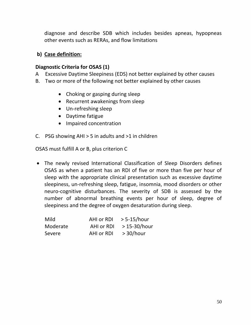
diagnose and describe SDB which includes besides apneas, hypopneas other events such as RERAs, and flow limitations
b) Case definition:
Diagnostic Criteria for OSAS (1) A Excessive Daytime Sleepiness (EDS) not better explained by other causes B. Two or more of the following not better explained by other causes
Choking or gasping during sleep
Recurrent awakenings from sleep
Un-refreshing sleep
Daytime fatigue
Impaired concentration
C. PSG showing AHI > 5 in adults and >1 in children
OSAS must fulfill A or B, plus criterion C
The newly revised International Classification of Sleep Disorders defines OSAS as when a patient has an RDI of five or more than five per hour of sleep with the appropriate clinical presentation such as excessive daytime sleepiness, un-refreshing sleep, fatigue, insomnia, mood disorders or other neuro-cognitive disturbances. The severity of SDB is assessed by the number of abnormal breathing events per hour of sleep, degree of sleepiness and the degree of oxygen desaturation during sleep.
Mild AHI or RDI > 5-15/hour Moderate AHI or RDI > 15-30/hour Severe AHI or RDI > 30/hour

51
Severity Grading of OSAS (criteria for children) (2)
AHI or RDI /hour
Adults Children
Mild: > 5-15 2-4
Moderate: >15-30 5-9
Severe: > 30 > 10
II. INCIDENCE OF THE CONDITION IN OUR COUNTRY The prevalence of SDB varies from region to region. It has been found to be present in 4% of men and 2% of women. In India various studies have shown the prevalence of SDB of 2.6% to 4.9% in adult males and 1% to 3.6% in adult females. About 4.8% children and 10.3% of elderly population in India have SDB. No gender difference in the prevalence of SDB has been observed in children and elderly subjects. The factors associated with the pathogenesis of SDB include anatomically small airway, obesity, and loss of upper airway motor tone during sleep and dysfunction of central respiratory control. The various risk factors are body habitus (especially percentage of predicted neck circumference), obesity, genetics, craniofacial morphology, chronic rhino-sinusitis with nasal obstruction, smoking and alcohol consumption. III. DIFFERENTIAL DIAGNOSIS
Narcolepsy
insomnia
Sleep hygiene disorder
Depression
Drug effect
OHS
Habitual snorers
Restless leg syndrome
IV. CLINICAL FEATURES

52
The night-time symptoms of SDB include:
Loud, disruptive snoring
Nocturnal pauses in breathing
Gasping or choking for air during sleep
Restless sleep
Nocturia
The daytime symptoms of SDB include: Feeling of excessive daytime sleepiness
Grogginess and morning headache
Memory or leaning problems
Not being able to concentrate at work
Depression and irritability
Sexual dysfunction
Dry throat upon awaking
Frequent nocturnal enuresis The symptoms seen in women include:
Report typical symptoms less frequently
Under-report snoring and underestimate symptom severity as compared to men
Non-specific somatic complaints such as insomnia, fatigue, myalgias, and morning headache
Amenorrhea and dysmenorrhea
Depression, anxiety and social isolation
Women tend to be more obese than men for the same degree of severity PRESENTATION IN CHILDREN Nocturnal symptoms in children include
Snoring
Restless sleep
Mouth breathing
Struggle to breathe/ gasp for air
Bed wetting
Daytime symptoms in children include

53
Feels depressed, sad or irritable
Sleepy during the day o Feels fatigued or tired during daytime o Excess activity, short attention span o Learning difficulties o Falling school performance o Difficulty to concentrate and memory lapses o Enlarged tonsils and adenoids
Consequences of SDB are as follows: Physiological Cardiovascular – Metabolic syndrome, hypertension, cerebrovascular
accidents, coronary artery disease Alterations in inflammatory biomarkers
Behavioral Tiredness in the morning EDS in permissive situation Increased risk of motor vehicle accidents Losses of concentration and productivity Epworth Sleepiness Score & psychomotor vigilance test are abnormal Behavioral effects can be variable amongst patients Behavioral measurement alone are insufficient
Social effects Principally related to snoring Disrupts social harmony by disturbance of bed-partner and others in
a home Falling asleep at social gatherings Subjective measures are commonly used Bed-partner scoring
V. PREVENTION AND COUNSELING
Risk factor reduction:
Weight reduction
Avoid alcohol and cigarette smoking
Treatment of nasal congestion

54
Surgical treatment of tonsillar and adenoid hypertrophy
Good sleep hygiene
Awareness about sleep disorders
Diagnosis and timely treatment of hypothyroidism VI. OPTIMAL DIAGNOSTIC CRITERIA, INVESTIGATIONS,
TREATMENT & REFERRAL CRITERIA *Situation 1: At Secondary Hospital/ Non-Metro situation: Optimal Standards of Treatment in Situations where technology and resources are limited
s) Clinical Diagnosis:
The diagnosis of SDB depends on properly taken history from the patient and the bed-partner regarding the typical daytime and night-time symptoms. Co-morbid conditions like hypertension, diabetes and other metabolic and hormonal problems. Good physical examination taking into account body mass index (BMI), neck length, percentage of predicted neck circumference (PPNC), cranio-facial anomalies like micrognathia and retrognathia, and evaluation of the upper airway using clinical examination scores, eg. Friedman score, X-ray cephalometry, CT scan, MRI, and videoendoscopy. For the localization of the site of the obstruction multi-channel pressure measurement has also been used with some success. The gold standard test for the diagnosis and severity assessment of SDB is clinical polysomnography (PSG); however, there are many practical considerations and clinical caveats in PSG interpretation in sleep related sleep disorders. History should include any history of sleep problem, sleep diary, medication history, medical problems, work history-shift work, alcohol or drug use, periodic limb movement disorders, dream history and teeth grinding. Physical examination should include measurement of body mass index (BMI; kg/m2), neck circumference and waist/hip ratio, examination of nose, oral

55
cavity, oropharynx, bony structure of head and neck. Sleep endoscopy is rarely needed to confirm airway collapse.
t) Investigations:
Thyroid function tests
Blood sugar (fasting and post-prandial)
Serum insulin (fasting and post-prandial)
HbA1c
Lipid profile
ECG and echocardiography
Pulmonary function testing (if overlap with chronic obstructive airway disease is suspected)
Arterial blood gases [if overlap with chronic obstructive airway disease or obesity-hypoventilation syndrome (OHS) is suspected]
Polysomnography o Full-night diagnostic study o Split-night with CPAP titration o Home study o Multiple Sleep Latency Testing (MSLT) if narcolepsy is
suspected Requirement for standard diagnostic nocturnal polysomnography (PSG) includes recording and analysis of the following parameters: EEG, (EOG), (EMG), oro-nasal airflow, chest wall effort, body position, snore microphone, ECG, and oxyhemoglobin saturation. The duration of a diagnostic NPSG is at least 6 h with the exception of the diagnostic portion of a split-night study, which is at least 2 h in duration. The various levels (types) of PSG are as follows
Type 1- gold standard, in-lab, attended PSG Type 2- Comprehensive portable monitoring, unattended Type 3- cardio-pulmonary sleep study or modified portable sleep apnea testing
Type 2 and Type 3 are acceptable in attended setting for patients without co-morbidity when scored by a trained physician Type 3 underestimates the severity of SDB Type 4- continuous single or dual bio-parameter recording -underestimates
severity

56
Indications of polysomnography are as follows:
For the diagnosis of SRBD and CPAP titration
Symptomatic patients with negative portable studies.
Full night PSG is required for CPAP titration of documented case of SDB; RDI > 15/hour or RDI > 5 with EDS
Evaluation of narcolepsy, and parasomnias
Before and after the application of dental appliances to assess the therapeutic benefits
Before and after upper airway surgery for snoring and SDB
After significant (10%) weight gain or weight loss
Patients with specific medical condition o Coronary artery disease o Congestive heart failure o History of stroke o Treatment of transient ischemic attack (TIA) o Significant tachy- or brady- arrhythmias
Indications for split-night study include:
AHI > 40/hour of at least 2 hours of sleep.
AHI between 20 and 40 in the presence of repetitive lengthy obstructions and major de-saturations.
CPAP titration must be carried out for more than 3 hours.
PSG documentation of abrogation of all events with CPAP.
u) Treatment: Treatment decision is based on evaluation of the patient’s history, anatomy, disease severity, symptoms and presence of co-morbidities. The severity of disease depends on
Severity of symptoms
Apnea hypopnea index (AHI)
Degree of oxygen desaturation o Minimum O2 desaturation o Sleep time spent below 90% oxygen saturation

57
Effect on sleep architecture o Presence or absence of REM and Delta sleep
Respiratory distress index (RDI) o Includes arousals with apneas and hypopneas
All modes of treatments should be discussed and these include behavioral treatments, use of oral appliances, positive airway pressure (PAP) therapy and surgical therapy while providing real information about success with each mode of treatment.
The behavioral modifications
Can reduce or eliminate OSA without the need for medication, device use or surgery.
o Sleep hygiene counseling o Sleep position therapy o Lifestyle modification and weight reduction o Avoidance of sedatives and alcohol o Smoking cessation o Avoidance of large meals before bedtime
Continuous positive airway pressure (CPAP) is the gold standard for treatment of OSAS. It acts as a pneumatic splint to keep airway patent. The benefits of CPAP include the following.
Minimally invasive and reversible
Reduction and/or reversal of OSA-related signs and symptoms Like snoring, excessive daytime sleepiness, un-refreshing or fragmented sleep, cognitive impairment
Decrease risk of accidents
Increased productivity
Decrease long-term complications of OSA like hypertension, heart disease, stroke, and death
Common adverse events of CPAP are as follows:
Minor (30-50% of Patients)
Inconvenience

58
Skin ulceration due to mask Poor mask fit Claustrophobia Various nasal problems (15-45%)
Major
Massive epistaxis Pneumocephalus – following recent surgery or trauma causing CSF leak.
Guidelines for titration
Patient should be evaluated for awake respiratory failure, congestive cardiac failure, and marked nocturnal hypoxemia.
CPAP titration should ideally be done in the hospital under supervision of a trained technician.
Patient should receive adequate PAP education, hands on demonstration, careful mask fitting and acclimatization
CPAP should be increased till all the obstructive events: apneas, hypopneas, RERAs, snoring and desaturation dissapear.
Recommended minimum pressure is 4 cm and maximum of 15 cm (<12 yrs) and 20 cm H2O (> 12 yrs)
CPAP pressure of 1 cm H2O should be increased with an interval no shorter than 5 min.
CPAP pressure should be increased by 1 cm H2O o 1 apnea (<12 yrs) or 2 apneas (> 12 yrs) o 1 hypopnea (<12 yrs) 3 hypopneas (> 12 yrs) o 3 RERAs (<12 yrs) 5 RERAs (> 12 yrs)
Use of BiPAP is recommended if CPAP titration pressure is > 15 cm H2O
Titration algorithm is same for split-night study Titration in patients with cardio respiratory failure
Requires close supervision by trained technician or physician while commencing CPAP.
Many patients might require endotracheal intubation or urgent tracheostomy.
The patient may become confused or delirious due to deranged blood gases.

59
In trained hands, judicious use of CPAP or BiPAP can control breathing disturbances.
Auto CPAP should not be used CPAP adherence
No clear field standard exist o Too few studies to define amount of adherence needed to treat
common sequelae
Average patient uses CPAP about 5 hours per night
Most clinicians generally recommend CPAP use for more than 4-5 hours per night on ≥ 70% of all nights
Predictors of CPAP adherence
Early use patterns predict long-term adherence o Patients appear to establish their patterns of use by the first
month (as early as 4 days) o Adherence at 1 month appears to predict adherence at 3
months o Since adherence is established by 3 months, alternative forms
of therapy should be considered for non-adherent patients Factors affecting CPAP adherence
Patient related factors:
Lesser severity of symptoms
Little or no perceived benefit from therapy
Failure to understand importance of or directions for CPAP use
Use of prescription/non-prescription drugs or alcohol
Lack of social support
Other medical illnesses or fatigue
Physical limitations (i.e. vision, hearing, hand coordination)
II. Therapy related factors:
Complexity of therapy/device use
Increased rate of adverse reactions that go unaddressed
Lack of efficacy
Expense of therapy
Chronic nature of illness

60
Compliance decreases over time III. Clinician related factors
Poor relationship with patient
Lack of clinician follow-up
Expression of doubt concerning therapeutic potential or creating falsely elevated expectations
Poor communication skills
Failure to identify concomitant use of alcohol and sedatives by the patient
Measures to improve CPAP adherence: Technological interventions:
Auto-titrating CPAP
Pressure delivery is auto-adjusted during changing airway conditions overnight
Bi-level positive pressure (BiPAP)
Set inhalation pressure with a lower set exhalation pressure Flexible pressure delivery (C-Flex, EPR)
Slight reduction in pressure during early exhalation Heated humidification
Decreases nasal/oral dryness for comfort
Improves nasal resistance Pressure ramp feature
CPAP starts at lower pressure (2-4 cmH2O) and gradually ramps up to prescribed pressure over set time period (5-45 min)
Behavioral interventions:
Patient education
Systematic desensitization and sensory awareness (for claustrophobia) o Wearing device for progressively longer periods
Cognitive behavioral therapy/Motivational Enhancement Therapy Reducing side effects:
PROBLEM SOLUTIONS
Skin breakdown/irritation • Refit mask • Try nasal pillows
Eye irritation • Refit mask • Saline eye drops

61
• Eye mask
Mouth leaks • Chin strap • Switch masks
Mask leaks/discomfort • Refit or switch masks
Nasal congestion • Heated humidification • Nasal saline sprays • Nasal steroids ± decongestants ± antihistamines • Correct any anatomic obstruction
Epistaxis • Heated humidifiers and saline
Rhinorrhea • Heated humidifier, nasal saline ± antihistamines
Aerophagia • Reduce CPAP pressure • Consider APAP, BiPAP
Claustrophobia • Switch to nasal pillows • Desensitization
Difficulty in exhalation • Ramp feature • Consider switching to APAP, BiPAP, flexible PAP • Consider re-titration
Treatment with oro-dental appliances: useful for mild to moderate cases of OSAS who are not tolerant/ willing to use CPAP treatment
Improve upper airway baseline dimensions o Pulling tongue forward – tongue retaining device o Moving mandible forward – nocturnal airway patient device
(NAPA)
Effectiveness of oral appliances o Significantly reduce or completely eliminate snoring o Significantly reduce AHI o Subjective improvement in daytime function but objective
evidence of improvement in sleepiness lacking o Adverse events include excessive salivation, temporo-mandibular
joint arthritis, and overall lower compliance (50%)

62
Patient selection for oro-dental appliances:
Factors associated with better response o Younger age o Lower BMI o Lower AHI o Good protrusive range o Presence of adequate healthy denture
However, some studies have demonstrated success even in patients with more severe OSA
Surgical treatment for OSAS
Patient selection for surgery depends on o OSA severity o Individual patient anatomy o Patients option for surgery o Presence of co-morbidities
Procedures used for localization of the site of obstruction o X-ray cephalometry, CT and MR imaging, video endoscopy under
sedation and multi channel pressure measurements.
Points of discussion while considering surgical option o Patient’s expectations and preferences o Patient’s expectation regarding cure of their snoring, apnea and
symptoms of tiredness. o Presence of co-morbidities
o Type of surgery o Pain and complication rates o Expected consequences o Morbidities o Cost o Need for evaluation following surgical procedure and possible need
for subsequent surgeries

63
Procedure on the upper airway, which may improve PAP use and compliance.
o Adeno-tonsillectomy o Nasal surgery for hypertrophied turbinate, deviated nasal septum or
nasal polyps.
Surgery that improves OSA without surgically altering the upper airway o Tracheostomy o Bariatric surgery
Surgery that directly alters the upper airway
Tracheostomy o Done on a temporary basis in the pre/peri-operative period o Not done routinely because of its associated hygiene and social
burden o Typically reserved for morbidly obese patients who can’t tolerate
PAP o Morbidly obese patients with OHS who require upper airway bypass
and nocturnal ventilation o Patients who are unable to tolerate PAP with significant co-morbidity
Bariatric surgery
Bariatric surgery has a potential to reduce body weight significantly as a significant number of OSA patients are obese.
Indications
BMI > 40
BMI > 35 with significant co-morbidities
Has the advantage of treating other co-morbidities associated with obesity
Surgery that alters the upper airway
Uvulopalatopharyngoplasty (UPPP)
Tongue advancement
Hyoid suspension
Maxilo-mandibular advancement (MMA)

64
ALGORITHM FOR TREATMENT OF OSA & TREATMENT GOALS
Mild (AHI >5-15) No symptoms Behavioral modification Symptoms Behavioral modification
Consider oral appliance Consider PAP Consider surgical intervention
Moderate (AHI >15-30) No symptoms Behavioral modification
Consider PAP Consider oral appliance Consider surgical intervention
Symptoms Behavioral modification PAP Surgical intervention for PAP failures
Consider oral appliance Severe (AHI>30) Symptoms or no symptoms Behavioral modification (rarely sufficient alone) PAP Surgical intervention for PAP failures
Consider tracheostomy if other treatments fail and significant symptoms or co-morbidities exist
Co-morbidities should also be taken into account when discussing the strength of recommendations
Patients with BMI >35 and co-morbidities or BMI>40 should be considered for bariatric surgery
Interventions should typically be applied in the order listed. Proceed down the list until success is reached.

65
Follow up Follow-up after medical and surgical intervention is critical to ensure that the patient has benefited from the chosen treatment and to provide an opportunity to implement further recommendations in case partial treatment response. v) Referral criteria:
Patient should be referred when following are present:
Severe OSAS/OHS having cardio-respiratory failure
Co-morbidities
Overlap syndrome (COPD or ILD with OSAS)
Poor compliance to CPAP treatment
Severe, intractable adverse events
Refusal for CPAP therapy
Consideration for oro-dental appliances and surgery
Requiring advanced investigations like nasal endoscopy, CT or MRI etc
Patients with complex sleep apnea
*Situation 2: At Super Specialty Facility in Metro location where higher-end technology is available
p) Clinical Diagnosis: Same as mentioned in Situation 1
q) Investigations:
Same as mentioned in Situation 1
Level I polysomnography Patients requiring advanced investigations like nasal endoscopy, CT or MRI etc
r) Treatment:
Titration and treatment of patients with severe cardio-respiratory failure Treatment and counseling of patients with poor CPAP adherence Oro-dental appliances and surgical treatment
Standard Operating procedure
66
As mentioned above
VII. WHO DOES WHAT? and TIMELINES
a. Doctor Diagnosis and Management including counseling b. Nurse Implementation of orders, monitoring of patients and
counseling c. Technician Investigations
VIII. FURTHER READING / REFERENCES 1. Kushida CA, Littner MR, Morgenthaler T, Alessi CA, Bailey D, Coleman J Jr, Friedman L,
Hirshkowitz M, Kapen S, Kramer M, Lee-Chiong T, Loube DL, Owens J, Pancer JP, Wise M. Practice parameters for the indications for polysomnography and related procedures: an update for 2005. Sleep. 2005 Apr;28(4):499-521
2. American Academy of Sleep Medicine. Sleep-related breathing disorders in adults: recommendations for syndrome definition and measurement techniques in clinical research. The report of an American Academy of Sleep Medicine task force. Sleep 1999; 22: 667-8

67
RESOURCES REQUIRED FOR ONE PATIENT
Situation HUMAN RESOURCES INVESTIGATIONS DRUGS & CONSUMABLES
EQUIPMENT
1 1. Physician with sleep medicine training
2. Nurse 3. Radiographer 4. Laboratory
technician 5. Biochemist 6. Sleep laboratory
technician 7. Cardiologist/
Physician with echocardiography training
1. Pulmonary function test
2. Arterial blood gas 3. Thyroid function
test 4. Blood sugar 5. Lipid profile 6. Electrocardiogram 7. Serum insulin 8. Echocardiography 9. Polysomnography
1. Tongue retaining devices
2. Nocturnal airway patient device
3. CPAP machine with humidifier
1. Polysomnography machine
2. Pulmonary function test machine
3. Pathology laboratory
4. Biochemistry laboratory
5. Echocardiography machine
6. ABG analyzer 7. Body composition
analyser
2 Above plus 1. Radiologist 2. ENT Surgeon 3. Anesthetist 4. Bariatric surgeon
Above plus 9. X-ray
cephalometry 10. Nasal/oral video
endoscopy 11. CT scan 12. MRI scan
As above Above plus 1. CT scan machine 2. MRI machine 3. Nasal/oral video
endoscope 4. Operation theatre 5. DEXA scan

68
PLEURAL DISEASES
I. WHEN TO SUSPECT/ RECOGNIZE?
i) Introduction:
There are 4 types of pleural diseases: pleurisy, pleural effusion, pneumothorax and pleural mesothelioma. Pleural mesothelioma is a malignant condition of pleura related to asbestos exposure occurring rather infrequently in our country compared to other lung cancers and is beyond the purview of this review.
j) Case definition: For both situations of care: 1. Pleurisy is defined as an acute inflammation of the pleura. The inflammation
can be primary or secondary due to spread from an adjacent structure. 1.1. Primarily, viral infections of the pleura result in pleurisy. Secondarily, the
pleura may be involved due to pneumonia or pulmonary infarction. The present review is restricted to ‘primary’ pleurisy.
2. Pleural effusion is defined as an excessive accumulation of fluid in the lungs.
There are mainly two types of pleural effusions: transudative and exudative pleural effusion. 2.1. Fundamentally, transudative pleural effusions reflect a systemic pathology
and exudative pleural effusions reflect a local pleural pathology. Generally, transudative effusions are usually bilateral, whereas exudative effusions are usually unilateral.
2.2. The common causes of transudative pleural effusions are congestive heart failure and hypoalbuminemic states (cirrhosis, nephrotic syndrome).
2.3. The common causes of exudative pleural effusions are tuberculosis, malignancy and traumatic hemothorax. Uncommon causes include pancreatitis, connective tissue diseases, non-traumatic hemothorax (embolism, malignancy) and chylothorax (tumors, filariasis)
3. Pneumothorax is defined as air in the pleural cavity resulting in collapse of the
whole lung or some part of the lung. It may be traumatic or spontaneous. 3.1. Traumatic pneumothorax can result in a life-threatening condition due to
tension pneumothorax.
69
3.2. Spontaneous pneumothorax may occur without any associated underlying lung pathology (primary) or may be associated with underlying lung pathology (secondary).
3.2.1. The common secondary causes include tuberculosis and chronic obstructive airway disease. Smoking and marfanoid habitus are important risk factors. Secondary pneumothorax is a more critical condition as the underlying lung is already compromised.
II. INCIDENCE OF THE CONDITION IN OUR COUNTRY No systematic robust epidemiological studies are available from India
III. DIFFERENTIAL DIAGNOSIS 1. Pleurisy
Cardiac: post-myocardial injury syndrome (Dressler syndrome)
Renal: chronic renal failure
2. Pleural effusion
Amebic liver abscess
Lower lobe collapse
Mass lesion
3. Pneumothorax
Pulmonary embolism
Acute coronary syndrome
Large bulla
IV. PREVENTION AND COUNSELING Optimal treatment of primary systemic illness. Abstinence from smoking, alcohol and other substance abuse. V. OPTIMAL DIAGNOSTIC CRITERIA, INVESTIGATIONS,
TREATMENT & REFERRAL CRITERIA *Situation 1: At Secondary Hospital: Optimal Standards of Treatment in Situations where technology and resources are limited

70
w) Clinical Diagnosis
Symptoms
1. Pleurisy: sharp pain in the lateral hemithorax characteristically increasing on inspiration and coughing.
2. Pleural effusion: may be asymptomatic or associated with dull aching pain on the affected site. Dyspnea, dry cough may also be present. Symptoms of underlying systemic pathology may be associated.
3. Pneumothorax: sudden onset excruciating pain associated with dyspnea especially in a smoker.
Signs
1. Pleurisy: acute history of fever, pleural rub. 2. Pleural effusion: findings of primary systemic disease, local
examination reveals decreased tactile fremitus, stony dull note on percussion and absent breath sounds on auscultation.
3. Pneumothorax: marfanoid habitus, local examination reveals decreased tactile fremitus, tympanitic note on percussion and absent breath sounds on auscultation.
x) Investigations
1. Pleurisy: No specific investigation per se. Patients with pleurisy normally
improve in 7-10 days. 2. Pleural effusion:
2.1 Chest X-ray (PA and lateral views of chest radiographs): 50 ml fluid can be appreciated on lateral chest radiograph as a meniscus in the posterior costophrenic angle. Lateral decubitus film helps in differentiating a loculated effusion from a free fluid effusion.
2.2 Ultrasound: USG helps in confirming a pleural effusion as compared to pleural thickening, which many times cannot be appreciated on a chest radiograph. It is also useful in diagnosing loculated effusions, subpulmonic and subdiaphragmtic pathology like an amebic liver abscess.
2.3 Pleural fluid (PF) evaluation

71
2.3.1 Diagnostic thoracocentesis performed with/without US guidance is the investigation of choice to differentiate between a transudative and exudative pleural effusion. This differentiation is the sheet anchor of further management.
2.3.2 Colour 2.3.2.1 Pale yellow: transudative 2.3.2.2 Turbid : exudative 2.3.2.3 Frank pus : empyema 2.3.2.4 Blood-like : traumatic tap, hemothorax 2.3.2.5 Milky : chylothorax
2.3.3 Biochemistry {Protein and lactate dehydrogenase (LDH)} Light’s criteria have a high (> 98%) sensitivity and specificity to classify PF. There are 3 criteria as follows: a. PF protein/serum protein> 0.5 b. PF LDH/ serum LDH> 0.6 c. PF LDH> 2/3 upper limit serum LDH Exudative PF: any one of 3 criteria present Transudative: even if one of the 3 criteria are absent
2.3.4 pH: normal PF pH is 7.64. 2.3.5 Cell cytology for polymorphonuclear or lymphocytic predominance 2.3.6 Malignant cytology (if available at secondary level) 2.3.7 Bacteriology: Gram stain and culture sensitivity in addition to pH are
very important investigations to decide regarding the management in cases of parapneumonic effusion vis-à-vis empyema.
2.3.8 Adenosine deaminase (ADA) levels (if available at the secondary level) Higher ADA levels with lymphocytosis point to a diagnosis of tubercular pleural effusion
2.3.9 Hematocrit (Hct): PF hct > 50% blood Hct signifies hemothorax differentiating from a traumatic thoracocentesis
3. Pneumothorax 3.1. Chest radiograph: collapsed lung at the hilum with absence of lung
markings in the periphery. 3.2. Important to evaluate the contralateral lung radiographically-normal or
abnormal for management.
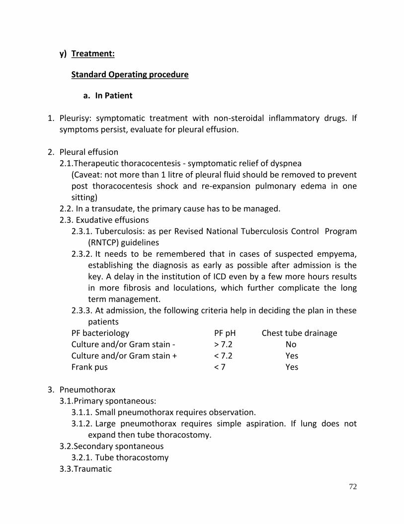
72
y) Treatment:
Standard Operating procedure
a. In Patient
1. Pleurisy: symptomatic treatment with non-steroidal inflammatory drugs. If symptoms persist, evaluate for pleural effusion.
2. Pleural effusion 2.1. Therapeutic thoracocentesis - symptomatic relief of dyspnea
(Caveat: not more than 1 litre of pleural fluid should be removed to prevent post thoracocentesis shock and re-expansion pulmonary edema in one sitting)
2.2. In a transudate, the primary cause has to be managed. 2.3. Exudative effusions
2.3.1. Tuberculosis: as per Revised National Tuberculosis Control Program (RNTCP) guidelines
2.3.2. It needs to be remembered that in cases of suspected empyema, establishing the diagnosis as early as possible after admission is the key. A delay in the institution of ICD even by a few more hours results in more fibrosis and loculations, which further complicate the long term management.
2.3.3. At admission, the following criteria help in deciding the plan in these patients
PF bacteriology PF pH Chest tube drainage Culture and/or Gram stain - > 7.2 No Culture and/or Gram stain + < 7.2 Yes Frank pus < 7 Yes
3. Pneumothorax
3.1. Primary spontaneous: 3.1.1. Small pneumothorax requires observation. 3.1.2. Large pneumothorax requires simple aspiration. If lung does not
expand then tube thoracostomy. 3.2. Secondary spontaneous
3.2.1. Tube thoracostomy 3.3. Traumatic

73
3.3.1. Depends on size- simple observation to thoracostomy 3.4. Tension
3.4.1. Medical emergency 3.4.2. Large bore needle in second intercostal space. If air gushes out, tube
thoracostomy
b. Out Patient Treatment of primary systemic illness
z) Referral criteria:
1. Diagnosis not clear 2. Response to therapy not optimal 3. Recurrent pneumothorax 4. Persistent air leak 5. Unexpanded lung after 3 days of tube thoracostomy
*Situation 2: At Tertiary hospital where higher-end technology is available
s) Clinical Diagnosis
As in Situation V.1 a) above
t) Investigations
10. Pleurisy: As in situation V.1b) above
11. Pleural effusion: As in situation V.1b)2. above PLUS 11.1. CT scan to accurately define the anatomy of lung parenchyma (lung
abscess) and pleura (empyema by the split pleura sign, loculated effusion, pleural masses)
11.2. Special investigations 11.2.1. PF triglyceride > 110 mg/dl for chylothorax 11.2.2. PF antinuclear factor for systemic lupus erythematosus (SLE)
[specific but not very sensitive]

74
11.2.3. PF Rheumatoid factor and anti-citrullinated protein antibody (anti-CCP)
11.2.4. PF amylase for pancreatitis 11.3. Pleural biopsy 11.4. Medical thoracoscopy and video-assisted thoracoscopy (also has
therapeutic potential)
12. Pneumothorax: as in situation V.1b)3. PLUS 12.1. Computed tomography to assess for blebs and associated pulmonary
disease
u) Treatment: Standard Operating procedure
a. In Patient
3. Pleurisy: As in situation V.1c)a.1 above
4. Pleural effusion: As in situation V.1c)a.2 above PLUS 4.1. Pleurodesis with doxycycline – recurrent malignant pleural effusion 4.2. Chest tube instilled fibrinolytic therapy (streptokinase) - parapneumonic
effusions 4.3. VATS (thoracotomy, if VATS not available)- non-resolving empyema
4.4. Pneumothorax as in V.1c)a.3 above PLUS
4.4.1. VATS with pleurodesis/bleb resection
b. Out Patient
Treatment of primary systemic illness VI. WHO DOES WHAT? and TIMELINES
a. Doctor: Diagnosis and Management including counseling b. Nurse: Implementation of orders, monitoring of patients and counseling c. Technician: Investigations

75
VII. FURTHER READING / REFERENCES
1. Light RW, MacGregor MI, Luchsinger PC, et al. Pleural effusions: The diagnostic separation of transudates and exudates. Ann Intern Med 1972;77:507-13 2. Colice GL, Curtis A, Deslauriers J, et al. Medical and surgical treatment of parapneumonic effusions: An evidence-based guideline [ACCP consensus statement]. Chest 2000;118:1158-71 5. Baumann, MH, Strange, C, Heffner, JE, et al. Management of spontaneous
pneumothorax: an American College of Chest Physicians Delphi consensus statement. Chest 2001;119:590-602
6. Seaton A. Diseases of the Pleura. In: Seaton A, Seaton D and Leitch AG
(editors). Crofton and Douglas's Respiratory Diseases, Fifth Edition. Oxford, UK: Blackwell Science Ltd. 2008.p.1152-81
7. Seaton D. Pneumothorax. In: Seaton A, Seaton D and Leitch AG (editors).
Crofton and Douglas's Respiratory Diseases, Fifth Edition. Oxford, UK: Blackwell Science Ltd. 2008.p.1182-211

76
RESOURCES REQUIRED FOR ONE PATIENT
Situation HUMAN RESOURCES
INVESTIGATIONS DRUGS & CONSUMABLES
EQUIPMENT
1 8. Physician 9. Surgeon 10. Nurse 11. Radiographer 12. Laboratory
technician
10. Chest radiograph 11. Pleural fluid
examination (total and differential cell count, malignant cytology, Mtb smear and culture, PCR, fungal smear and culture, protein, LDH, glucose, ADA)
12. USG chest and abdomen
13. Arterial blood gas analysis
14. Echocardiography 15. Blood
biochemistry
3. Anti-tuberculosis therapy (DOTS)
4. Intercostal tube
4. X-ray machine 5. USG machine 6. Cytopathology
and histopathology laboratories
7. Microbiology laboratory service
8. ABG analyzer 9. ECHO machine
2 Above plus 14. Radiologist 15. Pathologist 16. Thoracic
surgeon 17. Anesthetist 18. Nursing staff
trained in assisting thoracic surgery
Above plus 13. Malignant
cytology 14. Pleural fluid
triglyceride 15. Pleural fluid
antinuclear antibody, anti-CCP antibody
16. Markers for connective tissue diseases
17. Pleural Biopsy 18. Video-assisted
thoracoscopy 19. CT Scan
Above plus 11. Streptokinase 12. Doxycycline
Above plus 6. CT scan machine 7. Pleural biopsy
needle 8. Immunology
laboratory services
9. Video-assisted thoracoscope
10. Thoracic surgery operating theatre; recovery room and ICU

77
COMMUNITY ACQUIRED PNEUMONIA (Bacterial)
I. WHEN TO SUSPECT/ RECOGNIZE?
k) Introduction: Pneumonia, an infection of the pulmonary parenchyma is a commonly encountered, potentially life-threatening condition in clinical practice. In light of the recent pandemics of multidrug-resistant bacterial infections world-over, pneumonia has been categorized as either community-acquired pneumonia (CAP) or health care-associated pneumonia (HCAP). The subcategories of HCAP include hospital-acquired pneumonia (HAP) and ventilator-associated pneumonia (VAP). In spite of the availability of potent new antimicrobials, CAP is associated with considerable morbidity and mortality, particularly in elderly patients and those with significant co-morbid illnesses. l) Case definition: For both situations of care:
1. Symptoms and signs consistent with an acute lower respiratory tract infection (productive cough, rusty sputum, pleuritic chest pain, etc.); presence of at least one systemic manifestations (either a symptom complex of sweating, fever, shivers, aches and pains and/or temperature of >38 oC or more). 2. New focal signs on physical examination of the chest. 3. New chest radiograph evidence of pneumonic consolidation which is at least segmental or present in more than one lobe and is not known to be previously present for which there is no other explanation. 4. Confirmation of aetiological diagnosis by blood culture/sputum culture and/or molecular diagnostic methods.
II. INCIDENCE OF THE CONDITION IN OUR COUNTRY Estimates form the developed world suggests that CAP occurs in 8 to 15 per 1000 persons per year; the highest rates are at the extremes of age. No systematic studies have been carried out till date to define the incidence of CAP in India and reliable epidemiological data are lacking. III. DIFFERENTIAL DIAGNOSIS

78
1. Common
Pulmonary embolism/infarction
Pulmonary oedema
Bronchogenic carcinoma
Pulmonary tuberculosis
Bronchiectasis 2. Uncommon
Pulmonary eosinophilia/eosinophilic pneumonia
Cryptogenic organising pneumonia
Pulmonary alveolar haemorrhage
Foreign body
Congenital pulmonary abnormality (eg. lobar sequestration)
Radiation pneumonitis
IV. PREVENTION AND COUNSELING 1. Persons hospitalized with CAP who smoke should be counseled regarding smoking cessation. 2. Respiratory hygiene measures, including the use of hand hygiene and masks or tissues for patients with cough, should be used in outpatient settings and casualty as a means to reduce the spread of respiratory infections V. OPTIMAL DIAGNOSTIC CRITERIA, INVESTIGATIONS,
TREATMENT & REFERRAL CRITERIA *Situation 1: At Secondary Hospital: Optimal Standards of Treatment in Situations where technology and resources are limited aa) Clinical Diagnosis
Symptoms The onset of CAP is acute. Patients present with symptoms suggestive of acute lower respiratory tract infection, cough, rusty sputum, pleuritic chest pain, and constitutional symptoms, such as, fevers, sweating, chills, rigors, body aches and pains; temperature of >38 oC or more. Signs

79
Patients with CAP look sick and toxic. New and focal signs of pneumonic consolidation (reduced movements and expansion, increased tactile vocal fremitus, dull note on percussion, tubular bronchial breathing, increased vocal resonance, aegophony and whispering petriloquy; pleural rub) are present on physical examination of the chest. Signs due to complications, such as, pleural effusion, empyema, sepsis syndrome may also be present.
bb) Investigations 1. Chest radiograph: A chest radiograph is the cornerstone to establishing the diagnosis of CAP or an alternative diagnosis, and to assess the extent of the disease and the presence of complications such as pleural effusion, lung abscess etc. 2. In all admitted patients:
2. 1. Pulse oximetry: pulse oximtery is useful for assessment of gas-exchange status. 2.2. Sputum Gram’s stain, culture; smear examination for acid-fast bacilli; blood culture. 2.3. Full haemogram. 2.4. Serum biochemistry (blood urea, serum creatinine, liver function tests).
3. Arterial blood gas (ABG) analysis: ABG analysis is helpful in detecting acute respiratory failure that may complicate the course of CAP.
4. Assessment of severity: This is best assessed by CURB-65 scoring system [confusion, urea >7mmol/L, respiratory rate >30/min, blood pressure (systolic <90 mmHg or diastolic <60 mmHg), Age >65yrs] and guides admission to ICU and antibiotics administration. Pneumonia Severity Index (PSI) a prognostic model to identify those with low risk of death.
cc) Treatment:
Standard Operating procedure a. In-Patient 1. General therapeutic measures
1. SpO2 monitoring by pulse oximetry. 2. Adequate circulation and blood pressure is ensured using volume infusion and/or vasopressors.

80
3. Decision to admit 3.1. The decision to admit patients with one or more adverse factors (confusion, respiratory rate > 30/min; systolic blood pressure <90 mmHg or diastolic blood pressure <60 mmHg; age more than 65 years) without severity factors (hypoxaemia, bilateral or multilobar involvement on chest radiograph) should be individualized. 3.2. Patients with one or more severity factors listed above need hospitalization 3.3. Admission into an ICU: for patients with septic shock requiring vasopressors or with acute respiratory failure requiring intubation and mechanical ventilation, direct admission into ICU will be required. Admission to an ICU is also recommended for patients with 3 of the following minor criteria for severe CAP: respiratory rate >30
breaths/min; PaO2/FIO2 ratio 250; multilobar infiltrates; confusion/ disorientation; uraemia (blood urea nitrogen level >20 mg/dL); leukopenia (total leukocyte count, < 4000 cells/mm3), thrombocytopenia (platelet count <100,000/mm3), hypothermia (core temperature <36oC); and hypotension requiring aggressive fluid resuscitation.
4. Antibiotic treatment of the aetiological cause of CAP
4.1 In-patient antibiotic therapy 4.1.1 In-patients, non-ICU treatment
When admitted, these patients are treated with intravenous beta- lactam plus macrolide (Table 1 ).
Table 1 Choice of antibiotics for in-patient treatment of CAP
Choice of drug Daily dosage Intravenous beta-lactam Macrolide First choice Cefotaxime 2 g q8h Azithromycin 500 mg qd Second choice Ceftriaxone 1 g q12h Third choice Co-amoxyclav 1.2 g q8h
4.2 Duration of antibiotic therapy is 7 to 10 days based on clinical response
81
4.3 A switch to oral therapy can be made at 48-72 hours as most patients with CAP will have an adequate clinical response within 3 days, which includes improvement in cough and dyspnoea, absence of fever, decreasing white blood cell count and functioning gastrointestinal tract with adequate oral intake. 4.4 Patient may be discharged on the same day if medical and social factors are favorable. 4.5. Pleuritic pain should be relieved using drugs such as paracetamol.
2. Supplemental oxygen therapy In patients with hypoxaemia, spontaneous ventilation using a face mask with a high flow gas delivery system can be used to deliver a FIO2 of up to 0.5 to 0.6. b. Out Patient
Out-patient antibiotic therapy
The choice of antibiotics for out-patient treatment of CAP is as follows. For previously healthy patients and those who have not used antimicrobials within the previous 3 months, treatment with oral macrolide (e.g., azihtromycin) is indicated. For patients with comorbidities such as chronic heart, lung, liver or renal disease, diabetes mellitus, alcoholism, malignancies, asplenia, immunosuppressing conditions or use of immunosuppressing drugs, or use of antimicrobials within the previous 3 months, and patients in regions with a high rate (>25%) of infection with
high-level [minimum inhibitory concentration (MIC) 16 mg/mL] macrolide-resistant Streptococcus pneumoniae, oral treatment with co-amoxyclav in combination with macrolide is recommended.
dd) Referral criteria:
1. Patients presenting with two or more adverse factors (confusion, respiratory rate> 30/min; systolic blood pressure <90 mmHg or diastolic blood pressure <60 mmHg; age more than 65 years) or one or more severity factor (hypoxaemia, bilateral or multilobar involvement on chest radiograph).

82
2. Diagnosis not clear. 3. Patients not responding to therapy
*Situation 2: At Tertiary hospital where higher-end technology is available
v) Clinical Diagnosis
As in situation V.1 a) above
w) Investigations
As in situation V.1b) above PLUS 13. Two blood cultures (minimum 20 ml) 14. Sputum or other respiratory tract specimen for Gram stain, bacterial culture
and sensitivity tests; molecular diagnostic methods (e.g., polymerase chain reaction for respiratory pathogens)
15. Pleural fluid, if present, for microscopy, culture and pneumococcal antigen detection.
16. Investigations for Legionella pneumonia: 4.1 Urine for Legionella antigen 4.2 Sputum or other respiratory tract specimens (for Legionella culture and direct immunofluorescence) (if available)
5. Investigations for atypical bacterial pathogens: 5.1 If available, sputum or other respiratory sample for PCR or direct immunofluorescence (or other antigen detection test) for Mycoplasma pneumoniae, Chlamydia spp, Pneumocystis jirovecii (if at risk)
6. Imaging studies: ultrasonography of the chest is useful for detecting complications such as pleural effusion, empyema. In non-responders, computed tomography of the chest can help in detecting an underlying malignancy and complications, such as, lung abscess.
7. Bronchoscopic procedures: should selectively be used in individual patients for aetiological confirmation and ruling out an alternative diagnosis, such as, malignancy.
x) Treatment:
Standard Operating procedure a. In Patient
As in situation V.1c)a.1 above PLUS

83
1. General therapeutic measures 1.1. Patients should be assessed for volume depletion and may require intravenous fluids. 1.2. Prophylaxis of venous thromboembolism with low molecular weight heparin should be considered for all patients who are not fully mobile. 1.3. Nutritional support should be given during prolonged illness. 1.4. Airway clearance techniques should be considered if the patient has sputum and difficulty in expectoration or in the event of a pre-existing lung condition.
2 Antibiotic therapy As described under V.4 above PLUS 2.1 In-patients, ICU treatment
A beta-lactam (cefotaxime, ceftriaxone, or ampicillin-sulbactam) plus azithromycin are recommended.
For penicillin-allergic patients, a respiratory fluoroquinolone and aztreonam are recommended.
If Pseudomonas is a consideration, an antipseudomonal beta-lactam (e.g., piperacillin tazobactam, cefepime, imipenem, or meropenem) plus either ciprofloxacin or levofloxacin (750 mg) are recommended. or The above beta-lactam plus an aminoglycoside and azithromycin or The above beta-lactam plus an aminoglycoside and an antipseudomonal fluoroquinolone are recommended.
For penicillin-allergic patients, aztreonam may be substituted for above beta-lactam)
Prior to use of respiratory fluoroquinolones, active pulmonary tuberculosis should be excluded.
If community acquired methicillin-resistant Staphylococcus aureus is a consideration, vancomycin or linezolid can be added.
2. Supplemental oxygen therapy All patients should receive appropriate oxygen therapy with monitoring of oxygen saturation and inspired oxygen concentration with the aim to maintain PaO2 at 60 mm Hg or above and SpO2 between 94%–98%.

84
2.1. High concentrations of oxygen can safely be given in patients who are not at risk of hypercapnic respiratory failure. 2.2. Oxygen therapy in patients at risk of hypercapnic respiratory failure complicated by ventilatory failure should be guided by repeated ABG measurements.
3. Ventilatory support Patients with CAP who develop acute respiratory failure require mechanical ventilation for maintaining gas exchange with minimal complications.
4. Management of complications 4.1 Parapneumonic effusion/empyema
4.1.1. Early thoracocentesis is indicated for all patients with a parapneumonic effusion. 4.1.2. Patients found to have an empyema should have effective pleural fluid drainage and appropriate antibiotic therapy. Streptokinase may be instilled if loculation(s) present.
4.2 Lung abscess This condition is described in detail in another chapter
b. Out Patient Not applicable.
VI. WHO DOES WHAT? and TIMELINES a. Doctor: diagnosis and management including counseling b. Nurse: implementation of orders, monitoring of patients and
counseling c. Technician: investigations
VII. FURTHER READING / REFERENCES 1. Lim WS, Baudouin SV, George RC, Hill AT, Jamieson C, Le Jeune I, Macfarlane JT, Read RC, Roberts HJ, Levy ML, Wani M, Woodhead MA; Pneumonia Guidelines Committee of the BTS Standards of Care Committee. BTS guidelines for the management of community acquired pneumonia in adults: update 2009. Thorax 2009;64 Suppl 3:iii1-55. 2. Mandell LA, Wunderink RG, Anzueto A, Bartlett JG, Campbell GD, Dean NC, et al; Infectious Diseases Society of America; American Thoracic Society. Infectious Diseases Society of America/American Thoracic Society consensus guidelines on the management of community-acquired pneumonia in adults. Clin Infect Dis 2007;44 Suppl 2:S27-72.

85
3. Postgraduate Institute of Medical Education and Research Chandigarh: practice guidelines for management of community and hospital-acquired pneumonias - a consensus statement. Lung India 2006;23:115-20.
RESOURCES REQUIRED FOR ONE PATIENT Situation HUMAN
RESOURCES INVESTIGATIONS DRUGS &
CONSUMABLES EQUIPMENT
1 1.Physician 2.Nurse 3.Radiographer 4. Laboratory technician
1.Chest X-ray 2. USG chest 3. Pulse oximetry 4. ABG 5. Serum biochemistry 6. Hemogram 7. Sputum smear and culture 8. Sputum for AFB 9. Blood culture 10. HIV ELISA for high risk group
1.Oral and i.v. antibiotics 2. Streptokinase
1. X-ray machine 2. USG machine 3. Pulse oximeter 4. ABG machine 5. Oxygen cylinders 6. Nebulizers 7. Microbiology laboratory services 8. Hematology and biochemistry laboratory services
2 Above plus 1.Intensivist 2.Nurse and technician for assisting with bronchsocopy 3.Nursing staff trained in ICU care 4.Pathologist
Above plus 1.CT chest 2.Bronchoscopy
Above plus 1.Oxygen supply 2.Tubes and other disposables for thoracocentesis (when indicated)
Above plus 1.CT machine 2.Bronchoscope and facilities for bronchoscopy 3.Clinical pathology laboratory service 4.Microbiology service with conventional, serological and molecular diagnostic facilities for aetiological diagnosis confirmation 5.ICU 6.Noninvasive and invasive ventilators

86
PULMONARY HYPERTENSION
I. WHEN TO SUSPECT/ RECOGNIZE?
a) Introduction:
The diagnosis of pulmonary hypertension requires strong clinical suspicion in practice. It is usually found secondary to various systemic causes, the list of which is long but the usual causes include the rheumatic heart disease, and diseases of the respiratory system, including the obstructive airway diseases and the diffuse parenchymal pulmonary diseases. Dilation and hypertrophy of the right ventricle develops in response to diseases of the pulmonary vasculature and/or lung parenchyma, which is known as cor pulmonale. Pulmonary hypertension may also occur as primary idiopathic disease due to the mutation of certain genes, such as BMPR-2.
b) Case definition: Secondary hospital: presence of a pulmonary artery systolic pressure more than 40 mmHg at rest, calculated by the tricuspid velocity jet by trans-thoracic echocardiography. Super-speciality hospital: presence of a mean pulmonary artery pressure (PAP) of more than 25 mmHg at rest, pulmonary capillary wedge pressure < 15 mmHg, accompanied by increased pulmonary vascular resistance (PVR) (more than 160 dynes/s/cm5), by right heart catheterisation. II. INCIDENCE OF THE CONDITION IN OUR COUNTRY No systematic studies till date to define the incidence of pulmonary hypertension in our country
III. DIFFERENTIAL DIAGNOSIS 1. Pulmonary artery hypertension (PAH)
1.1 Idiopathic 1.2 Heritable
1.2.1 BMPR2 (chromosome 2, autosomal dominant)

87
1.2.2 ALK1, endoglin (with or without hereditary haemorrhagic telangiectasia)
1.3 a) Drugs: aminorex, fenfluramine, dexfenfluramine, cocaine,
phenylpropanolamine, amphetamines, L- tryptophan, methamphetamines, chemotherapeutic agents, fluoxetine, pergolide, oral contraceptives, oestrogen
b) Toxins: toxic rapeseed oil
1.4 Associated with (APAH) 1.4.1 Connective tissue diseases 1.4.2 HIV infection 1.4.3 Portal hypertension 1.4.4 Congenital heart disease 1.4.5 Chronic haemolytic anaemia: sickle cell anaemia 1.4.6 Schistosomiasis
1* Pulmonary veno-occlusive disease and/or pulmonary capillary haemangiomatosis 2. Pulmonary hypertension due to left heart disease
2.1 Systolic dysfunction 2.2 Diastolic dysfunction 2.3 Valvular heart disease
3. Pulmonary hypertension due to lung diseases and/or hypoxia
3.1 Chronic obstructive pulmonary disease (COPD) 3.2 Diffuse parenchymal lung disease (DPLD) 3.3 Other pulmonary diseases with mixed restrictive and obstructive
pattern 3.4 Sleep related breathing disorders (SRBDs) 3.5 Alveolar hypoventilation disorders 3.6 Chronic exposure to high altitude 3.7 Developmental abnormalities

88
4. Chronic thrombo-embolic pulmonary hypertension 5. Pulmonary hypertension with unclear and/or multifactorial mechanisms
5.1 Haematological disorders: myeloproliferative disorders, splenectomy. 5.2 Systemic disorders: sarcoidosis, pulmonary Langerhans cell histiocytosis, lymphangioleiomyomatosis, neurofibromatosis, vasculitis 5.3 Metabolic disorders: glycogen storage disease, Gaucher’s disease,
thyroid disorders 5.4 Others: tumoural obstruction, fibrosing mediastinitis, chronic renal
failure on dialysis
IV. PREVENTION AND COUNSELING Early diagnosis by strong clinical suspicion in clinical settings, and appropriate treatment of the underlying cause.
V. OPTIMAL DIAGNOSTIC CRITERIA, INVESTIGATIONS, TREATMENT & REFERRAL CRITERIA
*Situation 1: At Secondary Hospital/ Non-Metro situation: Optimal Standards of Treatment in Situations where technology and resources are limited ee) Clinical Diagnosis:
Patient presents with dyspnoea on exertion, fatigue, chest pain, palpitations, syncope, and lower extremity oedema. Examination reveals raised jugular pressure, left parasternal lift, right ventricular S4 , accentuated P2, early systolic click, midsystolic ejection murmur or early diastolic murmur at pulmonary area (Graham Steell) ff) Investigations: To confirm pulmonary hypertension, and determining the underlying cause.
Electrocardiogram
Chest radiograph
Trans-thoracic 2-D Doppler echocardiography

89
Pulmonary function test
Arterial blood gas analysis gg) Treatment:
General measures
Avoid heavy physical exertion or isometric exercise
Sodium restricted diet (less than 2.5 g per day)
Annual immunization against influenza
Avoid pregnancy (female patients)
Anticoagulants : idiopathic/chronic thrombo-embolism
Diuretics
Long-term oxygen supplementation to maintain oxygen saturation > 90% Specific treatment
Treat the underlying cause of pulmonary hypertension
Pulmonary hypertension-specific agents Sildenafil*
*20 mg 24 hourly gradually increased upto 20 mg 8 hourly with careful blood pressure monitoring. The drug can be used in PAH secondary to connective tissue diseases, chronic pulmonary thromboembolism and idiopathic pulmonary artery hypertension.
hh) Referral criteria:
Diagnosis not confirmed with echocardiography and patient requires right heart catheterisation
Underlying cause not clear, before labelling as idiopathic pulmonary artery hypertension
Worsening clinical condition of the patient
Suspected thrombo-embolism *Situation 2: At Super Specialty Facility in Metro location where higher-end technology is available
y) Clinical Diagnosis: Same as level 1

90
z) Investigations: Same as level 1, in addition
CT pulmonary angiography (CTPA)
Ventilation-perfusion scan
Multi-detector CT chest
Overnight supervised polysomnography
Connective tissue disease work up : ANA/RF/ANCA/ anticentromere antibodies
HIV ELISA, CD4 count, plasma viral load (screening for pulmonary hypertension not indicated routinely in ART clinics)
Functional tests : 6-minute-walk test, cardio-pulmonary exercise test
Right heart catheterization with acute vasodilator testing
aa) Treatment: Same as level 1, in addition
Pulmonary hypertension-specific agents Calcium channel blockers (only in patients with positive acute
vasodilator test) Sildenafil* 20 mg 24 hourly gradually increased upto 20 mg 8 hourly
with blood pressure monitoring Bosentan* 62.5 mg 12 hourly for 4 weeks, followed by
125 mg 12 hourly (monitor liver functions monthly)
* These drug can be used in PAH secondary to connective tissue diseases, chronic pulmonary thrombo-embolism and idiopathic pulmonary artery hypertension.

91
Surgical intervention
Pulmonary thrombo-endarterectomy for chronic thrombo-embolic pulmonary hypertension
Atrial septostomy
Lung and combined heart-lung transplantation (for refractory cases)
VI. WHO DOES WHAT? and TIMELINES a. Doctor
Screening, diagnosis, treatment and follow-up b. Nurse
Investigations and follow-up c. Technician
Investigations
VII. FURTHER READING / REFERENCES
1. Badesch DB, Abman SH, Simonneau G, et al. Medical therapy for pulmonary artery
hypertension: updated ACCP evidence-based clinical practice guidelines. Chest. 2007;131:1917–28.
2. Vallerie V. McLaughlin, Stephen L. Archer, David B. Badesch. ACCF/AHA Expert Consensus Document on Pulmonary Hypertension. J. Am. Coll. Cardiol.2009; 53; 1573-1619.
3. Galie N, Manes A, Negro L, et al. A meta-analysis of randomized controlled trials in pulmonary artery hypertension. Eur Heart J. 2009;30:394–403.
4. Christopher J Ryerson, Shalini Nayar, John R Swiston, Don D Sin. Pharmacotherapy in pulmonary artery hypertension: a systematic review and meta-analysis. Respiratory Research 2010, 11:12.
5. McLaughlin VV, McGoon MD. Pulmonary artery hypertension. Circulation. 2006;114:1417–31.
6. Simonneau G, Robbins I, Beghetti M, Channick RN, Delcroix M, Denton CP, Elliott CG, Gaine S, Gladwin MT, Jing ZC, Krowka MJ, Langleben D, Nakanishi N, Souza R. Updated clinical classification of pulmonary hypertension. J Am Coll Cardiol 2009; 54:S43–S54.
7. Guidelines for the diagnosis and treatment of pulmonary hypertension. The Task Force for the Diagnosis and Treatment of Pulmonary Hypertension of the European Society of Cardiology (ESC) and the European Respiratory Society (ERS), endorsed by the International Society of Heart and Lung Transplantation (ISHLT). European Heart Journal (2009) 30, 2493–2537. doi:10.1093/eurheartj/ehp297

92
RESOURCES REQUIRED FOR ONE PATIENT
Situation HUMAN
RESOURCES INVESTIGATIONS DRUGS &
CONSUMABLES EQUIPMENT
1 1.Physician 2.Nurse 3.Radiographer 4.Lab technician 5.Physician with training in echocardiography/ cardiologist
1.Electrocardiogram 2.Chest X-ray 3.Trans-thoracic 2-D echo 4. Arterial blood gas analysis 5. Pulmonary function test
Sildenafil 1.X-ray machine 2.ECG machine 3.ECHO machine 4.Spirometer along with 6-MWT facility 5.ABG analyzer
2 Above plus 1.Radiologist 2.Physician with sleep medicine training 3.Cardiologist 4.Cardiothoracic and vascular surgeon 5.Microbiologist 6.Nursing staff for OT, catheterization laboratory 7.Technician for sleep laboratory 8.Technician for clinical laboratory services 9.Nuclear Medicine expert
Above plus 1.Contrast-enhanced CT chest, High-resolution CT chest for underlying pulmonary parenchymal disease, CTPA (CT pulmonary angiography) 2.Ventilation-perfusion scan 3.Overnight polysomnography 4.Connective tissue disease work up : ANA/RF/ANCA/ anticentromere antibodies 5.HIV ELISA*, CD4 count, plasma viral load 6.Functional tests : 6-minute-walk test, cardio-pulmonary exercise test 7.Right heart catheterization with acute vasodilator testing
1.Sildenafil 2.Calcium channel blockers 3.Bosentan
Above plus 1.USG with Doppler machine 2.CT machine 3.Sleep laboratory 4.Rheumatology laboratory service with facilities for connective tissue disorders work-up 5.Cardiopulmonary exercise testing laboratory 6.Cardiac catheterization laboratory 7.ICTC centre for HIV testing, facilities for CD4 cell counts, plasma viral load estimation 8.CTVS operating theatre, recovery room and post-op facilities
93
SARCOIDOSIS
I. WHEN TO SUSPECT/ RECOGNIZE?
a) Introduction:
Sarcoidosis is a systemic disorder of unknown origin characterized by
granulomatous inflammation in a variety of organs, most commonly the lungs. It
affects people of all racial and ethnic groups. Sarcoidosis cases are increasingly
being reported from India since the second half of the last century. The accurate
diagnosis of sarcoidosis and the ability to differentiate it from tuberculosis are
challenges to physicians especially in countries with high prevalence of
tuberculosis.
b) Case definition (for both situations of care):
Patients with sarcoidosis may present to different specialties as sarcoidosis is a
multiorgan disorder. The clinical feature may vary depending on the ethnicity,
duration of illness, site and extent of organ involvement and activity of the
granulomatous process. Lung manifestations are seen in almost all (> 90%)
patients. Predominant pulmonary symptoms (30-50%) include dry cough [Indian
patients experience frequent exacerbation (especially dry cough and wheezing)
with seasonal change], dyspnea and chest pain.
Organs other than lungs, usually involved in sarcoidosis include skin (macules,
papules, plaques, erythema nodosum and rarely lupus pernio), joints (arthralgias
more common than arthritis), eye (anterior and posterior uveitis), liver and spleen
(granulomatous hepatitis, hepatic cirrhosis and portal hypertension), peripheral
lymph nodes, parotid, salivary glands (commonly parotid, rarely lacrimal and
submandibular glands), nervous system [cranial nerve palsies (facial nerve
involvement being commonest), headache, ataxia, cognitive dysfunction, weakness
and seizures), heart (conduction blocks, tachy- and brady- arrhythmias
cardiomyopathy with or without congestive cardiac failure, and sudden death),
kidney (hypercalciuria more common than hypercalcemia, and renal failure) and.
Sarcoidosis is an unusual cause of fever of unknown origin. Constitutional
symptoms such as fever, night sweats, fatigue and weakness, anorexia and weight
loss are found to be more common in Indian patients. Many asymptomatic patients
(30-50%) are discovered when a chest radiograph is obtained for screening
purposes. Tuberculin skin testing frequently reveals anergy.

94
II) Incidence of the condition in our country
The true burden of sarcoidosis in India is not clearly known as systematically
documented reliable epidemiological data are not available. In general, at a
secondary level hospital, 1-2 cases per month and at a specialty hospital, 1-3 cases
per week are likely to be seen.
III) Differential Diagnosis from other granulomatous diseases (as below)
1. Diseases due to Infective agents
a) Mycobacterial
i.Tuberculosis
ii.Atypical mycobacterial infection
b) Fungal
i. Histoplasmosis
ii. Coccidiodomycosis
c) Bacterial
i. Brucellosis
ii. Chlamydia infection
iii. Tularemia
d) Parasitic
i. Leishmaniasis
ii.Toxoplasmosis
2. Diseases due to exposure of organic or inorganic agents
a) Chronic hypersensitivity pneumonitis (Farmer’s
lung)
b) Pneumoconiosis (especially silco-tuberculosis)
c) Talc
d) Granulomatous diseases related to metals
i. Titanium
ii. Aluminium
iii. Zirconium
iv. Chronic beryllium disease
e) Methotrexate pneumonitis
3. Neoplasia
a) Lymphoma
b) Tumour-related granuloma
4. Autoimmune disorder
a) Granulomatous vasculitis (Wegener’s)

95
b) Primary biliary cirrhosis
c) Churg-Strauss syndrome
IV) Prevention and counseling
Sarcoidosis is considered as a benign disease with a tendency to wax and wane
either spontaneously or in response to treatment. It has been reported that 60-70%
of patients have spontaneous remission and 10-30% of patients have a chronic
course. Permanent sequelae are observed in 10-20 % of patients.
V) OPTIMAL DIAGNOSTIC CRITERIA, INVESTIGATIONS,
TREATMENT & REFERRAL CRITERIA
*Situation 1: At Secondary Hospital: Optimal Standards of Treatment in
Situations where technology and resources are limited
a) Diagnosis
Secondary hospital
1. History (occupational and environmental exposure; symptoms)
2. Physical examination with emphasis on lung, skin, eye, liver, heart, nervous
system and salivary glands
3. Peripheral blood counts (white blood cells, red blood cells, platelets)
4. Serum chemistry (calcium, liver enzymes, creatinine, blood urea nitrogen and
angiotensin converting enzyme)
5. Urine analysis (routine and 24-hr urinary calcium)
6. Chest radiograph (postero-anterior view)
7. Abdominal ultrasound
8. Tuberculin skin test
9. Spirometry
10. Electrocardiogram (ECG)
11. Biopsy of superficial organs (e.g. skin, lymph nodes)
A provisional diagnosis of sarcoidosis can be made based on clinic-radiographic
findings with a negative tuberculin skin test result. Sarcoidosis is a possibility
when chest radiograph shows bilateral hilar adenopathy in an asymptomatic
patient. In some cases, erythema nodosum and bilateral hilar adenopathy on chest
radiographs with fever and arthritis (Lofgren syndrome) suggests sarcoidosis. The
diagnosis can be established when clinic-radiographic findings are supported by
histological evidence of non-caseating granulomatous inflammation in one or more
of the involved organs; other causes of granulomas should be excluded. A
diagnosis of sarcoidosis is reasonably certain without biopsy in patients who
present with Löfgren's syndrome. In all other cases, a biopsy specimen should be

96
obtained from the involved organ that is most easily accessed, such as the skin,
peripheral lymph nodes, lacrimal glands, or conjunctiva. If diagnosis requires
pulmonary tissue, transbronchial biopsy by means of bronchoscopy has a
diagnostic yield of at least 85% when multiple lung segments are sampled. If
biopsy of a deep organ is required, the patient may require referral to a specialty
hospital.
*Situation 2: At Tertiary hospital where higher-end technology is available
In addition to the investigations suggested under secondary hospital, the following
investigations should be available at the super-specialty hospital.
1. Contrast enhanced and high resolution chest tomography.
2. 18
F-fluorodeoxyglucose positron-emission tomography (18
FDG PET-CT) may
be useful in assessing the extent of organ involvement and in pinpointing the
candidate organ(s) for diagnostic biopsy.
3. Pulmonary function tests [spirometry, lung volumes, carbon monoxide
diffusing capacity (DLCO), and 6-min walk test (6MWT)
4. Arterial blood gas analysis
5. Biopsy to obtain histological confirmation of non-caseating granulomas (with
special stains and cultures to exclude tuberculosis, fungal diseases and
malignancy)
6. Fibreoptic bronchoscopy including endobronchial and transbronchial lung
biopsy and bronchoalveolar lavage to rule out infectious etiology
7. Ophthalmic evaluation with slit-lamp examination
8. Other tests depending on clinical presentation and suspicion of extrathoracic
disease
9. Mediastinoscopy, video-assisted thoracoscopic surgery (VATS), open lung
biopsy
c) Treatment
Secondary hospital
Asymptomatic patients with normal lung function and patients with minimal, well
tolerated symptoms, mild functional abnormalities and with chest X-ray stage 1
should be observed without treatment because of the potential for spontaneous
improvement. These patients require treatment only if symptom develops or lung
function deteriorates. Patients with symptoms and with chest X-ray stages 2 to 4
can be treated with corticosteroids. Pulmonary sarcoidosis is treated with

97
prednisolone (30-40 mg/day, gradually tapered to 5-10 mg/day over 2-3 months;
Indian patients don’t require higher dosages). Treatment should be continued for a
minimum duration of 12 months. Patients with cough alone can be treated with
inhaled corticosteroids. Topical steroids have also been found to be useful in
some patients with skin sarcoidosis, nasal involvement, or uveitis.
Hydroxychloroquine should be added in patients with cutaneous and joints
manifestations. Sarcoidosis patients with multi-organ involvement, requiring
further investigations and alternate forms of treatment can be referred to a tertiary
care super specialty hospital.
Super-specialty hospital
In addition to the treatment recommended for patients at secondary hospital,
patients with severe ocular disease, neurosarcoidosis, cardiac sarcoidosis and
malignant hypercalcaemia require treatment with oral corticosteroids as the first-
line treatment. Several patients (~25%) relapse after discontinuation of
corticosteroid therapy and some with frequent relapses may require indefinite
therapy.
Patients with sarcoidosis requiring long-term corticosteroids therapy and those
with comorbidities e.g. diabetes, hypertension and who suffer from major side-
effects of steroids can be offered steroid-sparing alternative treatments. Relapses of
sarcoidosis may also require alternate steroid sparing drugs. Prednisolone
requirement has been found to be considerably reduced in patients receiving
methotrexate (5-15 mg weekly) for six months. Folic acid 1 mg/day may reduce
toxicity of methotrexate. Other cytotoxic drugs for sarcoidosis are azathioprine
(50-200 mg/day) and cyclophosphamide (50-150 mg/day). The antimalarial drugs
chloroquine and hydroxychloroquine (200-400 mg/day for 6 months) have been
found to be useful in lupus pernio, nasal sarcoidosis, disfiguring skin sarcoidosis
and hypercalcaemia. As hydroxychloroquine is found to be less toxic, it can be
used for prolonged periods without retinal damage. Hydroxychloroquine has also
been reported to be useful in chronic pulmonary sarcoidosis and also in
neurosarcoidosis failed on corticosteroid treatment. Refractory sarcoidosis can be
treated with infliximab intravenously 3-5 mg/kg (2wkly for 2 doses initially and
later on 3-10mg/kg every 4-8 wkly
VI) WHO DOES WHAT? and TIMELINES
a. Doctor Diagnosis and Management including counseling

98
b. Nurse Implementation of orders, monitoring of patients and
counseling
c. Technician Investigations
VII) FURTHER READING / REFERENCES
1. Baughman RP, Culver DA, Judson MA. A concise review of pulmonary sarcoidosis. Am
J Respir Crit Care Med 2011; 183: 573-81.
2. Gupta D, Sharma OP. Sarcoidosis. In: Textbook of Pulmonary and Critical Care
Medicine. M/s Jaypee Publishers, N. Delhi 2011 pp.1188-1216.
3. Jindal SK, Gupta D, Aggarwal AN. Sarcoidosis in India: Practical issues and difficulties
in diagnosis and management. Sarcoidosis Vasc Diffuse Lung Dis 2002; 19:176-84.
4. Hunninghake GW, Costabel U, Ando M, Baughman R, Cordier JF, du Bois R et al.
ATS/ERS/WASOG Statement on Sarcoidosis. Sarcoidosis Vasc Diffuse Lung Dis 1999;
16:149-73.
5. Sharma SK, Mohan A. Sarcoidosis: Global scenario and Indian perspective. Indian J Med
Res 2002; 116:221-47.
6. Iannuzzi MC, Rybicki BA, Teirstein AS. Sarcoidosis. N Engl J Med 2007; 357:2153-65
7. Dempsey OJ, Paterson EW, Kerr KM, Denison AR. Sarcoidosis. BMJ 2009; 339:b3206
8. Holmes J, Lazarus A. Sarcoidosis: extrathoracic manifestations. Dis Mon 2009; 55: 675-
92.
9. Sharma SK, Soneja M, Sharma A, Sharma M, Hari S. Rare manifestations of sarcoidosis.
Ind J Med Res 2012 (in press)

99
RESOURCES REQUIRED FOR ONE PATIENT
Situation HUMAN RESOURCES INVESTIGATIONS DRUGS &
CONSUMAB
LES
EQUIPMENT
1 1. Physician
2.Nurse
3.Laboratory
technician
4.Pulmonary function
test technician
5.Radiographer
1.Chest X-ray
2.Hemogram, serum
chemistry
3.Serum ACE levels
4.Pulmonary function tests
5.Electrocardiogram
6. Tuberculin skin test
7. ABG analysis
8. 2D-Echo
Oral
corticosteroi
ds
Tuberculin
PPD,
syringes
1.X-ray machine
2.Spirometer
3.EKG machine
4.Clinical laboratory
services including
serum ACE levels
5. ABG analyzer
6. ECHO machine
2 As above plus
1.Interventional
radiologist,
2.Thoracic surgeon
3.Anaesthetist
4.Pathologist
5.Nuclear medicine
expert
6.Opthalmologist
7. Nursing staff trained
in assisting thoracic
surgery
Above plus
1.Contrast-enhanced CT
chest and
High-resolution CT chest for
underlying pulmonary
parenchymal disease
2.Tissue biopsies
3.Ophthalmic evaluation
4.PET-CT scan (if available)
5.Mediastinoscopy, VATS
Above plus
methotrexat
e,
hydroxychl
oroquine,
azathioprine
,
cyclophosp
hamide,
infliximab
Above plus
1.CT machine
2.Bronchoscope
3.Pathology service
for interpretation of
histopathological
specimens
4.Ophthalmic
evaluation setup
including slit lamp
examination
5.Nucelar medicine
facility for PET-CT
(if available)
6.Mediastinoscope
7.Video-assisted
thoracoscope

100
RESPIRATORY FAILURE
I. WHEN TO SUSPECT/ RECOGNIZE?
a) Introduction:
The respiratory system performs the vital function of gas exchange. Respiratory
failure results when the respiratory system fails in one or both of its gas exchange
functions of oxygenation and carbon dioxide elimination. Depending on the
rapidity of development, respiratory failure can also be classified into (i) acute; (ii)
acute on chronic; and (iii) chronic respiratory failure. Acute respiratory distress
syndrome (ARDS) is a special type of acute respiratory failure and has been dealt
with in a separate chapter and will not be discussed in this chapter.
b) Case definition:
For both situations of care (mentioned below*)
Based on the arterial blood gas (ABG) analysis, respiratory failure may be
classified as (i) type I (hypoxaemic) respiratory failure; and (ii) type II
(hypercapnic) respiratory failure. Type I respiratory failure is characterized by
arterial oxygen tension (PaO2) less than 60 mm Hg with a normal or low partial
pressure of arterial carbon dioxide (PaCO2). Type II respiratory failure is
characterized by arterial hypoxaemia (PaO2 less than 60 mm Hg) and a PaCO2
greater than 50 mm Hg. Acute respiratory failure develops over minutes to hours;
chronic respiratory failure develops over several days or longer.
II. INCIDENCE OF THE CONDITION IN OUR COUNTRY
No systematic studies have been carried out till date to define the incidence of
acute or chronic respiratory failure in India and reliable epidemiological data are
lacking.
III. DIFFERENTIAL DIAGNOSIS
Even though, several conditions (e.g., bilateral diaphragmatic paralysis, obstructive
sleep apnoea) may mimic respiratory failure clinically, ABG analysis is essential
for establishing a diagnosis of respiratory failure.
101
IV. PREVENTION AND COUNSELING
1. Optimal treatment of underlying diseases that can result in respiratory failure.
2. Abstinence from smoking, alcohol and other substance abuse drugs.
3. Early diagnosis by a high index of clinical suspicion in relevant clinical settings
and institution of appropriate treatment of the underlying cause.
V. OPTIMAL DIAGNOSTIC CRITERIA, INVESTIGATIONS,
TREATMENT & REFERRAL CRITERIA
*Situation 1: At Secondary Hospital: Optimal Standards of Treatment in
Situations where technology and resources are limited
The following clinical scenarios raise the suspicion of respiratory failure.
However, ABG analysis is eventually required to diagnose respiratory failure.
a) Clinical Diagnosis
Acute respiratory failure
Symptoms
Symptoms at presentation in patients with acute respiratory failure can be due to
hypoxaemia, hypercapnia and the manifestations due to the aetiological cause.
1. Manifestations of hypoxaemia
1.1 Acute onset hypoxaemia results in dyspnoea, irritability, impaired
intellectual function, altered sensorium, progressing convulsions, coma and
death
1.2 Cardiac arrhythmias may develop especially if there is
dyselectrolytaemia or if the patient is also receiving diuretics or digitalis
2. Manifestations of hypercapnia
2.1 Patient with rapidly developing hypercapnia often manifests irritability,
confusion and somnolence.
2.2 The skin may be warm and flushed
2.3 Tachycardia may be present
2.4 The peripheral pulse may be bounding
2.5 Increased cerebral blood flow and intracranial tension may manifest as
papilloedema
2.6 Asterixis, tremor, myoclonic jerks, and seizures may also develop

102
2.7 Coma may ensue
3. Manifestations due to aetiological cause
3.1 Patients presenting with pneumonia may present with a history of a
febrile illness, cough and sputum production.
3.2 When acute respiratory failure develops in the setting of human
immunodeficiency virus (HIV) infection, or organ transplantation,
Pneumocystis pneumonia, bacterial and fungal pneumonias constitute
important treatable causes of acute respiratory failure.
3.3. In persons with acquired immunodeficiency syndrome (AIDS),
paradoxical worsening of opportunistic infections, termed immune
reconstitution inflammatory syndrome (IRIS) has been described shortly
after the initiation of highly active antiretroviral therapy (HAART).
3.4. A history of poisoning with substances such as organophosporus
pesticides, sedatives, hypnotics, and other drugs with potential for causing
respiratory depression may sometimes be evident.
3.5. In the setting of sepsis syndrome, trauma, pancreatitis, onset of acute
respiratory failure may indicate development of ARDS (also known as non-
cardiogenic pulmonary oedema)
Signs
1. Physical examination of the respiratory system may reveal localising clues
in patients with conditions such as pneumonia, pulmonary oedema, asthma,
or chronic obstructive pulmonary disease (COPD).
2. Central cyanosis, tachycardia and peripheral vasodilatation may be
evident.
3. Presence of pulsus alternans, evidence of left atrial hypertension in the
form of left parasternal heave, loud pulmonary component of the second
heart sound, gallop rhythm, cardiac murmurs, clicks, rub, may point to
cardiogenic pulmonary oedema as the cause of respiratory failure, while
their absence may point to ARDS.
4. Areflexic flaccid quadriparesis in a febrile patient should arouse a
suspicion of acute anterior poliomyelitis.
5. Ascending paralysis suggests a possibility of acute inflammatory
demyelinating polyneuropathy (AIDP).
6. Trismus, muscle spasms suggest a possibility of tetanus.
7. Acute respiratory failure requiring mechanical ventilation due to
myasthenic crisis may be the first manifestation of myasthenia gravis.

103
Chronic respiratory failure
Symptoms
Symptoms at presentation in patients with chronic respiratory failure can be due to
hypoxaemia, hypercapnia and the manifestations due to the aetiological cause
1. Manifestations of chronic hypoxemia
1.1 When it develops gradually, hypoxaemia is surprisingly well tolerated
and dyspnoea on exertion may be the only complaint.
1.2 Patients with chronic respiratory failure due to interstitial lung disease,
pneumoconiosis, fibrosing alveolitis often complain of progressively
worsening dyspnoea.
2. Manifestations of chronic hypercapnia
2.1 Headache on waking up in the morning may be a common complaint.
Signs
1. The skin may be warm and flushed
2. Tachycardia may be present
3. Peripheral pulse may be bounding.
4. Increased cerebral blood flow and intracranial tension may manifest as
papilloedema.
5. Asterixis, tremor, myoclonic jerks, and seizures may also develop.
6. Central cyanosis and signs of cor-pulmonale may also be evident.
7. Kyphoscoliosis, thoracic cage abnormalities, findings suggestive of
underlying causes such as hypothyroidism, motor neuron disease or
muscular dystrophy may be present.
b) Investigations
1. Pulse oximetry: pulse oximetry helps in detecting arterial hypoxemia
2. Chest radiograph: chest radiograph can help in identifying cause of respiratory
failure and ruling out other causes such as pneumothorax.
3. Echocardiography: useful in differentiating acute respiratory failure from
cardiogenic pulmonary oedema.
4. ABG analysis: ABG analysis is necessary to confirm the diagnosis of respiratory
failure

104
c) Treatment:
Standard Operating procedure
a. In Patient
General therapeutic measures
1. SpO2 monitoring by pulse oximetry
2. Treatment of the aetiological cause of acute respiratory failure (e.g.,
antibiotics for bacterial pneumonia).
3. In acute exacerbation of COPD, nebulized beta-2 agonists, ipratropium
and parenteral corticosteroid administration.
Supplemental oxygen therapy
Acute type I respiratory failure
1. Initially, spontaneous ventilation using a face mask with a high flow
gas delivery system can be used to deliver a FiO2 of up to 0.5 to 0.6.
2. Continuous positive airway pressure (CPAP) may be added to improve
PaO2 without increasing FIO2.
Acute type II respiratory failure
1. In patients with type II respiratory failure, high flow oxygen therapy
can be detrimental. Controlled oxygen delivery can be administered
through nasal prongs at 1 to 1.5 L/min or by a Venturi mask with the
flow set to deliver 24% to 28% oxygen (the Venturi mask has the
advantage of delivering a predictable FiO2, but the nasal prongs are better
tolerated by the patient).
b. Out Patient
Acute respiratory failure is managed in an intensive care (ICU) unit
setting.
d) Referral criteria:
1. Diagnosis not clear. Even though clinical manifestations of
hypoxemia and hypercapnia may raise the suspicion of respiratory
failure, ABG analysis is essential for diagnosing respiratory failure.
Hence, patients with suspicion of respiratory failure should be referred
to a tertiary hospital where higher-end technology is available.
2. Hemodynamic instability

105
*Situation 2: At Tertiary care hospital where higher-end technology is
available
a) Clinical Diagnosis
As in situation V.1 a) above
b) Investigations
1. As in situation V.1b) above PLUS
2. Computed tomography of chest
3. Tests for pulmonary thromboembolism
3.1 Doppler studies for deep vein thrombosis
3.2 CT pulmonary angiography
3.3 D-dimer estimation
3.4 V/Q scanning
4. Diagnostic testing to establish the aetiological cause of respiratory
failure
c) Treatment:
As in situation V.1 c) above PLUS
1. Respiratory stimulants
In patients with type II respiratory failure, hypercapnia and acidosis, who
are awaiting initiation of non-invasive ventilation (NIV), when NIV is
not available or is poorly tolerated respiratory stimulants such as
doxapram (initial dosage 5 mg/min, reducing according to the response to
1 to 3 mg/min; maximum dosage 600 mg) for short periods (24 to 36
hours) can be tried.
2. Ventilatory support
If the arterial hypoxemia does not get corrected with the measures
described above, ventilatory support may be required.
2.1 Non-invasive ventilation
2.1.1 NIV modalities such as continuous positive airway pressure
(CPAP) and bilevel positive airway pressure (BiPAP) are used in
the management of acute respiratory failure.
2.1.2 NIV has been particularly found to be useful in the
management of acute respiratory failure due to acute exacerbations

106
of COPD, decompensated congestive heart failure with mild to
moderate degree of pulmonary oedema, Pneumocysits pneumonia
and if severe co-morbid illnesses are present, among others.
However, NIV should be considered as a therapeutic option only at
centers equipped with facilities for tracheal intubation and
mechanical ventilation. Excessive secretions and unconscious
patient are contraindications for NIV.
2.2 Mechanical ventilation
2.2.1 When other measures fail to maintain adequate respiratory
effort, tracheal intubation and assisted mechanical ventilation will
be required.
2.2.2. The mode of ventilation should be individualised to suit the
needs of the patient. Following the initiation, the ventilator
settings should be adjusted based on the patient's underlying
disease process, gas exchange, lung mechanics, and response to
mechanical ventilation.
2.2.3. Aims of assisted ventilation in patients with respiratory
failure due to COPD and asthma include decreasing the load of the
respiratory muscles, ensuring adequate oxygenation, and
minimizing the development of dynamic hyperinflation and its
associated adverse consequences. Care must be exercised to avoid
causing further hyperinflation, and the set level of external PEEP
should always be less than the level of intrinsic PEEP.
2.2.4. In patients with acute severe asthma, development of
dynamic hyperinflation can be minimised by delivering the lowest
possible minute ventilation in the least possible time.
3. Weaning from mechanical ventilation
3.1 A patient who has stable underlying respiratory status, adequate
oxygenation, intact respiratory drive, and stable cardiovascular status
should be considered for weaning.
c. Out Patient
1. Acute respiratory failure is managed in an intensive care (ICU) unit
setting

107
2. Chronic respiratory failure can be managed in the out-patient setting,
ideally, at a tertiary health care level; however, many patients are
managed at secondary level health care also.
2.1 Drug treatment
Drugs, such as, including medroxyprogesterone, acetazolamide,
protriptyline, and almitrine bismesylate can be used for the management
of chronic ventilatory failure.
2.2 Domiciliary oxygen therapy
Patients with COPD and chronic respiratory failure must be encouraged
to ambulate. Controlled, low flow oxygen therapy (1 to 2 L/min) is
administered for at least 15 hours a day. Compressed gas cylinders, liquid
oxygen and oxygen concentrators have been used for domiciliary
oxygen therapy. Delivery devices that have been used include nasal
prongs, cannulae and face masks. Since long-term oxygen therapy
(LTOT) is expensive, oxygen conserving devices such as reservoir
oxygen
delivery; electromechanical pulsing devices; and trans-tracheal catheter
delivery can be used. For the outdoor activities, very light weight battery
operated portable oxygen concentrators can be used, but, these are 4-5
times more expensive than the usual concentrators. Alternatively, light-
weight oxygen cylinders which can be refilled by the oxygen
concentrators can be used.
VI. WHO DOES WHAT? and TIMELINES
a. Doctor. Diagnosis and management including counseling
b. Nurse. Implementation of orders, monitoring of patients and
counseling
c. Technician Investigations
d. Respiratory physiotherapist For administering supportive care to
patients admitted in ICU
VII. FURTHER READING / REFERENCES
1. Zimmerman JL. Respiratory failure. Blood Purif 2002;20:235-8.
2. Plant PK, Elliott MW. Chronic obstructive pulmonary disease. 9: management
of ventilatory failure in COPD. Thorax 2003;58:537-42.
3. Davidson AC. The pulmonary physician in critical care. 11: critical care
management of respiratory failure resulting from COPD. Thorax 2002;57:1079-
84.

108
SOURCES REQUIRED FOR ONE PATIENT
Situatio
n
HUMAN
RESOURCES
INVESTIGATION
S
DRUGS &
CONSUMABLES
EQUIPMENT
1 1. Physician
2. Nurse
3. Radiographer
4. Technicians for
performing
pulmonary
function testing
5. Physician with
training in
echocardiography
1. Pulse oximetry
2. Chest X-ray
3. ABG analysis
4. Echocardiography
5. Pulmonary function
testing
1. Oxygen
2. Inhaled
bronchodilators
(salbutamol,
ipratropium)
3. ICS (fluticasone/
budesonide)+ LABA
(formoterol/salmeterol
)
4. Parenteral and oral
bronchodilators
(theophylline,
terbutaline)
5. Parenteral and oral
steroids
(hydrocortisone,
prednisolone)
6. Parenteral and oral
antibiotics
1. Oxygen cylinder
2. Oxygen concentrator
3. Ventilator for NIV
4. Mechanical
ventilator
5. Nasal prongs
6. MC mask
7. X-ray machine
8. Pulse oximeter
9. Spirometer
10. ABG machine
11. Echocardiography
machine
2 Above plus
1. ICU staff with
pulmonary
training
2. Respiratory
physiotherapists
3. Radiologist
4. Nuclear medicine
expert
Above plus
1. Computed
tomography
including CT
pulmonary
angiography
2. Doppler studies
3. D-dimer estimation
4. V/Q scanning
Above plus
1. Respiratory stimulants
2. Higher antibiotics
Above plus
1. CT machine
2. USG with Doppler
machine
3. V/Q scanning
machine
4. Clinical pathology
laboratory service
for D-dimer
estimation
5. ICU set-up
6. Noninvasive and
invasive ventilators