standards for the diagnosis and treatment of patients with...
TRANSCRIPT

ATS/ ERS TASK FORCE
Standards for the diagnosis and treatment of patients with COPD:a summary of the ATS/ERS position paper
B.R. Celli*, W. MacNee*, and committee members
Committee members: A. Agusti, A. Anzueto, B. Berg, A.S. Buist, P.M.A. Calverley, N. Chavannes, T. Dillard, B. Fahy, A. Fein, J. Heffner,S. Lareau, P. Meek, F. Martinez, W. McNicholas, J. Muris, E. Austegard, R. Pauwels, S. Rennard, A. Rossi, N. Siafakas, B. Tiep, J. Vestbo,
E. Wouters, R. ZuWallack
*Pulmonary and Critical Care Division, St Elizabeth9s Medical Center, Tufts University School of Medicine, Boston, Massachusetts, USA.
#Respiratory Medicine ELEGI, Colt Research Lab Wilkie Building, Medical School, Teviot Place, Edinburgh, UK.CONTENTS
Background . . . . . . . . . . . . . . . . . . . . . . . . . . . . . 932
Goals and objectives. . . . . . . . . . . . . . . . . . . . . 933
Participants . . . . . . . . . . . . . . . . . . . . . . . . . . . 933
Evidence, methodology and validation . . . . . . . . 933
Concept of a "live", modular document . . . . . . . 933
Organisation of the document . . . . . . . . . . . . . . 933Definition of COPD . . . . . . . . . . . . . . . . . . . . . . . 933
Diagnosis of COPD . . . . . . . . . . . . . . . . . . . . . . . 933
Epidemiology, risk factors and natural history
of COPD. . . . . . . . . . . . . . . . . . . . . . . . . . . . . . . 934
Pathology and pathophysiology in COPD . . . . . . . . 934
Clinical assessment, testing and differential diagnosis of
COPD. . . . . . . . . . . . . . . . . . . . . . . . . . . . . . . . . 934
Medical history . . . . . . . . . . . . . . . . . . . . . . . . 935Physical signs. . . . . . . . . . . . . . . . . . . . . . . . . . 935
Smoking cessation. . . . . . . . . . . . . . . . . . . . . . . . . 935
Brief intervention . . . . . . . . . . . . . . . . . . . . . . . 935
Management of stable COPD: pharmacological therapy 936
Bronchodilators . . . . . . . . . . . . . . . . . . . . . . . . 936
Glucocorticoids . . . . . . . . . . . . . . . . . . . . . . . . 936
Outcomes of frequently used drugs . . . . . . . . . . 936
Combination therapy . . . . . . . . . . . . . . . . . . . . 937Management of stable COPD: long-term oxygen
therapy . . . . . . . . . . . . . . . . . . . . . . . . . . . . . . . . 937
Management of stable COPD: pulmonary
rehabilitation . . . . . . . . . . . . . . . . . . . . . . . . . . . . 937
Management of stable COPD: nutrition . . . . . . . . . 938
Management of stable COPD: surgery in and for
COPD. . . . . . . . . . . . . . . . . . . . . . . . . . . . . . . . . 938
Surgery in COPD . . . . . . . . . . . . . . . . . . . . . . . 938
Surgery for COPD . . . . . . . . . . . . . . . . . . . . . . 938
Management of stable COPD: sleep . . . . . . . . . . . . 938
Management of stable COPD: air-travel . . . . . . . . . 939Exacerbation of COPD: definition, evaluation and
treatment . . . . . . . . . . . . . . . . . . . . . . . . . . . . . . . 939
Definition . . . . . . . . . . . . . . . . . . . . . . . . . . . . 939
Assessment . . . . . . . . . . . . . . . . . . . . . . . . . . . 940
Indication for hospitalisation. . . . . . . . . . . . . . . 940
Indications for admission to specialised or intensive
care unit . . . . . . . . . . . . . . . . . . . . . . . . . . . . . 940
Treatment of exacerbations . . . . . . . . . . . . . . . . 940Exacerbation of COPD: inpatient oxygen therapy. . . 940
Setting and adjusting oxygen flow . . . . . . . . . . . 941
Monitoring following hospital discharge. . . . . . . 941
Exacerbation of COPD: assisted ventilation . . . . . . . 942
Indications for mechanical ventilation . . . . . . . . 942
Modes of mechanical ventilation . . . . . . . . . . . . 942
Criteria for hospital discharge . . . . . . . . . . . . . . 942
Follow-up evaluation . . . . . . . . . . . . . . . . . . . . 943Ethical and palliative care issues in COPD . . . . . . . 943
Integrated disease management for primary care in
COPD. . . . . . . . . . . . . . . . . . . . . . . . . . . . . . . . . 943
Referral indications . . . . . . . . . . . . . . . . . . . . . 943
Conclusions . . . . . . . . . . . . . . . . . . . . . . . . . . . . . 944
Background
The Standards for the Diagnosis and Treatment of Patientswith COPD document 2004 updates the position papers onchronic obstructive pulmonary disease (COPD) published bythe American Thoracic Society (ATS) and the EuropeanRespiratory Society (ERS) in 1995 [1, 2]. Both societies feltthe need to update the previous documents due to thefollowing. 1) The prevalence and overall importance ofCOPD as a health problem is increasing. 2) There have
been enough advances in the field to require an update,especially adapted to the particular needs of the ATS/ERSconstituency. 3) It allows for the creation of a "live" modulardocument based on the web; it should provide healthcareprofessionals and patients with a user friendly and reliableauthoritative source of information. 4) The care of COPDshould be comprehensive, is often multidisciplinary andrapidly changing. 5) Both the ATS and the ERS acknowledgethe recent dissemination of the Global Initiative of Obstruc-tive Lung Disease (GOLD) [3] as a major worldwide
Correspondence: B.R. Celli, St Elizabeth9s Medical Center, 736 Cambridge St, Boston. MA 02135, USA. Fax: 1 6175627756. E-mail:[email protected]
*Pulmonary and Critical Care Division, St Elizabeth9s Medical Center, Tufts University School of Medicine, Boston, Massachusetts, USA.#Respiratory Medicine ELEGI, Colt Research Lab Wilkie Building, Medical School, Teviot Place, Edinburgh, UK.
CONTENTS
Eur Respir J 2004; 23: 932–946DOI: 10.1183/09031936.04.00014304Printed in UK – all rights reserved
Copyright #ERS Journals Ltd 2004European Respiratory Journal
ISSN 0903-1936

contribution to the battle against COPD. However, somespecific requirements of the members of both societies requireadaptation of the broad GOLD initiative. Those requirementsinclude specific recommendations on oxygen therapy, pulmo-nary rehabilitation, noninvasive ventilation, surgery in andfor COPD, sleep, air travel, and end-of-life. In addition,special emphasis has been placed on issues related to the habitof smoking and its control.
Goals and objectives
The main goals of the updated document are to improvethe quality of care provided to patients with COPD and todevelop the project using a disease-oriented approach. Toachieve these goals, both organisations have developed amodular electronic web-based document with two compo-nents. 1) A component for health professionals that intendsto: raise awareness of COPD; inform on the latest advances inthe overall pathogenesis, diagnosis, monitoring and manage-ment of COPD; and promote the concept that COPD is atreatable disease. 2) A component for the patient that intendsto: provide practical information on all aspects of COPD; andpromote a healthy lifestyle to all patients afflicted with thedisease.
Participants
The committee members who were involved in the produc-tion of this document are clinicians, nurses, respiratory thera-pists and educators interested in the field of COPD. The currentStandards for the Diagnosis and Treatment of Patients withCOPD document is unique in that it also had input frompatients suffering from COPD. The committee members wereproposed and approved by the ATS and ERS. The memberswere selected because of their expertise and willingness toparticipate in the generation of the document. A uniquefeature of this project was the development of a patientdocument that could serve as a formal source of informationfor the patients, thereby making them partners in the effort todecrease the burden of the disease.
Evidence, methodology and validation
Several well-accepted guidelines served as the blueprint forthe document. Namely, the ATS and the ERS standards of1995 [1, 2] and the GOLD initiative published in 2001 [3]. Atthe initial meeting, each member of the committee wasassigned a specific section of the document and was asked toselect a subcommittee to gather literature and review theexisting evidence. The document was discussed in four groupmeetings, and the content and validity of each section wasthoroughly reviewed. The final statement is the product ofthose discussions and has been approved by all the membersof the committee. Several of the basic source documentsreviewed have used an evidence-based approach, and thecommittee utilised those references as a source of evidencewherever appropriate.
The draft document was reviewed by a diverse group ofexperts whose input was also considered. Peer review wasidentified by the ATS and ERS, and the final document wassubmitted for review and approval by the Board of Directorsof the ATS and the Executive Committee of the ERS.
Concept of a "live", modular document
Understanding that medicine and, in particular, the area ofCOPD is constantly undergoing changes, the ATS and theERS considered that it was time to develop new instrumentscapable of adjusting to the changes. As such, this is the firststatement conceived to be primarily based on the web andcapable of being changed as needed. To achieve this goal, theorganisations have developed a COPD task force composedof three members from each society whose office will last for3 yrs. The main task of the members is to constantly reviewadvances in the field of COPD and to propose changes to themodules of the document. As is customary in both organisa-tions, the need to do so may arise from the membershipthrough the current existing mechanisms. One of the membersof the task force from each society will represent the societyon the executive GOLD committee. The overall goal is toattempt to maintain a synchronous flow with the wider objec-tives of GOLD.
Organisation of the document
The document has two distinct components. The first,directed at patients and their needs, which can be accessedfrom the ATS/ERS website (www.copd-ats-ers.org) and fromthe website of each society (www.ersnet.org and www.thoracic.org), is not the subject of this summary. The second isdirected at healthcare practitioners and all those interested inthe professional issues related to COPD. This summaryhighlights the contents of the document for health practi-tioners, but the readers are encouraged to access thedocument via the website, where an easy navigational toolwill allow you to explore its contents. The reader is alsoencouraged to access the patient document in order tofamiliarise themselves with its content. It was designed forand with patients so as to serve as a reliable resource foreveryone.
Definition of COPD
Chronic obstructive pulmonary disease (COPD) is apreventable and treatable disease state characterised byairflow limitation that is not fully reversible. The airflowlimitation is usually progressive and is associated with anabnormal inflammatory response of the lungs to noxiousparticles or gases, primarily caused by cigarette smoking.Although COPD affects the lungs, it also produces significantsystemic consequences.
Diagnosis of COPD
The diagnosis of COPD should be considered in anypatient who has the following: symptoms of cough; sputumproduction; or dyspnoea; or history of exposure to risk fac-tors for the disease.
The diagnosis requires spirometry; a post-bronchodilatorforced expiratory volume in one second (FEV1)/forced vitalcapacity (FVC) f0.7 confirms the presence of airflowlimitation that is not fully reversible (table 1). Spirometryshould be obtained in all persons with the following history:exposure to cigarettes; and/or environmental or occupationalpollutants; and/or presence of cough, sputum production ordyspnoea. Spirometric classification has proved useful inpredicting health status [4], utilisation of healthcare resources[5], development of exacerbations [6, 7] and mortality [8] in
933ATS/ERS COPD STANDARDS

COPD. It is intended to be applicable to populations [9] andnot to substitute clinical judgment in the evaluation of theseverity of disease in individual patients.
It is accepted that a single measurement of FEV1 incom-pletely represents the complex clinical consequences of COPD.A staging system that could offer a composite picture ofdisease severity is highly desirable, although it is currentlyunavailable. However, spirometric classification is useful inpredicting outcomes such as health status and mortality, andshould be evaluated. In addition to the FEV1, the body massindex (BMI) [10, 11] and dyspnoea [12] have proved useful inpredicting outcomes such as survival, and this documentrecommends that they be evaluated in all patients.
BMI is easily obtained by dividing weight (in kg) overheight (in m2). Values v21 kg?m-2 are associated with incre-ased mortality.
Functional dyspnoea can be assessed by the MedicalResearch Council dyspnoea scale as follows. 0: not troubledwith breathlessness except with strenuous exercise. 1: troubledby shortness of breath when hurrying or walking up a slighthill. 2: walks slower than people of the same age due tobreathlessness or has to stop for breath when walking at ownpace on the level. 3: stops for breath after walking about100 m or after a few minutes on the level. 4: too breathless toleave the house or breathless when dressing or undressing.
Poorly reversible airflow limitation associated with bronch-iectasis, cystic fibrosis and fibrosis due to tuberculosis are notincluded in the definition of COPD, and should be consideredin its differential diagnosis.
Patients presenting with airflow limitation at a relativelyearly age (4th or 5th decade) and particularly those with afamily history of COPD should be tested for a1-antitrypsindeficiency.
Epidemiology, risk factors and natural history of COPD
COPD is a leading cause of morbidity and mortality world-wide, and results in an economic and social burden that isboth substantial and increasing. The prevalence and morbid-ity data greatly underestimate the total burden of COPDbecause the disease is usually not diagnosed until it isclinically apparent and moderately advanced. In peopleaged 25–75 yrs in the USA, the estimated prevalence ofmild COPD (defined as FEV1/FVC v70% and FEV1 o80%predicted) was 6.9% and of moderate COPD (defined asFEV1/FVCv70% and FEV1 f80% pred) was 6.6%, accordingto National Health and Nutrition Examination Survey(NHANES). COPD is the fourth-leading cause of death inthe USA and Europe, and COPD mortality in females hasmore than doubled over the last 20 yrs [13]. Currently, COPDis a more costly disease than asthma and, depending on
country, 50–75% of the costs are for services associated withexacerbations. Tobacco smoke is by far the most importantrisk factor for COPD worldwide. Other important risk factorsare occupational exposures, socio-economic status and gene-tic predisposition.
COPD has a variable natural history and not all individualsfollow the same course [14]. The often-quoted statistic thatonly 15–20% of smokers develop clinically significant COPDmay underestimate the toll of COPD.
It is increasingly apparent that COPD often has its rootsdecades before the onset of symptoms [15]. Impaired growthof lung function during childhood and adolescence, caused byrecurrent infections or tobacco smoking, may lead to lowermaximally attained lung function in early adulthood [16, 17].This abnormal growth will, often combined with a shortenedplateau phase in teenage smokers, increase the risk of COPD.
The risk factors for COPD are shown in table 2 and theyare separated into host factors and exposures.
Pathology and pathophysiology in COPD
COPD comprises pathological changes in four differentcompartments of the lungs (central airways, peripheral air-ways, lung parenchyma and pulmonary vasculature), whichare variably present in individuals with the disease [18–22].
Tobacco smoking is the main risk factor for COPD,although other inhaled noxious particles and gases may contri-bute. This causes an inflammatory response in the lungs,which is exaggerated in some smokers, and leads to thecharacteristic pathological lesions of COPD. In addition toinflammation, an imbalance of proteinases and antiprotei-nases in the lungs, and oxidative stress are also important inthe pathogenesis of COPD [23]. The different pathogenicmechanisms produce the pathological changes which, in turn,give rise to the following physiological abnormalities inCOPD: mucous hypersecretion and cilliary dysfunction;airflow limitation and hyperinflation; gas exchange abnormali-ties; pulmonary hypertension; and systemic effects [24, 25].
Clinical assessment, testing and differential diagnosis ofCOPD
COPD runs an insidious course, measured over years, withan often undiagnosed initial phase. Its presence can besuspected after a directed clinical evaluation and then con-firmed physiologically with simple spirometry. Chest radio-graphy helps in differential diagnosis (table 3), and other testsmay be useful to better determine the phenotype and physio-logical characteristics of individual patients.
Some patients with asthma cannot be distinguished from
Table 2. – Risk factors for chronic obstructive pulmonarydisease
Host factors Exposures
Genetic factors SmokingSex Socio-economic statusAirway hyperreactivity,
IgE and asthmaOccupationEnvironmental pollutionPerinatal events and childhood illnessRecurrent bronchopulmonary infectionsDiet
Ig: immunoglobulin.
Table 1. – Spirometric classification of chronic obstructivepulmonary disease (COPD)
Severity PostbronchodilatorFEV1/FVC
FEV1 % pred
At risk#w0.7 o80
Mild COPD f0.7 o80Moderate COPD f0.7 50–80Severe COPD f0.7 30–50Very severe COPD f0.7 v30
FEV1: forced expiratory volume in one second; FVC: forced vitalcapacity. #: patients who smoke or have exposure to pollutants, havecough, sputum or dyspnoea.
934 B.R. CELLI ET AL.

COPD with the current diagnostic tests. The management ofthese patients should be similar to that of asthma.
Medical history
A directed medical history should assess the followingissues: symptoms of cough, sputum production and dyspnoea;past medical history of asthma, allergies and other respiratory
diseases; family history of COPD or other respiratory diseases;co-morbidities; any unexplained weight loss; and exposure his-tory, smoking, and occupational and environmental exposures.
Physical signs
A normal physical examination is common in early COPD[13]. As the disease progresses, some signs become apparentand in advanced stages many are almost pathognomonic.Examination should aim at eliciting the presence of respiratoryand systemic effects of COPD. All patients should have theirrespiratory rate, weight and height, and BMI measured.
Smoking cessation
Cigarette smoking is an addiction and a chronic relapsingdisorder, and is regarded as a primary disorder by theDepartment of Health and Human Services Guidelines in theUSA [26, 27] and by the World Health Organization (WHO).Therefore, treating tobacco use and dependence should beregarded as a primary and specific intervention. Smokingshould be routinely evaluated whenever a patient presents to ahealthcare facility and all smokers should be offered the bestchance to treat this disorder.
The most comprehensive of the guidelines prepared onsmoking cessation is "Treating Tobacco Use and Depen-dence", an evidence-based guideline sponsored by the USDepartment of Health and Human Services and released in2000, which updates the previous evidence-based guideline"Smoking Cessation" released in 1996. The guideline and themeta-analyses on which it is based are available online [28].The key findings of this report are summarised in table 4.
Brief intervention
The key steps in brief intervention are as follows. Ask:systematically, identify all tobacco users at every visit,implement an office-wide system that ensures that tobaccouse is queried and documented for every patient at every clinicvisit. Advise: strongly urge all tobacco users to quit, in a clear,strong and personalised manner. Assess: determine will-ingness to make a quit attempt. Assist: help the patient witha quit plan, provide practical counselling, provide treatmentand social support, help the patient obtain extra treatmentand social support, recommend the use of approved pharma-cotherapy (except in special circumstances), and providesupplementary materials. Arrange: schedule follow-up contact,either in person or via the telephone.
Permanent remissions can be achieved in a substantialpercentage of smokers with currently available treatments.Successful treatment of this disorder can have a substantialbenefit in reducing many secondary complications of whichCOPD is one. All patients willing to make a serious attempt
Table 3. – Differential diagnosis of chronic obstructivepulmonary disease (COPD)
Diagnosis Suggestive features
COPD Mid-life onsetSlowly progressing symptomsLong history of smoking
Asthma Early onsetVarying symptomsSymptoms during the night/early
morningPresence of allergy, rhinitis and/or
eczemaA family historyAirflow limitation that is largely
reversibleCongestive heart failure Fine basilar crackles on auscultation
Dilated heart on chest radiographyPulmonary oedemaVolume restriction not airflow
limitation on pulmonary functiontests
Bronchiectasis Large volume of purulent sputumCommonly associated with bacterial
infectionCoarse crackles/clubbing on
auscultationBronchial dilation and bronchial wall
thickening on chest radiography/CTTuberculosis Onset at all ages
Lung infiltrate on chest radiographyMicrobiological confirmationHigh local prevalence of tuberculosis
Obliterative bronchiolitis Younger onset and in nonsmokersHistory of rheumatoid arthritis/fume
exposureHypodense areas on expiration on
CT suggestive of bronchiolitisDiffuse panbronchiolitis Effects mostly male nonsmokers
Almost all have chronic sinusitisDiffuse small centrilobular nodular
opacities and hyperinflation onchest radiography and HRCT
CT: computed tomography; HRCT: high resolution computed to-mography.
Table 4. – Key points of the Treating Tobacco Use and Dependence guidelines
Tobacco dependence is a chronic condition that warrants repeated treatment until long-term or permanent abstinence is achievedEffective treatments for tobacco dependence exist and all tobacco users should be offered these treatmentsClinicians and healthcare delivery systems must institutionalise the consistent identification, documentation and treatment of every tobacco
user at every visitBrief tobacco dependence intervention is effective and every tobacco user should be offered at least brief interventionThere is a strong dose-response relationship between the intensity of tobacco dependence counselling and its effectivenessThree types of counselling were found to be especially effective: practical counselling, social support as part of treatment and social
support arranged outside treatmentFive first-line pharmacotherapies for tobacco dependence are effective: bupropion SR, nicotine gum, nicotine inhaler, nicotine nasal
spray and nicotine patch, and at least one of these medications should be prescribed in the absence of contraindicationsTobacco-dependence treatments are cost effective relative to other medical and disease prevention interventions
935ATS/ERS COPD STANDARDS

to quit should be offered pharmacological support (nicotinereplacement therapy and/or bupropion) [26, 27]. Smokingcessation activities and support for its implementation shouldbe integrated into the healthcare system.
Management of stable COPD: pharmacological therapy
Effective medications for COPD are available and allpatients who are symptomatic merit a trial of drug treatment[1–3]. The medications for COPD currently available can reduceor abolish symptoms, increase exercise capacity, reduce thenumber and severity of exacerbations, and improve healthstatus. At present, no treatment has modified the rate ofdecline in lung function. The inhaled route is preferred.
The change in lung function after brief treatment with anydrug does not help in predicting other clinically relatedoutcomes. Changes in FEV1 following bronchodilator thera-py can be small but are often accompanied by larger changesin lung volumes, which contribute to a reduction in perceivedbreathlessness. Combining different agents produces a greaterchange in spirometry and symptoms than single agents alone.
Bronchodilators
Three types of bronchodilator are in common clinical use:b-agonists, anticholinergic drugs and methylxanthines. Despitesubstantial differences in their site of action within the celland some evidence for nonbronchodilator activity with someclasses of drug, the most important consequence of broncho-dilator therapy appears to be airway smooth muscle relaxa-tion and improved lung emptying during tidal breathing. Theresultant increase in FEV1 may be relatively small but is oftenaccompanied by larger changes in lung volumes [29], with a
reduction in residual volume and/or a delay of the onset ofdynamic hyperinflation during exercise. Both of these changescontribute to a reduction in perceived breathlessness [30, 31].In general, the more advanced the COPD, the more importantthe changes in lung volume become relative to those in FEV1.The clinical use of bronchodilator drugs is illustrated infigure 1.
Short-acting bronchodilators can increase exercise tole-rance acutely [30, 31]. Long-acting inhaled b-agonists improvehealth status possibly to a greater extent than regular short-acting anticholinergics [32], reduce symptoms, rescue medica-tion use and increase time between exacerbations comparedwith placebo [33–35]. Combining short-acting bronchodilatoragents (salbutamol (albuterol)/ipratropium) produces agreater change in spirometry than either agent alone [34].Combining long-acting b-agonists and ipratropium leads tofewer exacerbations than either drug alone. No good com-parative data between different long-acting b-agonists arepresently available, although it is likely that their effects willbe similar. Combining long-acting b-agonists and theophyl-line appears to produce a greater spirometric change thaneither drug alone [35]. Tiotropium improves health status andreduces exacerbations and hospitalisations compared withboth placebo and regular ipratropium [36, 37].
Theophylline is a weak bronchodilator, which may havesome anti-inflammatory properties. Its narrow therapeuticindex and complex pharmacokinetics make its use difficult,but modern slow-release preparations have improved thisproblem and lead to more stable plasma levels. Generally,therapeutic levels should be measured and patients should bekept on the lowest effective dose (recommended serum level8–14 mg?dL-1).
Glucocorticoids
Glucocorticoids act at multiple points within the inflam-matory cascade, although their effects in COPD are moremodest as compared with bronchial asthma. Data from largepatient studies suggest that inhaled corticosteroids can pro-duce a small increase in postbronchodilator FEV1 and a smallreduction in bronchial reactivity in stable COPD [38–40]. Inpatients with more advanced disease (usually classified as anFEV1 v50% pred) there is evidence that the number ofexacerbations per year and the rate of deterioration in healthstatus can be reduced by inhaled corticosteroids in COPD[38]. Evidence from four large prospective 3-yr studies hasshown no effect of inhaled corticosteroids on rate of changeof FEV1 in any severity of COPD [38–41].
When therapy is thought to be ineffective, a trial of with-drawing treatment is reasonable. Some patients will exacer-bate when this occurs, which is a reason for re-instituting thistherapy [42]. The results of forthcoming large randomisedtrials with mortality as an outcome will help clarify the role ofinhaled glucocorticoids in COPD.
Outcomes of frequently used drugs
Table 5 summarises the effects of frequently used medica-tions in patients with COPD. The evidence level was obtainedfrom the GOLD document [3] using the same grade ofevidence, as follows. Grade A: randomised clinical trial (RCT),rich body of data. Grade B: RCT, limited body of data.Grade C: nonrandomised trials, observational studies. GradeD: panel consensus.
������������������� ��
��������������������������������������������������������������
������������������������������������������������
�� !��������������"�������� ����������������������
#� !�$�� !���������%�������������&��
#������'������(
��������&������$���'���������������#� !�%���
#������'������(��� �������(
�$��'�����������������������
)��
)��
Fig. 1. – Algorithm for pharmacological treatment of chronic obstruc-tive pulmonary disease (COPD). SA-BD: short-acting bronchodilator;LA-BD: long-acting bronchodilator; ICS: inhaled corticosteroid.Assess effectiveness by treatment response criteria. If forced expira-tory volume v50% predicted and exacerbations of COPD requiring acourse of oral corticosteroid or antibiotic occurred at least oncewithin the last year, consider adding regular ICS. Always ensure thepatient can use an inhaled device effectively and understands itspurpose. If an ICS and a long-acting b-agonist are used, prescribe acombination inhaler.
936 B.R. CELLI ET AL.

Combination therapy
Combining medications of different classes seems a con-venient way of delivering treatment and obtaining betterresults. This includes better lung function and improvedsymptoms [43–45].
Data from trials combining long-acting inhaled b-agonistsand inhaled corticosteroids show a significant additionaleffect on pulmonary function and a reduction in symptoms inthose receiving combination therapy compared with its com-ponents [45]. The largest effects in terms of exacerbations andhealth status are seen in patients with an FEV1 v50% pred,where combining treatment is clearly better than eithercomponent drug used alone.
Management of stable COPD: long-term oxygen therapy
Supplemental long-term oxygen therapy (LTOT) improvessurvival, exercise, sleep and cognitive performance in hypox-aemic patients [1–3, 45–50]. Reversal of hypoxaemia super-sedes concerns about carbon dioxide (CO2) retention.
Arterial blood gas (ABG) assessment is the preferredmethod to determine oxygen need because it includes acid-base information. Arterial oxygen saturation as measured bypulse oximetry (Sp,O2) is adequate for trending. Physiologicalindications for oxygen include a arterial oxygen tension(Pa,O2) v7.3 kPa (55 mmHg). The therapeutic goal is tomaintain Sp,O2 w90% during rest, sleep and exertion. Ifoxygen is prescribed during an exacerbation, ABG should berechecked in 30–90 days. Withdrawal of oxygen because ofimproved Pa,O2 in patients whose need for oxygen wasdetermined when in a stable state may be detrimental.
Active patients require portable oxygen. Oxygen sourcesinclude gas, liquid and concentrator; while oxygen deliverymethods include nasal continuous flow, pulse demand, reser-voir cannulae and transtracheal catheters [51]. Patient educa-tion improves compliance.
Figure 2 shows a flow chart for prescribing home oxygentherapy.
Management of stable COPD: pulmonary rehabilitation
Pulmonary rehabilitation is defined as "a multidisciplinaryprogramme of care for patients with chronic respiratoryimpairment that is individually tailored and designed tooptimise physical and social performance and autonomy"[52].
Pulmonary rehabilitation results in improvements in multi-ple outcome areas of considerable importance to the patient,including dyspnoea, exercise ability, health status andhealthcare utilisation [53–57]. These positive effects occur
despite the fact that it has a minimal effect on pulmonaryfunction measurements. This reflects the fact that much of themorbidity from COPD results from secondary conditions,which are often treatable if recognised. Examples of thesetreatable conditions are cardiac deconditioning, peripheralmuscle dysfunction, and a reduction in total and lean bodymass, anxiety and poor coping skills. Elements of compre-hensive pulmonary rehabilitation, including promoting ahealthy lifestyle, stressing adherence to therapy and encourag-ing physical activity, should be incorporated into the careof all patients with COPD. Pulmonary rehabilitation is amultidisciplinary programme of care that is individuallytailored and designed to optimise physical and socialperformance, and autonomy.
Pulmonary rehabilitation should be considered for patientswith COPD who have dyspnoea or other respiratory symp-toms, reduced exercise tolerance, a restriction in activitiesbecause of their disease, or impaired health status. There are
Table 5. – Effect of commonly used medications on important clinical outcomes in chronic obstructive pulmonary disease
FEV1 Lung volume Dyspnoea HRQoL AE Exerciseendurance
Diseasemodifierby FEV1
Mortality Side-effects
Short-acting b-agonists Yes (A) Yes (B) Yes (A) NA NA Yes (B) NA NA SomeIpratropium bromide Yes (A) Yes (B) Yes (A) No (B) Yes (B) Yes (B) No NA SomeLong-acting b-agonists Yes (A) Yes (A) Yes (A) Yes (A) Yes (A) Yes (B) No NA MinimalTiotropium Yes (A) Yes (A) Yes (A) Yes (A) Yes (A) Yes (B) NA NA MinimalInhaled corticosteroids Yes (A) NA Yes (B) Yes (A) Yes (A) NA No NA SomeTheophylline Yes (A) Yes (B) Yes (A) Yes (B) NA Yes (B) NA NA Important
FEV1: forced expiratory volume in one second; HRQoL: health-related quality of life; AE: exacerbation of COPD; NA: evidence not available.GOLD grade levels are indicated in brackets (see text for explanation).
*�������������������������������������&���������������������'����
�� +�,-".������ +�,//0��
�� +1-".2-"/����%��������������������������������������������
��������
�������'���������� +�3/����4� +�35607
����������������������������
8���������������4� +�35607
����������9�#:��� 9
�������9�#:��� 9
*���������������������������'����(
���������#8 8
;��������!<.6256���
�� +�,-".����-".2-"/���%������������������������������������������������������(
�����������#8 8
=�
)��
=�
���������#8 8
)��
Fig. 2. – A flow chart for prescribing long-term oxygen therapy(LTOT). Pa,O2: arterial oxygen tension; Sa,O2: arterial oxygen satura-tion; ABG: arterial blood gases.
937ATS/ERS COPD STANDARDS

no specific pulmonary function inclusion criteria that indicatethe need for pulmonary rehabilitation, since symptomsand functional limitations direct the need for pulmonaryrehabilitation.
The pulmonary rehabilitation programme includes exercisetraining, education, psychosocial/behavioural intervention,nutritional therapy, outcome assessment and promotion oflong-term adherence to the rehabilitation recommendations.
Management of stable COPD: nutrition
Weight loss, as well as a depletion of fat-free mass (FFM),may be observed in stable COPD patients, irrespective of thedegree of airflow limitation, and being underweight is associ-ated with an increased mortality risk [58].
Nutritional screening is recommended in the assessment ofCOPD. Simple screening can be based on measurements ofBMI and weight change. Patients are considered under-weight (BMI v21 kg?m-2; age w50 yrs), normal weight (BMI21–25 kg?m-2), overweight (BMI 25–30 kg?m-2) or obese(BMI o30 kg?m-2). Criteria to define weight loss are weightloss w10% in the past 6 months or w5% in the past month.
Weight loss and particularly muscle wasting contributesignificantly to morbidity, disability and handicap in COPDpatients. Weight loss and loss in fat mass is primarily theresult of a negative balance between dietary intake and energyexpenditure, while muscle wasting is a consequence of animpaired balance between protein synthesis and protein break-down. In advanced stages of COPD, both energy balance andprotein balance are disturbed. Therefore, nutritional therapymay only be effective if combined with exercise or otheranabolic stimuli [59, 60].
Management of stable COPD: surgery in and for COPD
Surgery in COPD
Patients with a diagnosis of COPD have a 2.7–4.7-foldincreased risk of postoperative pulmonary complications[61–63]. However, COPD is not an absolute contraindicationto any surgery. The most important concept determining therisk of surgery is that the further the procedure from thediaphragm, the lower the pulmonary complication rate.Although the value of pre-operative pulmonary functiontesting in general surgery is debatable, pre-operative pulmo-nary function studies have a well-documented role in theevaluation of patients undergoing lung surgery [64–67].
Smoking cessation at least 4–8 weeks pre-operatively andoptimisation of lung function can decrease post-operativecomplications. In addition, early mobilisation, deep breath-ing, intermittent positive-pressure breathing, incentive spiro-metry and effective analgesia may decrease post-operativecomplications.
An algorithm for pre-operative evaluation of patientsundergoing lung resection is shown in figure 3.
Surgery for COPD
Bullectomy, lung volume reduction surgery and lungtransplantation may result in improved spirometry, lungvolumes, exercise capacity, dyspnoea, health-related qualityof life and possibly survival in highly selected patients [68–69].Factors associated with a favourable or unfavourable out-come in bullectomy are shown in table 6.
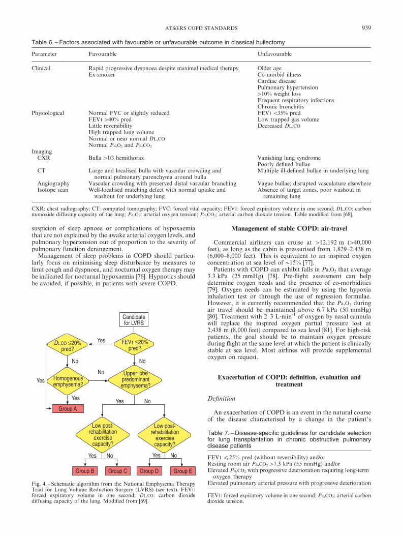
The recently completed National Emphysema Therapy
Trial (NETT) showed benefits for a subset of patientswith nonhomogenous emphysema. Figure 4 summarises thestratification of the patients and the results of the trial foreach of the groups. Group B is comprised of those patientswith nonhomogenous emphysema of upper lobe predomi-nance and limited exercise performance after pre-operativecomprehensive rehabilitation. Group C corresponds topatients with predominant upper lobe emphysema and goodpost-rehabilitation exercise capacity. Group D corresponds tothose patients with homogenous emphysema and low post-rehabilitation exercise capacity. Group E was characterisedby homogeneous emphysema and good post-rehabilitationexercise capacity. Finally, group A corresponds to those patientswith a very high risk for lung reduction surgery [70].
The results of this trial showed that patients in group Bwho underwent surgery had a lower mortality, better exercisecapacity and health status than patients randomised tomedical therapy. The operated patients in groups C and Ddid not benefit from improved survival but had significantimprovements in exercise capacity and health status com-pared to patients randomised to medical therapy. The patientsin group E had higher mortality and would, therefore, not becandidates for LVRS. The results in this group are similar tothose observed in the highest risk group (A) who should notbe considered for surgery.
Lung transplantation results in improved pulmonaryfunction, exercise capacity and quality of life, however, itseffects on survival remain controversial [71]. Specific guide-lines for lung transplantation in COPD are shown in table 7.
Management of stable COPD: sleep
Sleep in COPD is associated with oxygen desaturation,which is predominantly due to the disease itself rather than tosleep apnoea [72]. The desaturation during sleep may begreater than during maximum exercise [73]. In COPD, sleepquality is markedly impaired, both subjectively and objec-tively [74]. However, sleep apnoea syndrome is about asprevalent in COPD as in a general population of similar age,but oxygen desaturation during sleep is more pronouncedwhen the two conditions co-exist [75].
The clinical assessment in all patients with COPD shouldinclude questions about sleep quality and possible co-existingsleep apnoea syndrome. Sleep studies are not indicated inCOPD except in special circumstances. These include: a clinical
������������#��
>?@9�,A60�������#�� �,A60����
B���������������������������
C�������&�������������������
����������&��������������
>?@9�3A60������#�� �3A60����
����>?@9�3D60�����
����#�� �3D60����
��������������
����>?@9�,D60�����������#�� �,D60����
E���������'���������4���������'
���������7
�� +����39F��#:�� 9:��� 9��������� +���396��#:�� 9:��� 9
�����������'�3.���������4FD������7
Fig. 3. – Algorithm for pre-operative testing for lung resection. DL,CO:carbon dioxide diffusing capacity of the lung; FEV1: forced expira-tory volume in one second; ppo: predicted postoperative; V9O2,max:maximum oxygen consumption.
938 B.R. CELLI ET AL.
suspicion of sleep apnoea or complications of hypoxaemiathat are not explained by the awake arterial oxygen levels, andpulmonary hypertension out of proportion to the severity ofpulmonary function derangement.
Management of sleep problems in COPD should particu-larly focus on minimising sleep disturbance by measures tolimit cough and dyspnoea, and nocturnal oxygen therapy maybe indicated for nocturnal hypoxaemia [76]. Hypnotics shouldbe avoided, if possible, in patients with severe COPD.
Management of stable COPD: air-travel
Commercial airliners can cruise at w12,192 m (w40,000feet), as long as the cabin is pressurised from 1,829–2,438 m(6,000–8,000 feet). This is equivalent to an inspired oxygenconcentration at sea level ofy15% [77].
Patients with COPD can exhibit falls in Pa,O2 that average3.3 kPa (25 mmHg) [78]. Pre-flight assessment can helpdetermine oxygen needs and the presence of co-morbidities[79]. Oxygen needs can be estimated by using the hypoxiainhalation test or through the use of regression formulae.However, it is currently recommended that the Pa,O2 duringair travel should be maintained above 6.7 kPa (50 mmHg)[80]. Treatment with 2–3 L?min-1 of oxygen by nasal cannulawill replace the inspired oxygen partial pressure lost at2,438 m (8,000 feet) compared to sea level [81]. For high-riskpatients, the goal should be to maintain oxygen pressureduring flight at the same level at which the patient is clinicallystable at sea level. Most airlines will provide supplementaloxygen on request.
Exacerbation of COPD: definition, evaluation andtreatment
Definition
An exacerbation of COPD is an event in the natural courseof the disease characterised by a change in the patient9s
Table 6. – Factors associated with favourable or unfavourable outcome in classical bullectomy
Parameter Favourable Unfavourable
Clinical Rapid progressive dyspnoea despite maximal medical therapy Older ageEx-smoker Co-morbid illness
Cardiac diseasePulmonary hypertensionw10% weight lossFrequent respiratory infectionsChronic bronchitis
Physiological Normal FVC or slightly reduced FEV1 v35% predFEV1 w40% pred Low trapped gas volumeLittle reversibility Decreased DL,CO
High trapped lung volumeNormal or near normal DL,CO
Normal Pa,O2 and Pa,CO2
ImagingCXR Bulla w1/3 hemithorax Vanishing lung syndrome
Poorly defined bullaeCT Large and localised bulla with vascular crowding and
normal pulmonary parenchyma around bullaMultiple ill-defined bullae in underlying lung
Angiography Vascular crowding with preserved distal vascular branching Vague bullae; disrupted vasculature elsewhereIsotope scan Well-localised matching defect with normal uptake and
washout for underlying lungAbsence of target zones, poor washout in
remaining lung
CXR: chest radiography; CT: computed tomography; FVC: forced vital capacity; FEV1: forced expiratory volume in one second; DL,CO: carbonmonoxide diffusing capacity of the lung; Pa,O2; arterial oxygen tension; Pa,CO2; arterial carbon dioxide tension. Table modified from [68].
#�� ��+60���(
)�� >?@9��+60���(
*�����������������(
<������
B�������'������������������(
#������� ���'��������
��������������(
<�����! <������ <������ <�����?
)�� )��
)��)�� =� =�
=�
=�
=�=�
)��
���������#@;�
#������� ���'��������
��������������(
Fig. 4. – Schematic algorithm from the National Emphysema TherapyTrial for Lung Volume Reduction Surgery (LVRS) (see text). FEV1:forced expiratory volume in one second; DL,CO: carbon dioxidediffusing capacity of the lung. Modified from [69].
Table 7. – Disease-specific guidelines for candidate selectionfor lung transplantation in chronic obstructive pulmonarydisease patients
FEV1 f25% pred (without reversibility) and/orResting room air Pa,CO2 w7.3 kPa (55 mmHg) and/orElevated Pa,CO2 with progressive deterioration requiring long-term
oxygen therapyElevated pulmonary arterial pressure with progressive deterioration
FEV1: forced expiratory volume in one second; Pa,CO2: arterial carbondioxide tension.
939ATS/ERS COPD STANDARDS

baseline dyspnoea, cough and/or sputum beyond day-to-dayvariability sufficient to warrant a change in management.
There is no agreed classification of exacerbations. Thefollowing operational classification of severity can help rankthe clinical relevance of the episode and its outcome. Level I:treated at home. Level II: requires hospitalisation. Level III:leads to respiratory failure.
Assessment
Several clinical elements must be considered when evaluat-ing patients with exacerbations. These include the severity ofthe underlying COPD, the presence of co-morbidity and thehistory of previous exacerbations. The physical examinationshould evaluate the effect of the episode on the haemody-namic and respiratory systems. The diagnostic procedures tobe performed depend on the setting of the evaluation [82, 83].
The pharmacological treatment of patients with an exacer-bation of COPD is based on the same medications utilised inthe management of the stable patient [1–3]. However, theevidence supports the use of systemic glucocorticosteroids[84–88].
Table 8 shows the elements of the clinical evaluation anddiagnostic procedures that are usually informative in patientswith exacerbations according to the severity of the episode.
Indication for hospitalisation
Table 9 provides reasonable guidelines for patient hospita-lisation. Based on expert consensus, they consider the severityof the underlying respiratory dysfunction, progression ofsymptoms, response to outpatient therapy, existence of co-morbid conditions and the availability of adequate home care.
Indications for admission to specialised or intensive careunit
The severity of respiratory dysfunction dictates the need foradmission to an intensive care unit (ICU). Depending on theresources available within an institution, admission ofpatients with severe exacerbations of COPD to intermediateor special respiratory care units may be appropriate if
personnel, skills and equipment exist to identify and manageacute respiratory failure successfully.
Indications for ICU or special care unit admission includethe following: impending or actual respiratory failure; pre-sence of other end-organ dysfunction, i.e. shock, renal, liveror neurological disturbance; and/or haemodynamic instability.
Treatment of exacerbations
The treatment of exacerbations has to be based on theclinical presentation of the patient, as shown in tables 10, 11and 12.
Exacerbation of COPD: inpatient oxygen therapy
During a severe exacerbation, ABGs should be monitoredfor Pa,O2, arterial carbon dioxide tension (Pa,CO2) and pH.The Sp,O2 should be monitored for trending and adjustingoxygen settings. The goal of inpatient oxygen therapy is tomaintain Pa,O2w8 kPa (60 mmHg) or Sp,O2w90% in order toprevent tissue hypoxia and preserve cellular oxygenation. Dueto the shape of the oxyhaemoglobin dissociation curve, increa-sing the Pa,O2 to values much greater than 8 kPa (60 mmHg)confers little added benefit (1–2 vol %) and may increase therisk of CO2 retention, which may lead to respiratory acidosis
Table 8. – Clinical history, physical findings and diagnostic procedures in patients with exacerbation of chronic obstructivepulmonary disease (COPD)
Level I Level II Level III
Clinical historyCo-morbid conditions# z zzz zzzHistory of frequent exacerbations z zzz zzzSeverity of COPD Mild/moderate Moderate/severe Severe
Physical findingsHaemodynamic evaluation Stable Stable Stable/unstableUse accessory respiratory muscles, tachypnoea Not present zz zzzPersistent symptoms after initial therapy No zz zzz
Diagnostic proceduresOxygen saturation Yes Yes YesArterial blood gases No Yes YesChest radiograph No Yes YesBlood tests} No Yes YesSerum drug concentrationsz If applicable If applicable If applicableSputum gram stain and culture No§ Yes YesElectrocardiogram No Yes Yes
z: unlikely to be present; zz: likely to be present;zzz: very likely to be present. #: the more common co-morbid conditions associated with poorprognosis in exacerbations are congestive heart failure, coronary artery disease, diabetes mellitus, renal and liver failure; }: blood tests include cellblood count, serum electrolytes, renal and liver function; z: serum drug concentrations, consider if patients are using theophylline, warfarin,carbamezepine, digoxin; §: consider if patient has recently been on antibiotics.
Table 9. – Indications for hospitalisation of patients with aCOPD exacerbation
The presence of high-risk co-morbid conditions, includingpneumonia, cardiac arrhythmia, congestive heart failure,diabetes mellitus, renal or liver failure
Inadequate response of symptoms to outpatient managementMarked increase in dyspnoeaInability to eat or sleep due to symptomsWorsening hypoxaemiaWorsening hypercapniaChanges in mental statusInability of the patient to care for her/himself (lack of home support)Uncertain diagnosisInadequate home care
940 B.R. CELLI ET AL.

[89, 90]. The main delivery devices include nasal cannula andventuri masks. Alternative delivery devices include nonre-breather masks, reservoir cannulae, nasal cannulae or transtra-cheal catheters.
As a general principle, prevention of tissue hypoxiasupercedes CO2 retention concerns. If CO2 retention occurs,monitor for acidemia. If acidemia occurs, consider noninva-sive or invasive mechanical ventilation.
Setting and adjusting oxygen flow
Figure 5 shows an algorithm for correcting hypoxaemia inthe COPD patient.
Monitoring following hospital discharge
Patients may be started on oxygen for the first time duringhospitalisation for an acute exacerbation and discharged before
recovery is complete. Patients with hypoxaemia at dischargemay require short-term oxygen therapy as the effects of theexacerbation are clearing. After 30–90 days, oxygen may nolonger be required; thus re-evaluation of the patient9s oxygen
Table 10. – Level I: outpatient treatment
Patient educationCheck inhalation techniqueConsider use of spacer devices
BronchodilatorsShort-acting b2-agonist# and/or ipratropium MDI with spacer
or hand-held nebuliser as neededConsider adding long-acting bronchodilator if patient is not
using oneCorticosteroids (the actual dose may vary)
Prednisone 30–40 mg orally?day-1 for 10–14 daysConsider using an inhaled corticosteroid
AntibioticsMay be initiated in patients with altered sputum characteristicsz
Choice should be based on local bacterial resistance patternsAmoxicillin/ampicillin}, cephalosporinsDoxycyclineMacrolides§
If the patient has failed prior antibiotic therapy consider:Amoxicillin/clavulanateRespiratory fluoroquinolonesƒ
MDI: metered-dose inhaler. #: salbutamol (albuterol), terbutaline; z:purulence and/or volume; }: depending on local prevalence of bacterialb-lactamases; §: azithromycin, clarithromycin, dirithromycin, roxithro-mycin; ƒ: gatifloxacin, levofloxacin, moxifloxacin.
Table 11. – Level II: treatment for hospitalised patient
BronchodilatorsShort-acting b2-agonist and/orIpratropium MDI with spacer or hand-held nebuliser as needed
Supplemental oxygen (if saturation v90%)Corticosteroids
If patient tolerates, prednisone 30–40 mg orally?day-1 for10–14 days
If patient can not tolerate oral intake, equivalent dose i.v. forup to 14 days
Consider using inhaled corticosteroids by MDI or hand-heldnebuliser
Antibiotics (based on local bacteria resistance patterns)May be initiated in patients that have a change in their sputum
characteristics#
Choice should be based on local bacteria resistance patternsAmoxicillin/clavulanateRespiratory fluoroquinolones (gatifloxacin, levofloxacin,
moxifloxacin)If Pseudomonas spp. and/or other Enterobactereaces spp. are
suspected, consider combination therapy
MDI: metered-dose inhaler. #: purulence and/or volume.
Table 12. – Level III: treatment in patients requiring special orintensive care unit
Supplemental oxygenVentilatory supportBronchodilators
Short-acting b2-agonist (salbutamol (albuterol)) and ipratropiumMDI with spacer, two puffs every 2–4 h
If the patient is on the ventilator, consider MDI administrationConsider long-acting b-agonist
CorticosteroidsIf patient tolerates oral medications, prednisone 30–40 mg
orally?day-1 for 10–14 daysIf patient cannot tolerate, give the equivalent dose i.v. for up to
14 daysConsider using inhaled corticosteroids by MDI or hand-held
nebuliserAntibiotics (based on local bacteria resistance patterns)
Choice should be based on local bacteria resistance patternsAmoxicillin/clavulanateRespiratory fluoroquinolones (gatifloxacin, levofloxacin,
moxifloxacin)If Pseudomonas spp. and/or other Enterobactereaces spp. are
suspected, consider combination therapy
MDI: metered-dose inhaler.
������������� '�����!<!����� +
��������� +�3/����G���� +������ +�560
*��������(4��� +�3A"-���7
=������������������������
;��������!<���92+��
*��������(��� +�3A"-���
E������ +�� +�560
�*�,-".F(4������� +3/���7
E������ +�� +�560
;��������!<���+��
�*�,-".F(4������� +3/���7
=������������������������
����������������&���������=��@��������'����
=� )��
)��
=�
=�
)��
)��
=�
Fig. 5. – Algorithm to correct hypoxaemia in an acutely ill chronicobstructive pulmonary disease patient. ABG: arterial blood gas; Pa,O2:arterial oxygen tension; O2: oxygen; Sa,O2: arterial oxygen saturation;Pa,CO2: arterial carbon dioxide tension; NPPV: noninvasive positivepressure ventilation.
941ATS/ERS COPD STANDARDS

and medical status should be completed. If the patient nolonger meets the prescribing criteria for LTOT, oxygen shouldbe discontinued, as there is no proven survival benefit forpatients with mild hypoxaemia [91]. Patients recovering froman exacerbation may benefit from pulmonary rehabilitation.
Some patients who needed oxygen prior to hospitalisationmay, over time, increase their Pa,O2 to the point that they nolonger qualify for oxygen. This phenomenon is thought to bedue to a reparative effect of LTOT. Withdrawing oxygenfrom these patients may negate the reparative effect and causethe patient9s status to deteriorate to the point of meeting thephysiological requirement for oxygen. Consequently, thesepatients should continue their oxygen therapy without inter-ruption, as withdrawing their oxygen might be detrimental[92, 93].
Exacerbation of COPD: assisted ventilation
Mechanical ventilation can be administered via noninvasiveor invasive ventilation. Noninvasive is preferred wheneverpossible. Mechanical ventilation, either "invasive" or "non-invasive", is not a therapy but it is a form of life support untilthe cause underlying the acute respiratory failure is reversedwith medical therapy [94–96]. Patients considered for mechan-ical ventilation should have a measurement of ABGs.
Indications for mechanical ventilation
The institution of mechanical ventilation should be consi-dered when, despite optimal medical therapy and oxygenadministration there is acidosis (pHv7.35) and hypercapnia(Pa,CO2 w6–8 kPa (45–60 mmHg)) and respiratory frequencyw24 breaths?min-1.
Modes of mechanical ventilation
Mechanical ventilation can be delivered as follows. 1)Through an endotracheal tube, by passing the upper airway,i.e. "conventional" or "invasive" mechanical ventilation. 2)Without the use of an endotracheal tube, i.e. "noninvasive"mechanical ventilation or NIMV, which can be instituted intwo modes: noninvasive positive pressure ventilation (NPPV)(nasal or face masks); or negative pressure ventilation (e.g.iron lung, not recommended).
Noninvasive positive pressure ventilation. NPPV is by far themost popular mode of providing noninvasive ventilation. It istypically administered as a combination of continuous positiveairway pressure (CPAP) plus pressure support ventilation(PSV) [94–98]. ABG improve because of an increase in alveolarventilation without significant modifications in the alveolarventilation/perfusion mismatching and gas exchange in thelungs [99].
ABGs are fundamental for the correct assessment andguidance of therapy. Once baseline ABGs are obtained, if thepH isv7.35 in the presence of hypercapnia, NPPV should bedelivered in a controlled environment such as intermediateICUs and/or high-dependency units. If the pH isv7.25, NPPVshould be administered in the ICU and intubation should bereadily available. The combination of some CPAP (e.g.4–8 cmH2O) and PSV (e.g. 10–15 cmH2O) provides the mosteffective mode of NPPV.
Patients meeting exclusion criteria should be considered forimmediate intubation and ICU admission. In the first hours,
NPPV requires the same level of supervision as conventionalmechanical ventilation.
Contraindications for NPPV include the following: respira-tory arrest; cardiovascular instability (hypotension, arrhyth-mias, myocardial infarction); impaired mental status,somnolence, inability to cooperate; copious and/or viscoussecretions with high aspiration risk; recent facial or gastro-oesophageal surgery; craniofacial trauma and/or fixed naso-pharyngeal abnormality; burns; and extreme obesity.
NPPV can be considered successful when ABGs and pHimprove, dyspnoea is relieved, the acute episode resolveswithout the need of endotracheal intubation, mechanicalventilation can be discontinued and the patient is dischargedfrom the hospital.
One-year mortality was reported to be lower in patientsreceiving NPPV for exacerbations of COPD, as compared toboth conventional mechanical ventilation [100] and optimalmedical therapy alone [101].
Figure 6 illustrates a useful flow-chart for the use of NPPVin exacerbation of COPD complicated by acute respiratoryfailure.
Invasive ventilation. Intubation should be considered inpatients with the following. 1) NPPV failure: worsening ofABGs and or pH in 1–2 h; lack of improvement in ABGs andor pH after 4 h. 2) Severe acidosis (pHv7.25) and hypercapnia(Pa,CO2w8 kPa (60 mmHg)). 3) Life-threatening hypoxaemia(arterial oxygen tension/inspiratory oxygen fractionv26.6 kPa(200 mmHg)). 4) Tachypnoea w35 breaths?min-1.
Criteria for hospital discharge
As a general rule, patients hospitalised for an acuteexacerbation can be considered for discharge once the reasons
?����'��������� �����H�������&�����������������
�����������������=��@(
)�� =�
I�����
����'��E@
=��@���������������
�����&���������*����� +
�������������
)�� =�
I��������������������������
���������=��@����'��E@�D/��
+ ��8 ��'������
������� >�����
������������E@ =��@
Fig. 6. – Flow-chart for the use of noninvasive positive pressureventilation (NPPV) during exacerbation of chronic obstructive pulmo-nary disease (COPD) complicated by acute respiratory failure. MV:mechanical ventilation; Pa,CO2: arterial carbon dioxide tension.
942 B.R. CELLI ET AL.

for admission are controlled and/or reversed. Based onconsensus, conditions that need to be met when consideringpatients for discharge include: symptoms returning to base-line, including eating, sleeping, etc.; haemodynamic stability;oxygenation returning to baseline; inhaled b-agonist therapyrequired less frequently; ability to resume ambulation; abilityto eat and sleep without frequent awakening by dyspnoea;off-parenteral therapy for 12–24 h; patient (or home care-giver) understands correct use of medications; follow-up andhomecare arrangements have been completed (e.g. visitingnurse, oxygen delivery, meal provisions etc.).
Follow-up evaluation
Once discharged, the patient should be followed. There areno studies that have addressed the specific schedules morelikely to result in a positive outcome, but patients withfrequent exacerbations are more likely to relapse. Likewise,patients who have developed respiratory failure requiringadmission to an ICU carry a very high mortality risk. Basedon this, the guidelines for the re-evaluation of patientsadmitted for exacerbation of COPD should include: reassess-ment within 4 weeks; evaluation of improvement in symptomsand physical exam; assessment of need for supplementaloxygen; repeat examination if previous abnormalities werepresent; assessment of ability of the patient to cope with theenvironment; an understanding and re-adjustment of thetreatment regimen.
Ethical and palliative care issues in COPD
Patients with COPD experience acute exacerbations of theirdisease, which may produce respiratory failure and a possibleneed for either ventilatory support or accepting death. Noclinical features can identify patients with respiratory failurewho will experience more burden than benefit from life sup-portive care.
If the patient has, or can have, clear preferences abouttreatment, respect for the patient requires that care providersgive effect to the patient9s views. Autonomy of the patient isthe predominant ethical principle that drives end-of-lifedecision-making in many societies.
All healthcare providers should assist patients during stableperiods of health to think about their advance care planningby initiating discussions about end-of-life care. These discus-sions should prepare patients with advanced COPD for alife-threatening exacerbation of their chronic disease, whileassisting them to go on living and enjoying life. Pulmonaryrehabilitation provides an important opportunity to assistadvance care planning for patients with moderate-to-severeCOPD. Educational programmes on advance care planningwithin pulmonary rehabilitation increase the adoption rate forinstruments of advance care planning and patient-physiciandiscussion about end-of-life care [102]. Patients who choose torefuse life supportive care or have it withdrawn require expertdelivery of palliative care.
Patients with COPD sometimes qualify for formal hospiceservices, especially when they are having repeated exacerba-tions and very poor measures on tests of pulmonary function.Nevertheless, many patients will have a fatal exacerbationwithin a short time of having fairly good function, so onecannot wait to consider using hospice until death is nearlycertain. Opportunities for hospice care are frequently neg-lected for patients coming to the end of life with COPD
[103, 104]. Neglect in offering patients and their familiesappropriate resources for supportive end-of-life care results inunnecessary admissions to acute care hospitals for worseningrespiratory symptoms.
Integrated disease management for primary care inCOPD
Disease management can be regarded as an integrated andsystematic approach in which healthcare providers worktogether in a coordinated and cooperative manner to producean optimal outcome for a particular patient with COPD,throughout the entire continuum of care [105]. The concept ofthe disease as a continuum is depicted in figure 7. This figurealso represents the navigation tool for the web-basedStandards for the Diagnosis and Treatment of Patients withCOPD document 2004.
Integrated care for COPD involves the patient and a teamof clinical professionals working in primary care, cooperatingwith secondary care and rehabilitation services. Optimaldisease management involves redesigning standard medicalcare to integrate rehabilitative elements into a system ofpatient self-management and promotion of a healthy lifestyle.
The following aspects are important: smoking cessation forall patients who smoke; early diagnosis and secondaryprevention (healthy lifestyle, vaccinations, exercise); educa-tion and self-management; pulmonary rehabilitation; mon-itoring and early recognition of exacerbation; implementationof rapid action plan; careful attention to end-of-life issues;palliative care.
Referral indications
Referral to specialist care is indicated for COPD patientswith the following: disease onset at age v40 yrs; frequentexacerbations (two or more per year) despite adequatetreatment; rapidly progressive course of disease (decline inFEV1, progressive dyspnoea, decreased exercise tolerance,unintentional weight loss; severe COPD (FEV1v50% pred)despite optimal treatment; need for LTOT; onset of co-morbid illness (osteoporosis, heart failure, bronchiectasis,lung cancer); evaluation for surgery.
�������������������
������� ���������� ?����'����� ;���������������
�����&�������
����������������
���������������
������������'��������
������������
��������>?@9
������������������
Fig. 7. – Continuum of care for chronic obstructive pulmonary disease(COPD). FEV1: forced expiratory volume in one second.
943ATS/ERS COPD STANDARDS

Conclusions
This summary presents an overview of the entire documentfor the health practitioner, which is readily available online(www.copd-ats-ers, www.ersnet.org and www.thoracic.org).This summary does not include any reference to the patientdocument, which, due to its inherent characteristics, cannotbe presented in a conventional format. The readers areencouraged to visit the website to access both full documents.
The committee that developed this document fully under-stands that the field is rapidly changing and that individualcomponents of this document need to be updated periodicallyas the need arises. However, the modular and flexible designallows for this to occur easier than ever before. The challengefor the future is to develop mechanisms to permit the updatedflow of valid scientific information to reach all who need it.
References
1. American Thoracic Society. Standards for the diagnosis andcare of patients with chronic obstructive pulmonary disease.Am J Respir Crit Care Med 1995; 152: S77–S121.
2. Siafakas NM, Vermeire P, Pride NB, et al. Optimalassessment and management of chronic obstructive pulmo-nary disease (COPD). The European Respiratory SocietyTask Force. Eur Respir J 1995; 8: 1398–1420.
3. Pauwels RA, Buist AS, Calverley PM, Jenkins CR, Hurd SS,the GOLD Scientific Committee. Global strategy for thediagnosis, management, and prevention of chronic obstruc-tive pulmonary disease. NHLBI/WHO Global Initiative forChronic Obstructive Lung Disease (GOLD) Workshopsummary. Am J Respir Crit Care Med 2001; 163: 1256–1276.
4. Ferrer M, Alonso J, Morera J, et al. Chronic obstructivepulmonary disease and health related quality of life. Ann IntMed 1997; 127: 1072–1079.
5. Friedman M, Serby C, Menjoge S, Wilson J, Hilleman D,Witek T. Pharmacoeconomic evaluation of a combination ofipratropium plus albuterol compared with ipratropium aloneand albuterol alone in COPD. Chest 1999; 115: 635–641.
6. Burge PS, Calverley PM, Jones PW, Spencer S, AndersonJA, Maslen TK. Randomised, double blind, placebocontrolled study of fluticasone propionate in patients withmoderate to severe chronic obstructive pulmonary disease:the ISOLDE trial. BMJ 2000; 320: 1297–1303.
7. Dewan N, Rafique S, Kanwar B, et al. Acute exacerbation ofCOPD. Factors associated with poor treatment outcome.Chest 2000; 117: 662–671.
8. Anthonisen NR, Wright EC, Hodgkin JE, the IPPB TrialGroup. Prognosis in chronic obstructive pulmonary disease.Am Rev Respir Dis 1986; 133: 14–20.
9. Celli B, Halbert R, Isonaka S, Schau B. Population impact ofdifferent definitions of airway obstruction. Eur Respir J 2003;22: 268–273.
10. Schols AM, Slangen J, Volovics L, Wouters EF. Weight lossis a reversible factor in the prognosis of chronic obstructivepulmonary disease. Am J Respir Crit Care Med 1998; 157:1791–1797.
11. Landbo C, Prescott E, Lange P, Vestbo J, Almdal TP.Prognostic value of nutritional status in chronic obstructivepulmonary disease. Am J Respir Crit Care Med 1999; 160:1856–1861.
12. Nishimura K, Izumi T, Tsukino M, Oga T. Dyspnea is abetter predictor of 5-year survival than airway obstruction inpatients with COPD. Chest 2002; 121: 1434–1440.
13. Mannino DM, Homa DM, Akinbami LJ, Ford ES, ReddSC. Chronic obstructive pulmonary disease surveillance –United States, 1971–2000. MMWR 2002; 51: 1–16.
14. Prescott E. Tobacco-related diseases: the role of gender. DanMed Bull 2000; 47: 115–131.
15. Anto JM, Vermeire P, Vestbo J, Sunyer J. Epidemiology ofchronic obstructive pulmonary disease. Eur Respir J 2001;17: 982–994.
16. Fletcher CM, Tinker CM, Peto R, Speizer FE. The naturalhistory of chronic bronchitis and emphysema. Oxford,Oxford University Press, 1976.
17. Gold DR, Wang X, Wipyj D, Speizer FE, Ware JH, DockeryDW. Effects of cigarette smoking on lung function inadolescent boys and girls. N Engl J Med 1996; 335: 931–937.
18. Saetta M, Di Stefano A, Turato G, et al. CD8zT-lymphocytes in peripheral airways of smokers with chronicobstructive pulmonary disease. Am J Respir Crit Care Med1998; 157: 822–826.
19. Rennard SI. Inflammation and repair processes in chronicobstructive pulmonary disease. Am J Respir Crit Care Med1999; 160: S12–S16.
20. Peinado VI, Barbera JA, Abate P, et al. Inflammatoryreaction in pulmonary muscular arteries of patients withmild chronic obstructive pulmonary disease. Am J RespirCrit Care Med 1999; 159: 1605–1611.
21. Rodriguez-Roisin R, MacNee W. Pathophysiology ofchronic obstructive pulmonary disease. Eur Respir Mono1998; 3: 107–126.
22. O9Shaughnessy TC, Ansari TW, Barnes NC, Jeffery PK.Inflammation in bronchial biopsies of subjects with chronicbronchitis: inverse relationship of CD8zT lymphocytes withFEV1. Am J Respir Crit Care Med 1997; 155: 852–857.
23. Repine JE, Bast A, Lankhorst I. Oxidative stress in chronicobstructive pulmonary disease. Oxidative Stress StudyGroup. Am J Respir Crit Care Med 1997; 156: 341–357.
24. Matsuba K, Wright JL, Wiggs BR, Pare PD, Hogg JC. Thechanges in airways structure associated with reduced forcedexpiratory volume in one second. Eur Respir J 1989; 2: 834–839.
25. O9Donnell DE, Revill SM, Webb KA. Dynamic hyperinfla-tion and exercise intolerance in chronic obstructive pulmo-nary disease. Am J Respir Crit Care Med 2001; 164: 770–777.
26. Fiore MC, Bailey WC, Cohen SJ. Smoking cessation.Guideline technical report no. 18. Publication No. AHCPR97-Noo4. Rockville, MD, US Department of Health andHuman Services, Public Health Service, Agency for HealthCare Policy and Research, October 1997.
27. Fiore MC. US public health service clinical practice guide-line: treating tobacco use and dependence. Respir Care 2000;45: 1200–1262.
28. Office of the Surgeon General. Tobacco Cessation Guidelinewww.surgeongeneral.gov/tobacco/default.htm. Data lastupdated: continuous.
29. Celli B, ZuWallack R, Wang S, Kesten S. Improvement inresting inspiratory capacity and hyperinflation with tiotro-pium in COPD patients with increased static lung volumes.Chest 2003; 124: 1743–1748.
30. O9Donnell DE, Lam M, Webb KA. Spirometric correlates ofimprovement in exercise performance after anticholinergictherapy in chronic obstructive pulmonary disease. AmJ Respir Crit Care Med 1999; 160: 542–549.
31. Belman MJ, Botnick WC, Shin JW. Inhaled bronchodilatorsreduce dynamic hyperinflation during exercise in patientswith chronic obstructive pulmonary disease. Am J RespirCrit Care Med 1996; 153: 967–975.
32. Dahl R, Greefhorst LA, Nowak D, et al. Inhaled formoteroldry powder versus ipratropium in chronic obstructivepulmonary disease. Am J Respir Crit Care Med 2001; 164:778–784.
33. Jones PW, Bosh TK. Quality of life changes in COPDpatients treated with salmeterol. Am J Respir Crit Care Med1997; 155: 1283–1289.
34. Combivent trialists. In chronic obstructive pulmonarydisease, a combination of ipratropium and albuterol ismore effective than either agent alone. An 85-day multicentertrial. COMBIVENT Inhalation Aerosol Study Group. Chest1994; 105: 1411–1419.
35. Zuwallack RL, Mahler DA, Reilly D, et al. Salmeterol plus
944 B.R. CELLI ET AL.

theophylline combination therapy in the treatment ofCOPD. Chest 2001; 119: 1661–1670.
36. Casaburi R, Mahler DA, Jones PW, et al. A long-termevaluation of once daily inhaled tiotropium in chronicobstructive pulmonary disease. Eur Respir J 2002; 19: 217–224.
37. Vicken W, Van Noord JA, Greefhorst AP, et al., on behalf ofthe Dutch/Belgian Tiotropium Study Group. Improvedhealth outcomes in patients with COPD during 1 yeartreatment with tiotropium. Eur Respir J 2002; 19: 209–216.
38. Burge PS, Calverley PM, Jones PW, Spencer S, AndersonJA, Maslen TK. Randomised, double blind, placebocontrolled study of fluticasone propionate in patients withmoderate to severe chronic obstructive pulmonary disease:the ISOLDE trial. BMJ 2000; 320: 1297–1303.
39. Pauwels RA, Lofdahl C-G, Laitinen LA, et al. Long-termtreatment with inhaled budesonide in persons with mildchronic obstructive pulmonary disease who continue smok-ing. New Engl J Med 1999; 340: 1948–1953.
40. The Lung Health Study Research Group. Effect of inhaledtriamcinolone on the decline in pulmonary function inchronic obstructive pulmonary disease. New Engl J Med2000; 343: 1902–1909.
41. Vestbo J, Sorensen T, Lange P, Brix A, Torre P, Viskum K.Long-term effect of inhaled budesonide in mild andmoderate chronic obstructive pulmonary disease: A rando-mised controlled trial. Lancet 1999; 353: 1819–1823.
42. Jarad NA, Wedzicha JA, Burge PS, Calverley PMA. Anobservational study of inhaled corticosteroid withdrawal instable chronic obstructive pulmonary disease. Respir Med1999; 93: 161–168.
43. Calverley PM, Boonsawat W, Cseke Z, et al. Maintenancetherapy with budesonide and formoterol in chronic obstruc-tive pulmonary disease. Eur Respir J 2003; 22: 912–919.
44. Szafranski W, Cukier A, Ramirez A, et al. Efficacy andsafety of budesonide/formoterol in the management ofCOPD. Eur Respir J 2003; 21: 74–81.
45. Calverley P, Pauwels R, Vestbo J, et al. Combined salmeteroland fluticasone in the treatment of chronic obstructivepulmonary disease: a randomised controlled trial. Lancet2003; 361: 449–456.
46. Report of the Medical Research Council Working Party.Long-term domiciliary oxygen therapy in chronic hypoxiccor pulmonale complicating chronic bronchitis and emphy-sema. Lancet 1981; 1: 681–685.
47. Nocturnal Oxygen Therapy Trial Group. Continuous ornocturnal oxygen therapy in hypoxemic chronic obstructivelung disease. Ann Intern Med 1980; 93: 391–398.
48. Weitzenblum E, Sautegeau A, Ehrhart M, Mammosser M,Pelletier A. Long-term oxygen therapy can reverse theprogression of pulmonary hypertension in patients withchronic obstructive pulmonary disease. Am Rev Respir Dis1985; 131: 493–498.
49. Oswald-Mammosser M, Weitzenblum E, Quoix E, et al.Prognostic factors in COPD patients receiving long-termoxygen therapy: importance of pulmonary artery pressure.Chest 1995; 107: 1193–1198.
50. Zielinski J, Tobiasz M, Hawrylkiewicz I, Sliwinksi P,Palasiewicz G. Effects of long-term oxygen therapy onpulmonary hemodynamics in COPD patients: a 6-yearprospective study. Chest 1998; 113: 65–70.
51. Tiep BL, Barnett J, Schiffman G, Sanchez O, Carter R.Maintaining oxygenation via demand oxygen delivery duringrest and exercise. Respir Care 2002; 47: 887–892.
52. Pulmonary rehabilitation: official statement of the AmericanThoracic Society. Am J Respir Crit Care Med 1999; 159:1666–1682.
53. Goldstein RS, Gort EH, Stubbing D, et al. Randomisedcontrolled trial of respiratory rehabilitation. Lancet 1994;344: 1394–1397.
54. Reardon J, Awad E, Normandin E, Vale F, Clark B,ZuWallack RL. The effect of comprehensive outpatient
pulmonary rehabilitation on dyspnea. Chest 1994; 105: 1046–1052.
55. Ries AL, Kaplan RM, Limberg TM, Prewitt LM. Effects ofpulmonary rehabilitation on physiologic and psychosocialoutcomes in patients with chronic obstructive pulmonarydisease. Ann Intern Med 1995; 122: 823–832.
56. Wijkstra PJ, van der Mark TW, Kraan J, van Altena R,Koeter GH, Postma DS. Effects of home rehabilitation onphysical performance in patients with chronic obstructivepulmonary disease (COPD). Eur Respir J 1996; 9: 104–110.
57. Strijbos JH, Postma DS, van Altena R, Gimeno F, KoeterGH. A comparison between an outpatient hospital-basedpulmonary rehabilitation program and a home-care pulmo-nary rehabilitation program in patients with COPD. Afollow-up of 18 months. Chest 1996; 109: 366–372.
58. Schols AMWJ, Soeters PB, Dingemans AMC, Mostert R,Frantzen PJ, Wouters EF. Prevalence and characteristics ofnutritional depletion in patients with stable COPD eligiblefor pulmonary rehabilitation. Am Rev Respir Dis 1993; 147:1151–1156.
59. Schols AM, Soeters PB, Mostert R, et al. Physiologic effectsof nutritional support and anabolic steroids in patients withchronic obstructive pulmonary disease: A randomisedcontrolled trial. Am J Respir Crit Care Med 1995; 152:1248–1274.
60. Creutzberg EL, Wouters EFM, Mostert R, et al. Efficacy ofnutritional supplementation therapy in depleted patientswith chronic obstructive pulmonary disease. Nutrition 2003;19: 120–127.
61. Arozullah AM, Khuri SF, Henderson WG, Daley J.Development and validation of a multifactorial risk indexfor predicting postoperative pneumonia after major non-cardiac surgery. Ann Intern Med 2001; 135: 847–857.
62. Smetana GW. Preoperative pulmonary evaluation. N EnglJ Med 1999; 340: 937–944.
63. Trayner E Jr, Celli BR. Postoperative pulmonary complica-tions. Med Clin North Am 2001; 85: 1129–1139.
64. Weisman IM. Cardiopulmonary exercise testing in thepreoperative assessment for lung resection surgery. SeminThorac Cardiovasc Surg 2001; 13: 116–125.
65. Martinez FJ, Iannettoni M, Paine III R. Medical evaluationand management of the lung cancer patient prior to surgery,radiation or chemotherapy. In: Pass H, Mitchell J, JohnsonD, Turrisi A, eds. Lung cancer: principles and practice.Philadelphia, PA, Lippincott Williams & Williams, 2000;pp. 649–681.
66. Bolliger CT, Jordan P, Soler M, et al. Pulmonary functionand exercise capacity after lung resection. Eur Respir J 1996;9: 415–421.
67. Bolliger CT, Perruchoud AP. Functional evaluation of thelung resection candidate. Eur Respir J 1998; 11: 198–212.
68. Martinez FJ. Surgical therapy for chronic obstructivepulmonary disease: conventional bullectomy and lungvolume reduction surgery in the absence of giant bullae.Semin Respir Crit Care Med 1999; 20: 351–364.
69. Snider GL. Reduction pneumoplasty for giant bullousemphysema. Implications for surgical treatment of nonbul-lous emphysema. Chest 1996; 109: 540–548.
70. National Emphysema Treatment Trial Research Group. Arandomized trial comparing lung-volume-reduction surgerywith medical therapy for severe emphysema. N Engl J Med2003; 348: 2059–2073.
71. American Thoracic Society. International guidelines for theselection of lung transplant candidates. Am J Respir CritCare Med 1998; 158: 335–339.
72. Hudgel DW, Martin RJ, Capehart M, Johnson B, Hill P.Contribution of hypoventilation to sleep oxygen desaturationin chronic obstructive pulmonary disease. J Appl Physiol1983; 55: 669–677.
73. Mulloy E, McNicholas WT. Ventilation and gas exchangeduring sleep and exercise in patients with severe COPD.Chest 1996; 109: 387–394.
945ATS/ERS COPD STANDARDS
74. Klink M, Quan S. Prevalence of reported sleep disturbancesin a general population and their relationship to obstructiveairways diseases. Chest 1987; 91: 540–546.
75. Sanders MH, Newman AB, Haggerty CL, et al. Sleep andsleep-disordered breathing in adults with predominantly mildobstructive airway disease. Am J Respir Crit Care Med 2003;167: 7–14.
76. Fletcher EC, Luckett RA, Goodnight-White S, Miller CC,Qian W, Costarangos-Galarza C. A double-blind trial ofnocturnal supplemental oxygen for sleep desaturation inpatients with chronic obstructive pulmonary disease and adaytime Pa,O2 above 60 mm Hg. Am Rev Respir Dis 1992;145: 1070–1076.
77. Dillard TA, Berg BW, Rajagopal KR, Dooley JW, MehmWJ. Hypoxemia during air travel in patients with chronicobstructive pulmonary disease. Ann Intern Med 1989; 111:362–367.
78. Christensen CC, Ryg M, Refvem OK, et al. Development ofsevere hypoxaemia in chronic obstructive pulmonary diseasepatients at 2438 m (8000 ft) altitude. Eur Respir J 2000; 15:635–639.
79. Gong H, Tashkin DP, Lee EY, Simmons MS. Hypoxia-altitude simulation test. Am Rev Respir Dis 1984; 130: 980–986.
80. AMA Commission on Emergency Medical Services. Medicalaspects of transportation aboard commercial aircraft. JAMA1982; 247: 1007–1011.
81. Berg BW, Dillard TA, Rajagopal KR, Mehm WJ. Oxygensupplementation during air travel in patients with chronicobstructive pulmonary disease. Chest 1992; 101: 638–641.
82. Emerman CL, Cydulka RAK. Evaluation of high-yieldcriteria for chest radiography in acute exacerbation ofchronic obstructive pulmonary disease. Ann Emerg Med1993; 22: 680–684.
83. O9Brien C, Guest PF. Physiological and radiologicalcharacterization of patients diagnosed with chronic obstruc-tive pulmonary disease in primary care. Thorax 2000; 55:631–632.
84. Albert RK, Martin TR, Lewis SW. Controlled clinical trialof methylprednisolone in patients with chronic bronchitisand acute respiratory insufficiency. Ann Intern Med 1980; 92:753–758.
85. Niewhoehner DE, Erbland ML, Deupree RH, et al. Effect ofsystemic glucocorticoids on exacerbations of chronicobstructive pulmonary disease. Department of VeteransAffairs Cooperative Study Group. N Engl J Med 1999;340: 1941–1947.
86. Davies L, Angus RM, Calverley PM. Oral corticosteroids inpatients admitted to hospital with exacerbations of chronicobstructive pulmonary disease: a prospective randomizedcontrolled trial. Lancet 1999; 345: 456–460.
87. Thompson WH, Nielson CP, Carvalho P, et al. Controlledtrial of oral prednisone in outpatients with acute COPDexacerbation. Am J Respir Crit Care Med 1996; 154: 407–412.
88. Aaron S, Vandenheeur K, Hebert P, et al. Outpatient oralprednisone after emergency treatment of chronic obstructivepulmonary disease. N Engl J Med 2003; 348: 2618–2625.
89. Aubier M, Murciano D, Milie-Emili M, et al. Effects of theadministration of oxygen therapy on ventilation and bloodgases in patients with chronic obstructive pulmonary disease
during acute respiratory failure. Am Rev Respir Dis 1980;122: 747–754.
90. Dumont CP, Tiep BL. Using a reservoir nasal cannula inacute care. Critical Care Nurse 2002; 22: 41–46.
91. Gorecka D, Gorzelak K, Sliwinski P, Tobiasz M, Zielinski J.Effect of long term oxygen therapy on survival in patientswith chronic obstructive pulmonary disease with moderatehypoxaemia. Thorax 1997; 52: 674–679.
92. O9Donohue WJ. Effect of arterial oxygen therapy onincreasing arterial oxygen tension in hypoxemia patientswith stable chronic obstructive pulmonary disease whilebreathing ambient air. Chest 1991; 100: 968–972.
93. Oba Y, Salzman GA, Willsie SK. Re-evaluation ofcontinuous oxygen therapy after initial prescription inpatients with chronic obstructive pulmonary disease. RespirCare 2000; 45: 401–406.
94. International Consensus Conferences in Intensive CareMedicine: noninvasive positive pressure ventilation in acuterespiratory failure. Am J Respir Crit Care Med 2001; 163:283–291.
95. BTS Guideline. Non invasive ventilation in acute respiratoryfailure. British Thoracic Society Standards of Care Commit-tee. Thorax 2002; 57: 192–211.
96. Mehta S, Hill NS. Non invasive ventilation. State of the Art.Am J Respir Crit Care Med 2001; 163: 540–577.
97. Lightowler JV, Wedzicha JA, Elliot M, Ram SF. Noninvasive positive pressure ventilation to treat respiratoryfailure resulting from exacerbations of chronic obstructivepulmonary disease: Cochrane systematic review and meta-analysis. BMJ 2003; 326: 185–189.
98. Rossi A, Appendini L, Roca J. Physiological aspects ofnoninvasive positive pressure ventilation. Eur Respir Mon2001; 16: 1–10.
99. Diaz O, Iglesia R, Ferrer M, et al. Effects of non invasiveventilation on pulmonary gas exchange and hemodynamicsduricng acute hypercapnic exacerbations of chronic obstruc-tive pulmonary disease. Am J Respir Crit Care Med 1997;156: 1840–1845.
100. Conti G, Antonelli M, Navalesi P, et al. Noninvaisve vsconventional mechanical ventilation in patients with chronicobstructive pulmonary disease after failure of medicaltreatment in the ward: a randomized trial. Intensive CareMed 2002; 28: 1701–1707.
101. Plant PK, Owen JL, Elliott MW. Non-invasive ventilation inacute exacerbations of chronic obstructive pulmonarydisease: long term survival and predictors of in-hospitaloutcome. Thorax 2001; 56: 708–712.
102. Heffner JE, Fahy B, Hilling L, Barbieri C. Outcomes ofadvance directive education of pulmonary rehabilitationpatients. Am J Respir Crit Care Med 1997; 155: 1055–1059.
103. Emanuel EJ, Fairclough DL, Slutsman J, Alpert H, BaldwinD, Emanuel LL. Assistance from family members, friends,paid care givers, and volunteers in the care of terminally illpatients. N Engl J Med 1999; 341: 956–963.
104. Christakis NA, Escarce JJ. Survival of Medicare patientsafter enrollment in hospice programs. N Engl J Med 1996;335: 172–178.
105. Epstein RS, Sherwood LM. From outcomes research todisease management: a guide for the perplexed. Ann InternMed 1996; 124: 832–837.
946 B.R. CELLI ET AL.