standards of practice - international academy of
TRANSCRIPT

Standards
of
Practice
Current: Jan 20, 2021
International Academy of Biological Dentistry and Medicine © 2021

Definition of Biological Dentistry Patient records
Nutrition Muscle Testing
EAV Myofunctional/Cranial Sacral
Neural Therapy Tongue Diagnosis
Biological Terrain Assessment Acupuncture Homeopathy
Compatibility testing Standards Procedure for Amalgam Removal
Standards for Surgical Cleanout Ozone
DNA testing Airway/Sleep Appliances
Fotana Laser Detox PRP
IV for sedation or nutritional support Amalgam Meter
Note
The IABDM Standards of Practice is a living document, continually open to discussion and revision. The most current edition will be available through the Academy website
(iabdm.org) at all times.
To comment, recommend changes or otherwise contribute to this document, IABDM members should contact Executive Director Dr. Dawn Ewing ([email protected] or
281-251-4411).
Biological Dentistry A Definition & Introductory Considerations
A specialty within the dental profession, biological dentistry is concerned with the
whole-body effects of all dental materials, techniques, and procedures. It unites the best
clinical practices and technologies of western dentistry and medicine with a wide array of
modalities beyond the horizon of conventional practice. For biological dentistry
acknowledges, appreciates, and considers the complex and dynamic relationships
between oral health and systemic health within the context of the whole person. These
things are inseparable.
Optimal health and wellness are intimately related to which and how dental
materials, techniques and care are provided. We intend to be minimally invasive yet
appropriately active.
Biological dentists may be general dentists, periodontists, orthodontists, oral
surgeons or pedodontists. In addition to training in their chosen specialty, they also have
extensive training in both dental toxicology and specific healing modalities beyond those
of western dentistry. The latter include – but are not limited to – Traditional Chinese
Medicine (TCM), Airway Management, Ayurveda, herbology, homeopathy, iridology and
energy medicine. Specific modalities will vary from dentist to dentist, but all are
incorporated into treatment for the betterment of the patient. For the word “biological”
refers to life. Any protocol followed must be one designed of components that sustain life
or improve the quality of life for individuals pursuing treatment.
These individuals, of course, are wonderfully – and often troublingly – unique, and
this fact must be taken into consideration, especially as it relates to the permanent
placement of dental materials. Experienced practitioners have learned to deal with the fact
that no single dental material or anesthetic can be appropriate for all patients.

Though each practitioner has their favorite materials for strength, beauty and
chemical metabolism, material testing for each patient is preferred over blanket approval
of any one material. The ideal treatment is customized treatment, tailored to the individual
patient’s biochemistry, energetic profile, current health status, needs, values, desires and
goals.
The issue of materials applies to root canal treated teeth, as well, but there is a
more fundamental consideration: A tooth without vessels and nerves is, by definition,
dead, and keeping that tooth in the body is a burden with which that body must deal.
Discussion of “compatible” root canal filling materials and possible canal sterilization by
laser or irrigants such as ozone are interesting but fail to address the issue of burden.
Each dentist and patient may decide for themselves if the “saving” of a tooth is worth its
potential impact on overall health.
Some physicians recommend that their patients have their dentist sign an
agreement to use only the medicaments, treatments and protocols the physician favors.
While well-intentioned, such prescription is inappropriate in light of the above issues. The
biological dentist is in the best position to determine suitable treatment based on the
patient’s unique and specific health situation.
What Is Biological Dentistry? By Felix Liao, DDS
Contributed by the author
“Biological” means life and health enhancing. Biological dentistry has one aim: to support and promote total health with a healthier teeth, gums, and mouth. Teeth and mouth are connected to the whole body through bones, blood, lymph, fascia, emotions, food, water, air and more. Biological dentistry is concerned with
A. The whole-body effects of oral-facial structure, teeth/gum/jaw infections, dental materials, dental treatment and non-treatment.
B. The effect of a patient’s food, lifestyle, environment and systemic health on their teeth, gums, jaws, muscles, joints, saliva and associated oral tissues and structures.
C. The effect of all health care interventions on patient’s oral health.
In short, biological dentists are dental physicians who subscribe to the goals of restoring and sustaining whole body health by
• Promoting lifestyle, health and preventive dental practices.
• Relieving or reducing body burdens such as infections, inflammation, toxic materials, electromagnetic disturbances, malocclusion and associated head-neck-jaw pain, snoring, sleep apnea and more, with clinical dental care.
• Educating patients on the systemic hazards of mouth breathing, food allergies,
nutritional deficiencies, postural distortion and psycho-emotional distress and disorders.
• Educating all health professionals on oral contributions to systemic ills, and vice versa.
To promote total health through oral care, biological dental physicians have training beyond dental school in integrative and alternative modalities. Examples can include but are not limited to
1. Traditional Chinese Medicine and Electrodermal Assessment 2. Homeopathy, energy medicine and biological terrain assessment 3. Cranial-jaw-spinal orthopedics 4. Sleep and Airway medicine 5. Nutrition and lifestyle impacts on adults’ oral health 6. Nutrition, allergies and food sensitivities on children’s dental-facial development 7. Oral-Systemic links on all mouth-body connections 8. Emotional and post-traumatic stress on mouth-brain connections 9. Toxicology, environmental medicine and detoxification support 10. Muscle and joint physiology, myofascial release and myofunctional therapy 11. Neural therapy

12. Surgical and non-surgical treatment of bone infections
The purpose of these additional modalities is to make the mouth fit to support total health, and vice versa. Examples from biological dental physicians’ collective experience include:
• Jaw pain in patients with undiagnosed gluten sensitivity.
• Heavy tongue coating as an indication of digestive insufficiency.
• Teeth grinding in patients with an undiagnosed sleep breathing disorder.
• Late eruption of adult teeth as an indication of low iodine levels, which can affect growth and development.
Every IABDM member is invited and encouraged to add to this Clinical Experience Bank. Email your contributions to [email protected] We share such clinical experience with each other and other interested health care professionals to promote mutual referrals to achieve functional and total health for patients.

Standards Of Practice
The Standards of Practice (SOPs) for the International Academy of Biological
Dentistry and Medicine (IABDM) have been compiled and peer-reviewed by members of
the organization.
The Standards are available on our website (iabdm.org) and are updated as
needed. Members will be notified by email and an announcement will be posted on the
website whenever any such changes have been made.
The IABDM encourages new members to read this document completely and
direct any questions or concerns with the Academy’s Executive Director, Dawn Ewing
While it may be impossible to construct a complete list of protocols for the typical
biological dental or medical office, we can highlight those based on a common paradigm:
treating the cause of symptoms with the aim of alleviating disease, restoring and
sustaining health.
This stands in stark contrast to non-biological practice, which privileges the
control of symptoms, improvement of aesthetics and continuance of life regardless of its
quality.
We likewise share a deep and constant belief in the Hippocratic injunction: First,
do no harm.
Thus, the biological dental office is one in which the dentist pays most attention
to how toxic materials and infections are removed from the mouth and how new
restorative materials will affect the body. Many biological dental offices also monitor and
treat the patient as they undergo detoxification and recuperation.
The biological medical office is one in which the physician pays most attention to
systems in a state of disease that have caused the patient’s body to be out of balance

with itself or its environment. The biological physician incorporates nutrition, energy
medicine and detoxification in the restoration of complete health.
Following the information on records gathering below, the rest of this document
consists of synopses of each type of modality commonly used by biological practitioners,
along with references and resources for further study. This collection of information is
not intended to be exhaustive but illustrative of the best science supporting biological
dentistry and medicine as they are practiced around the world today.
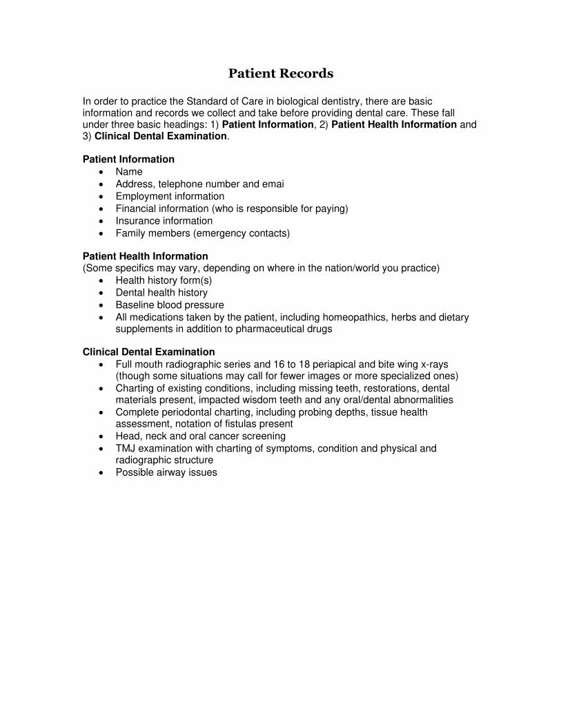
Patient Records
In order to practice the Standard of Care in biological dentistry, there are basic information and records we collect and take before providing dental care. These fall under three basic headings: 1) Patient Information, 2) Patient Health Information and 3) Clinical Dental Examination. Patient Information
• Name • Address, telephone number and emai • Employment information • Financial information (who is responsible for paying) • Insurance information • Family members (emergency contacts)
Patient Health Information (Some specifics may vary, depending on where in the nation/world you practice)
• Health history form(s) • Dental health history • Baseline blood pressure • All medications taken by the patient, including homeopathics, herbs and dietary
supplements in addition to pharmaceutical drugs Clinical Dental Examination
• Full mouth radiographic series and 16 to 18 periapical and bite wing x-rays (though some situations may call for fewer images or more specialized ones)
• Charting of existing conditions, including missing teeth, restorations, dental materials present, impacted wisdom teeth and any oral/dental abnormalities
• Complete periodontal charting, including probing depths, tissue health assessment, notation of fistulas present
• Head, neck and oral cancer screening • TMJ examination with charting of symptoms, condition and physical and
radiographic structure • Possible airway issues

Nutrition Nutrition includes matters of ingestion, digestion, absorption and metabolism of nutrients, food and water. A goal of prevention of caries, periodontal disease and bone loss may include addressing hormonal issues, as well. Differential diagnosis can be made by laboratory analysis of blood, urine, saliva and hair. EAV or muscle testing may be helpful in determining nutritional needs. Uses We now recognize high blood sugar problems and genetic ones like MTHFR may interfere with our common goal. Deficient diets are a contributing factor to bone loss and periodontal disease, as well as decay. Balancing body chemistry can play a role in TMJ and myofascial healing. Training The IABDM recognizes that most schools are lacking in hours dedicated to nutrition and strongly recommends members take classes to increase their awareness. Resources Course by Dr. Al Danenburg https://iabdm.org/?s=nutrition+course Weston Price http://www.westonaprice.org/ Price Pottinger Foundation http://ppnf.org/ Bibliography
1. Page M. Your Body Is Your Best Doctor. New Canaan, CT: Keats Publishing; 1972.
2. Cheraskin E, Ringsdorf WM. Diet and Disease. New Canaan, CT: Keats Publishing; 1980.
3. Page M. Degeneration Regeneration. St. Petersburg Beach, FL: Nutritional Development; 1949
4. Mindell E. Vitamin Bible. New York, NY: Warner; 1985.
5. Erasmus U. Fats and Oils. Vancouver, BC: Alive Books, 1986.
6. Appleton N. Lick the Sugar Habit. Price/Pottenger.
7. Pottenger FM Jr. Pottenger Cats Study in Nutrition. Price/Pottenger.

Applied Kinesiology, Muscle Testing, Autonomic Response Testing, Matrix Reflex Testing, Bio-energetic Response
Testing
Applied Kineiology (muscle testing) is derived from the work of George Goodheart, DC (1964).
Matrix Response Testing Method (MRT, formerly known as AM-FM) Source: http://www.radicalmedicine.com/MRT_technique.html
Introduction MRT is an energetic testing method that evaluates changes in patients' connective tissue, also known as the "ground substance" or "matrix" tissue. MRT assists doctors and practitioners in determining the underlying cause of a patient's pain, dysfunction or disease, and the most specific and optimal form of treatment. In 1998, Dr. Williams developed the Matrix Reflex Testing method. This energetic testing technique evolved from a combination of the reflex arm length test (AR) originated by Raphael van Assche, DO, from Austria; an assessment of the body's electromagnetic field adapted from a French physician, Paul Nogier, MD; the use of hand modes and six-channel entry patterns modified from Alan Beardall, DC; and Dr. Williams' own original contributions (Mode Cards, distinction between right versus left arm shortening, Gel and Sol Matrix Testing, etc.). The Reflex Arm Measurement Component of MRT
The reflex arm length test is conducted by comparing a patient's arm lengths with the elbows relaxed and slightly flexed. Analogous to the kinesiological phenomena of a muscle changing from strong to weak when challenged with a toxic stressor, a shift from even arms to a short arm in the MRT technique also indicates a positive test. For example, a wheat vial placed within the energy field of a patient with a wheat allergy would elicit a short arm reflex response. In contrast to the autonomic nervous system-mediated kinesiology testing, the MRT method primarily assesses the
functioning of the body's connective tissue. Deane Juhan, in his excellent book Job's Body, describes this most ubiquitous tissue:
The look and feel of connective tissue is familiar to any cook: It is the whitish, glossy sac which surrounds each individual muscle in a carcass, the smooth, slick covering over raw bones, the membranes that encase the internal organs and line the body cavities, the tough tendons, ligaments, and bursae which cook up into gristle. (p. 61)
Connective tissue thus has the firmer texture of tendons, ligaments and membranes, as well as being quite fluid, ranging from a viscous gelatin-like state to watery. This aspect of the connective tissue that has variable fluidity is known as the ground substance, or matrix tissue. Often referred to as our internal ocean, this matrix mesh - much like egg-white in consistency - surrounds and bathes "every nook and cranny" in our bodies

(Juhan, p. 60). Within this matrix resides protein-sugar molecules, or proteoglycans, that react so instantly and uniformly to stress, that the matrix tissue has been referred to as a liquid crystal. It is therefore easy to understand its very direct communication with the cell membranes, which have also been likened to liquid crystals that act as semiconductors (Lipton, The Biology of Belief, 2005, p. 90). Thus, in contrast to what most of us learned in school, far from being simply a filling material or connecting tissue, the matrix tissue serves a multitude of other important functions in the body. For example, it is the strategic site where essential metabolic processes take place; that is, where nutrients and hormones cross from the capillaries to the cells, and wastes from cellular activity cross back. Disturbances within this capillary bed-cell matrix tissue area have been recognized as the starting point of many diseases. Therefore, the matrix connective tissue is primarily affected in every type of disease, and in turn, can play the most significant role in the healing process (Pischinger & Heine, 1991, p. 17). In his article, "Quantum Coherence and Conscious Experience," Mae-Wan Ho summarizes the very essential functioning of the matrix tissue most succinctly:
Connective tissues make up the bulk of all multicellular animals. They are flexible, highly responsive, yet ordered phases which are connected, via transmembrane proteins to the intracellular matrices of individual cells. The extracellular and intracellular matrices together constitute an excitable continuum for rapid intercommunication permeating the entire organism, enabling it to function as a coherent whole. The existence of this liquid crystalline continuum has been directly demonstrated in all live organisms...It constitutes a "body consciousness" that precedes the nervous system in evolution...[and] works in tandem with, and independently of the nervous system."
In the MRT test, toxins or degenerative processes in the matrix are identified by a change in arm length. For example, patients who have mercury amalgam fillings have this most toxic metal (second only to plutonium) diffused throughout their matrix tissue. When a vial of mercury is placed on these patients' energy field, it can elicit a positive short arm response. This short arm reaction is a measure of the reflexive contractile response of the proteoglycans in the matrix tissue, responding instantly and uniformly throughout the body to a recognized toxic stressor.
This response is quite immediate, as observed in the MRT method as well as in the laboratory described by Dr. Alfred Pischinger and Professor Hartmut Heine in Matrix and Matrix Regulation:
As a result of their electrical lability, proteoglycans react to every quality of stimulus with depolarization and can transmit this in the ground system [matrix] as a chain reaction. (p. 114)
And since the depolarization of these protein-sugar molecules "can spread suddenly throughout the entire system," and "the extracellular matrix permeates the extracellular spaces of the entire organism, reaches every cell, and always reacts uniformly," it is clear why the whole body responds so instantaneously to any toxic challenge (pp. 18 &

19). The actual mechanics of the arm length change occurs through the lability of the matrix tissue's unique reflex ability to change from gel (more viscous) to sol (more liquid) in response to stress (Oschman, J. Energy Medicine, 2000, p. 170). Benefits of Matrix Reflex Testing MRT is generally a more sensitive method of testing than kinesiology, and thus serves as a useful ancillary test for many kinesiologists. Modern electron microscopy studies have found that the nerves do not actually touch the cells, but that their neurotransmitters are carried from the blind nerve endings through the matrix tissue to their target cells. Through its matrix-mediated assessment, the MRT method therefore more directly measures the body's extracellular as well as intracellular environments, in contrast to the ANS (autonomic nervous system)-mediated muscle testing. Further, since the matrix tissue is also in direct contact with the hormones through the capillary beds, the subtle disturbances in hormonal functioning can also best be measured through this more sensitive test. This fact, that neither the blood nor the nervous system anatomically contact or innervate the cells, has actually been known as far back as 1845, as Dr. Pischinger and Dr. Heine elucidate in Matrix and Matrix Regulation:
C.B. Reichert (1845) also understood the connective tissue [matrix] not only as a mechanically binding but organically vital (!) medium, and recognized that the nerves and vessels do not actually come into direct contact with the functioning cells at any point in the body, but that the connective tissue is the mediating member, the bearer of the nerve and nutrition flow, and that the reciprocal effects pass through it everywhere. Only the connective tissue has direct contact with all parts of the body. (pp. 14 & 15)
The superior sensitivity of the Matrix Reflex Testing has been borne out countless times clinically, when strong defensive presentations such as "sympathicotonia" (systemic hypertonic muscle responses) or "crashing" (the loss of clarity during a treatment) are able to be assessed through MRT, when further muscle testing is not possible. Additionally, an important and extremely common pathological presentation known as oscillation - when the proteoglycans in the matrix tissue are in electrical chaos and become disordered due to some acutely disturbing factor - can only be diagnosed and treated through MRT. Oscillation is not possible to assess through muscle testing, or through other energetic testing methods such as electroacupuncture. However, although arm reflex testing has been proven more sensitive than muscle testing, kinesiology still remains as a valuable ancillary testing technique for many MRT practitioners, and the dramatic change from a weak to a strong muscle with the correct treatment continues to be an excellent form of in vivo biofeedback for both patients and doctors. One final benefit of MRT is that it is relatively easy to learn. Unlike kinesiology or the even-more-difficult-to-master Auriculomedicine technique (from Nogier), the basics of reflex arm measurement testing can be learned in weeks, and one can even become quite proficient at it within a year. It should be further noted that doctors and practitioners with a background in muscle testing (AK, CK, NK, etc.) master the MRT technique exceptionally fast. The Field Measurement Component of Matrix Reflex Testing

An even more subtle and sensitive parameter of MRT is the field measurement. This test measures the electromagnetic field, or EMF, that surrounds every living thing. Unlike the astral and mental planes that are described in esoteric schools as extending from four to six feet out from the body, the EMF very closely surrounds the physical body. This field that lies within millimeters of the physical body is not so esoteric however, and in fact, has been well documented through various scientific instrumentation, particularly Kirlian photography. Recent research has found that this EMF is actually another manifestation of the body's liquid crystal continuum, or matrix, resulting in the direct current (DC) electrodynamic field that permeates the entire body as well as the borders of the body, or EMF (Ho, 1997). Thus, the EMF provides an even more sensitive measurement of the body's matrix tissue, or "body consciousness" (Ho, p. 1997). The measurement of this electromagnetic field, also known as the etheric field, the body double, or the Wei Qi (protective energy) in Chinese Medicine, was first utilized in energetic testing by Dr. Paul Nogier in his Auriculomedicine technique (Nogier also originated auriculotherapy). In MRT, the EMF can be measured through the therapy localization of the ulnar bone (used as a microrepresentation of the body's EMF), using the reflex arm measurement test to note change when the exact distance of the EMF is located. In health, the EMF should be very close - within millimeters - of the physical body. In dysfunction and disease however, the EMF drifts out from the body, sometimes as far as four feet away. It is explained in Traditional Chinese Medicine (TCM), that when this normally protective Wei Qi field of energy is dispersed outward the body can no longer defend itself against "external pernicious influences." These pernicious forces in TCM classically included the elements of wind, damp, cold and heat. Unfortunately nowadays, we face even more pernicious and formidable influences, including poisoning from heavy metals (mercury, nickel, aluminum, etc.) and chemicals (propylene glycol, benzene, toluene, etc.); foreign microbes from vaccinations (as well as the aluminum and thimerosal included); pathogenic bacteria manufactured in insidiously silent dental, tonsil, sinus and genital focal infections; and the chronic inflammatory by-products from the use of excessive pharmaceutical (and street) drugs, needless surgeries (anesthetic is a proven carcinogen) and devitalized refined foods.
MRT thus provides practitioners with a second measuring system to more precisely and accurately evaluate patients' issues. For example, the aforementioned patient who responded with a right short arm from the challenge of a mercury vial may next respond with even arms with the challenge of some form of appropriate treatment, such as a bottle of chlorella (which helps bind toxic metals in the body and remove them primarily through the colon). This is a classic "2-Point," first identified by Dr. George Goodheart, which occurs in MRT as well as kinesiological testing.
The field measurement test can help further evaluate if this product is exactly right for the patient. For example, if this particular brand of chlorella is not perfect for the patient, then the field measurement test will not test well, that is, not close enough to the body.

And since the doctor or practitioner is searching for the most perfect supplement to mitigate mercury poisoning, an EMF test that is still approximately 8 inches away from the body, for example, indicates this brand of chlorella is not good enough and should not be prescribed. However, when the practitioner tests another form of algae, say "Alginate Plus" (an alginate that has proved superior to chlorella and has some milk thistle herb in it which helps activate bile and further flush toxic metals from the body), the patient responds optimally - with a tight and protective electromagnetic field. Thus, the field measurement test is like the second part of a "one-two punch" in diagnostic assessment. And when both the reflex arm measurement and the field measurement are used in tandem, doctors and practitioners can more accurately assess dysfunction and disease in patients, as well as more precisely fine-tune the most appropriate customized holistic treatments. The 6-Channel Adaptation Testing Another important component of modern Matrix Reflex Testing is the use of the 6-channel adaptation patterns. The six channels, based on the ancient Chinese text Shang Han Lun, represent six different energies comprised of one arm and one leg meridian. For example, the Tai Yang, or Greater Yang, channel is composed of the bladder meridian (on the leg) and the small intestine meridian (on the arm), and is regarded in this system as one long yang meridian. The six channels react to, defend against, and adapt to stress. The late great Alan Beardall, DC, who originated Clinical Kinesiology, mapped out these 6-channel patterns through the measurement of leg and arm length (not the reflex arm test but a measurement of the arms extended in a straight-arm, traction-like manner). He used to use all six at once as an entry into deeper ("core-level") diagnostic testing. Dr. Williams has found that the use of these 6-channels individually is also valuable and fit perfectly into the MRT technique. For example, when a patient who presents initially in Lesser Yin with a right short leg (lumbosacral contraction pattern) and a left short arm (cervicothoracic contraction pattern), the legs and arms can be "evened" with either the associated Kidney 3 or Heart 7 auriculotherapy point (according to Bahr). By clearing this 6-channel adaptation pattern, the true cause of the short leg/short arm adaptation can then be elicited and treated. Thus, this ancient Chinese philosophy and acupuncture technique can be used to help clear adaptation patterns that the body has utilized defensively when it was under stress - often holding on to these patterns for years and decades - that often obscure accurate energetic testing. Dr. Williams also discovered over several years of clinical research in working with these 6-channel adaptive patterns another key factor that has since greatly influenced the Matrix Reflex Testing method. She found that whenever the MRT test displayed a left short arm, that this was signaling an underlying 6-channel distortion pattern that needed to be cleared before one could gain clarity and accuracy in testing. In contrast, the response of a right short arm may indicate an underlying 6-channel adaptation pattern, or be an entirely appropriate (non-adaptive) response to a challenge (a positive hand chakra in the patient's dominant hand can differentiate an adaptive versus normal response). In conclusion, it was found that it does matter whether a patient's arm goes right or left short, and the only appropriate clear response in the MRT method is a right short arm. Thus, the 6-channel adaptation procedure has been extremely useful in MRT,

by alerting the practitioner of an underlying distortion pattern that when treated can bring the patient back into clarity. Further, a left short arm or oscillation response can also signal that the patient is not "on line" and not communicating congruently and clearly. This valuable clinical observation has resulted in significantly more profound treatments that are received "whole-bodily," and disseminated more fully and deeply throughout the system via the ubiquitous liquid crystal matrix tissue. The Use of Hand Modes Hand modes were originated by Alan Beardall, DC, in 1978, and evolved into a primary component of his muscle testing technique, Clinical Kinesiology. After his death in 1987, more modes were developed by Richard Holding, DO; Gary Klepper, DC; Solihin Thom, DO; Rene Espy, DC; Robert Shane; Louisa Williams, MS, DC, ND; and others. The use of hand modes as diagnostic and therapeutic filters greatly augments the specificity of diagnosis and the effectiveness of therapeutic intervention. In 1998, Dr. Williams developed a set of Mode Cards containing almost 400 diagnostic and therapeutic modes, which help further facilitate diagnostic and therapeutic assessment in the MRT technique, as well as in kinesiology and other energetic testing methods.
Summary of Matrix Reflex Testing When well-trained and knowledgeable physicians and practitioners carefully assess the state of the very ubiquitous and highly sensitive matrix tissue through the reflex arm length test and the measurement of the EMF, in conjunction with clearing any 6-channel adaptations that may present initially or arise during treatment, and furtherutilize the highly precise diagnostic Mode Cards, optimum energetic testing results. Holistic doctors and practitioners - in combination with an extensive history and physical exam and appropriate laboratory and x-ray tests - experience notably greater accuracy in their diagnostic testing as well as more profound, effective, and in-depth treatments. Just as with the Neural Kinesiology method (co-authored by Klinghardt/Williams, now taught as ART by Klinghardt), toxic metals and chemicals, primary food allergies, nutritional deficiencies, dominant foci, and other major factors that contribute to chronic disease and dysfunction still "hold up" as essential to diagnose and treat for optimal wellness. In addition, Dr. Williams has also included other "sine qua non" treatments that can be more accurately assessed with the MRT method, including the use of auriculotherapy (according to Nogier), drainage (gemmotherapy embryonic herbal remedies), Schuessler cell salts and San Pharma isopathic drops (especially to heal bone and soft tissue in dental focal infections), therapeutic foods and probiotics (Bioimmersion.com), and constitutional homeopathy (according to the Sankaran System). Uses
• To determine proper arch relationship for occlusion and centric positioning in fixed and removable appliances.
• To test for biocompatibility or allergy of dental materials and other substances. • To test for nutritional needs. • To use in differential diagnosis of dental disturbances.
Additional uses are being developed as knowledge increases. Training

The IABDM recommends a basic session that includes proper muscle testing dynamics and the philosophy of muscle testing. We strongly suggest – at minimum – a 24 hour course. Additional coursework only improves the practitioner’s skill level. Education
• Marin Naturopathic Medicine http://www.marinnaturopathicmedicine.com http://www.radicalmedicine.com
Resources
• International Academy of Applied Kinesiology http://www.icak.com/
• Applied Kinesiology Synopsis & Volume II by David S. Walther, DC, Systems DC (1988)
• You’ll Be Better: The Story of Applied Kinesiology by George Goodheart, Jr., DC,
AK Printing (2000)
• Dental Kinesiology by George Eversaul, PhD, DK Research (1978)
• Cranial Dental Sacral Complex by Gerald Smith, DDS, G.H. Smith (1983)

Electro-Acupuncture after Voll (EAV), Electrodermal Assessment
According to Mosby’s Medical Dictionary (8th edition), Electroacupuncture after Voll (EAV) is a system of diagnosis and treatment based on the measurement of the electrical characteristics of acupoints, with the results being used to determine specific remedies.
In the late 1940s, Dr. Reinhold Voll, a German physician, anatomy professor and acupuncturist, developed the forerunner of the EAV units of today, the Dermatron. This device and its progeny are basically ohmmeters. Voll found that there is significantly less resistance over an acupuncture point than over skin in general – 10,000 ohms over a normal acupuncture point, which is represented as 50 on a 100 point scale. When inflammation is present, there is less resistance and thus a higher reading. When degenerative changes occur, there is more resistance and a lower reading. Using the Dermatron, Voll was able to determine that every tooth and its surrounding structure relates to specific organs, tissues, vertebra, and muscles. In the United States, the information gathered through EAV is considered data. It may not be considered as the sole means to make a diagnosis. Just as you need more data than a thermometer measures to determine if a patient has the flu or meningitis, you need more than EAV data to diagnose. Uses
• Determination of imbalances via resistance to areas of the body. • Biocompatibility testing of dental materials. • Nutritional screening. • Homeopathic matching.
Training Effective use of EAV takes time to master. A number of courses are available for developing knowledge and skills:
• Diploma in Bio Electric Functions Diagnostics (EAV Testing) http://homotoxicologycourses.turningpointtraining.org/Bio_Electric_Functions_Diagnostics_EAV_Testing
• EAV Intensive http://www.periodensystem.ch/courses.html
• Physica Energetics BioTesting/EAV http://www.physicaenergetics.com/index.php?target=pages&page_id=eav_classes
• AIBS Advanced EAV/EDS Screening Skills
http://aoibs.com/sessions.htm
• KT seminars https://ktdentalseminars.mykajabi.com/store

• IHT Basic & Advanced Training https://www.ihtbio.com/bioscanmsa-patient-compliance/
Bibliography
1. Tsuei JJ The past, present, and future of the electrodermal screening system (EDSS). J of Adv in Med.
2. Calabrese C, Bie, I, Pollisar N, Tjaden C, Brewitt B. A reliability study of
electrodermal screening instrument. Bastyr University Research Institute.
3. Diamond WJ, Cowden WL, Goldberg B. An Alternative Medicine Definitive Guide to Cancer. Future Medicine, 1997.
4. Voll R., Sarkisyanz H., Scott-Morley AJ (transl.). The 850 EAV Measurement
Points of the Meridians and Vessels Including Secondary Vessels. Medizinisch Literarische Verlagsgesellschaft, 1983.
5. Voll, R. Topographic Positions of the Measurement Points in Electro-
Acupuncture. Medizinisch Literarische Verla, 1977.
6. Tsuei JJ, Lehman CW, Lam FMK, Zhu DAH. A food allergy study using the EAV Acupuncture Technique. Available: http://www.biomeridian.com/allergy-study.htm
7. Breiner M. Whole-Body Dentistry. Quantum Health Press, 2011.
Research Overview
40 Years of Electro Accupuncture According to Voll (EAV): Summary of Studies and Scientific Publications by Bernhard A. Weber, Marburg Source: http://www.naturmednet.de/Studien/eavengl.htm [Some spelling has been corrected from the original – Ed.] Concerning electro acupuncture of Voll there are 9 universities dissertations and numerous studies by which the diagnostic possibilities of EAV are confirmed (Berlin, Heidelberg, München, Würzburg, Witten/Herdecke, Moskau, Taipeh/Taiwan, Utrecht NL, Honolulu/USA). The argument of missing proofs is no more to be justified. The successful development and international circulation of EAV unfortunately is still in contradiction to acceptance of this procedure by the majority of doctors and medical health insurances. These assembled studies and scientific publications show, that all bases but the holistic medical combination of diagnostic and therapy, too, were meanwhile examined and confirmed to be successful. The stock-taking of 1992 of the University Witten/Herdecke to research situation of electro acupuncture according to Voll now requires fundamental supplements (lit. 1). All colleagues are asked to send in new and lacking articles and studies for later enlargement.

1 - 40 High schools and universities and made up studies and dissertations basic research, diagnostic and therapeutic studies, individual cases Private studies and evaluations basic research, diagnostic studies and therapeutic studies Representation of individual cases Teacher’s books Literature The following studies and scientific work for diagnostic and therapy with electro-acupuncture according to Voll are represented to us and give suggestions to effectiveness, possibilities and limitations. A: Studies and dissertations drawn up in universities Basic research A-1: This first scientific dissertation "About the electric reaction of special segments of the skin" by Dipl. Ing. C.-E. Overhof, TH Karlsruhe, 1960 is unconnected with EAV, but the procedure of diagnosis electro neural therapy by Croon. He suggests by measuring exact skin points the condition of the health of the inner organs. Overhof concludes by this measuring that the membrane of cells is measured and that there exists a system, which is combined the skin area. The definite difference of the measuring results with the healthy and sick can be confirmed. The possibility of a normalizing through purposeful electric simulating therapy is mentioned. Because of the similar technic a transfer to electro acupuncture by Voll appears to be possible. A-2: In 1987 H. Heine histologically pointed out the morphology of the acupuncture points as perforation of fascides of the connecting tissue-vessel-nerves of the superficial fascial of the body and facilitated this way a physical explanation for the electric measurement of acupuncture. Thereby the relation between skin points and distant organs could be understood. Further examinations in 1990 and 1993 completed the check-ups. Attestation of several other universities followed (M. Eggerbacher, 1991, Vienna; Zerlauth, 1992, Munich) Heine, H.: Anatomical Structure of Acupuncture Points (Deutsche Zeitschrift für Akupunktur 2/1988, S. 26 - 30) A-3: Heine, H., Koenig, L.: "Morphologische Grundlagen der Elektroakupunktur nach Voll" (Deutsche Zeitschrift für Akupunktur 37, 1/1994, S. 3 - 11) Basical work concerning acupuncture point and test for medicine. A-4: "Elektroherd - Realität oder Hypothese?" by E. Sonnabend, H. Kurz and Chr. Redl, Zahnklinik University Munich.

Electroacupuncture as a diagnostic method of the objectivity so called focuses is regarded as a method of outsiders and of different importance, the authors summarize their critical experiences in a study as a relevance, which is a technique distant of orthodox medicine. They could indicate possibilities of electro acupuncture, but they pleaded to clarify by intensive research. Results: The summary of our results is represented in illustration no. 15: In 59 % of cases (Column D) the results of the electroacupuncture test were congruent with positive, clinical and roentgenologic test. In 26 % of the cases (Column C) the negative, clinical, roentgenological and electroacupuncture test were congruent, i. e. in 85 % of the cases corresponded. In 9 % of the cases (Column B) there was a clinical and roentgenological test, but not electroacupuncture, and in 6 % the result was exactly opposite. For us there are no doubts concerning the meaningfulness of this method of examination because of the considerable correlating results by 85 %. Summary: For electroacupuncture as a method to diagnose focuses parts served as testing procedure and it’s evaluation as a relevant diagnosis, but there are always still some hypothetical and unknown parts. Therefore further research is necessary before one can support without reservation electroacupuncture to diagnose focuses. A-5: American works of the University Honolulu/Hawaii, USA Electroacupuncture according to Voll will now indicate as epidermal screening test (EDST). The Allergy Study by f. Lam, J. Tsuei, 1982, of electroacupuncture, enforced by the University of Hawaii in USA shows in comparison with five acknowledged test procedures of orthodox medicine accordance in allergy test by about 80 percent. Therefore electroacupuncture by Voll is regarded as a very valuable method of diagnosis. A-6: The article "Case Findings from a Family Practitioner’s Office Using EAV" from the American Journal of Acupuncture, (Vol. II, N1, 1983, page 23 - 29) by Prof. J. Tsuei and F. Lam describes eleven patients with different diagnosis tested by acupuncture of Voll. The internal specialists tests affirmed the found stress (laboratory, roentgen, histology etc.). Six found melanomas were treated with operation and orthodox medicine, with metastatic condition shorten the life span of patients in five cases would seize already early stage of the diseases and thereby long time positive results could be attained. (Quot. A-3) A-7: A Food Allergy Study Utilizing the EAV Acupuncture Technique, (J. Tsuei, C. Lehmann, F. Lam, D. Zhao, University of Hawaii, USA, Am. J. of Acupuncture Vol. 12, No. 2, 1984) Six procedures for allergies were set for 30 patients. In comparison with Skin-RAST- and IgE-Test electroacupuncture shows a good accordance with other methods. A simple

and reliable allergy test was found thereby, especially for food allergies (German translation by Institut für Naturheilverfahren in Marburg, Uferstraße 4, 35037 Marburg available). A-8: Evoked Electrical conductivity on the Lung Acupuncture Points in Healthy Individuals and Confirmed Lung Cancer Patients (S. G. Sullivan, D. Eggleston, J. Martinoff, R. Kroenig, U. C. L. A. School of Medicine, Los Angeles, Am. J. of Acupuncture, Vol. 13, No. 3, 1985) 30 patients showed in comparison of x-ray picture and mesurement by electroacupuncture on the lung point on the hand between 26 "healthy lungs" and four carcinoma patients in a blind test (dividing screen with limited view of the hands shows a clearly positive correlation between the measuring points. Several other works about special skin points with less electrical resistance were referred in this study. A-9: L. Klinger, Heidelberg, in 1987 measuring with EAV could make significant distinctions between patients with healthy and diseased lungs as TBC and lung carcinoma. (Z. Allg. Med. 63.563-567) The measuring points as the basis of EAV hereby could be documented. A-10: H. Gloerfeld, Marburg, made a research in 1987 in his dissertation about EAV acupuncture points in the face (2), on the hands (2) and one "undefinite" point (forehead) on healthy persons during the day. Thereby was applied unusual strong pressure to 12 N (Norm 1,5 by VRT to maximum 6 by the EAV) (page 58). The expressions conductivity and resistance of the skin are confused (p. 60). At first the measurements were applied in an improper way with dry skin. They were compared with results by Gloerfield using instead of water a paste to make a contact, which is unusual. Both ways of measurement are inconsistent with the method applied in EAV. The differences of the normal range for hand-hand in the beginning test which were observed and the measurement of the acupuncture point are therefore an invalid objection. They rather determine the normal value of "Electroacupuncture according to Gloerfeld". Small raise of the point value is completely overestimated by the alleged healthy patients. The more important symptom of the descending indicator of sick persons and the test of medicine or resonance are not examined at all. Falsely they start out from the descending indicator by hand-hand normal range (p. 89) which is not described in the literature of EAV and plainly shows the misunderstanding. Minor swaying by repeated measurements during the day are not recognized as biorhythm. The examination of a method of measurement needs to be understood in theory and practice, to be able to appreciate the procedure critically. A dissertation, which is not able to demonstrate the foregoing for EAV is not worthy to be taken seriously. Similarly you could reject pulse, blood pressure and blood sugar measurement, as here, too, a biorhythm is to be observed. The extensive literature of studies with scientific evaluation, some with big number of patients was neglected. A-11: J. J. Tsuei, C. Chung, F. Lam and M. Mi examine in "Studies of Bioenergie in Healthy Subjekts" 1988 at the Center of Eastern und Western Medicine of the University

of Honolulu (USA) 483 healthy patients in Taiwan with EAV. The possibilities of organ diagnosis of acupuncture points in a shortened test of only 20 minutes were examined. There were altogether three tests, each patient was examined by two doctors to compare the values. The claim of a comparing study on sick persons was made. (It shall be published in 1996.) A-12: "Study on Bioenergy in Diabetes Mellitus Patients" Am. J. of Acupuncture 1989, 17 (1) 31 - 38 by J. Tsuei, F. Lam and Z. Zhao Comparing measurement of 95 patients without Diabetes mellitus and 55 patient with Diabetes mellitus. The pancreas measuring points showed by heightened value and falling indicator with high accuracy for the disease. Therefore the EAV is considered as an effective and worthy method of diagnosis. A-13: The "Study on the Bioenergetic Measurement of Acupuncture Points for Determination of Correct Dosages of Allopathic or Homeopathic Medicines in Treatment of ‘Diabetes Mellitus’" in Am. J. of Acupuncture, Vol. 18, No. 2, 1990 by F. Lam, J. Tsuei and Z. Zhao of the University of Hawaii, USA, tested on 55 patients with the help of EAV-medication test the optimal doses of insulin or oral antidiabetic. The severity of pancreatic damage could hereby be determined. Without the optimal doses of medication could be determined before the patient took it, who usually has to be tested during several days to become adjusted to it. To determine it, measuring points had to be equalized. (German Translation: Institut für Naturheilverfahren, Uferstraße 4, 35037 Marburg, published in Panta, Z. f. bio. Funktionsdiagnostik, Haug-Verlag) A-14: The dissertation "Electroacupuncture - modern development for diagnostic and therapeutic possibilities by enlarged procedures" by Monika Vogl (1991) at the University Würzburg - gives the overviews with the description of the consisting methods EAV, BFD and Vegatest method. The necessity of further scientific evaluations is stressed. Private tests did not take place. A-15: Electroacupuncture tests at the University Utrecht in the Netherlands. The double blind study of the Netherlands "Homeopathic medicines in closed phials tested by changes in the conductivity of the skin: a critical evaluation. Blind testing and partial elucidation of the mechaniques" by R. van Wijk of the University Utrecht 1992, 80 pages, represents a comprehensive test, to affirm the testing of medication by EAV scientifically. It was confirmed, accertained statistically, that the changing of the skin’s electrical conductivity on the acupuncture points can be arrived, like found by the testing of medicine through fitting test ampoules. The artificial poisoning by Diphenyl (preservative for citrus fruit) was equalized in experimental test with the homeopathic medicine Sulphur D12, the falling indicator during the measuring could be abolished. The results in double blind test were significant, the failure rate was remarkable. The artificial testing situation could be the reason. Therapy studies with complete "wholeness" diagnose and therapy should follow here. They require much larger demand, for which up to now no institution was found. A-16 to A-20: Further tests by Mrs. Prof. J. Tsuei of the Yang-Ming University in Taiwan, the promise to publish in German was accomplished:

A-16: Tsuei, J. J., C. Chun and C. Y. Lu. "Study of Pesticide Residues in the bodies of Workers at a Chemical Factory by Bionergetic Measurements". R. O. C. National Science Reports, Apr. 1988 - Mar. 1989 - Comparative-descriptive study, N=162 - Only available in Chines (Übersetzung geplant, Institut f. N. Marburg) A-17: Chang, Y. and J. J. Tsuei. "Correlation Study between Acupuncture Points, Meridians and Internal Organs of Rats by Bioenergetic Measurements". R. O. C. National Science Council Reports, Aug. 1988 - July 1989 - Descriptive study - Abstract currently available in English A-18: Lui, W. C. and J. J. Tsuei. "Bioenergetic Measurements of Patients with Chronic Fatigue Syndrome". Scientific Reports of the Foundation for East-West Medicine, 1990 - Comparative-descriptive study, N=10 - Abstract currently available in English A-19: Tsuei, J. J. and P. Chang. "A Comparative Study of Herbal to Allopathic Treatments for Allergic Rhinitis". Paper presented to the Association of Allergy and Asthma of the Republic of China, No. 1991 - Descriptive study, N=60 - Abstract currently available in English A-20: Tsuei, J. J. and F. M. K. Lam Jr. "Observation in the Clinical Application of Electroacupuncture According to Voll".The third joint conferences of the World Congress of Clinical Medicine and Pharmacy and the International Symposium on Acupuncture and Moxibustion R. O. C., Program and Abstract of Papers, Nov. 25 - 27, 1990, pages 127 - 128 - Presentation - Abstract currently available in English A-21: Chen, K. C., et al. "Transient Responses of an Human Body to a Small DC Voltage and Electrical Properties of Meridians". Paper presented to the WHO International Congress on Traditional Medicine (Beijing) Oct. 21, 1991 - Descriptive study - Synopsis currently available in English** A-22: Tsuei, J. J. "The Clinical Value of Electrodermal Screening Test". Paper presented to the WHO International Congress on Traditional Medicine (Beijing) Oct. 21, 1991 - Presentation - Synopsis currently available in English** A-23: Tsuei, J. J., W. K. Wang and P. T. Yang. "The Study of Bioenergetic Screening Model for Hypertension". R. O. C. National Science Council Reports, June 1991 - Nov. 1992 - Case control study, N=405 - Synopsis currently available in English A-24: Tsuei, J. J., W. K. Wang, K. G. Chen. "Comparative Study of 400 Subjects Electro-dermal Screening Test with Contemporary Routine Physical Examination, Including:

Urine, Stool, Biochemistry, X-ray, EKG, and Dental Evaluation, and Traditional clinical Diagnosis". R. O. C. National Science Council Reports, Aug. 1992 - July 1993 - Comparative-descriptive study, N=139 - Abstract currently available in English A-25: Chen, S. Y., C. T. Liu. "Study of Galvanic Dental Voltages; The Relationship of Buccal Currents and Voltages in the Mouth and the Meridian System of the Body". R. O. C. National Science Reports, Aug. 1992 - July 1993 - Comparative-descriptive, N=160 - Abstract currently available in English A-26: Tsuei, J. J. "The Past, Present and Future of the Electrodermal Screening System (EDSS)". Journal of Advancement in Medicine, Winter 1995 - Review article, 53 references - In English **Available in: Tsuei, Julia J., editor. International Congress on Traditional Medicine (Beijing) ‘91, Symposium & Workshop on October 21, 1991, Modern Interpretation of "Qi" and "Blood" - Bioenergetic Medicine, Taipei: Foundation For East-West Medicine, 1991 A-21: J. J. Tsuei, National Yangming University in Taipei in Taiwan makes a summary in 1995 with "The Past, Present and Future of the Electrodermal Screening System (EDSS)" of the foregoing studies and reports the positive possibilities of EAV. (Journal of Advancement in Medicine, Vol. 8, No. 4, 1995, p. 217 - 232, Human Sciences Press, Inc.) Since 1987 Prof. Dr. Maiwald in Würzburg was very preoccupied with the possibilities of the bioenergetic of regulations of procedure. Under his direction appeared five inaugural dissertations concerning the procedures of measurement BFD and VRT, which are related to EAV. (Quoted from P. Pflaum, Medizin transparent, 1 - 1996) A-27: Bürk, Jörg Martin: The BFD (Bioelectric function and regulation diagnosis) as method-testing of its reproducibility, dependability and clearness of healthy volunteers within individual test. Med. Diss. Würzburg, 1991. A-28: Pflaum, Peter: Tests on reproducibility of bioelectric measurement results on skin points of the procedure of the bioelectronic function and regulation diagnostic (BFD). Zahnmed. Diss. Würzburg, 1992. In circadian procedure during 40 hours rhythms with different maximal and minimal can be observed, they are independent of the chosen measure point, but have a timely parallel procedure. They don’t correlat with the traditional Chinese organ clock but with the physiological productivity curve. (Quot. by P. Pflaum, Medizin transparent, 1 - 1996) A-29: Schmitz, Olaf: Untersuchung zur Objektivierung der Quecksilberbelastung als Ursache bei Symptomen der Colitis ulcerosa bzw. des Morbus Crohn. Med. Diss. Würzburg, 1991. In a general practice office were found the following data with Colitis ulcerosa resp. M. Crohn, tested by the VRT-method (before Vega-Test), similar to the EAV method,

treated among others with Mercury solubilis. Through a retrospective questioning the subjective success of treatment was determined, to be able to find a possible correlation between the material amalgam incrimination and disease. A possible correlation is predicted to be possible. Schmitz expresses himself very restraining to the topic and the strong points of VRT. Before the background of the effects from acute and chronic poisoning from quicksilver to stomach and large intestinal track as well as the immun modular effect of quicksilver ions as antigen you need not to be astonished at all about the correlation of amalgam incrimination of Colitis ulcerosa resp. M Crohn, as it is demonstratable by VRT. Dental patients before and after amalgam sanitation should be tested.(Quot. P. Pflaum, Medizin transparent 1 - 1996) A-30: Umhöfer, Elke: Vergleichbarkeit der Ergebnisse einer Zahn-Herdsuche durchgeführt mit konventionellen Untersuchungsmethoden und mit Methoden der Bioelektrischen Funktions- und Regulationsdiagnostik.Zahnmed. Diss. Würzburg, 1991. As method of BFD the electro skin test was used. The discovery of tooth focuses by conventional methods always was successful by roentgenological changes or clinical symptoms. By 87,5 % they were affirmed by EHT. Apart from this further findings were made by EHT (chronic infections condition in the beginning stage). 27 % of the dental defects only could be found by EHT. So this reflex zone test as a helpful completion seeking focuses obtains ist justifiability. The possibilities of obstruction from the skin reaction due to blockage are mentioned. Umhöfer represents the diagnostic meaning of BFD in form of electro skin test (EHT) searching for tooth focuses. Whereby the use of EHT is recommended not only for dentists, but also for colleagues who practice holistically, without the dentists possibilities for diagnosis at their disposal. It should be mentioned, that by the others methods of BFD a diagnosis of focuses and defects in ZMK is possible. (Quot. P. Pflaum, Medizin transparent, 1 - 1996) A-31: O. Bergsmann, University of Vienna, represents with the book "Elektrodiagnostik" a reference source for basic fundamentals and differences of electro acupuncture procedures. (Wiener Internationale Akademie für Ganzheitsmedizin, Facultas-Verlag, Wien 1992) A-32: O. Bergsmann, F. Perger represent in "Risk factor: focus" besides other procedures the EAV as a diagnostic procedure. (Wiener Internationale Akademie für Ganzheitsmedizin, Facultas-Verlag, Wien 1993) A-33: Schurk, H.-E., Wiegele, B. "Physical Basis of EAV". Results of the first dissertation of the FH Augsburg, Panta 3, Quartal 1994, Vol. 3, p. 49 - 54. Order of experiment:
1. Development of an electric model of acupuncture with the aim to judge objectively the measuring instruments concerning their grade of quality in measurement and dynamic.
2. Construction of a laboratory system for reproducible measurements with the aim, to identify and to exclude exterior influences on the measurement like the pressure points of the electrode, the skin conductivity etc. The basis for an

objective assessment of the testing instruments by EAV and for the possibility of reproducible measurements by electroacupuncture seem to have a solid base through an excellent basis work.
A-34: Wiegele, B., Hefele, K. "Prüfplatz zur Untersuchung des Meß- und Anzeigeverhaltens von EAV-Geräten", Panta 6, Heft 3 (1995), pages 62 - 68. Summary: In this article is represented a testing place by PC for EAV instruments, by it is possible to examine and to compare them concerning their measurement and indication. The testing place is based upon an electric model, which imitates the statistic and dynamic conduct of acupuncture. Opposite to the human acupuncture point this does not change for a long time. The first experiments indicate, that the maximal and the down pointing of the indicator of the different EAV instruments partly agree very well. The upswing of the indicator of the various instruments differ considerably. A-35: Study to the theme "Chinese Organ hour" A testing by EAV during 24 hours by S. Eisenmann showed the Biorhythm of the acupuncture points (dissertation in evaluation). Details of this work of the basic research of EAV were published in the article in the Journal of acupuncture by Prof. G. Hildebrandt, Marburg. A-36: Pilot double blind study with EAV to the biocompatibility of dental metals and the exact measurement by Prof. Dr. med. dent. Siebert, F. University Berlin, 1996. Lecture to the meeting: 40 years EAV in Fulda 1996. Five testers (Barthelmi, Heinrici, Huf, Leiner, Thürhow), tested persons, each by two testers to control after one week the toleration of dental metals. Examinations accompanied with x-ray to the teeth, skin allergy test, immunological status, switching in the physio energetic test. The positive results were represented. A-37: Therapeutic study with 4000 patients, EAV testing, J. J. Tsuei, Taiwan, publishing announced for 1996 A-38: Bullemer, M.: Development of a laboratory system to implement reproducible measurements of bioelectric signals in EAV and the regulation and registration of physical significant influences. Diploma work, FH Augsburg, 28.7.1995 A-39: Schurk, H.-E., Bullemer, M.: Correlation between distinction of indicator and the pressing of the electrodes, Panta 6, Karl F. Haug Verlag, Heidelberg 1995 A-40: Prof. Jounoussov, orthopedic, University of Moscow. Study about the therapeutic success in a rehabilitation clinic with EAV starting examination in Zeitschrift für Naturheilkunde, 9, 1996 A-41: Comparison of diagnosis of EAV-laboratory with toxins, University of Heidelberg, I. Gerhard, Langetepe, 1996, announced. The acupuncture point as well as the ascertainable disease of the pertaining organ by measuring the conductivity could be proved. By several studies they succeeded in verifying the resonant or medicine test, which is the second part of EAV. The result of

this supported diagnosis for many acute but especially chronically sick patients shows multiple causes of incrimination whose therapy is only possible by these cause findings. Conclusion: Nine universities with dissertations and numerous studies affirm the possibilities of EAV for diagnosis. (Berlin, Heidelberg, München, Würzburg, Witten/Herdecke, Moskau, Taiwan, Utrecht, NL, USA) The argument of missing scientific proofs no longer can be justified. B 1-1X Private studies and evaluations Basic research - diagnostic studies - therapeutic studies Numerous descriptions concerning the development, basis and use of EAV are to be found in the literature of Voll, Thomsen, Rossmann, Kramer and Türk. Especially the therapy effects are represented in the following studies: B: Vill, Hermann Dr. med. Nosoden therapy by heart disease with clinical verifying in testing of medicine, Nosoden therapy and mesenchymal purification by R. Voll, Vol. 14, 2. Sonderheft/MLV, Hamburg 1964 From 391 patients with heart disease are represented the rates of healing and improving using EAV for Nosoden therapy. EKG and x-ray changes were thereby compared. . B: R. Voll, F. Kramer and J. Thomsen report in 1968 in "Histologic Statistic and Casuistic Articles to Odontogene Focuses" more than 400 mutual cases, which were controlled by x-ray and partly histologically. (6. Sonderheft in: Int. Ges. für EAV, ML-Verlag Uelzen) F. Kramer points out the high score with patients in a part group with histological post examination comparing EAV and histological results by tooth focuses with chronical maxillary ostitis and chronic pulpitis. These results in x-ray were to be registrated only in a third of the cases. There were very few bacterial results. Various disease cases after removal the tooth focus were represented. J. Thomsen reports three cases with bacteriologic results by these tooth focuses. B 2: E. Höllischer found by 420 cases of chronic disease by electro neural therapy according to Croon and EAV an accumulation of incrimination of pancreas (116 patients) by infections, silver amalgam, insecticides and other chemical substances. Treatment concepts were represented. ("Toxic environmental incrimination for pancreas in diagnosis and therapy" in Sonderheft 8: "Diagnostik und Therapie der Umweltbelastung in der Sprechstunde", R. Voll 1976) B 4: K. Beisch and D. Bloess represented in 1979 "Ein Wirksamkeitsnachweis homöopathischer Medikamente am Beispiel der Nosoden" a study of regular physiology in testing of EAV. (ML-Verlag Uelzen) The effect of nosodes, the homeopathic microorganisms in high potency is reported as specific for the system and reproducible. The EAV represents an unrenouncable for the causal therapy of acute and especially of chronic diseases, because the correction of disturbed ... is possible. Twelve patients and their treatment are presented.
B: In the field study about conductivity measuring in EAV (Magazin for orthodox medicine 1979, 52, 304 - 311) by Siegfried Häussler, Wolfgang Köpcke and Karl Überla, where 18 established medical doctors took part in a study with 609 mal patients. The measurement conductivity was determined by the doctor and the physician’s assistant, that the exact procedure of measurement could be affirmed. The exactness of the conductivity definitely is sufficient and is in same range like regular standard examination, i. e. the measurement of the blood pressure. The correlation of the conductivity concerning wheater and age was only provable. Furthers examinations are necessary to substantiate further hypothesis and to examine the clinical relevance. B: H. Rossmann. Statistic evaluation of measurement by EAV, Biological Medicine 4 (1985) shows the improvement of the measurement value of a standardized measuring protocol, which corresponds to the improvement of the patient’s condition. B: H. Rossmann, Popp: Statistic of EAV, 1 & 2. Ärztezeitung f. Nat. 1 and 9, 1986 B: H. Rossmann: Is EAV to be proved statically? Accupuncture theory and praxis 4 (1986) B: L. Koenig: The meaning of a systemic finding results for the chance of success of the isopathic therapy by EAV. Ärztez. f. Nat. 30 (1989), p. 614 - 629 B: Höllischer, E., Mehlhardt, W., Popp, F. A., Schmidt, H. G.: Statistic analysis of resistance measurement on special skin points. By 22 persons, randomly selected was measured the resistance 4 times in sequence of 4 weeks of each 212 main points according to Croon’s measuring. Phys. Med. and Reh. 9/79, p. 472 - 475 B: Höllischer, E., Mehlhardt, W.: Examination of objectivity of EAV testing medicine by measuring the emission of biophotons - a provisional announcement in Ärztezeitschrift für Naturheilverfahren, 6/1981. Two days’ cucumber sprouts were poisoned by a Heparin solution of 0,1 g/l. After 145 hours the light emission was measured, one without additional homeopathic Heparin potencies, the other with following potencies: D3, D10, D12, D15, D30. Twenty experimental procedures demonstrate with sprouts of cucumbers and beans, that the emission of photons is significant higher without extra homeopathic potencies. It is to be seen the same measurement with additional homeopathic emission, especially with D12. B: "Hyperactive children - hypermobil kidney; the test results of examination by EAV of 65 children", Zeitschrift für biometrische Systemdiagnostik und Regulationstherapie, Panta, No. 1, 1992, p. 13 - 17, represents the most extensive work of testing medicine in Germany. The differences to not hyperactive children were clear. The diet, avoiding intolerable food, often containing phosphate, showed the disappearing of this extremely irritating disturbed behavior and also the immediate return with wrong diet. The causes of food intolerance were tested, complementary therapy with homeopathic elements were recommended. By avoiding consequently there was an essential improvement to be registrated: extreme agitation, aggressions and concentration disturbance until failure at school disappeared. B: J. Fonk reports in her book: intestinal parasites (Darmparasitose) the central disturbance of immune system numerous cases of chronic disease caused by parasite incrimination.
B: Fonk, Ingrid: Zahnsanierung - Ein gesundheitliches Risiko? (Dental rehabilitation - a risk for health?) Ärztezeitschrift für Naturheilverfahren 6, p. 478 - 484, ML-Verlag, Uelzen 1991, 174 patients. This work deals with the problem of toleration of material for teeth. It shows, that in principle there is no dental material with the possibility to become a serious disruptive factor by electrophysical, toxic and allergic processes for the immune system. Criterions for tolerance are discussed. As minimal demand for artificial material in medical field is to be absolutely free from polymeria. The "over all disturbance factor" dental material is represented in 174 patients with chronic diseases from different specialties, who are resistant as where as far the conventional medicine but for a large part of natural healing procedure, too. The EAV is under impression of the authors the only method to show the relations and to help the patients in concern. B: Fonk, Ingrid: Seronegative Toxoplasmose. What the modern laboratory is able to do in case of insuffency of immune system? In: Voll, R.: New results of research by EAV. ML-Verlag, Uelzen, 1987 (116 patients) In the cases of 116 chronic patients the results of EAV were compared with those of laboratory. The laboratory results, assuming an intact immune system is besides two cases with bacteriological cystitis useless. In the other hand the EAV as diagnostic procedure independent from the immune reaction brings a lot of data. This is not only a diagnosis but a systematic therapy, whereby the extent of therapy depends on the ability of the immune system to regenerate. A typical case of chronic posterior uveitis was presented. Independently from the respective symptoms it is possible to prove a case of a typical constellation of findings. A common characteristic is the infections susceptibility and a triade disturbance in the ENT, intestinal, kidney and urological system. Overall symptom is a weakness of susceptibility of these patients is discussed, as a consequence of therapy with dominant suppressive medicine. Here I want to point out to dyslexic children and failure at school. Who experiences how these children develop by EAV therapy physically, spiritually and mentally and how their chances for profession and future improve by better achievement, there will be no doubt. B: Basic Research G. S. Hanzl: In his book: "The new medical paradigma", Haug-Verlag 1995 he succeeds to define the physical scientific bases of EAV. According to cybernetic definition from health and disease, the presentation of regular circulation function and the disease making disturbing influence is shown, that systems with positive feedback tend to chaotic degeneration systems with negative feedback tend to solidification. The syntheses of both systems is necessary. (Quot. I. Ruf) B: A comparative study of electro diagnosis according to Croon and Voll - medical examination (Autumn 1992) by D. Danz, P. Rohsmann and B. A. Weber (Institut f. Naturheilverfahren) is confined in the diagnostic part of both natural healing procedure of chronic patients in the for holistic medicine Dr. Walb in Homberg/Ohm. The high correlation of the strong point of both procedures is important for internist examinations of great value for holistic medical diagnosis. The possibilities of differential diagnosis to test medicine by EAV hereby were only used in a small scope (to be evaluated).

B: Blind study for resonant test by EAV (Voll) This important diagnostic comparison between testing resonance or medicine and conditions of blind study with 51 patients could be proved successfully. The correspondance of both procedures was 92 %. D. Danz, D. Leber, R. Schneider, B. A. Weber: Homeopathic diagnostic comparison with EAV in a blind study. Ärztezeitung f. Naturheilverfahren 9 (1993), ML-Verlag (Institut für Naturheilverfahren, Marburg). B: PCB study (Institut für Naturheilverfahren, Marburg) The incrimination of the air in rooms of a children’s cradle could be proved. All 17 workers and children had an individual molasting factor when they were tested by EAV. In the comparing group only two women were incriminated. Hostly there were a lot of other incriminations or focuses (to be evaluated). B: H. Vill reported in 1995 in his speech "Essential features with chronic patients and geriatric patients" 776 patients with chronical dental focuses, tonsil focuses, which were tested by BFD and treated by homeopathy. (Script with graphs by Int.Forschungsgemeinschaft für BFD). Huf, Lübeck, speech year’s meeting, Int. G. f. EAV, 1995, Köln, odontogene focuses, about 700 cases, text to be prepared (pers. Mitt. 6/96) B: Therapy study "Acupuncture and electro acupuncture for migrane and headache" comparing study between acupuncture and electropuncture diagnosis with natural therapy; Mrs. Dr. med. Yarong Xiao, B. A. Weber (Z. Ä. f. Naturheilverfahren 7, 1996) Comparing examination to the therapy of headache and migraine (50 patients) and electro acupuncture by Voll (49 patients) under special consideration of the therapy blockades of amalgam and dysbiosis. Combination of the procedures with acute and chronic diseases. The possible combination of these weo procedures was very helpful in acute cases, concerning the acupuncture and the therapeutic blockades, especially with EAV for the small subgroups of patients, who were treated by both procedures. A parallel examination by EAV of 50 acupuncture patients showed a very similar spectrum of incriminations to the 49 patients of the amalgam study. For both patients’ groups the incriminations, which are represented, are to be understood as the couse or partial cause for headache and migraine. Both nature healing procedures are able to in cases of chronic, by orthodox medicine only ... to suppress or enable to heal. It was impressive for the patients to see the rapid relief by practicing acupuncture to remove pain. (Institut für Naturheilverfahren, Marburg) submitted by: Zeitschrift Ärzte für Naturheilverfahren 1996. B: Therapy study Amalgam study in Marburg, (Institut für Naturheilverfahren) B. A. Weber, R. Schneider in U. Hofmann, edition 1996 in the adviser: "Sick by Amalgam - and what then?", GeMUT-Publishing House Marburg.

In 1996 the Marburg Amalgam study was published and for the first 130 patients, who removed amalgam and had a detoxification, it was possible to state reltively sure, that: 80,4 % of the patients felt an improvement of their troubles after removing amalgam and detoxification during 3 - 6 months main method of examination: EAV Single symptoms - improvement in percent: Allergies 60,4 %, chronical infections 79,2 %, chronical headache 77,5 %, neurological symptoms 73,1 %. The only procedure of examination, "accepted", the allergy test for amalgam, was only positive for 13,1 % of the patients, i. e. in it’s meaningfulness rather worthless for the patients. The treatment of consequent diseases of amalgam: intestinal dysbiosis and chronical infections of the nasal sinus was practised for 82 patients of the Amalgam study in Marburg. Intestinal mycosis, often attested by natural healing tests were proved in more than 90 % of the cases. Perhaps the far-reaching change of our eating habits with an increase of an average of 100 g sugar per day, ten times more than our ancestors, the epidemic of dental caries and harzardous waste amalgam will show another consequence. The aim of the following pilot study will investigate if the frequent treated eye disease dry eye is a consequence of changed eating habits, too. "Dry eye" - Keratoconjunctivitis sicca Diagnosis and Therapy Pilot study with 36 patients Bernhard A. Weber (Institut für Naturheilverfahren, Marburg) submitted to Magazin for regulation medicine 1996 Summary Natural healing diagnosis and therapy are able to alleviate the symptoms of the eye disease conjunctivitis sicca, which is mostly chronic, in addition it is the main causes and with the patients’ motivation it can help to detoxicate. In our estimation the tests of orthodox medicine DMOS test, epicutaneous test, fecal test, blood test, for chemical charges and allergies of nutrition are more expensive and insecure. The pilot study "Dry eye" frequently showed for almost all patients as the causes charges of heavy metal (83 %; Amalgam, Copper, Palladium, seldom lead), toxins of charges of intestinal mycosis (91 %) and incompatible nutrition. The comparison with a control group. of patients with essential lower charge and the success ot therapy confirm the results. EAV enables to test the single organ eye using the acupuncture point at the hand with the resonance test to find charges and recommendation of therapy.

The causes need to be eliminated if diseases and taking medicine is to be avoided for years. As the heavy metal charges of teeth are frequent and their following diseases: intestinal mycosis toxins and allergies are the main cause. B: Mehlhardt, W.: "Electro physical basic knowledge about the acupuncture points" Different starting tests to distinguish "RST" reaction places of the skin from normal skin places "HST", for example after scaling of the skin by Wolf HST and RST are tested. HST shows an even reduction of resistance, the RST the resistance doesn’t show any resistance at first, only at the point of the scaling skin to the wet place it suddenly is considerably reduced. It was shown, that the specific "resistance" under the acupuncture point is substantially lower than of the surrounding area. That means the answer to stimulation electrical tension is higher when the electricity flows and the transport of ions is more intensive. B: Rossmann, H. "Statistic evaluations of EAV measurement", Biologische Medizin, 4/85 B: V. P. Karp, D. S. Chernavski and A. P. Nikitin published their own experiences in Russia about EAV in their article "Procedures of kinetic diagnosis of EA and application to estimate the condition of patients" in Regulations Medizin 1/1996 Morell, Franz, The changing of erythro sedimentation, the pH, rH2 and rho values in blood by tested and injected medicine. 6 cases with laboratory control of the success of therapy, in testing medicine, nosoden therapy and purging of mesenchym by R. Voll, Volume 14, 2. Sonderheft/MLV, Hamburg, 1964. A detailed description of various individual cases. Numerous other cases are published in the magazines Panta, Regulation medicine, Biological Medicine, GZM-Praxis and Science, Medizin and in other periodical for physical medicine…..
Examination of Muscles, TENS, Myofunctional Therapy, Myofascial Release,
Cranial Sacral Assessment, Oralfascial The fascia is a fibrous membrane from embryological tissue that reorganizes along the lines of tension imposed on the body. The fibrous contraction has the potential to alter organ and tissue physiology. Uses
• TMD • Chronic head, neck or body pain • Patients complaining of dizziness, vertigo, difficulty swallowing • Patients with restricted range of motion
Education The training necessary to begin this work will vary. Lack of expertise may limit the therapeutic benefit, but injury to a patient is unlikely.
• Academy of Orofacial Myofunctional Therapy A Comprehensive Course in Orofacial Myofunctional Therapy http://www.myoacademy.net/course
• Upledger Institute International
http://www.upledger.com/takeAClass.asp Bibliography
1. Barnes J. Myofascial Release: The Search for Excellence. Rehabilitation Services, 1990.
2. Simons DG, Travell JG, Simons LS, Cummings BD. Myofascial Pain and
Dysfunction: The Trigger Point Manual (2 volumes). Lippincott Williams & Wilkins, 1998.
3. Upledger JE. Craniosacral Therapy II: Beyond the Dura. Eastland, 1987.
4. Juhan D. Job’s Body. Barrytown/Station Hill Press, 2003.
Neural Therapy
Adapted from Klinghardt Academy materials: Uses:
The injection of an anesthetic into a trigger point for diagnostic purposes
Neural therapy is an injection treatment that stimulates healing. Developed in Europe in the early 1900s, neural therapy has continued to grow in use. It is an effective treatment for chronic pain, especially when practiced by a well-trained physician. Neural therapy is also a wonderful complementary procedure for use with prolotherapy, a treatment that stimulates healing of ligaments, tendons and joints. Neural Therapy (NT) is a treatment of dysfunction(s) within the autonomic nervous system (ANS). It was developed in Germany in the early part of this century by two physicians, Walter and Ferdinand Huneke. Historically, it involves the injection of scars, glands, trigger points, acupuncture points, vascular structures, ligaments and autonomic ganglia with procaine. Non-invasive modalities include iontophoresis, electro-block and infrared therapy. According to Ferdinand Huneke, there can be interference fields that cause ANS dysfunction – an “interference field” being any pathologically damaged tissue which acts as a stimulus to the ANS. Neural Therapy corrects the dysfunction by stopping the interference field. Once this occurs, organs, glands and tissues can function better, eliminating chronic disease conditions. One possible explanation for this improvement is that NT increases the circulation to the injured organs including the thyroid and adrenal glands, liver and kidneys. Dr. Huneke taught that most interference fields are found in the head region, with the tonsils and teeth being most common. A bout of tonsillitis or a tonsillectomy, for instance, can be the start of an interference field in the tonsils. Likewise, an infection or a root canal can set up an interference field in a tooth. Scars are the next most common interference field. Any scar, no matter how small or old, even if it dates back to early childhood, may be an interference field. Uses
• Chronic head, neck and face pain • TMJ • Tinnitus • Vertigo • Restricted range of motion • Focal infections
Education Training is required after licensure (MD, DO, DDS, DMD, ND). In Germany, NT is available from many orthodox medical practitioners. Elsewhere, it is practiced by a wide variety of practitioners, including physicians, osteopaths, dentists, naturopaths, chiropractors and acupuncturists. The academy suggests training in the use of
prolotherapy

• American Academy of Neural Therapy
http://www.klinghardtacademy.com/Neural-Therapy/
. Phil Mollica and Robert Harris
course [email protected]://www.oxygenhealingtherapies.com/Dental_Ozone
_Therapy_Course_Speaker.htm Bibliography
1. Kidd RF. Neural Therapy: Applied Neurophysiology and Other Topics. GeneralStore PublishingHouse, 2005.
2. Helwig D. Neural therapy. Gale Encyclopedia of Alternative Medicine. Gacl,
2005. Available at: http://www.encyclopedia.com/doc/1G2-3435100563.html. Accessibility verified September 19, 2013.
3. Dosch P. Manual of Neural Therapy According to Huneke, 11th Edition. Haug,
1984.
4. Hauser R. Prolo Your Pain Away: Curing Chronic Pain with Prolotherapy. Beulah Land Press, 1998.
5. Fischer L. Neurotherapie nach Huneke. Hippokrates Verlag, 1998.
Chinese Tongue Diagnosis The tongue is the most visible internal organ of the body and an excellent indicator of general health. Because it has a high metabolic rate and a rich blood supply, changes that occur through the system can and are seen on the tongue. Lack of nutrients such as Vitamin B, C, zinc and iron all cause oxidation or a change in the cells. The taste cells will change, and patients will report a “metallic taste.” A normal tongue is light red in color with a thin white coating. It is neither too wet nor too dry, and without cracks or crevices. Specific changes reflect specific pathologies. Uses Traditional Chinese Medicine recognizes over 200 diseases that can be diagnosed from the tongue. Education Online classes and books are available. Bibliography
• Zeines V. Your Tongue Never Lies: Amazing Health Secrets the Tongue Can Reveal. Xlibris, 2010.
• Burkhart NW. The tongue: A window to other organs. RDH Magazine 2011;
31(5). Available at http://www.rdhmag.com/articles/print/volume-31/issue-5/columns/the-tongue-a-window-to-other-organs.html. Accessibility verified September 19, 2013.
• Tongue diagnosis: A diagnostic tool in Traditional Chinese Medicine. About
Alternative Medicine. Available at: .http://altmedicine.about.com/library/weekly/bl_Tongue Diagnosis.htm Accessibility verified September 19, 2013.

Biological Terrain Assessment
From “An Introduction to Biological Terrain” by Han van de Braak:
Biological Terrain Analysis (BTA) was invented by professor of hydrology Prof Louis-Claude Vincent, whose Bioelectronimètre was first used in France in 1946. His method forces one to take a contextual, broad-spectrum view beyond any chronic symptomatology a patient presents. Vincent found that the defining triad of pH, rH2 (oxidation-reduction potential at the given pH) and Ohms resistance was as equally appropriate to human health as he had found it to be to testing water quality.
Vincent’s research in France – where he established his reference bandwidths – struck a chord with eminent doctors in Germany like Dr.phil. Dr.med. Bach, Dr.med. Reinhold Voll valued technique in Germany used by medical physicians, dentists, veterinary surgeons, pharmacists and naturopathic physicians alike. Since, Vincent’s technique has been adopted in many countries around the world most notably in the USA.
Uses In broadest terms, BTA shows which biological systems are in good shape and which are vulnerable, weakened or compromised.The individual’s values are compared to the benchmark values for health to determine what degree of dis-regulation the individual is experiencing. It gives us our first – and best – glimpse of what is going on behind or actually causing the symptoms. Although BTA does not diagnose specific conditions or disease processes, it does provide extremely valuable information about the individual’s biochemistry. Education
A variety of courses are available for learning how to use this evaluative tool.
• Biomedx http://biomedx.com/
Bibliography Verigin GM. How Illness Happens: An Introduction to the Biological Terrain. Available at: http://www.biologicaldentalhealth.com/Data/terrain.pdf. Accessibility verified September 19, 2013. Verigin GM. A biological dentist’s perspective on living systems. Biosis 2010; 28-30. Part 1 available at: http://www.biologicaldentalhealth.com/dr-verigins-biodental-library/33.html#DVC; Part 2 available at: http://www.biologicaldentalhealth.com/dr-verigins-biodental-library/32.html#DVC; Part 3 available at: http://www.biologicaldental health.com/dr-verigins-biodental-library/31.html#MCS. Accessibility verified September 19, 2013. Verigin GM. Healing as process. Biosis 2010-11; 31-33. Part 1 available at: http://www.biologicaldentalhealth.com/dr-verigins-biodental-library/7.html#DVC; Part 2 available at: http://www.biologicaldentalhealth.com/component/content/article/
102.html#dvc; Part 3 available at: http://www.biologicaldentalhealth.com/component/ content/article/103.html#dvc. Accessibility verified September 19, 2013. Van de Braak H. An introduction to biological terrain. CAM 2002; May: 12+. Available at: http://www.purewatersystems.com/Biological%20Terrain.pdf. Accessibility verified September 19, 2013. Van Wijk R., Van Wijk EPA. Biophotons in diagnostics progress and expectations. The Bridge 2012; April: 14-23. Available at: http://www.biologicaldentalhealth.com/ bridge_bioterrain.pdf. Accessibility verified September 19, 2013. Biomedx. Understanding biological terrain for health. Available at: http://biomedx.com/ bioterrain/page4.html. Accessibility verified September 19, 2013. Biomedx. Classical BTA review. Available at: http://biomedx.com/bioterrain/page5.html. Accessibility verified September 19, 2013.
Acupuncture
Oral acupuncture is a form of acupuncture performed in the oral cavity. As Dr. med. Jochen M. Gleditsch presented to the Academy in 1989, the acupuncture points he located in the oral mucous membrane “can be used to influence disorders of various parts of the body.” Because it is not therapeutically practical to apply acupuncture needles in the mouth, Dr. Gleditsch taught that they should be stimulated by means of superficial injections into the oral mucous membrane, using natural saline solution, low percentage local anesthetics (.5 to 1%) or a mix of both. “Homeopathic or other physiological dilutions may be added,” he said, “depending on the basic functional disorder involved.” The points may also be stimulated by laser irradiation. Additionally,
Oral Acupuncture is comparable to Ear Acupuncture. Pain conditions as well as blocked movements of all parts of the spine and of the joints improve considerably. For some reason, however, there are patients who respond better to Oral Acupuncture and others to auriculotherapy, and others to traditional acupuncture.
Uses
• Pain • Limited range of motion • Facial paralysis • TMJ • Xerostoma
Education The IABDM strongly suggests coursework be taken to develop utmost precision in delivering therapy.
• Health CMi Acupuncture CE Online Courses http://www.healthcmi.com/acupunctureceuscontinuingeducationonline
Bibliography Gleditsch J. Oral acupuncture. From Neural Therapy, Reflex Zones and Somatotopies: A Key to the Diagnostic and Therapeutic Understanding of Man’s Ills, a seminar guide compiled by the American Academy of Biological Dentistry, June 1989. Available at: http://biologicaldentalhealth.com/dr-verigins-biodental-library/89.html. Accessibility verified September 19, 2013. Gleditsch,J. Oral acupuncture. Available at: http://med-vetacupuncture.org/english/ icmart/icm98abs/abs2.html. Accessibility verified September 19, 2013. Kramer F. Energetic interrelations between maxilla-dental region and whole organism. From Neural Therapy, Reflex Zones and Somatotopies: A Key to the Diagnostic and Therapeutic Understanding of Man’s Ills, a seminar guide compiled by the American
Academy of Biological Dentistry, June 1989. Available at: http://biologicaldentalhealth. com/dr-verigins-biodental-library/84.html. Accessibility verified September 19, 2013. Robinson NG. Acupuncture for oral problems. Available at: http://csuvets.colostate.edu/ pain/Articlespdf/Acupuncture%20for%20Oral%20Problems.pdf. Accessibility verified September 19, 2013. Schmid-Schwap M, et al. Oral acupuncture in the therapy of craniomandibular dysfunction syndrome – a randomized controlled trial. Wien Kiln Wochenschr. 2006; 118(1-2): 36-42.
Homeopathy (Classical and Combination), Flower Essences and Herbology
Mosby’s Medical Dictionary (8th edition) defines homeopathy as
a system of therapeutics based on the theory that "like cures like." The theory was advanced in the late eighteenth century by Dr. Samuel Hahnemann, who believed that a large amount of a particular drug may cause symptoms of a disease whereas moderate dosage may reduce those symptoms; thus some disease symptoms may be treated by very small doses of medicine. In practice, homeopathists dilute drugs with milk sugar in ratios of 1 to 10 to achieve the smallest dose of a drug that seems necessary to control the symptoms in a patient and prescribe only one medication at a time.
Homeopathic treatment may involve single remedies or multiple remedies combined into a single dosage. Uses
• Pain • TMJ • Periodontal conditions • Recurrent decay • Oral yeast infections • Head and neck trauma • Postoperative pain • Nervous patients
Education At minimum, one 24 hour course should be completed, but longer courses of study are encouraged.
• British Institute of Homeopathy Diploma Course in Dental Homeopathy (Online course, up to 2 years) http://bihmalaysia.page.tl/Diploma-Course-in-Dental-Homeopathy.htm
• KT seminars Introduction to Homeopathy for Dental Office
https://ktdentalseminars.mykajabi.com/offers/LhvHqkWz/checkout
• Diploma in Dental Homeopathy (Online course, 220 hours) http://www.worldwidehealth.com/event.php?id=1792&categoryID=64
Bibliography Lessell CB. Textbook of Dental Homeopathy. Saffron Walden, 2004.
Boericke W., and Boericke, OE. Pocket Manual of Homeopathic Materia Medica. Motilal Banarsidass, 1993. Kent JT. Repertory of the Homeopathic Materia Medica. B Jain, 2007.

Kent JT. Lectures on Homeopathic Materia Medica. B Jain, 2005. Medhurst R. Homeopathy in dentistry. Hpathy Ezine 2010. Available at: http://hpathy.com/homeopathy-papers/homeopathy-in-dentistry/. Accessibility verified September 19, 2013. Mathie RT, Farrer S. Outcomes from homeopathic prescribing in dental practice: A prospective, research-targeted pilot study. Homeopathy 2007; 96(2): 74-81. Available at http://www.homeopathyjournal.net/article/S1475-4916(07)00013-6/abstract. Accessibility verified September 19, 2013.

Determination of Compatible Dental Materials
Galvanic Reaction of Dental Materials and it’s Toxic Effects
Summary
Since the early use of metal implants in humans, corrosion has been a
recognized problem in medicine and dentistry. The oral cavity has been deemed
the most hostile environment in the human body. It is only recently, that
research is showing the extent of how the multitude of dental materials corrode,
due to galvanic reactions in the oral cavity. While some metals are essential to
normal biological functions like copper, zinc, iron, manganese, and cobalt,
other metals such as nickel, titanium, mercury, and aluminum, can be toxic to
humans. The complexity of galvanic corrosion cannot be understated, nor can
the various types of complications that can arise, from this phenomenon.
Metal toxicity can affect the kidney, liver, heart, and the immune and nervous
systems. This should be of concern for all medical and dental professionals,
since these restorative materials are implanted for long term use. With most
dental materials, function, strength, cost and longevity are typically the only
concerns of the dentist, however, what has been emerging in the scientific
literature shows that it is imperative that this paradigm changes soon and that
biocompatibility of materials should be assessed for each individual patient.
Dental alloys are used in common dental restorations such as; dental amalgam,
implants, solders, prosthodontic appliances and orthodontics. Galvanic
corrosion is a topic that is known in dentistry, but rarely considered when
restoring teeth. Many dental materials were originally thought to be inert, such
as dental amalgam and titanium implants, however, it has now been proven that
there are no dental materials that are completely inert. What is currently being
investigated is the impact of galvanic corrosion of these common dental
materials, not only causing failure of the restorations, themselves, but also the
toxicity of these materials, when they translocate throughout the body.
There are six basic factors involved in galvanic corrosion:
(1) potentials
(2) polarization
(3) electrode areas

(4) resistance and galvanic current
(5) the electrolyte medium
(6) aeration, diffusion and agitation of the electrolyte
Galvanic coupling is a galvanic cell in which the more negative metal (anode)
is the less corrosion resistant metal than the more positive metal (cathode).
Geis-Gerstorfer et al. believes that the galvanic corrosion of dental devices is
important in two respects: 1) the biological effects which may result from the
dissolution of alloys and 2) the current flow resulting from galvanic cell that
could cause bone destruction.
Some of the dental alloys are comprised of the following elements:
Gold
Aluminum
Silver
Beryllium
Boron
Carbon
Cobalt
Chromium
Copper
Iron
Nickel
Mercury
Titanium
Indium

Iridium
Palladium
Platinum
Tantalum
Silicon
Tin
Rhodium
Manganese
Molybdenum
Gallium
Vanadium
Tungsten
Tantalum
Zirconium
Many of the above elements are known to be toxic to humans, especially due to
bioaccumulation in the body. Metals like nickel, cobalt, beryllium, gold and
palladium rank high in the allergy hit lists. Nickel leads the position on this list.
There are several types of corrosion observed in dental materials. These include
galvanic, fretting, crevice corrosion, environmentally induced cracking (EIC)
and microbiology induced corrosion. These factors can release ion and metal
debris have been associated with cytotoxicity and inhibition of cell
differentiation. Corrosion of implants is multifactorial, including electrical,
chemical, and mechanical factors, which have an effect on the peri-implant
tissues and microbiota.
There are numerous influences that can create acceleration of oral galvanism
such; as the various metals present, air, water, hot or cold liquids and food,

acidic drinks, mastication, brushing teeth, and fluoride, which is extremely
reactive and found in many dental products, as well as in drinking water.
Some common symptoms of oral galvanism are: metallic taste, discoloration of
teeth, galvanic pain, oral lesions, cariogenesis, allergic hypersensitive reactions,
dermatitis and stomatitis, endodontic failures, dental implant rejection,
tumorgenisis, and carcinogenisis.
Allergic reaction to metal can occur, which can develop into a hypersensitive
outcome, especially when there is a chronic exposure. There are external
exposures to metals from common sources such as; jewelry, watches, coins,
and even makeup. General signs of metal hypersensitivity include chronic
fatigue, depression, or fibromyalgia (pain without a known cause). Oral
lichenoid lesions are commonly found when an individual has a metal allergy
due to chronic exposure of metal implants. There are reported cases of implant
rejection that have occurred, even though a patient had no prior hypersensitive
reaction.
The more diverse metals that are in the mouth, the greater the reactivity and the
greater corrosion. Some commonly used dental materials such dental amalgam,
has been used for almost 200 years. It is a combination of mercury, tin, zinc,
silver and copper. Titanium implants have become an extremely popular
restorative treatment that is comprised of titanium, aluminum, vanadium, iron
and oxygen. In a study by Johansson et al. the effect of surface treatments and
electrode area size on the corrosion of cast and machined titanium in contact
with conventional and high copper amalgams in saline solutions with and
without fluoride ions was investigated. They found that conventional amalgam
corroded more than high copper amalgams in contact with titanium in saline
solutions. Also, surface preparations and fluorides effected the electrochemical
activity of titanium. Increased level of titanium was released in tissue adjacent
to titanium implants. Tissue reaction to released titanium varied from a mild to
more severe inflammatory response. When these two dental materials are
present in the oral cavity, the corrosion that takes place can release metallic
ions, which can interact with the oral tissue to generate redness, swelling, and
infection. Oral lesions can then occur.
Oral galvanism can also cause a reduction in mechanical strength, such as
direct failure of the alloy, loosening of crowns and bridges, reduction in bond
strengths of metal brackets bonded to teeth, and also degradation of porcelain
fused to metal restorations. It should, however, be understood that the different
corrosion types occur independent of each other but at the same time, and in

some instances complementary to each other or an extension of an earlier type
of corrosion.
Additionally, corrosion that is associated with microbiology has been
extensively researched. It has been observed that the microorganisms lead to
the corrosion of metal alloys when they are immersed in aqueous conditions.
Loading forces on the crown under function may cause the bending of the
components or movements inside the implant abutment, with the formation of a
micro or larger gaps and with access of bacteria and fluids. Chang et al.,
reported the corrosion of metal dental alloy materials increases in the presence
of the streptococcus mutans, where the byproducts trigger an inflammatory
process that renders the peri-implant environment acidic and corrosive. These
biological events make the peri-implant environment acidic and provide an
environment conducive to corrosion with the participation of anaerobic
bacteria. The interaction of the anaerobic and aerobic bacteria is a complex
mechanism in various zones that favor the corrosion process. These phenomena
of acidic waste products of bacteria and microbes synergistically contributing
to the corrosion of metal surfaces are called microbial corrosion.
Electromagnetic radiation (EMR) is energy in waves emitted from a source.
Electromagnetic field (EMF) is a physical field produced by electrically
charged objects and is strongest at its source. Almost all metals in the EMF are
affected by the EMR. This includes the amalgam fillings in particular and other
metal restorations and components of prosthesis and appliances and the dental
implants, which also induces galvanic deterioration. Most people are unaware
of the ill effects of EMR to dental restorations and health. Some of the
symptoms associated with electro-hypersensitivity include: neurological
symptoms, cardiac symptoms, ophthalmologic symptoms, dryness of lips,
tongue, mouth, eyes; deteriorating fillings; ringing in the ears etc.
A study by Podzimek et al. investigated immune markers in oral discomfort
patients before and after elimination of oral galvanism.
MATERIAL AND METHODS: The levels of IgA1, IgA2, sIgA, lysozyme and
antiIgA/ HSP60 were evaluated in representative samples from 159 patients
with galvanism, from 177 patients without galvanism and in two control
groups. All the participants underwent personal history taking, clinical
examination, galvanic currents measurement and saliva collection.
RESULTS: Electro active dental materials were removed in 30 patients. There
was a significant increase in IgA2 level, a significant decrease in
antiIgA/HSP60 levels and an increase in IgA1, sIgA and lysozyme levels found

after the removal of electro active restorations. Morphological symptoms
disappeared in 70% of the treated patients.
CONCLUSION: The study confirmed that pathologic galvanic phenomena
influences the immune defense reactions in the oral cavity and thus may cause
the symptoms of oral discomfort. A measurement of the galvanism and a
subsequent removal of electro active restorations should become a common
therapeutic.
The objective of a study by Prochazkova et al. was to discover if metal alloys in
the oral cavity was a cause of oral discomfort in sensitive patients.
PATIENT GROUPS AND METHODOLOGY: Three hundred fifty-seven
persons with dental metal restorations were divided into groups according to
abnormal values of galvanic currents and by oral discomfort. In all persons a
detailed examination of the oral cavity was performed, and galvanic currents
were measured. In one hundred fifty-nine patients’ abnormal galvanic currents were found. Measurement of metallic elements in saliva was performed in these
patients and in a group of 21 healthy volunteers without any metals in the oral
cavity. Thirty-three patients agreed to treatment which involved removal of the
causative alloys and their replacement by non-metallic restorations.
RESULTS: No correlation was found between the values of measured currents
and the number of teeth treated by metal restorations. However, patients with
metal restorations had significantly higher contents not only of mercury, but
also of tin, silver, copper, and gold in the saliva than patients without metallic
restorations. After removal of the electro-active restorations, both the contents
of metals in saliva and galvanic currents decreased in comparison with the
levels before the treatment.
CONCLUSIONS: Galvanic effects as well as metal particles may induce a
series of local or systemic pathological phenomena in sensitive individuals. The
occurrence of pathologically acting galvanic effects is influenced not only by
the composition and combination of different dental alloys, but to a significant
degree also by the quality of used materials and processing.
Katehsari and Niedermeier’s ground breaking research investigated the solubility of dental core build-up materials in electric fields in a pseudo-
realistic galvanic experimental setup.
Materials and methods: The composite Admira (Voco, Cuxhaven, Germany)
and two glass ionomer cements, Ketac Molar and Photac (3M-Espe, Seefeld,

Germany), were examined. Test specimens were exposed to electric field
strengths of 10e27 V/m in 0.9% saline solution. After 1, 2, 3, and 24 h, 2ml of
the electrolyte was removed for analysis. Aluminum and calcium were selected
as parameters to measure the solubility of the products. Differences between
the test samples and controls were ascertained using the two sample t-test.
Results: For all of the test groups, Admira demonstrated minimal solubility
compared to Ketac and Photac. However, after 24 h in an electric field of 27
V/m, Admira demonstrated the highest increase in solubility compared to the
controls (3.47 vs control at 0.76 mmol/l). The second highest increase yielded
the conventional glass ionomer cement Ketac (8.62 vs control at 5.28 mmol/l),
and a minor increase in solubility showed the composite-based glass ionomer
material Photac (38.73 vs control at 31.78 mmol/l) compared to the controls.
Conclusion: This study demonstrated that galvanic processes increase the
solubility of glass ionomer and composite. Therefore, the time of storage,
electric field strength, and contact of the material with the electrodes
significantly influenced their solubility.
As mentioned above, there is a wide variety of metal alloys in dental materials.
These materials are implanted for long term use in the oral cavity, which is the
most hostile environment in the human body. Due to the plethora of mixed
metals found in dental restorations, galvanic corrosion can and does occur.
Galvanic corrosion can occur from one’s diet such as drinking hot/cold beverages to eating acidic food, to the use of fluoride dental products, and other
factors as well. The problems that can arise from this exposure include; failure
of the dental restorations, metallic taste, discoloration of teeth, galvanic pain,
oral lesions, cariogenesis, allergic hypersensitive reactions, dermatitis and
stomatitis, endodontic failures, dental implant rejection, tumorgenisis,
carcinogenesis, neurological symptoms, cardiac symptoms, ophthalmologic
symptoms, dryness of lips, tongue, mouth, eyes; deteriorating fillings; ringing
in the ears etc. As more research is published on galvanic reaction in the oral
cavity, it is highly suggested nonmetal restorative dental materials should be
used and that biocompatibility testing be performed prior to any dental
restoration procedure to provide the least reactive restorative materials for the
patient.
Compatibility of dental materials can be accomplished through a number of modalities, including
• Blood Compatibility Testing (Biocomp, Melissa)

• Electro Acupuncture by Voll,
• Muscle testing Uses: To determine sensitivities or allergies to materials Resources: Biocomp https://biocomplabs.com/ Melissa https://melisa.org/ References Articles about titanium sensitivity and other metals https://melisa.org/articles/

Standard Procedures for Removing Toxic Materials from the Mouth
Protect Protocol Methods to Reduce Mercury Vapor Exposure to Both Patient & Operators During Amalgam Removal Additional member commentary in italics
1. Eye protection for our patients, as well as dentist and staff.
2. Optional pre-treatment rinse with oral detox, then suctioned out.
3. Patient in dental chair covered with a body-length disposable drape. Paper coverings should also be placed over the patient’s face while amalgams are being removed.
4. Alternative oxygen source provided to patient via nasal mask, which ideally has a
one-way valve and vacuum within the mask; non-rebreather gas masks for dental personnel.
5. Isolate treatment area with a properly fitted, non-latex dental dam. The dam
should be coated with HgX cream to lessen the amount of mercury vapor absorbed. If dam use not possible, multiple high- and low-volume suctions can be used. Two high-volume suctions are in place on the “tooth side” of the barrier, and a low-volume suction is placed in the mouth, on the “airway” side of the barrier. Mercury vapors that could pass through or around the rubber dam are thus vacuumed into our office filters and not inhaled by our patients.
Evidence of its protective benefit: As a follow-up to Molin’s 1990 study, Berglund and Molin demonstrated that the use of a rubber dam eliminated the spike in plasma mercury one day after amalgam removal, as well as the spike in urine mercury ten days afterward. Use a saliva ejector behind the dam to evacuate air that may contain mercury vapor. Nitrile dams are better vapor barriers than latex. In both cases, copious amounts of cool water are used, with the spray focused on the junction of the handpiece bur and the filling being removed. If you keep it under a constant water spray while cutting, you will keep the temperature down, and reduce the vapor pressure within the mercury.
6. Oral rinse and detox after the last mercury filling has been removed or between
removals.
7. A saliva ejector placed under the dental dam to pick up mercury vapors that will otherwise pass through.
8. Surround-type or two properly placed high volume suctions are best.

Studies indicate that at-source capture and filtration of aerosols escaping the oral cavity during an amalgam removal procedure is the first line of defense against mercury vapor exposure to the doctor, staff and patient. When the Dental Aerosol Vacuum (DentAirVac, or DAV) is in use, mercury vapor readings are maintained well below allowable exposure levels. However, when the DAV is removed during the amalgam removal procedure, the mercury vapor exceeds maximum exposure levels by 10x and up. Concentration of particulate, odors and vapors is at its highest within about a 12" radius of the oral cavity. At-source vacuums should be designed to capture this concentrated miasmic aerosol with enough cubic feet/minute (cfm) air flow to contain 27 micron and finer particulate and redirect this aerosol into a series of filters to remove all particulate, odors and vapors before reintroducing the filtered air back into the operatory. The DAV VII Turbo system uses a powerful fan motor rated at 770 cfm, and the Turbo+ is rated at 850 cfm. An 18" long liner is placed inside the intake hose at the collection cup and is designed to keep the inside of the hose clean of moisture and oral aerosol debris such as amalgam dust. There are 3 more filters in the vacuum unit itself: 1) A prefilter pad captures particulate down to about 10 microns; 2) A HEPA filter captures particulate down to 0.3 microns; 3) A carbon filter is chemically treated to specifically adsorb mercury and other dental specific vapors and odors. Other uses for the DAV include filtration of air abrasion dust, laser smoke plume, airborne viruses, prophy powder, ozone gas, BPA dust, V.O.C.s and much more.
9. Sectioning mercury fillings into as large of “chunks” as possible with a new
carbide cutting burr and abundant rinsing. Cutting the amalgam with a dental bur produces very small particles with vastly increased surface area and vastly increased potential for subjecting the people present to a mercury exposure. By removing an old amalgam in big chunks, you will aerosolize less of the contents than if you grind it all away.
10. An ionizing filtering system and mercury vapor filtering system to purify the air.
11. After mercury removal, dental personnel remove gloves, wash hands thoroughly
and reglove.
12. All drapes, gloves, and other protective coverings should be removed and taken out of the office.
13. For high- and low-volume suction at the chair, have a centralized mercury
separator. Best units collect 99% of mercury before it goes to waste water. Dentistry is the only industry that is unregulated about mercury pollution. We must regulate ourselves voluntarily.
The IABDM recommends the use of M.A.R.S. Bio-Med’s LibertyBOSS separator because of its ability to filter dissolvable mercury and because there is no staff exposure to mercury when replacing the unit.
14. Consider appropriate nutritional and physical support protocols under the care of
a knowledgeable practitioner.
15. Feel comfortable and confident that the particular restorative materials used have been evaluated for safest use. Use the most compatible and technically appropriate materials.
Mercury Safe Dentistry: What You Need to Know By Tom McGuire, DDS Contributed by the author There are a number of things to consider if you want to make your dental practice as occupationally Mercury Safe as possible. It is essential to understand Why you should make your practice Mercury Safe, What you need in regard to equipment and products, and How you can put it all together in the most efficient and cost-effective way possible. It is also important to understand that making your office Mercury Safe has nothing to do with the amalgam filling controversy - whether or not amalgam fillings are a health hazard. Another important factor to consider is that because of OSHAs existing regulations all dental offices will have to be Mercury Safe in the near future. In fact, even with the FDA new position banning amalgam use in select groups - you will still have to make your office Mercury Safe because of OSHA. All of these - and more - will be discussed here. The bottom line here is that even though you aren't yet required to make your office Mercury Safe - it is simply the right thing to do - for yourself, your staff, the environment, and the patient. The unsafe removal of amalgam fillings generates excessive and unnecessary toxic levels of mercury vapor - and once that is understood it is difficult to imagine why any dentist not wanting to make his or her office Mercury Safe. This article will introduce you to the concept and philosophy of Mercury Safe Dentistry. Where necessary I'll include resources that you can easily access to provide additional information and support. The first thing is to understand the significant difference between Mercury Safe and Mercury Free. The Difference Between Mercury Free and Mercury Safe Strictly speaking, the term “Mercury Free” refers to dentists who do not put amalgam fillings in their patients’ teeth. This term was first used over 40 years ago by dentists who wanted to distinguish themselves from other dentists who believed that amalgams were safe and continued to place them. However, even then the term Mercury Free wasn’t a truly accurate description because dentists who didn’t put in amalgam fillings still had to remove them – and the unsafe

removal process released excessive and harmful amounts of toxic mercury vapor. But being Mercury Free was a good beginning. Over time, dentists who were Mercury Free developed equipment and safe removal protocols that allowed them to dramatically minimize a patient’s exposure to mercury during the removal process. In effect, using these protocols meant that their practices were now also Mercury Safe. Yet they continued to only use the term Mercury Free to describe what they did. However, things have changed and that term no longer works! Today it is not enough for a dentist who is both Amalgam Free and Mercury Safe to just promote his or her practice as being Mercury Free. Why? Recently a survey showed that 52% of general dentists no longer use amalgam and are referring to themselves as being Mercury Free. But, and this is important for patients (and dentists) to know, not because they were concerned about safely removing mercury amalgams – but primarily because they no longer felt amalgam was a good filling material when compared to the newer composite fillings. This of course has created a dilemma for patients who believed that dentists who said they were Mercury (Amalgam) Free also meant they used protocols to safely remove amalgam fillings. But patients are catching on and now look for dentists who are not only Mercury Free, but will safely remove their amalgam fillings. More and more patients and are asking this question of the dentist: “Are you both Mercury Free and Mercury Safe?” Bottom line . . . patients can longer assume that a dentist who advertises his or her practice as being Amalgam (Mercury) Free, is also Mercury Safe! So it is no longer enough just to say you are Mercury Free if you want patients to know you are also Mercury Safe. The Main Difference between a Mercury Safe Dentist and a Dentist who is only Mercury Free Unsafe removal of amalgam fillings can generate huge amounts of toxic mercury vapor, easily over 200 times more than the maximum levels of mercury vapor allowed by all government regulatory agencies. What really separates Mercury Safe dentists from those who are only Mercury Free, is their understanding that:
• When unsafely removed, amalgam fillings release huge amounts of poisonous mercury vapor.
• The mercury released from amalgam fillings negatively affects themselves, the patient, the staff, and the environment.
• Patients absolutely need to be protected from exposure to toxic mercury vapor during the amalgam removal process.
Mercury Safe dentists also have the specialized equipment, training, experience, and skills necessary to dramatically minimize exposure to mercury during amalgam removal. Thus, if you are serious about protecting yourself, your staff, your patients, and the environment from excessive and unnecessary occupational exposure to mercury vapor at the dental office - you must commit to making sure your office is not just Mercury Free - but also Mercury Safe! Occupationally Mercury Safe and the Amalgam Filling Controversy Next, it is very important to understand that making your office occupationally Mercury Safe has nothing to do with whether or not the mercury released from amalgam fillings is

a health hazard for those who have them in their teeth. They are totally separate issues - but for years these two were pretty much lumped together and a clear distinction wasn't made. While it is true they both are related to amalgam fillings each is unique. For over 200 years there has been a controversy regarding amalgam fillings and the effect of the mercury released to those who have them their teeth. There are those who strongly believe amalgams are a health hazard and those who just as strongly believe they are not. Certainly there isn't a consensus and while I personally believe that they are a health hazard - the battle still wages. However, there is no controversy and there is complete consensus about the need and importance of making the dental office occupationally Mercury Safe. Taking this action is also supported by the ADA, which I will discuss shortly, and its support is significant for many reasons. Briefly, the difference between the two is that the issue of whether or not amalgam fillings are a health hazard is directly related to the mercury released from the fillings and the effect of that mercury on the person with the amalgams. The occupational issue is specifically related to the mercury released from amalgam fillings during the various procedures done at the dental office and its impact on the dentist, staff, patient, and environment; including placing, unsafely removing, and polishing them. Becoming Mercury Safe Becoming Mercury Free is simply a decision one makes to not place mercury amalgam fillings. Making your practice fully Mercury Safe is not difficult but does involve a greater commitment, both in time and money. Once you are clear about Why you should be Mercury Safe; you will need to know What you need to do to make your practice fully Mercury Safe and then How to implement the safe removal protocols once that has been accomplished. There are three distinct aspects to making your practice fully Mercury Safe, protecting;
1. The Patient 2. The Environment 3. The Dentist and Office Staff
While they do overlap, they are not the same and it is important to understand the differences. For example, you could install a mercury separator and make your office environmentally safe. Or you could make your practice Mercury Safe for the patient and do nothing to make it safe for the environment or the dentist and staff. You could even take steps to make it safe for the dentist and staff and not make it safe for the environment or the patient. I believe there are many dental offices whose practice is only Mercury Safe for the patient - and in areas where separators have been mandated, Mercury Safe for the environment and the patient. One of the reasons for this is that for a long time all the

efforts that were made relating to Mercury Safe Dentistry were directed at protecting the patient. There is nothing wrong with this as far as it goes. Dentists can and do promote their practices as Mercury Safe for the patient even if they aren't fully Mercury Safe in regards to protecting themselves and the staff. It is fantastic for the patient and as I consider Mercury Safe Dentistry to be the new growth area in dentistry it is critical to protect the patient. But if you want to be fully Mercury Safe it isn't enough to just protect the patient. The good news is that dentists finally caught on and realized that their own occupational exposure to mercury was exponentially greater than any patient. This awareness eventually lead to the equipment and protocols that also included protecting dentists, their staff, and the environment. Thus, the key here is that to make your practice as Mercury Safe as is possible today - you will have to make the choice as to which of the three listed above that you are committing to. If you commit to all three you will need to take all of the steps necessary to make it happen. This is a summary of what you will have to commit to make your practice Fully Mercury Safe:
• Learn Why you need to make your practice occupationally Mercury Safe • Learn What you what you need to protect each of the three segments • Learn How to maximize the protection • Purchase all the equipment and products necessary • Set your office up to be Mercury Safe • Educate your staff • Educate your patients • Understand and follow the established Mercury Safe removal protocols
Why Your Practice Should be Mercury Safe Being Mercury Safe should be the cornerstone of the modern dental practice. In my opinion, every dentist who is considering making their practices Mercury Safe needs to understand Why it is in their best interests to do so. To that end Dr. Paul Rubin and I produced a website that explains in detail all the reasons Why your dental practice should be fully Mercury Safe. (The website www.newdirectionsdentistry.com will also give you access to our DVD Course: How to Make Your Practice Mercury Safe: Minimizing Occupational Exposure to Mercury in the Dental Office. This is the only course of its kind and is the A to Z guide necessary to make your practice Mercury Safe. I will introduce the DVD Course itself a little later.) Other Important Considerations The most obvious reasons have been discussed - making your practice Mercury Safe for yourself, your staff, your patients, and the environment. But there are other important considerations that go far beyond the basic protocols.

Here are some other reasons Why every dentist - regardless of how one personally feels about the amalgam filling controversy - should consider making their practices Mercury Safe - as soon as possible.
• Make your practice OSHA compliant • Minimize the risk of lawsuits • Promote your practice as Mercury Safe • Generate new patients • Increase revenues
Everything you will need to know about Why you should make your practice Mercury Safe can be found on our website: www.newdirectionsdentistry.com. However, I feel a brief discussion about making your practice Mercury Safe so you can be OSHA compliant is warranted here. OSHA Compliance OSHA is the Federal regulatory agency whose mandate it is to protect the employee from all harmful toxins and poisons (including mercury) and unsafe equipment at the workplace. Every dental office has had to deal with OSHA at some point. If a business is not in compliance and has to be monitored by OSHA the process the business has to go through is extraordinarily time intensive - involving a significant amount of monitoring and paper work. OSHA's mandate has no requirement to protect dentists, patients, or the environment; only employees, such as assistants and other office staff. The ADA and State Boards place no restrictions on placing amalgams or removing amalgams. Also there are no Federal or State regulations against putting in amalgams or unsafely removing them - but that doesn't mean dental offices are not regulated or off the hook. It only means that existing regulations are not being enforced. OSHA already has regulations regarding the amount of mercury vapor in the workplace that it considers unsafe. Thus any business that uses mercury can be monitored by OSHA to insure that employee exposure to it will not exceed a set level of mercury vapor. This is already the law of the land. The key point here is that although dental offices (collectively as an industry) are the second biggest users of elemental mercury - they are the only industry using mercury that is not monitored by OSHA for mercury vapor. So what does this mean to a dental office that is not occupationally Mercury Safe? It means you are most definitely skating on thin ice. Just because OSHA hasn't yet seen fit to actively monitor the dental office for mercury vapor doesn't mean it can't and won't in the future. If it does you will wish that your office was already Mercury Safe. In addition, the public is becoming increasingly more and more aware of this issue and there is more and more information being provided by the media concerning this topic. Thus, in my opinion, it won't be long now until OSHA expands its monitoring to include the dental office. After all, it is mandated to protect all employees who are subjected to excessive amounts of mercury vapor at the workplace - including dental offices. Its

mandate does not say - all employees except those working in dental offices. It is already the law and that fact just needs to be pointed out - not proven. What we do know is that OSHA's permissible exposure limit (PEL) for mercury vapor is 100mcg (micrograms per cubic meter of air). If OSHA monitored a business using mercury and found that the average levels exceeded that amount everyone would have to leave that facility until it was safe to return. Measure OSHA's level against the amount of mercury vapor that can be released when unsafely removing an amalgam filling (more than 3000mcg, depending on the source of information). It is easy to see why an office that is not safely removing amalgams would never be OSHA compliant for its ceiling limit for mercury. Even polishing an amalgam can release at least 8 times more mercury vapor than allowed in the workplace. Minimizing a Lawsuit OSHA can issue a citation, file a lawsuit, and even fine you. There is also a direct relationship between not being OSHA compliant and possibly being sued by an employee. For example, if a dental assistant discovered she was being exposed to unnecessary levels of mercury vapor at the office that exceeded OSHA limits, you didn't inform her of the risks, and you didn't take the necessary steps to protect her at the workplace - she could file a lawsuit against you. As I said, there are many other important reasons to make your practice occupationally mercury safe but this one would have to be high on the list. I should also point out that there have been many studies done about the harm to employees exposed to occupational mercury at the workplace, including dentists and their staff. Some of these studies are available on the New Directions Dentistry website. (You can access them by going to www.newdirectionsdentistry.com and clicking on Links to Hg Studies in the Main Menu.) American Dental Association & Mercury Safe Dentistry As I mentioned previously, there is a huge distinction between whether or not the mercury released from amalgam fillings pose a health risk for those with them in their teeth - and making a dental office occupationally Mercury Safe. In fact, the ADA's position on both issues dramatizes this distinction. The ADA adamantly defends the use of mercury amalgam fillings and states that they are a practical, inexpensive, and a safe dental filling material. Its position on amalgam fillings is very clear! Yet, it just as convincingly promotes safe handling of mercury in the dental office. While they may call it Dental Mercury Hygiene Recommendations - there is little difference between their position and the one we take and call Mercury Safe Dentistry. Scientifically the ADA has no choice in the matter as, given abundant number of scientific studies and OSHA's position, there is no way to defend intentionally making the workplace unsafe for employees regarding mercury exposure. Which in turn means the ADA cannot even hint that that the elemental mercury used in dentistry is somehow safer than the elemental mercury used in other industries. About 75% of what the ADA recommends supports the philosophy behind the Mercury Safe Removal Protocols. It even recommends that the dental office periodically use a mercury vapor analyzer to test the office for mercury vapor levels. The only real

difference between what we recommend and what the ADA recommends is that it doesn't extend their concern, or protective protocols, to patients who are having amalgam placed or removed. The ADA's stated position on occupationally Mercury Safe Dentistry is found in a position paper from the ADA Council on Scientific Affairs, published in the JADA in 1999. This is not so easy to find on the ADA's website but you can access it at www.newdirectionsdentistry.com. Use the Main Menu and scroll to Links to Hg Studies. In addition, the ADA has also added its support for mercury separators to its Best Management Practices. Thus, the ADA supports protecting the dentist, the office staff, and the environment. The only one not on our list is protection for the patient while having an amalgam placed or removed. Bottom Line: You are "Safe" Being Mercury Safe Over the years I've consulted with hundreds of dentists who finally understood that there are short and long-term health consequences directly related to on-going exposure to mercury at the office - and then wanted to learn how to make their practices as Mercury Safe as possible. They became even more concerned when I explained the other important reasons to do so, mentioned above. But no matter how concerned they were and how much they wanted to move forward, everyone of them expressed a sincere concern that if they made their practices Mercury Safe they would get hassled but the State Dental Boards. Not just hassled, but possibly lose their license to practice dentistry. This is a legitimate concern and most of them had heard horror stories about dentists losing their licenses just for talking to patients about the potential health hazards of mercury amalgam fillings. This concern and confusion existed because most dentists did not understand the difference between the amalgam filling controversy and practicing Mercury Safe Dentistry. Of course as I explained above - this is a non-issue and there is no risk involved with the dental boards for making your practice Mercury Safe. After all, doing so is supported by the ADA! Plus it will also make your office compliant for mercury with OSHA. In short, you can feel completely safe and protected from any State Dental Board when you make your practice Mercury Safe. What You Can Say About Mercury Amalgam Fillings You don't even have to say anything to patients about how you personally feel about amalgam fillings if you don't want to. The way around this is to provide patients with factual information about mercury amalgam fillings from a credible third-party source - which takes you off the hook. However, you should be clear that if a patient asks you how you feel about amalgams you can express your opinion. Why is this? The boards are concerned about you giving your opinion about amalgam fillings without being asked. For example, if you tell patients that you think they should have their amalgams removed because you feel the mercury exposure from them is detrimental to their health - and the patient agrees to have them removed and replaced with another filling material - you could be charged with malpractice. This is because the ADA considers amalgam to be a good and safe filling material. Thus, if you get patients

to remove amalgams and have them replaced with a more expensive filling material, you would - according to Code-of-Ethics - have done so to make a profit. And that, in their opinion, is a no - no. Marketing and Promoting Your Mercury Safe Practice I consider Mercury Safe Dentistry to be the new growth area in dentistry. This is also discussed on the New Directions Dentistry website but it is a very important aspect of becoming Mercury Safe. There is nothing wrong with generating revenues by doing the right thing. It also means that by cost-effectively marketing our practice, you will be able to quickly pay back the capital investment it takes to make your practice Mercury Safe. Is essence, it is a profitable investment in your dental practice. By maximizing your Internet exposure and learning how to effectively promote your Mercury Safe practice in your community - particularly in getting health practitioners to refer patients to your practice - you will generate a steady flow of new patients and significantly increase revenues. This is not a theory - it works - and a segment of the New Directions Dentistry DVD Course is devoted to this subject. What and How to Make Your Practice as Mercury Safe as Possible While becoming Mercury Free only involves making a decision to do so, becoming Mercury Safe is more involved. Hopefully you have already committed to becoming a Mercury Safe practice and the next step is learning How to make that happen - and What you need to fully implement the Safe Removal Protocols. There are two ways to go about this. Start from scratch and devote hundreds of hours to researching it - or eliminate that extra time and go to the source. That source is the DVD Course: How to Make Your Practice Mercury Safe: Minimizing Occupational Exposure to Mercury in the Dental Office. This unique DVD is the only one of its kind available to dentists and includes everything you need to know to minimize occupational mercury exposure and to easily and quickly become a Mercury Safe Practice. It is a 4.5 hour course and presents the information in a logical and step-by-step format. This includes demonstrations, presentation of the very best equipment and products, visuals of how you implement the protocols, staff education, and much more. It also offers 6 Hours of CE credit. The DVD was taken from a live Seminar and will explain;
• The safe amalgam removal protocols, equipment and products needed to make your practice Mercury Safe;
• Where to purchase the equipment and products (selected exhibitors demonstrate their products in the DVD Course);
• How to help ensure your practice is protected from potential lawsuits; • The most cost-effective way to educate your staff and patients about the value
and benefits of Mercury Safe Dentistry; • How to add new patients and increase revenues by cost-effectively promoting
your Mercury Safe practice; • How to create a powerful Internet presence needed to access the 80% of those
searching the Internet to find a Mercury Safe practice;
• How to educate your patients about the value and importance of Mercury Safe Dentistry and why you choose to add it to your practice.
Because it is in a DVD format the course is by far the easiest, quickest, and most cost-effective way to provide the information presented at the seminar to dentists who want to make their practices Mercury Safe as quickly - and as quickly as possible. One of the key benefits of the DVD format is that it makes the entire seminar available to you and your staff immediately - without the expensive of travel and down time at the office to attend a seminar. It also allows you and your staff to view it at your leisure and to be able to refer back to it at any time. Educating Your Patients about Mercury Safe Dentistry There are a number of ways to do this. You or your staff can take valuable office time explaining this to patients and answering their questions. Or you can provide them with other sources of information and have them learn about it - away from the dental office. I learned a long time ago that while you have to treat the patient at the dental office you don't have to teach them there. We make the following patient handouts available. Why My Practice is Mercury Safe One of the first things you can do is let All of your patients know why you are practicing Mercury Free and Mercury Safe Dentistry. After all you may have patients for whom you placed amalgam fillings and new patients who didn't come to your office to have their amalgams removed. Providing this information will not only educate your patients about this subject but serve as a way to promote your Mercury Safe practice. We have prepared a patient handout that will serve this purpose for you and you can order it from us by calling toll free, 800-335-7755. Just ask for the Patient Handout: Why My Practice is Mercury Safe. The Difference Between Mercury Free and Mercury Safe Dentists This patient handout explains the difference between Mercury Free and Mercury Safe dentists. Once you become Mercury Safe you will want all of your patients to know what that difference is. As with the above handout, it will serve to promote your Mercury Safe practice. You can access it by calling the number above. What the Patient Needs to Know about Amalgam Fillings The second level of patient education is informing them about mercury amalgam fillings. As I mentioned earlier, the most effective way to minimize any possible risk factor of educating your patients about the health hazards of mercury amalgam fillings is by providing them with information - in a way they can understand - from a credible, third-party, source. I recommend utilizing my book, The Poison in Your Teeth: Mercury Amalgam (Silver) Fillings: Hazardous to Your Health as that primary source. You can access it at www.mercurysafedentists.com. The Patient's Guide to Safe Amalgam Removal Protocols There is a lot of information out there for the patient about what constitutes the safest way to remove amalgam fillings. Some of them aren't true, or no longer true. Some are incomplete and confusing. Some just aren't accurate. For example, for a long time it was believed that a latex rubber dam was the most effective protection against mercury vapor for the patient when an amalgam is being
removed. This is no longer true and Dr. Paul Rubin does an excellent demonstration as to why in the DVD Course. A rubber dam will help protect against amalgam particles (particulate) being swallowed but mercury vapor easily passes through a latex dam. However, many patients erroneously think you aren't really protecting them unless you use a rubber dam. Once your practice is fully Mercury Safe it is in your best interests - and your patients - to provide them with the list of Safe Removal Guidelines. You can personalize this list to reflect the protocols you choose to use. You can add it to your website or provide it as a patient handout. This will provide a number of benefits for you and the patient.
• They will know exactly what you do regarding safely removing mercury amalgam fillings;
• It will allow them to see how you may differ from dental offices who aren't fully Mercury Safe;
• It will be a great marketing tool.
What I'm providing here is a model, or template, that you can utilize to provide this information to your patients. I realize there is no standard, State Board, or legal requirement, for you to make your practice Mercury Safe. Some dentists will maximize the protocols and others less - this is solely up to you . In addition, I wanted to make it clear to patients about the different protocols and how they affect them. Keep in mind that there is a difference between what you need to know, and do, to make your practice mercury safe and what you want the patient to know. The DVD Course will explain that to you. Safe Removal Guidelines for Amalgam Fillings: For the Patient The purpose of the safe amalgam filling removal protocol is to protect the patient from excessive and unnecessary exposure to the mercury vapor released during their removal. The protocols listed below are designed to protect the patient, the environment, and the dentist and staff. What you need to know. Fifty-percent of an amalgam filling is mercury. The amount of mercury vapor released from an amalgam filling is directly proportional to the temperature. Thus, the higher the temperature the more mercury vapor is released. The purpose behind our safe removal protocols is to not only lower the temperature of an amalgam during its removal but to capture whatever mercury vapor escapes during that process. 1. Keep the fillings cool during removal Drilling out an amalgam filling generates a tremendous amount of heat, which causes a significant increase in the release of mercury, both as a vapor and as amalgam particles, during the entire removal process. Cooling the filling with water and air while drilling substantially reduces the amount of mercury vapor the filling releases. 2. Cutting the Amalgam into Chunks Mercury Safe dentists use a removal process that’s commonly referred to as chunking. This involves less drilling because the dentist only drills enough to cut the filling into chunks/sections, which can then be easily removed by a hand instrument or

suction. Both chunking and keeping the filling cool during removal are very important and Mercury Safe dentists follow this procedure. 3. Use a high-volume evacuator Mercury Safe dentists use a powerful high-volume evacuator/suction system. This is an important tool in minimizing the patient’s exposure to mercury vapor and amalgam particles. The evacuator tip should be kept close to the filling during the entire time the filling is being removed. This helps capture more of the mercury vapor and particles. All mercury safe dentists will follow this procedure. 4. Provide the patient with an alternative source of air All patient should be provided with an alternative air source - delivered through a nasal hood that covers the nostrils - while their amalgam fillings are being removed. This nasal hood provides compressed air from a tank, air from a source outside the office, or oxygen from a tank. Patients should breathe through their nose/nostrils and make every effort to avoid breathing through the mouth while mercury amalgam silver fillings are being removed. If this is strictly followed it will maximize the patient's protection - regardless if all of the other protective protocols are utilized. An alternative source of air isn’t necessary after the removal process is completed and the tooth is being prepared for the new filling and while the new filling is placed but some dentists may still utilize it. ****Some Mercury Safe dentists do not feel mercury amalgam fillings should be removed during pregnancy or during nursing. Others feel it may be better if they are safely removed. But if a patient decides they should be removed, an alternative source of air should absolutely be provided when removing amalgam fillings from the teeth of pregnant and nursing mothers, patients who have multiple allergies and sensitivities, immune system problems, or any other health issues that could be related to chronic mercury poisoning. 5. Using a rubber dam A rubber dam isolates the tooth or teeth being worked on. It was believed that the rubber dam would protect the patient from breathing mercury vapor in through the mouth. We now know that mercury vapor can readily pass through a rubber dam made out of latex, the most commonly used rubber dam material. In addition, if the patient breathes through the mouth during the removal process - mercury vapor will not only pass through the rubber dam - but will also pass over and around the rubber dam. Many patients have heard that the rubber dam offers a great deal of protection and should absolutely be included as part of the safe removal protocol. Some people will even insist on its use to protect them from mercury vapor. Because the rubber dam does not protect you from inhaling mercury vapor through the mouth during the removal process, it does not absolutely have to be used when removing amalgam fillings. If a composite filling is used to replace a mercury amalgam filling, the rubber dam offers an isolated and dry field for placing the composite filling. The dentist will decide when it is or isn't necessary. It does take some time to place and remove the rubber dam, it can be a little uncomfortable, and some patients simply cannot tolerate its use. There are other exceptions to its use, including the position or location of some teeth, particularly 3rd molars or so-called wisdom teeth, may make it impossible to place a rubber dam. Incorporating the first 4 steps listed above will provide the greatest source of protection from mercury vapor for the patient. But while the rubber dam offers little protection

against mercury vapor I believe that it does make it easier to evacuate the filling material and prevent amalgam particles from being swallowed. A Note to the Patient Whether your dentist uses a rubber dam or not you should always focus on breathing through your nose/nostrils during the entire time the amalgam fillings are being removed. I can't emphasize the importance of this enough! 6. Remove gloves and clean the patient’s mouth Once an amalgam filling(s) has been removed and replaced, the dentist and the assistant should remove and dispose of their gloves and the rubber dam, and thoroughly rinse and vacuum the patient’s entire mouth for at least 15 seconds. This will help remove amalgam particles and residual mercury vapor from the mouth. The patient should make every effort not to swallow during the rinsing procedure. I also suggest that after the rinsing procedure, the patient use a small amount of water and gargle as far back into his throat as possible. Again, the patient should not swallow this watery residue! Instead, he or she should spit it into a sink or cup. All mercury free dentists should follow this procedure but it won't hurt to remind him or her. (Some dentists offer a mouth rinse that can capture mercury and, if used, the same rinsing and gargling procedures as explained above should be followed.) 7. Immediately clean up After the fillings have been removed and replaced, the dentist or dental assistant should immediately remove and dispose of the patient’s protective covering and thoroughly clean the patient's face and neck. All mercury safe dentists should routinely do this but remind the assistant if she forgets. 8. Use additional air purification Some mercury safe dentists use an additional air vacuum/filtering system that’s placed as close to the patient’s mouth as is practical. The more popular ones resemble an elephant’s trunk and have openings about 4 inches in diameter. More and more Mercury Safe dentists are using this type of mercury capturing system and while it’s a positive addition to the removal protocol, it is more important for the dentist and assistant than the patient. 9. Filtering air in the operatory There are a number of effective ways to filter/capture mercury in the air in the dental office. I’m not making specific recommendations. But many mercury safe dental offices filter the office air, as they work in it all day and it’s to their benefit to do so. This is more important for the dentist and staff than for the patient. *****Although Mercury Safe offices don’t place amalgam fillings, they certainly are required to remove them. Keep in mind that you will only be at the office for a short period of time but the dentist and his or her staff will be removing these fillings many times throughout the day. I believe all mercury safe dentists would want to take the necessary precautions to protect themselves and their entire staff from excessive exposure to mercury. But keep in mind that the first 4 protocols are the most important for the patient's protection.
10. Use activated charcoal There’s some evidence that activated charcoal taken 10-15 minutes before amalgam removal can bind smaller particles of swallowed mercury, allowing them to be harmlessly passed out of the intestine via the feces. I consider this to be optional…. Comments As you can see, the amalgam removal procedure in a Mercury Safe office is vastly different from what you have experienced at a dental office that is not Mercury Safe. The above protocols are only presented as guidelines for the safe removal of mercury amalgam fillings. It should be noted that all of the above procedures are not weighted equally in regard to how much protection from mercury vapor and particles they will provide the patient. Some are more important than others but I believe it is absolutely necessary for Steps 1, 2, 3, and 4 to be taken. For example, it is much more important to the patient to breathe an alternative source of air than to take activated charcoal so you wouldn't want to trade taking charcoal for an alternative source of air. It is also more beneficial to the patient if the dentist keeps the fillings cool, uses a high speed evacuator and a rubber dam, than filtering the dental office air. Again, not all the above procedures are absolutely necessary to safely remove amalgam fillings but the more of them your mercury dentist uses the better. Always ask the dentist if you are unsure about any procedure he or she is using. What's Next If you are new to the concept of Mercury Safe dentistry you should now have a better understanding of this philosophy. You will also have sources of information that you can readily access to help guide you along the path to become a Mercury Safe Practice. From my many years of experience in this area I can assure you that if you make a commitment to become Mercury Safe you will absolutely be doing the right thing. It is not only the right and ethical thing to do for your patients, your staff, and the environment but the right thing for yourself, your practice, and your health. I also want to mention the role the IABDM can play in this process. I am a member of that wonderful organization and their purpose is to support its members in every aspect of what I call holistic/healthy dentistry. If you haven't done so, I encourage you to go to its website and learn more about them. It is an organization that will support you in the process of making your practice Mercury Safe, healthy, and offers an environment where you can be in contact with like minded dentists. Education: DVD set www.newdirectionsdentistry.com Online and in person classes and info on what to purchase first https://ktdentalseminars.mykajabi.com/offers/CMqHB4FU/checkout Resources:
FDA new stance on amalgam https://www.fda.gov/news-events/press-announcements/fda-issues-recommendations-certain-high-risk-groups-regarding-mercury-containing-dental-amalgam#:~:text=The%20FDA%20is%20not%20recommending,tooth%20structure%2C%20potentially%20resulting%20in Bibliography
1. Taylor J. Mercury Toxicity. Scripps, 1988.
2. Fasciana GS. Are Your Dental Fillings Hurting You? Health Challenge Press, 1986.
3. Chew CL, Soh G, Lee AS, Yeoh TS. Long-term dissolution of mercury from a
non-mercury-releasing amalgam. Clin Prev Dent 1991; 13(3): 5-7.
4. Geijersstam E, Sandborgh-Englund G, Jonsson F, Ekstrand J. Mercury uptake and kinetics after ingestion of dental amalgam. J Dent Res 2001; 80: 1793-1796.
5. Molin M, Bergman B, Marklund SL, Schutz A, Skervfing S. Mercury, selenium,
and glutathione peroxidease before and after amalgam removal in man. Acta Odontol Scand 1990; 48(3): 189-202.
6. Snapp KR, et al. The contribution of dental amalgam to mercury in blood. J Dent
Res 1989; 68(5):780-5.
7. Richardson GM. Inhalation of mercury-contaminated particulate matter by dentists: An overlooked occupational risk. Hum Ecol Risk Assess 2003; 9:1519-1531.
8. Stonehouse CA, Newman AP. Mercury vapour release from a dental
aspirator. Brit Dental J. 2001; 190: 558-560.
9. Berglund A, Molin M. Mercury levels in plasma and urine after removal of all amalgam restorations: The effect of using rubber dams. Dent Mater 1997; 13: 297-304.
10. Nimmo A, Werley MS, Martin JS, Tansy MF. Particulate inhalation during the removal of amalgam restorations. J Prosthet Dent 1990; 63(2):228-33.

Surgical Clean out of Bone Marrow Edema, Osteomyelitis, Neuralgia Inducing Cavitational Osteonecrosis in Jaws,
Non-vital Teeth (Root Canals)
Diagnosis of such dental foci may be determined through a combination of modalities, including digital x-rays, cone beam x-rays, palpation, muscle testing, EAV, thermography, pulp testing and the pencil brush test. Education The surgeon should have completed a post graduate course on open bone biopsy with removal of periodontal ligament.
Bibliography
1. Levy TE, Huggins HA. Routine dental extractions routinely produce cavitations. J of Adv Med 1996; 9(4): 235-249. Available at: http://biologicaldentalhealth.com/Data/ cavitations.pdf. Accessibility verified September 19, 2013.
2. Bouquot JE, Roberts AM, Person P, and Christian J. The histopathology of
neuralgia-inducing cavitational osteonecrosis (NICO). J Dent Res 1989; 68:952.
3. Bouquot J, Christian J. Long-term effects of jawbone curettage on the pain of facial neuralgia; treatment results in neuralgia-inducing cavitational osteonecrosis. Oral Surg Oral Med Oral Pathol 1991; 72:582.
4. Bouquot JE. More on Neuralgia-inducing cavitational osteonecrosis, NICO (reply
to letter to the editor). Oral Surg Oral Med Oral Pathol 1992; 74:348-350.
5. Bouquot JE, Roberts A. NICO (neuralgia-inducing cavitational osteonecrosis): radiographic appearance of the "invisible" osteomyelitis. Oral Surg Oral Med Oral Pathol 1992; 74: 600.
6. Bouquot JE, Roberts AM, Person P, Christian J. NICO (neuralgia-inducing
cavitational osteonecrosis): osteomyelitis in 224 jawbone samples from patients with facial neuralgia. Oral Surg Oral Med Oral Pathol 1992; 73:307-319.
7. McMahon R, Bouquot J. Corticosteroid use and jawbone osteonecrosis. J Facial
Pain, 1993.
8. Bouquot J. In review of NICO (neuralgia-inducing cavitational osteonecrosis), the invisible "osteomyelitis" of the jaws. Proceedings of Parker Mahan International Conference on Facial Pain. Pantke Institute, Gainesville, Florida; April, 1994.
9. Bouquot JE. Ischemia and infarction of the jaws - the "phantom" pain of NICO. J
Craniomand Pract 1994; 12:138-139.
10. Bouquot JE. NICO (neuralgia-inducing cavitational osteonecrosis) of the jawbones. In: Meinig GE. Root canal cover-up, 2nd ed. Ojai, California, Bion Publ.; 1994.

11. McMahon R, Bouquot J, Mahan P, Gremillion H. Elevated serum peripheral nerve anti-myelin antibody titers in atypical facial pain patients with NICO. J Orofacial Pain 1994; 8:104.
12. Bouquot J, Christian J. Long-term effects of jawbone curettage on the pain of
facial neuralgia. J Oral Maxillofac Surg 1995; 53:387-397.
13. Glueck CJ, McMahon RE, Bouquot JE, Rabinovich B. Hypofibrinolysis and thrombophilia, pathophysiologic etiologies of osteonecrosis of the jaws and atypical facial neuralgia. J Orofacial Pain, 1995; 9:103.
14. Glueck CJ, McMahon RE, Bouquot JE, Stroop D, Trent T, Freiberg R, Wang S.
Resistance to activated protein C, estrogens, and osteonecrosis of the jaws. J Invest Med 1995; 43 (suppl): 234A.
15. Glueck CJ, Gruppo R, McMahon RE, Bouquot JE, Stroop D, Becker A, Tracy T,
Wang P. Anticardiolipin antibodies and osteonecrosis of the jaws. J Invest Med 1995; 43 (suppl 3): 459A.
16. Gruppo R, Glueck C, McMahon R, Bouquot J. Anticardiolipin antibodies,
thrombophilia and hypofibrinolysis in Neuralgia-Inducing Cavitational Osteonecrosis of jaw. J Oral Maxillofac Surg 1995; 53(suppl): 84-85.
17. McMahon R, Glueck C, Bouquot J, Rabinovich B. Protein C disorders are risk
factors for alveolar osteonecrosis and chronic facial pain. J Oral Maxillofac Surg 1995; 53(suppl): 169-170.
18. Bouquot J, McMahon R. Ischemic osteonecrosis of the jaws in 2,023 patients
with facial pain. J Oral Pathol Med 1996; 25:271.
19. Glueck CJ, McMahon RE, Bouquot J, Tracy T, Seive-Smith L, Wang P. Treatment of thrombophilia and hypofibrinolysis ameliorates osteonecrosis of the jaws. J Investig Med 1996; 44:375.
20. Glueck CJ, McMahon RE, Bouquot JE, et al. Thrombophilia, hypofibrinolysis and
osteonecrosis of the jaws. Oral Surg Oral Med Oral Pathol 1996; 81:557-566.
21. Gruppo R, Glueck CJ, McMahon RE, Bouquot J, Rabinovich BA, Becker A, Tracy T, Wang P. The pathophysiology of osteonecrosis of the jaw: anticardiolipin antibodies, thrombophilia, and hypofibrinolysis. J Lab Clin Med 1996; 127:481-488.
22. Bouquot JE, McMahon RE. Ischemic osteonecrosis in facial pain syndromes; A
review of NICO (neuralgia-inducing cavitational osteonecrosis) based on experience with more than 2,000 patients. TMDiary 1996; 8:32-39.
23. Bouquot JE, McMahon RE. Ischemic alveolar osteonecrosis in 2,023 patients
with chronic facial pain. J Orofacial Pain 1997; 11:180.
24. Bouquot JE, McMahon RE. Ischemic osteonecrosis. Oral Surg Oral Med Oral Pathol Oral Radiol Endod 1997; 84:229-230.
25. Glueck CJ, Freiberg R, Gruppo R, Crawford A, Roy D, Brandt G, McMahon RE, Bouquot J, Tracy T, Stroop D, Wang P, Becker A. Thrombophilia and hypofibrinolysis: reversible pathogenetic etiologies of osteonecrosis. In: Urbaniak JR, Jones, JP Jr (eds). Osteonecrosis: etiology, diagnosis, and treatment. American Academy of Orthopaedic Surgeons; Chicago, Illinois; 1997:105-110.
26. Glueck CJ, McMahon RE, Bouquot JE. The treatment of thrombophilia and
hypofibrinolysis ameliorates the pain of osteonecrosis of the jaws. J Orofacial Pain 1997; 11:180.
27. Glueck CJ, McMahon RE, Bouquot JE, Triplett DA, Gruppo R, Wang P.
Heterozygosity for the Leiden mutation V gene, a common pathoetiology for osteonecrosis of the jaw with thrombophilia augmented by exogenous estrogens. J Lab Clin Med 1997; 130:540-543.
28. McMahon RE, Glueck CJ, Bouquot J. Protein C disorders are risk factors for
alveolar osteonecrosis and chronic facial pain. J Orofacial Pain 1997; 11:179.
29. McMahon RE, Glueck CJ, Bouquot JE, Triplett. The prevalence of factor V Leiden and methylene-tetrahydrofolate reductase (MTHFR) gene mutations in 87 patients with osteonecrosis of the jaws and chronic facial pain. J Orofacial Pain 1997; 11:179-180.
30. Triplett, McMahon RE, Bouquot JE, Glueck CJ. The mutant factor V Leiden gene
and thrombophilic resistance to activated protein C in patients with ischemic osteonecrosis of the jaws. J Orofacial Pain 1997; 11:179.
31. Bouquot J, LaMarche M. Subpontic osteonecrosis: imaging and microscopic
features in 38 patients with "idiopathic" chronic pain. Oral Surg Oral Med Oral Pathol Oral Radiol Endod 1998; 86:209-210.
32. Bouquot J, McMahon R. Bone marrow edema syndrome: new disease or early
presentation of ischemic osteonecrosis? J Oral Pathol Med 1998; 27:346.
33. Glueck CJ, McMahon R, Bouquot J, Tracy T, Sieve-Smith L, Wang P. A preliminary pilot study of treatment of thrombophilia and hypofibrinolysis and amelioration of the pain of osteonecrosis of the jaws. Oral Surg Oral Med Oral Pathol Oral Radiol Endod 1998; 85:64-73.
34. McMahon RE, Bouquot JE, Glueck CJ. Exogenous estrogen may exacerbate
thrombophilia, impair bone healing and contribute to development of chronic facial pain. J Craniomand Pract 1998; 16:143-153.
35. McMahon R, Bouquot J, Mahan P, Saxen M. Elevated anti-myelin antibodies in
patients with maxillofacial osteonecrosis (NICO). J Oral Pathol Med 1998; 27:345-346.
36. Bouquot JE, LaMarche MG. Ischemic osteonecrosis under fixed partial denture
pontics: radiographic and microscopic features in 38 patients with chronic pain. J Prosthet Dent 1999; 81:148-158.
37. Adams WR, Spolnick KJ, Bouquot JE. Maxillofacial osteonecrosis in a patient with multiple facial pains. J Oral Pathol Med 1999; 28:423-432.
38. Bouquot JE. Maxillofacial osteonecrosis. www.maxillofacialcenter.com/NICO/. Uploaded May, 2000.
39. Bouquot JE, McMahon RE. Neuropathic pain in maxillofacial osteonecrosis. J
Oral Maxillofac Surg 2000; 58:1003-1020
40. Bouquot J, McMahon R The hollow tuberosity – clinicopathologic review of 1057 biopsied cases. J Oral Pathol Med 2000; 29:345.
41. Bouquot J, Wrobleski G, Fenton S. The most common osteonecrosis?
Prevalence of maxillofacial osteonecrosis (MFO). J Oral Pathol Med 2000; 29:345.
42. Bouquot J, McMahon R. Bone marrow edema in maxillofacial osteonecrosis –
clinicopathologic review of 2432 cases. Oral Surg Oral Med Oral Pathol Oral Radiol Endod 2000; 90:491.
43. Bouquot JE. Neuropathic pain in maxillofacial osteonecrosis. Proceedings of the
8th Salzburg Weekend Seminar, European Association for Cranio-Maxillofacial Surgery. Salzburg, Austria; October, 2000.
44. Bouquot JE, LaMarche MG. Ischemic osteonecrosis under fixed partial denture
pontics: radiographic and microscopic features in 38 patients with chronic pain. 2000 Yearbook of Dentistry. Year Book Med. Publ, Inc, Chicago, 2000.
45. Bouquot J, Martin W, Wrobleski G. Computer-based thru-transmission
sonography (CTS) imaging of ischemic osteonecrosis of the jaws – a preliminary investigation of 6 cadaver jaws and 15 pain patients. Oral Surg Oral Med Oral Pathol Oral Radiol Endod 2001; 92:550.
46. Bouquot J, Adams W, Spolnik K, Deardorf K. Technetium-99m MDP (tech99)
radioisotope bone scans & bone biopsies in 56 patients with chronic facial pain. Oral Surg Oral Med Oral Pathol Oral Radiol Endod 2001; 92:543.
47. Bouquot JE, Margolis M, Shankland WE II. Report to the FDA. Through-
transmission sonography ("TTS") – A new technology for the evaluation of jawbone density and dessication. Comparison with pantographic radiographs at 167 biopsied sites and correlation with histopathology of 285 scanned alveolar sites. McKenna & Cunea, Inc.; Washington, DC; November, 2001.
48. Bouquot JE, Shankland WE II, Margolis M. Report to the Federal Drug
Administration: Through-transmission sonography (TTS) for detection of low bone density of the jaws. Comparison with radiology for 92 osteoporotic alveolar sites with histopathologic confirmation. McKenna & Cunea, Inc.; Washington, DC; December, 2001.
49. Bouquot J, Shankland W, Margolis M. Through-transmission sonography (TTS) –
new technology for evaluation of bone density and desiccation. Comparison with

radiology of 170 biopsied alveolar sites of osteoporotic and ischemic damage. Oral Surg, Oral Med Oral Pathol Oral Radiol Endod 2002; 93:413-414
50. Bouquot JE, Shankland WE II, Margolis M, Glaros W. Through-transmission
Alveolar Ultrasonography (TAU) – new technology for detection of low bone density of the jaws. Comparison with radiology for 92 osteoporotic alveolar sites with histopathologic confirmation. J Oral Pathol Med 2002; 31:289-290.
51. Bouquot JE, McMahon RE. Intravascular clots in biopsy samples of ischemic
osteonecrosis – comparison with periapical inflammatory lesions. J Oral Pathol Med 2002; 31:290.
52. Bouquot J, Margolis M, Shankland W, Imbeau J. Through-transmission Alveolar
Ultrasonography (TAU) – A new technology for evaluation of medullary diseases. Correlation with histopathology of 285 scanned jaw sites. Oral Surg, Oral Med Oral Pathol Oral Radiol Endod 2002; 94:210.
53. Bouquot JE, Rohrer M, McMahon RE, Boc T. Focal osteoporotic marrow defect
(FOMD) – literature review and report of 596 new cases. Oral Surg, Oral Med Oral Pathol Oral Radiol Endod 2002; 94:211.
54. Bouquot JE, McMahon RE. The pain of maxillofacial osteonecrosis. In: Haddox D
(ed.). Orofacial pain -- guidelines for assessment, diagnosis, and management, 2nd edition. Chicago, Quintessence Publ, 2002:
Ozone In a Dental Office
Uses of ozone in an office: Injection, lavage, pocket insufflation, topical application, rinses Education: We suggest taking a course and buying equipment that attaches to an oxygen tank to be able to make Medical grade ozone.
The following courses are accepted.
1. Eric Zaremski [email protected] 415-519-0849 cell
https://ozoneoils.com/in-office-dental-ozone-training/
2. Phil Mollica and Robert Harris’ course [email protected]://www.oxygenhealingtherapies.com/Dental
_Ozone_Therapy_Course_Speaker.htm
3. Bill Domb https://ozonewithoutborders.ngo/product/dr-bill-domb/
4. KT seminars https://ktdentalseminars.mykajabi.com/offers/eozaP2Lg/checkout

DNA Testing
Uses:
DNA testing can now be offered to assist with knowing what bacteria is linked with
cardiovascular disease.
DNA testing of pathology can now be offered on jaw bone and on teeth.
Resources: The Oral Panel Test identifies bacteria, viruses, fungi and parasites in removed teeth, blood, tissue, dental implants, bone grafts or other biological samples. It tests for 88 different pathogens, including tetanus, botulism, diphtheria, HPV 16 and HPV 18, Candida albicans and more. Using PCR, one of the most advanced tools in molecular biology today, specificity of this test is one in a trillion or greater and the sensitivity of the test is the ability to detect the DNA of between 1 and 10 microbes.
https://dnaconnexions.com/
MyPerioPath® is our most widely used test for the detection of oral pathogens that
cause gum disease and threaten oral & systemic health. MyPerioPath® provides early
warning of oral pathogens to enable the personalization of periodontal treatment. The
pathogens tested for in MyPerioPath® can help determine if a patient is at increased risk
of Cardiovascular Disease, Diabetes, Stroke and birth complications. MyPerioPath® test
identifies the following type(s) of oral bacteria
https://www.oraldna.com/
Airway, Tongue Tie, Lip Tie, Snoring
Why Alignment & Airway for IABDM Dentists
Uses:
A biological office should be able to offer analysis of airway issues
that involve the mouth, tongue, teeth, frenum, palate. This may
involve a sleep study, CBCT scan, models and therapy if needed.
The future of dentistry is in airway, sleep, and bruxing solution beyond night guard and mandibular appliances. 25% of population have sleep apnea, and they are in your office. What percentage of your patients have chronic neck-shoulder-back pain and fatigue? Dentists-turned AMDs (Airway-centered Mouth Doctor™) are in the most strategic position to upgrade life quality.

Education:
For the CEU: 24 hours of webinars + 30 hours of hands-on training that can
be taken in bite-size chunks, e.g., 3 hours a week gets an AMD-in-training to
start doing cases in 2 months.
Credentials:
1. Integrative Mouth Consultants for RDH's and assistants;
2. Alignment & Airway Mouth Doctors (AMD) for doctors;
3. Certified AMDs: do 20 cases and complete an oral defense with Dr.
Felix to qualify as entry-level instructor to mentor the next wave of
new AMDs.
http://www.holisticmouthsolutions.com/dental/
Vivos Theapeutics courses
https://www.vivoslife.com/for-healthcare-professionals.html
Fotana Laser Uses:
• Open airway by reducing palate and uvula, wound care, gingival reduction, A full range of hard- and soft-tissue treatments
• Extremely precise hard-tissue cutting and ablation • Little or no bleeding surgical procedures, with simultaneous disinfection • Tooth whitening • Periodontal therapy • Laser assisted ceramic bonding • Pain management
Education: Requires hands on training
https://www.fotona.com/en/workshops/
Detoxification Most patients will need some assistance with the detoxification phase of treatment. Modalities include the following which will involve practitioners other than the dentist. Our medical members will prepare the patient for amalgam removal or surgery and will detox them after dental procedures.
• Oral supplementation • Toxic Metal Hair testing • Toxic Metal urine challenge
• IV chelation
• Oral chelation
• Sauna
• Salt bathing
• Dry skin brushing
• Hot-cold water therapy
• Photon beam generation
• Lymphatic drainage
• Acupressure
• Chinese foot pads
• Foot detoxification baths
Protein Rich Fibrin (PRF)/ Platelet Rich
Plasma(PRP) Use: To fill a surgical void and provide a barrier. Platelet-rich plasma, or PRP, is a
substance that’s thought to promote healing when injected. Plasma is a component of
your blood that contains special “factors,” or proteins, that help your blood to clot. It also
contains proteins that support cell growth. Researchers have produced PRP by isolating

plasma from blood and concentrating it. Because the tissue growth factors are more
concentrated, researchers think the body’s tissues may heal faster.
Platelet-rich fibrin From Wikipedia, the free encyclopedia Platelet-rich fibrin (PRF) or leukocyte- and platelet-rich fibrin (L-PRF) is a second-generation PRP where autologous platelets and leukocytes are present in a complex fibrin matrix[1][2] to accelerate the healing of soft and hard tissue[3] and is used as a tissue-engineering scaffold for endodontics.[4] To obtain PRF, required quantity of blood is drawn quickly into test tubes without an anticoagulant and centrifuged immediately. Blood can be centrifuged using a tabletop centrifuge for at least 10 min at 3000 revolution per minute. The resultant product consists of the following three layers; topmost layer consisting of platelet poor plasma, PRF clot in the middle, and red blood cells (RBC) at the bottom. PRF is available as a fibrin clot. PRF clot can be removed from the test tube using a sterile tweezer-like instrument. After lifting, the RBC layer attached to the PRF clot can be carefully removed using a sterilized scissor.[5] Platelet activation in response to tissue damage occurs during the process of making PRF release several biologically active proteins including; platelet alpha granules, platelet-derived growth factor (PGDF), transforming growth factors-β (TGF-β), vascular endothelial growth factor (VEGF), and epidermal growth factor.[6] Actually, the platelets and leukocyte cytokines play important parts in role of this biomaterial, but the fibrin matrix supporting them is the most helpful in constituting the determining elements responsible for real therapeutic potential of PRF. Cytokines are immediately used and destroyed in a healing wound. The harmony between cytokines and their supporting fibrin matrix has much more importance than any other platelet derivatives.[7]
Dentistry Ridge preservation Ridge preservation (Colloquially Socket preservation), a procedure to reduce bone loss after tooth extraction to preserve the dental alveolus (containing the tooth socket) in the alveolar bone. A platelet-rich fibrin (PRF) membrane containing bone growth enhancing elements can be stitched over the wound or a graft material or scaffold is placed in the socket of an extracted tooth at the time of extraction. The socket is then directly closed with stitches or covered with a non-resorbable or resorbable membrane and sutured. Sinus lift A platelet-rich fibrin can be used if a sinus lift is required for a dental implant.[8][9] Periodontal regeneration Reproduction or reconstitution of a lost or injured part to restore the architecture and function of the periodontium becomes the integral part of comprehensive periodontal therapy. Conventional open flap debridement falls short of regenerating tissues destroyed by the disease. Platelet derived growth factor along with bone morphogenetic proteins are among the most researched

growth factors in periodontal regeneration.[10][11] Platelet rich fibrin showed significant improvement in clinical periodontal parameter as well as in radiograph when compared with open flap debridement alone in a meta analysis.[12] Several bone graft materials have been used in the treatment of infrabony defects. Demineralized freeze dried bone allograft (DFDBA) has been histologically proven to be the material of choice for regeneration. Platelet-rich fibrin has shown significant results comparable to DFDBA for periodontal regeneration.[13] One of the most common aesthetic problem encountered in the field of periodontology is gingival recession, which is, perceived by the patients as increase in length of teeth. Though connective tissue graft is a gold standard procedure, PRF can be used as an alternative procedure by keeping patient's comfort in mind.[14] Guided bone and tissue regeneration PRF is used in guided bone and tissue regeneration.[15]
Education: The Academy suggests learning how to draw blood. This is something
the dentist will do. The Academy recognizes that Dentists do not have a ”Duty to Delegate” like an MD. This means staff even if trained may not do this procedure
under the dentists license. To avoid this, the office could employ an MD as the
medical director of the practice. That way any staff is actually drawing blood under
that physicians license. Alternatives are, using a mobile IV nurse who is under their
own medical director.
Requirements: Use of a centrifuge
Resources:
How to draw blood
http://www.phleb.com/phlebotomyonlinetrainingape.html
The academy has a video on how to create the prf after the blood
draw. https://www.youtube.com/watch?v=sLclBMMp7ns
References:
1. Naik B, Karunakar P, Jayadev M, Marshal VR (2013). "Role of Platelet rich fibrin in wound healing: A critical review". J Conserv Dent. 16 (4): 284–93. doi:10.4103/0972-0707.114344. PMC 3740636. PMID 23956527.
2. ^ Dohan Ehrenfest, David M.; Rasmusson, Lars; Albrektsson, Tomas (2009). "Classification of platelet concentrates: from pure platelet-rich plasma (P-PRP) to leucocyte- and platelet-rich fibrin (L-PRF)". Trends in Biotechnology. 27 (3): 158–167. doi:10.1016/j.tibtech.2008.11.009. ISSN 0167-7799. PMID 19187989.
3. ^ Dohan, David M.; Choukroun, Joseph; Diss, Antoine; Dohan, Steve L.; Dohan, Anthony J.J.; Mouhyi, Jaafar; Gogly, Bruno (2006). "Platelet-rich fibrin (PRF): A second-generation platelet concentrate. Part I: Technological concepts and evolution". Oral Surgery, Oral Medicine, Oral Pathology, Oral Radiology, and Endodontology. 101 (3): e37–e44. doi:10.1016/j.tripleo.2005.07.008. ISSN 1079-2104. PMID 16504849.
4. ^ Hotwani, Kavita; Sharma, Krishna (2014). "Platelet rich fibrin - a novel acumen into regenerative endodontic therapy". Restorative Dentistry & Endodontics. 39 (1): 1–6. doi:10.5395/rde.2014.39.1.1. ISSN 2234-7658. PMC 3916499. PMID 24516822.
5. ^ Mufti, Sonam (2017). "Comparative Evaluation of Platelet-Rich Fibrin with Connective Tissue Grafts in the Treatment of Miller's Class I Gingival Recessions". Contemporary Clinical Dentistry. 8 (4): 531–537. doi:10.4103/ccd.ccd_325_17. PMC 5754972. PMID 29326502.

6. ^ Dohan, DM; Choukroun, J (2006). "Platelet-rich fibrin (PRF): a second-generation platelet concentrate. Part II: platelet-related biologic features". Oral Surgery, Oral Medicine, Oral Pathology, Oral Radiology, and Endodontics. 101 (3): e45–e50. doi:10.1016/j.tripleo.2005.07.009. PMID 16504850.
7. ^ Dohan, DM (March 2006). "Platelet-rich fibrin (PRF): a second-generation platelet concentrate. Part I: technological concepts and evolution". Oral Surgery, Oral Medicine, Oral Pathology, Oral Radiology and Endodontics. 101 (3): 37–44. doi:10.1016/j.tripleo.2005.07.008. PMID 16504849.
8. ^ Tajima, Nobutaka; Ohba, Seigo; Sawase, Takashi; Asahina, Izumi (2013). "Evaluation of Sinus Floor Augmentation with Simultaneous Implant Placement Using Platelet-Rich Fibrin as Sole Grafting Material". The International Journal of Oral & Maxillofacial Implants. 28 (1): 77–83. doi:10.11607/jomi.2613. ISSN 0882-2786. PMID 23377050.
9. ^ Simonpieri, Alain; Choukroun, Joseph; Corso, Marco Del; Sammartino, Gilberto; Ehrenfest, David M. Dohan (2011). "Simultaneous Sinus-Lift and Implantation Using Microthreaded Implants and Leukocyte- and Platelet-Rich Fibrin as Sole Grafting Material: A Six-Year Experience". Implant Dentistry. 20 (1): 2–12. doi:10.1097/ID.0b013e3181faa8af. ISSN 1056-6163. PMID 21278521.
10. Darby, IB (2013). "A systematic review of the use of growth factors in human periodontal regeneration". J Periodontol. 84 (4): 465–476. doi:10.1902/jop.2012.120145. PMID 22612370.
11. Kumar, RV (2013). "Platelet rich fibrin: A new paradigm in periodontal regeneration". Cell Tissue Bank. 14 (3): 453–463. doi:10.1007/s10561-012-9349-6. PMID 23143637.
12. Shah, Monali (2014). "Effectiveness of autologous platelet-rich fibrin in the treatment of intra-bony defects: A systematic review and meta-analysis". J Indian Soc Periodontol. 18 (6): 698–704. doi:10.4103/0972-124X.147400. PMC 4296452. PMID 25624624.
13. Shah, Monali (2015). "Comparative evaluation of platelet-rich fibrin with demineralized freeze-dried bone allograft in periodontal infrabony defects: A randomized controlled clinical study". J Indian Soc Periodontol. 19 (1): 56–60. doi:10.4103/0972-124X.145803. PMC 4365159. PMID 25810594.
14. Mufti, Sonam; Shah, Monali (2017). "Comparative Evaluation of Platelet-Rich Fibrin with Connective Tissue Grafts in the Treatment of Miller's Class I Gingival Recessions". Contemp Clin Dent. 8 (4): 531–537. doi:10.4103/ccd.ccd_325_17. PMC 5754972. PMID 29326502.
15. Khiste, Sujeet Vinayak; Naik Tari, Ritam (2013). "Platelet-Rich Fibrin as a Biofuel for Tissue Regeneration". ISRN Biomaterials. 2013: 1–6. doi:10.5402/2013/627367. ISSN 2314-4025.
16. Li, Qi; Pan, Shuang; Dangaria, Smit J.; Gopinathan, Gokul; Kolokythas, Antonia; Chu, Shunli; Geng, Yajun; Zhou, Yanmin; Luan, Xianghong (2013). "Platelet-Rich Fibrin Promotes Periodontal Regeneration and Enhances Alveolar Bone Augmentation". BioMed Research International. 2013: 1–13. doi:10.1155/2013/638043. ISSN 2314-6133. PMC 3622372. PMID 23586051.
17. Johns, DextonAntony; Vidyanath, S; Kumar, MRamesh; Shivashankar, VasundaraYayathi (2012). "Platelet Rich Fibrin in the revitalization of tooth with necrotic pulp and open apex". Journal of Conservative Dentistry. 15 (4): 395–8. doi:10.4103/0972-0707.101926. ISSN 0972-0707. PMC 3482758. PMID 23112492.
IV for Nutritional Support
It is well known that “scurvy”, was caused by the lack of vitamin C, which was common amongst sailors during the 16th to 18th centuries. The major
indicators of scurvy, was identified in the oral cavity that included swollen and
bleeding gums, tooth loss, and delays in wound healing. While the study of
vitamin C and its health benefits have been investigated for many decades, the
resurgence of its benefits not only in integrative medicine and dentistry, but
also in conventional medicine can no longer be ignored. Beyond simply
preventing scurvy by the correction of vitamin C deficiency, supraphysiological
vitamin C levels can exert very strong multifaceted effects.
Medicine and Dentistry have long understood the need for anti-oxidants from
our diet in order to control the amount of cellular damage due to oxidation.
Oxidation and reduction, the removing of electrons and the giving of electrons,
occurs continually and is kept in balance by the body. This occurs so long as
the amount of oxidation, usually caused by toxins and microorganisms, does
not exceed the bodies’ ability to restore electrons back via the chemical process known as reduction. The whole process of giving and taking of electrons is
really the definition of life, since every biochemical reaction requires the
movement of these electrons. A shortage of electrons in the body is essentially
the definition of illness or disease. When the body completely runs out of
electrons, the body dies. It is really that simple!
Vitamin C deficiency is reported to occur more frequently in patients with
chronic disease, major depression, and in hospitalized patients, postsurgical
patients, and smokers. Lee et al. (2017) found in their large population study of
10,930 adults over 19 years old, that those with inadequate dietary vitamin C
intake were more likely to have periodontitis. The link was in a biological-
gradient relationship and highlighted in adults aged 30 to 49 years.
Biological dentists spend most of their time restoring health to patients by first
removing toxic materials from the patients’ mouth. During the removal process, both the patient and the staff are exposed to these toxic substances. It has been
established, that toxins entering the blood stream immediately bind to electrons,
creating an environment where there is more oxidation taking place than
reduction, therefore, depleting the bodies bank of electrons. It is our
responsibility to lessen the amount of damage done by this exposure. Vitamin
C acts as a major donor of electrons, therefore it has been classified as a key
anti-oxidant. It would be most effective when removing toxic materials from
the mouth if and only if, a significant level of Vitamin C in the plasma could be
reached.
Research on the importance and practical use of vitamin C, both orally and
intravenously has been growing tremendously over the last several years,
especially with the recent COVID-19 pandemic. The antiviral properties of
vitamin C have been known for almost 80 years and has been used to treat

hepatitis, herpes, chickenpox and measles, infectious mononucleosis,
trichinosis, urethritis, arthritis, and cancer. The principal mechanism of actions
of IV vitamin C, helps to make the immune system stronger, reduces the
cytokine storms and inhibits oxidative processes. Studies have confirmed that
plasma concentrations of vitamin C can be much higher
with IV administration than that of oral administration, and is well-tolerated in
clinical trials. Vitamin C’s ability to scavenge reactive oxygen species (ROS) and increase nitric oxide production through induction of endothelial nitric
oxide synthase have made it a focus of interest as a cardiovascular therapy
adjunct.
As stated previously, the effectiveness of vitamin C is dependent on the levels
of vitamin C within the plasma. The bioavailability of vitamin C absorption
was conducted in a landmark study on humans. They found the percentage (%)
of vitamin C that reaches the systemic circulation, was 100% for a single oral
dose of 200 mg, but decreased to 33% with a single dose of 1250 mg. This was
because the intestinal SVCT1 transporter achieves maximal saturation around
500–1000 mg. Furthermore, the bioavailability did not decrease linearly
because of alterations in renal vitamin C excretion. Vitamin C administered via
IV however, can bypass the intestinal limitations to achieve a 70-fold higher
plasma concentration. In contrast to oral intake, IV vitamin C can achieve a
peak plasma concentration up to 15,000 uM. It is imperative to note that most
of the earlier trials did not appreciate the differential pharmacokinetics of
vitamin C and made assumptions that the plasma levels achieved with oral
administration are equivalent to the intravenous administration. Not all vitamin
C treatments, however, are created equal. Because of limitations in
bioavailability, oral administration does not allow for the therapeutic plasma
levels required in critical conditions such as sepsis, septic shock, and acute
respiratory distress syndrome.
As mentioned above, wound healing is affected when vitamin C is lacking. A
study by Miron et al. (2020) investigated vitamin C as a key factor in post-
extraction healing and found that wound healing involved a series of complex
stages. They included; hemostasis, inflammation, re-epithelialization, neo-
vascularization, collagen deposition and matrix remodeling with scar
formation. A high microbial load can severely diminish acute or chronic wound
healing, which is especially challenging in the oral cavity. Their preliminary
study showed that vitamin C contributes to the healing of the post- extraction
wound and also to the resumption of a normal microcirculation in the
traumatized tissues. They concluded that the human body consumes higher
amounts of ascorbic acid after dental extraction and that the administration of
vitamin C is a necessity if a faster healing of post-extraction wounds are
desired. They also found that the vitamin C requirement at tissue level is
directly proportional to the wound size and the healing time required.
Patients with moderate to severe periodontal disease are usually given 25-50
grams per IV bag. Those who are also fighting cancer would do better with 50-
75 grams. IV vitamin C increases the effect of chemotherapeutic agents without
increasing side effects. Your average Myers cocktail has at least 5 grams of
vitamin C along with other essential nutrients.
Calabrese produced a list of chemicals that are denatured by vitamin C:
Benzene
Vinyl chloride
Cobalt
Lead
Organophosphates
Chloroform
Arsenic
Cyanide
Mercury
Industrial hydrocarbons
Polychlorinated bisphenols
Cadmium
Fluoride
Silica
The biological practioners will use IV vitamin C for the following conditions:
Periodontal disease

Glaucoma
Physical stress
Heat
Cold
Bone fractures
Arthritis
Ocular hypertension
Bronchitis
Mental illness
Cancer
Atherosclerosis
Connective tissue disease
Food allergies
Ulcers
Multiple Sclerosis
Anaphylactic shock
The risks of IV vitamin C therapy are no different than the risks for any other
IV therapy. There is always the risk of infection, infiltration of blood vessels,
bleeding, sclerosing of the continual irritated veins and of course there is the
potential for allergic reaction to corn (if the vitamin C is made from corn).
There are a few contraindications that must be addressed such as; high dose IV
vitamin C is contraindicated for patients with renal dysfunction due to the
inability of their kidneys to clear high circulating concentrations. The second
contraindication is related to patients with G6PD deficiency. G6PD deficiency
creates an inability to detoxify products of oxidative stress generated by high
dose IVC administration. The population most at risk for G6PD deficiency are

American Blacks, Black Africans, Mediterranean’s, East Indians, American Indians, and South East Asians. Caution is also required for patients requiring
regular glucose monitoring due to the potential for IV vitamin C to interfere
with point-of-care glucose monitors. In other words, because of the similarity
in their chemical configuration, the monitors will recognize the IV vitamin C as
glucose and give the patient an incorrect high glucose reading.
Other than these three contraindications, high dose vitamin C has been found to
provide a cheap, strong and multifaceted antioxidant, and is tolerated very well
in most individuals. The practitioner is able to maintain fluids, maintain
nutritional support, and provide a port of entry for emergency drugs. It needs to
be considered as part of a complete protocol for patient protection.
Required training: Must be preformed by the Dentist as Dentists do not gave
a duty to delegate to a staff member. If the dentist wants a staff member that
has been trained in phlebotomy to Start the IV, The Academy suggests the
dentist network with an MD and have that MD be the Medical Director for the
practice.
Video on VIT C IV is through IABDM https://www.youtube.com/watch?v=VQ2C3DuPfuc&list=PLuBKkyNpTSyqlG0lcLmVl6Yqf
6IurCdM5&index=20
References:
1) Role of vitamin C in wound healing after dental implant surgery in patients
treated with bone grafts and patients with chronic periodontitis. Li et al.
https://pubmed.ncbi.nlm.nih.gov/30039526/
2) Intravenous vitamin C for reduction of cytokines storm in acute respiratory
distress syndrome. Boretti et al.
https://www.ncbi.nlm.nih.gov/pmc/articles/PMC7172861/
3) Vitamin C in the critically ill - indications and controversies. Nabzdyk et al.
https://www.ncbi.nlm.nih.gov/pmc/articles/PMC6201324/
4) Intravenous vitamin C in the supportive care of cancer patients: a review and
rational approach. Klimant et al.
https://www.ncbi.nlm.nih.gov/pmc/articles/PMC5927785/
5) The association of dietary vitamin C intake with periodontitis among Korean
adults: Results from KNHANES Ⅳ. Lee et al.https://journals.plos.org/plosone/article?id=10.1371/journal.pone.0177074
6) Vitamin C revisited. Oudemans-van Straaten et al.
https://link.springer.com/article/10.1186/s13054-014-0460-x
7) Evidence is stronger than you think: a meta-analysis of vitamin C use in
patients with sepsis. Jing Li https://link.springer.com/article/10.1186/s13054-
018-2191-x
8) The Emerging Role of Vitamin C as a Treatment for Sepsis. Kashiouris et al.
https://www.mdpi.com/2072-6643/12/2/292/htm
9) The Emerging Role of Vitamin C in the Prevention and Treatment of
COVID-19. Carr et al. https://www.mdpi.com/2072-6643/12/11/3286/htm
10) Intravenous Vitamin C for Cancer Therapy – Identifying the Current Gaps
in Our Knowledge. Carr et al.
https://www.frontiersin.org/articles/10.3389/fphys.2018.01182/full
11) From COVID to Cancer, is Vitamin C the Answer? Tibau & Grube.
https://juniperpublishers.com/gjo/GJO.MS.ID.556085.php
12) Intravenous high-dose vitamin C for the treatment of severe COVID-19:
study protocol for a multicentre randomised controlled trial. Liu et al.
https://bmjopen.bmj.com/content/10/7/e039519.full
13) Intravenous Vitamin C in Cancer. Traub et al.
https://www.naturalmedicinejournal.com/journal/2014-02/intravenous-vitamin-
c-cancer
14) Making sense of early high-dose intravenous vitamin C in
ischemia/reperfusion injury. Spoelstra-de Man et al.
https://ccforum.biomedcentral.com/articles/10.1186/s13054-018-1996-y
15) Vitamin C Pharmacokinetics: Implications for Oral and Intravenous Use.
Padayatty et al. https://www.acpjournals.org/doi/10.7326/0003-4819-140-7-
200404060-00010
16) Vitamin C, a Key Factor in Post-extraction Healing? A Preliminary Study.
Miron et al. https://revistadechimie.ro/pdf/30%20MIRON.pdf
17) Intravenous Nutrient Therapy: the “Myears’ Cocktail”. Gaby AR. https://loudounholistichealthpartners.com/wp-content/uploads/2017/02/Myers-
Cocktail.pdf
18) The Truth About Vitamin C and Cancer. Holford
https://www.patrickholford.com/advice/truth-about-vitamin-c-cancer
19) Ascorbic acid: New role of an age-old micronutrient in the management of
periodontal disease in older adults. Alagl et al.
20) An Insight and Update on the Analgesic Properties of Vitamin C.
Chaitanya NCSK, et al.
https://www.ncbi.nlm.nih.gov/pmc/articles/PMC6142887/#ref57
21) The Relationship between Vitamin C and Periodontal Diseases: A
Systematic Review. Tada et al.
https://www.ncbi.nlm.nih.gov/pmc/articles/PMC6678404/
22) Prana and electrons in health and beyond. Srinivasan
https://www.ncbi.nlm.nih.gov/pmc/articles/PMC4097910/
23) Earthing: Health Implications of Reconnecting the Human Body to the
Earth's Surface Electrons. Chevalier et al.
https://www.hindawi.com/journals/jeph/2012/291541/
24) Glut-1 explains the evolutionary advantage of the loss of endogenous
vitamin C-synthesis. Hornung et al.
25) Vitamin C physiology: the known and the unknown and Goldilocks.
Padayatty et al.
CONSCIOUS SEDATION The definition of conscious sedation is an induced state of sedation
characterized by a minimally depressed consciousness such that the patient is
able to continuously and independently maintain a patent airway, retain
protective reflexes, and remain responsive to verbal commands and physical
stimulation. Conscious sedation is used both in medical and dental procedures.
It is often used in dentistry, for a variety of reasons.

According to Kapur et al. (2018) the list of indications, preparation and
challenges, for conscious sedation are as follows:
Indications for Conscious Sedation
1. Dental phobia and anxiety
2. Traumatic and long dental procedures
3. Medical conditions aggravated by stress such as angina, asthma and
epilepsy1
4. Children more than 1 year of age
5. Mentally challenged individuals
6. Ineffective local anesthesia due to any reason.
Since so many of the procedures done in the office of a biological dental office
are long procedures, it is highly recommended to sedate a patient when any
procedure extends over a two-hour time frame.
Preparation for conscious sedation involves the preparation of patients as well
as preparation in an operating area to meet any unforeseen challenges.
Patient preparation
a. Consent for treatment: Valid informed consent is necessary for all patients
receiving dental care under conscious sedation, and this should be confirmed in
writing. In case of children, valid consent should be signed by the legal
guardian
Presedation checkup: Patient's detailed history and examination are performed
so as to classify according to the American Society of Anesthesiologists (ASA)
classification. Only patients who satisfy the criteria of ASA Grade I and II
should be considered for sedation in dental surgery outside hospital by the
average trained dentist. Patients who fall into the criteria of ASA grade III
should be treated only by those practitioners with extensive experience. For
pediatric patients, it is recommended that only the ASA Grade I patients are
sedated outside a hospital environment.
Challenges

1. Shared airway between the dentist and the anesthesiologist
2. Phobia and anxiety
3. Coexisting medical conditions such as cardiac anomalies, mental instability,
and epilepsy
4. Chances of arrhythmias during surgery due to trigeminal nerve stimulation
5. Enlarged tonsils and adenoids in children likely to precipitate respiratory
obstruction
6. Risk of patient losing consciousness, respiratory, and cardiovascular
depression
7. Vasovagal syncope due to the dependent position of legs in dental chair.
The anesthesiologist should be well prepared to face and tackle all the
anticipated challenges as enumerated above. A detailed and thorough pre-
sedation checkup comprising assessment of airway, cardiorespiratory system,
any congenital abnormality, medication history, and allergy must be done. The
operating area should be well equipped with all the resuscitation drugs and
equipment required to resuscitate the patient in case of emergency. This is also
true for offices that do not provide conscious sedation because the truth is that
anyone at any time, can have a cardio-event and be in need of excellent
emergency care. The doctors who provide conscious sedation request a more
detailed health history of their patients and are better prepared to handle
emergencies.
While conscious sedation is commonly used to aid in the reduction of patient
pain and anxiety, it is very useful in encouraging patient cooperation and
improving overall patient satisfaction with dental treatment. A higher
percentage of patients are willing to undergo necessary procedures knowing
that they will be in a comfortable sedated state while having the dentistry
completed.
Training: The Academy recognized that each state may have different
requirements to be able to do conscious sedation. Basic CPR, ACLS and
courses designed for dentists are a minimum.
References:

1) Comparing efficacy and safety of four intravenous sedation regimens in
dental outpatients. Dionned et al.
https://www.sciencedirect.com/science/article/abs/pii/S0002817714617396
2) Conscious Sedation in Dentistry. Kapur et al.
https://www.ncbi.nlm.nih.gov/pmc/articles/PMC6327823/
3) Conscious Sedation in Dentistry. Fiorillo https://www.mdpi.com/1010-
660X/55/12/778/htm
4) Current methods of sedation in dental patients - a systematic review of the
literature. Corcuera-Flores et al.
https://www.ncbi.nlm.nih.gov/pmc/articles/PMC5005095/
5) Techniques to administer oral, inhalational, and IV sedation in dentistry.
Harbuz et al
Amalgameter/Ritameter Galvanic Reaction of Dental Materials and it’s Toxic Effects
An Introduction:
Since the early use of metal implants in humans, corrosion has been a
recognized problem in medicine and dentistry. The complexity of galvanic
corrosion cannot be understated, nor can the various types of complications that
can arise, from this phenomenon. The more diverse metals that are in the
mouth, the greater the reactivity and the greater corrosion. Some commonly
used dental materials such dental amalgam, has been used for almost 200 years.
It is a combination of mercury, tin, zinc, silver and copper. The protocol
developed by Dr. Hal Huggins in the early 1980’s insisted that the doctor would measure the electro galvanism of all of the amalgam fillings prior to
removal. The quadrant that contained the highest negative electric charge
would be the section that would have those amalgams removed first. His
thinking at that time was that the removal of the lesser charged fillings would
give the fillings with the higher charged fillings more power. This was
especially an important consideration during the time that conscious sedation
was not available, because fillings would be removed over several visits, with
time in between for the electro galvanism in the mouth to change and
potentially cause damage. The measurements are taken with what we then

called the “Rita Meter” or OHM meter. This important piece of equipment can be purchased from Huggins Applied Healing.
There are a wide variety of metal alloys in dental materials. These materials are
implanted for long term use in the oral cavity, which is the most hostile
environment in the human body. Due to the plethora of mixed metals found in
dental restorations, galvanic corrosion can and does occur. Galvanic corrosion
can occur from external exposure to EMF’s, one’s diet, such as drinking hot/cold beverages, to eating acidic food, to the use of fluoride dental products,
and the continuous energetic field created by the patient’s own meridians, and
other factors as well. The problems that can arise from this exposure include;
failure of the dental restorations, metallic taste, discoloration of teeth, galvanic
pain, oral lesions, cariogenesis, allergic hypersensitive reactions, dermatitis and
stomatitis, endodontic failures, dental implant rejection, tumorgenisis,
carcinogenesis, neurological symptoms, cardiac symptoms, ophthalmologic
symptoms, dryness of lips, tongue, mouth, eyes; deteriorating fillings; ringing
in the ears etc. As more research is published on galvanic reaction in the oral
cavity, it is highly suggested nonmetal restorative dental materials should be
used and that biocompatibility testing be performed prior to any dental
restoration procedure to provide the least reactive restorative materials for the
patient.
For more information, please read full article “Galvanic Reaction of Dental Materials and It’s Toxic Effects”