starvation survival and recovery in serum of candida albicans compared with enterococcus faecalis
TRANSCRIPT
Starvation survival and recovery in serum of Candida albicanscompared with Enterococcus faecalis
Donna Richards, BDSc, GradDipClinDent, DCD,a John K. Davies, BSc, PhD,b andDavid Figdor, MDSc, FRACDS, DipEndo, PhD, FASM,a,b Melbourne, AustraliaUNIVERSITY OF MELBOURNE AND MONASH UNIVERSITY
Objective. Candida albicans has been a common isolate in posttreatment disease, usually as a monoinfection of theroot filled canal. A factor likely to contribute to its pathogenic potential in posttreatment infection is an ability toendure starvation and use serum as a nutritional source. This study evaluated the starvation-survival behavior, growth,and recovery in human serum of C. albicans and compared it with Enterococcus faecalis.Study design. Varying cell densities of C. albicans and E. faecalis were suspended in 5% human serum or water for4-6 months. Starvation recovery was assessed by addition of 50% serum to starved cells. Cell survival was monitoredby periodic removal of aliquots and viable counts.Results. Initial cell density was important for starvation survival. Candida albicans and E. faecalis survived starvationin water for 6 months when the starting cell density was �105 and �108 colony-forming units (cfu)/mL, respectively.Both species thrived in 5% serum from low initial densities (�102 and �104 cfu/mL for C. albicans and E. faecalis,respectively), and starvation-state cells recovered on addition of 50% serum.Conclusion. Candida albicans is well suited for survival in nutrient-limited conditions and can use serum as a sourceof nutrition and for recovery from starvation. These findings parallel the behavior of E. faecalis, which possesses asimilar capacity for starvation survival and growth in serum, traits that are of likely importance for their participation in
posttreatment infection. (Oral Surg Oral Med Oral Pathol Oral Radiol Endod 2010;110:125-130)The nutrient-rich milieu of the untreated infected pulpspace typically supports a diverse polymicrobial mixdominated by anaerobes,1,2 in contrast to posttreatmentinfection, where the nutrient-limited environmentwithin the obturated root canal usually contains a lim-ited assortment of predominantly gram-positive facul-tative anaerobes of mostly single species.3,4 Enterococ-cus faecalis has been repeatedly implicated as thepredominant species in monoinfection of teeth withpersistent disease, but a relatively high prevalence ofstreptococci and yeast species have also been recoveredfrom root-filled teeth with persistent apical periodonti-tis.3-7
The reduced assortment of species in teeth withpersistent apical periodontitis3-8 points to a strong se-lection pressure favoring those species suited to sur-vival in the nutrient-limited obturated root canal. Astarvation-survival strategy would allow microorgan-
Supported by the Australian Society of Endodontology and Mel-bourne Dental School, University of Melbourne.aMelbourne Dental School, University of Melbourne.bDepartment of Microbiology, Monash University.Received for publication Jan 15, 2010; returned for revision Mar 6,2010; accepted for publication Mar 9, 2010.1079-2104/$ - see front matter© 2010 Mosby, Inc. All rights reserved.
doi:10.1016/j.tripleo.2010.03.007isms to persist in the root-filled canal and to later, withaccess to nutrients, recover from the starved state.
Characterization of the starvation-survival responseof E. faecalis has shown its ability to withstand starva-tion in water- or nutrient-limited media and that star-vation-state cells could recover upon the addition ofhuman serum.9 The starvation-survival capacity ofother species implicated in persistent apical periodon-titis is essentially unexplored, yet further investigationshould provide a better understanding of the role ofspecific pathogenic properties necessary for involve-ment in posttreatment disease.
Candida albicans is a common inhabitant of the oralcavity (31%-44% of healthy subjects).10,11 Yet in theuntreated infected root canal, it is an infrequent partic-ipant (2%-7%), as reported by both culture-based11-13
and polymerase chain reaction (PCR)-based14,15 stud-ies, with 1 exception16 (21%). In posttreatment infec-tions, a higher prevalence of 3%-18% with C. albicanshas been described using culture methods3-7,11,17,18 andof 6%-9% by PCR.19,20 Potential pathogenic propertiesthat favor a higher prevalence of fungi in posttreatmentinfection have been described in several reviews andinclude resistance to antimicrobial treatment and den-tine adhesion and invasion.21-24
We hypothesized that other species found in monoin-fections of root canals with persistent disease might
also possess a starvation survival ability similar to125
OOOOE126 Richards et al. July 2010
E. faecalis. Therefore, this study sought to characterizethe starvation-survival response of C. albicans and itsability to recover from starvation in the presence ofhuman serum. These observations were compared tothe starvation-survival and serum-recovery kinetics ofE. faecalis.
MATERIALS AND METHODSMicroorganisms and culture conditions
Candida albicans (strain MFC14.2, MicrobiologyDepartment, Monash University) and E. faecalis JH2-2,derived from the parental strain JH2,25 were grown inbrain-heart infusion (BHI) broth, incubated with shak-ing, or on BHI agar plates (Oxoid, Basingstoke, U.K.)in an aerobic environment at 37°C. All experimentswere performed in triplicate, except long-term starva-tion of C. albicans with a starting cell density of 104
colony-forming units (cfu)/mL, which was performedin duplicate.
Growth and starvation of cellsFor starvation assays, cells were harvested by cen-
trifugation (3,200g, 10 min), washed twice in phos-phate-buffered saline solution (PBS), and resuspendedin sterile distilled water to a final suspension of 103-109
cfu/mL. Cell density was confirmed by viable counts.Starvation-survival kinetics were followed for 6months.
Growth, survival, and recovery in human serumSera from 3 healthy human adults were pooled, in-
activated at 56°C for 30 minutes, and stored at �80°Cuntil used. Growth in pooled human serum (PHS) wasdetermined by inoculation of mid–log-phase cells into5% PHS (diluted in PBS). Long-term survival wasdetermined for cell densities of 103-106 cfu/mL in 5%PHS, with stationary incubation at 37°C for 4 months.
Recovery of 7- and 14-day starved cells was assessedby serum supplementation (50% concentration afteraddition) to starvation cultures followed by incubationat 37°C. At preset intervals, aliquots were removed andsurvival determined by viable counts of serial dilutionsin PBS and plating on BHI agar.
RESULTSStarvation-survival kinetics
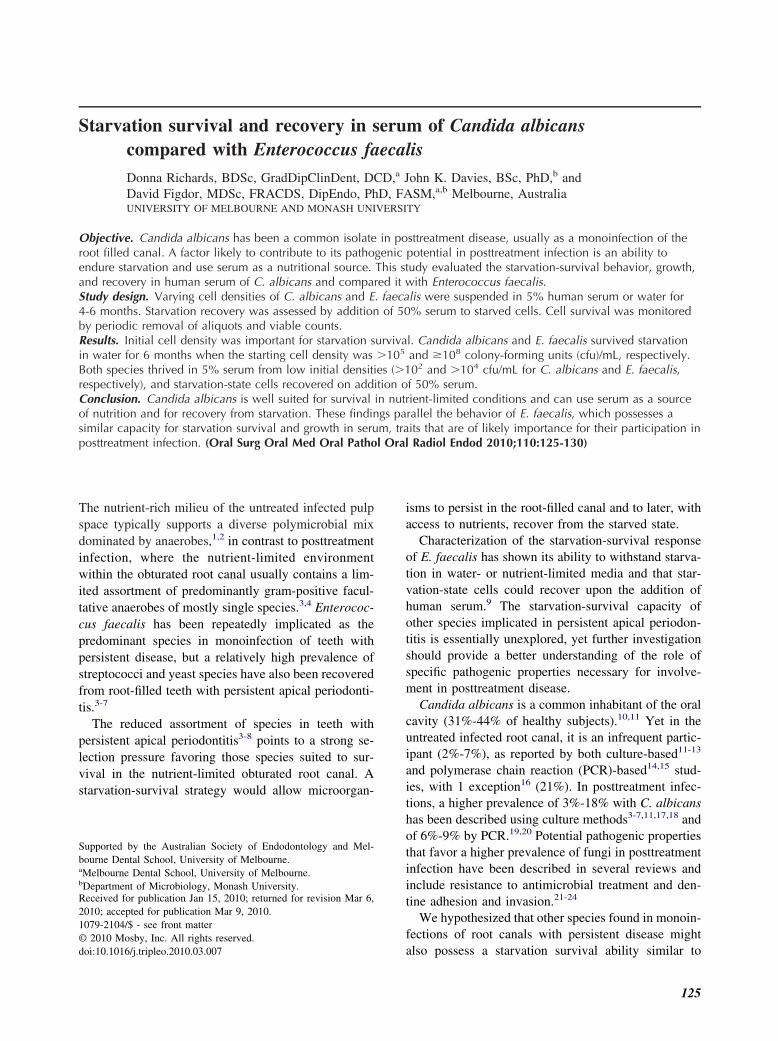
The kinetics of starvation survival for the loweststarting cell density of C. albicans and E. faecalis thatsurvived in water for 6 months are shown in Fig. 1.Candida albicans survived 6 months’ starvation in wa-ter if the initial cell density was �105 cfu/mL. Withstarting densities of 106-109 cfu/mL, there was a grad-ual decline in cell numbers, but at 6 months there was
still a viable cell population of �105 cfu/mL (data notshown). Candida albicans did not survive starvationbeyond 3 weeks if the starting density was �104
cfu/mL (data not shown).Enterococcus faecalis survived starvation in water
for 6 months if the initial cell density was �108 cfu/mLat the onset of starvation. At lower densities (106 and107 cfu/mL) cell survival was short-lived and no cellswere recovered at 5 and at 56 days, respectively (datanot shown).
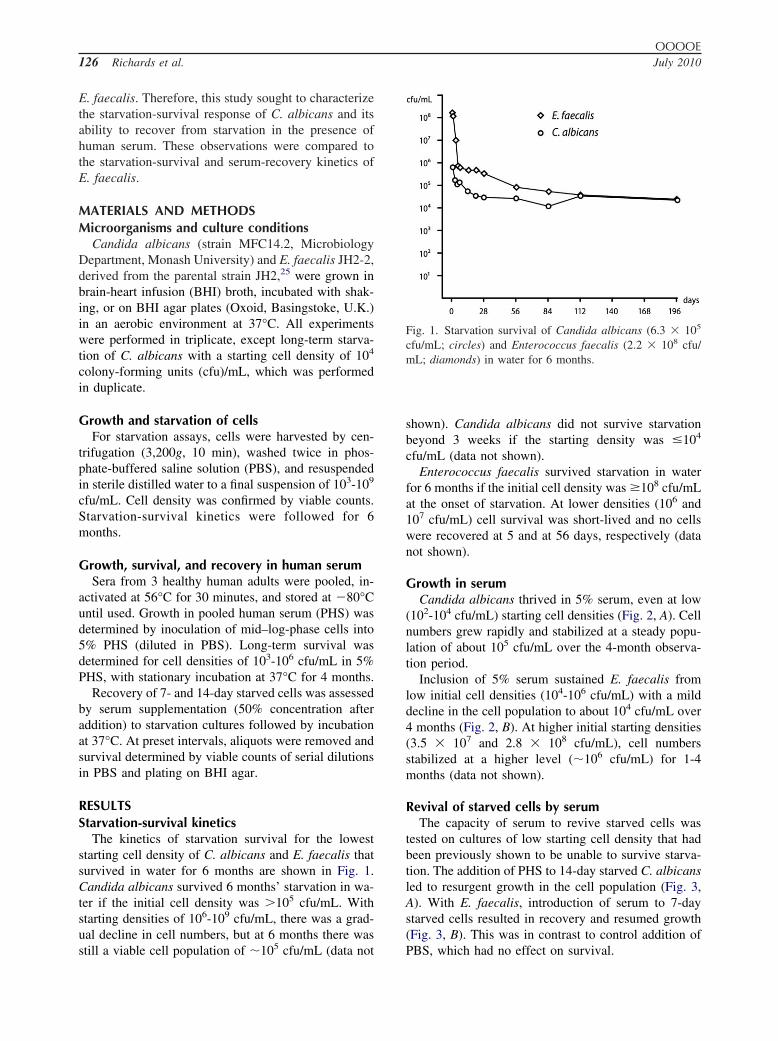
Growth in serumCandida albicans thrived in 5% serum, even at low
(102-104 cfu/mL) starting cell densities (Fig. 2, A). Cellnumbers grew rapidly and stabilized at a steady popu-lation of about 105 cfu/mL over the 4-month observa-tion period.
Inclusion of 5% serum sustained E. faecalis fromlow initial cell densities (104-106 cfu/mL) with a milddecline in the cell population to about 104 cfu/mL over4 months (Fig. 2, B). At higher initial starting densities(3.5 � 107 and 2.8 � 108 cfu/mL), cell numbersstabilized at a higher level (�106 cfu/mL) for 1-4months (data not shown).
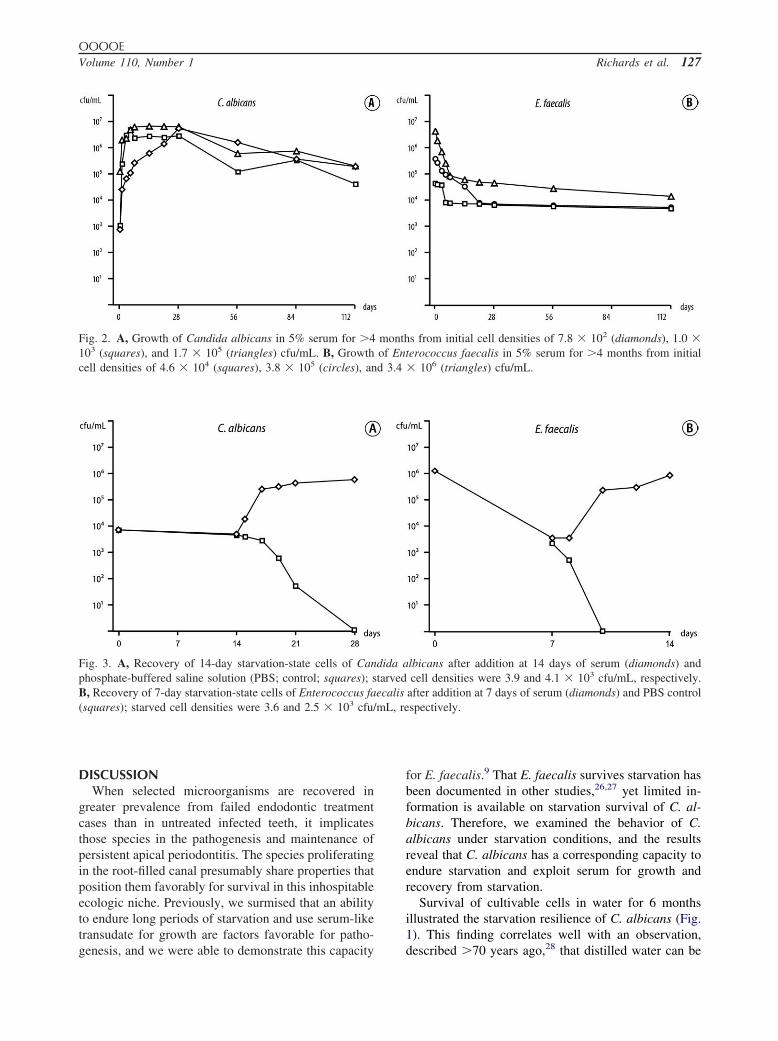
Revival of starved cells by serumThe capacity of serum to revive starved cells was
tested on cultures of low starting cell density that hadbeen previously shown to be unable to survive starva-tion. The addition of PHS to 14-day starved C. albicansled to resurgent growth in the cell population (Fig. 3,A). With E. faecalis, introduction of serum to 7-daystarved cells resulted in recovery and resumed growth(Fig. 3, B). This was in contrast to control addition of
Fig. 1. Starvation survival of Candida albicans (6.3 � 105
cfu/mL; circles) and Enterococcus faecalis (2.2 � 108 cfu/mL; diamonds) in water for 6 months.
PBS, which had no effect on survival.
d 3.4
mL, re
OOOOEVolume 110, Number 1 Richards et al. 127
DISCUSSIONWhen selected microorganisms are recovered in
greater prevalence from failed endodontic treatmentcases than in untreated infected teeth, it implicatesthose species in the pathogenesis and maintenance ofpersistent apical periodontitis. The species proliferatingin the root-filled canal presumably share properties thatposition them favorably for survival in this inhospitableecologic niche. Previously, we surmised that an abilityto endure long periods of starvation and use serum-liketransudate for growth are factors favorable for patho-
Fig. 2. A, Growth of Candida albicans in 5% serum for �4103 (squares), and 1.7 � 105 (triangles) cfu/mL. B, Growthcell densities of 4.6 � 104 (squares), 3.8 � 105 (circles), an
Fig. 3. A, Recovery of 14-day starvation-state cells of Canphosphate-buffered saline solution (PBS; control; squares); sB, Recovery of 7-day starvation-state cells of Enterococcus fa(squares); starved cell densities were 3.6 and 2.5 � 103 cfu/
genesis, and we were able to demonstrate this capacity
for E. faecalis.9 That E. faecalis survives starvation hasbeen documented in other studies,26,27 yet limited in-formation is available on starvation survival of C. al-bicans. Therefore, we examined the behavior of C.albicans under starvation conditions, and the resultsreveal that C. albicans has a corresponding capacity toendure starvation and exploit serum for growth andrecovery from starvation.
Survival of cultivable cells in water for 6 monthsillustrated the starvation resilience of C. albicans (Fig.1). This finding correlates well with an observation,
s from initial cell densities of 7.8 � 102 (diamonds), 1.0 �erococcus faecalis in 5% serum for �4 months from initial� 106 (triangles) cfu/mL.
lbicans after addition at 14 days of serum (diamonds) andcell densities were 3.9 and 4.1 � 103 cfu/mL, respectively.
after addition at 7 days of serum (diamonds) and PBS controlspectively.
monthof Ent
dida atarvedecalis
described �70 years ago,28 that distilled water can be

OOOOE128 Richards et al. July 2010
used as a storage method for yeasts and further reportsshowing long-term survival of C. albicans in distilledwater for 3-10 years.29-31 Cell density at starvationonset was a significant factor for survival, where �105
cfu/mL was favorable for survival but at lower celldensities (�104 cfu/mL) C. albicans could not survivestarvation beyond 3 weeks. The capacity of C. albicansfor starvation survival compares favorably with E. fae-calis, which can endure many months of starvation.9,27
Starvation survival for E. faecalis is also highly con-tingent on cell density at starvation onset.9,32 Interest-ingly, C. albicans survived starvation from a signifi-cantly lower initial cell density than E. faecalis (105
compared with 108 cfu/mL, respectively), which sug-gests a superior capacity for sustaining itself in limitednumbers under nutrient-limited conditions.
Although there is no information regarding potentialnutrition sources available for microorganisms embed-ded in the obturated canal, it is not unreasonable tothink that tissue fluid derived from the periapical tissuesmay enter the root canal space to provide suitablesubstrate. Indeed, ultrastructural investigations of mi-crobes involved in failed treatment cases have demon-strated that they are predominantly located in the apicalregion of the root canal space in voids or accessorycanals.33,34 The proximity of microorganisms to theperiapical granuloma is consistent with the idea thatthese microorganisms may be activated by adjacenthost molecules35 and derive their nutrition from peria-pical tissue fluid. Therefore, we evaluated the potentialof C. albicans and E. faecalis to use serum for survivaland for recovery of starved cells.
Human serum sustained growth of both species formore than 6 months. Just 5% serum was enough tosupport growth of low numbers (7.8 � 102 cfu/mL) ofC. albicans. Similarly, the results for growth of E.faecalis in human serum showed that 5% serum sus-tained the cells from low starting cell densities (4.6 �104 cfu/mL) for �4 months, which correlated well withthe findings of a previous study.9 Thus, the availabilityof even a low concentration of serum has the potentialto dramatically prolong survival of low cell numbers ofboth species, compared with the requirement for asignificantly higher starting density to endure absolutestarvation.
In starvation-recovery experiments, starved C. albi-cans cells were rapidly revived by addition of 50%serum. Similarly, starved E. faecalis recovered with thenutritional support of serum, as shown previously.9
These results illustrate that the gradual demise of asmall population of starved cells can be effectivelyreversed if they have the fortune to encounter a nutri-
tional upshift from serum.Environmental conditions, e.g., interaction with cal-cium and collagen components in dentin,36 have thepotential to influence fungal behavior and growth form.Candida albicans grows in different forms, such asgerm tubes, yeasts (blastospores), pseudo- and truehyphae, and chlamydospores, and, depending on theenvironmental cues, switching may occur among thesemorphotypes (except chlamydospores).36 Starvationand revival of starved C. albicans cells may inducemorphologic switching, including to a hyphal growthform where cells remain attached to each other afterdivision.24,37,38 Because cell survival was determinedby enumeration of colony-forming units on non-Can-dida–specific plates, there was a possibility of an un-derestimation of cell numbers if starved C. albicanswere not discrete single cells but had switched to achained filamentous form.
Cell survival was assessed by colony growth onplates, which remains the gold standard for assessmentof cell viability. Alternative approaches, such as cellstaining, offer the potential of defining viable cells byvisible fluorescence of intact cell membranes but havetheir own shortcomings, including nonspecific bind-ing39 and the potential for false association with via-bility.40
It is worth noting that a single strain each was se-lected for study and that other strains may show differ-ent morphologic, physiologic, and phenotypic proper-ties. Nevertheless, in an earlier study, E. faecalisshowed similar starvation survival kinetics when 2strains were compared9 and the present results for C.albicans are consistent with previous studies that dem-onstrate long-term survival of the species in water.29-31
In the root-filled canal, microorganisms may be in-terred within dentin, filling material, adjacent voids, oranatomic ramifications separate from the main rootcanal. Nutrient availability at these sites is likely tovary from substrate replete to complete starvation. Asshown recently, some prevalent endodontic pathogenscannot survive starvation, and the prospects of survivaldepend on a higher level of serum as a nutritionalsource.41 Whether individual species will endure andhave the possibility to participate in posttreatment dis-ease depends on many factors, but the present findings,in conjunction with other studies,9,41 show that cellnumbers, starvation-survival capacity of the species,and availability of even low amounts of serum willlikely influence their fate.
In conclusion, this study has shown that C. albicansexhibits starvation survival behavior similar to E. fae-calis. Both species are capable of starvation survival for�6 months and are able to use low levels of serum for
growth. These characteristics are conducive to species
OOOOEVolume 110, Number 1 Richards et al. 129
survival and contribution to posttreatment apical peri-odontitis.
The authors thank Dr. Sally Turner for research adviceand Prof. Göran Sundqvist for valuable criticism.
REFERENCES1. Sundqvist G. Bacteriological studies of necrotic dental pulps.
Umeå University odontological dissertations no. 7. Umeå (Swe-den): Umeå University; 1976.
2. Sundqvist G. Taxonomy, ecology, and pathogenicity of the rootcanal flora. Oral Surg Oral Med Oral Pathol Oral Radiol Endod1994;78:522-30.
3. Sundqvist G, Figdor D, Persson S, Sjögren U. Microbiologicanalysis of teeth with failed endodontic treatment and the out-come of conservative re-treatment. Oral Surg Oral Med OralPathol Oral Radiol Endod 1998;85:86-93.
4. Molander A, Reit C, Dahlén G, Kvist T. Microbiological statusof root-filled teeth with apical periodontitis. Int Endod J1998;31:1-7.
5. Hancock HH, III, Sigurdsson A, Trope M, Moiseiwitsch J. Bac-teria isolated after unsuccessful endodontic treatment in a NorthAmerican population. Oral Surg Oral Med Oral Pathol OralRadiol Endod 2001;91:579-86.
6. Peciuliene V, Reynaud AH, Balciuniene I, Haapasalo M. Isola-tion of yeasts and enteric bacteria in root-filled teeth with chronicapical periodontitis. Int Endod J 2001;34:429-34.
7. Pinheiro ET, Gomes BP, Ferraz CC, Sousa EL, Teixeira FB,Souza-Filho FJ. Microorganisms from canals of root-filled teethwith periapical lesions. Int Endod J 2003;36:1-11.
8. Peciuliene V, Balciuniene I, Eriksen HM, Haapasalo M. Isolationof Enterococcus faecalis in previously root-filled canals in aLithuanian population. J Endod 2000;26:593-5.
9. Figdor D, Davies JK, Sundqvist G. Starvation survival, growthand recovery of Enterococcus faecalis in human serum. OralMicrobiol Immunol 2003;18:234-9.
10. Arendorf TM, Walker DM. The prevalence and intra-oral distri-bution of Candida albicans in man. Arch Oral Biol 1980;25:1-10.
11. Egan MW, Spratt DA, Ng YL, Lam JM, Moles DR, GulabivalaK. Prevalence of yeasts in saliva and root canals of teeth asso-ciated with apical periodontitis. Int Endod J 2002;35:321-9.
12. Lana MA, Ribeiro-Sobrinho AP, Stehling R, Garcia GD, SilvaBK, Hamdan JS, et al. Microorganisms isolated from root canalspresenting necrotic pulp and their drug susceptibility in vitro.Oral Microbiol Immunol 2001;16:100-5.
13. Möller ÅJR. Microbiological examination of root canals andperiapical tissues of human teeth. Methodological studies. Od-ontol Tidsk 1966;74:(Suppl):1-380.
14. Siqueira JF Jr, Rôças IN, Moraes SR, Santos KR. Direct ampli-fication of rRNA gene sequences for identification of selectedoral pathogens in root canal infections. Int Endod J 2002;35:345-51.
15. Siqueira JF Jr, Rôças IN, Lopes HP. Patterns of microbial col-onization in primary root canal infections. Oral Surg Oral MedOral Pathol Oral Radiol Endod 2002;93:174-8.
16. Baumgartner JC, Watts CM, Xia T. Occurrence of Candidaalbicans in infections of endodontic origin. J Endod 2000;26:695-8.
17. Cheung GS, Ho MW. Microbial flora of root canal-treated teethassociated with asymptomatic periapical radiolucent lesions.
Oral Microbiol Immunol 2001;16:332-7.18. Waltimo TM, Sirén EK, Torkko HL, Olsen I, Haapasalo MP.Fungi in therapy-resistant apical periodontitis. Int Endod J1997;30:96-101.
19. Siqueira JF Jr, Rôças IN. Polymerase chain reaction-based anal-ysis of microorganisms associated with failed endodontic treat-ment. Oral Surg Oral Med Oral Pathol Oral Radiol Endod2004;97:85-94.
20. Rôças IN, Hülsmann M, Siqueira JF, Jr. Microorganisms in rootcanal-treated teeth from a German population. J Endod 2008;34:926-31.
21. Waltimo TM, Sen BH, Meurman JH, Ørstavik D, Haapasalo MP.Yeasts in apical periodontitis. Crit Rev Oral Biol Med 2003;14:128-37.
22. Siqueira JF, Jr, Sen BH. Fungi in endodontic infections.Oral Surg Oral Med Oral Pathol Oral Radiol Endod 2004;97:632-41.
23. McCullough MJ, Ross BC, Reade PC. Candida albicans: areview of its history, taxonomy, epidemiology, virulence at-tributes, and methods of strain differentiation. Int J Oral Maxil-lofac Surg 1996;25:136-44.
24. Calderone R, Suzuki S, Cannon R, Cho T, Boyd D, Calera J, etal. Candida albicans: adherence, signaling and virulence. MedMycol 2000;38 Suppl 1:125-37.
25. Jacob AE, Hobbs SJ. Conjugal transfer of plasmid-borne multi-ple antibiotic resistance in Streptococcus faecalis var. zymo-genes. J Bacteriol 1974;117:360-72.
26. Giard JC, Hartke A, Flahaut S, Boutibonnes P, Auffray Y.Glucose starvation response in Enterococcus faecalis JH2-2:survival and protein analysis. Res Microbiol 1997;148:27-35.
27. Sedgley CM, Lennan SL, Appelbe OK. Survival of Enterococcusfaecalis in root canals ex vivo. Int Endod J 2005;38:735-42.
28. Castellani A. The viability of some pathogenic fungi in steriledistilled water. J Trop Med Hyg 1939;42:225-6.
29. Hartung de Capriles C, Mata S, Middelveen M. Preservation offungi in water (Castellani): 20 years. Mycopathologia 1989;106:73-9.
30. McGinnis MR, Padhye AA, Ajello L. Storage of stock cultures offilamentous fungi, yeasts, and some aerobic actinomycetes insterile distilled water. Appl Microbiol 1974;28:218-22.
31. Odds FC. Long-term laboratory preservation of pathogenicyeasts in water. J Med Vet Mycol 1991;29:413-5.
32. del Mar Lleò M, Pierobon S, Tafi MC, Signoretto C, Canepari P.mRNA detection by reverse transcription-PCR for monitoringviability over time in an Enterococcus faecalis viable but non-culturable population maintained in a laboratory microcosm.Appl Environ Microbiol 2000;66:4564-7.
33. Nair PNR, Sjögren U, Krey G, Kahnberg K-E, Sundqvist G.Intraradicular bacteria and fungi in root-filled, asymptomatichuman teeth with therapy-resistant periapical lesions: a long-term light and electron microscopic follow-up study. J Endod1990;16:580-8.
34. Nair PNR. On the causes of persistent apical periodontitis: areview. Int Endod J 2006;39:249-81.
35. Pendrak ML, Yan SS, Roberts DD. Sensing the host environ-ment: recognition of hemoglobin by the pathogenic yeast Can-dida albicans. Arch Biochem Biophys 2004;426:148-56.
36. Sen BH, Safavi KE, Spangberg LS. Growth patterns of Candidaalbicans in relation to radicular dentin. Oral Surg Oral Med OralPathol Oral Radiol Endod 1997;84:68-73.
37. Brown DH Jr, Giusani AD, Chen X, Kumamoto CA. Filamen-tous growth of Candida albicans in response to physical envi-ronmental cues and its regulation by the unique CZF1 gene. Mol
Microbiol 1999;34:651-62.
OOOOE130 Richards et al. July 2010
38. Tripathi G, Wiltshire C, Macaskill S, Tournu H, Budge S, BrownAJ. Gcn4 co-ordinates morphogenetic and metabolic responsesto amino acid starvation in Candida albicans. EMBO J 2002;21:5448-56.
39. Biggerstaff JP, Le Puil M, Weidow BL, Prater J, Glass K,Radosevich M, et al. New methodology for viability testing inenvironmental samples. Mol Cell Probes 2006;20:141-6.
40. Renye JA, Jr, Piggot PJ, Daneo-Moore L, Buttaro BA. Persis-tence of Streptococcus mutans in stationary-phase batch culturesand biofilms. Appl Environ Microbiol 2004;70:6181-7.
41. Brundin M, Figdor D, Sundqvist G, Sjögren U. Starvation
response and growth in serum of Fusobacterium nucleatum,Peptostreptococcus anaerobius, Prevotella intermedia, andPseudoramibacter alactolyticus. Oral Surg Oral Med Oral PatholOral Radiol Endod 2009;108:129-34.
Reprint requests:
Dr. David Figdor517 St Kilda RoadMelbourne, VIC 3004Australia
[email protected]