state of the art - ubc critical care medicine
TRANSCRIPT

State of the Art
Ischemia–Reperfusion–induced Lung InjuryMarc de Perrot, Mingyao Liu, Thomas K. Waddell, and Shaf Keshavjee
Toronto Lung Transplant Program and Thoracic Surgery Research Laboratory, Toronto General Hospital, University Health Network,University of Toronto, Toronto, Ontario, Canada
Ischemia–reperfusion–induced lung injury is characterized by non- diseases. The last decade has been marked by both a significantspecific alveolar damage, lung edema, and hypoxemia occurring increase in the number of centers performing lung transplanta-within 72 hours after lung transplantation. The most severe form tion and in the number of recipients on the waiting list. Themay lead to primary graft failure and remains a significant cause Registry of the International Society for Heart and Lung Trans-of morbidity and mortality after lung transplantation. Over the past plantation reported in 2002 that almost 15,000 lung transplantsdecade, better understanding of the mechanisms of ischemia– have been performed worldwide and that more than 1,500 lungreperfusion injury, improvements in the technique of lung preserva- transplants are performed annually (1).tion, and the development of a new preservation solution specifi- Despite refinements in lung preservation and improvementscally for the lung have been associated with a reduction in the
in surgical techniques and perioperative care, ischemia–reper-incidence of primary graft failure from approximately 30 to 15%fusion–induced lung injury remains a significant cause of earlyor less. Several strategies have also been introduced into clinicalmorbidity and mortality after lung transplantation. The syn-practice for the prevention and treatment of ischemia–reperfusion–drome typically occurs within the first 72 hours after transplanta-induced lung injury with various degrees of success. However, onlytion and is characterized by nonspecific alveolar damage, lungthree randomized, double-blinded, placebo-controlled trials onedema, and hypoxemia. The clinical spectrum can range fromischemia–reperfusion–induced lung injury have been reported inmild hypoxemia associated with few infiltrates on chest X-raythe literature. In the future, the development of new agents andto a picture similar to full-blown acute respiratory distress syn-their application in prospective clinical trials are to be expected to
prevent the occurrence of this potentially devastating complication drome requiring positive pressure ventilation, pharmacologicand to further improve the success of lung transplantation. therapy, and occasionally extracorporeal membrane oxygen-
ation (2). A number of terms have been used to describe thisKeywords: lung transplantation; primary graft failure; acute lung injury; syndrome, but ischemia–reperfusion injury is most commonlyearly graft dysfunction; lung preservation used, with primary graft failure attributed to the most severe
form of injury that frequently leads to death or prolonged me-CONTENTS chanical ventilation beyond 72 hours (Table 1). In addition to
significant morbidity and mortality in the early postoperativeDonor Lung Assessmentperiod, severe ischemia–reperfusion injury can also be associatedEffect of Cold Ischemic Storagewith an increased risk of acute rejection that may lead to graftOxidative Stressdysfunction in the long term (3).Sodium Pump Inactivation
Primary graft failure is the end-result of a series of hits oc-Intracellular Calcium Overloadcurring from the time of brain death to the time of lung reperfu-Iron Releasesion after transplantation. Ischemia–reperfusion injury has beenCell Deathidentified as the main cause of primary graft failure. However,Consequences of Ischemia and Reperfusionother injuries occurring in the donor before the retrieval proce-Upregulation of Molecules on Cell Surface Membranedure can contribute to and amplify the lesions of ischemia andRelease of Proinflammatory Mediatorsreperfusion (Figure 1). Attention of lung transplant physiciansLeukocyte Activationhas therefore been focused on selective assessment of donorStrategies to Prevent Lung Dysfunctionlungs, effective technique of lung preservation, and careful man-Method of Lung Preservation and Reperfusionagement of transplanted lungs after reperfusion to reduce theClinical Evidence in Prevention and Treatment of Lungseverity of ischemia–reperfusion injury and the incidence of pri-Reperfusion Injurymary graft failure. Donor lung assessment is an attempt to selectFuture Strategies
Conclusions lungs that will be able to handle a period of several hours ofischemia without significant impairment in their function after
Since 1983, lung transplantation has enjoyed increasing success reperfusion. Unfortunately, currently only 10 to 30% of donorand has become the mainstay of therapy for most end-stage lung lungs are judged suitable for transplantation (4).
Lungs that have been selected for transplantation are gener-ally flushed with a preservation solution and hypothermicallypreserved to decrease their metabolic rate and energy require-
(Received in original form July 8, 2002; accepted in final form December 3, 2002) ment until implantation in the recipient. The period of coldCorrespondence and requests for reprints should be addressed to Shaf Keshavjee, ischemic storage is kept as short as possible and usually rangesM.D., Toronto Lung Transplant Program, Toronto General Hospital, 200 Elizabeth between 4 and 8 hours according to the location of the donor.Street, EN 10-224, Toronto, ON, M5G 2C4 Canada. E-mail: Shaf.Keshavjee@uhn. Although hypothermia is essential for organ storage, it is associ-on.ca
ated with a series of events such as oxidative stress, sodiumAm J Respir Crit Care Med Vol 167. pp 490–511, 2003
pump inactivation, intracellular calcium overload, iron release,DOI: 10.1164/rccm.200207-670SOInternet address: www.atsjournals.org and induction of cell death that may induce upregulation of

State of the Art 491
TABLE 1. TERMS USED TO DESCRIBEISCHEMIA–REPERFUSION–INDUCED LUNG INJURY
Reimplantation edemaReimplantation responseReperfusion injuryReperfusion edemaPrimary graft failureEarly graft dysfunction
molecules on the cell surface membrane and the release of proin-flammatory mediators that will eventually activate passenger(donor) and recipient leukocytes after reperfusion. Prolongedischemia may also result in a “no-reflow phenomenon” demon-strated by significant microvascular damages leading to persis-tent blood flow obstruction and subsequent ischemia despitereperfusion.
Over the past decade, numerous studies have been performedto optimize the technique of lung preservation. A new preserva-tion solution, which combines a low potassium concentrationand dextran, has also been developed specifically for the lungs Figure 1. Ischemia–reperfusion–induced lung injury may be aggravated(5, 6). Several strategies for the prevention and treatment of by a number of events occurring in the donor before lung retrieval.ischemia–reperfusion–induced lung injury have been introducedinto clinical practice and have translated into a reduction in theincidence of severe ischemia–reperfusion injury from approxi-mately 30 to 15% or less (7, 8). without significant effect on early outcome (10–13). The presence
This review will initially focus on donor lung assessment, then of bilateral infiltrates on chest X-rays, persistent pus at bronchos-copy, and signs of bronchoaspiration remain, however, strictthe effect of cold ischemic storage with its consequences aftercontraindications to the use of donor lungs for transplantationreperfusion will be reviewed, and finally the technique of lung(14). Table 2 defines the criteria for ideal and extended donorspreservation and the current strategies for prevention and treat-as well as some factors considered to be strict contraindicationsment of ischemia–reperfusion–induced lung injury will be pre-to the use of donor lungs for transplantation. It is recognized,sented.however, that one may chose to accept increased risk in usinglungs for recipients who are desperately ill.DONOR LUNG ASSESSMENT
The deleterious effect of brain stem death on organ functionThe success of lung preservation primarily depends on proper has been increasingly recognized over the last few years. Brainorgan selection. Currently, the parameters used to assess donor death can induce disruption in homeostatic regulation with pro-lungs are based on donor history, arterial blood gases, chest found disturbances in endocrine function and an intense in-X-ray appearance, bronchoscopy findings, and physical examina- flammatory reaction that may reduce the tolerance of the organstion of the lung at the time of retrieval (9). These parameters to handle a period of ischemia (15–17). Follette and colleaguesattempt to determine function and viability of the lungs, but have shown that a bolus of steroids (methylprednisolone approx-their accuracy in determining the risk of reperfusion injury is not imately 15 mg/kg) administered to all donors after brain deathoptimal and several centers have extended their donor selection declaration can improve PaO2 and increase lung donor recovery
(18). The steroid bolus can potentially reduce the inflammatorycriteria to the use of nonideal (i.e., extended or marginal) donors
TABLE 2. IDEAL, EXTENDED, AND MARGINAL DONOR SELECTION CRITERIA SUGGESTED BY THE TORONTO LUNGTRANSPLANT GROUP
Standard Criteria Extended Criteria ContraindicationsSelection Criteria (Ideal Donors) (Extended Donors) (Marginal Donors)
ABO compatibility Identical Compatible IncompatibleDonor history
Age, yr � 55 � 55Smoking history, pack-years � 20 � 20Chest trauma No trauma Localized trauma Extensive lung traumaDuration of mechanical ventilation, h � 48 � 48History of asthma No YesHistory of cancer No (except low-grade skin cancer Primary central nervous History of cancer
and carcinoma in situ) system tumorsSputum gram stain Negative PositiveOxygenation, mm Hg* � 300 � 300Chest X-ray Clear Localized abnormality Diffuse infiltratesBronchoscopy Clear Secretions in main airways Persistent pus/signs of aspiration
* Last blood gas performed in the operating room with an FIO2of 100% and positive end-expiratory pressure of 5 cm H2O.

492 AMERICAN JOURNAL OF RESPIRATORY AND CRITICAL CARE MEDICINE VOL 167 2003
reaction and compensate for the deficit in hypophyseal hormones Hence, in the lung, the oxidative stress resulting from ischemiashould be distinguished from the oxidative stress resulting fromobserved after brain death.
Comparison of organ donation from living and cadaveric hypoxia.Hypoxia and, ultimately, anoxia result in a sharp decrease ofdonors presents a unique opportunity to study the effect of brain
death on clinical outcome. Some authors have shown that kidney adenosine triphosphate (ATP) and a corresponding increase inthe ATP degradation product hypoxanthine, which generatesbiopsies from cadaveric kidney donors had significantly higher
levels of inflammatory cytokines, adhesion molecules, and HLA- superoxide when oxygen is reintroduced with reperfusion and/or ventilation. This phenomenon can occur in the lung whenDR than biopsies from living donors, and the expression of these
markers on tubular cells before transplantation was associated alveolar oxygen tension drops below 7 mm Hg during ischemia(31). It can be blocked by inhibitors of xanthine oxidase suchwith a higher incidence of primary graft dysfunction and early
acute rejection (19–21). In human lung transplantation, the che- as allopurinol (32, 33).Ischemia is characterized by the absence of blood flow intomokine interleukin (IL)-8 has been shown to be upregulated in
bronchoalveolar lavage and lung tissue from brain-dead donors, the lung, which can cause lipid peroxidation and oxidant injurydespite the presence of oxygen (29, 32). The mechanism of oxida-and the level was found to significantly correlate with the inci-
dence of primary graft failure after reperfusion (22, 23). Hence, tive stress is different from that occurring during anoxia–reoxygenation because it is not associated with ATP depletion,there is growing body of evidence suggesting that cadaveric
donors are exposed to inflammatory events due to brain death, and it can occur during the storage period (29, 30, 32). In addi-tion, it cannot be blocked by inhibitors of xanthine oxidaseprolonged intubation, episodes of infection and/or hypotension(32, 34).that may increase organ susceptibility to ischemia–reperfusion
The endothelium appears to be one of the predominantinjury and alloimmune responses. In the future, methods tosources of oxidants during nonhypoxic lung ischemia (34). Endo-rapidly assess the degree of inflammation in the lung, for instancethelial cells are highly sensitive to physical forces resulting fromby measuring the levels of proinflammatory cytokines and/orblood flow variation and are able to transform these mechanicaladhesion molecules may be extremely useful to determine theforces into electrical and biochemical signals (mechanotransduc-type of lung suitable for transplantation and the potential toler-tion) (35). The absence of the mechanical component of flowance to prolonged ischemia. These methods would help to reduceduring lung ischemia stimulates membrane depolarization ofthe incidence of primary graft failure and to optimize the useendothelial cells with the activation of NADPH oxidase, nuclearof organs available for transplantation.factor-�B, and calcium/calmodulin-dependent nitric oxide syn-thase (NOS) (34, 36). Other cells such as macrophages and/orEFFECT OF COLD ISCHEMIC STORAGEmarginated neutrophils, which are known to have a high
Hypothermia decreases metabolic rate. Therefore, biochemical NADPH oxidase activity, could also contribute to the lungreactions are reduced and the rate of degradation of essential oxidant burden that takes place during the ischemic storagecellular components necessary for organ viability is reduced. (37, 38).Most enzyme systems show a 1.5- to 2.0-fold decrease in activity
Sodium Pump Inactivationfor every 10�C decrease in temperature (24). However, althoughhypothermia is essential during organ storage, a number of The sodium (Na�/K�-ATPase) pump is important to preserveevents can still occur leading to activation of inflammatory medi- proper intracellular electrolyte concentration (high K�, lowators that are ultimately deleterious to the preserved organ at Na�) and to maintain adequate clearance of alveolar fluid. Hypo-the time of reperfusion. thermic storage results in the loss of function of the sodium
pump, which returns to normal activity with rewarming to 37�COxidative Stressif the epithelial cells are not damaged (39). The loss of function
Oxidative stress is characterized by the formation of reactive of the sodium pump results in accumulation of sodium in theoxygen species such as superoxide anion, hydrogen peroxide, cell resulting in cell swelling. This is associated with an influxand hydroxyl radical (25). These molecules, in particular the of chloride inside the cell and an efflux of K� out of the cell.hydroxyl radical, are highly unstable and react with the first Preservation solutions contain electrolytes and colloid to createstructure they encounter, usually the lipid component of the cell an osmotic pressure gradient in an attempt to prevent hypother-membrane. Cell injury produced by lipid peroxidation can range mia-induced cell swelling. Preservation of organs at 10�C hasfrom increased permeability to cell lysis. The generation of intra- been proved to be superior than at 4�C, and this has been attrib-cellular oxygen species has been found to be present in most lung uted to better preservation of function of the Na�/K�-ATPaseparenchymal cells, including endothelial cells, Type II alveolar activity (40). The sodium pump activity has also been shown toepithelial cells, Clara cells, and ciliated airway epithelial cells as resume better functional activity at the time of rewarming if thewell as in alveolar macrophages (26). Two important mechanisms lungs are preserved with extracellular-type preservation solu-lead to the production of reactive oxygen species (Figure 2). tions that contain low K� and high Na� concentrations (41).One results from the accumulation of hypoxanthine and the
Intracellular Calcium Overloadconversion of the enzyme xanthine dehydrogenase into xanthineoxidase during anoxia, with the degradation of hypoxanthine Hypothermic storage alters calcium metabolism in cells both byinto superoxide after reoxygenation (27). The other mechanism release of calcium from intracellular depots and by pathologicdepends on the NADPH oxidase system, which is present mainly influx through the plasma membrane (24). The alteration of pHon the membrane surface of neutrophils and monocytes/macro- and intracellular calcium concentration disrupt many intracellu-phages and catalyzes the reduction of oxygen into hydrogen lar processes causing cellular damage (24). Elevated cytosolicperoxide and superoxide anion (27). calcium can also enhance the conversion of xanthine dehydroge-
Commonly, ischemia–reperfusion corresponds to anoxia– nase to xanthine oxidase and potentiate the damaging effect ofreoxygenation in organ transplantation. However, the lung has free radicals on mitochondria (25).to be considered differently because it contains oxygen in the Support of a role for calcium overload in the mechanismalveoli during ischemic preservation. Alveolar oxygen helps of ischemia–reperfusion injury has been demonstrated by the
protective effect of verapamil, a calcium channel blocker, onmaintains aerobic metabolism and prevents hypoxia (28–30).

State of the Art 493
Figure 2. Formation of reactive oxygen species during isch-emia–reperfusion and anoxia–reoxygenation of the lung. Thelung has to be considered differently than any other organsbecause it contains oxygen in the alveoli during the ischemicperiod. Hence, the oxidative stress resulting from ischemiashould be distinguished from the oxidative stress resultingfrom hypoxia.
ischemic injury (42). The effect has been found to be optimal Cell Deathwhen it is administered to the donor before lung retrieval be- Using in situ terminal deoxynucleotidyl transferase-mediatedcause it can reduce lipid peroxidation during ischemia and pre- deoxyuridine triphosphate nick end-labeling staining as a markervent endothelial damage after reperfusion (42, 43). Similar re- of apoptosis, we have observed in human lung transplantationsults have been observed with other calcium channel blockers that lungs with excellent function and good clinical outcomesuch as nifedipine and diltiazem (44). have up to 30% of their cells undergoing apoptosis within 2
hours of reperfusion (54). Similar findings have been observedIron Releaseexperimentally after 6 and 12 hours of cold ischemic time in
Although iron is an essential element for all living cells, it can rats, whereas longer ischemic times were associated with a pre-be highly toxic under pathophysiologic or stress conditions be- ponderance of necrotic cells in lung tissue (55). In contrast tocause of its ability to participate in the generation of powerful necrosis, apoptosis is not present during ischemia, its presenceoxidants. In its free form, iron can cycle between the oxidized peaks rapidly after reperfusion and does not correlate with lung(Fe3�) and reduced state (Fe2�), and catalyze the transformation function (54–56).of hydrogen peroxide and superoxide into the highly reactive Apoptosis induction is triggered and modulated by two path-hydroxyl radical through the Fenton reaction (Figure 3). In addi- ways (Figure 4). The intrinsic pathway involves the mitochondriation, free iron facilitates the decomposition of lipid hydroperox- and is activated by reactive oxygen species, whereas the extrinsicides and accelerates the nonenzymatic oxidation of glutathione. pathway is activated by the ligation of death receptors withFree iron can be released from ferritin and cytochrome P-450 their ligands—such as tumor necrosis factor (TNF) with TNF-during ischemia by a number of factors such as acidosis, proteoly-
receptors and Fas with Fas-ligand (57). Although the first path-sis, and superoxide (45–47). In addition to tissue oxidation, ironway is activated in the early phase after reperfusion, the secondcan be released into the circulation where it can potentiallymay take up to several hours to induce apoptosis (58).activate platelet aggregation (46, 48).
Whether apoptotic cells have a deleterious impact on organThe importance of iron in promoting ischemia–reperfusionfunction remains controversial. Some authors have demon-injury has been demonstrated by the increased injury observedstrated that ischemia–reperfusion injury of kidneys and heartsin iron-supplemented tissue and by the protection offered byis reduced when antiapoptotic agents are injected before reperfu-the iron chelator, deferoxamine (45, 49, 50). Recently, a novelsion in mice models of warm ischemia (59, 60). However, otheriron chelator (desferriexochelin 772SM) has been shown to en-investigators have argued that by blocking the apoptotic molecu-hance the effect of a P-selectin antagonist in preventing isch-lar cascade after a period of brain ischemia, injured cells mayemia–reperfusion injury in a rat liver model (51). Lazaroids,not be able to recover but may instead continue to release proin-which are aminosteroids inhibiting iron-dependent lipid peroxi-flammatory agents and subsequently die by necrosis, a mode ofdation, have also shown good results in protecting the lung fromcell death more injurious to surrounding tissue (61). We haveischemia–reperfusion injury in most studies (52, 53).observed that for a similar amount of dead cells in the trans-planted lung, the presence of apoptotic cells was associated withbetter lung function than if the cells had died by necrosis (62).
Figure 3. Fenton reaction.CONSEQUENCES OF ISCHEMIA AND REPERFUSIONIron can cycle between the
oxidized and reduced state, Upregulation of Molecules on Cell Surface Membraneand catalyze the transforma-
Adhesion molecules. Adhesion molecules can be differentiatedtion of hydrogen peroxide andinto three major families, the selectins, the immunoglobulin su-superoxide into the highly re-
active hydroxyl radical. perfamily, and the integrins. Leukocyte emigration involves the

494 AMERICAN JOURNAL OF RESPIRATORY AND CRITICAL CARE MEDICINE VOL 167 2003
Figure 4. After receiving a death signal, cells can undergo either pro-grammed cell death or necrotic cell death. During the course of themost quiescent form of programmed cell death, “classical” apoptosis,caspase 8 and/or caspase 9 are activated through the external pathway(A ) or the mitochondrial pathway (B ), respectively. Both pathways leadto the activation of caspase 3. Cells, which fail to execute the “classical”apoptotic process, may either be salvaged and return to function, or
Figure 5. The potential mechanism of interaction between leukocyteundergo apoptosis-like or necrosis-like programmed cell death.activation and cytokine release during ischemia and reperfusion of thelung. Ischemia triggers the activation of passenger macrophages, whichrelease proinflammatory cytokines and mediate reperfusion injury dur-ing the early phase of reperfusion. IL-8, IL-12, IL-18, TNF-�, and IFN-�
sequential events of rolling, adherence, activation, and extrava- will then activate recipient neutrophils and T-lymphocytes, which willsation. Leukocyte rolling is dependent on selectin-mediated in- trigger the delayed phase of reperfusion injury and perpetuate lungteraction between endothelial cells (P-selectin and E-selectin) tissue damage. T-lymphocytes infiltrate lung tissue more rapidly thanand leukocytes (L-selectin). Firm adherence and activation of neutrophils and may also participate in the activation of recipient neutro-leukocytes occur when leukocyte �1-integrin or �2-integrin phils after reperfusion.binds to endothelial cells expressing intercellular adhesionmolecule-1 or vascular endothelial adhesion molecule-1, respec-tively. Finally, leukocyte extravasation into the tissue is de-pendent on integrin-immunoglobulin interactions, involving in- contact phase and the intrinsic pathway of the coagulation sys-tercellular adhesion molecule-1 and platelet endothelial cell tem, has been shown to improve early lung function and toadhesion molecule-1. reduce ischemia–reperfusion injury in a dog model of single lung
Adhesion molecules are upregulated on pulmonary endothe- transplantation (75). C1-esterase inhibitor has also been used tolial cells during ischemia, and blockade of adhesion molecules treat lung graft failure in two patients, but further clinical studiessuch as P-selectin, intercellular adhesion molecule-1, and CD18 are required to prove its efficiency (76).(�-chain of the �2-integrin) at the time of reperfusion can reduce Recent experiments have shown that mice placed in a hypoxiclung reperfusion injury (63–67). E-selectin and L-selectin block- environment suppress their fibrinolytic axis by increasing macro-ade may also be beneficial after several hours of reperfusion phage release of plasminogen activator inhibitor-1 and decreas-when neutrophils have a preponderant role (64, 68, 69). The use ing macrophage release of tissue-type plasminogen activator andof biostable analogs of the oligosaccharides Lewis X and Lewis urokinase-type plasminogen activator (77). Additional studies inA, which are potent ligands for selectin adhesion molecules, mice have shown that the beneficial effect of heme oxygenase-1,have also been shown to reduce ischemia–reperfusion injury carbon monoxide, and IL-10 during lung ischemia is partiallywhen given before reperfusion (70–72). mediated by their ability to potentiate the fibrinolytic axis (78,
Prothrombotic and antifibrinolytic factors. Hypoxia can in- 79). The role of prothrombotic and antifibrinolytic agents isduce endothelial cells and macrophages to develop procoagulant a relatively new area of investigation, and further studies areproperties, which may contribute to the formation of microvascu- required to determine more precisely the role of fibrinolyticlar thrombosis and impede the return of blood flow after reperfu- agents in ischemia–reperfusion injury of the lung.sion. In vitro studies have shown that endothelial cells subjected
Release of Proinflammatory Mediatorsto hypoxia can suppress their production of the anticoagulantCytokines. Clinical and experimental studies have shown thatcofactor thrombomodulin and increase their production of aischemia–reperfusion of solid organs such as the kidney (80),membrane-associated factor X activator (73). Tissue factor hasliver (81), heart (82), and lung (83) induces a rapid release ofalso been shown to be upregulated on endothelial cells and
macrophages by hypoxia and to play a significant role in modulat- proinflammatory cytokines (Table 3). In human lung trans-plantation, measurable amounts of pro- and antiinflammatorying ischemia–reperfusion injury in a warm ischemia liver model
(74). The administration of C1-esterase inhibitor, which inhibits cytokines such as TNF-�, IFN-�, IL-8, IL-10, IL-12, and IL-18can be measured in lung tissue during the cold ischemic time andthe classic pathway of the complement system as well as the

State of the Art 495
TABLE 3. SOURCE AND FUNCTION OF CYTOKINES POTENTIALLY INVOLVED IN REPERFUSIONINJURY DURING LUNG TRANSPLANTATION
Cytokine Main Cell Source Function
TNF-� Macrophages, lymphocytes ProinflammatoryIFN-� Lymphocytes ProinflammatoryMCP-1 Immune cells, and lung epithelial cells Macrophage chemotaxisIL-1� Macrophages, fibroblasts ProinflammatoryIL-2 Lymphocytes T cell proliferationIL-6 Macrophages, endothelial cells, and epithelial cells ProinflammatoryIL-8 Macrophages, epithelial cells, and fibroblasts Neutrophil chemotaxisIL-10 Macrophages, lymphocytes AntiinflammatoryIL-12 Macrophages T cell activationIL-18 Macrophages T cell activation
Definition of abbreviations: IL interleukin; MCP-1 macrophage chemoattractant protein-1; TNF tumor necrosis factor.
after reperfusion (23). Although most cytokine levels decreased hand, catalyzes leukotrienes such as leukotriene-B4, C4, D4, andafter reperfusion, the chemokine IL-8 significantly increased after E4, which can increase capillary permeability.reperfusion. Donor parameters including oxygen tension, cause Phospholipase A2 comprises a constantly growing family ofof brain death, smoking history, positive sputum cultures, and enzymes that have been divided into subgroups based on struc-time on ventilator did not appear to influence the cytokine levels. tural homology and numbered by their order of discovery (88).However, the age of the donor was inversely correlated with the These enzymes differ in cellular localization and mechanisms oflevels of IL-10 after reperfusion. Because IL-10 is an important release (88). Recently, Group II secretory phospholipase A2 hasantiinflammatory cytokine, this may explain why lungs from been found to play a major role in acute lung injury. Its levelolder donors might be more susceptible to ischemia–reperfusion has been found to be elevated in bronchoalveolar lavage fluidinjury and are associated with higher postoperative mortality from humans with acute respiratory distress syndrome (89), andrates (84). animal studies have shown that this form of phospholipase A2
A striking relationship between IL-8 levels and graft function induces acute lung injury after acid aspiration (90), intrachealcan also be observed after human lung transplantation (23). IL-8, injection of lipopolysaccharides (91), and after intestinal isch-which is a potent chemokine-promoting neutrophil migration emia–reperfusion injury (92). In addition, Group II secretoryand activation, rapidly increased after reperfusion. IL-8 levels phospholipase A2 has been shown to directly mediate surfactantin lung tissue 2 hours after reperfusion negatively correlated dysfunction in guinea pigs (91).with lung function assessed by the PaO2/FiO2 ratio and the mean To date, only few studies have analyzed the effect of phospho-airway pressure, and positively correlated with the Acute Physi- lipase A2 inhibitors in lung ischemia–reperfusion injury (93, 94).ology and Chronic Health Evaluation Score during the first 24 However, these inhibitors were not specific for Group II secre-postoperative hours in the intensive care unit (23). In addition, tory phospholipase A2, and they may well have blocked thewe and others have shown that high levels of IL-8 in donor lung generation of some PGs such as PGE2 and PGI2. Specific Grouptissue or bronchoalveolar lavage are associated with an increased II secretory phospholipase A2 inhibitors have been developedrisk of death from primary graft dysfunction after transplantation recently, and further studies should help elucidate this issue in(22, 23). The potential importance of IL-8 has also been demon- the future (95).strated in patients with acute respiratory distress syndrome (85) Platelet-activating factor can be released by a wide varietyand in clinical liver transplantation (86). In addition, Sekido of cells including macrophages, platelets, endothelial cells, mastand colleagues (87) have shown that the intravenous administra- cells, and neutrophils. It exerts its biological effects by activatingtion of anti–IL-8 antibody at the beginning of the reperfusion
the platelet-activating factor receptors, which consequently acti-period markedly reduces lung injury and neutrophil infiltrationvate leukocytes, stimulate platelet aggregation, and induce the3 hours after reperfusion in a rabbit model of warm lung isch-release of cytokines and the expression of cell adhesion mole-emia. The potential mechanism of interaction between leukocytecules (96). Platelet-activating factor has been difficult to analyzeactivation and cytokine release in ischemia–reperfusion injurybecause it is rapidly degraded by tissue and plasma platelet-during lung transplantation is shown in Figure 5.activating factor acetylhydrolases. Because there are no specificLipids. Cellular injury is accompanied by a rapid remodelinginhibitors for the biosynthesis of platelet-activating factor, mostof membrane lipids with the generation of bioactive lipids thatstudies have shown the importance of platelet-activating factorcan serve as both intra- and/or extracellular mediators. Phospho-by blocking its receptor.lipases such as phospholipase A2, phospholipase C, phospholi-
Platelet-activating factor has been shown to play a critical rolepase D, and sphingomyelinase play a pivotal role in the genera-in initiating lung injury. The most direct evidence was publishedtion of these lipid mediators. Among them, phospholipase A2recently by Nagase and colleagues who demonstrated that plate-has been detected in a wide variety of inflammatory conditionslet-activating factor receptor knockout mice developed less se-such as ischemia–reperfusion.vere acute lung injury after acid aspiration, whereas the overex-The activation of phospholipase A2 induces the productionpression of platelet-activating factor receptor in transgenic miceof platelet-activating factor, an extraordinarily potent mediatorexaggerated the injury (97). A number of studies have demon-of inflammation, and mobilizes arachidonic acid from the mem-strated that the administration of antagonists of platelet-activat-brane lipid pool, which will then be degraded by two majoring factor during the ischemic storage and after reperfusionpathways into eicosanoids. The potent vaso- and bronchocons-reduce ischemia–reperfusion injury and improve lung functiontrictor thromboxane A2 as well as various prostaglandins (PGs)(98–100). Similar results have been observed when platelet-acti-such as PGD2, PGE2, PGF2, and PGI2 are produced via the cyclo-
oxygenase pathway. The lipoxygenase pathway, on the other vating factor acetylhydrolase was administered to the flush solu-

496 AMERICAN JOURNAL OF RESPIRATORY AND CRITICAL CARE MEDICINE VOL 167 2003
tion and after reperfusion to increase the degradation rate of tion in the expression of inducible NOS and a lower proportionof apoptotic cells in the lung after reperfusion (121).the molecule (101).
Arachidonic acid metabolites such as leukotrienes and throm-Leukocyte Activationboxanes have been shown to increase in the lung during isch-
emia–reperfusion injury in a dog model of warm ischemia (102, Experimental and clinical evidence suggest that ischemia–103). Thromboxanes may contribute to reperfusion injury and reperfusion injury occurs in a biphasic pattern. The early phaseexacerbate lung edema (104). In addition, mast cells, which are of reperfusion, which depends primarily on donor characteristics,known to release large amounts of leukotrienes and histamine, and the delayed phase of reperfusion, which occurs over theare increased in number after lung ischemia and reperfusion ensuing 24 hours and depends primarily on recipient factors(102). The administration of mast cell membrane-stabilizing (122). Donor/passenger macrophages are activated during isch-agents have also been shown to improve lung function after emia and mediate the early phase of reperfusion injury, whereasreperfusion, indirectly demonstrating the importance of leuko- recipient lymphocytes and neutrophils are primarily involved intrienes (105, 106). the delayed phase of reperfusion injury (123–126). The recruit-
Complement. Studies in ischemia–reperfusion injury of the ment of lymphocytes and neutrophils into the lung results fromlung have shown that activation of the complement system after the release of cytokines and other mediators before and afterreperfusion may lead to cellular injury through direct and indi- reperfusion (23). The potential mechanism of interaction be-rect mechanisms (107, 108). Products of complement activation tween the cytokine release and the activation of macrophages,cause smooth muscle contraction and increased vascular perme- lymphocytes, and neutrophils during ischemia–reperfusion in-ability and induce degranulation of phagocytic cells, mast cells, jury in lung transplantation is shown in Figure 5.and basophils (109). The activated complement fragment C5a Macrophages. Alveolar macrophages can produce a largeis also capable of amplifying the inflammatory response via its number of cytokines and procoagulant agents in vitro in responsechemoattractant properties, its induction of granule secretion in to oxidative stress (74, 127). In an in vivo model of warm isch-phagocytes, and its ability to induce neutrophil and monocyte/ emia, Eppinger and colleagues demonstrated the importance ofmacrophage generation of toxic oxygen metabolites (110). Acti- TNF-�, IFN-�, and macrophage chemoattractant protein-1 invation of complement fragments C3 and C5 is also essential for the early phase of reperfusion and suggested that alveolar macro-the activation of the complement cascade and the generation of phages could have an important role immediately after reperfu-the membrane attack complex, which leads to direct cell lysis sion (126). Fiser and colleagues recently confirmed this hypothe-(111). sis by specifically inhibiting pulmonary passenger macrophages
Complement receptor-1 is a natural complement antagonist with gadolinium chloride injected into the donor before a periodthat has been cloned and the transmembrane portion removed of cold ischemia (125). They showed that lungs, in which passen-to obtain a soluble form of complement receptor-1. This soluble ger macrophages were inhibited, had significantly better functionform suppresses complement activation in vivo by inhibiting C3 immediately after reperfusion and this was independent of neu-and C5 convertases, which prevent the activation of both the trophil inhibition.classic and alternative pathways. In a swine single lung transplant Lymphocytes. Evidence suggests that lymphocytes may havemodel, we and others have shown that the administration of an important role in ischemia–reperfusion injury. Richter andsoluble complement receptor-1 to the recipient before reperfu- colleagues demonstrated that human lung donor parenchymasion reduced lung edema, decreased neutrophil accumulation, contains a large number of passenger macrophages and activatedand improved oxygenation of the transplanted lung (112, 113). lymphocytes, among which T cells and natural killer cells pre-Recently, Stammberger and colleagues have demonstrated in a dominate (128). Similar findings have been observed in liverrat lung transplant model that the administration of a molecule transplantation with a large number of activated CD8� T cells,combining soluble complement receptor-1 with sialyl Lewis X, � T cells, and natural killer cells being transmitted with thea selectin receptor antagonist, can achieve even better results liver graft to the recipient (129–132). Although the role of thesethan the administration of soluble complement receptor-1 alone passenger lymphocytes has not been extensively explored in(72). This study highlights the fact that several pathways may the setting of ischemia–reperfusion injury, recent studies haveneed to be blocked to address the redundancy of the inflamma- demonstrated that nude mice, CD4�/CD8� knockout mice, andtory system. CD4� depleted mice have significantly less severe reperfusion
Endothelin. Endothelins are powerful vasoconstrictors—10 injury of the liver and kidney than control mice (123, 133, 134).times more active than angiotensin II or vasopressin (114). Three In addition, Clavien and colleagues have shown in an ex vivoisoforms have been described in human and other mammals, model of liver reperfusion that cold preservation induces anendothelin-1, endothelin-2, and endothelin-3, of which endothe- increase in lymphocyte adherence within the first 10 minuteslin-1 has been most extensively studied because it is released by of reperfusion and that these infiltrating lymphocytes could beendothelial cells and smooth muscle cells and its expression is important in mediating graft dysfunction (135). We have recentlypredominant in the lung (114). In addition to being a potent demonstrated by flow cytometry in a rat lung transplant modelvasoconstrictor, endothelin-1 can stimulate the production of that recipient CD4� T cells rapidly accumulate in lung tissuecytokines by monocytes/macrophages and promote the retention after reperfusion. CD4� T cells then upregulate CD25, a poten-of neutrophils in the lung (115). tial marker of activation, and participate in ischemia–reperfusion
Clinical and experimental studies in lung transplantation have injury in the delayed phase of reperfusion by releasing IFN-�shown that endothelin-1 can accumulate in lung tissue before (136).and during the first few hours after reperfusion (116, 117). High Neutrophils. Neutrophils progressively infiltrate the trans-levels of endothelin-1 can then lead to an increased expression planted lung during the initial 24 hours of reperfusion (137).
Although they certainly play an important role in perpetuatingof vascular endothelial growth factor and increase vascular per-meability (118). The role of endothelin-1 in ischemia–reper- reperfusion injury, their function in the early phase of reperfu-
sion is less predominant. Using an isolated rat lung perfusionfusion injury has been demonstrated by the improvement in lungfunction when endothelin receptor antagonists are administered model, Deeb and colleagues demonstrated that the addition of
neutrophils to the perfusion system was not necessary for thebefore or during reperfusion (100, 119, 120). The administrationof endothelin-1 receptor antagonist was associated with a reduc- induction of reperfusion injury after a period of warm ischemia

State of the Art 497
TABLE 4. COMPOSITION OF PRESERVATION SOLUTIONS
Composition Euro-Collins University of Wisconsin Celsior LPD-Glucose (Perfadex)
Sodium 10 28 100 138Potassium 115 125 15 6Chloride 15 0 41.5 142Magnesium 0 0 13 0.8Sulfate 0 4 0 0.8Phosphate 57.5 25 0 0.8Calcium 0 0 0.26 0.3Bicarbonate 10 5 0 1Dextran 40 0 0 0 50Glucose 3.5 0 0 0.9Raffinose 0 30 0 0Lactobionate 0 100 80 0Glutathione 0 3 3 0Adenosine 0 5 0 0Allopurinol 0 1 0 0Pentafraction 0 50 0 0Glutamate 0 0 20 0Histidine 0 0 30 0Mannitol 0 0 60 0
Definition of abbreviation: LPD low-potassium dextran.All units are in mmol/L except Dextran 40, glucose, and pentafraction, which are in g/L.
(138). Following this line of experimentation, the same group in the inflated lungs and allowed safe extension of the ischemictime to 24 hours in dogs (28). Steen and colleagues, as welldemonstrated that reperfusion injury exhibits a bimodal pattern,
consisting of neutrophil-independent events during the first few as other groups, repeated these experiments and found safepulmonary preservation for 12 to 24 hours with LPD-glucose inhours of reperfusion and of neutrophil-mediated events after 4
hours of reperfusion (124). Further studies with specific antibod- porcine, canine, and primate models of left single and doublelung transplantation (144–147).ies against neutrophils have confirmed these findings and show
that other leukocytes such as macrophages have a more impor- Ultrastructural analyses have shown significantly better con-servation of lung integrity with extracellular-type preservationtant role in the early phase of reperfusion (125, 139, 140).solutions than with intracellular-based solutions (148). Betterultrastructural appearance may not translate into better lungSTRATEGIES TO PREVENT LUNG DYSFUNCTIONfunction after short ischemic periods, but after prolonged isch-
Method of Lung Preservation and Reperfusion emic time, i.e., 8 hours or longer, lungs preserved with LPDLung preservation solution. Currently, the vast majority of cen- solution have always shown better lung function than lungs pre-ters have adopted a single pulmonary artery flush to preserve served with intracellular-type preservation solutions (149–151).the lungs because of its technical simplicity (141). Preservation Celsior, which is an extracellular-type preservation solutionsolutions that have been studied include mainly intracellular- specifically developed for the heart, has also been shown totype solutions (high K�, low Na� solutions) such as Euro-Collins achieve satisfactory results in lung preservation (152–155). Someand University of Wisconsin solution, and extracellular-type so- authors have suggested that Celsior might even be better thanlutions (low K�, high Na� solutions) such as low-potassium dex- LPD in lung preservation (156, 157). Celsior, in contrast to LPD,tran (LPD) and Celsior (Table 4). Historically, Euro-Collins was contains high amounts of reduced glutathione, histidine, anddeveloped for kidney preservation, University of Wisconsin for lactobionate, which may play an important role in the prevention
of free radical injury (158). Future studies should determineliver preservation, and Celsior for heart preservation. LPD isthe only solution that has been specifically developed for lung if the addition of antioxidants and/or radical scavengers could
further enhance the quality of preservation with LPD solution.preservation. LPD-glucose solution (Perfadex; Vitrolife, Goteb-org, Sweden) has been approved for clinical practice, and many As previously mentioned, the beneficial effect of preservation
with LPD is due to the combination of both a low potassiumcenters have switched to the use of LPD-glucose as their clinicallung preservation solution. concentration and the presence of dextran (142). The low potas-
sium concentration may be less detrimental to the functionalThe concept of using a modified extracellular fluid solutionto preserve the lung was developed in Japan in the mid-1980s. and structural integrity of endothelial cells, which may thus lead
to less production of oxidants (34, 37, 38) and release of lessFujimura and colleagues demonstrated that a modified extracel-lular solution was superior to the intracellularly based Euro- pulmonary vasoconstrictors (143, 159–161). Dextran 40 is a macro-
molecule with an average molecular weight of 40,000 D exertingCollins solution for prolonged lung allograft preservation (5).After these experiments, Keshavjee and colleagues demon- an oncotic pressure of 24 mm Hg when diluted at a concentration
of 5% (162). Dextran improves erythrocyte deformability, pre-strated that the association of low-potassium (4 mmol/L) anddextran 40 reliably and reproducibly provided significantly better vents erythrocyte aggregation, and induces disaggregation of
already aggregated cells, in addition to an antithrombotic effectlung function than Euro-Collins after 12 hours of ischemic timein a canine single lung transplantation model (6). The same induced by coating endothelial surfaces and platelets (142).
These effects improve pulmonary microcirculation and preservegroup further demonstrated that both dextran 40 and the low-potassium concentration were critical components of the LPD the endothelial–epithelial barrier, which may secondarily pre-
vent the no-reflow phenomenon and reduce the degree of watersolution (142, 143). After these experiments, Date and colleaguesobserved that the addition of 1% of glucose to the LPD solution and protein extravasation at the time of reperfusion (163). In
addition, in vitro studies have demonstrated that LPD solutionprovided a substrate for the aerobic metabolism that takes place
498 AMERICAN JOURNAL OF RESPIRATORY AND CRITICAL CARE MEDICINE VOL 167 2003
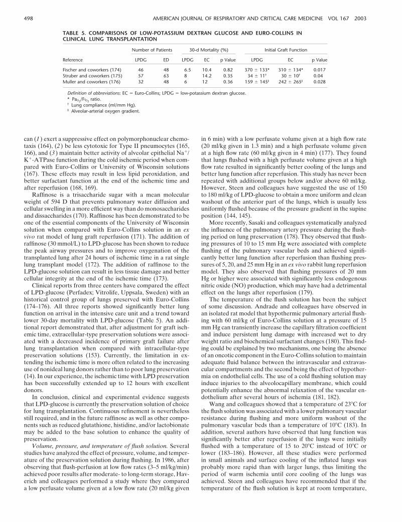
TABLE 5. COMPARISONS OF LOW-POTASSIUM DEXTRAN GLUCOSE AND EURO-COLLINS INCLINICAL LUNG TRANSPLANTATION
Number of Patients 30-d Mortality (%) Initial Graft Function
Reference LPDG ED LPDG EC p Value LPDG EC p Value
Fischer and coworkers (174) 46 48 6.5 10.4 0.82 370 � 133* 310 � 134* 0.017Struber and coworkers (175) 57 63 8 14.2 0.35 34 � 11† 30 � 10† 0.04Muller and coworkers (176) 32 48 6 12 0.36 159 � 145‡ 242 � 265‡ 0.028
Definition of abbreviations: EC Euro-Collins; LPDG low-potassium dextran glucose.* PaO2
/FIO2ratio.
† Lung compliance (ml/mm Hg).‡ Alveolar-arterial oxygen gradient.
can (1) exert a suppressive effect on polymorphonuclear chemo- in 6 min) with a low perfusate volume given at a high flow rate(20 ml/kg given in 1.3 min) and a high perfusate volume giventaxis (164), (2) be less cytotoxic for Type II pneumocytes (165,
166), and (3) maintain better activity of alveolar epithelial Na�/ at a high flow rate (60 ml/kg given in 4 min) (177). They foundthat lungs flushed with a high perfusate volume given at a highK�-ATPase function during the cold ischemic period when com-
pared with Euro-Collins or University of Wisconsin solutions flow rate resulted in significantly better cooling of the lungs andbetter lung function after reperfusion. This study has never been(167). These effects may result in less lipid peroxidation, and
better surfactant function at the end of the ischemic time and repeated with additional groups below and/or above 60 ml/kg.However, Steen and colleagues have suggested the use of 150after reperfusion (168, 169).
Raffinose is a trisaccharide sugar with a mean molecular to 180 ml/kg of LPD-glucose to obtain a more uniform and cleanwashout of the anterior part of the lungs, which is usually lessweight of 594 D that prevents pulmonary water diffusion and
cellular swelling in a more efficient way than do monosaccharides uniformly flushed because of the pressure gradient in the supineposition (144, 145).and dissaccharides (170). Raffinose has been demonstrated to be
one of the essential components of the University of Wisconsin More recently, Sasaki and colleagues systematically analyzedthe influence of the pulmonary artery pressure during the flush-solution when compared with Euro-Collins solution in an ex
vivo rat model of lung graft reperfusion (171). The addition of ing period on lung preservation (178). They observed that flush-ing pressures of 10 to 15 mm Hg were associated with completeraffinose (30 mmol/L) to LPD-glucose has been shown to reduce
the peak airway pressures and to improve oxygenation of the flushing of the pulmonary vascular beds and achieved signifi-cantly better lung function after reperfusion than flushing pres-transplanted lung after 24 hours of ischemic time in a rat single
lung transplant model (172). The addition of raffinose to the sures of 5, 20, and 25 mm Hg in an ex vivo rabbit lung reperfusionmodel. They also observed that flushing pressures of 20 mmLPD-glucose solution can result in less tissue damage and better
cellular integrity at the end of the ischemic time (173). Hg or higher were associated with significantly less endogenousnitric oxide (NO) production, which may have had a detrimentalClinical reports from three centers have compared the effect
of LPD-glucose (Perfadex; Vitrolife, Uppsala, Sweden) with an effect on the lungs after reperfusion (179).The temperature of the flush solution has been the subjecthistorical control group of lungs preserved with Euro-Collins
(174–176). All three reports showed significantly better lung of some discussion. Andrade and colleagues have observed inan isolated rat model that hypothermic pulmonary arterial flush-function on arrival in the intensive care unit and a trend toward
lower 30-day mortality with LPD-glucose (Table 5). An addi- ing with 60 ml/kg of Euro-Collins solution at a pressure of 15mm Hg can transiently increase the capillary filtration coefficienttional report demonstrated that, after adjustment for graft isch-
emic time, extracellular-type preservation solutions were associ- and induce persistent lung damage with increased wet to dryweight ratio and biochemical surfactant changes (180). This find-ated with a decreased incidence of primary graft failure after
lung transplantation when compared with intracellular-type ing could be explained by two mechanisms, one being the absenceof an oncotic component in the Euro-Collins solution to maintainpreservation solutions (153). Currently, the limitation in ex-
tending the ischemic time is more often related to the increasing adequate fluid balance between the intravascular and extravas-cular compartments and the second being the effect of hypother-use of nonideal lung donors rather than to poor lung preservation
(14). In our experience, the ischemic time with LPD preservation mia on endothelial cells. The use of a cold flushing solution mayinduce injuries to the alveolocapillary membrane, which couldhas been successfully extended up to 12 hours with excellent
donors. potentially enhance the abnormal relaxation of the vascular en-dothelium after several hours of ischemia (181, 182).In conclusion, clinical and experimental evidence suggests
that LPD-glucose is currently the preservation solution of choice Wang and colleagues showed that a temperature of 23�C forthe flush solution was associated with a lower pulmonary vascularfor lung transplantation. Continuous refinement is nevertheless
still required, and in the future raffinose as well as other compo- resistance during flushing and more uniform washout of thepulmonary vascular beds than a temperature of 10�C (183). Innents such as reduced glutathione, histidine, and/or lactobionate
may be added to the base solution to enhance the quality of addition, several authors have observed that lung function wassignificantly better after reperfusion if the lungs were initiallypreservation.
Volume, pressure, and temperature of flush solution. Several flushed with a temperature of 15 to 20�C instead of 10�C orlower (183–186). However, all these studies were performedstudies have analyzed the effect of pressure, volume, and temper-
ature of the preservation solution during flushing. In 1986, after in small animals and surface cooling of the inflated lungs wasprobably more rapid than with larger lungs, thus limiting theobserving that flush-perfusion at low flow rates (3–5 ml/kg/min)
achieved poor results after moderate- to long-term storage, Hav- period of warm ischemia until core cooling of the lungs wasachieved. Steen and colleagues have recommended that if theerich and colleagues performed a study where they compared
a low perfusate volume given at a low flow rate (20 ml/kg given temperature of the flush solution is kept at room temperature,

State of the Art 499
TABLE 6. CURRENT RECOMMENDATIONS FOR LUNGthen the lungs should be maintained in a collapsed state duringPRESERVATION FROM THE TORONTO LUNGcold storage to reduce the core temperature quicker by avoidingTRANSPLANT GROUPthe insulating effect of air (145).
Ultrastructural analysis of the lungs at various time points Volume of flush solution, ml/kg 50–60during the preservation period shows that the injuries induced PA pressure during flush delivery, mm Hg 10–15
Temperature of flush solution, �C 4–8by the flush itself appear to be minimal when compared with theLung ventilation VT: 10 ml/kg and PEEP: 5 cm H2Oinsult induced by ischemia on the endothelial–epithelial barrierOxygenation � 50% FIO2(187, 188). Hence, despite some potential injuries induced byLung inflation (airway pressure), cm H2O 15–20
cold flushing, it appears that this contribution to the total injury Storage temperature, �C 4–8is minimal when compared with the insult induced by ischemia.
Definition of abbreviations: PA pulmonary artery; PEEP positive end-expir-Flushing the lungs with a hypothermic preservation solutionatory pressure.should therefore still be recommended.
Inflation, oxygenation, and storage temperature. Althoughatelectatic lungs can be preserved at cold temperature for 5 to6 hours in humans and for up to 24 hours in pigs (189, 190),
tion at 10�C achieved better results than preservation at 4 orthere have been a large number of experiments since the early15�C and higher (40, 202, 205, 206). However, these findings1970s suggesting that preservation of the lung is improved whenwere not confirmed by other groups (207, 208). In addition,they are inflated with oxygen (191). Expansion of the lungs withlungs preserved at 10�C require a greater amount of metabolicoxygen during the ischemic period protects the lung from injurysubstrate, and the risk of lung injury can increase extremelyby three mechanisms: (1) it maintains some aerobic metabolism,rapidly if the temperature rises above 10�C during preservation(2) it preserves the integrity of pulmonary surfactant, and (3)(204). Hence, if a 10�C preservation temperature were used, theit preserves epithelial fluid transport.temperature of the organs would have to be constantly moni-During ischemia, lungs inflated with air are still able to con-tored because of the narrow margin of safety. For this reason, wesume oxygen and to produce energy through the more efficientrecommend preservation of the lungs at a temperature rangingaerobic metabolic pathway, which prevents the accumulationbetween 4 and 8�C (Table 6).of cellular metabolites and delay cell death (192, 193). Hence,
Retrograde flush and late reflush. Retrograde flush, whichalveolocapillary membranes are better preserved and therefers to the administration of the flush solution through the leftamount of total protein and lactate dehydrogenase in the bron-atrial appendage or the pulmonary veins, and drainage throughchoalveolar lavage fluid are significantly lower than if the lungsthe pulmonary artery, has been described for lung and heart–lungwere preserved in a complete atelectatic state or inflated withtransplantation (209, 210). The technique adds the potential100% nitrogen (193, 194). As well, static pulmonary complianceadvantages of flushing both the bronchial and pulmonary vesselsand surfactant secretion remain significantly better if the lungsand of limiting the effect of pulmonary arterial vasoconstrictionare preserved in an inflated instead of a deflated state (193–195).on the distribution of the flush solution. Experimentally, a retro-In addition, Sakuma and colleagues have recently demonstratedgrade flush has been found to improve lung preservation whenthat lung deflation decreases alveolar fluid clearance, whereascompared with an anterograde flush. This effect was attributed tofluid clearance was maintained in inflated lungs, independentlymore effective clearance of red blood cells within the capillaries,of the presence of oxygen (196).better distribution of the flush solution along the tracheobron-Atelectasis is also associated with higher pulmonary vascularchial tree, and less severe impairment of surfactant functionresistance and poorer distribution of the lung preservation solu-(157, 197, 211, 212). However, despite the retrograde flush, pre-tion (197, 198). Hence, a recruitment maneuver before flushingtreatment with PGE1 was still helpful in improving pulmonarythe lungs is certainly an effective measure. However, overdisten-dynamic compliance after reperfusion (213). After these results,sion of the lung by either static inflation, high Vt, or high positiveseveral groups have adopted a combined procedure with anend-expiratory pressure has been shown to be detrimental duringanterograde flush through the pulmonary artery followed by amechanical ventilation, and there is evidence suggesting thatretrograde flush through each of the pulmonary veins in situhyperinflation during storage increases the pulmonary capillarywhile the lungs are still ventilated (214, 215).filtration coefficient (199–201). In rat experiments, we and others
Late reflush was initially described in kidney transplantationhave observed that lung inflation during storage should be lim-and refers to the administration of a second flush immediatelyited to 50% of the total lung capacity or to an airway pressurebefore implantation of the graft (216). This method has beenof 10 to 15 cm H2O to avoid barotrauma (195, 202). In ourshown to wash out inflammatory agents and to improve post-clinical practice, we perform a recruitment maneuver to fullytransplant graft function by limiting cell damage after reperfu-re-expand the lung before flushing them, and we ventilate thesion (116, 216–218). The University of North Carolina has devel-lungs with a Vt of 10 ml/kg and a positive end-expiratory pres-oped a specific extracellular solution for late reflush (Carolinasure of 5 cm H2O during the flushing period. The lungs are thenrinse solution) to replenish important substrates and provideinflated with a sustained peak airway pressure of a maximumantioxidants and vasodilators to the graft before reperfusion toof 15 to 20 cm H2O before tracheal crossclamping in an effort
to obtain complete lung expansion but avoid overdistension. limit cell injury (219). This solution has been shown to be supe-rior to Euro-Collins for late reflush in an ex vivo model of lungIt should be noted that overinflated lungs may be exposed to
significantly more overdistension if they are transported in air- reperfusion (218). In clinical lung transplantation, Venuta andcolleagues have completed a study with 14 patients demonstra-planes because of the potentially lower atmospheric pressure
during the flight. ting that the addition of a late retrograde reflush with LPD-glucose to an anterograde flush was associated with improvedOxygen is required during storage to support aerobic metabo-
lism (192, 202, 203). However, an FiO2 greater than 50% may be lung function when compared with an anterograde flush only(215). Future studies are required to determine whether theassociated with more lipid peroxidation during lung storage (29,
192, 202, 204). Hence, inflation with an oxygen fraction of 50% improvement in lung function that they observed was due to theretrograde flush and/or to the late reflush effect.or less is usually recommended in clinical practice.
Several experimental studies have shown that lung preserva- Low reperfusion pressure and protective ventilation. The pul-
500 AMERICAN JOURNAL OF RESPIRATORY AND CRITICAL CARE MEDICINE VOL 167 2003
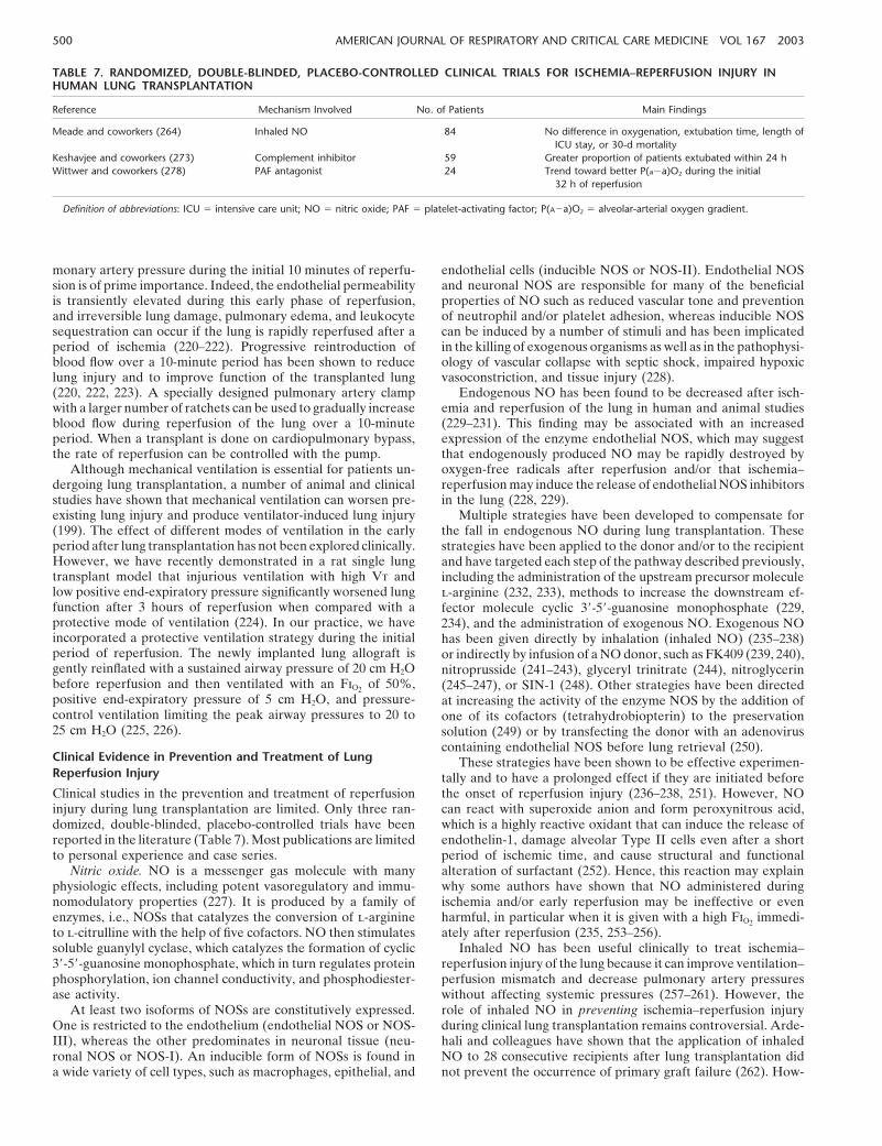
TABLE 7. RANDOMIZED, DOUBLE-BLINDED, PLACEBO-CONTROLLED CLINICAL TRIALS FOR ISCHEMIA–REPERFUSION INJURY INHUMAN LUNG TRANSPLANTATION
Reference Mechanism Involved No. of Patients Main Findings
Meade and coworkers (264) Inhaled NO 84 No difference in oxygenation, extubation time, length ofICU stay, or 30-d mortality
Keshavjee and coworkers (273) Complement inhibitor 59 Greater proportion of patients extubated within 24 hWittwer and coworkers (278) PAF antagonist 24 Trend toward better P(a a)O2 during the initial
32 h of reperfusion
Definition of abbreviations: ICU intensive care unit; NO nitric oxide; PAF platelet-activating factor; P(A a)O2 alveolar-arterial oxygen gradient.
monary artery pressure during the initial 10 minutes of reperfu- endothelial cells (inducible NOS or NOS-II). Endothelial NOSand neuronal NOS are responsible for many of the beneficialsion is of prime importance. Indeed, the endothelial permeability
is transiently elevated during this early phase of reperfusion, properties of NO such as reduced vascular tone and preventionof neutrophil and/or platelet adhesion, whereas inducible NOSand irreversible lung damage, pulmonary edema, and leukocyte
sequestration can occur if the lung is rapidly reperfused after a can be induced by a number of stimuli and has been implicatedin the killing of exogenous organisms as well as in the pathophysi-period of ischemia (220–222). Progressive reintroduction of
blood flow over a 10-minute period has been shown to reduce ology of vascular collapse with septic shock, impaired hypoxicvasoconstriction, and tissue injury (228).lung injury and to improve function of the transplanted lung
(220, 222, 223). A specially designed pulmonary artery clamp Endogenous NO has been found to be decreased after isch-emia and reperfusion of the lung in human and animal studieswith a larger number of ratchets can be used to gradually increase
blood flow during reperfusion of the lung over a 10-minute (229–231). This finding may be associated with an increasedexpression of the enzyme endothelial NOS, which may suggestperiod. When a transplant is done on cardiopulmonary bypass,
the rate of reperfusion can be controlled with the pump. that endogenously produced NO may be rapidly destroyed byoxygen-free radicals after reperfusion and/or that ischemia–Although mechanical ventilation is essential for patients un-
dergoing lung transplantation, a number of animal and clinical reperfusion may induce the release of endothelial NOS inhibitorsin the lung (228, 229).studies have shown that mechanical ventilation can worsen pre-
existing lung injury and produce ventilator-induced lung injury Multiple strategies have been developed to compensate forthe fall in endogenous NO during lung transplantation. These(199). The effect of different modes of ventilation in the early
period after lung transplantation has not been explored clinically. strategies have been applied to the donor and/or to the recipientHowever, we have recently demonstrated in a rat single lung and have targeted each step of the pathway described previously,transplant model that injurious ventilation with high Vt and including the administration of the upstream precursor moleculelow positive end-expiratory pressure significantly worsened lung l-arginine (232, 233), methods to increase the downstream ef-function after 3 hours of reperfusion when compared with a fector molecule cyclic 3�-5�-guanosine monophosphate (229,protective mode of ventilation (224). In our practice, we have 234), and the administration of exogenous NO. Exogenous NOincorporated a protective ventilation strategy during the initial has been given directly by inhalation (inhaled NO) (235–238)period of reperfusion. The newly implanted lung allograft is or indirectly by infusion of a NO donor, such as FK409 (239, 240),gently reinflated with a sustained airway pressure of 20 cm H2O nitroprusside (241–243), glyceryl trinitrate (244), nitroglycerinbefore reperfusion and then ventilated with an FiO2 of 50%, (245–247), or SIN-1 (248). Other strategies have been directedpositive end-expiratory pressure of 5 cm H2O, and pressure- at increasing the activity of the enzyme NOS by the addition ofcontrol ventilation limiting the peak airway pressures to 20 to one of its cofactors (tetrahydrobiopterin) to the preservation25 cm H2O (225, 226). solution (249) or by transfecting the donor with an adenovirus
containing endothelial NOS before lung retrieval (250).Clinical Evidence in Prevention and Treatment of Lung These strategies have been shown to be effective experimen-Reperfusion Injury tally and to have a prolonged effect if they are initiated before
the onset of reperfusion injury (236–238, 251). However, NOClinical studies in the prevention and treatment of reperfusioninjury during lung transplantation are limited. Only three ran- can react with superoxide anion and form peroxynitrous acid,
which is a highly reactive oxidant that can induce the release ofdomized, double-blinded, placebo-controlled trials have beenreported in the literature (Table 7). Most publications are limited endothelin-1, damage alveolar Type II cells even after a short
period of ischemic time, and cause structural and functionalto personal experience and case series.Nitric oxide. NO is a messenger gas molecule with many alteration of surfactant (252). Hence, this reaction may explain
why some authors have shown that NO administered duringphysiologic effects, including potent vasoregulatory and immu-nomodulatory properties (227). It is produced by a family of ischemia and/or early reperfusion may be ineffective or even
harmful, in particular when it is given with a high FiO2 immedi-enzymes, i.e., NOSs that catalyzes the conversion of l-arginineto l-citrulline with the help of five cofactors. NO then stimulates ately after reperfusion (235, 253–256).
Inhaled NO has been useful clinically to treat ischemia–soluble guanylyl cyclase, which catalyzes the formation of cyclic3�-5�-guanosine monophosphate, which in turn regulates protein reperfusion injury of the lung because it can improve ventilation–
perfusion mismatch and decrease pulmonary artery pressuresphosphorylation, ion channel conductivity, and phosphodiester-ase activity. without affecting systemic pressures (257–261). However, the
role of inhaled NO in preventing ischemia–reperfusion injuryAt least two isoforms of NOSs are constitutively expressed.One is restricted to the endothelium (endothelial NOS or NOS- during clinical lung transplantation remains controversial. Arde-
hali and colleagues have shown that the application of inhaledIII), whereas the other predominates in neuronal tissue (neu-ronal NOS or NOS-I). An inducible form of NOSs is found in NO to 28 consecutive recipients after lung transplantation did
not prevent the occurrence of primary graft failure (262). How-a wide variety of cell types, such as macrophages, epithelial, and

State of the Art 501
ever, Thabut and colleagues reported that the administration of nary bypass, but the results did not reach statistical significancebecause of the small number of patients (n 12). This likelyinhaled NO in combination with pentoxifylline at the time of
reperfusion in 23 patients reduced the incidence of ischemia– reflects the added potential benefit of inhibiting complementactivation related to cardiopulmonary bypass. The results ofreperfusion injury when compared with two historical control
groups (263). Our group has recently completed a randomized, Phase III trials in cardiac surgery should confirm whether com-plement inhibition with the soluble complement receptor-1 isdouble-blinded, placebo-controlled trial of inhaled NO adminis-
tered to lung transplant recipients, starting 10 minutes after protective when patients are placed on cardiopulmonary bypasscircuits (275).reperfusion for a minimum of 6 hours (264). Among a total
of 84 recipients, we observed no significant differences in the Cardiopulmonary bypass is known to activate the release ofmediators and to stimulate the activation of complement factors.immediate oxygenation, time to extubation, length of stay in the
intensive care unit, or 30-day mortality (Table 7). In conclusion, We therefore limit the use of cardiopulmonary bypass to recipi-ents with pulmonary hypertension and to those who cannotalthough inhaled NO therapy can be useful in improving gas
exchange in cases of established reperfusion injury, the role for tolerate unilateral ventilation or perfusion (276). Some centers,however, routinely perform lung transplantation using cardio-NO in the prevention of ischemia–reperfusion injury has yet to
be demonstrated in clinical lung transplantation. pulmonary bypass with good results (277). One potentially bene-ficial effect of cardiopulmonary bypass is the ability to reperfuseProstaglandins. PGE1 has been shown to be beneficial when
added to intracellular preservation solutions such as Euro-Col- the newly implanted lungs with controlled pulmonary arterypressures over a prolonged period of time.lins and University of Wisconsin (184, 207, 265). The beneficial
effect of PGE1 is attributed to its vasodilator properties that Antagonist of platelet-activating factor. Wittwer and col-leagues have recently reported their clinical experience with anmay lead to better distribution of the preservation solution and
to the stimulation of cyclic-3�,5�adenosine monophosphate– antagonist of platelet-activating factor (BN52021, Ginkolide B)in 24 patients randomly assigned to a high dose of antagonistdependent protein kinase during the cold ischemic time, which
may reduce endothelial permeability, neutrophil adhesion, and in the flush solution and after reperfusion (n 8), a low doseof antagonist in the flush solution and after reperfusion (n 8),platelet aggregation on reperfusion (265).
The continuous intravenous administration of PGE1 to the and a control group (n 8) (278). They observed a trend towarda better alveolar-arterial oxygen gradient within the first 32 hoursrecipient during the early phase of reperfusion has been shown
to reduce ischemia–reperfusion injury of the lung in animal mod- after reperfusion and better chest X-ray score in the two groupsreceiving the antagonist (Table 7). In clinical kidney transplanta-els of lung transplantation (266, 267). Although this effect can
be partially attributed to the vasodilator property of PGE1 during tion, a randomized, double-blinded, single center trial with 29recipients showed a significant reduction in the incidence ofthe initial 10 minutes of reperfusion (268), after a longer period
of reperfusion a continuous PGE1 infusion achieved significantly primary graft failure after transplantation in the group of patientsreceiving the antagonist of the platelet-activating factor (279).better lung function than other vasodilator agents such as prosta-
cyclin and nitroprusside (269). Hence, the continuous infusion These promising results from single centers should encouragelarge multicenter trials.of PGE1 clearly has a beneficial role on ischemia–reperfusion
injury, some of which can be attributable to its antiinflammatory Surfactant therapy. Pulmonary surfactant consists of approxi-mately 90% lipids, mainly saturated phosphatidylcholine, andeffects. Indeed, the continuous administration of PGE1 during
reperfusion is associated with a shift from a proinflammatory approximately 10% proteins, including the surfactant apopro-teins-A, B, C, and D. Type II pneumocytes synthesize, store,cytokine profile including TNF-�, IFN-�, and IL-12 to an antiin-
flammatory cytokine profile with increased IL-10 in a rat lung secrete, and to a large extent recycle pulmonary surfactants(280). The surfactant pool can be separated into the intracellulartransplant model (266). Other effects of PGE1, such as its antiag-
gregant action on platelets, may also potentially explain its bene- surfactant, represented by the lamellar bodies of Type II pneu-mocytes, and the intra-alveolar surfactant, which consists of sev-ficial role (270, 271).
On the basis of experimental evidence, some centers routinely eral subtypes, including freshly secreted lamellar body-likeforms, tubular myelin, the alveolar lining layer, and small unila-use an infusion of PGE1 during the postoperative period after
lung transplantation, whereas others reserve PGE1 infusion for mellar vesicles. Bronchoalveolar lavage studies usually refer totwo subfractions of the intra-alveolar surfactant, large aggregatesthe treatment of severe reperfusion injury (272). Prospective
randomized trials are required to determine whether routine or heavy forms, largely corresponding to tubular myelin,whichare highly active in decreasing the alveolar surface tension, andPGE1 has an overall beneficial effect in the postoperative course
during clinical lung transplantation. Such studies may use the small aggregates or light forms, largely corresponding to de-graded and inactive small unilamellar vesicles (281).newly developed aerosolized form of PGE1, which has been
shown experimentally to reduce ischemia–reperfusion injury of Surfactant dysfunction has been shown to occur during isch-emia–reperfusion injury of the lung (281, 282). Ultrastructuralthe lung without having the systemic hypotensive side effect of
intravenous PGE1 (243). analyses have shown an increase in the small to large surfactantaggregate ratio, an increase in sphingomyelin, and a decrease inComplement inhibition. After the successful experimental ap-
plication of the complement inhibitor, soluble complement phosphatidylglycerol and phosphatidylcholine, which correlatedwith decreased pulmonary compliance and lung oxygenationreceptor-1 (112), we performed a multicenter randomized, dou-
ble-blinded, placebo-controlled trial that included 59 lung trans- (281, 283, 284). These changes were also associated with a deficitin surfactant adsorption and a decrease in surfactant apopro-plant recipients (273, 274). Among 29 patients receiving a dose
of soluble complement receptor-1 before reperfusion, 14 (48%) tein-A (284–286). Alveolar surfactant dysfunction may occurdespite the absence of plasma protein leakage or changes inwere extubated within 24 hours, which was significantly better
than in the control arm with only 6 patients extubated out of a lamellar bodies of Type II pneumocytes (281, 287). The dysfunc-tion is most likely the result of numerous insults occurring duringtotal of 30 (20%). In addition, the overall duration of mechanical
ventilation and length of intensive care unit stay tended to be lung storage such as the production of phospholipase A2, me-chanical distorsion, altered phospholipid metabolism, reducedshorter in the group receiving the therapeutic drug (Table 7).
The effect of soluble complement receptor-1 appeared to be production of surfactant apoprotein-A, and/or accumulation ofC-reactive protein (284, 285, 288). Although some alterations instronger in the group of patients who underwent cardiopulmo-

502 AMERICAN JOURNAL OF RESPIRATORY AND CRITICAL CARE MEDICINE VOL 167 2003
surfactant can be observed immediately after pulmonary artery the lung from ischemia–reperfusion injury through suppressionof the antifibrinolytic pathway (78).flushing, most of the alterations have been shown to progres-
Carbon monoxide, like NO, is a messenger gas molecule thatsively increase during ischemic storage and to be significantlycan regulate vasomotor tone through the production of cyclicless with extracelullar-type preservation solutions (282, 283, 286,3�-5�-guanosine monophosphate (305). However, increasing evi-288).dence suggests that carbon monoxide is an extremely potentExperimental studies and clinical observations have foundantiinflammatory and antiapoptotic molecule that mediates itsthat exogenous surfactant therapy can improve pulmonary func-cytoprotective effect through the activation of the mitogen-acti-tion after lung transplantation (289–294). The administration ofvated protein kinase pathway, independent of the NO/cyclic 3�-exogenous surfactant is associated with a higher amount of to-5�-guanosine monophosphate pathway (306). The administrationtal surfactant phospholipid, a higher percentage of the heavyof low levels of inhaled carbon monoxide has been shown tosubtype of surfactant, a normalized percentage of phosphatidyl-decrease lung inflammation induced by ischemia, hyperoxia, andcholine, and a higher amount of endogenous surfactant apo-aeroallergen (78, 307, 308). The levels of inhaled carbon monox-protein-A, which has been shown to improve oxygenation andide in these studies ranged between 50 and 500 parts per million,compliance of the transplanted lung (291, 292). Exogenous surfac-which appears to be significantly lower than the known toxictant has also been shown to enhance immediate recovery fromlevel of carbon monoxide (297).transplantation injury and to be persistently beneficial for endoge-
The effect of heme oxygenase-1 and its metabolites derivesnous surfactant metabolism up to one week after transplantationfrom a fine balance between cytoprotection and cytotoxicity.(295). Exogenous surfactant given to the donor before retrievalIndeed, carbon monoxide and bilirubin are known to be poten-has been associated with better and more reliable results thantially toxic by, respectively, dissociating oxygen from hemoglobinwhen it was administered just before or immediately after reper-and by causing kernicterus (309). In addition, overexpression offusion (290, 291, 293, 296). Struber and coworkers have success-heme oxygenase-1 may be associated with an excessive accumu-fully used a nebulized synthetic surfactant in several patientslation of free iron, which may catalyze the formation of hydroxylwith reperfusion injury after lung transplantation (294). Theyradical through the Fenton reaction, and result in increasedobserved rapid improvement in pulmonary compliance and inoxidative stress (310). Future studies are required to determinealveolar-arterial oxygen gradient, leading to extubation withinthe role of manipulation of this novel pathway in protecting thea few days after the application (294). In the future, these promis-lung from ischemia–reperfusion injury.ing results need to be confirmed by a prospective, randomized
Preconditioning. Tissues exposed to one insult can developtrial.tolerance to a subsequent injury. This biological adaptationforms the basis of the concept of preconditioning. Various typesFuture Strategiesof preconditioning have been used in vivo to protect the lungHeme oxygenase pathway. Heme oxygenases catalyze the con-from ischemia–reperfusion injury. For instance, short periods ofversion of heme into biliverdin, carbon monoxide, and free iron.ischemia (ischemic preconditioning) (311–314), increased tem-The free iron is then sequestred into ferritin, whereas biliverdinperature (hyperthermic preconditioning) (315, 316), and admin-
is metabolized into bilirubin. Heme oxygenase consists of three istration of pharmacologic agents (chemical preconditioning)isoforms, the inducible heme oxygenase-1, also known as heat (317, 318) have been shown to be successful in reducing lungshock protein 32, and two constitutive isoforms, heme oxyge- injury in most cases (319).nase-2, and heme oxygenase-3. Heme oxygenase-1 activity has The mechanism by which preconditioning confers protectionbeen shown to provide potent cytoprotective effects against a is not well understood. Hyperthermia (approximately 5�C abovevariety of agents causing oxidative stress in vivo and in vitro normal temperature) was initially shown to upregulate the syn-(297). This finding is also supported by recent observations from thesis of a family of proteins, named heat shock proteins thatheme oxygenase-1 deficient mice and humans exhibiting in- confer protection against a variety of stresses, including isch-creased susceptibility to oxidative stress among other abnormali- emia–reperfusion injury (320). Although ischemic precondi-ties (298, 299). tioning and some cytoprotective agents have also been shown
Heme oxygenase-1 is rapidly upregulated on reperfusion, and to upregulate the production of heat shock protein, the mecha-its overexpression has been shown to confer marked cytoprotec- nism that renders the organ resistant to ischemia–reperfusiontion against ischemia–reperfusion injury. Indeed, after a period injury is certainly more complex and involves other moleculesof myocardial ischemia, transgenic mice overexpressing heme (321). Recent studies have incriminated reactive oxygen species,oxygenase-1 have better cardiac function and reduced infarct ATP-sensitive potassium channel openers, protein kinase C, pro-size when compared with wild-type mice, whereas heterozygous tein tyrosine kinase, and nuclear factor-�B as potential intracel-(HO-1�/ ) mice with a 40% reduction in the expression of heme lular signal transduction pathways of ischemic preconditioningoxygenase-1 display reduced ventricular recovery and increased (322–324). Currently, ischemic preconditioning has been showninfarct size when compared with wild-type mice (300, 301). Simi- to be effective clinically in hepatic resection (325) and in coro-lar findings have been observed in a liver model of ischemia– nary artery bypass graft surgery (326), but its role remains un-reperfusion injury when the expression of heme oxygenase-1 proven in clinical lung transplantation.was induced by cobalt protoporphyrin or by adenoviral-medi- Gene therapy. The use of gene therapy in the transplantationated heme oxygenase-1 gene transfer (302). setting is potentiated because immunosuppressive therapy
The mechanism by which selective overexpression of heme allows effective and repeated transfection with current genera-oxygenase-1 confers protection against ischemia–reperfusion in- tion of viral vectors (327, 328). Multiple strategies have beenjury remains poorly understood and may be mediated by each used experimentally to transfect donor lungs. Genes have beenof the three by-products generated by the enzyme. Free iron administered to the donor before lung retrieval (329), on theactivates the production of ferritin, which may mediate cytopro- back table during the cold ischemic time (330, 331), or to thetection against ischemia–reperfusion injury (303). Bilirubin is a recipient after reperfusion (332). They have been delivered intra-known antioxidant that has been shown to protect isolated per- vascularly (333), intramuscularly (332), or transtracheally (329)fused rat hearts when heme oxygenase-1 was inhibited (304). in a naked form (334) or with the help of a vector, either viral
(335) or nonviral (336).Finally, carbon monoxide has recently been shown to protect

State of the Art 503
increases risk of late bronchiolitis obliterans syndrome. Ann ThoracWe have demonstrated that transfection of the donor lungSurg 2002;73:1041–1047.is possible through the transtracheal route using a second-gener-
4. Ware LB, Wang Y, Fang X, Warnock M, Sakuma T, Hall TS, Matthayation adenoviral vector without contaminating other organs suchM. Assessment of lungs rejected for transplantation and implications
as the heart, liver, or kidneys (329). Because the transfection for donor selection. Lancet 2002;360:619–620.rate is significantly decreased at cold temperature (337, 330), 5. Fujimura S, Handa M, Kondo T, Ichinose T, Shiraishi Y, Nakada T.
Successful 48-hour simple hypothermic preservation of canine lungthis mode of administration is useful in that it allows for efficienttransplants. Transplant Proc 1987;19:1334–1336.transfection before retrieving and cooling the lungs. Transtra-
6. Keshavjee SH, Yamazaki F, Cardoso PF, McRitchie DI, Patterson GA,cheal administration of the gene coding for the antiinflammatoryCooper JD. A method for safe twelve-hour pulmonary preservation.cytokine, human IL-10, to the donor 12 to 24 hours before lungJ Thorac Cardiovasc Surg 1989;98:529–534.
retrieval reduces ischemia–reperfusion injury and improves lung 7. Meyers BF, Sundt TM, Henry S, Trulock EP, Guthrie T, Cooper JD,function in a rat single lung transplant model (335, 338). IL-10 Patterson GA. Selective use of extracorporeal membrane oxygen-
ation is warranted after lung transplantation. J Thorac Cardiovascis an antiinflammatory cytokine that exerts antiinflammatorySurg 2000;120:20–26.and immunosupressive effects on a large variety of cells including
8. Fiser SM, Kron IL, McLendon Long S, Kaza AK, Kern JA, Tribblemacrophages, lymphocytes, and neutrophils, and it has beenCG. Early intervention after severe oxygenation elevation improvesshown to be beneficial in various models of ischemia–reperfusionsurvival following lung transplantation. J Heart Lung Transplant 2001;
injury (339). We are currently performing similar experiments 20:631–636.in large animal models with endoscopic delivery of adenoviral- 9. Sundaresan S, Trachiotis GD, Aoe M, Patterson GA, Cooper JD. Donor
lung procurement: assessment and operative technique. Ann Thoracmediated human IL-10 gene to the donor (340). Once optimalSurg 1993;56:1409–1413.gene delivery to large animals can be achieved, human lung
10. Sundaresan S, Semenkovich J, Ochoa L, Richardson G, Trulock EP,protection from ischemia–reperfusion and immunologic injuryCooper JD, Patterson GA. Successful outcome of lung transplanta-by gene therapy may soon be possible.tion is not compromised by the use of marginal donor lungs. J Thorac
As researches continue to improve our understanding of the Cardiovasc Surg 1995;109:1075–1079.mechanisms of injury related to lung preservation and as the 11. Kron IL, Tribble CG, Kern JA, Daniel TM, Rose CE, Truwit JD,
Blackbourne LH, Bergin JD. Successful transplantation of marginallygenes that control these processes are identified, we will moveacceptable thoracic organs. Ann Surg 1993;217:518–522.closer to the ultimate strategy in preservation-related lung injury.
12. Bhorade SM, Vigneswaran W, McCabe MA, Garrity ER. LiberalizationGene-based therapy promises the exciting potential to geneti-of donor criteria may expand the donor pool without adverse conse-cally modify organs to withstand the stresses of the transplantquence in lung transplantation. J Heart Lung Transplant 2000;19:
process. 1199–1204.13. Gabbay E, Williams TJ, Griffiths AP, Macfarlane LM, Kotsimbos TC,
Esmore DS, Snell GI. Maximizing the utilization of donor organsCONCLUSIONSoffered for lung transplantation. Am J Respir Crit Care Med 1999;160:
Better understanding of the mechanisms of ischemia–reperfu- 265–271.14. Pierre AF, Sekine Y, Hutcheon MA, Waddell TK, Keshavjee SH. Mar-sion injury, improvement in the technique of lung preservation,
ginal donor lungs: a reassessment. J Thorac Cardiovasc Surg 2002;123:and the recent introduction of a new preservation solution spe-421–428.cifically developed for the lungs have helped to reduce the inci-
15. Kusaka M, Pratschke J, Wilhelm MJ, Ziai F, Zandi-Nejad K, Mackenziedence of ischemia–reperfusion–induced lung injury and the de-HS, Hancock WW, Tilney NL. Activation of inflammatory mediators
velopment of primary graft failure after lung transplantation. In in rat renal isografts by donor brain death. Transplantation 2000;69:the future, the development of new agents and their application 405–410.in prospective clinical trials are to be expected to prevent the 16. Bittner HB, Kendall SW, Chen EP, Craig D, Van Trigt P. The effects
of brain death on cardiopulmonary hemodynamics and pulmonaryoccurrence of this potentially devastating complication and toblood flow characteristics. Chest 1995;108:1358–1363.further improve the success of lung transplantation.
17. Mertes PM, el Abassi K, Jaboin Y, Burtin P, Pinelli G, Carteaux JP,One of the major upcoming challenges will be to improveBurlet C, Boulange M, Villemot JP. Changes in hemodynamic andthe number of donor lungs available for transplantation. Al-metabolic parameters following induced brain death in the pig. Trans-
though the number of patients on the waiting list is constantly plantation 1994;58:414–418.increasing, only 10 to 30% of the donor lungs are currently used 18. Follette DM, Rudich SM, Babcock WD. Improved oxygenation andfor transplantation. Hence, the development of new strategies increased lung donor recovery with high-dose steroid administration
after brain death. J Heart Lung Transplant 1998;17:423–429.to repair and improve the quality of the lungs could have a19. Kim YS, Lim CS, Kim S, Lee JS, Lee S, Kim ST, Kim HJ, Chae DW.tremendous impact on the number of transplants performed. In
Cadaveric renal allograft at the time of implantation has the similaraddition, an improved understanding of the mechanisms in-immunological features with the rejecting allograft. Transplantationvolved during lung preservation may help to elucidate the poten-2000;70:1080–1085.
tial link between acute lung injury and chronic graft dysfunction. 20. Schwarz C, Regele H, Steininger R, Hansmann C, Mayer G, OberbauerIn the future, genetic analysis using novel technologies such as R. The contribution of adhesion molecule expression in donor kidneymicroarray analysis will help to determine which genes play a biopsies to early allograft dysfunction. Transplantation 2001;71:1666–
1670.role in the transplantation process. Hopefully, this will provide21. Koo DD, Welsh KI, McLaren AJ, Roake JA, Morris PJ, Fuggle SV.new insights into the mechanisms of injury and reveal potential
Cadaver versus living donor kidneys: impact of donor factors onnew strategies to improve lung preservation.antigen induction before transplantation. Kidney Int 1999;56:1551–1559.
References 22. Fisher AJ, Donnelly SC, Hirani N, Haslett C, Strieter RM, Dark JH,Corris PA. Elevated levels of interleukin-8 in donor lungs is associated1. Hertz M, Taylor D, Trulock E, Boucek M, Mohacsi P, Edwards L, Keckwith early graft failure after lung transplantation. Am J Respir CritB. The Registry of the International Society for Heart and LungCare Med 2001;163:259–265.Transplantation: nineteenth official report: 2002. J Heart Lung Trans-
23. de Perrot M, Sekine Y, Fischer S, Waddell TK, McRae K, Liu M, Wigleplant 2002;21:950–970.DA, Keshavjee S. Interleukin-8 release during early reperfusion pre-2. King RC, Binns OA, Rodriguez F, Kanithanon RC, Daniel TM, Spotnitzdicts graft function in human lung transplantation. Am J Respir CritWD, Tribble CG, Kron IL. Reperfusion injury significantly impactsCare Med 2002;165:211–215.clinical outcome after pulmonary transplantation. Ann Thorac Surg
24. Clavien PA, Harvey PR, Strasberg SM. Preservation and reperfusion2000;69:1681–1685.injuries in liver allografts: an overview and synthesis of current studies.3. Fiser SM, Tribble CG, Long SM, Kaza AK, Kern JA, Jones DR, Robbins
MK, Kron IL. Ischemia-reperfusion injury after lung transplantation Transplantation 1992;53:957–978.

504 AMERICAN JOURNAL OF RESPIRATORY AND CRITICAL CARE MEDICINE VOL 167 2003
25. McCord JM. Oxygen-derived free radicals in postischemic tissue injury. aggravated by an iron supplemented diet and is partly prevented bysimultaneous antioxidant supplementation. Res Commun Mol PatholN Engl J Med 1985;312:159–163.
26. Al Mehdi AB, Shuman H, Fisher AB. Intracellular generation of reac- Pharmacol 1994;86:273–285.51. Amersi F, Dulkanchainun T, Nelson SK, Farmer DG, Kato H, Zaky J,tive oxygen species during nonhypoxic lung ischemia. Am J Physiol
1997;272:L294–L300. Melinek J, Shaw GD, Kupiec-Weglinski JW, Horwitz LD, et al. Anovel iron chelator in combination with a P-selectin antagonist pre-27. Kelly RF. Current strategies in lung preservation. J Lab Clin Med 2000;
136:427–440. vents ischemia/reperfusion injury in a rat liver model. Transplantation2001;71:112–118.28. Date H, Matsumura A, Manchester JK, Obo H, Lima O, Cooper JM,
Sundaresan S, Lowry OH, Cooper JD. Evaluation of lung metabolism 52. Takeyoshi I, Iwanami K, Kamoshita N, Takahashi T, Kobayashi J,Tomizawa N, Kawashima Y, Matsumoto K, Morishita Y. Effect ofduring successful twenty-four-hour canine lung preservation. J Thorac
Cardiovasc Surg 1993;105:480–491. lazaroid U-74389G on pulmonary ischemia-reperfusion injury in dogs.J Invest Surg 2001;14:83–92.29. Fisher AB, Dodia C, Tan ZT, Ayene I, Eckenhoff RG. Oxygen-depen-
dent lipid peroxidation during lung ischemia. J Clin Invest 1991;88: 53. Tanoue Y, Morita S, Ochiai Y, Zhang QW, Hisahara M, Miyamoto K,Nishida T, Kawachi Y, Tominaga R, Yasui H. Successful twenty-674–679.
30. Eckenhoff RG, Dodia C, Tan Z, Fisher AB. Oxygen-dependent reperfu- four-hour canine lung preservation with lazaroid U74500A. J HeartLung Transplant 1996;15:43–50.sion injury in the isolated rat lung. J Appl Physiol 1992;72:1454–1460.
31. Fisher AB, Dodia C. Lung as a model for evaluation of critical intracellu- 54. Fischer S, Cassivi SD, Xavier AM, Cardella JA, Cutz E, Edwards V, LiuM, Keshavjee S. Cell death in human lung transplantation: apoptosislar PO2 and PCO. Am J Physiol 1981;241:E47–E50.
32. Zhao G, Al Mehdi AB, Fisher AB. Anoxia-reoxygenation versus isch- induction in human lungs during ischemia and after transplantation.Ann Surg 2000;231:424–431.emia in isolated rat lungs. Am J Physiol 1997;273:L1112–L1117.
33. Kennedy TP, Rao NV, Hopkins C, Pennington L, Tolley E, Hoidal JR. 55. Fischer S, Maclean AA, Liu M, Cardella JA, Slutsky AS, Suga M,Moreira JF, Keshavjee S. Dynamic changes in apoptotic and necroticRole of reactive oxygen species in reperfusion injury of the rabbit
lung. J Clin Invest 1989;83:1326–1335. cell death correlate with severity of ischemia-reperfusion injury inlung transplantation. Am J Respir Crit Care Med 2000;162:1932–1939.34. Al Mehdi AB, Zhao G, Dodia C, Tozawa K, Costa K, Muzykantov V,
Ross C, Blecha F, Dinauer M, Fisher AB. Endothelial NADPH oxi- 56. Stammberger U, Gaspert A, Hillinger S, Vogt P, Odermatt B, WederW, Schmid RA. Apoptosis induced by ischemia and reperfusion indase as the source of oxidants in lungs exposed to ischemia or high
K�. Circ Res 1998;83:730–737. experimental lung transplantation. Ann Thorac Surg 2000;69:1532–1536.35. Lansman JB. Endothelial mechanosensors: going with the flow. Nature
1988;331:481–482. 57. Denecker G, Vercammen D, Declercq W, Vandenabeele P. Apoptoticand necrotic cell death induced by death domain receptors. Cell Mol36. Al Mehdi AB, Zhao G, Fisher AB. ATP-independent membrane depo-
larization with ischemia in the oxygen-ventilated isolated rat lung. Life Sci 2001;58:356–370.58. Kuwano K, Hara N. Signal transduction pathways of apoptosis andAm J Respir Cell Mol Biol 1998;18:653–661.
37. Henderson LM, Chappell JB, Jones OT. Superoxide generation by the inflammation induced by the tumor necrosis factor receptor family.Am J Respir Cell Mol Biol 2000;22:147–149.electrogenic NADPH oxidase of human neutrophils is limited by the
movement of a compensating charge. Biochem J 1988;255:285–290. 59. Daemen MA, van’t Veer C, Denecker G, Heemskerk VH, Wolfs TG,Clauss M, Vandenabeele P, Buurman WA. Inhibition of apoptosis38. Kitagawa S, Johnston RB Jr. Relationship between membrane potential
changes and superoxide-releasing capacity in resident and activated induced by ischemia-reperfusion prevents inflammation. J Clin Invest1999;104:541–549.mouse peritoneal macrophages. J Immunol 1985;135:3417–3423.
39. Ware LB, Golden JA, Finkbeiner WE, Matthay MA. Alveolar epithelial 60. Yaoita H, Ogawa K, Maehara K, Maruyama Y. Attenuation of ischemia/reperfusion injury in rats by a caspase inhibitor. Circulation 1998;97:fluid transport capacity in reperfusion lung injury after lung trans-
plantation. Am J Respir Crit Care Med 1999;159:980–988. 276–281.61. Hartmann A. Antiapoptotic agents in brain ischemia. N Engl J Med 2000;40. Wang LS, Yoshikawa K, Miyoshi S, Nakamoto K, Hsieh CM, Yamazaki
F, Guerreiro Cardoso PF, Schaefers HJ, Brito J, Keshavjee S, et al. 342:823.62. Fischer S, de Perrot M, Liu M, MacLean AA, Cardella JA, Imai Y,The effect of ischemic time and temperature on lung preservation in
a simple ex vivo rabbit model used for functional assessment. J Thorac Suga M, Keshavjee S. IL-10 gene transfection of donor lungs amelio-rates post-transplant cell death by a switch from cellular necrosis toCardiovasc Surg 1989;98:333–342.
41. Sugita M, Suzuki S, Kondo T, Noda M, Fujimura S. Transalveolar fluid apoptosis. J Thorac Cardiovasc Surg (In press)63. Minamiya Y, Tozawa K, Kitamura M, Saito S, Ogawa J. Platelet-activat-absorption ability in rat lungs preserved with Euro-Collins solution
and EP4 solution. Transplantation 1999;67:349–354. ing factor mediates intercellular adhesion molecule-1-dependent radi-cal production in the nonhypoxic ischemia rat lung. Am J Respir Cell42. Yokomise H, Ueno T, Yamazaki F, Keshavjee S, Slutsky A, Patterson
GA. The effect and optimal time of administration of verapamil on Mol Biol 1998;19:150–157.64. Moore TM, Khimenko P, Adkins WK, Miyasaka M, Taylor AE. Adhe-lung preservation. Transplantation 1990;49:1039–1043.
43. Pickford MA, Gower JD, Dore C, Fryer PR, Green CJ. Lipid peroxida- sion molecules contribute to ischemia and reperfusion-induced injuryin the isolated rat lung. J Appl Physiol 1995;78:2245–2252.tion and ultrastructural changes in rat lung isografts after single-
passage organ flush and 48-hour cold storage with and without one- 65. Naka Y, Toda K, Kayano K, Oz MC, Pinsky JD. Failure to expressthe P-selectin gene or P-selectin blockade confers early pulmonaryhour reperfusion in vivo. Transplantation 1990;50:210–218.
44. Karck M, Haverich A. Nifedipine and diltiazem reduce pulmonary protection after lung ischemia or transplantation. Proc Natl Acad SciUSA 1997;94:757–761.edema formation during postischemic reperfusion of the rabbit lung.
Res Exp Med (Berl) 1992;192:137–144. 66. Kapelanski DP, Iguchi A, Niles SD, Mao HZ. Lung reperfusion injuryis reduced by inhibiting a CD18-dependent mechanism. J Heart Lung45. Zhao G, Ayene IS, Fisher AB. Role of iron in ischemia–reperfusion
oxidative injury of rat lungs. Am J Respir Cell Mol Biol 1997;16:293– Transplant 1993;12:294–306.67. DeMeester SR, Molinari MA, Shiraishi T, Okabayashi K, Manchester299.
46. Huang YT, Ghio AJ, Nozik-Grayck E, Piantadosi CA. Vascular release JK, Wick MR, Cooper JD, Patterson GA. Attenuation of rat lungisograft reperfusion injury with a combination of anti-ICAM-1 andof nonheme iron in perfused rabbit lungs. Am J Physiol Lung Cell
Mol Physiol 2001;280:L474–L481. anti-beta2 integrin monoclonal antibodies. Transplantation 1996;62:1477–1485.47. Bysani GK, Kennedy TP, Ky N, Rao NV, Blaze CA, Hoidal JR. Role
of cytochrome P-450 in reperfusion injury of the rabbit lung. J Clin 68. Steinberg JB, Mao HZ, Niles SD, Jutila MA, Kapelanski DP. Survivalin lung reperfusion injury is improved by an antibody that binds andInvest 1990;86:1434–1441.
48. Pratico D, Pasin M, Barry OP, Ghiselli A, Sabatino G, Iuliano L, Fitz- inhibits L- and E-selectin. J Heart Lung Transplant 1994;13:306–318.69. Demertzis S, Langer F, Graeter T, Dwenger A, Georg T, Schafers HJ.Gerald GA, Violi F. Iron-dependent human platelet activation and
hydroxyl radical formation: involvement of protein kinase C. Circula- Amelioration of lung reperfusion injury. Eur J Cardiothorac Surg1999;16:174–180.tion 1999;99:3118–3124.
49. Qayumi AK, Jamieson WR, Poostizadeh A, Germann E, Gillespie KD. 70. Reignier J, Sellak H, Lemoine R, Lubineau A, Mazmanian GM, DetruitH, Chapelier A, Herve P. Prevention of ischemia-reperfusion lungComparison of new iron chelating agents in the prevention of isch-
emia/reperfusion injury: a swine model of heart-lung transplantation. injury by sulfated Lewis(a) pentasaccharide: The Paris-Sud UniversityLung Transplantation Group. J Appl Physiol 1997;82:1058–1063.J Invest Surg 1992;5:115–127.
50. van Jaarsveld H, Kuyl JM, Wiid NM. Ischemia/reperfusion injury is 71. Schmid RA, Yamashita M, Boasquevisque CH, Ando K, Fujino S,

State of the Art 505
by type-II phospholipase A2 and inhibited by a direct surfactantPhillips L, Cooper JD, Patterson GA. Carbohydrate selectin inhibitorprotein A-phospholipase A2 protein interaction. J Clin Invest 1998;CY-1503 reduces neutrophil migration and reperfusion injury in ca-102:1152–1160.nine pulmonary allografts. J Heart Lung Transplant 1997;16:1054–
92. Koike K, Yamamoto Y, Hori Y, Ono T. Group IIA phospholipase A21061.mediates lung injury in intestinal ischemia-reperfusion. Ann Surg72. Stammberger U, Hamacher J, Hillinger S, Schmid RA. sCR1sLe amelio-2000;232:90–97.rates ischemia/reperfusion injury in experimental lung transplanta-
93. Shen CY, Wang D, Chang ML, Hsu K. Protective effect of mepacrinetion. J Thorac Cardiovasc Surg 2000;120:1078–1084.on hypoxia-reoxygenation-induced acute lung injury in rats. J Appl73. Ogawa S, Gerlach H, Esposito C, Pasagian-Macaulay A, Brett J, SternPhysiol 1995;78:225–231.D. Hypoxia modulates the barrier and coagulant function of cultured
94. Nagahiro I, Aoe M, Yamashita M, Date H, Andou A, Shimizu N. EPC-bovine endothelium: increased monolayer permeability and inductionK1 is effective in lung preservation in an ex vivo rabbit lung perfusionof procoagulant properties. J Clin Invest 1990;85:1090–1098.model. Ann Thorac Surg 1997;63:954–959.74. Compeau CG, Ma J, DeCampos KN, Waddell TK, Brisseau GF, Slutsky
95. Furue S, Mikawa K, Nishina K, Shiga M, Ueno M, Tomita Y, KuwabaraAS, Rotstein OD. In situ ischemia and hypoxia enhance alveolarK, Teshirogi I, Ono T, Hori Y, et al. Therapeutic time-window of amacrophage tissue factor expression. Am J Respir Cell Mol Biolgroup IIA phospholipase A2 inhibitor in rabbit acute lung injury:1994;11:446–455.correlation with lung surfactant protection. Crit Care Med 2001;29:75. Salvatierra A, Velasco F, Rodriguez M, Alvarez A, Lopez-Pedrera R,719–727.Ramirez R, Carracedo J, Lopez-Rubio F, Lopez-Pujol A, Guerrero
96. Miotla JM, Jeffery PK, Hellewell PG. Platelet-activating factor plays aR. C1-esterase inhibitor prevents early pulmonary dysfunction afterpivotal role in the induction of experimental lung injury. Am J Respirlung transplantation in the dog. Am J Respir Crit Care Med 1997;Cell Mol Biol 1998;18:197–204.155:1147–1154.
97. Nagase T, Ishii S, Kume K, Uozumi N, Izumi T, Ouchi Y, Shimizu T.76. Struber M, Hagl C, Hirt SW, Cremer J, Harringer W, Haverich A. C1-Platelet-activating factor mediates acid-induced lung injury in geneti-esterase inhibitor in graft failure after lung transplantation. Intensivecally engineered mice. J Clin Invest 1999;104:1071–1076.Care Med 1999;25:1315–1318.
98. Corcoran PC, Wang Y, Katz NM, Rajan SS, Analouei AR, Foegh77. Pinsky DJ, Liao H, Lawson CA, Yan SF, Chen J, Carmeliet P, LoskutoffML, Wallace RB. Platelet activating factor antagonist enhances lungDJ, Stern DM. Coordinated induction of plasminogen activatorpreservation in a canine model of single lung allotransplantation. Jinhibitor-1 (PAI-1) and inhibition of plasminogen activator gene ex-Thorac Cardiovasc Surg 1992;104:66–72.pression by hypoxia promotes pulmonary vascular fibrin deposition.
99. Kawahara K, Tagawa T, Takahashi T, Akamine S, Nakamura A, Yama-J Clin Invest 1998;102:919–928. moto S, Muraoka S, Tomita M. The effect of the platelet-activating
78. Fujita T, Toda K, Karimova A, Yan SF, Naka Y, Yet SF, Pinsky DJ. factor inhibitor TCV-309 on reperfusion injury in a canine model ofParadoxical rescue from ischemic lung injury by inhaled carbon mon- ischemic lung. Transplantation 1993;55:1438–1439.oxide driven by derepression of fibrinolysis. Nat Med 2001;7:598–604. 100. Stammberger U, Carboni GL, Hillinger S, Schneiter D, Weder W,
79. Okada K, Fujita T, Minamoto K, Liao H, Naka Y, Pinsky DJ. Potentia- Schmid RA. Combined treatment with endothelin- and PAF-antago-tion of endogenous fibrinolysis and rescue from lung ischemia/reper- nists reduces posttransplant lung ischemia/reperfusion injury. J Heartfusion injury in interleukin (IL)-10-reconstituted IL-10 null mice. J Lung Transplant 1999;18:862–868.Biol Chem 2000;275:21468–21476. 101. Kim JD, Baker CJ, Roberts RF, Darbinian SH, Marcus KA, Quardt
80. Lemay S, Rabb H, Postler G, Singh AK. Prominent and sustained up- SM, Starnes VA, Barr ML. Platelet activating factor acetylhydrolaseregulation of gp130-signaling cytokines and the chemokine MIP-2 in decreases lung reperfusion injury. Ann Thorac Surg 2000;70:423–428.murine renal ischemia-reperfusion injury. Transplantation 2000;69: 102. Su M, Chi EY, Bishop MJ, Henderson WR Jr. Lung mast cells increase959–963. in number and degranulate during pulmonary artery occlusion/reper-
81. Gerlach J, Jorres A, Nohr R, Zeilinger K, Spatkowski G, Neuhaus P. fusion injury in dogs. Am Rev Respir Dis 1993;147:448–456.Local liberation of cytokines during liver preservation. Transpl Int 103. Shimizu N, Kita T, Aoe M, Nakata M, Miyai Y, Teramoto S. Changes1999;12:261–265. in levels of arachidonic acid metabolites in blood and bronchoalveolar
82. Oz MC, Liao H, Naka Y, Seldomridge A, Becker DN, Michler RE, lavage fluid after warm ischemia-reperfusion of lung. Acta Med Okay-Smith CR, Rose EA, Stern DM, Pinsky DJ. Ischemia-induced ama 1991;45:417–422.interleukin-8 release after human heart transplantation: a potential 104. Zamora CA, Baron DA, Heffner JE. Thromboxane contributes to pul-role for endothelial cells. Circulation 1995;92:II428–II432. monary hypertension in ischemia-reperfusion lung injury. J Appl
83. Serrick C, Adoumie R, Giaid A, Shennib H. The early release of Physiol 1993;74:224–229.interleukin-2, tumor necrosis factor-alpha and interferon-gamma 105. Vural KM, Liao H, Oz MC, Pinsky DJ. Effects of mast cell membraneafter ischemia reperfusion injury in the lung allograft. Transplantation stabilizing agents in a rat lung ischemia-reperfusion model. Ann
Thorac Surg 2000;69:228–232.1994;58:1158–1162.84. Hosenpud JD, Bennett LE, Keck BM, Boucek MM, Novick JR. The 106. Barr ML, Carey JN, Nishanian GP, Roberts RF, Sakamaki Y, Darbinian
SH, Starnes VA. Addition of a mast cell stabilizing compound toRegistry of the International Society for Heart and Lung Transplanta-tion: seventeenth official report: 2000. J Heart Lung Transplant 2000; organ preservation solutions decreases lung reperfusion injury. J
Thorac Cardiovasc Surg 1998;115:631–636.19:909–931.85. Miller EJ, Cohen AB, Matthay MA. Increased interleukin-8 concentra- 107. Naka Y, Marsh HC, Scesney SM, Oz MC, Pinsky DJ. Complement
activation as a cause for primary graft failure in an isogeneic rattions in the pulmonary edema fluid of patients with acute respiratorydistress syndrome from sepsis. Crit Care Med 1996;24:1448–1454. model of hypothermic lung preservation and transplantation. Trans-
plantation 1997;64:1248–1255.86. Mueller AR, Platz KP, Haak M, Undi H, Muller C, Kottgen E, Weide-mann H, Neuhaus P. The release of cytokines, adhesion molecules, 108. Bishop MJ, Giclas PC, Guidotti SM, Su ML, Chi EY. Complement
activation is a secondary rather than a causative factor in rabbitand extracellular matrix parameters during and after reperfusion inhuman liver transplantation. Transplantation 1996;62:1118–1126. pulmonary artery ischemia/reperfusion injury. Am Rev Respir Dis
1991;143:386–390.87. Sekido N, Mukaida N, Harada A, Nakanishi I, Watanabe Y, MatsushimaK. Prevention of lung reperfusion injury in rabbits by a monoclonal 109. Frank MM. Complement in the pathophysiology of human disease. N
Engl J Med 1987;316:1525–1530.antibody against interleukin-8. Nature 1993;365:654–657.88. Tischfield JA. A reassessment of the low molecular weight phospholi- 110. Ivey CL, Williams FM, Collins PD, Jose PJ, Williams TJ. Neutrophil
chemoattractants generated in two phases during reperfusion of isch-pase A2 gene family in mammals. J Biol Chem 1997;272:17247–17250.89. Kim DK, Fukuda T, Thompson BT, Cockrill B, Hales C, Bonventre emic myocardium in the rabbit: evidence for a role for C5a and
interleukin-8. J Clin Invest 1995;95:2720–2728.JV. Bronchoalveolar lavage fluid phospholipase A2 activities are in-creased in human adult respiratory distress syndrome. Am J Physiol 111. Zhou W, Farrar CA, Abe K, Pratt JR, Marsh JE, Wang Y, Stahl GL,
Sacks SH. Predominant role for C5b-9 in renal ischemia/reperfusion1995;269:L109–L118.90. Furue S, Kuwabara K, Mikawa K, Nishina K, Shiga M, Maekawa N, injury. J Clin Invest 2000;105:1363–1371.
112. Pierre AF, Xavier AM, Liu M, Cassivi SD, Lindsay TF, Marsh HC,Ueno M, Chikazawa Y, Ono T, Hori Y, et al. Crucial role of groupIIA phospholipase A(2) in oleic acid-induced acute lung injury in Slutsky AS, Keshavjee SH. Effect of complement inhibition with
soluble complement receptor 1 on pig allotransplant lung function.rabbits. Am J Respir Crit Care Med 1999;160:1292–1302.91. Arbibe L, Koumanov K, Vial D, Rougeot C, Faure G, Havet N, Longa- Transplantation 1998;66:723–732.
113. Schmid RA, Zollinger A, Singer T, Hillinger S, Leon-Wyss JR, Schobcre S, Vargaftig BB, Bereziat G, Voelker DR, et al. Generation oflyso-phospholipids from surfactant in acute lung injury is mediated OM, Hogasen K, Zund G, Patterson GA, Weder W. Effect of soluble

506 AMERICAN JOURNAL OF RESPIRATORY AND CRITICAL CARE MEDICINE VOL 167 2003
complement receptor type 1 on reperfusion edema and neutrophil SM. Lymphocyte adherence in the reperfused rat liver: mechanismsand effects. Hepatology 1993;17:131–142.migration after lung allotransplantation in swine. J Thorac Cardiovasc
Surg 1998;116:90–97. 136. de Perrot M, Young K, Imai Y, Fischer S, Liu M, Waddell TK, Zhang L,Keshavjee S. Involvement of donor T cells in the regulation of isch-114. Boscoe MJ, Goodwin AT, Amrani M, Yacoub MH. Endothelins and
the lung. Int J Biochem Cell Biol 2000;32:41–62. emia-reperfusion injury during lung transplantation [abstract]. J HeartLung Transplant 2002;21:163–164.115. Sato Y, Hogg JC, English D, van Eeden SF. Endothelin-1 changes
polymorphonuclear leukocytes’ deformability and CD11b expression 137. Adoumie R, Serrick C, Giaid A, Shennib H. Early cellular events inthe lung allograft. Ann Thorac Surg 1992;54:1071–1076.and promotes their retention in the lung. Am J Respir Cell Mol Biol
2000;23:404–410. 138. Deeb GM, Grum CM, Lynch MJ, Guynn TP, Gallagher KP, LjungmanAG, Bolling SF, Morganroth ML. Neutrophils are not necessary for116. Taghavi S, Abraham D, Riml P, Paulus P, Schafer R, Klepetko W,
Aharinejad S. Co-expression of endothelin-1 and vascular endothelial induction of ischemia-reperfusion lung injury. J Appl Physiol 1990;68:374–381.growth factor mediates increased vascular permeability in lung grafts
before reperfusion. J Heart Lung Transplant 2002;21:600–603. 139. Steimle CN, Guynn TP, Morganroth ML, Bolling SF, Carr K, DeebGM. Neutrophils are not necessary for ischemia-reperfusion lung117. Shennib H, Serrick C, Saleh D, Adoumie R, Stewart DJ, Giaid A.
Alterations in bronchoalveolar lavage and plasma endothelin-1 levels injury. Ann Thorac Surg 1992;53:64–72.140. Lu YT, Hellewell PG, Evans TW. Ischemia-reperfusion lung injury:early after lung transplantation. Transplantation 1995;59:994–998.
118. Abraham D, Taghavi S, Riml P, Paulus P, Hofmann M, Baumann C, contribution of ischemia, neutrophils, and hydrostatic pressure. AmJ Physiol 1997;273:L46–L54.Kocher A, Klepetko W, Aharinejad S. VEGF-A and –C but not –B
mediate incraesed vascular permeability in preserved lung grafts. 141. Hopkinson DN, Bhabra MS, Hooper TL. Pulmonary graft preservation:a worldwide survey of current clinical practice. J Heart Lung Trans-Transplantation 2002;73:1703–1706.
119. Shennib H, Lee AG, Kuang JQ, Yanagisawa M, Ohlstein EH, Giaid plant 1998;17:525–531.142. Keshavjee SH, Yamazaki F, Yokomise H, Cardoso PF, Mullen JB,A. Efficacy of administering an endothelin-receptor antagonist
(SB209670) in ameliorating ischemia-reperfusion injury in lung allo- Slutsky AS, Patterson GA. The role of dextran 40 and potassium inextended hypothermic lung preservation for transplantation. J Thoracgrafts. Am J Respir Crit Care Med 1998;157:1975–1981.
120. Mizutani H, Minamoto K, Aoe M, Yamashita M, Date H, Andou A, Cardiovasc Surg 1992;103:314–325.143. Yamazaki F, Yokomise H, Keshavjee SH, Miyoshi S, Cardoso PF, Slut-Shimizu N. Expression of endothelin-1 and effects of an endothelin
receptor antagonist, TAK-044, at reperfusion after cold preservation sky AS, Patterson GA. The superiority of an extracellular fluid solu-tion over Euro-Collins’ solution for pulmonary preservation. Trans-in a canine lung transplantation model. J Heart Lung Transplant
1998;17:835–845. plantation 1990;49:690–694.144. Steen S, Sjoberg T, Massa G, Ericsson L, Lindberg L. Safe pulmonary121. Shaw MJ, Shennib H, Bousette N, Ohlstein EH, Giaid A. Effect of
endothelin receptor antagonist on lung allograft apoptosis and NOSII preservation for 12 hours with low-potassium-dextran solution. AnnThorac Surg 1993;55:434–440.expression. Ann Thorac Surg 2001;72:386–390.
122. Sommers KE, Griffith BP, Hardesty RL, Keenan RJ. Early lung allograft 145. Steen S, Kimblad PO, Sjoberg T, Lindberg L, Ingemansson R, MassaG. Safe lung preservation for twenty-four hours with Perfadex. Annfunction in twin recipients from the same donor: risk factor analysis.
Ann Thorac Surg 1996;62:784–790. Thorac Surg 1994;57:450–457.146. Date H, Izumi S, Miyade Y, Andou A, Shimizu N, Teramoto S. Success-123. Zwacka RM, Zhang Y, Halldorson J, Schlossberg H, Dudus L, Engel-
hardt JF. CD4(�) T-lymphocytes mediate ischemia/reperfusion- ful canine bilateral single-lung transplantation after 21-hour lung pres-ervation. Ann Thorac Surg 1995;59:336–341.induced inflammatory responses in mouse liver. J Clin Invest 1997;
100:279–289. 147. Sundaresan S, Lima O, Date H, Matsumura A, Tsuji H, Obo H, AoeM, Mizuta T, Cooper JD. Lung preservation with low-potassium124. Eppinger MJ, Jones ML, Deeb GM, Bolling SF, Ward PA. Pattern of
injury and the role of neutrophils in reperfusion injury of rat lung. J dextran flush in a primate bilateral transplant model. Ann ThoracSurg 1993;56:1129–1135.Surg Res 1995;58:713–718.
125. Fiser SM, Tribble CG, Long SM, Kaza AK, Cope JT, Laubach VE, Kern 148. Soccal PM, Gasche Y, Pache JC, Schneuwly O, Slosman DO, MorelDR, Spiliopoulos A, Suter PM, Nicod LP. Matrix metalloproteinasesJA, Kron IL. Lung transplant reperfusion injury involves pulmonary
macrophages and circulating leukocytes in a biphasic response. J correlate with alveolar-capillary permeability alteration in lung isch-emia-reperfusion injury. Transplantation 2000;70:998–1005.Thorac Cardiovasc Surg 2001;121:1069–1075.
126. Eppinger MJ, Deeb GM, Bolling SF, Ward PA. Mediators of ischemia- 149. Hausen B, Beuke M, Schroeder F, Poets CF, Hewitt C, DelRossi AJ,Schafers HJ. In vivo measurement of lung preservation solution effi-reperfusion injury of rat lung. Am J Pathol 1997;150:1773–1784.
127. Naidu BV, Krishnadasan B, Byrne K, Farr AL, Rosengart M, Verrier cacy: comparison of LPD, UW, EC and low K�-EC following shortand extended ischemia. Eur J Cardiothorac Surg 1997;12:771–779.ED, Mulligan MS. Regulation of chemokine expression by cyclospo-
rine A in alveolar macrophages exposed to hypoxia and reoxygena- 150. King RC, Binns OA, Kanithanon RC, Parrino PE, Reece TB, Maliszew-skyj JD, Shockey KS, Tribble CG, Kron IL. Acellular low-potassiumtion. Ann Thorac Surg 2002;74:899–905.
128. Richter N, Raddatz G, Steinhoff G, Schafers HJ, Schlitt HJ. Transmis- dextran preserves pulmonary function afer 48 hours of ischemia. AnnThorac Surg 1997;64:795–800.sion of donor lymphocytes in clinical lung transplantation. Transpl
Int 1994;7:414–419. 151. Chien S, Zhang F, Niu W, Tseng MT, Gray L Jr. Comparison of Univer-sity of Wisconsin, Euro-Collins, low-potassium dextran, and krebs-129. Jonsson JR, Hogan PG, Balderson GA, Ooi LL, Lynch SV, Strong RW,
Powell EE. Human liver transplant perfusate: an abundant source of henseleit solutions for hypothermic lung preservation. J ThoracCardiovasc Surg 2000;119:921–930.donor liver- associated leukocytes. Hepatology 1997;26:1111–1114.
130. Schlitt HJ, Kanehiro H, Raddatz G, Steinhoff G, Richter N, Nashan B, 152. Roberts RF, Nishanian GP, Carey JN, Sakamaki Y, Starnes VA, BarrML. A comparison of the new preservation solution Celsior to Euro-Ringe B, Wonigeit K, Pichlmayr R. Persistence of donor lymphocytes
in liver allograft recipients. Transplantation 1993;56:1001–1007. Collins and University of Wisconsin solutions in lung reperfusioninjury. Transplantation 1999;67:152–155.131. Navarro F, Portales P, Candon S, Pruvot FR, Pageaux G, Fabre JM,
Domergue J, Clot J. Natural killer cell and alphabeta and gammadelta 153. Thabut G, Vinatier I, Brugiere O, Leseche G, Loirat P, Bisson A, MartyJ, Fournier M, Mal H. Influence of preservation solution on earlylymphocyte traffic into the liver graft immediately after liver trans-
plantation. Transplantation 2000;69:633–639. graft failure in clinical lung transplantation. Am J Respir Crit CareMed 2001;164:1204–1208.132. Navarro F, Portales P, Pageaux JP, Perrigault PF, Fabre JM, Domergue
J, Clot J. Activated sub-populations of lymphocytes and natural killer 154. Xiong L, Legagneux J, Wassef M, Oubenaissa A, Detruit H, Mouas C,Menasche P. Protective effects of Celsior in lung transplantation. Jcells in normal liver and liver grafts before transplantation. Liver
1998;18:259–263. Heart Lung Transplant 1999;18:320–327.155. Reignier J, Mazmanian M, Chapelier A, Alberici G, Menasche P, Weiss133. Le Moine O, Louis H, Demols A, Desalle F, Demoor F, Quertinmont
E, Goldman M, Deviere J. Cold liver ischemia-reperfusion injury M, Herve P. Evaluation of a new preservation solution: Celsior in theisolated rat lung: Paris-Sud University Lung Transplatation Group. Jcritically depends on liver T cells and is improved by donor pretreat-
ment with interleukin 10 in mice. Hepatology 2000;31:1266–1274. Heart Lung Transplant 1995;14:601–604.156. Wittwer T, Wahlers T, Fehrenbach A, Elki S, Haverich A. Improvement134. Burne MJ, Daniels F, El Ghandour A, Mauiyyedi S, Colvin RB, O’Don-
nell MP, Rabb H. Identification of the CD4(�) T cell as a major of pulmonary preservation with Celsior and Perfadex: impact of stor-age time on early post-ischemic lung function. J Heart Lung Trans-pathogenic factor in ischemic acute renal failure. J Clin Invest 2001;
108:1283–1290. plant 1999;18:1198–1201.157. Wittwer T, Fehrenbach A, Meyer D, Brandes H, Albes J, Richter J,135. Clavien PA, Harvey PR, Sanabria JR, Cywes R, Levy GA, Strasberg

State of the Art 507
Wahlers T. Retrograde flush perfusion with low-potassium solutions arterial pressure during flushing on lung preservation. Transplantation1996;61:22–27.for improvement of experimental pulmonary preservation. J Heart
Lung Transplant 2000;19:976–983. 179. Tanaka H, Chiba Y, Sasaki M, Matsukawa S, Muraoka R. Relationshipbetween flushing pressure and nitric oxide production in preserved158. Keshavjee SH, McRitchie DI, Vittorini T, Rotstein OD, Slutsky AS,
Patterson GA. Improved lung preservation with dextran 40 is not lungs. Transplantation 1998;65:460–464.180. Andrade RS, Wangensteen OD, Jo JK, Tsai MY, Bolman RM III.mediated by a superoxide radical scavenging mechanism. J Thorac
Cardiovasc Surg 1992;103:326–328. Effect of hypothermic pulmonary artery flushing on capillary filtrationcoefficient. Transplantation 2000;70:267–271.159. Sasaki S, McCully JD, Alessandrini F, LoCicero J III. Impact of initial
flush potassium concentration on the adequacy of lung preservation. 181. Kimblad PO, Sjoberg T, Steen S. Pulmonary vascular resistance relatedto endothelial function after lung transplantation. Ann Thorac SurgJ Thorac Cardiovasc Surg 1995;109:1090–1095.
160. Kimblad PO, Sjoberg T, Massa G, Solem JO, Steen S. High potassium 1994;58:416–420.182. Kimblad PO, Massa G, Sjoberg T, Steen S. Endothelium-dependentcontents in organ preservation solutions cause strong pulmonary vaso-
contraction. Ann Thorac Surg 1991;52:523–528. relaxation in pulmonary arteries after lung preservation and trans-plantation. Ann Thorac Surg 1993;56:1329–1333.161. Bando T, Albes JM, Fehrenbach H, Nusse T, Schafers HJ, Wahlers T.
Influence of the potassium concentration on functional and structural 183. Wang LS, Nakamoto K, Hsieh CM, Miyoshi S, Cooper JD. Influenceof temperature of flushing solution on lung preservation. Ann Thoracpreservation of the lung: where is the optimum? J Heart Lung Trans-
plant 1998;17:715–724. Surg 1993;55:711–715.184. Chiang CH, Wu K, Yu CP, Yan HC, Perng WC, Wu CP. Hypothermia162. Spaggiari L, Bobbio P. Dextran 40 at 2% versus 5% in low-potassium
solutions: which is best? Ann Thorac Surg 1994;58:1784–1786. and prostaglandin E(1) produce synergistic attenuation of ischemia-reperfusion lung injury. Am J Respir Crit Care Med 1999;160:1319–163. Schneuwly OD, Licker M, Pastor CM, Schweizer A, Slosman DO, Ka-
panci Y, Nicod LP, Robert J, Spiliopoulos A, Morel DR. Beneficial 1323.185. Albes JM, Fischer F, Bando T, Heinemann MK, Scheule A, Wahlerseffects of leukocyte-depleted blood and low-potassium dextran solu-
tions on microvascular permeability in preserved porcine lung. Am T. Influence of the perfusate temperature on lung preservation: isthere an optimum? Eur Surg Res 1997;29:5–11.J Respir Crit Care Med 1999;160:689–697.
164. Sakamaki F, Hoffmann H, Munzing S, Krombach F, Messmer K, Schil- 186. Wittwer T, Wahlers T, Fehrenbach A, Cornelius JF, Elki S, Ochs M,Fehrenbach H, Albes J, Haverich A, Richter J. Combined use ofdberg FW. Effects of lung preservation solutions on PMN activation
in vitro. Transpl Int 1999;12:113–121. prostacyclin and higher perfusate temperatures further enhances thesuperior lung preservation by Celsior solution in the isolated rat lung.165. Maccherini M, Keshavjee SH, Slutsky AS, Patterson GA, Edelson JD.
The effect of low-potassium-dextran versus Euro-Collins solution for J Heart Lung Transplant 1999;18:684–692.187. Fehrenbach H, Riemann D, Wahlers T, Hirt SW, Haverich A, Richterpreservation of isolated type II pneumocytes. Transplantation 1991;
52:621–626. J. Scanning and transmission electron microscopy of human donorlungs: fine structure of the pulmonary parenchyma following preser-166. Carbognani P, Rusca M, Solli P, Spaggiari L, Alessandrini F, Ferrari
C, Cattelani L, Dal Corso H, Bobbio P. Pneumocytes type II ultra- vation and ischemia. Acta Anat (Basel) 1994;151:220–231.188. Muller C, Hoffmann H, Bittmann I, Isselhard W, Messmer K, Dienem-structural modifications after storage in preservation solutions for
transplantation. Eur Surg Res 1997;29:319–326. ann H, Schildberg FW. Hypothermic storage alone in lung preserva-tion for transplantation: a metabolic, light microscopic, and functional167. Suzuki S, Inoue K, Sugita M, Tsubochi H, Kondo T, Fujimura S. Effects
of EP4 solution and LPD solution vs Euro-Collins solution on Na(�)/ analysis after 18 hours of preservation. Transplantation 1997;63:625–630.K(�)-ATPase activity in rat alveolar type II cells and human alveolar
epithelial cell line A549 cells. J Heart Lung Transplant 2000;19:887– 189. The Toronto Lung Transplant Group. Experience with single-lung trans-plantation for pulmonary fibrosis. JAMA 1988;259:2258–2262.893.
168. Sakamaki F, Hoffmann H, Muller C, Dienemann H, Messmer K, Schil- 190. Steen S, Sjoberg T, Ingemansson R, Lindberg L. Efficacy of topicalcooling in lung preservation: is a reappraisal due? Ann Thorac Surgdberg FW. Reduced lipid peroxidation and ischemia-reperfusion in-
jury after lung transplantation using low-potassium dextran solution 1994;58:1657–1663.191. Haverich A, Scott WC, Jamieson SW. Twenty years of lung preservation:for lung preservation. Am J Respir Crit Care Med 1997;156:1073–1081.
169. Struber M, Hohlfeld JM, Fraund S, Kim P, Warnecke G, Haverich A. a review. J Heart Transplant 1985;4:234–240.192. Date H, Matsumura A, Manchester JK, Cooper JM, Lowry OH, CooperLow-potassium dextran solution ameliorates reperfusion injury of
the lung and protects surfactant function. J Thorac Cardiovasc Surg JD. Changes in alveolar oxygen and carbon dioxide concentrationand oxygen consumption during lung preservation: the maintenance2000;120:566–572.
170. Hopkinson DN, Odom NJ, Bridgewater BJ, Hooper TL. Comparison of aerobic metabolism during lung preservation. J Thorac CardiovascSurg 1993;105:492–501.of saccharides as osmotic impermeants during hypothermic lung graft
preservation. Transplantation 1996;61:1667–1671. 193. Akashi A, Nakahara K, Kamiike W, Matsumura A, Hatanaka N, Kawas-hima Y, Yoshida Y, Tagawa K. Attenuation of warm ischemic injury171. Hopkinson DN, Odom NJ, Bridgewater BJ, Hooper TL. University of
Wisconsin solution for lung graft preservation: which components of rat lung by inflation with room air: assessment of cellular compo-nents and the surfactant in the bronchoalveolar lavage fluid in relationare important? J Heart Lung Transplant 1994;13:990–997.
172. Fischer S, Hopkinson D, Liu M, Keshavjee S. Raffinose improves the to changes in cellular adenosine triphosphate. Transplantation 1993;55:24–30.function of rat pulmonary grafts stored for twenty-four hours in low-
potassium dextran solution. J Thorac Cardiovasc Surg 2000;119:488– 194. Fukuse T, Hirata T, Nakamura T, Kawashima M, Hitomi S, Wada H.Influence of deflated and anaerobic conditions during cold storage492.
173. Fischer S, Hopkinson D, Liu M, Maclean AA, Edwards V, Cutz E, on rat lungs. Am J Respir Crit Care Med 1999;160:621–627.195. DeCampos KN, Keshavjee S, Liu M, Slutsky AS. Optimal inflationKeshavjee S. Raffinose improves 24-hour lung preservation in low
potassium dextran glucose solution: a histologic and ultrastructural volume for hypothermic preservation of rat lungs. J Heart LungTransplant 1998;17:599–607.analysis. Ann Thorac Surg 2001;71:1140–1145.
174. Fischer S, Matte-Martyn A, de Perrot M, Waddell TK, Sekine Y, Hut- 196. Sakuma T, Tsukano C, Ishigaki M, Nambu Y, Osanai K, Toga H,Takahashi K, Ohya N, Kurihara T, Nishio M, et al. Lung deflationcheon M, Keshavjee S. Low-potassium dextran preservation solution
improves lung function after human lung transplantation. J Thorac impairs alveolar epithelial fluid transport in ischemic rabbit and ratlungs. Transplantation 2000;69:1785–1793.Cardiovasc Surg 2001;121:594–596.
175. Struber M, Wilhelmi M, Harringer W, Niedermeyer J, Anssar M, 197. Baretti R, Bitu-Moreno J, Beyersdorf, Matheis G, Francischetti I, Kreit-mayr B. Distribution of lung preservation solutions in parenchymaKunsebeck A, Schmitto JD, Haverich A. Flush perfusion with low
potassium dextran solution improves early graft function in clinical and airways: influence of atelectasis and route of delivery. J HeartLung Transplant 1995;14:80–91.lung transplantation. Eur J Cardiothorac Surg 2001;19:190–194.
176. Muller C, Furst H, Reichenspurner H, Briegel J, Groh J, Reichart 198. Unruh H, Hoppensack M, Oppenheimer L. Vascular properties of ca-nine lungs perfused with Eurocollins solution and prostacyclin. AnnB. Lung procurement by low-potassium dextran and the effect on
preservation injury: Munich Lung Transplant Group. Transplantation Thorac Surg 1990;49:292–298.199. Dos Santos CC, Slutsky AS. Invited review: mechanisms of ventilator-1999;68:1139–1143.
177. Haverich A, Aziz S, Scott WC, Jamieson SW, Shumway NE. Improved induced lung injury: a perspective. J Appl Physiol 2000;89:1645–1655.200. Haniuda M, Hasegawa S, Shiraishi T, Dresler CM, Cooper JD, Pattersonlung preservation using Euro-Collins solution for flush-perfusion.
Thorac Cardiovasc Surg 1986;34:368–376. GA. Effects of inflation volume during lung preservation on pulmo-nary capillary permeability. J Thorac Cardiovasc Surg 1996;112:85–93.178. Sasaki M, Muraoka R, Chiba Y, Hiramatu Y. Influence of pulmonary

508 AMERICAN JOURNAL OF RESPIRATORY AND CRITICAL CARE MEDICINE VOL 167 2003
201. Aoe M, Okabayashi K, Cooper JD, Patterson GA. Hyperinflation of 223. Halldorsson A, Kronon M, Allen BS, Bolling KS, Wang T, Rahman S,canine lung allografts during storage increases reperfusion pulmonary Feinberg H. Controlled reperfusion after lung ischemia: implicationsedema. J Thorac Cardiovasc Surg 1996;112:94–102. for improved function after lung transplantation. J Thorac Cardiovasc
202. Kayano K, Toda K, Naka Y, Pinsky DJ. Identification of optimal condi- Surg 1998;115:415–424.tions for lung graft storage with Euro-Collins solution by use of a rat 224. de Perrot M, Imai Y, Ranieri VM, Waddell TK, Liu M, McRae K,orthotopic lung transplant model. Circulation 1999;100:II257–II261. Zhang H, Slutsky AS, Keshavjee S. Impact of ventilator induced
203. Weder W, Harper B, Shimokawa S, Miyoshi S, Date H, Schreinemakers lung injury on the development of reperfusion injury in a rat lungH, Egan T, Cooper JD. Influence of intraalveolar oxygen concentra- transplant model. J Thorac Cardiovasc Surg 2002;124:1137–1144.tion on lung preservation in a rabbit model. J Thorac Cardiovasc 225. McRae KM. Pulmonary transplantation. Curr Opin Anesthesiol 2000;13:Surg 1991;101:1037–1043. 53–59.
204. Haniuda M, Dresler CM, Mizuta T, Cooper JD, Patterson GA. Free 226. DeCampos KN, Keshavjee S, Slutsky AS, Liu M. Alveolar recruitmentradical-mediated vascular injury in lungs preserved at moderate hypo- prevents rapid-reperfusion-induced injury of lung transplants. J Heartthermia. Ann Thorac Surg 1995;60:1376–1381. Lung Transplant 1999;18:1096–1102.
205. Date H, Lima O, Matsumura A, Tsuji H, d’Avignon DA, Cooper JD. 227. Meyer KC, Love RB, Zimmerman JJ. The therapeutic potential of nitricIn a canine model, lung preservation at 10 degrees C is superior to oxide in lung transplantation. Chest 1998;113:1360–1371.that at 4 degrees C: a comparison of two preservation temperatures 228. Liu M, Tremblay L, Cassivi SD, Bai XH, Mourgeon E, Pierre AF,on lung function and on adenosine triphosphate level measured by Slutsky AS, Post M, Keshavjee S. Alterations of nitric oxide synthasephosphorus 31-nuclear magnetic resonance. J Thorac Cardiovasc Surg expression and activity during rat lung transplantation. Am J Physiol1992;103:773–780. Lung Cell Mol Physiol 2000;278:L1071–L1081.
206. Ueno T, Yokomise H, Oka T, Puskas J, Mayer E, Slutsky AS, Patterson 229. Pinsky DJ, Naka Y, Chowdhury NC, Liao H, Oz MC, Michler RE,GA. The effect of PGE1 and temperature on lung function following Kubaszewski E, Malinski T, Stern DM. The nitric oxide/cyclic GMPpreservation. Transplantation 1991;52:626–630. pathway in organ transplantation: critical role in successful lung pres-
207. Mayer E, Puskas JD, Cardoso PF, Shi S, Slutsky AS, Patterson GA. ervation. Proc Natl Acad Sci USA 1994;91:12086–12090.Reliable eighteen-hour lung preservation at 4 degrees and 10 degrees 230. Marczin N, Riedel B, Gal J, Polak J, Yacoub M. Exhaled nitric oxideC by pulmonary artery flush after high-dose prostaglandin E1 admin- during lung transplantation. Lancet 1997;350:1681–1682.istration. J Thorac Cardiovasc Surg 1992;103:1136–1142. 231. Le Cras TD, McMurtry IF. Nitric oxide production in the hypoxic lung.
208. Kirk AJ, Colquhoun IW, Dark JH. Lung preservation: a review of Am J Physiol Lung Cell Mol Physiol 2001;280:L575–L582.current practice and future directions. Ann Thorac Surg 1993;56:990– 232. Vainikka T, Heikkila L, Kukkonen S, Toivonen HJ. L-arginine in lung1000. graft preservation and reperfusion. J Heart Lung Transplant 2001;20:
209. Sarsam MA, Yonan NA, Deiraniya AK, Rahman AN. Retrograde pul- 559–567.monaryplegia for lung preservation in clinical transplantation: a new 233. Shiraishi Y, Lee JR, Laks H, Waters PF, Meneshian A, Blitz A, Johnsontechnique. J Heart Lung Transplant 1993;12:494–498. K, Lam L, Chang PA. L-arginine administration during reperfusion
210. Varela A, Montero C, Cordoba M, Serrano-Fiz S, Burgos R, Tellez JC, improves pulmonary function. Ann Thorac Surg 1996;62:1580–1586.Tebar E, Tellez G, Ugarte J. Clinical experience with retrograde lung 234. Sandera P, Hillinger S, Stammberger U, Schoedon G, Zalunardo M,preservation. Transpl Int 1996;9:S296–S298. Weder W, Schmid RA. 8-Br-cyclic GMP given during reperfusion
211. Struber M, Hohlfeld JM, Kofidis T, Warnecke G, Niedermeyer J, Som- improves post-transplant lung edema and free radical injury. J Heartmer SP, Haverich A. Surfactant function in lung transplantation after Lung Transplant 2000;19:173–178.24 hours of ischemia: advantage of retrograde flush perfusion for 235. Murakami S, Bacha EA, GMazmanian GM, Detruit H, Chapelier A,preservation. J Thorac Cardiovasc Surg 2002;123:98–103. Dartevelle P, Herve P. Effects of various timings and concentrations
212. Varela A, Montero CG, Cordoba M, Antequera A, Perez M, Tabuenca of inhaled nitric oxide in lung ischemia-reperfusion: The Paris-SudMJ, Ortiz BJ, Tendillo FJ, Mascias A, Santos M, et al. Improved University Lung Transplantation Group. Am J Respir Crit Care Meddistribution of pulmonary flush solution to the tracheobronchial wall 1997;156:454–458.in pulmonary transplantation. Eur Surg Res 1997;29:1–4. 236. Bhabra MS, Hopkinson DN, Shaw TE, Hooper TL. Low-dose nitric
213. Chen CZ, Gallagher RC, Ardery P, Dyckman W, Donabue S, Low HB. oxide inhalation during initial reperfusion enhances rat lung graftRetrograde flush and cold storage for twenty-two to twenty-five hours
function. Ann Thorac Surg 1997;63:339–344.lung preservation with and without prostaglandin E1. J Heart Lung237. Bacha EA, Herve P, Murakami S, Chapelier A, Mazmanian GM, deTransplant 1997;16:658–666.
Montpreville V, Detruit H, Libert JM, Dartevelle P. Lasting beneficial214. Varela A, Cordoba M, Serrano-Fiz S, Burgos R, Montero CG, Tellezeffect of short-term inhaled nitric oxide on graft function after lungG, Novoa N, Castedo E, Tebar E, Tellez J, et al. Early lung allografttransplantation: Paris-Sud University Lung Transplantation Group.function after retrograde and antegrade preservation. J ThoracJ Thorac Cardiovasc Surg 1996;112:590–598.Cardiovasc Surg 1997;114:1119–1120.
238. Okabayashi K, Triantafillou AN, Yamashita M, Aoe M, DeMeester215. Venuta F, Rendina EA, Bufi M, Della RG, De Giacomo T, CostaSR, Cooper JD, Patterson GA. Inhaled nitric oxide improves lungMG, Pugliese F, Coccia C, Ciccone AM, Coloni GF. Preimplantationallograft function after prolonged storage. J Thorac Cardiovasc Surgretrograde pneumoplegia in clinical lung transplantation. J Thorac1996;112:293–299.Cardiovasc Surg 1999;118:107–114.
239. Takeyoshi I, Otani Y, Yoshinari D, Kawashima Y, Ohwada S, Matsu-216. Parrott NR, Forsythe JL, Matthews JN, Lennard TW, Rigg KM, Proudmoto K, Morishita Y. Beneficial effects of novel nitric oxide donorG, Taylor RM. Late perfusion: a simple remedy for renal allograft(FK409) on pulmonary ischemia-reperfusion injury in rats. J Heartprimary nonfunction. Transplantation 1990;49:913–915.Lung Transplant 2000;19:185–192.217. Moriyasu K, McKeown PP, Novitzky D, Snow TR. Beneficial effect of
240. Sunose Y, Takeyoshi I, Ohwada S, Iwazaki S, Aiba M, Tomizawa N,initial warm crystalloid reperfusion in 6-hour lung preservation. JTsutsumi H, Oriuchi N, Matsumoto K, Morishita Y. The effect ofHeart Lung Transplant 1995;14:699–705.FK409—a nitric oxide donor—on canine lung transplantation. J Heart218. Serrick CJ, Jamjoum A, Reis A, Giaid A, Shennib H. Amelioration ofLung Transplant 2000;19:298–309.pulmonary allograft injury by administering a second rinse solution.
241. King RC, Binns OA, Kanithanon RC, Cope JT, Chun RL, ShockeyJ Thorac Cardiovasc Surg 1996;112:1010–1016.KS, Tribble CG, Kron IL. Low-dose sodium nitroprusside reduces219. Gao WS, Takei Y, Marzi I, Lindert KA, Caldwell-Kenkel JC, Currinpulmonary reperfusion injury. Ann Thorac Surg 1997;63:1398–1404.RT, Tanaka Y, Lemasters JJ, Thurman RG. Carolina rinse solution:
242. Yamashita M, Schmid RA, Ando K, Cooper JD, Patterson GA. Nitro-a new strategy to increase survival time after orthotopic liver trans-prusside ameliorates lung allograft reperfusion injury. Ann Thoracplantation in the rat. Transplantation 1991;52:417–424.Surg 1996;62:791–796.220. Pierre AF, DeCampos KN, Liu M, Edwards V, Cutz E, Slutsky AS,
243. Lockinger A, Schutte H, Walmrath D, Seeger W, Grimminger F. Protec-Keshavjee SH. Rapid reperfusion causes stress failure in ischemic rattion against gas exchange abnormalities by pre-aerosolized PGE1,lungs. J Thorac Cardiovasc Surg 1998;116:932–942.iloprost and nitroprusside in lung ischemia-reperfusion. Transplanta-221. Bhabra MS, Hopkinson DN, Shaw TE, Onwu N, Hooper TL. Controlledtion 2001;71:185–193.reperfusion protects lung grafts during a transient early increase in
244. Bhabra MS, Hopkinson DN, Shaw TE, Hooper TL. Attenuation of lungpermeability. Ann Thorac Surg 1998;65:187–192.graft reperfusion injury by a nitric oxide donor. J Thorac Cardiovasc222. Clark SC, Sudarshan C, Khanna R, Roughan J, Flecknell PA, DarkSurg 1997;113:327–333.JH. Controlled reperfusion and pentoxifylline modulate reperfusion
245. Kawashima M, Bando T, Nakamura T, Isowa N, Liu M, Toyokuni S,injury after single lung transplantation. J Thorac Cardiovasc Surg1998;115:1335–1341. Hitomi S, Wada H. Cytoprotective effects of nitroglycerin in ischemia-

State of the Art 509
reperfusion-induced lung injury. Am J Respir Crit Care Med 2000; DJ. cAMP-mediated vascular protection in an orthotopic rat lung161:935–943. transplant model: insights into the mechanism of action of prostaglan-
246. Kayano K, Toda K, Naka Y, Okada K, Oz MC, Pinsky DJ. Superior din E1 to improve lung preservation. Circ Res 1996;79:773–783.protection in orthotopic rat lung transplantation with cyclic adenosine 266. de Perrot M, Fischer S, Liu M, Jin R, Bai XH, Waddell TK, Keshavjee S.monophosphate and nitroglycerin-containing preservation solution. Prostaglandin E1 protects lung transplants from ischemia-reperfusionJ Thorac Cardiovasc Surg 1999;118:135–144. injury: a shift from pro- to anti-inflammatory cytokines. Transplanta-
247. Naka Y, Chowdhury NC, Liao H, Roy DK, Oz MC, Michler RE, Pinsky tion 2001;72:1505–1512.DJ. Enhanced preservation of orthotopically transplanted rat lungs 267. Aoe M, Trachiotis GD, Okabayashi K, Manchester JK, Lowry OH,by nitroglycerin but not hydralazine: requirement for graft vascular Cooper JD, Patterson GA. Administration of prostaglandin E1 afterhomeostasis beyond harvest vasodilation. Circ Res 1995;76:900–906. lung transplantation improves early graft function. Ann Thorac Surg
248. Clark SC, Sudarshan C, Roughan J, Flecknell PA, Dark JH. Modulation 1994;58:655–661.of reperfusion injury after single lung transplantation by pentoxifyl- 268. DeCampos KN, Keshavjee S, Liu M, Slutsky AS. Prevention of rapidline, inositol polyanions, and sin-1. J Thorac Cardiovasc Surg 1999; reperfusion-induced lung injury with prostaglandin E1 during the117:556–564. initial period of reperfusion. J Heart Lung Transplant 1998;17:1121–
249. Schmid RA, Hillinger S, Walter R, Zollinger A, Stammberger U, Speich 1128.R, Schaffner A, Weder W, Schoedon G. The nitric oxide synthase 269. Matsuzaki Y, Waddell TK, Puskas JD, Hirai T, Nakajima S, Slutskycofactor tetrahydrobiopterin reduces allograft ischemia-reperfusion AS, Patterson GA. Amelioration of post-ischemic lung reperfusioninjury after lung transplantation. J Thorac Cardiovasc Surg 1999; injury by prostaglandin E1. Am Rev Respir Dis 1993;148:882–889.118:726–732. 270. Himmelreich G, Hundt K, Neuhaus P, Bechstein WO, Roissant R, Riess
250. Suda T, Mora BN, D’Ovidio F, Cooper JA, Hiratsuka M, Zhang W, H. Evidence that intraoperative prostaglandin E1 infusion reducesMohanakumar T, Patterson GA. In vivo adenovirus-mediated endo- impaired platelet aggregation after reperfusion in orthotopic liverthelial nitric oxide synthase gene transfer ameliorates lung allograft transplantation. Transplantation 1993;55:819–826.ischemia-reperfusion injury. J Thorac Cardiovasc Surg 2000;119:297– 271. Okada Y, Marchevsky AM, Kass RM, Matloff JM, Jordan SC. A stable304. prostacyclin analog, beraprost sodium, attenuates platelet accumula-
251. Chetham PM, Sefton WD, Bridges JP, Stevens T, McMurtry IF. Inhaled tion and preservation-reperfusion injury of isografts in a rat modelnitric oxide pretreatment but not posttreatment attenuates ischemia- of lung transplantation. Transplantation 1998;66:1132–1136.reperfusion-induced pulmonary microvascular leak. Anesthesiology 272. Christie JD, Bavaria JE, Palevsky HI, Litzky L, Blumenthal NP, Kaiser1997;86:895–902. LR, Kotloff RM. Primary graft failure following lung transplantation.
252. Fehrenbach A, Wittwer T, Meyer D, von Vietinghoff S, Viehover M, Chest 1998;114:51–60.Fehrenbach H, Richter J, Wahlers T. Nitroglycerin alters alveolar 273. Keshavjee SH, Davis RD, Zamora MR, Schulman L, Levin J, Ryan U,type II cell ultrastructure after ischemia and reperfusion. J Heart Patterson GA. Inhibition of complement in human lung transplantLung Transplant 2001;20:876–888. reperfusion injury: a multicenter clinical trial [abstract]. J Heart Lung
253. Hausen B, Muller P, Bahra M, Ramsamooj R, Hewitt CW. Donor Transplant 1998;13:254.pretreatment with intravenous prostacyclin versus inhaled nitric oxide 274. Zamora MR, Davis RD, Keshavjee SH, Schulman L, Levin J, Ryanin experimental lung transplantation. Transplantation 1996;62:1714– U, Patterson GA. Complement inhibition attenuates human lung1719. transplant reperfusion injury: a multicenter trial. Chest 1999;116:46S.
254. Schutte H, Mayer K, Burger H, Witzenrath M, Gessler T, Seeger W, 275. Rioux P. TP-10 (AVANT immunotherapeutics). Curr Opin InvestigGrimminger F. Endogenous nitric oxide synthesis and vascular leak- Drugs 2001;2:364–371.age in ischemic-reperfused rabbit lungs. Am J Respir Crit Care Med 276. McRae K. Con: lung transplantation should not be routinely performed2001;164:412–418. with cardiopulmonary bypass. J Cardiothorac Vasc Anesth 2000;14:
255. Bhabra MS, Hopkinson DN, Shaw TE, Hooper TL. Modulation of lung 746–750.reperfusion injury by nitric oxide: impact of inspired oxygen fraction. 277. Marczin N, Royston D, Yacoub M. Pro: lung transplantation should beTransplantation 1999;68:1238–1243. routinely performed with cardiopulmonary bypass. J Cardiothorac
256. Eppinger MJ, Ward PA, Jones ML, Bolling SF, Deeb GM. DisparateVasc Anesth 2000;14:739–745.
effects of nitric oxide on lung ischemia-reperfusion injury. Ann Thorac278. Wittwer T, Grote M, Oppelt P, Franke U, Schaefers HJ, Wahlers T.
Surg 1995;60:1169–1175.Impact of PAF antagonist BN 52021 (Ginkolide B) on post-ischemic257. Adatia I, Lillehei C, Arnold JH, Thompson JE, Palazzo R, Fackler JC,graft function in clinical lung transplantation. J Heart Lung TransplantWessel DL. Inhaled nitric oxide in the treatment of postoperative2001;20:358–363.graft dysfunction after lung transplantation. Ann Thorac Surg 1994;
279. Grino JM. BN 52021: a platelet activating factor antagonist for pre-57:1311–1318.venting post-transplant renal failure: a double-blind, randomized258. Date H, Triantafillou AN, Trulock EP, Pohl MS, Cooper JD, Pattersonstudy: the BN 52021 Study Group in Renal Transplantation. AnnGA. Inhaled nitric oxide reduces human lung allograft dysfunction.Intern Med 1994;121:345–347.J Thorac Cardiovasc Surg 1996;111:913–919.
280. Novick RJ, Possmayer F, Veldhuizen RA, Menkis AH, McKenzie FN.259. Hermle G, Schutte H, Walmrath D, Geiger K, Seeger W, GrimmingerSurfactant analysis and replacement therapy: a future tool of the lungF. Ventilation-perfusion mismatch after lung ischemia-reperfusion:transplant surgeon? Ann Thorac Surg 1991;52:1194–1200.protective effect of nitric oxide. Am J Respir Crit Care Med 1999;
281. Ochs M, Nenadic I, Fehrenbach A, Albes JM, Wahlers T, Richter J,160:1179–1187.Fehrenbach H. Ultrastructural alterations in intraalveolar surfactant260. Kemming GI, Merkel MJ, Schallerer A, Habler OP, Kleen MS, Hallersubtypes after experimental ischemia and reperfusion. Am J RespirM, Briegel J, Vogelmeier C, Furst H, Reichart B, et al. Inhaled nitricCrit Care Med 1999;160:718–724.oxide (NO) for the treatment of early allograft failure after lung
282. Andrade RS, Solien EE, Wangensteen OD, Tsai MY, Kshettry VR,transplantation: Munich Lung Transplant Group. Intensive Care MedBolman RM III. Surfactant dysfunction in lung preservation. Trans-1998;24:1173–1180.plantation 1995;60:536–541.261. Macdonald P, Mundy J, Rogers P, Harrison G, Branch J, Glanville A,
283. Ochs M, Fehrenbach H, Nenadic I, Bando T, Fehrenbach A, Schepel-Keogh A, Spratt P. Successful treatment of life-threatening acutemann D, Albes JM, Wahlers T, Richter J. Preservation of intraal-reperfusion injury after lung transplantation with inhaled nitric oxide.veolar surfactant in a rat lung ischaemia/reperfusion injury model.J Thorac Cardiovasc Surg 1995;110:861–863.Eur Respir J 2000;15:526–531.262. Ardehali A, Laks H, Levine M, Shpiner R, Ross D, Watson LD, Shvartz
284. Veldhuizen RA, Lee J, Sandler D, Hull W, Whitsett JA, Lewis J, Poss-O, Sangwan S, Waters PF. A prospective trial of inhaled nitric oxidemayer F, Novick RJ. Alterations in pulmonary surfactant compositionin clinical lung transplantation. Transplantation 2001;72:112–115.and activity after experimental lung transplantation. Am Rev Respir263. Thabut G, Brugiere O, Leseche G, Stern JB, Fradj K, Herve P, JebrakDis 1993;148:208–215.G, Marty J, Fournier M, Mal H. Preventive effect of inhaled nitric
285. Casals C, Varela A, Ruano ML, Valino F, Perez-Gil J, Torre N, Jorgeoxide and pentoxifylline on ischemia/reperfusion injury after lungE, Tendillo F, Castillo-Olivares JL. Increase of C-reactive proteintransplantation. Transplantation 2001;71:1295–1300.and decrease of surfactant protein A in surfactant after lung trans-264. Meade M, Granton JT, Matte-Martyn A, McRae K, Cripps PM, Weaverplantation. Am J Respir Crit Care Med 1998;157:43–49.B, Keshavjee SH. A randomized trial of inhaled nitric oxide to prevent
286. Fehrenbach A, Ochs M, Warnecke T, Wahlers T, Wittwer T, Schmiedlreperfusion injury following lung transplantation [abstract]. J HeartA, Elki S, Meyer D, Richter J, Fehrenbach H. Beneficial effect ofLung Transplant 2001;20:254–255.
265. Naka Y, Roy DK, Liao H, Chowdhury NC, Michler RE, Oz MC, Pinsky lung preservation is related to ultrastructural integrity of tubular

510 AMERICAN JOURNAL OF RESPIRATORY AND CRITICAL CARE MEDICINE VOL 167 2003
myelin after experimental ischemia and reperfusion. Am J Respir Crit KN, Schmechel DE, Piantadosi CA. Inhaled carbon monoxide andhyperoxic lung injury in rats. Am J Physiol Lung Cell Mol PhysiolCare Med 2000;161:2058–2065.
287. Ochs M, Fehrenbach H, Richter J. Ultrastructure of canine type II 2001;281:L949–L957.309. Choi AM. Heme oxygenase-1 protects the heart. Circ Res 2001;89:105–pneumocytes during hypothermic ischemia of the lung: a study by
means of conventional and energy filtering transmission electron mi- 107.310. Suttner DM, Dennery PA. Reversal of HO-1 related cytoprotectioncroscopy and stereology. Anat Rec 2001;263:118–126.
288. Erasmus ME, Petersen AH, Oetomo SB, Prop J. The function of surfac- with increased expression is due to reactive iron. FASEB J 1999;13:1800–1809.tant is impaired during the reimplantation response in rat lung trans-
plants. J Heart Lung Transplant 1994;13:791–802. 311. Du ZY, Hicks M, Winlaw D, Spratt P, Macdonald P. Ischemic precondi-tioning enhances donor lung preservation in the rat. J Heart Lung289. Buchanan SA, MMauney MC, Parekh VI, DeLima NF, Binns OA,
Cope JT, Shockey KS, Tribble CG, Kron IL. Intratracheal surfactant Transplant 1996;15:1258–1267.312. Friedrich I, Spillner J, Lu EX, Bartling B, Barnscheid M, Sablotzki A,administration preserves airway compliance during lung reperfusion.
Ann Thorac Surg 1996;62:1617–1621. Schade U, Reidemeister JC, Silber RE, Gunther A, et al. Ischemicpre-conditioning of 5 minutes but not of 10 minutes improves lung290. Hausen B, Rohde R, Hewitt CW, Schroeder F, Beuke M, Ramsamooj
R, Schafers HJ, Borst HG. Exogenous surfactant treatment before function after warm ischemia in a canine model. J Heart Lung Trans-plant 2001;20:985–995.and after sixteen hours of ischemia in experimental lung transplanta-
tion. J Thorac Cardiovasc Surg 1997;113:1050–1058. 313. Gasparri RI, Jannis NC, Flameng WJ, Lerut TE, Van Raemdonck DE.Ischemic preconditioning enhances donor lung preservation in the291. Novick RJ, MacDonald J, Veldhuizen RA, Wan F, Duplan J, Denning L,
Possmayer F, Gilpin AA, Yao LJ, Bjarneson D, et al. Evaluation of rabbit. Eur J Cardiothorac Surg 1999;16:639–646.314. Li G, Chen S, Lou W, Lu E. Protective effects of ischemic precondi-surfactant treatment strategies after prolonged graft storage in lung
transplantation. Am J Respir Crit Care Med 1996;154:98–104. tioning on donor lung in canine lung transplantation. Chest 1998;113:1356–1359.292. Erasmus ME, Petersen AH, Hofstede G, Haagsman HP, Bambang
OS, Prop J. Surfactant treatment before reperfusion improves the 315. Javadpour M, Kelly CJ, Chen G, Stokes K, Leahy A, Bouchier-HayesDJ. Thermotolerance induces heat shock protein 72 expression andimmediate function of lung transplants in rats. Am J Respir Crit Care
Med 1996;153:665–670. protects against ischaemia-reperfusion-induced lung injury. Br J Surg1998;85:943–946.293. Hohlfeld JM, Struber M, Ahlf K, Hoeper MM, Fraund S, Krug N,
Warnecke G, Harringer W, Haverich A, Fabel H. Exogenous surfac- 316. Hiratsuka M, Yano M, Mora BN, Nagahiro I, Cooper JD, PattersonGA. Heat shock pretreatment protects pulmonary isografts fromtant improves survival and surfactant function in ischaemia-reperfu-
sion injury in minipigs. Eur Respir J 1999;13:1037–1043. subsequent ischemia-reperfusion injury. J Heart Lung Transplant 1998;17:1238–1246.294. Struber M, Hirt SW, Cremer J, Harringer W, Haverich A. Surfactant
replacement in reperfusion injury after clinical lung transplantation. 317. Hirata T, Fukuse T, Ishikawa S, Hanaoka S, Chen Q, Shoji T, Wada H.“Chemical preconditioning” by 3-nitropropionate reduces ischemia-Intensive Care Med 1999;25:862–864.
295. Erasmus ME, Hofstede GJ, Petersen AH, Haagsman HP, Oetomo SB, reperfusion injury in cardiac-arrested rat lungs. Transplantation 2001;71:352–359.Prop J. Effects of early surfactant treatment persisting for one week
after lung transplantation in rats. Am J Respir Crit Care Med 1997; 318. Schutte H, Witzenrath M, Mayer K, Rosseau S, Seeger W, GrimmingerF. Short-term “preconditioning” with inhaled nitric oxide protects156:567–572.
296. Novick RJ, Veldhuizen RA, Possmayer F, Lee J, Sandler D, Lewis JF. rabbit lungs against ischemia-reperfusion injury. Transplantation 2001;2:1363–1370.Exogenous surfactant therapy in thirty-eight hour lung graft preserva-
tion for transplantation. J Thorac Cardiovasc Surg 1994;108:259–268. 319. Waddell TK, Hirai T, Piovesan J, Oka T, Puskas JD, Patterson GA,Slutsky AS. The effect of heat shock on immediate post-preservation297. Otterbein LE, Choi AM. Heme oxygenase: colors of defense against
cellular stress. Am J Physiol Lung Cell Mol Physiol 2000;279:L1029– lung function. Clin Invest Med 1994;17:405–413.320. Benjamin IJ, McMillan DR. Stress (heat shock) proteins: molecularL1037.
298. Poss KD, Tonegawa S. Reduced stress defense in heme oxygenase 1- chaperones in cardiovascular biology and disease. Circ Res 1998;83:117–132.deficient cells. Proc Natl Acad Sci USA 1997;94:10925–10930.
299. Yachie A, Niida Y, Wada T, Igarashi N, Kaneda H, Toma T, Ohta K, 321. Maulik N, Engelman RM, Wei Z, Liu X, Rousou JA, Flack JE, DeatonDW, Das DK. Drug-induced heat-shock preconditioning improvesKasahara Y, Koizumi S. Oxidative stress causes enhanced endothelial
cell injury in human heme oxygenase-1 deficiency. J Clin Invest postischemic ventricular recovery after cardiopulmonary bypass. Cir-culation 1995;92:II381–II388.1999;103:129–135.
300. Yet SF, Tian R, Layne MD, Wang ZY, Maemura K, Solovyeva M, Ith 322. Xuan YT, Tang XL, Banerjee S, Takano H, Li RC, Han H, Qiu Y, LiJJ, Bolli R. Nuclear factor-kappaB plays an essential role in theB, Melo LG, Zhang L, Ingwall JS, et al. Cardiac-specific expression
of heme oxygenase-1 protects against ischemia and reperfusion injury late phase of ischemic preconditioning in conscious rabbits. Circ Res1999;84:1095–1109.in transgenic mice. Circ Res 2001;89:168–173.
301. Yoshida T, Maulik N, Ho YS, Alam J, Das DK. H(mox-1) constitutes 323. Fukuse T, Hirata T, Omasa M, Wada H. Effect of adenosine triphos-phate-sensitive potassium channel openers on lung preservation. Aman adaptive response to effect antioxidant cardioprotection: a study
with transgenic mice heterozygous for targeted disruption of the heme J Respir Crit Care Med 2002;165:1511–1515.324. Neely CF, Keith IM. A1 adenosine receptor antagonists block ischemia-oxygenase-1 gene. Circulation 2001;103:1695–1701.
302. Amersi F, Buelow R, Kato H, Ke B, Coito AJ, Shen XD, Zhao D, Zaky reperfusion injury of the lung. Am J Physiol 1995;268:L1036–L1046.325. Clavien PA, Yadav S, Sindram D, Bentley RC. Protective effects ofJ, Melinek J, Lassman CR, et al. Upregulation of heme oxygenase-1
protects genetically fat Zucker rat livers from ischemia/reperfusion ischemic preconditioning for liver resection performed under inflowocclusion in humans. Ann Surg 2000;232:155–162.injury. J Clin Invest 1999;104:1631–1639.
303. Balla G, Jacob HS, Balla J, Rosenberg M, Nath K, Apple F, Eaton JW, 326. Wu ZK, Tarkka MR, Eloranta J, Pehkonen E, Kaukinen L, EHonkonenEL, Kaukinen S. Effect of ischemic preconditioning on myocardialVercellotti GM. Ferritin: a cytoprotective antioxidant strategem of
endothelium. J Biol Chem 1992;267:18148–18153. protection in coronary artery bypass graft patients: can the free radi-cals act as a trigger for ischemic preconditioning? Chest 2001;119:304. Clark JE, Foresti R, Sarathchandra P, Kaur H, Green CJ, Motterlini
R. Heme oxygenase-1-derived bilirubin ameliorates postischemic 1061–1068.327. Cassivi SD, Liu M, Boehler A, Pierre A, Tanswell AK, O’Brodovichmyocardial dysfunction. Am J Physiol Heart Circ Physiol 2000;278:
H643–H651. H, Mullen JB, Slutsky AS, Keshavjee SH. Transplant immunosup-pression increases and prolongs transgene expression following ade-305. Karimova A, Pinsky DJ. The endothelial response to oxygen depriva-
tion: biology and clinical implications. Intensive Care Med 2001;27: noviral-mediated transfection of rat lungs. J Heart Lung Transplant2000;19:984–994.19–31.
306. Otterbein LE, Bach FH, Alam J, Soares M, Tao LH, Wysk M, Davis 328. Cassivi SD, Liu M, Boehler A, Tanswell AK, Slutsky AS, KeshavjeeS, Todd TRJ. Transgene expression after adenovirus-mediated re-RJ, Flavell RA, Choi AM. Carbon monoxide has anti-inflammatory
effects involving the mitogen-activated protein kinase pathway. Nat transfection of rat lungs is increased and prolonged by transplantimmunosuppression. J Thorac Cardiovasc Surg 1999;117:1–7.Med 2000;6:422–428.
307. Chapman JT, Otterbein LE, Elias JA, Choi AM. Carbon monoxide 329. Cassivi SD, Cardella JA, Fischer S, Liu M, Slutsky AS, KeshavjeeS. Transtracheal gene transfection of donor lungs prior to organattenuates aeroallergen-induced inflammation in mice. Am J Physiol
Lung Cell Mol Physiol 2001;281:L209–L216. procurement increases transgene levels at reperfusion and followingtransplantation. J Heart Lung Transplant 1999;18:1181–1188.308. Clayton CE, Carraway MS, Suliman HB, Thalmann ED, Thalmann
State of the Art 511
330. Yano M, Hiratsuka M, Mora BN, Scheule RK, Patterson GA. Transfec- liorates ischemia-reperfusion injury and improves early posttrans-plant graft function in the rat. Hum Gene Ther 2001;12:1513–1526.tion of pulmonary artery segments in lung isografts during storage.
336. Yano M, Mora BN, Ritter JM, Scheule RK, Yew NS, MohanakumarAnn Thorac Surg 1999;68:1810–1814.T, Patterson GA. Ex vivo transfection of transforming growth factor-331. Yano M, Hiratsuka M, Nagahiro I, Mora BN, Scheule RK, Pattersonbeta1 gene to pulmonary artery segments in lung grafts. J ThoracGA. Ex vivo transfection of pulmonary artery segments in lung iso-Cardiovasc Surg 1999;117:705–713.grafts. Ann Thorac Surg 1999;68:1805–1809. 337. Chapelier A, Danel C, Mazmanian M, Bacha EA, Sellak H, Gilbert
332. Suda T, D’Ovidio F, Daddi N, Ritter JH, Mohanakumar T, Patterson MA, Herve P, Lemarchand P. Gene therapy in lung transplantation:GA. Recipient intramuscular gene transfer of active transforming feasibility of ex vivo adenovirus-mediated gene transfer to the graft.growth factor-beta1 attenuates acute lung rejection. Ann Thorac Surg Hum Gene Ther 1996;7:1837–1845.2001;71:1651–1656. 338. Tagawa T, Suda T, Daddi N, Kozower BD, Kanaan SA, Mohanakumar
333. Itano H, Zhang W, Ritter JH, McCarthy TJ, Mohanakumar T, Patterson T, Patterson GA. Low-dose endobronchial gene transfer to amelio-rate lung graft ischemia-reperfusion injury. J Thorac Cardiovasc SurgGA. Adenovirus-mediated gene transfer of human interleukin 102002;123:795–802.ameliorates reperfusion injury of rat lung isografts. J Thorac Cardio-
339. Eppinger MJ, Ward PA, Bolling SF, Deeb GM. Regulatory effects ofvasc Surg 2000;120:947–956.interleukin-10 on lung ischemia-reperfusion injury. J Thorac Cardio-334. D’Ovidio F, Daddi N, Suda T, Grapperhaus K, Patterson GA. Efficientvasc Surg 1996;112:1301–1305.naked plasmid cotransfection of lung grafts by extended lung/plasmid 340. Martins SC, de Perrot M, Imai Y, Chaparro A, Sakiyama S, Quadri M,
exposure time. Ann Thorac Surg 2001;71:1817–1823. Segall L, Waddell TK, Liu M, Keshavjee S. Endoscopic delivery of335. Fischer S, Liu M, Maclean AA, de Perrot M, Ho M, Cardella JA, Zhang adenoviral-mediated human interleukin-10 gene to the donor im-
XM, Bai XH, Suga M, Imai Y, et al. In vivo transtracheal adenovirus- proves post transplant lung function in a large animal model [ab-stract]. J Heart Lung Transplant 2002;21:212.mediated transfer of human interleukin-10 gene to donor lungs ame-