static-content.springer.com10.1186... · web viewsmooth muscle ciliary body √ supplementary...
TRANSCRIPT

Additional Files:
Supplementary Table1. Ocular PRDM5 expression and embryonic origin of ocular structure. PRDM5 is expressed predominantly in tissues of neuroectodermal origin.
Superficial epithelium Mesenchyme Neuro-epithelium
Ectoderm PRDM5 Neural crest PRDM5 Mesoderm PRDM5 Neural plate PRDM5
Corneal epithelium √ Corneal stroma - Blood vessel endothelium - Sensory retina √
INL nuclei √
Corneal endothelium - ONL nuclei √
GCL nuclei √
Lens - Bruch’s membrane - Nerve fiber layer √
Rods and cones √
Pigmentary retina -
RPE nuclei -
RPE cytoplasm -
Bruch’s membrane - Iris stroma - Lens capsule √
Dura mater -
Smooth muscle ciliary body √
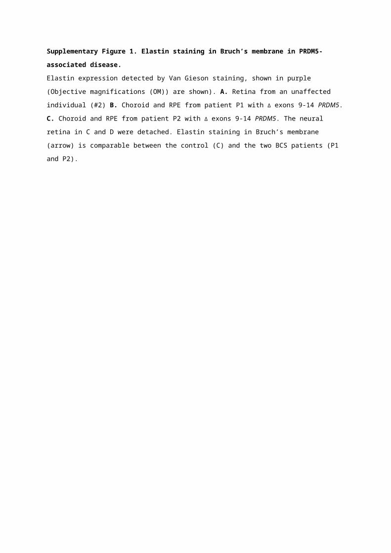
Supplementary Figure 1. Elastin staining in Bruch’s membrane in PRDM5-associated disease. Elastin expression detected by Van Gieson staining, shown in purple (Objective magnifications (OM))
are shown). A. Retina from an unaffected individual (#2) B. Choroid and RPE from patient P1 with Δ
exons 9-14 PRDM5. C. Choroid and RPE from patient P2 with Δ exons 9-14 PRDM5. The neural
retina in C and D were detached. Elastin staining in Bruch’s membrane (arrow) is comparable between
the control (C) and the two BCS patients (P1 and P2).
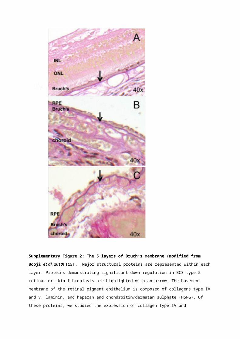
Supplementary Figure 2: The 5 layers of Bruch’s membrane (modified from Booji et al, 2010) [15]. Major structural proteins are represented within each layer. Proteins demonstrating significant
down-regulation in BCS-type 2 retinas or skin fibroblasts are highlighted with an arrow. The basement
membrane of the retinal pigment epithelium is composed of collagens type IV and V, laminin, and
heparan and chondroitin/dermatan sulphate (HSPG). Of these proteins, we studied the expression of
collagen type IV and demonstrated large downregulation of collagen type IV in PRDM5-associated
disease. The inner collagenous layer of Bruch’s membrane is composed primarily of collagens type I,
III, V and fibronectin, all of which were either downregulated in Bruch’s membrane of patient retinas
carrying the deletion of exons 9-14 PRDM5 mutation (P1 and P2), or the skin fibroblasts of a patient
lacking PRDM5 (P4) (collagens type I and III). The lack of PRDM5 also appeared to result in the
disorganized expression of collagen type V in skin fibroblasts. The elastin layer consists predominantly
of elastin fibres, collagens type IV and VI, collagen type XVIII and fibronectin. The basement
membrane of the choriocapillaris is predominantly composed of laminin, heparan sulphate and
collagens type IV, V and VI. BM: Bruch’s membrane; RPE: retinal pigment epithelium.
Supplementary Figure 3. qPCR assessment of target gene ITGA8 demonstrating fold change in
mRNA expression in dermal fibroblasts isolated from BCS patients with different mutations: PRDM5
p.Arg590* (P3) and PRDM5 internal deletion of exons 9 to 14 (P1), versus sex and age-matched
control fibroblast cell lines in the logarithmic scale (Log2RQ). mRNA levels were normalized to
GAPDH expression as described in the methods section. The Y-axis represents fold change (RQ) in
gene expression determined by the 2−ΔΔCt method. The X-axis shows the target gene assessed. Error
bars represent the 95% confidence interval around the mean. ITGA8 transcript levels in cell lines of
patients with PRDM5 mutations are decreased relative to control fibroblast cell lines. The p-value for
the paired two-tailed t-test reaches statistical significance (P<0.01).
ITA8 p.Arg590*/cont ITA8 9-14/contΔ
-4.5
-4
-3.5
-3
-2.5
-2
-1.5
-1
-0.5
0
Fold
Ch
ange
Gen
e E
xpre
ssio
n
(lo
g2)