static-curis.ku.dk a hybrid carrageenan containing both b-carrageenan and k/b-carrageenan motifs....
TRANSCRIPT

u n i ve r s i t y o f co pe n h ag e n
Københavns Universitet
A novel enzyme portfolio for red algal polysaccharide degradation in the marinebacterium Paraglaciecola hydrolytica S66T encoded in a sizeable polysaccharideutilization locusSchultz-Johansen, Mikkel; Bech, Pernille Kjersgaard; Hennessy, Rosanna Catherine; Glaring,Mikkel Andreas; Barbeyron, Tristan; Czjzek, Mirjam; Stougaard, PeterPublished in:Frontiers in Microbiology
DOI:10.3389/fmicb.2018.00839
Publication date:2018
Document VersionPublisher's PDF, also known as Version of record
Citation for published version (APA):Schultz-Johansen, M., Bech, P. K., Hennessy, R. C., Glaring, M. A., Barbeyron, T., Czjzek, M., & Stougaard, P.(2018). A novel enzyme portfolio for red algal polysaccharide degradation in the marine bacteriumParaglaciecola hydrolytica S66T encoded in a sizeable polysaccharide utilization locus. Frontiers inMicrobiology, 9, [839]. https://doi.org/10.3389/fmicb.2018.00839
Download date: 17. mar.. 2019

fmicb-09-00839 April 30, 2018 Time: 15:30 # 1
ORIGINAL RESEARCHpublished: 03 May 2018
doi: 10.3389/fmicb.2018.00839
Edited by:Satoshi Tsuneda,
Waseda University, Japan
Reviewed by:Thomas Schweder,
University of Greifswald, GermanyMatthias Wietz,
University of Oldenburg, Germany
*Correspondence:Peter [email protected]
†These authors have contributedequally to this work.
Specialty section:This article was submitted to
Microbial Physiology and Metabolism,a section of the journal
Frontiers in Microbiology
Received: 04 December 2017Accepted: 12 April 2018Published: 03 May 2018
Citation:Schultz-Johansen M, Bech PK,
Hennessy RC, Glaring MA,Barbeyron T, Czjzek M and
Stougaard P (2018) A Novel EnzymePortfolio for Red Algal PolysaccharideDegradation in the Marine Bacterium
Paraglaciecola hydrolytica S66T
Encoded in a Sizeable PolysaccharideUtilization Locus.
Front. Microbiol. 9:839.doi: 10.3389/fmicb.2018.00839
A Novel Enzyme Portfolio for RedAlgal Polysaccharide Degradation inthe Marine Bacterium Paraglaciecolahydrolytica S66T Encoded in aSizeable Polysaccharide UtilizationLocusMikkel Schultz-Johansen1†, Pernille K. Bech1†, Rosanna C. Hennessy1,Mikkel A. Glaring1, Tristan Barbeyron2, Mirjam Czjzek2 and Peter Stougaard1*
1 Section for Microbial Ecology and Biotechnology, Department of Plant and Environmental Sciences, University ofCopenhagen, Frederiksberg, Denmark, 2 Sorbonne Université, CNRS, Integrative Biology of Marine Models (LBI2M),Station Biologique de Roscoff (SBR), Roscoff, France
Marine microbes are a rich source of enzymes for the degradation of diversepolysaccharides. Paraglaciecola hydrolytica S66T is a marine bacterium capable ofhydrolyzing polysaccharides found in the cell wall of red macroalgae. In this study, weapplied an approach combining genomic mining with functional analysis to uncoverthe potential of this bacterium to produce enzymes for the hydrolysis of complexmarine polysaccharides. A special feature of P. hydrolytica S66T is the presence of alarge genomic region harboring an array of carbohydrate-active enzymes (CAZymes)notably agarases and carrageenases. Based on a first functional characterizationcombined with a comparative sequence analysis, we confirmed the enzymatic activitiesof several enzymes required for red algal polysaccharide degradation by the bacterium.In particular, we report for the first time, the discovery of novel enzyme activities targetingfurcellaran, a hybrid carrageenan containing both β-carrageenan and κ/β-carrageenanmotifs. Some of these enzymes represent a new subfamily within the CAZy classification.From the combined analyses, we propose models for the complete degradation ofagar and κ/β-type carrageenan by P. hydrolytica S66T. The novel enzymes describedhere may find value in new bio-based industries and advance our understandingof the mechanisms responsible for recycling of red algal polysaccharides in marineecosystems.
Keywords: marine bacteria, carbohydrate-active enzymes, algal polysaccharides, agarase, carrageenase,furcellaran, GH16
INTRODUCTION
Macroalgae contain complex and diverse polysaccharides (e.g., agars, carrageenans, alginates,and fucoidans), which constitute a carbon source for a range of marine bacteria (Martin et al.,2014). Hence, understanding the bacterial mechanisms for polysaccharide degradation is crucial todecipher carbon fluxes in marine ecosystems (Azam and Malfatti, 2007; Arnosti, 2011). Bacteria of
Frontiers in Microbiology | www.frontiersin.org 1 May 2018 | Volume 9 | Article 839

fmicb-09-00839 April 30, 2018 Time: 15:30 # 2
Schultz-Johansen et al. Red Algal Degradation in P. hydrolytica S66T
the phyla Bacteroidetes and Proteobacteria are often associatedwith macro- and microalgae and can degrade a range of marinepolymeric carbon (Martin et al., 2015). Community-based studieshave shown that Bacteroidetes are the first taxon to respondto algal blooms (Teeling et al., 2012, 2016), followed later byAlpha- and Gammaproteobacteria. Accordingly, Bacteroidetesgenerally encode a larger enzymatic potential for carbohydratedegradation than other bacterial phyla (Barbeyron et al., 2016b).These Carbohydrate Active enZymes (CAZymes) often clusterin polysaccharide utilization loci (PULs), which by definitioncontain the entire capacities for sensing, degradation, transport,and metabolism of a given polysaccharide (Grondin et al., 2017).In Bacteroidetes, PULs have traditionally been recognized by thepresence of tandem SusC/SusD-like proteins encoding a TonB-dependent transporter and a carbohydrate-binding lipoprotein,respectively (Martens et al., 2011; Terrapon and Henrissat, 2014).More recent results show that the PUL paradigm can be extendedand also covers degradation systems lacking SusC/SusD pairs(Ficko-Blean et al., 2017; Grondin et al., 2017); e.g., those presentin Gammaproteobacteria (Neumann et al., 2015).
Degradation of various algal polysaccharides such asalginate, agar, and carrageenan have been investigated throughgenomic and functional analysis, in particular, of the marineFlavobacterium Zobellia galactanivorans (Hehemann et al., 2012;Thomas et al., 2012, 2017; Barbeyron et al., 2016b; Ficko-Bleanet al., 2017). Likewise, similar enzyme systems have beendescribed in other Bacteroidetes members such as Gramellaforsetii (Bauer et al., 2006; Kabisch et al., 2014), Formosaagariphila (Mann et al., 2013), and Polaribacter spp. (Xinget al., 2015). Gammaproteobacteria have also been connectedto polymer degradation. Mesocosm studies have indicatedthat members of the Alteromonadaceae family dominate thebacterial community during algal polysaccharide degradationin some ecological niches (Wietz et al., 2015; von Scheibneret al., 2017). For instance, Alteromonas macleodii was found tobe abundant during alginate degradation (Wietz et al., 2015)and subsequent studies have verified the presence of alginateutilization systems in some ecotypes (Neumann et al., 2015).Likewise, Glaciecola agarilytica dominated an agar-degradingcommunity (Wietz et al., 2015) in accordance with the agarolyticphenotype reported for this bacterium (Yong et al., 2007). Severalother genera within the Alteromonadaceae family have beenconnected with agarolytic activities such as Agarivorans (Kimet al., 2016), Gayadomonas (Chi et al., 2013), and Catenovulum(Yan et al., 2011), however, little is known about the responsibleenzymatic systems within these taxa. Paraglaciecola hydrolyticaS66T represents a newly isolated Gammaproteobacterium of theAlteromonadaceae family, which has the ability to degrade bothagar and carrageenan (Bech et al., 2017). These abilities havebeen highlighted by the detection of various genomic systemsfor polysaccharide degradation (Schultz-Johansen et al., 2016).More insight into the enzymatic machineries of these taxonomicgroups may result in a deeper understanding of their role inmarine ecosystems.
Agars and carrageenans are major cell wall polysaccharidesin different phyla of red macroalgae. In their simplest forms,these polysaccharides are composed of a backbone built
from disaccharide blocks (-bioses, Supplementary Figure 1)of D-galactose and 3,6-anhydrogalactose (L-AHG in agar andD-AHG in carrageenan) and the resulting galactan backbone istermed agarose and β-carrageenan, respectively (Knutsen et al.,1994). However, the galactan backbone often adapts a diverserange of modifications including sulfation, methylation, andpyruvation (Usov, 2011). For example, in agar from Porphyraspp. L-AHG is replaced by L-galactose-6-sulfate; a polymer alsoknown as porphyran (Anderson and Rees, 1965). Likewise,different types of carrageenans have been defined, based onthe position and number of sulfate groups along the galactanbackbone, the presence of which influences the rheologicalproperties and industrial utilization (Campo et al., 2009); themost common types are κ- and ι-carrageenans. While industriallyexploited carrageenophyte red algae contain carrageenans with amajority of sulfated κ- and ι-type (Kappaphycus and Eucheumaspp.), most natural carrageenans are complex in structure, withvarying compositional patterns of disaccharide building blocksalong the polymeric chain. For example, furcellaran (extractedfrom Furcellaria lumbricalis) is a common name for hybridcarrageenan composed mainly of κ- and β-type disaccharidemotifs (Knutsen et al., 1990; Yang et al., 2011).
According to the CAZy database (Lombard et al., 2014),enzymes hydrolyzing agar belong to glycoside hydrolase (GH)families GH16, GH50, GH86, GH96, GH117, and GH118,whereas carrageenases are found in families GH16, GH82,GH127, GH129, and GH150 (Michel et al., 2006; Martin et al.,2014; Ficko-Blean et al., 2017). In addition, auxiliary enzymessuch as sulfatases may assist in the degradation of sulfated algalpolysaccharides (Helbert, 2017).
The identification of PULs has been widely used in genome-based studies to predict algal polysaccharide degradation inmarine bacteria (Mann et al., 2013; Sun et al., 2016; Zhuet al., 2016; Gao et al., 2017). While such analyses give overallhints about carbohydrate metabolism, the lack of characterizedclose homologs for the individual proteins makes it difficult topredict the fine substrate specificities. Thus, mining of PULscombined with experimental validation (functional screening)are required to advance our understanding of substrate diversitiesand the detailed mechanisms used by microbes for thebreakdown of complex carbohydrates. Indeed, such an approachhas successfully been used for the discovery of new enzymeactivities targeting marine polysaccharides (Hehemann et al.,2010; Rebuffet et al., 2011; Foran et al., 2017).
Besides the understanding of their important biologicalroles, oligosaccharides obtained from algal polysaccharides haverecently received increased interest due to their biomedicalpotential (Hu et al., 2006; Enoki et al., 2012; Morya et al., 2012;Kalitnik et al., 2016). In this context, novel enzymes are importantfor advancing the bioconversion of algae into biologicallyactive compounds with potential for industrial applications. Inaddition, the discovery of enzymes having novel activities canserve as tools for structural elucidation of complex carbohydrates(Aarstad et al., 2012).
In this study, we have investigated enzymes involved inred algal degradation by P. hydrolytica S66T and we providetentative models for the catabolism of agar and carrageenan
Frontiers in Microbiology | www.frontiersin.org 2 May 2018 | Volume 9 | Article 839

fmicb-09-00839 April 30, 2018 Time: 15:30 # 3
Schultz-Johansen et al. Red Algal Degradation in P. hydrolytica S66T
in this marine bacterium. In line with earlier studies wedemonstrate that novel enzyme specificities can be discoveredfrom PULs containing unannotated genes or distant homologsto characterized CAZymes. In particular, we report the findingof hitherto unknown furcellaranase-active enzymes, which arelikely to represent new GH families or subfamilies withinthe CAZy classification. The novel enzymes described heremay find value in new bio-based industries and advance ourunderstanding of the mechanisms responsible for recycling of redalgal polysaccharides in marine ecosystems.
MATERIALS AND METHODS
Data AvailabilityP. hydrolytica S66T was isolated and characterized in aprevious study (Bech et al., 2017). The draft genome sequenceof P. hydrolytica S66T is available from Genbank, acc. no.LSNE00000000 (Schultz-Johansen et al., 2016). The resultspresented here are based on protein predictions and locus tagsgenerated by RAST (Aziz et al., 2008), and are accessible on theRAST guest account under Genome ID 89404.41 (using login andpassword ‘guest’). For convenience the RAST generated locus tagprefix (fig|89404.4.peg.) was renamed to ‘Ph’ in the current study.
Sequence AnalysesCarbohydrate active enzymes and sulfatases were identified inthe P. hydrolytica S66T genome using Hidden Markov Modelsearches against local versions of the dbCAN 5.0 (Yin et al.,2012) and Pfam (Finn et al., 2016) databases. Predicted proteinsequences were compared with homologs at NCBI using BLASTpsearches against the PDB and nr databases. Sulfatase homologswere also compared in the SulfAtlas database2 using the built-inBLAST function (Barbeyron et al., 2016a). Manual comparisonand synteny of bacterial genomes was assessed using the MaGeinterface3 on the MicroScope platform (Vallenet et al., 2009).N-terminal secretion signals were predicted using SignalP 4.1(Petersen et al., 2011) and LipoP 1.0 (Juncker et al., 2003).Multiple alignments and phylogenetic trees were constructedusing MEGA6 (Tamura et al., 2013). The integrated ClustalWalgorithm was used for aligning GH16 protein sequences. Thealignment was manually inspected and then used to construct anunrooted maximum likelihood phylogenetic tree based on 1,000bootstrap iterations.
Gene Cloning and Production ofRecombinant Enzymes for FunctionalScreeningOligonucleotides used in this study are listed in SupplementaryTable 1. In general, the primers were designed to target thefull gene sequence without any predicted signal peptides. Thegene encoding Ph1586 was predicted from an ORF of ∼5
1http://rast.nmpdr.org2http://abims.sb-roscoff.fr/sulfatlas/3http://www.genoscope.cns.fr/agc/mage
kb and consequently only the proposed catalytic region wasPCR amplified. Genomic DNA (gDNA) was extracted fromP. hydrolytica S66T as described before (Schultz-Johansen et al.,2016) and used as template DNA in PCR reactions. PCRsof enzyme encoding genes were carried out with PhusionDNA polymerase (Novagen) or, for reactions with uracil-containing primers, the Pfu-X7 polymerase (Nørholm, 2010),kindly provided by Prof. Halkier, University of Copenhagen. PCRproducts were purified using the QIAquick PCR purification Kit(Qiagen, Germany). The Ph1631 gene construct was cloned intothe NdeI and XhoI sites of a pET22b vector (Novagen). Theremaining gene constructs were inserted into vectors (pET9a-USER-1 and pET9a-USER-2) modified for USERTM cloningaccording to the procedures described by Nour-Eldin et al.(2010). In brief, to construct the pET9a-USER-1 vector, a genecassette was assembled from oligonucleotides (SupplementaryTable 1) and cloned into the NdeI and BamHI sites of a linearizedpET9a vector (Novagen). The resulting pET9a-USER-1 featureda USER compatible cloning cassette in addition to a C-terminalocta-histidine tag, and was linearized with PacI and nt.BbvCIprior to cloning. The pET9a-USER-2 vector was generated asa ‘USER fusion’ product (Geu-Flores et al., 2007) by PCRamplification of pET9a-USER-1 using the primers specified inSupplementary Table 1. Assembly of purified PCRs and pET9a-USER vectors was carried out with USERTM enzyme (NEB) aspreviously described (Nour-Eldin et al., 2010).
Vector constructs were transformed into E. coli TOP10 cellsand incubated overnight at 37◦C on LB agar with ampicillin(100 µg/mL) for pET22b and kanamycin (50 µg/mL) for pET-USER constructs. Recombinant colonies were picked from platesand cultivated in LB broth with appropriate antibiotics at 37◦Covernight with shaking. Recombinant plasmids were purifiedusing the MiniPrep Plasmid DNA Purification Kit (Qiagen) andverified by DNA sequencing before production of recombinantenzymes.
For enzyme production, recombinant plasmids weretransformed into E. coli BL21(DE3) cells (as described above).Single colonies were picked from plates and cultivated in 20 mLZYP-5052 autoinduction medium (Studier, 2005) supplementedwith antibiotics for 3 days at 20◦C with shaking. Cells werecollected by centrifugation and resuspended in 0.5 mL lysisbuffer (50 mM HEPES, pH = 8, 100 mM NaCl or 50 mMTris-HCl, pH = 8, 100 mM NaCl). For Ph1656, Ph1659,Ph1663, and Ph1675, the lysis buffer also contained 5 mM CaCl2and for Ph1664 the lysis buffer contained 300 mM NaCl and5 mM MgCl2. Cell lysis was performed in a FastPrep-24TM
5G bead beater (MP Biomedicals) using glass beads size 212–300 µm/425–600 µm in the ratio 1:1 (Sigma-Aldrich). Aftercell disruption, the soluble and insoluble protein fractions wereseparated by centrifugation.
Enzyme Reactions and ActivityVisualizationCrude recombinant cell lysates were screened for activityagainst algal substrates; agar, κ-carrageenan, ι-carrageenan(Sigma-Aldrich), agarose (Invitrogen, United Kingdom,<0.351% sulfate), porphyran (prepared as described in
Frontiers in Microbiology | www.frontiersin.org 3 May 2018 | Volume 9 | Article 839

fmicb-09-00839 April 30, 2018 Time: 15:30 # 4
Schultz-Johansen et al. Red Algal Degradation in P. hydrolytica S66T
Hehemann et al., 2010), and furcellaran (Est-Agar, Kärla,Estonia). Agar-type oligosaccharides were obtained from Aglyco(Beijing, China) and L-AHG used as a standard was a kind giftfrom Prof. Kyoung Heon Kim (Korea University, South Korea).Carrageenan-type oligosaccharides were obtained from Dextra(Reading, United Kingdom).
For a 100 µL enzyme reaction, 10 µL crude cell lysate wasadded to polysaccharides (final concentration 0.05% w/v) oroligosaccharides (2 µg/µL) in the corresponding cell lysis buffer.Samples were incubated at 25◦C overnight and degradationproducts from enzymatic reactions were analyzed by thin layerchromatography (TLC) or fluorophore-assisted-carbohydrate-electrophoresis (FACE). TLC was performed on silica gels 60F254 (Merck) using n-butanol/acetic acid/water (2:1:1 v/v/v) asa running solvent and plates were developed on a hot plateafter spraying with orcinol. FACE was performed using labelingfluorophores 8-aminonaphthalene-1,3,6-trisulfonate (ANTS) or2-aminoacridone (AMAC) as described before (Starr et al., 1996;Hehemann et al., 2010). Briefly, enzyme reactions were dried in aspeed-vac centrifuge and the pellets were dissolved in 2 µL ANTSsolution (0.15 M ANTS in acetic acid/water (3:17, v/v) and 5µL freshly prepared 1 M sodium cyanoborohydride in DMSO)or AMAC solution (0.1 M AMAC in acetic acid/DMSO (3:17,v/v) and 5 µL freshly prepared 1 M sodium cyanoborohydridein water). After derivatization at 37◦C for 20 h, the sampleswere subjected to electrophoresis through a 6% stacking and 27%running polyacrylamide gel at 4◦C for 30 min at 15 mA followedby 30 mA for 2 h. Fluorescently labeled oligosaccharides in thepolyacrylamide gel were visualized under UV light.
RNA Extraction and ReverseTranscription PCR (RT-PCR)For RNA extraction, strain S66T was grown in Minimal MarineMedium (Thomas et al., 2011), supplemented with 2 g/Lagarose, agar, porphyran, or glucose that had been sterilizedby autoclaving. Cells were cultivated at 20◦C for 5 days withshaking (200 rpm) and total RNA was extracted using theZR Fungal/Bacterial RNA MiniPrepTM kit according to themanufacturer’s instructions (Zymo Research, United States).RNA purity and concentration were determined using aQubit R© RNA HR Assay Kit (Invitrogen) and RNA qualitywas assessed by agarose gel electrophoresis. ContaminatingDNA was removed using the DNA-freeTM DNA removal kit(Ambion) according to the manufacturer’s instructions. RNA waschecked for gDNA contamination by PCR prior to performingcDNA synthesis. Reverse transcription was performed with50 ng total RNA of each sample using a GoScript R© ReverseTranscription System (Promega) using non-sequence specificprimers according to manufacturer instructions. The obtainedcDNA was stored at −20◦C and used as template in subsequentPCR reactions. Target-specific primers were designed using CLC(Main Workbench) software (CLC BIO, Aarhus, Denmark)to amplify selected genes encoding enzymes predicted to beinvolved in polysaccharide degradation (Supplementary Table 2).PCRs were performed using the GoTag R© DNA polymerase(Promega) with 10 µM of each primer and 1 µL RNA or gDNA
(control) in a total reaction volume of 24 µL using the followingcycling parameters: 98◦C for 2 min, 30 cycles of 10 s at 98◦C, 30 sat 55◦C, and 30 s at 72◦C, followed by a final step at 72◦C for5 min. PCR products (4 µL) were visualized on agarose gels andsemi-quantitatively assessed. All experiments were performedwith biological triplicates.
RESULTS AND DISCUSSION
The Potential for PolysaccharideDegradation by P. hydrolytica S66T
P. hydrolytica S66T (from now on referred to as S66T) is capableof degrading several plant- and algal-derived polysaccharides,most notably red algal polysaccharides ranging from low-sulfate containing agarose to sulfated agar, porphyran, andκ-carrageenan (Bech et al., 2017). These phenotypic traitscorrelate with a preliminary survey of the S66T genome encodinga large number of putative enzymes with predicted functionsrelated to carbohydrate utilization (Schultz-Johansen et al., 2016).In this study, a more extensive analysis of the carbohydrateutilization potential was carried out (Supplementary DataSheet 1). This revealed a total of 270 CAZy modules inthe S66T genome, 188 of which were assigned to proteinfamilies involved in carbohydrate degradation, i.e., GHs,polysaccharide lyases (PLs) and carbohydrate esterases (CEs).Notably, the CAZyme potential of S66T exceeded that of anotherGammaproteobacterium P. atlantica, which is considered a“super-degrader” (Barbeyron et al., 2016b). Additional enzymesincluded 35 glycosyl transferases (GTs), 11 auxiliary activities(AAs), and 36 unique proteins associated with 46 carbohydrate-binding modules (CBMs). Twelve putative sulfatases werealso identified, and except for one candidate (S4 β-lactamasefamily) these represented various subfamilies within the S1formylglycine-dependent sulfatases according to the SulfAtlasdatabase (Barbeyron et al., 2016a). The existence of secretionsignals in all S1 sulfatases suggests a potential function againstexternal substrates and supports a proposed role in scavenging ofsulfated carbohydrates for most of these sulfatases.
A closer investigation of the CAZyme repertoire revealedseveral candidates belonging to GH families with characterizedred algae-specific members: Eight GH16 enzymes (β-agarases,κ-carrageenases, β-porphyranases or glucanases), fourGH50 enzymes (β-agarases), one GH82 (ι-carrageenase),two GH86 (β-agarases or β-porphyranases), one GH117(α-1,3-(3,6-anhydro)-L-galactosidase), and one GH127 (α-1,3-(3,6-anhydro)-D-galactosidase). No representatives fromagarolytic families GH96 (α-agarases) or GH118 (β-agarases)were identified.
All Genes for Red Algal PolysaccharideDegradation Are Organized in a Single,Large Gene ClusterAll CAZymes with a potential activity against red algalpolysaccharides were clustered in a contiguous genomic regionspanning ∼167,000 bp (Figure 1). The GH genes in this
Frontiers in Microbiology | www.frontiersin.org 4 May 2018 | Volume 9 | Article 839

fmicb-09-00839 April 30, 2018 Time: 15:30 # 5
Schultz-Johansen et al. Red Algal Degradation in P. hydrolytica S66T
FIGURE 1 | Genomic organization of a ∼167 kb PUL-like region in P. hydrolytica S66T encoding the proposed enzyme repertoire for complete degradation of agarand carrageenan. Putative gene functions are color-coded and regions flanking putative GHs are highlighted. Locus tags are indicated on the genes and proteinfamily/type is indicated next to the genes, if available. Black and red bars indicate genes for metabolism of L- and D-AHG, respectively. Agarolytic andcarrageenolytic PUL segments are shaded in yellow and green, respectively.
region co-localized with transporters (TonB and TRAP),sulfatases and dehydrogenases. Despite the absence of SusC/SusDhomologs this genomic organization resembled that of PULs inBacteroidetes (Martens et al., 2009). TonB dependent receptors inthis region are likely to contribute to oligosaccharide detectionand transport in analogy to the bacteroidetal SusC/SusD system(Blanvillain et al., 2007; Thomas et al., 2012; Neumann et al.,2015). Based on sequence-based and functional analyses, at leasttwo overall sub-PULs could be assigned: one dedicated to agardegradation (Ph1566–Ph1636) and the other responsible forcarrageenan degradation (Ph1646–Ph1681).
Agarolytic Degradation SystemHomology searches against the GenBank nr database, usingproteins encoded in the PUL-like regions, predominantlyidentified proteins with uncharacterized functions as the closestrelatives. Sequence comparison against characterized proteinsfrom PDB revealed that GHs located in the first part of thePUL-like region (Ph1586–Ph1636) displayed sequence similarityto characterized β-agarases (Ph1631_GH16, Ph1589_GH50,Ph1609_GH50, Ph1624_GH50, and Ph1636_GH50),β-porphyranases (Ph1586_GH86, Ph1607_GH86) and oneα-1,3-(3,6-anhydro)-L-galactosidase (Ph1615_GH117) (Figure 1and Supplementary Table 3). In the near upstream region, wealso identified a candidate homolog to a characterized agarolyticβ-galactosidase (Ph1566_GH2). Ph1566_GH2 displayed 40%sequence identity to VejABG, which has previously been shownto release galactose from the non-reducing end of agaro-oligosaccharides (Lee C.H. et al., 2014). RT-PCR experimentssupported an agarolytic role, since the GH genes tested in therange Ph1586–Ph1636 (except Ph1615) were induced in responseto agarose and porphyran substrates (Supplementary Figure 2A).
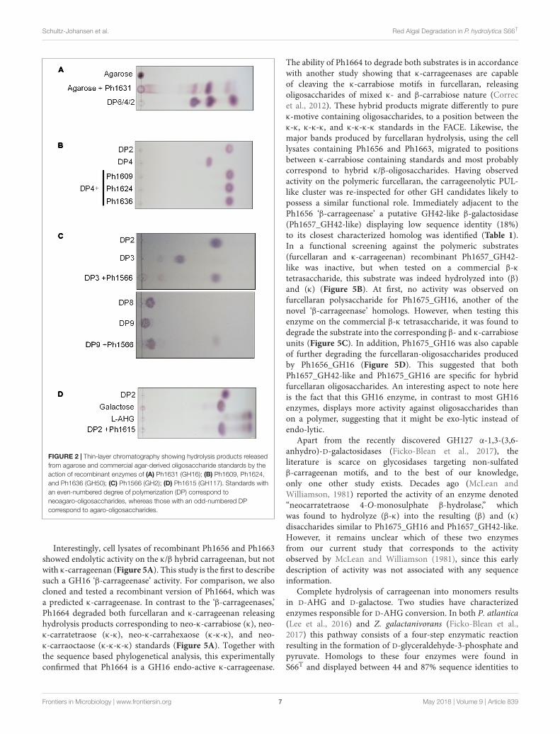
The agarolytic role of six GHs was confirmed in apreliminary functional characterization (Table 1). E. coli celllysates expressing the genes encoding agarolytic GHs wereincubated with agar-type substrates and enzymatic degradationwas visualized by TLC (Figure 2). TLC analysis showedthat Ph1631 degraded agarose into primarily neoagarotetraose,with some neoagarobiose and neoagarohexaose also produced(Figure 2A). In addition, Ph1631 also displayed activityagainst porphyran (but only little, compared to agarose) andneoagaro-oligosaccharides, releasing neoagarotetraose and asmall amount of neoagarobiose as the major products (data
not shown). This suggested that Ph1631 is a GH16 β-agarasemost likely exhibiting endolytic activity. Ph1609, Ph1624, andPh1636 (GH50) showed activity on agar and agarose, as wellas on neoagaro-oligosaccharides down to neoagarotetraose.The main hydrolysis products of all three enzymes wereneoagarobiose indicating that Ph1609, Ph1624, and Ph1636represent GH50 exo-β-agarases (Figure 2B). Ph1566 (GH2)released galactose from the non-reducing end of odd-numberedagaro-oligosaccharides (Figure 2C) but was unable to hydrolyzeeven-numbered neo-oligosaccharides and lactose (data notshown). This confirmed that Ph1566_GH2 shares the functionof VejABG from Vibrio sp. EJY3 which until this date wasthe only report of an agarolytic GH2 β-galactosidase (Lee C.H.et al., 2014). Finally, Ph1615 (GH117) was able to hydrolyzeneoagarobiose yielding L-AHG and galactose confirming thatPh1615 was an α-1,3-(3,6-anhydro)-L-galactosidase (Rebuffetet al., 2011) (Figure 2D).
Complete hydrolysis of agar into monomers results in L-AHGand D-galactose. Interestingly, close homologs to characterizedenzymes responsible for metabolism of L-AHG (Lee S.B. et al.,2014; Yun et al., 2015) were also identified in the PUL-likecluster (Figure 1 and Supplementary Table 3). In both Vibriosp. EJY3 and P. atlantica T6c, the first two steps in L-AHGmetabolism involves an L-AHG dehydrogenase that oxidizeL-AHG to AHGA and an L-AHG cycloisomerase that isomerizeAHGA into L-KDGal (Lee S.B. et al., 2014; Yun et al., 2015).Homologs of these enzymes (71–87% identity) are also found inS66T. In Vibrio sp. EJY3, L-KDGal is believed to enter the centralmetabolism after these two steps (Yun et al., 2015), whereas inP. atlantica, L-KDGal is further processed to KDPG by a three-step reaction before entering the Entner–Doudoroff pathway (LeeS.B. et al., 2014). These three latter enzymes have homologs inS66T (49–89% identity) and in addition (except for the KDGkinase), the L-AHG pathways from P. atlantica and S66T sharecomplete synteny (Supplementary Figure 3A). Therefore, S66T
is most likely to metabolize L-AHG according to the enzymaticpathway reported for P. atlantica T6c (Lee S.B. et al., 2014).
Based on the functional validated enzymes and sequence-based analyses, we can account for the complete metabolicagarose pathway in S66T (Figure 3). At least, the agarolyticsystem includes an endo-acting GH16 β-agarase (Ph1631)that produces neoagaro-oligosaccharides, which are furtherprocessed by GH50 β-agarases (Ph1609, Ph1624, and Ph1636)
Frontiers in Microbiology | www.frontiersin.org 5 May 2018 | Volume 9 | Article 839
fmicb-09-00839 April 30, 2018 Time: 15:30 # 6
Schultz-Johansen et al. Red Algal Degradation in P. hydrolytica S66T
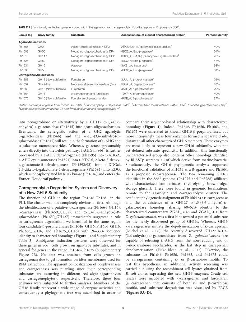
TABLE 1 | Functionally verified enzymes encoded within the agarolytic and carrageenolytic PUL-like regions in P. hydrolytica S66T.
Locus tag CAZy family Substrate Accession no. of closest characterized protein Percent identity
Agarolytic activities
Ph1566 GH2 Agaro-oligosaccharides ≥ DP3 AEX22320.1; Agarolytic β-galactosidase1 40%
Ph1609 GH50 Neoagaro-oligosaccharides ≥ DP4 4BQ2_A; Exo-β-agarase2 61%
Ph1615 GH117 Neoagaro-oligosaccharides ≥ DP2 3R4Y_A; α-1,3-(3,6-anhydro)-L-galactosidase2 73%
Ph1624 GH50 Neoagaro-oligosaccharides ≥ DP4 4BQ2_A; Exo-β-agarase2 47%
Ph1631 GH16 Agarose 3WZ1_A; β-agarase3 68%
Ph1636 GH50 Neoagaro-oligosaccharides ≥ DP4 4BQ2_A; Exo-β-agarase2 31%
Carrageenolytic activities
Ph1656 GH16 (New subfamily) Furcellaran 3JUU_A; β-porphyranase4 26%
Ph1657 GH42-like Neocarratetraose monosulfate (β-κ ) 5DFA _A; β-galactosidase5 18%
Ph1663 GH16 (New subfamily) Furcellaran 4ATE_A; β-porphyranase4 29%
Ph1664 GH16 κ-carrageenan and furcellaran 1DYP_A; κ-carrageenase6 40%
Ph1675 GH16 (New subfamily) Furcellaran-oligosaccharides 4ATE_A; β-porphyranase4 27%
Protein homologs originate from 1Vibrio sp. EJY3, 2Saccharophagus degradans 2-40T, 3Microbulbifer thermotolerans JAMB A94T, 4Zobellia galactanivorans DsijT,5Geobacillus stearothermophilus T6 and 6Pseudoalteromonas carrageenovora 9T.
into neoagarobiose or alternatively by a GH117 α-1,3-(3,6-anhydro)-L-galactosidase (Ph1615) into agaro-oligosaccharides.Eventually, the synergistic action of a GH2 agarolyticβ-galactosidase (Ph1566) and the α-1,3-(3,6-anhydro)-L-galactosidase (Ph1615) will result in the formation of L-AHG andD-galactose monosaccharides. Whereas, galactose presumablyenters directly into the Leloir pathway, L-AHG in S66T is furtherprocessed by a L-AHG dehydrogenase (Ph1595) into L-AHGA,L-AHG cycloisomerase (Ph1591) into L-KDGal, 2-keto-3-deoxy-L-galactonate-5-dehydrogenase (Ph1592/93) into L-DDGal,2,5-diketo-L-galactonate-5-dehydrogenase (Ph1694) into KDG,which is phosphorylated by KDG kinase (Ph1616) and enters theEntner–Doudoroff pathway.
Carrageenolytic Degradation System and Discoveryof a New GH16 SubfamilyThe function of GHs in the region Ph1646–Ph1681 in thePUL-like cluster was not completely obvious at first. Althoughthe identification of a putative κ-carrageenase (Ph1664_GH16),ι-carrageenase (Ph1659_GH82), and α-1,3-(3,6-anhydro)-D-galactosidase (Ph1650_GH127) immediately suggested a rolein carrageenan degradation, we identified in the same regionfour candidate β-porphyranases (Ph1646_GH16, Ph1656_GH16,Ph1663_GH16, and Ph1675_GH16) with 26–33% sequenceidentity to characterized homologs (Figure 1 and SupplementaryTable 3). Ambiguous induction patterns were observed forthese genes in S66T cells grown on agar-type substrates, and ingeneral for genes in the range Ph1646–Ph1675 (SupplementaryFigure 2B). No data was obtained from cells grown oncarrageenan due to gel formation on filter membranes used forRNA extraction. The apparent co-localization of porphyranasesand carrageenases was puzzling since their correspondingsubstrates are occurring in different red algae (agarophytesand carrageenophytes), respectively. Therefore, these fourenzymes were subjected to further analyses. Members of theGH16 family represent a wide range of enzyme activities andconsequently a phylogenetic tree was established in order to
compare their sequence-based relationship with characterizedhomologs (Figure 4). Indeed, Ph1646, Ph1656, Ph1663, andPh1675 were unrelated to known GH16 β-porphyranases, butmore intriguingly these four enzymes formed a separate clade,distant to all other characterized GH16 members. These enzymesare most likely to represent a new GH16 subfamily, with notyet defined substrate specificity. In addition, this functionallyuncharacterized group also contains other homologs identifiedby BLASTp searches, all of which derive from marine bacteria.Simultaneously, the GH16 phylogenetic analysis supportedthe functional validation of Ph1631 as a β-agarase and Ph1664as a proposed κ-carrageenase. The two remaining GH16sidentified in the S66T genome (Ph1275 and Ph3268) affiliatedwith characterized laminarinases (hydrolyzing brown algalstorage glucan). These were found in genomic localizationsremote to the agarolytic and carrageenolytic clusters. Theconfident phylogenetic assignment of Ph1664 as a κ-carrageenaseand the co-existence of a GH127 α-1,3-(3,6-anhydro)-D-galactosidase homolog (sharing 60–62% identity to thecharacterized counterparts ZGAL_3148 and ZGAL_3150 fromZ. galactanivorans), was a first hint toward a potential substratefor the newly discovered group of GH16s. Whereas, GH16κ-carrageenases initiate the depolymerization of κ-carrageenan(Michel et al., 2006), the recently discovered GH127 α-1,3-(3,6-anhydro)-D-galactosidases from Z. galactanivorans arecapable of releasing D-AHG from the non-reducing end ofβ-neocarrabiose saccharides, as the last step in carrageenandepolymerization (Ficko-Blean et al., 2017). Likewise, thesubstrate for Ph1646, Ph1656, Ph1663, and Ph1675 couldbe carrageenans containing κ- or β-carrabiose motifs. Totest this hypothesis, an additional activity screening wascarried out using the recombinant cell lysates obtained fromE. coli clones expressing the new GH16 enzymes. Crude celllysates were incubated with κ-carrageenan and furcellaran(a carrageenan that consists of both κ- and β-carrabiosemotifs), and substrate degradation was visualized by FACE(Figures 5A–D).
Frontiers in Microbiology | www.frontiersin.org 6 May 2018 | Volume 9 | Article 839
fmicb-09-00839 April 30, 2018 Time: 15:30 # 7
Schultz-Johansen et al. Red Algal Degradation in P. hydrolytica S66T
FIGURE 2 | Thin-layer chromatography showing hydrolysis products releasedfrom agarose and commercial agar-derived oligosaccharide standards by theaction of recombinant enzymes of (A) Ph1631 (GH16); (B) Ph1609, Ph1624,and Ph1636 (GH50); (C) Ph1566 (GH2); (D) Ph1615 (GH117). Standards withan even-numbered degree of polymerization (DP) correspond toneoagaro-oligosaccharides, whereas those with an odd-numbered DPcorrespond to agaro-oligosaccharides.
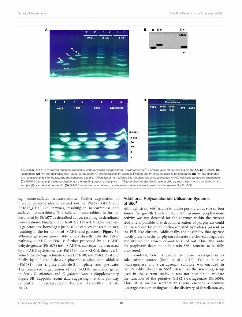
Interestingly, cell lysates of recombinant Ph1656 and Ph1663showed endolytic activity on the κ/β hybrid carrageenan, but notwith κ-carrageenan (Figure 5A). This study is the first to describesuch a GH16 ‘β-carrageenase’ activity. For comparison, we alsocloned and tested a recombinant version of Ph1664, which wasa predicted κ-carrageenase. In contrast to the ‘β-carrageenases,’Ph1664 degraded both furcellaran and κ-carrageenan releasinghydrolysis products corresponding to neo-κ-carrabiose (κ), neo-κ-carratetraose (κ-κ), neo-κ-carrahexaose (κ-κ-κ), and neo-κ-carraoctaose (κ-κ-κ-κ) standards (Figure 5A). Together withthe sequence based phylogenetical analysis, this experimentallyconfirmed that Ph1664 is a GH16 endo-active κ-carrageenase.
The ability of Ph1664 to degrade both substrates is in accordancewith another study showing that κ-carrageenases are capableof cleaving the κ-carrabiose motifs in furcellaran, releasingoligosaccharides of mixed κ- and β-carrabiose nature (Correcet al., 2012). These hybrid products migrate differently to pureκ-motive containing oligosaccharides, to a position between theκ-κ, κ-κ-κ, and κ-κ-κ-κ standards in the FACE. Likewise, themajor bands produced by furcellaran hydrolysis, using the celllysates containing Ph1656 and Ph1663, migrated to positionsbetween κ-carrabiose containing standards and most probablycorrespond to hybrid κ/β-oligosaccharides. Having observedactivity on the polymeric furcellaran, the carrageenolytic PUL-like cluster was re-inspected for other GH candidates likely topossess a similar functional role. Immediately adjacent to thePh1656 ‘β-carrageenase’ a putative GH42-like β-galactosidase(Ph1657_GH42-like) displaying low sequence identity (18%)to its closest characterized homolog was identified (Table 1).In a functional screening against the polymeric substrates(furcellaran and κ-carrageenan) recombinant Ph1657_GH42-like was inactive, but when tested on a commercial β-κtetrasaccharide, this substrate was indeed hydrolyzed into (β)and (κ) (Figure 5B). At first, no activity was observed onfurcellaran polysaccharide for Ph1675_GH16, another of thenovel ‘β-carrageenase’ homologs. However, when testing thisenzyme on the commercial β-κ tetrasaccharide, it was found todegrade the substrate into the corresponding β- and κ-carrabioseunits (Figure 5C). In addition, Ph1675_GH16 was also capableof further degrading the furcellaran-oligosaccharides producedby Ph1656_GH16 (Figure 5D). This suggested that bothPh1657_GH42-like and Ph1675_GH16 are specific for hybridfurcellaran oligosaccharides. An interesting aspect to note hereis the fact that this GH16 enzyme, in contrast to most GH16enzymes, displays more activity against oligosaccharides thanon a polymer, suggesting that it might be exo-lytic instead ofendo-lytic.
Apart from the recently discovered GH127 α-1,3-(3,6-anhydro)-D-galactosidases (Ficko-Blean et al., 2017), theliterature is scarce on glycosidases targeting non-sulfatedβ-carrageenan motifs, and to the best of our knowledge,only one other study exists. Decades ago (McLean andWilliamson, 1981) reported the activity of an enzyme denoted“neocarratetraose 4-O-monosulphate β-hydrolase,” whichwas found to hydrolyze (β-κ) into the resulting (β) and (κ)disaccharides similar to Ph1675_GH16 and Ph1657_GH42-like.However, it remains unclear which of these two enzymesfrom our current study that corresponds to the activityobserved by McLean and Williamson (1981), since this earlydescription of activity was not associated with any sequenceinformation.
Complete hydrolysis of carrageenan into monomers resultsin D-AHG and D-galactose. Two studies have characterizedenzymes responsible for D-AHG conversion. In both P. atlantica(Lee et al., 2016) and Z. galactanivorans (Ficko-Blean et al.,2017) this pathway consists of a four-step enzymatic reactionresulting in the formation of D-glyceraldehyde-3-phosphate andpyruvate. Homologs to these four enzymes were found inS66T and displayed between 44 and 87% sequence identities to
Frontiers in Microbiology | www.frontiersin.org 7 May 2018 | Volume 9 | Article 839

fmicb-09-00839 April 30, 2018 Time: 15:30 # 8
Schultz-Johansen et al. Red Algal Degradation in P. hydrolytica S66T
FIGURE 3 | Tentative pathway for agarose degradation in P. hydrolytica S66T. Enzymes are indicated at each reaction step together with CAZy family if applicable.Solid arrows represent the proposed pathway of P. hydrolytica S66T based on functional (black) and bioinformatics (gray) analyses. The dashed arrow represents analternative catabolic fate of L-KDGal as reported elsewhere. NAOS, neoagaro-oligosaccharides; AOS, agaro-oligosaccharides; L-AHG, 3,6-anhydro-L-galactose;Gal, D-galactose; L-AHGA, 3,6-anhydro-L-galactonate; L-KDGal, 2-keto-3-deoxy-L-galactonate; L-DDGal, 2,5-diketo-3-deoxy- L-galactonate; KDG,2-keto-3-deoxy-D-gluconate; KDPG, 2-keto-3-deoxy-6-phospho-D-gluconate.
the characterized counterparts from P. atlantica. In addition,conserved gene synteny was observed for the D-AHG pathwayin all three organisms (Supplementary Figure 3B). Moreover,four sulfatases belonging to the S1_19 sulfatase subfamilywere identified interspersed between the GHs. This subfamilycontains characterized members (Patl_0889 and Patl_0895 fromP. atlantica T6c) which cleave off the sulfate from galactose-4-sulfate in ι- and κ-carrageenans, respectively (Prechouxet al., 2013, 2016). Patl_0889 shared 76% identity with thePh1648 sulfatase from S66T and Patl_0895 showed 83% toPh1677. Both Ph1648 and Ph1677 sulfatases were located inthe carrageenolytic part of the PUL-like cluster. Also, sulfatasegenes located in the PUL-like cluster were in general inducedwith the increased degree of sulfatation in the substrates
tested (Supplementary Figure 2C). Altogether, this supported afunction in the removal of sulfate groups from the backbone ofcarrageenan, agar and/or porphyran as reported elsewhere (Kimet al., 2004; Prechoux et al., 2013, 2016; Wang et al., 2015).
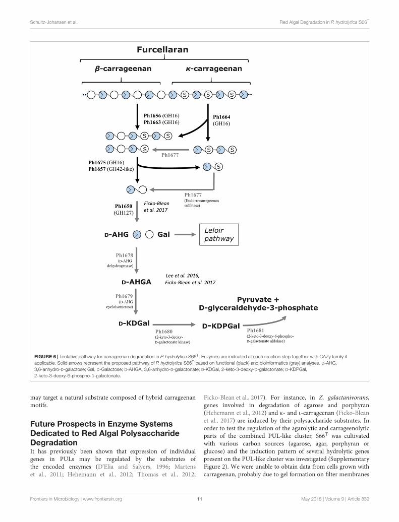
On the basis of the current discoveries, we are able forthe first time to propose a model for furcellaran degradationin a marine bacterium (Figure 6). In S66T, it is assumedthat the GHs in the carrageenolytic PUL-like cluster each arespecific for a particular part in furcellaran. The κ-carrageenase(Ph1664_GH16) targets the κ-carrageenan part in furcellaranreleasing κ-carrageenan oligosaccharides that are furtherdesulfated through the action of sulfatases. The proposedsulfatase activity remains to be established. However, a goodcandidate would be Ph1677, which displays high sequence
Frontiers in Microbiology | www.frontiersin.org 8 May 2018 | Volume 9 | Article 839

fmicb-09-00839 April 30, 2018 Time: 15:30 # 9
Schultz-Johansen et al. Red Algal Degradation in P. hydrolytica S66T
FIGURE 4 | Sequence-based relationship of GH16 family proteins from P. hydrolytica S66T (black triangles) and homologs obtained from public databases. Ph1646,Ph1656, Ph1663, and Ph1675 are part of a separate phylogenetic clade forming a new GH16 subfamily. Accession number and taxonomy is indicated for eachprotein. The β-agarases, β-porphyrases, κ-carrageenases, and laminarinases represent characterized enzymes according to the CAZy database.
identity (83%) to a characterized endo-κ-carrageenan sulfatase(Prechoux et al., 2016). The proposed reaction product ofthe action of the κ-carrageenase Ph1664_GH16 togetherwith sulfatase Ph1677 is mono-sulfated neocarratetraose
and neocarrabiose. In addition, enzymes such as the heredescribed furcellaranases, Ph1656_GH16 and Ph1663_GH16,can hydrolyze the β-carrageenan or hybrid part of furcellaran,releasing partially sulfated carrageenan oligosaccharides,
Frontiers in Microbiology | www.frontiersin.org 9 May 2018 | Volume 9 | Article 839
fmicb-09-00839 April 30, 2018 Time: 15:30 # 10
Schultz-Johansen et al. Red Algal Degradation in P. hydrolytica S66T
FIGURE 5 | FACE of hydrolysis products released by carrageenolytic enzymes from P. hydrolytica S66T. Samples were prepared using ANTS (A,C,D) or AMAC (B)fluorophore. (A) Ph1664 degrades both kappa-carrageenan (C) and furcellaran (F), whereas Ph1656 and Ph1663 are specific for furcellaran. (B) Ph1657 degradesβ-κ tetrasaccharide into the resulting disaccharides β and κ. ∗(Migration of non-sulfated β is not observed since uncharged AMAC was used as labeling fluorophore).(C) Ph1675 degrades β-κ tetrasaccharide into the resulting disaccharides β and κ. Oligosaccharides standards were applied as substrates in a mix containing κ, κ-κand β-κ (1) or κ-κ-κ and κ-κ-κ-κ (2). (D) Ph1675 is inactive on furcellaran, but degrades the furcellaran-oligosaccharides released by Ph1656.
e.g., mono-sulfated neocarratetraose. Further degradation ofthese oligosaccharides is carried out by Ph1675_GH16 andPh1657_GH42-like enzymes, resulting in neocarrabiose andsulfated neocarrabiose. The sulfated neocarrabiose is furtherdesulfated by Ph1677 as described above, resulting in desulfatedneocarrabiose. Finally, the Ph1650_GH127 α-1,3-(3,6-anhydro)-D-galactosidase homolog is proposed to catalyze the reaction stepresulting in the formation of D-AHG and galactose (Figure 6).Whereas galactose presumably enters directly into the Leloirpathway, D-AHG in S66T is further processed by a D-AHGdehydrogenase (Ph1678) into D-AHGA, subsequently processedby a D-AHG cycloisomerase (Ph1679) into D-KDGal, then by a 2-keto-3-deoxy-D-galactonate kinase (Ph1680) into D-KDPGal andfinally by a 2-keto-3-deoxy-6-phospho-D-galactonate aldolase(Ph1681) into D-glyceraldehyde-3-phosphate and pyruvate.The conserved organization of the D-AHG metabolic genesin S66T, P. atlantica and Z. galactanivorans (SupplementaryFigure 3B) supports recent data suggesting that this pathwayis central in carrageenolytic bacteria (Ficko-Blean et al.,2017).
Additional Polysaccharide Utilization Systemsof S66T
Although strain S66T is able to utilize porphyran as sole carbonsource for growth (Bech et al., 2017), genuine porphyranaseactivity was not detected for the enzymes within the currentstudy. It is possible that depolymerization of porphyran couldbe carried out by other uncharacterized hydrolases present inthe PUL-like clusters. Additionally, the possibility that agarosemotifs present in the porphyran substrate are cleaved by agarasesand utilized for growth cannot be ruled out. Thus, the routefor porphyran degradation in strain S66T remains to be fullyuncovered.
In contrast, S66T is unable to utilize ι-carrageenan assole carbon source (Bech et al., 2017). Yet, a putativeι-carrageenase and ι-carrageenan sulfatase was encoded inthe PUL-like cluster in S66T. Based on the screening setupused in the current study, it was not possible to validatethe function of the putative GH82 ι-carrageenase (Ph1659).Thus, it is unclear whether this gene encodes a genuineι-carrageenase or, analogous to the discovery of furcellaranases,
Frontiers in Microbiology | www.frontiersin.org 10 May 2018 | Volume 9 | Article 839
fmicb-09-00839 April 30, 2018 Time: 15:30 # 11
Schultz-Johansen et al. Red Algal Degradation in P. hydrolytica S66T
FIGURE 6 | Tentative pathway for carrageenan degradation in P. hydrolytica S66T. Enzymes are indicated at each reaction step together with CAZy family ifapplicable. Solid arrows represent the proposed pathway of P. hydrolytica S66T based on functional (black) and bioinformatics (gray) analyses. D-AHG,3,6-anhydro-D-galactose; Gal, D-Galactose; D-AHGA, 3,6-anhydro-D-galactonate; D-KDGal, 2-keto-3-deoxy-D-galactonate; D-KDPGal,2-keto-3-deoxy-6-phospho-D-galactonate.
may target a natural substrate composed of hybrid carrageenanmotifs.
Future Prospects in Enzyme SystemsDedicated to Red Algal PolysaccharideDegradationIt has previously been shown that expression of individualgenes in PULs may be regulated by the substrates ofthe encoded enzymes (D’Elia and Salyers, 1996; Martenset al., 2011; Hehemann et al., 2012; Thomas et al., 2012;
Ficko-Blean et al., 2017). For instance, in Z. galactanivorans,genes involved in degradation of agarose and porphyran(Hehemann et al., 2012) and κ- and ι-carrageenan (Ficko-Bleanet al., 2017) are induced by their polysaccharide substrates. Inorder to test the regulation of the agarolytic and carrageenolyticparts of the combined PUL-like cluster, S66T was cultivatedwith various carbon sources (agarose, agar, porphyran orglucose) and the induction pattern of several hydrolytic genespresent on the PUL-like cluster was investigated (SupplementaryFigure 2). We were unable to obtain data from cells grown withcarrageenan, probably due to gel formation on filter membranes
Frontiers in Microbiology | www.frontiersin.org 11 May 2018 | Volume 9 | Article 839

fmicb-09-00839 April 30, 2018 Time: 15:30 # 12
Schultz-Johansen et al. Red Algal Degradation in P. hydrolytica S66T
used for RNA extraction. More importantly, the formation ofcell aggregates during cultivation of S66T on all the substratesprevented optical density comparison of the growth. Instead,this was gauged by the amount of extracted RNA, whichwas comparable for all treatments (Supplementary Table 4).Consequently, the gene regulation could only be preliminaryevaluated.
Overall, our results seem to point toward an even morecomplex genetic organization of polysaccharide utilizingsystems than those reported to date (Ficko-Blean et al.,2017; Grondin et al., 2017). In the here presented case, theco-localization of agar-specific genes with more complexcarrageenan degrading genes in the same genetic region, thebi-directional orientation of the genes, and the preliminarygene expression analyses suggest a succession of evolutionaryevents of gene shuffling and/or horizontal gene transfer,possibly leading to a complex cross-regulation of agarasesand carrageenases in S66T. Interestingly, this observationis in-line with recent genome wide transcriptomic analysesof Z. galactanivorans that show common signatures whengrowing on chemically divergent polysaccharides from thesame algal phylum (Thomas et al., 2017). In our study, bothagar-specific and carrageenan-specific genes are regulated byagar and agarose suggesting that the regulation pattern of thisPUL-like cluster might be very complex. However, due to thetechnical limitations in this experiment, we do not yet haveaccess to regulation patterns in the presence of carrageenanand more thorough investigations are needed in order to fullyunderstand the gene regulation in the complex PUL-like clusterin S66T. We are currently testing different growth conditionsin order to prevent the formation of cell aggregates, whichwould allow more comprehensive transcriptomic analyses in thefuture.
The size of the PUL-like region in S66T is unusual and, toour knowledge, one of the biggest reported to date. Thus, theevolutionary events leading to the assembly of this PUL seemintriguing. Although, it was outside the scope of the present studyto analyze this in detail, initial observations suggest that manyof the genes in the PUL-like region may have been horizontallytransferred from other Gammaproteobacteria. This is supportedby the presence of at least one mobile element within the PULand DNA/RNA-modifying genes in the flanking regions (notshown).
Functional screening combined with phylogenetic analysesrevealed that Ph1656, Ph1663, Ph1675 and possibly Ph1646constitute a new GH16 subfamily with substrate preference forvarious stretches of furcellaran – a hybrid carrageenan composedof κ-type and non-sulfated β-type carrabiose motifs. Likewise,Ph1657 was found to cleave a tetrasaccharide (β-κ) into itsresulting disaccharide moieties. The exact functionalities are yetto be investigated in detail, but results from the preliminarilyscreening point toward a group of enzymes acting on thenon-sulfated β-carrageenan motifs (since κ-carrageenan was notdegraded).
GH16 family enzymes share a β-jellyroll fold and are believedto have evolved from a common ancestor into several individualsubfamilies with enzyme activities targeting both marine and
terrestrial substrates (Barbeyron et al., 1998; Michel et al., 2006;Eklöf et al., 2013). Thus, detailed biochemical characterization ofthe novel GH16 members from P. hydrolytica S66T is of specialinterest and should provide new valuable information about theirfunctional roles in the degradation of red algal polysaccharides,as well as the evolutionary relationship between GH16 familyenzymes.
Considering the results presented here, it is interestingto note that the furcellaran active GH16 and GH42-likemembers are absent in the newly characterized carrageenolyticsystem of Z. galactanivorans (except for a genuine GH16κ-carrageenase). In brief, the carrageenolytic system ofZ. galactanivorans is composed of a core PUL comprisingthe D-AHG metabolizing pathway, several sulfatases and α-1,3-(3,6-anhydro)-D-galactosidases, in addition to other genes(including endolytic carrageenases and transporters) encoded ingenomic regions outside the core PUL (Ficko-Blean et al., 2017).On the other hand, Ficko-Blean et al. (2017) showed that GH16and GH42-like homologs with unknown functions are foundin carrageenolytic PULs of other marine bacteria includingmembers of the Alteromonadaceae family to which P. hydrolyticaS66T belongs. We therefore speculate that the furcellaranaseactivities demonstrated in the present study may correspondto these uncharacterized CAZymes. If so, our findings maycontribute to a better understanding of carrageenan degradationin marine bacteria.
In addition to the GHs described in this study, othernew enzyme activities may be found in the red algal PUL-like clusters. In this context, a GH29 (fucosidases) and aGH63 (glucosidases) located in the agarolytic part of the genecluster are interesting targets for future characterization studies,since members of these CAZyme families are not obviouslyinvolved in hydrolysis of red algal polysaccharides and maytherefore represent new enzyme activities. This rationale hasearlier led to the discovery of novel enzyme activities actingon algal carbohydrates (Rebuffet et al., 2011; Ficko-Bleanet al., 2017). Thus, we expect that future research in otherprotein complements in the S66T PUL-like clusters may revealadditional functional novelties related to agar and carrageenandegradation.
AUTHOR CONTRIBUTIONS
MS-J, PB, MG, TB, MC, and PS conceptualized and designedthe studies. MS-J, PB, and TB performed the bioinformatic andphylogenetic analyses. PB and MS-J cloned and characterizedthe enzymes. RH and PB performed the RT-PCR analyses. MS-Jwrote the manuscript with contributions from all authors. All theauthors read and approved the final manuscript.
FUNDING
This work was supported by grants from the Novo NordiskFoundation (Grant No. NNF12OC0000797) and The Danish
Frontiers in Microbiology | www.frontiersin.org 12 May 2018 | Volume 9 | Article 839

fmicb-09-00839 April 30, 2018 Time: 15:30 # 13
Schultz-Johansen et al. Red Algal Degradation in P. hydrolytica S66T
Council for Independent Research, Technology and ProductionSciences (0602-02399B) to PS.
ACKNOWLEDGMENTS
We thank Prof. Kyoung Heon Kim, Department ofBiotechnology, Korea University, for providing the 3,6-anhydro-L-galactose and the group of Prof. Barbara Halkier, University
of Copenhagen, for Pfu-X7 polymerase. Line Christiansen isthanked for assisting in the characterization of enzymes.
SUPPLEMENTARY MATERIAL
The Supplementary Material for this article can be foundonline at: https://www.frontiersin.org/articles/10.3389/fmicb.2018.00839/full#supplementary-material
REFERENCESAarstad, O. A., Tondervik, A., Sletta, H., and Skjak-Braek, G. (2012). Alginate
sequencing: an analysis of block distribution in alginates using specificalginate degrading enzymes. Biomacromolecules 13, 106–116. doi: 10.1021/bm2013026
Anderson, N. S., and Rees, D. A. (1965). Porphyran - a polysaccharidewith a masked repeating structure. J. Chem. Soc. 5880–5887. doi: 10.1039/jr9650005880
Arnosti, C. (2011). Microbial extracellular enzymes and the marine carbon cycle.Ann. Rev. Mar. Sci. 3, 401–425. doi: 10.1146/annurev-marine-120709-142731
Azam, F., and Malfatti, F. (2007). Microbial structuring of marine ecosystems. Nat.Rev. Microbiol. 5, 782–791. doi: 10.1038/nrmicro1747
Aziz, R. K., Bartels, D., Best, A. A., DeJongh, M., Disz, T., Edwards, R. A., et al.(2008). The RAST Server: rapid annotations using subsystems technology. BMCGenomics 9:75. doi: 10.1186/1471-2164-9-75
Barbeyron, T., Brillet-Gueguen, L., Carre, W., Carriere, C., Caron, C., Czjzek, M.,et al. (2016a). Matching the diversity of sulfated biomolecules: creation of aclassification database for sulfatases reflecting their substrate specificity. PLoSOne 11:e0164846. doi: 10.1371/journal.pone.0164846
Barbeyron, T., Gerard, A., Potin, P., Henrissat, B., and Kloareg, B. (1998). Thekappa-carrageenase of the marine bacterium Cytophaga drobachiensis.Structural and phylogenetic relationships within family-16 glycosidehydrolases. Mol. Biol. Evol. 15, 528–537.
Barbeyron, T., Thomas, F., Barbe, V., Teeling, H., Schenowitz, C., Dossat, C.,et al. (2016b). Habitat and taxon as driving forces of carbohydrate catabolismin marine heterotrophic bacteria: example of the model algae-associatedbacterium Zobellia galactanivorans DsijT. Environ. Microbiol. 18, 4610–4627.doi: 10.1111/1462-2920.13584
Bauer, M., Kube, M., Teeling, H., Richter, M., Lombardot, T., Allers, E., et al. (2006).Whole genome analysis of the marine Bacteroidetes ’Gramella forsetii’ revealsadaptations to degradation of polymeric organic matter. Environ. Microbiol. 8,2201–2213. doi: 10.1111/j.1462-2920.2006.01152.x
Bech, P. K., Schultz-Johansen, M., Glaring, M. A., Barbeyron, T., Czjzek, M.,and Stougaard, P. (2017). Paraglaciecola hydrolytica sp. nov., a bacterium withhydrolytic activity against multiple seaweed-derived polysaccharides. Int. J. Syst.Evol. Microbiol. 67, 2242–2247. doi: 10.1099/ijsem.0.001933
Blanvillain, S., Meyer, D., Boulanger, A., Lautier, M., Guynet, C., Denance, N.,et al. (2007). Plant carbohydrate scavenging through tonB-dependent receptors:a feature shared by phytopathogenic and aquatic bacteria. PLoS One 2:e224.doi: 10.1371/journal.pone.0000224
Campo, V. L., Kawano, D. F., da Silva, D. B., and Carvalho, I. (2009).Carrageenans: biological properties, chemical modifications and structuralanalysis - A review. Carbohydr. Polym. 77, 167–180. doi: 10.1016/j.carbpol.2009.01.020
Chi, W. J., Park, J. S., Kwak, M. J., Kim, J. F., Chang, Y. K., and Hong, S. K. (2013).Isolation and characterization of a novel agar-degrading marine bacterium,Gayadomonas joobiniege gen, nov, sp. nov., from the Southern Sea, Korea.J. Microbiol. Biotechnol. 23, 1509–1518.
Correc, G., Barabanova, A., Tuvikene, R., Truus, K., Yermak, I., and Helbert, W.(2012). Comparison of the structures of hybrid kappa-/beta-carrageenansextracted from Furcellaria lumbricalis and Tichocarpus crinitus. Carbohydr.Polym. 88, 31–36. doi: 10.1016/j.carbpol.2011.11.052
D’Elia, J. N., and Salyers, A. A. (1996). Effect of regulatory protein levelson utilization of starch by Bacteroides thetaiotaomicron. J. Bacteriol. 178,7180–7186.
Eklöf, J. M., Shojania, S., Okon, M., McIntosh, L. P., and Brumer, H. (2013).Structure-function analysis of a broad specificity Populus trichocarpa endo-beta-glucanase reveals an evolutionary link between bacterial licheninases andplant XTH gene products. J. Biol. Chem. 288, 15786–15799. doi: 10.1074/jbc.M113.462887
Enoki, T., Tominaga, T., Takashima, F., Ohnogi, H., Sagawa, H., and Kato, I. (2012).Anti-tumor-promoting activities of agaro-oligosaccharides on two-stage mouseskin carcinogenesis. Biol. Pharm. Bull. 35, 1145–1149. doi: 10.1248/bpb.b12-00188
Ficko-Blean, E., Prechoux, A., Thomas, F., Rochat, T., Larocque, R., Zhu, Y.,et al. (2017). Carrageenan catabolism is encoded by a complex regulon inmarine heterotrophic bacteria. Nat. Commun. 8:1685. doi: 10.1038/s41467-017-01832-6
Finn, R. D., Coggill, P., Eberhardt, R. Y., Eddy, S. R., Mistry, J., Mitchell, A. L., et al.(2016). The Pfam protein families database: towards a more sustainable future.Nucleic Acids Res. 44, D279–D285. doi: 10.1093/nar/gkv1344
Foran, E., Buravenkov, V., Kopel, M., Mizrahi, N., Shoshani, S., Helbert, W., et al.(2017). Functional characterization of a novel “ulvan utilization loci” found inAlteromonas sp LOR genome. Algal Res. 25, 39–46. doi: 10.1016/j.algal.2017.04.036
Gao, B., Jin, M., Li, L., Qu, W., and Zeng, R. (2017). Genome sequencing revealsthe complex polysaccharide-degrading ability of novel deep-sea bacteriumFlammeovirga pacifica WPAGA1. Front. Microbiol. 8:600. doi: 10.3389/fmicb.2017.00600
Geu-Flores, F., Nour-Eldin, H. H., Nielsen, M. T., and Halkier, B. A. (2007).USER fusion: a rapid and efficient method for simultaneous fusion andcloning of multiple PCR products. Nucleic Acids Res. 35:e55. doi: 10.1093/nar/gkm106
Grondin, J. M., Tamura, K., Dejean, G., Abbott, D. W., and Brumer, H. (2017).Polysaccharide utilization loci: fueling microbial communities. J. Bacteriol.199:e00860-16. doi: 10.1128/JB.00860-16
Hehemann, J. H., Correc, G., Barbeyron, T., Helbert, W., Czjzek, M., and Michel, G.(2010). Transfer of carbohydrate-active enzymes from marine bacteria toJapanese gut microbiota. Nature 464, 908–912. doi: 10.1038/nature08937
Hehemann, J. H., Correc, G., Thomas, F., Bernard, T., Barbeyron, T., Jam, M., et al.(2012). Biochemical and structural characterization of the complex agarolyticenzyme system from the marine bacterium Zobellia galactanivorans. J. Biol.Chem. 287, 30571–30584. doi: 10.1074/jbc.M112.377184
Helbert, W. (2017). Marine polysaccharide sulfatases. Front. Mar. Sci. 4:6.doi: 10.3389/fmars.2017.00006
Hu, B., Gong, Q., Wang, Y., Ma, Y., Li, J., and Yu, W. (2006). Prebiotic effectsof neoagaro-oligosaccharides prepared by enzymatic hydrolysis of agarose.Anaerobe 12, 260–266. doi: 10.1016/j.anaerobe.2006.07.005
Juncker, A. S., Willenbrock, H., Von Heijne, G., Brunak, S., Nielsen, H., andKrogh, A. (2003). Prediction of lipoprotein signal peptides in Gram-negativebacteria. Protein Sci. 12, 1652–1662. doi: 10.1110/ps.0303703
Kabisch, A., Otto, A., Konig, S., Becher, D., Albrecht, D., Schuler, M., et al. (2014).Functional characterization of polysaccharide utilization loci in the marineBacteroidetes ’Gramella forsetii’, KT0803. ISME J. 8, 1492–1502. doi: 10.1038/ismej.2014.4
Kalitnik, A. A., Anastyuk, S. D., Sokolova, E. V., Kravchenko, A. O., Khasina, E. I.,and Yermak, I. M. (2016). Oligosaccharides of kappa/beta-carrageenan from thered alga Tichocarpus crinitus and their ability to induce interleukin 10. J. Appl.Phycol. 28, 545–553. doi: 10.1007/s10811-015-0577-6
Kim, J. H., Byun, D. S., Godber, J. S., Choi, J. S., Choi, W. C., and Kim, H. R.(2004). Purification and characterization of arylsulfatase from Sphingomonas
Frontiers in Microbiology | www.frontiersin.org 13 May 2018 | Volume 9 | Article 839

fmicb-09-00839 April 30, 2018 Time: 15:30 # 14
Schultz-Johansen et al. Red Algal Degradation in P. hydrolytica S66T
sp AS6330. Appl. Microbiol. Biotechnol. 63, 553–559. doi: 10.1007/s00253-003-1463-8
Kim, S. G., Pheng, S., Lee, Y. J., Eom, M. K., and Shin, D. H. (2016). Agarivoransaestuarii sp nov., an agar-degrading bacterium isolated from a tidal flat. Int. J.Syst. Evol. Microbiol. 66, 3119–3124. doi: 10.1099/ijsem.0.001155
Knutsen, S. H., Myslabodski, D. E., and Grasdalen, H. (1990). Characterization ofcarrageenan fractions from Norwegian Furcellaria-lumbricalis (Huds) Lamourby H-1-Nmr spectroscopy. Carbohydr. Res. 206, 367–372. doi: 10.1016/0008-6215(90)80076-f
Knutsen, S. H., Myslabodski, D. E., Larsen, B., and Usov, A. I. (1994). A modifiedsystem of nomenclature for red algal galactans. Bot. Mar. 37, 163–169.doi: 10.1515/botm.1994.37.2.163
Lee, C. H., Kim, H. T., Yun, E. J., Lee, A. R., Kim, S. R., Kim, J. H., et al.(2014). A novel agarolytic beta-galactosidase acts on agarooligosaccharides forcomplete hydrolysis of agarose into monomers. Appl. Environ. Microbiol. 80,5965–5973. doi: 10.1128/AEM.01577-14
Lee, S. B., Cho, S. J., Kim, J. A., Lee, S. Y., Kim, S. M., and Lim, H. S.(2014). Metabolic pathway of 3,6-anhydro-L-galactose in agar-degradingmicroorganisms. Biotechnol. Bioprocess Eng. 19, 866–878. doi: 10.1007/s12257-014-0622-3
Lee, S. B., Kim, J. A., and Lim, H. S. (2016). Metabolic pathway of3,6-anhydro-D-galactose in carrageenan-degrading microorganisms. Appl.Microbiol. Biotechnol. 100, 4109–4121. doi: 10.1007/s00253-016-7346-6
Lombard, V., Golaconda Ramulu, H., Drula, E., Coutinho, P. M., and Henrissat, B.(2014). The carbohydrate-active enzymes database (CAZy) in 2013. NucleicAcids Res. 42, D490–D495. doi: 10.1093/nar/gkt1178
Mann, A. J., Hahnke, R. L., Huang, S., Werner, J., Xing, P., Barbeyron, T., et al.(2013). The genome of the alga-associated marine flavobacterium Formosaagariphila KMM 3901T reveals a broad potential for degradation of algalpolysaccharides. Appl. Environ. Microbiol. 79, 6813–6822. doi: 10.1128/AEM.01937-13
Martens, E. C., Koropatkin, N. M., Smith, T. J., and Gordon, J. I. (2009). Complexglycan catabolism by the human gut microbiota: the Bacteroidetes Sus-likeparadigm. J. Biol. Chem. 284, 24673–24677. doi: 10.1074/jbc.R109.022848
Martens, E. C., Lowe, E. C., Chiang, H., Pudlo, N. A., Wu, M., McNulty, N. P.,et al. (2011). Recognition and degradation of plant cell wall polysaccharides bytwo human gut symbionts. PLoS Biol. 9:e1001221. doi: 10.1371/journal.pbio.1001221
Martin, M., Barbeyron, T., Martin, R., Portetelle, D., Michel, G., and Vandenbol, M.(2015). The cultivable surface microbiota of the brown alga Ascophyllumnodosum is Enriched in Macroalgal-polysaccharide-degrading bacteria. Front.Microbiol. 6:1487. doi: 10.3389/fmicb.2015.01487
Martin, M., Portetelle, D., Michel, G., and Vandenbol, M. (2014). Microorganismsliving on macroalgae: diversity, interactions, and biotechnological applications.Appl. Microbiol. Biotechnol. 98, 2917–2935. doi: 10.1007/s00253-014-5557-2
McLean, M. W., and Williamson, F. B. (1981). Neocarratetraose 4-O-monosulphate beta-hydrolase from Pseudomonas carrageenovora. Eur. J.Biochem. 113, 447–456.
Michel, G., Nyval-Collen, P., Barbeyron, T., Czjzek, M., and Helbert, W. (2006).Bioconversion of red seaweed galactans: a focus on bacterial agarases andcarrageenases. Appl. Microbiol. Biotechnol. 71, 23–33. doi: 10.1007/s00253-006-0377-7
Morya, V. K., Kim, J., and Kim, E. K. (2012). Algal fucoidan: structural and size-dependent bioactivities and their perspectives. Appl. Microbiol. Biotechnol. 93,71–82. doi: 10.1007/s00253-011-3666-8
Neumann, A. M., Balmonte, J. P., Berger, M., Giebel, H. A., Arnosti, C., Voget, S.,et al. (2015). Different utilization of alginate and other algal polysaccharidesby marine Alteromonas macleodii ecotypes. Environ. Microbiol. 17, 3857–3868.doi: 10.1111/1462-2920.12862
Nørholm, M. H. (2010). A mutant Pfu DNA polymerase designed for advanceduracil-excision DNA engineering. BMC Biotechnol. 10:21. doi: 10.1186/1472-6750-10-21
Nour-Eldin, H. H., Geu-Flores, F., and Halkier, B. A. (2010). USER cloning andUSER fusion: the ideal cloning techniques for small and big laboratories.Methods Mol. Biol. 643, 185–200. doi: 10.1007/978-1-60761-723-5_13
Petersen, T. N., Brunak, S., von Heijne, G., and Nielsen, H. (2011). SignalP 4.0:discriminating signal peptides from transmembrane regions. Nat. Methods 8,785–786. doi: 10.1038/nmeth.1701
Prechoux, A., Genicot, S., Rogniaux, H., and Helbert, W. (2013). Controllingcarrageenan structure using a novel formylglycine-dependent sulfatase, anendo-4S-iota-carrageenan sulfatase. Mar. Biotechnol. 15, 265–274. doi: 10.1007/s10126-012-9483-y
Prechoux, A., Genicot, S., Rogniaux, H., and Helbert, W. (2016). Enzyme-assistedpreparation of furcellaran-like kappa-/beta-Carrageenan. Mar. Biotechnol. 18,133–143. doi: 10.1007/s10126-015-9675-3
Rebuffet, E., Groisillier, A., Thompson, A., Jeudy, A., Barbeyron, T., Czjzek, M.,et al. (2011). Discovery and structural characterization of a novel glycosidasefamily of marine origin. Environ. Microbiol. 13, 1253–1270. doi: 10.1111/j.1462-2920.2011.02426.x
Schultz-Johansen, M., Glaring, M. A., Bech, P. K., and Stougaard, P. (2016). Draftgenome sequence of a novel marine bacterium, Paraglaciecola sp. strain S66,with hydrolytic activity against seaweed polysaccharides. Genome Announc.4:e00304-16. doi: 10.1128/genomeA.00304-16
Starr, C. M., Masada, R. I., Hague, C., Skop, E., and Klock, J. C. (1996).Fluorophore-assisted carbohydrate electrophoresis in the separation, analysis,and sequencing of carbohydrates. J. Chromatogr. A 720, 295–321.
Studier, F. W. (2005). Protein production by auto-induction in high densityshaking cultures. Protein Expr. Purif. 41, 207–234.
Sun, C., Fu, G. Y., Zhang, C. Y., Hu, J., Xu, L., Wang, R. J., et al. (2016).Isolation and complete genome sequence of Algibacter alginolytica sp. nov.,a novel seaweed-degrading Bacteroidetes bacterium with diverse putativepolysaccharide utilization loci. Appl. Environ. Microbiol. 82, 2975–2987.doi: 10.1128/AEM.00204-16
Tamura, K., Stecher, G., Peterson, D., Filipski, A., and Kumar, S. (2013). MEGA6:molecular evolutionary genetics analysis version 6.0. Mol. Biol. Evol. 30,2725–2729. doi: 10.1093/molbev/mst197
Teeling, H., Fuchs, B. M., Becher, D., Klockow, C., Gardebrecht, A., Bennke,C. M., et al. (2012). Substrate-controlled succession of marine bacterioplanktonpopulations induced by a phytoplankton bloom. Science 336, 608–611.doi: 10.1126/science.1218344
Teeling, H., Fuchs, B. M., Bennke, C. M., Kruger, K., Chafee, M., Kappelmann, L.,et al. (2016). Recurring patterns in bacterioplankton dynamics during coastalspring algae blooms. Elife 5:e11888. doi: 10.7554/eLife.11888
Terrapon, N., and Henrissat, B. (2014). How do gut microbes break down dietaryfiber? Trends Biochem. Sci. 39, 156–158. doi: 10.1016/j.tibs.2014.02.005
Thomas, F., Barbeyron, T., and Michel, G. (2011). Evaluation of referencegenes for real-time quantitative PCR in the marine flavobacterium Zobelliagalactanivorans. J. Microbiol. Methods 84, 61–66. doi: 10.1016/j.mimet.2010.10.016
Thomas, F., Barbeyron, T., Tonon, T., Genicot, S., Czjzek, M., and Michel, G.(2012). Characterization of the first alginolytic operons in a marine bacterium:from their emergence in marine Flavobacteriia to their independent transfersto marine Proteobacteria and human gut Bacteroides. Environ. Microbiol. 14,2379–2394. doi: 10.1111/j.1462-2920.2012.02751.x
Thomas, F., Bordron, P., Eveillard, D., and Michel, G. (2017). Gene expressionanalysis of Zobellia galactanivorans during the degradation of algalpolysaccharides reveals both substrate-specific and shared transcriptome-wideresponses. Front. Microbiol. 8:1808. doi: 10.3389/fmicb.2017.01808
Usov, A. I. (2011). Polysaccharides of the red algae. Adv. Carbohydr. Chem.Biochem. 65, 115–217. doi: 10.1016/B978-0-12-385520-6.00004-2
Vallenet, D., Engelen, S., Mornico, D., Cruveiller, S., Fleury, L., Lajus, A.,et al. (2009). MicroScope: a platform for microbial genome annotation andcomparative genomics. Database 2009:ba021. doi: 10.1093/database/bap021
von Scheibner, M., Sommer, U., and Jurgens, K. (2017). Tight coupling ofGlaciecola spp. and diatoms during cold-water phytoplankton spring blooms.Front. Microbiol. 8:27. doi: 10.3389/fmicb.2017.00027
Wang, X. Y., Duan, D. L., Xu, J. C., Gao, X., and Fu, X. T. (2015). Characterizationof a novel alkaline arylsulfatase from Marinomonas sp FW-1 and its applicationin the desulfation of red seaweed agar. J. Ind. Microbiol. Biotechnol. 42,1353–1362. doi: 10.1007/s10295-015-1625-6
Wietz, M., Wemheuer, B., Simon, H., Giebel, H. A., Seibt, M. A., Daniel, R., et al.(2015). Bacterial community dynamics during polysaccharide degradation atcontrasting sites in the Southern and Atlantic Oceans. Environ. Microbiol. 17,3822–3831. doi: 10.1111/1462-2920.12842
Xing, P., Hahnke, R. L., Unfried, F., Markert, S., Huang, S., Barbeyron, T., et al.(2015). Niches of two polysaccharide-degrading Polaribacter isolates from the
Frontiers in Microbiology | www.frontiersin.org 14 May 2018 | Volume 9 | Article 839

fmicb-09-00839 April 30, 2018 Time: 15:30 # 15
Schultz-Johansen et al. Red Algal Degradation in P. hydrolytica S66T
North Sea during a spring diatom bloom. ISME J. 9, 1410–1422. doi: 10.1038/ismej.2014.225
Yan, S. L., Yu, M., Wang, Y., Shen, C., and Zhang, X. H. (2011). Catenovulumagarivorans gen. nov., sp nov., a peritrichously flagellated, chain-forming, agar-hydrolysing gammaproteobacterium from seawater. Int. J. Syst. Evol. Microbiol.61, 2866–2873. doi: 10.1099/ijs.0.027565-0
Yang, B., Yu, G. L., Zhao, X., Ren, W. N., Jiao, G. L., Fang, L. H., et al.(2011). Structural characterisation and bioactivities of hybrid carrageenan-like sulphated galactan from red alga Furcellaria lumbricalis. Food Chem. 124,50–57. doi: 10.1016/j.foodchem.2010.05.102
Yin, Y., Mao, X., Yang, J., Chen, X., Mao, F., and Xu, Y. (2012). dbCAN: a webresource for automated carbohydrate-active enzyme annotation. Nucleic AcidsRes. 40, W445–W451. doi: 10.1093/nar/gks479
Yong, J. J., Park, S. J., Kim, H. J., and Rhee, S. K. (2007). Glaciecola agarilytica sp.nov., an agar-digesting marine bacterium from the East Sea, Korea. Int. J. Syst.Evol. Microbiol. 57(Pt 5), 951–953. doi: 10.1099/ijs.0.64723-0
Yun, E. J., Lee, S., Kim, H. T., Pelton, J. G., Kim, S., Ko, H. J., et al. (2015). Thenovel catabolic pathway of 3,6-anhydro-L-galactose, the main component of
red macroalgae, in a marine bacterium. Environ. Microbiol. 17, 1677–1688.doi: 10.1111/1462-2920.12607
Zhu, Y., Chen, P., Bao, Y., Men, Y., Zeng, Y., Yang, J., et al. (2016). Completegenome sequence and transcriptomic analysis of a novel marine strain Bacillusweihaiensis reveals the mechanism of brown algae degradation. Sci. Rep.6:38248. doi: 10.1038/srep38248
Conflict of Interest Statement: The authors declare that the research wasconducted in the absence of any commercial or financial relationships that couldbe construed as a potential conflict of interest.
Copyright © 2018 Schultz-Johansen, Bech, Hennessy, Glaring, Barbeyron, Czjzek andStougaard. This is an open-access article distributed under the terms of the CreativeCommons Attribution License (CC BY). The use, distribution or reproduction inother forums is permitted, provided the original author(s) and the copyright ownerare credited and that the original publication in this journal is cited, in accordancewith accepted academic practice. No use, distribution or reproduction is permittedwhich does not comply with these terms.
Frontiers in Microbiology | www.frontiersin.org 15 May 2018 | Volume 9 | Article 839