status of gpcr modeling and docking as reflected by...
TRANSCRIPT

Structure
Article
Status of GPCR Modeling and Docking as Reflectedby Community-wide GPCR Dock 2010 AssessmentIrina Kufareva,1 Manuel Rueda,1 Vsevolod Katritch,1,2 GPCR Dock 2010 participants, Raymond C. Stevens,3,*and Ruben Abagyan1,2,*1Skaggs School of Pharmacy and Pharmaceutical Sciences, University of California, San Diego, La Jolla, CA 92039, USA2San Diego Supercomputer Center, La Jolla, CA 92039, USA3Department of Molecular Biology, The Scripps Research Institute, La Jolla, CA 92037, USA
*Correspondence: [email protected] (R.C.S.), [email protected] (R.A.)
DOI 10.1016/j.str.2011.05.012
SUMMARY
The community-wide GPCR Dock assessment isconducted to evaluate the status of molecularmodeling and ligand docking for human G protein-coupled receptors. The present round of the assess-ment was based on the recent structures of dopa-mine D3 and CXCR4 chemokine receptors bound tosmall molecule antagonists and CXCR4 with a syn-thetic cyclopeptide. Thirty-five groups submittedtheir receptor-ligand complex structure predictionsprior to the release of the crystallographic coordi-nates. With closely related homology modeling tem-plates, as for dopamine D3 receptor, and with incor-poration of biochemical and QSAR data, moderncomputational techniques predicted complex detailswith accuracy approaching experimental. In con-trast, CXCR4 complexes that had less-characterizedinteractions and only distant homology to the knownGPCR structures still remained very challenging. Theassessment results provide guidance for modelingand crystallographic communities in method devel-opment and target selection for further expansion ofthe structural coverage of the GPCR universe.
INTRODUCTION
The G protein-coupled receptor (GPCR) superfamily has more
than 800 members in the human genome (Fredriksson et al.,
2003; Ono et al., 2005), detecting a variety of extracellular chem-
ical, biological, or physical signals that are critical for human
biology and disease (Lagerstrom and Schioth, 2008). Under-
standing their three-dimensional (3D) structures will help under-
standing their function and will enable the development of new
therapeutic molecules. However, the dynamical nature of these
membrane proteins makes them notoriously difficult crystalliza-
tion targets (Cherezov et al., 2010). Prior to the summer of 2010,
only four vertebrate GPCRs yielded to crystallization efforts:
bovine rhodopsin (bRho) in liganded (Palczewski et al., 2000)
and ligand-free (opsin) (Park et al., 2008; Scheerer et al., 2008)
forms; human b2 (Cherezov et al., 2007) and turkey b1 (Warne
et al., 2008) adrenergic receptors; and human A2A adenosine
1108 Structure 19, 1108–1126, August 10, 2011 ª2011 Elsevier Ltd A
receptor (A2AAR) (Jaakola et al., 2008). Comparative analysis of
these structures demonstrated that, despite the conserved
seven transmembrane (7TM) topology, structural determinants
of ligand interaction are strikingly diverse between distantly
related GPCRs, even within class A where all the structures
solved so far belong, and that factors contributing to reshaping
of the ligand binding pockets include helical shifts, turns, tilts,
and kinks, as well as conformations of highly variable extracel-
lular loops. Due to such structural diversity, it is impossible to
expand the atomic details of ligand binding elucidated by crys-
tallography to cover all other members of the GPCR family.
Continuous improvement of molecular modeling and docking
algorithms andmethodology (Abagyan et al., 1997; Abagyan and
Totrov, 1994; Abel et al., 2010; Barth et al., 2009; Bottegoni et al.,
2008, 2009; Brylinski and Skolnick, 2008; Case et al., 2005; Cav-
asotto et al., 2005, 2008; Chen et al., 2003; Davis and Baker,
2009; Eswar et al., 2006; Katritch et al., 2010; Lang et al.,
2009; Morris et al., 2009; Vaidehi et al., 2002; Verdonk et al.,
2003; Yarov-Yarovoy et al., 2006; Zhou andGilson, 2009) is com-
plemented by great advances in computing resources and tech-
nologies. The recent decades were marked by an increase of
approximately four orders of magnitude in individual computing
power and storage, as well as the development of large super-
computers and specialized hardware (e.g., Shaw et al., 2010).
The amount of protein structural data in the Protein Data Bank
(PDB) has been growing exponentially providing invaluable infor-
mation about molecular interactions and, with increasing struc-
tural coverage of the mammalian proteome, relevant templates
for modeling by homology. Despite all these advances, modern
theoretical methods still fail to produce models of experimental
accuracy and/or ‘‘dockable’’ quality in many real-life cases,
e.g., in the absence of close structural homology templates.
The community-wide GPCR Dock assessment has the goal
of monitoring the progress of molecular modeling and ligand
docking for GPCR targets. The first round of assessment was
performed in 2008 when the structure of the A2AAR was solved
(Jaakola et al., 2008); 29 groups attempted to predict atomic
details of its interaction with a small molecule antagonist. The
most accurate models were built by homology with the b2 adren-
ergic receptor (b2AR) structure that shares �35% sequence
identity with A2AAR in the transmembrane (TM) domain. These
models were able to correctly predict about one-half of intermo-
lecular contacts (Michino et al., 2009). The present round of the
assessment, GPCR Dock 2010, is particularly exciting because
the modeled complexes represent three distinct classes and
ll rights reserved

Structure
Community-wide GPCR Dock 2010 Assessment
three levels of difficulty: (1) dopamine D3 receptor in complex
with eticlopride, which is a small molecule in a small molecule
pocket with two close homologymodeling templates; (2) chemo-
kine receptor CXCR4 bound to isothiourea IT1t, which is a small
molecule in a large peptide-binding pocket with more distant
templates; and (3) CXCR4/CVX15, which is the first GPCR
complex with a peptide analog. Prediction of the ligand binding
pose and its interactions within the receptor-binding pocket
constituted the main focus of the assessment. As a secondary
target, the prediction of the overall structure of the TM bundle
was also evaluated.
Comprehensive analysis of the 275 GPCR complex models
submitted for the assessment helped elucidate the current
trends in GPCR modeling and docking. In particular we found
that reliable homology modeling requires at least 35%–40%
sequence identity between target and template, and that in
such close homology cases, the combination of modern mod-
eling techniques with biochemical and QSAR studies allows
complex detail prediction with accuracy approaching experi-
mental. The results of this experiment outline the boundaries
for computational expansion of the sparse GPCR structural
information onto the new GPCR family members and their inter-
actions with small molecules or peptides. They also define the
‘‘white spots’’ on theGPCRmap that are in biggest need of being
addressed by crystallography.
RESULTS
GPCRDock2010: Description andSubmission StatisticsThe GPCR modeling and docking assessment 2010 was per-
formed for three separate ligand-receptor structures: human
dopamine D3 receptor bound to eticlopride (D3/eticlopride);
human chemokine receptor CXCR4 bound to an isothiourea
derivative IT1t (CXCR4/IT1t) (Thoma et al., 2008); and CXCR4
bound to the CVX15 peptide (Arg-Arg-Nal-Cys-Tyr-Gln-Lys-
dPro-Pro-Tyr-Arg-Cit-Cys-Arg-Gly-dPro) (CXCR4/CVX15). For
these 3 targets, 117, 103, and 55 unique interpretable models
were submitted by 32, 25, and 19 groups, respectively. The list
of participating groups, names, and affiliations is given in Table 1.
The models were assessed by several independent criteria
that were applied to the following structural features: (1) the
TM bundle structure, (2) structure of the second extracellular
loop (ECL2), (3) definition of the binding pocket, (4) geometry
of the binding site residues, (5) ligand position, and (6) the atomic
contacts between the ligand and the receptor. The last two
criteria, ligand position and ligand/pocket atomic contacts, con-
stituted the primary focus of the present assessment; therefore,
they were converted into a single Z-score and used for final
model ranking.
One characteristic of GPCR Dock 2010 was the availability of
multiple experimental 3D structures for two out of three target
categories. Wu et al. (2010) have obtained asmany as four struc-
tures of the CXCR4/IT1t complex, with the total of eight chains
related by noncrystallographic symmetry (NCS), all slightly
different in the position and atomic contacts of the compound.
Similarly, the single structure of the D3/eticlopride complex
(Chien et al., 2010) contains two chains. The models submitted
for the assessment were compared to all relevant target struc-
tures, and the structure resulting in most favorable values of
Structure 19, 1108–
the assessment criteria (i.e., lowest TM bundle rmsd, lowest
ECL2 rmsd, or highest Z-score) was chosen for each model.
TM Bundle and ECL2 PredictionGiven the conserved 7TM topology and the availability of several
GPCR structures that can be used as templates for homology
modeling, structure prediction for TM helices is a less chal-
lenging task than for loops or ligand/receptor complexes, and
is usually performed with reasonable accuracy. Figure 1A illus-
trates the levels of TM region sequence identity of the two recep-
tors in this assessment to the available structural templates, and
the corresponding root-mean-square deviation (rmsd) values for
the TM backbone atoms. Dopamine D3 receptor belongs to the
aminergic family of GPCRs; two other aminergic receptors,
turkey b1AR or human b2AR, could be used as reliable homology
modeling templates, with the level of TM sequence identity of D3
to b1AR as high as 41% and the backbone atom rmsd as low as
1.41 A. In contrast, for CXCR4 the highest sequence identity
template (b1AR) is only 25.5% identical with 2.84 A TM backbone
rmsd, and the lowest TM backbone rmsd (observed between
CXCR4 and bRho) is only 2.3 A. Consequently, CXCR4 appears
to be a more difficult modeling target than D3.
Target difficulty was evaluated using standard CASP
measures (Cozzetto et al., 2009) and the LGA (Local-Global
Alignment) package (Zemla, 2003) kindly provided by Dr. Adam
Zemla. At least 94% of target-template Ca atom pairs fell within
5 A following the LGA superposition in the case of D3 (data for
best templates, i.e., b1AR and b2AR structures), with the
sequence identity of the superimposed regions exceeding
35% and LGA score of 86.8%. The corresponding numbers for
CXCR4 were 84% Ca atom pairs, 20% sequence identity, and
the LGA score of 61.3%. This analysis places both targets in
the easy region on the CASP difficulty scale with CXCR4 being
more challenging.
The estimated GPCR Dock 2010 target difficulty correlates
with the assessment results: the lowest TM backbone rmsd
achieved by several groups is 1.26 A in the case of the D3/
eticlopride complex (UMich-Zhang #3), whereas for the CXCR4/
IT1t and CXCR4/CVX15 complexes, the best rmsd was 2.05 A
(UMich-Pogozheva #1, UMich-Zhang #2), and 2.53 A (UMich-
Pogozheva #1), respectively. The corresponding median values
are 1.70, 2.75, and 3.28 A (Figure 1B; see source data in Table
S1 available online).
One challenge in modeling the TM domain of CXCR4 was rep-
resented by the fact that this receptor, aswell asmany other che-
mokine receptors, has a conserved proline-induced kink in helix
II, the so-called TXP motif (Deville et al., 2009; Govaerts et al.,
2001; Kellenberger et al., 2007; Rey et al., 2010). A proline-
related distortion observed in the corresponding regions of the
available homology modeling templates, b-adrenergic and
adenosine receptors (but not in bRho), represents a bulge rather
than a typical kink (Rey et al., 2010). Sequence alignment in the
region is not straightforward and requires introduction of a one-
residue gap (Figure 2A), which allows for �100� rotation of the
top portion of helix II and places W94 and D97 (Figure 2B), the
critical residues known to interact with CXCR4 antagonists
(Wong et al., 2008), inside the binding pocket. We found that in
almost 50% of the CXCR4 models submitted for the assess-
ment, the rotation of the top part of helix II is within 20� from
1126, August 10, 2011 ª2011 Elsevier Ltd All rights reserved 1109

Table 1. Participants of GPCR Dock 2010
Group Name/ID # Names Department Institution E-mail
PharmaDesign
(0400)
Yasushi Yoshikawa Research & Development
Division
PharmaDesign Inc.,
Tokyo, Japan
Toshio Furuya
UMich-Zhang
(0460)
Huisun Lee Center for Computational
Medicine and
Bioinformatics
University of Michigan,
Ann Arbor, MI, USA
Ambrish Roy
John Grime
Joseph Rebehmed
Yang Zhang
VU-MedChem
(1006)
Luc Roumen Department of Medicinal
Chemistry
VU University, Amsterdam,
The Netherlands
[email protected] J.P. de Esch
Rob Leurs
Chris de Graaf
Soochow
(1135/4416)
Youyong Li Institute of Functional
Nano & Soft Materials
(FUNSOM) and Jiangsu
Key Laboratory for
Carbon-Based
Functional Materials &
Devices
Soochow University,
Suzhou, China
Tingjun Hou
UCSF-Shoichet-2
(1178)
Michael M. Mysingera Department of
Pharmaceutical
Chemistry
University of California
San Francisco,
San Francisco, CA
Dahlia R. Weissa
John J. Irwin
Brian K. Shoichet
Monash-Yuriev
(1180)
Fiona M. McRobb Medicinal Chemistry
and Drug Action,
Monash Institute of
Pharmaceutical
Sciences,
Monash University,
Melbourne, Australia
[email protected] Capuano
Ian T. Crosby
David K. Chalmersa
Elizabeth Yurieva
WUStL (1285) Qi Wang Division of Radiological
Sciences, Mallinckrodt
Institute of Radiology
Washington University,
St. Louis, MO
Robert H. Mach
David E. Reichert
Strasbourg (1576) Gwo-Yu Chuang Structural
Chemogenomics
University of Strasbourg,
Illkirch, France
Didier Rognan
Monash-Sexton-1
(1487)
John Simms Pharmacology Monash University,
Melbourne, Australia
Patrick Sexton
Monash-Sexton-2
(1813)
Denise Wootten Pharmacology Monash University,
Melbourne, Australia
John Simms
Patrick Sexton
Warsaw (2211) Dorota Latek Faculty of Chemistry University of Warsaw,
Warsaw, Poland
Umesh
Ghoshdastider
Slawomir Filipek
LenServer (2364) LenServer School of Computer
Science
and Technology
Soochow University,
Suzhou, China
Caltech (2556) Andrea Kirkpatrick Chemistry Caltech, Pasadena,
CA, USA
[email protected] Trzaskowski
Adam Griffith
Soo-Kyung Kim
Ravinder Abrol
William A. Goddard III
Structure
Community-wide GPCR Dock 2010 Assessment
1110 Structure 19, 1108–1126, August 10, 2011 ª2011 Elsevier Ltd All rights reserved

Table 1. Continued
Group Name/ID # Names Department Institution E-mail
COH-Vaidehi
(2560)
Nagarajan Vaidehi Division of Immunology Beckman Research Institute
of the City of Hope, Duarte,
CA, USA
Alfonso Lam
Supriyo Bhattacharya
Hubert Li
Gouthaman Balaraman
Michiel Niesen
Evotec (2632) Sandeep Pal Computational chemistry
and Molecular Modeling
Evotec Ltd., Abingdon, UK [email protected]
RHUL (2866) Yrii Vorobjev2 1Department of Computer
Science, Royal Holloway
1Royal Holloway, University
of London, Egham, UK;2Softberry Inc., Mount Kisco,
NY, USA
Natalia Bakulina2
Victor Solovyev1
Schrodinger
(3041)
Thijs Beuming1 1Laboratory of Biological
Modeling; 2National
Institute of Diabetes
and Digestive and
Kidney Diseases; 3Weill
Cornell Medical College
1Schrodinger Inc.,
New York, NY;2NIH, Bethesda, MD, USA;3Cornell University,
New York, NY
Stefano Costanzi2
Lei Shi3
Chris Higgs1
Noeris Salam1
Dmitry Lupyan1
Woody Sherman1
UNC (3532) Feng Ding Biochemistry and
Biophysics
University of North Carolina,
Chapel Hill, NC, USA
Pradeep Kota
Srinivas
Ramachandran
Nikolay V. Dokholyan
UCSF-Shoichet-1
(3646)
Jens Carlssona Department of
Pharmaceutical
Chemistry
University of California
San Francisco,
San Francisco, CA
[email protected] G. Colemana
Hao Fan
Avner Schlessinger
John J. Irwin
Andrej Sali
Brian K. Shoichet
QUB (3682) Irina Tikhonova School of Pharmacy,
Medical Biology Centre
Queen’s University,
Belfast, UK
Umich-Pogozheva Irina Pogozheva Department of Medicinal
Chemistry, College
of Pharmacy
University of Michigan,
Ann Arbor, MI, USA
(3713/7425)
Andrei Lomize
Monash-Hall
(3801)
Nathan E. Hall Drug Discovery Biology,
Monash Institute for
Pharmaceutical Sciences
Monash University,
Parkville, Australia
KIAS (4374) Muhammad
Muddassar1–3
1School of Computational
Sciences; 2Center for
Computational Medicine
and Bioinformatics;3Center for
Neuromedicine
1Korea Institute for
Advanced Study, Seoul,
Korea; 2University of
Michigan, Ann Arbor, MI,
USA; 3Korea Institute
of Science and Technology,
Seoul, Korea
Yang Zhang2
Ae Nim Pae3
Jooyoung Lee1
PompeuFabra
(5084)
Laura Lopez Computer Assisted
Drug Design
Pompeu Fabra University,
Barcelona, Spain
Cristian Obiol-Pardo
Jana Selent
Sydney (5207) Sadia Mahboob Biomolecular Structure
and Informatics, Faculty
of Pharmacy
University of Sydney,
Sydney, Australia
Tim Werner
W. Bret Church
(Continued on next page)
Structure
Community-wide GPCR Dock 2010 Assessment
Structure 19, 1108–1126, August 10, 2011 ª2011 Elsevier Ltd All rights reserved 1111

Table 1. Continued
Group Name/ID # Names Department Institution E-mail
GaTech (5334) Michal Brylinski Center for the Study of
Systems Biology
Georgia Institute of
Technology, Atlanta, GA
Tadashi Ando
Aysam Guerler
Hongyi Zhou
Jeffrey Skolnick
Helsinki-Xhaard
(5508)
Henri Xhaard Centre for Drug Research,
Faculty of Pharmacy
University of Helsinki,
Helsinki, Finland
Stockholm (6006) Wiktor Jurkowski Center of Biomembrane
Research
Stockholm University,
Stockholm, Sweden
Arne Elofsson
UNSW (7141) Ahsan K. Murad School of Medical
Sciences
University of New South Wales,
Sydney, Australia
Malgorzata Drwal
Tom B. Dupree
Renate Griffith
UNM (7334) Liliana
Ostopovici-Halip1
1Institute of Chemistry;2Division of Biocomputing
1Romanian Academy,
Timisoara, Romania;2University of New Mexico,
Albuquerque, NM, USA
Cristian Bologa2
Baylor-Barth
(7533)
K.M. Chen1 1Verna and Marrs
McLean Department of
Biochemistry and
Molecular Biology;2Department of
Pharmacology
Baylor College of
Medicine, Houston, TX
J. Sun2
Patrick Barth1,2
UWash (7571) Vladimir
Yarov-Yarovoy
Pharmacology University of Washington,
Seattle, WA
David Baker
CDD-CMBI (8004) Bas Vroling Computational Drug
Discovery Group,
Centre for Molecular and
Biomolecular Informatics
Radboud University Nijmegen
Medical Centre, Nijmegen,
The Netherlands
Marijn P.A. Sanders
Sander B. Nabuurs
MolLife (8241) Gregory
V. Nikiforovich
MolLife Design LLC,
St. Louis, MO
aEqual contributors.
Structure
Community-wide GPCR Dock 2010 Assessment
the target structure (Figure 2C; source data in Table S1). There-
fore, the modeling community appears well aware of the
possible helical shifts between distantly related GPCRs and is
capable of modeling such a shift with a relatively high degree
of accuracy.
In addition to the TM helical bundle, we assessed the correct-
ness of prediction of ECL2. Although none of the submitted
models had its ECL2 within 2 A rmsd from the crystal structure,
D3 models were closer to this goal with the best ECL2 backbone
rmsd of 2.69 A (WUStL #2–3, Monash-Hall #4, RHUL all). The
closest ECL2 predictions for CXCR4/IT1t and CXCR4/CVX15
were at 4.32 A (Baylor-Barth #4) and 6.61 A (PharmaDesign #1,
UMich-Zhang #2) from their respective target structures. The
median ECL2 rmsd values were 4.11, 9.19, and 9.70 A for
D3/eticlopride, CXCR4/IT1t, and CXCR4/CVX15, respectively.
Eight groups predicted the b-hairpin fold of the second extracel-
lular loops in CXCR4 (45 models of both CXCR4 complexes), but
inmost of thesemodels, the loopwasplaceddeeply in thepocket
similarly to bRho ECL2, and as in bRho, the hydrogen bond
pattern was shifted compared to the CXCR4 structures. Quite
surprisingly, the precise CXCR4 ECL2 b strand hydrogen bond
1112 Structure 19, 1108–1126, August 10, 2011 ª2011 Elsevier Ltd A
connectivity was only observed in the least accurate set of
models (LenServer #1–5, TM rmsdof >11 A, ECL2 rmsdof >24 A).
The protein prediction Z-score calculated by averaging TM
and ECL2 prediction Z-scores is given in Table S1. The positive
side of the Z-score distribution was relatively compact with no
models scoring above 2 SDs from the average in any of the three
assessments: the highest protein prediction Z-scores were 1.11
for D3/eticlopride (WUStL #3), 1.72 for CXCR4/IT1t (Baylor-Barth
#4), and 1.59 for CXCR4/CVX15 (UMich-Zhang #2). Models
generated ab initio were noticeably less accurate than homology
models. Formost CXCR4models, authors had to intervene in the
modeling procedure by manually adjusting either target-tem-
plate sequence alignment, or helix I and II conformation of the
obtained 3D model.
To evaluate the results in context of CASP achievements, we
assessed the models by the main CASP measure, GDT (Global
Distance Test) total score (TS). The average/best GDT-TS values
were 73%/80% for D3 and 55%/60% for CXCR4, which is within
the range of prediction accuracy observed in CASP for targets of
similar difficulty (Kryshtafovych et al., 2005, 2009). GDT-TS
improvement over the naive model obtained by copying the
ll rights reserved

Figure 1. Increasing Modeling Difficulty of Targets in GPCR Dock 2010 Correlated with the Decrease in Prediction Accuracy
(A) Sequence and structural similarity of the assessment targets to the available homology modeling templates: crystal structures of bRho (Palczewski et al.,
2000); opsin (Park et al., 2008; Scheerer et al., 2008); human b2AR (Cherezov et al., 2007); turkey b1AR (Warne et al., 2008); and human A2AAR (Jaakola et al.,
2008). For CXCR4 the highest sequence similarity template (b1AR) is only�25% identical and structurally quite dissimilar from the target. For D3, b1AR and b2AR
represent relatively high sequence and structural similarity templates. The values were obtained by comparison with PDB entries 1u19 (bRho), 2vt4 (b1AR), 2rh1
(b2AR), 3eml (A2AAR), and 3cap (opsin).
(B) Scatter plots of model TM domain rmsd and ECL2 rmsd values comparison with the target structures for the three assessment targets. Source data for these
plots can be found in Table S1.
Structure
Community-wide GPCR Dock 2010 Assessment
coordinates of a single-best template was observed inmore than
30 D3 models (maximal GDT-TS improvement of 2.7%), but only
in 2–4CXCR4models (numberofmodels variesdependingon the
target structure, maximal GDT-TS improvement of 3.4%). There-
fore, by the CASP measures, the progress in protein modeling
achieved in GPCR Dock 2010 is rather modest. However, it is
important to realize that CASP measures primarily focus on the
backbone prediction, are equally influenced by the binding
pocket residues and distant regions irrelevant for interactions
with the ligand, and evaluate model accuracy at a relatively low
resolution; for example, GDT-TS calculates the fraction of Ca
atoms that fit under distance cutoffs of 2 A, 4 A, etc. It is well
known that energy-based ligand docking requires higher accu-
racy: even 1 A deviation of a single side chain in the binding
pocket can lead to incorrect ligandpositioningandscoring (Erick-
son et al., 2004). Therefore, in this assessment we directly evalu-
ated the ligand binding prediction accuracy as described below.
Pocket DefinitionSelection of binding site residues represents a critical step in
prediction of protein/ligand complex structures. Unguided,
‘‘blind’’ ligand docking to a distant homology model of the target
often results in unrealistic ligand positions outside of the TM
bundle, in the middle of the tentative lipid bilayer or even on
the intracellular side of the protein. Luckily, residues forming
the orthosteric-binding pocket in class A GPCRs can, to some
extent, be inferred by distant homology with the available struc-
tural templates. Site-directed mutagenesis studies on both
Structure 19, 1108–
CXCR4 and D3 provide additional guidance in binding pocket
residue selection. In some cases, energy-based sampling and
refinement of the complex models can further improve the initial
prediction.
The best models in terms of pocket definition predicted as
much as 81% of the pocket surface area for D3/eticlopride (Cal-
tech #2 in comparison with chain B in PDB 3pbl), 49% for
CXCR4/IT1t (PharmaDesign #4 in comparison with chain A in
PDB 3odu), and 49% for CXCR4/CVX15 (PharmaDesign #1).
The median values were 52%, 30%, and 12%, respectively (Fig-
ure 3; source data in Table S2). The lower prediction accuracy for
CXCR4 correlates not only with more distant homology but also
with the larger size and less-defined composition of its binding
pocket. The pocket was lined by multiple polar residues, many
of which were shown to play critical roles in ligand binding
and/or signaling; however, no direct mutagenesis data were
available for IT1t. In contrast to CXCR4, evolution of D3 as
a receptor for endogenous dopamine resulted in a small well-
defined pocket with a single acidic residue, D1103.32, known to
make the critical interaction with positively charged amines in
the ligands.
Ligand Rmsd and Ligand-Pocket ContactsCorrect prediction of the ligand binding pose and its atomic
contacts with the pocket side chains was the primary goal of
the GPCR Dock 2010 participants. The eticlopride molecule is
engaged in 62–65 atomic contacts with the 15 residues in the
two D3 pocket structures (here, contacts are defined as a pair of
1126, August 10, 2011 ª2011 Elsevier Ltd All rights reserved 1113

Figure 2. Modeling the Proline-Induced Kink in Helix II of CXCR4
(A) Helix II sequence alignment between CXCR4 and the four homology modeling templates available in PDB at the time of the assessment. The two adrenergic
receptors and the adenosine receptor, but not bRho, have a proline-induced bulge in helix II, whereas CXCR4 has a proline kink. Consequently, a one-residue
alignment gap is necessary to correctly model this kink and orient W94 and D97 toward the binding pocket.
(B) Comparison of the top part of helix II (viewed along the helix from the extracellular side) between the CXCR4/IT1t target structure and a representative model.
(C) Scatter plot of the rotation angles at the top of helix II with respect to the target structure for all models submitted to CXCR4/IT1t and CXCR4/CVX15
assessments. Source data are given in Table S1.
Structure
Community-wide GPCR Dock 2010 Assessment
atoms at the distance of%4 A). Themean and standard deviation
(SD) of ligand contact strength with each of the pocket residues
are shown in Figures 4A and 4D. Similarly, IT1t makes from 46
to 64 contacts with the neighboring residues in different CXCR4/
IT1t complex structures; the list of interacting residues includes
W94, D97, A98, W102, V112, H113, Y116, R183, I185, C186,
D187,R188,andE288; in somechains the ligandalsohasnonzero
contact strength with E32, K38, and Y255 (Figures 4B and 4E).
Finally, the CVX15 peptide makes 181 atomic contacts (%4 A)
with 26 CXCR4 residues (Figures 4C and 4F). The models sub-
mitted for the assessment were assessed for the ability to repro-
ducesomeor all of thesecontacts,whilemaintaininga reasonably
accurate placement of the ligand in the binding site.
Figure 3. Prediction of Binding Pocket Area by the Models of the Thre
Source data for these plots can be found in Table S2.
1114 Structure 19, 1108–1126, August 10, 2011 ª2011 Elsevier Ltd A
To assist evaluation of accuracy of a model with a given
number of correct contacts and/or ligand rmsd, it is useful to
know the experimentally observed distribution of the corre-
sponding parameters, for example, between multiple structures
of a single complex that are related by NCS. The shaded plot
background in Figure 5A represents the result of comparison
of NCS-related molecules in a large subset of PDB structures.
It shows that due to natural protein flexibility and experimental
resolution limits, multiple structures of the same complex are ex-
pected to differ on average by 0.2–0.6 A in ligand rmsd and share
80%–90% of ligand/pocket atomics contacts, with the marginal
cases reaching 1.5 A rmsd and 65% of shared contacts. On the
same plot we provide the scatter of cross-comparison of the
e Assessment Targets
ll rights reserved

Figure 4. Ligand-Pocket Atomic Contacts for the Three Targets of GPCR Dock 2010
(A–C) Per-residue contact strengths (average and SD for the multiple target structures where available).
(D–F) Target complex structures. Ligands are shown as yellow sticks; interacting pocket residues are in black.
Structure
Community-wide GPCR Dock 2010 Assessment
target structures: the two chains in the structures of D3/eticlopr-
ide differ by 0.37 A in ligand rmsd and share 80% of contacts,
whereas between some CXCR4/IT1t complexes, ligand rmsd
reaches 0.8 A, and common contacts are only 53% (this low
number is in part due to disordered parts of the pocket in PDB
entries 3oe6, 3oe8, and 3oe9).
As Figure 5A illustrates, ligand rmsd weakly correlates with
the number of correct atomic contacts for a small fraction
of high-accuracy models (source data in Table S2). For the
majority of the set, the two measures represent complementary
criteria of the model accuracy. For D3, as many as 23 submitted
models correctly predicted the ligand position with rmsd <2.5 A;
however, these models demonstrate a wide range of atomic
contact predictions. The best model, PompeuFabra #3, has
the strikingly low rmsd of 0.96 A from the ligand in chain B of
the D3 crystal structure, and correctly reproduces as much as
58% of the contacts, thus scoring first by both criteria and ap-
proaching the level of similarity to the target structures that
can be expected between NCS-related molecules in a single
X-ray structure. The lowest ligand rmsd achieved for the
CXCR4/IT1t complex is 2.47 A, but the corresponding model
(COH-Vaidehi #1) reproduces only 18% of contacts. The small
Structure 19, 1108–
number of correct contacts in this model is explained by the
large deviations in the pocket side chains: 6.01 A for all 13
pocket residues, and 3.2 A for the 7 pocket residues in the TM
region. The model that ranks first in terms of ligand-pocket
contacts (VU-MedChem #5, 36% of correctly predicted con-
tacts) demonstrates a ligand rmsd of 4.88 A. Pocket residue
rmsd for this model is slightly lower, 4.94 A for the entire pocket,
and 3.04 A for its TM part. Prediction of the CXCR4/CVX15
complex apparently represented the biggest challenge because
none of the models is closer than 8 A rmsd to the target struc-
ture, with the highest ratio of correctly predicted contacts being
as low as 7%. Median ligand rmsd values are 3.85 A for D3/
eticlopride, 8.00 A for CXCR4/IT1t, and 15.03 A for CXCR4/
CVX15. The challenge of peptide docking is partially associated
with the larger size and greater number of rotatable bonds,
which not only increase the computational complexity of the
problem (Erickson et al., 2004) but also make it less amenable
for site-directed biochemical characterization: unlike for eti-
clopride, or even for IT1t, no definite spatial restraints could
be identified for CVX15.
Figure 6 shows the distribution of pocket residue rmsd values
for all competing models (source data in Table S2). We found no
1126, August 10, 2011 ª2011 Elsevier Ltd All rights reserved 1115

Figure 5. Prediction of Ligand Pose and Its Atomic Contactswith the Pocket Residues for the Three Targets in GPCRDock 2010 Assessment
(A) Scatter plots of ligand rmsd and correctly reproduced contact strength ratio values for themodels of the three assessment targets (source data are provided in
Table S2). The shaded plot background represents the distribution of the corresponding parameters for pairs of NCS-related molecules in a large subset of PDB
structures.
(B and C) Superimposition of the topmodels that correctly reproduced the critical hydrogen bonding interaction of the ligand with D110 in D3/eticlopride complex
(B) and with E288 in CXCR4/IT1t complex (C). Capturing these interactions does not correlate with the correctness of the ligand pose in case of CXCR4 and only
weakly correlates in case of D3.
Structure
Community-wide GPCR Dock 2010 Assessment
correlation between the pocket prediction accuracy and the
ligand docking accuracy (data not shown). In some cases the
assessment participants were able tomodel the pocket relatively
closely (e.g., �1.9–2 A from the crystal structure in case of
CXCR4IT/1t models GaTech #1 and Soochow #1–3) but failed
to dock the ligand sufficiently accurately. On the contrary,
biochemistry-driven model generation and selection helped
generate approximately correct ligand poses even in some
partially incorrect pockets.
Despite the fact that only one model of the CXCR4/IT1t
complex predicted more than 20% of the native contacts, 22
of the 103 models captured, at least partially, the critical con-
tacts that the ligand makes with the side chains of E288 or
D97. However, these predictions did not correlate with the
correctness of the ligand position or with the total number of pre-
dicted contacts, with the exception of the two models described
above (Figure 5C). This was also the case for the predictions of
D3/eticlopride complex, though variability of the ligand poses
capturing the critical interaction of the charged amine group
with D110 was not as striking (Figure 5B). This can be explained
by the fact that in most cases (Supplemental Experimental
Procedures), the key contacts were deduced from mutagenesis
or other experimental studies and imposed as restraints or selec-
1116 Structure 19, 1108–1126, August 10, 2011 ª2011 Elsevier Ltd A
tion criteria in the modeling procedures, rather than reproduced
as a result of ab initio ligand docking.
Model Ranking by GroupsThe participants of GPCR Dock 2010 were allowed to submit at
most five models for each of the targets, and were asked to rank
their models according to their confidence in the correctness of
the prediction. Therefore, we could evaluate the reliability of
complex scoring functions used by the assessment participants
and their ability to rank the correct models first.
Although the distribution of Z-scores for models ranked first
does appear somewhat skewed toward higher values for the
D3/eticlopride and CXCR4/IT1t models (Figure 7; source data
in Table S2), in most cases the best models were not ranked first
by their authors. Groups from COH and CDD-CMBI make
a notable exception: the most accurate COH-Vaidehi models
in both CXCR/IT1t and D3/eticlopride assessments were ranked
first; their second, third, and fifth models of CXCR/IT1t complex
also scored high, and so did their third model in D3/eticlopride
assessment. Accuracy of all five CDD-CMBI D3/eticlopride
models was above 1 standard deviation (SD) from the mean,
and they ranked their best model first. Finally, the Helsinki-
Xhaard group submitted a single model to the D3/eticlopride
ll rights reserved

Figure 6. Scatter Plots of the Model Binding Pocket Residue Rmsd from the Target Structures for the Three Assessment Targets
Source data for these plots can be found in Table S2.
Structure
Community-wide GPCR Dock 2010 Assessment
competition that appeared to be within the top most accurate
models in the assessment. However, Helsinki-Xhaard did not
use any formal scoring functions to rank their potential solutions;
pose selection and ranking of the ligand binding pose was per-
formed manually, taking into account the existing mutagenesis
data (a detailed description of the method is given in Supple-
mental Experimental Procedures). In the case of the COH-Vai-
dehi group, the ligand poses were ranked by their all-atom force
field-binding energies (Supplemental Experimental Procedures);
the five solutions submitted for the competition were selected
from the ten top-scoring poses manually by agreement with
mutagenesis and biochemistry data (Rosenkilde et al., 2007;
Wong et al., 2008). The CDD-CMBI group ranked their solutions
with a consensus scoring function incorporating FlexX and PLP
docking scores, geometrical quality indicators, as well as molec-
ular dynamics force field-interaction energies (Nabuurs et al.,
2007).
Analysis of Best ModelsParticipants of GPCR Dock 2010 employed a variety of methods
for model generation. Although most models were built by ho-
mology, several groups used ab initio approaches for modeling
of the TM domain and/or loops. Prior to ligand docking, the
models were optimized by molecular dynamics or Monte Carlo
sampling. Selection of a few representative models from gener-
ated ensembles was performedwith either force field-interaction
energies or knowledge-based potentials. The ab initio models
generally tend to be less accurate than the homology models
(Table S1); however, ligand pose prediction accuracy does not
significantly correlate with the protein prediction accuracy. In
most cases human intervention was used in the modeling pro-
cess, at the stages of building the sequence alignment, protein
modeling, ligand docking, or answer selection. Consequently,
expert knowledge of the subject area, chemical intuition, and a
Structure 19, 1108–
degree of ingenuity seem to be at least as important in success-
ful GPCR/ligand complex modeling as advanced modeling algo-
rithms and tools. Among the automated procedures, selecting
the model by its ability to correctly dock and score the active
ligand(s), a.k.a. ligand-guided modeling, appeared to be a
common and successful approach. Another fruitful ligand-
guided technique employed by several groups was based on
pharmacophore representation of multiple existing ligands.
Cumulative Z-scores obtained from ligand rmsd and atomic
contacts measurements were used for overall model ranking
and identification of most accurate models. Models with
Z-scores above 1 are listed in Table 2; the complete model list
can be found in Tables S1 and S2. The detailed characterization
of the top models from each assessment is presented below.
The highest degree of accuracy achieved in the D3/eticlopride
assessment is represented by model #3 by PompeuFabra (Fig-
ure 8A). It ranked first both in terms of ligand pose (0.96 A rmsd
to the crystal structure) and atomic contacts (58% correct
contacts). The ligand is in a correct conformation (0.34 A rmsd
after ligand structure superimposition) though translated by
�0.8 A with respect to its position in the target structure. The
correctly predicted residue contacts include charged amine
interaction with D110, as well as contacts with V111, V189,
S192, S193, F345, F346, H349, and Y373 (9 out of 15 target
contact residues). The contact with I183 was not reproduced
due to �3.5 A shift in the predicted position of ECL2, whereas
other contacts were probably missed because of the ligand
shift. Of the TM bundle, 63% is superimposable onto the target
structure with a backbone rmsd of 0.52 A, but the ECL2 rmsd is
about 2.87 A. The errors in ECL2 placement lead to an overall
pocket residue rmsd of 1.5 A in this model, whereas the TM
part of the pocket is only 1.16 A from the target structure. In
the docking procedure, eticlopride was restricted to form
a salt bridge between the positively charged nitrogen and the
1126, August 10, 2011 ª2011 Elsevier Ltd All rights reserved 1117

Figure 7. Distribution of Z-Scores for Models that Were Ranked 1st, 2nd, etc. by Their Authors
Although on average, models ranked first by the authors tend to bemarginally more accurate, the best models in D3/eticlopride (A), CXCR4/IT1t (B), and CXCR4/
CVX15 (C) assessments were assigned ranks 3, 5, and 5, by their authors, respectively. Source data can be found in Table S2.
Structure
Community-wide GPCR Dock 2010 Assessment
carboxylate of Asp1103.32. The poses were relaxed and refined
using MD simulations of ECL2, binding site, and the ligand,
alone and in combinations. Final rescoring was performed using
a consensus of several available scoring functions, taking into
account the formation of two intramolecular hydrogen bonds
in eticlopride.
Model #1 by CDD-CMBI (Figure 8A) is just as good in terms of
ligand/pocket contacts (57% of correct contacts, 12 of 15 resi-
dues) but has errors in ligand conformation, including the incor-
rect orientation of the 5-ethyl attachment on the benzene ring as
well as �60� rotation of the ethylpyrrolidin ring (ligand heavy
atom rmsd of 2.13 A). Therefore, it illustrates that ligand heavy
atom rmsd is a measure dominated bymost deviating fragments
of the molecule; as such, rmsd may not be the best choice for
evaluating the correctness of docking poses and must be com-
1118 Structure 19, 1108–1126, August 10, 2011 ª2011 Elsevier Ltd A
plemented by other measures such as atomic contacts. Struc-
ture-based pharmacophores, complementary to known active
compounds, were used to generate initial low-resolution poses,
which were subsequently redocked and optimized to generate
the final models. Model #1 by COH-Vaidehi (Figure 8A) also
features a very accurate ligand placement (1.22 A rmsd) but
has a slightly lower number of correct contacts (51% contacts,
involving 10 of 15 residues). Pose selection here was performed
using existing mutagenesis data.
In all cases, correct ligand placement and contacts were
achieved despite errors in prediction of ECL2: none of the top-
scoring models had an ECL2 backbone rmsd of lower than 2.8 A,
and none predicts the ligand interaction with I183 in that loop.
For CXCR4/IT1t complex, VU-MedChemmodel #5 (Figure 8B)
predicted the largest number of correct ligand-pocket contacts
ll rights reserved

Table 2. GPCR Dock 2010 Models that Score One or More SD above the Average
Group Model
TM Rmsd, A
(# residues)
Fraction TM
Superimposed,
A (rmsd)
ECL2
Rmsd (A)
Rmsd, A
(# residues)
TM Pocket
Rmsd, A
(# residues)
W94, D97
Rotation (�)
Fraction
of Pocket
Predicted
Ligand
Rmsd
(A)
Atomic Contacts
(# residues)/
Reference
Correct Contact
Strength
(% reference)
Critical
Contacts Z-Score
D3/Eticlopride
PompeuFabra 3 1.38 (205) 63% (0.52) 2.87 1.50 (15) 1.16 (14) n/a 63% 0.96 36 (9)/65 (15) .95 (58%) D110 2.39
CDD-CMBI 1 1.95 (205) 66% (0.66) 4.38 2.25 (15) 1.67 (14) n/a 50% 2.13 36 (12)/65 (15) .75 (57%) D110 2.05
COH-Vaidehi 1 1.58 (205) 69% (0.79) 4.11 2.04 (15) 1.36 (14) n/a 59% 1.22 31 (10)/65 (15) .78 (51%) D110 2.00
UCSF-Shoichet-1 4 1.45 (205) 65% (0.60) 3.53 1.53 (15) 1.41 (14) n/a 74% 1.23 29 (10)/65 (15) .46 (47%) D110 1.85
Schrodinger 5 1.41 (205) 60% (0.53) 12.57 1.57 (15) 1.48 (14) n/a 38% 1.77 23 (11)/65 (15) .94 (45%) D110a 1.63
CDD-CMBI 2 1.95 (205) 67% (0.66) 4.38 2.24 (15) 1.67 (14) n/a 46% 2.27 26 (11)/65 (15) .5 (44%) D110a 1.49
CDD-CMBI 3 1.95 (205) 66% (0.65) 4.35 2.17 (15) 1.59 (14) n/a 43% 2.38 30 (11)/65 (15) .1 (44%) D110 1.44
Warsaw 4 2.13 (205) 62% (0.64) 4.71 1.68 (15) 1.64 (14) n/a 52% 2.34 27 (9)/65 (15) .47 (39%) 1.22
CDD-CMBI 4 1.94 (205) 67% (0.66) 4.17 1.99 (15) 1.62 (14) n/a 35% 2.13 19 (8)/65 (15) .84 (36%) 1.16
CDD-CMBI 5 1.94 (205) 67% (0.67) 4.00 1.87 (15) 1.67 (14) n/a 41% 2.44 21 (8)/65 (15) .57 (37%) D110a 1.14
Helsinki-Xhaard 1 1.55 (205) 67% (0.67) 3.74 1.43 (15) 1.29 (14) n/a 47% 3.42 21 (7)/65 (15) .95 (42%) D110a 1.13
COH-Vaidehi 2 1.57 (205) 70% (0.80) 4.01 2.16 (15) 1.50 (14) n/a 57% 2.96 25 (7)/65 (15) .75 (39%) D110 1.10
Monash-Sexton-2 3 1.61 (205) 63% (0.62) 4.81 2.47 (15) 2.30 (14) n/a 52% 1.94 17 (6)/65 (15) .51 (33%) 1.05
Schrodinger 1 1.43 (205) 63% (0.55) n/ab 1.50 (15) 1.42 (14) n/a 61% 2.24 19 (10)/65 (15) .81 (33%) D110 1.01
CXCR4/IT1t
VU-MedChem 5 2.21 (204) 64% (1.14) 7.42 4.94 (13) 3.04 (7) 22.9, 43.9 47% 4.88 19 (5)/64 (13) .16 (36%) D97, E288 3.49
COH-Vaidehi 1 4.16 (204) 44% (1.09) 9.16 6.01 (13) 3.20 (7) 0, �1.4 47% 2.47 9 (2)/64 (13) .56 (18%) D97 2.19
COH-Vaidehi 2 4.15 (204) 44% (1.08) 9.19 5.87 (13) 3.07 (7) �0.2, 2. 43% 2.88 8 (4)/64 (13) 93 (17%) D97 2.02
COH-Vaidehi 3 5.28 (204) 18% (1.56) 10.42 7.58 (13) 6.00 (7) 92, 0.1 36% 6.86 10 (2)/64 (13) .28 (17%) D97 1.47
UCSF-Shoichet-2 5 2.79 (204) 67% (1.15) n/ab 2.15 (8) 2.18 (7) 31.5, �0.4 40% 6.14 7 (4)/64 (13) 22 (15%) E288 1.41
COH-Vaidehi 5 4.16 (204) 44% (1.09) 9.16 5.98 (13) 3.21 (7) 0, �1.4 38% 5.17 7 (4)/64 (13) 7 (13%) D97 1.32
GaTech 3 2.98 (204) 57% (1.30) 12.05 8.05 (13) 1.90 (7) 3.7, 7.4 30% 7.49 6 (2)/64 (13) 58 (14%) E288a 1.10
GaTech 2 2.98 (204) 57% (1.30) 12.05 8.05 (13) 1.90 (7) 3.7, 7.4 29% 6.41 5 (2)/64 (13) 41 (12%) 1.08
Caltech 2 3.44 (204) 35% (1.33) 11.19 6.93 (13) 3.47 (7) �2.9, 13.8 30% 9.46 9 (2)/64 (13) 91 (17%) E288 1.01
CXCR4/CVX15
UMich-Zhang 5 2.88 (204) 53% (1.08) 8.19 6.73 (26) 4.11 (12) 58.8, 57.5 26% 8.88 7 (4)/181 (26) .64 (6%) n/a 2.40
COH-Vaidehi 4 4.99 (204) 49% (1.03) 9.30 9.05 (26) 5.19 (12) 0, 4.4 37% 15.61 11 (1)/181 (26) .96 (7%) n/a 1.92
COH-Vaidehi 3 4.99 (204) 49% (1.03) 9.30 9.20 (26) 5.37 (12) 0, 4.4 34% 13.90 6 (5)/181 (26) 51 (5%) n/a 1.52
UMich-Zhang 4 3.22 (204) 51% (1.01) 8.73 7.31 (26) 5.25 (12) 66, 65.1 35% 17.06 8 (5)/181 (26) .43 (6%) n/a 1.34
UMich-Zhang 1 2.82 (204) 60% (1.18) 6.78 6.40 (26) 3.66 (12) 73.1, 67.3 41% 10.53 6 (1)/181 (26) 49 (3%) n/a 1.21
MolLife 2 3.88 (149) 32% (1.56) 9.96 5.78 (25) 3.84 (12) 60.6, 92.3 43% 11.85c 4 (1)/181 (26) 14 (3%) n/a 1.18
The results of analysis of these and all other models can be found in Tables S1 and S2. n/a, not applicable.aContact geometry incompatible with hydrogen bond formation.b ECL2 not modeled or incomplete.c Ligand structure errors.
Stru
cture
Community
-wideGPCRDock2010Assessment
Stru
cture
19,1108–1126,August10,2011ª2011ElsevierLtd
Allrig
hts
reserved
1119
39
38
34
32
30
30
30
26
24
25
28
26
22
22
21
10
9.
10
9.
7.
8.
7.
9.
11
12
9.
10
5.
6.

Figure 8. Top-Scoring Models of the Three GPCR Dock 2010 Assessment Targets
These and other models can also be viewed in interactive mode on the assessment results page at http://ablab.ucsd.edu/GPCRDock2010/.
(A) Most accurate models of D3/eticlopride complex: up to 60% of ligand-pocket contacts are reproduced correctly, and the ligand pose and orientation closely
resemble those observed in the X-ray structure.
Structure
Community-wide GPCR Dock 2010 Assessment
1120 Structure 19, 1108–1126, August 10, 2011 ª2011 Elsevier Ltd All rights reserved

Structure
Community-wide GPCR Dock 2010 Assessment
(36%). Although only 5 of 13 residues interacting with the ligand
were identified, the critical hydrogen bonding interactions with
D97 and E288 were captured correctly. In the process of model
building, authors formulated several hypotheses about interac-
tions of the basic centers in IT1t with acidic residues in the
CXCR4 TM domain (D972.63, D1714.60, D2626.58, and E2887.39)
whose importance was also confirmed by mutagenesis (Wong
et al., 2008); they consequently chose ligand poses that satisfied
some of these hypotheses (de Graaf and Rognan, 2009). Despite
the presence of a proline-imposed kink (TXP motif) in helix II, its
top, including D97, is bent outward by about 5 A and slightly
rotated in comparison with the crystal structure. As a result,
the ligand is shifted in the direction of helix II and III by approxi-
mately 5.5 A. However, its orientation is similar to the crystallo-
graphic position. Overall topology of the TM domain (modeled
by homology from b2AR) is correct, with 64% of the helices
closely superimposable with the target structure (partial back-
bone rmsd of 1.14 A). The extracellular loops are not predicted
correctly, e.g., ECL2 backbone rmsd is 7.42 A.
COH-Vaidehimodels #1and#2 (Figure8B)weremost accurate
in terms of ligand rmsd. The dicyclohexylthiourea portion of the
ligand was docked correctly, whereas the imidazothiazole
system was flipped, leading to overall ligand heavy atom rmsd
of2.47and2.88 A formodels#1and#2, respectively. The location
of thebindingpocketwasselectedbasedonmutationdata for cy-
clam and noncyclam compounds (Rosenkilde et al., 2007; Wong
et al., 2008). The models were selected by optimal protein-ligand
contacts from the top docked poses ranked by all-atom binding
energies (Supplemental ExperimentalProcedures).Quite surpris-
ingly, model #1 reproduced only 18% of correct contacts, which
involved two residues, D97 and V112. Inmodel #2, some of these
contacts were lost, but one contact with W94 and two contacts
with Y116 were captured, leading to the total of 17% contacts
with four residues. Both models rank relatively low in terms of
protein structure prediction: they have the Z-score of �1.08,
with about 44% of the TM domain superimposable with the
crystal structure (rmsdof 1.09 and 1.08 A, respectively); however,
the most significant deviations are found on the cytoplasmic side
and on the extracellular sides of helices I, IV, and V, i.e., in the
regions not involved in ligand binding. Although extracellular
loops are far from crystallographic conformation, the kink in helix
II is modeled very accurately (about 2� rotation forW94 and D97),
placing the ligand binding part of helix II within 1.3 A of the target
position. These authors generated the TM domain model by
homology using A2AAR and b2AR crystal structures as templates,
and adjusted the rotational orientation of helices II and IV using
a previously published LITICON protocol (Bhattacharya and Vai-
dehi, 2010) so that D97 and D171 are completely inside the
binding pocket.
Modeling the CXCR4/CVX15 peptide complex represented
the biggest challenge of GPCR Dock 2010. The top model of
this complex (#5 by UMich-Zhang, Figure 8C) has the Z-score
of 2.4, thus far exceeding other models in accuracy; however,
this accuracy is far from either experimental or even modeling
(B) Top-scoringmodels of the CXCR4/IT1t complex. Although themodels reprodu
case even with Glu288, very few other contacts are captured, and the ligand orie
(C) Top-scoring model of the CXCR4/CVX15 complex. Relative orientation of the
the experimental structure. Only a few residue contacts are captured.
Structure 19, 1108–
accuracy in the other two assessments. Only the overall orienta-
tion and topology of the peptide are predicted correctly,
including the b-hairpin fold, solvent exposed crown and buried
termini, and N to C direction. The peptide rmsd from the target
structure is almost 9 A, and only 6% of the contacts are
captured in this model: Arg1 of the peptide with D187, Nal3
with Q200, and Arg14 with D262. In the process of model gener-
ation, the peptide b-hairpin conformation was imposed and
maintained using distance restraints; docking poses were
generated by random translation and rotation in binding site
and assessed on the basis of shape and chemical feature
complementarity and ability to satisfy the experimental
restraints. The b-hairpin fold of ECL2 is predicted, though the
two b strands are shifted with respect to each other (e.g., the
backbone carbonyl of D187 makes a hydrogen bond with
S178 instead of N176). The position of ECL2 sinking in the
pocket is incorrect, rather resembling its bRho template. The
TM domain rmsd is 53% superimposable with the structure
(partial rmsd of 1.08 A), though top parts for all helices except
helix II are shifted by 2–3 A.
DISCUSSION
Along with GPCR Dock 2008, the present assessment helps to
define the boundaries for reliable structural modeling of GPCRs
and their complexes with ligands. It illustrates that the avail-
ability of an experimentally solved homology modeling template
with �35%–40% sequence identity to the target (that prefer-
ably belongs to the same GPCR family) enables the computa-
tional groups to produce complex models that approach the
level of accuracy observed in the experiment. By projecting
this result onto the GPCR phylogenetic tree, we can pinpoint
the targets that may be modeled sufficiently accurately from
the existing structural templates (Figure 9). In contrast, distant
homology modeling (�25%–30% between target and template,
template and target belonging to different families) still needs
much improvement to reach docking application accuracy
comparable to the crystal structure. The modeling accuracy
for the three GPCR targets evaluated so far decreases in the
order D3 > A2AAR > CXCR4, which follows the order of corre-
sponding target-template phylogenetic distances. Although
prediction accuracy depends on a variety of additional factors,
e.g., shape and hydrophobicity of the pocket, this circumstan-
tial correlation reflects the general tendency in homology
modeling and should be taken into account for selection of
structural templates for modeling and targets for crystallization
efforts.
As expected, modeling variable and flexible regions such as
ECL2 represents the most difficult task. In many cases
(CXCR4/IT1t and D3/eticlopride), the ligand binding pocket is
mostly formed by the TM residues, and therefore, ligand position
and contacts can be roughly predicted based on the TM domain
only. However, even for this region, ambiguities in target-tem-
plate sequence alignment (like in case of CXCR4 helix II kink)
ce the critical hydrogen bonding interaction of the ligandwith Asp97, and in one
ntation is only approximately correct.
peptide and its b-hairpin fold are reproduced with some degree of similarity to
1126, August 10, 2011 ª2011 Elsevier Ltd All rights reserved 1121
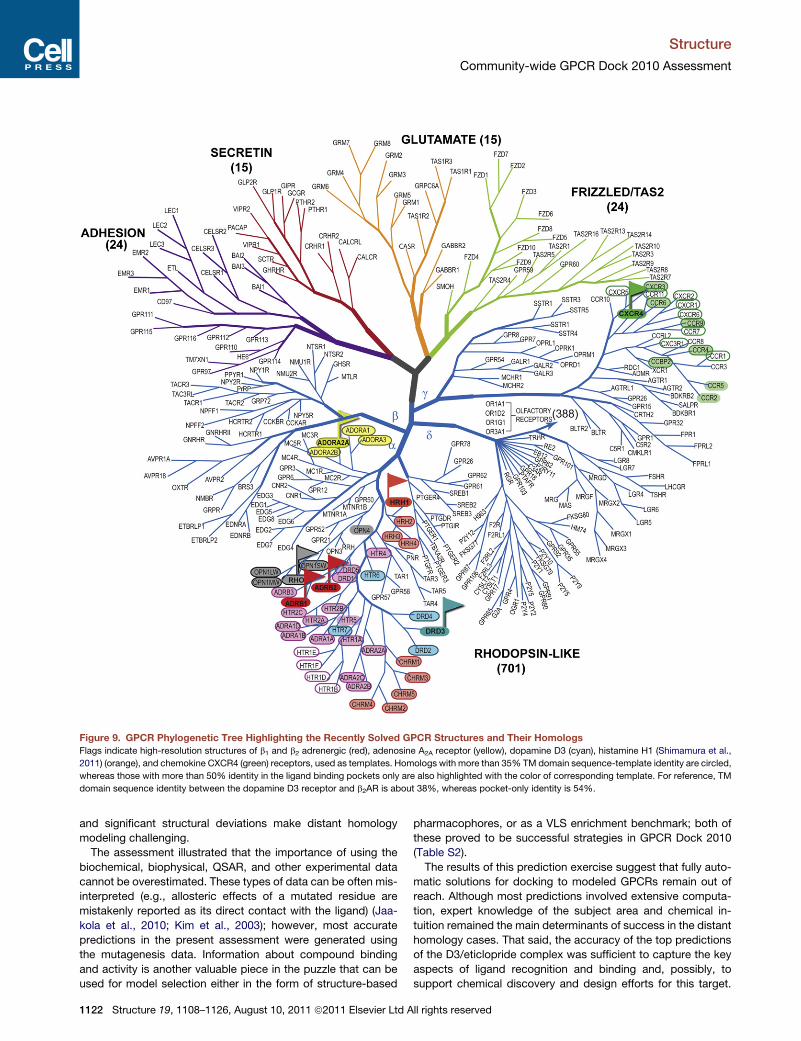
Figure 9. GPCR Phylogenetic Tree Highlighting the Recently Solved GPCR Structures and Their Homologs
Flags indicate high-resolution structures of b1 and b2 adrenergic (red), adenosine A2A receptor (yellow), dopamine D3 (cyan), histamine H1 (Shimamura et al.,
2011) (orange), and chemokine CXCR4 (green) receptors, used as templates. Homologs with more than 35% TM domain sequence-template identity are circled,
whereas those with more than 50% identity in the ligand binding pockets only are also highlighted with the color of corresponding template. For reference, TM
domain sequence identity between the dopamine D3 receptor and b2AR is about 38%, whereas pocket-only identity is 54%.
Structure
Community-wide GPCR Dock 2010 Assessment
and significant structural deviations make distant homology
modeling challenging.
The assessment illustrated that the importance of using the
biochemical, biophysical, QSAR, and other experimental data
cannot be overestimated. These types of data can be often mis-
interpreted (e.g., allosteric effects of a mutated residue are
mistakenly reported as its direct contact with the ligand) (Jaa-
kola et al., 2010; Kim et al., 2003); however, most accurate
predictions in the present assessment were generated using
the mutagenesis data. Information about compound binding
and activity is another valuable piece in the puzzle that can be
used for model selection either in the form of structure-based
1122 Structure 19, 1108–1126, August 10, 2011 ª2011 Elsevier Ltd A
pharmacophores, or as a VLS enrichment benchmark; both of
these proved to be successful strategies in GPCR Dock 2010
(Table S2).
The results of this prediction exercise suggest that fully auto-
matic solutions for docking to modeled GPCRs remain out of
reach. Although most predictions involved extensive computa-
tion, expert knowledge of the subject area and chemical in-
tuition remained the main determinants of success in the distant
homology cases. That said, the accuracy of the top predictions
of the D3/eticlopride complex was sufficient to capture the key
aspects of ligand recognition and binding and, possibly, to
support chemical discovery and design efforts for this target.
ll rights reserved

Structure
Community-wide GPCR Dock 2010 Assessment
In the past, few if any efforts of de novo protein-ligand complex
predictions led to models that were subsequently confirmed at
atomic resolution, far less for targets as challenging as mem-
brane receptors. The partial successes observed in GPCR Dock
are a testament to the confidence that the field has gained, and
that some previously inaccessible targets, with human input,
now fall into its remit.
ConclusionThree novel crystal structures in GPCR Dock 2010 dramatically
expanded the range of assessed modeling problems, which
now includes relatively close homologs within a GPCR family,
distant homology with a different branch of the class A GPCR
tree, aswell as apeptide-receptor complex. For a closehomology
case, represented by dopamine D3 receptor with �40% se-
quence identity to adrenergic structural templates, many groups
show reasonably accurate ligand docking results, with the top
models approaching accuracy of ligand placement in the crystal
structures and potentially providing the basis for structure-based
chemical discovery efforts. For more distant homology, repre-
sented by CXCR4 target with �25% sequence identity to the 3D
templates, only threegroupscapturedoverall positionof the small
molecule ligand (rmsd <5 A), and only one identified more than
20% ligand-receptor atomic contacts, pointing to a spectrum of
challenges for such modeling. Thus, even when ligand binding is
largely defined by structurally conserved TM regions of GPCRs,
a modeler has to deal with substantial variations in kinks and
helical structure in the binding pocket region, which impacts
quality of docking. Although some aspects of these variations
(e.g., CXCR4 helix II kink and rotation) can be modeled with the
help of experimentally derived restraints, others are likely to go
undefined. Finally, for the most challenging case of CXCR4-
peptide complex, the modeling is further complicated by the
fact that peptide interactions are defined by highly variable extra-
cellular loops and N terminus of the receptor, which are not
amenable to accurate predictions so far.
Like the previous assessment, the results of GPCR Dock 2010
demonstrate the advantage of hypothesis-driven approaches
that take maximum advantage of available experimental infor-
mation about the target and its ligands. Improving coverage of
the GPCR family with experimentally determined structures,
continued efforts for biophysical characterization of GPCRs
and their complexes with ligands, and of course improved
conformational modeling that makes good use of these data
will help to advance the field toward comprehensive under-
standing of GPCR structural diversity.
EXPERIMENTAL PROCEDURES
Data Collection and Filtering
GPCRDock 2010 registration andmodel submission systemwas implemented
and made available online at http://gpcr.scripps.edu/GPCRDock2010/.
Authors were requested to submit at most five models for each target they
chose to model, in the PDB format, using the provided PDB file templates. At
the analysis stage, the models were checked for correctness of the protein
and ligand covalent geometry and bond connectivity. In an attempt to be
inclusive, we did not discard any models with errors in ligand covalent
geometry; most of these errors could be unambiguously fixed, and the remain-
ing cases (2 D3/eticlopride, 5 CXCR4/IT1t, and 18 CXCR4/CVX15 models)
were compared to the target structures by the maximal common ligand
substructure.
Structure 19, 1108–
Evaluation of Structure Prediction of the TM Helix Bundle
and the Second Extracellular Loop
For all spatial comparison criteria, the protein molecule of eachmodel was first
superimposed onto the backbone Ca, C, and N atoms of the TM helices of the
target structure. TM regions in both target receptors were defined by residue
stretches 1.30–1.60, 2.37–2.66, 3.22–3.54, 4.38–4.61, 5.37–5.64, 6.28–6.60,
and 7.31–7.55 in Ballesteros-Weinstein notation (Ballesteros and Weinstein,
1995). In this notation a single most conserved residue among the class A
GPCRs is designated x.50, where x is the TM helix number; all other residues
on that helix are numbered relative to this conserved position. Superimposition
was performed using an adaptive algorithm that iteratively finds the region of
higher similarity by assigning distance-dependent Gaussian weights to devi-
ating fragments of the structure (Abagyan and Kufareva, 2009; Bottegoni
et al., 2009). Application of this algorithm ensured that the superimposition
quality was not dominated by a single flexible and/or poorly predicted part,
e.g., one deviating part of a helix.
For the superimposed TMbundles, rmsd of Ca, C, and N atoms of themodel
from their respective counterparts in the target structure was calculated. The
fraction of TM bundle for which high-quality superimposition was found (<2 A
rmsd) and the corresponding partial rmsd were also reported. With the same
superimposition of TM helices, we calculated rmsd of backbone atoms of
the model’s ECL2 from that of the target structure. We chose to focus on
ECL2 rather than on all extracellular parts of the protein because of its size
and the critical role it plays in ligand binding for many GPCRs. ECL2 was
defined by residues F171–N185 in D3 and by residues A174–E179, R183–
N192 in CXCR4. The tip of ECL2 b-hairpin (residues A180, D181, and D182)
was omitted from ECL2 comparison for CXCR4 because this region was disor-
dered in the majority of target structures, and was themost flexible in others as
demonstrated by its structural variability and high B factor values.
For CXCR4, additional attention was paid to the rotation of the top part of
helix II that carries two critical ligand-interacting residues, W94 and D97.
The accurate modeling of this region by homology with the available structural
templates (bRho, b1AR and b2AR, and A2AAR) required introduction of a one-
residue gap in the alignment that results in �100� rotation in the top of helix II
(residues 91–100) and orients W94 and D97 toward the binding pocket (Fig-
ure 2B). To assess the extent of rotation in helix II, the TM domain of each
model was superimposed onto the target structure as described above; the
model was then translated in space to ensure the optimal overlay of the helical
axis of the top part of its helix II with the corresponding axis in the target. The
two angles were measured: one angle between the projections of W94 Cb
atoms onto the plane perpendicular to the helical axes, and another angle
between the projections of D97 Cb atoms.
Evaluation of Binding Pocket Predictions
Similarity of the predicted to the experimental pocket residue content was as-
sessed by calculating and comparing the residue backbone and side-chain
surface areas that become solvent inaccessible in the presence of the ligand
in the target structures and in themodels. Accessible surface area calculations
were performed using the Shrake and Rupley algorithm implemented in ICM
(Abagyan et al., 1994, 2009). A binding pocket was formalized as vector P of
length 2n, where n is the number of residues in the protein, with components
P[2i � 1] and P[2i] equal to the decrease in accessible backbone and side-
chain areas of the i-th residue upon ligand binding. For each target struc-
ture/model pair with pockets PR and PM, a pocket similarity vector PRXM
was constructed using PRXM[i] = Min(PR[i], PM[i]). The weight of this similarity
vector was calculated as jPRXMj = SiPRXM[i] and compared to the weight of
the target pocket, jPRj. The result was reported as a real number continuously
distributed on the interval [0,1], or in percentages. This number has a meaning
of recall (or coverage) in statistical classification terminology (TP/(TP+FN)); the
value corresponding to precision (TP/(TP+FP)) can be obtained by comparing
jPRXMj to theweight of themodel pocket vector, jPMj. However, we did not use
the pocket precision value in model evaluation and do not report it in the
present publication.
Similarity of the pocket residue conformations was evaluated by measuring
rmsd between the heavy atoms of the residues that constituted the binding
pockets in the target structures. For D3 and CXCR4/IT1t complexes, binding
pockets were defined as the sets of residues for which the average contact
strength with the ligand exceeded the value corresponding to the distance
1126, August 10, 2011 ª2011 Elsevier Ltd All rights reserved 1123

Figure 10. Definition and Properties of Atomic Contact Strength Function with and without a Continuous Decrease Margin
(A) Atomic contact strength function definition for two atoms.
(B) Zero margin (a.k.a. hard cutoff, black curve) contact definition leads to unstable behavior of the function and its intolerance to even the smallest changes in
side chain and ligand conformations. The continuous decrease margin approach is devoid of this instability (colored curves).
Structure
Community-wide GPCR Dock 2010 Assessment
of 4 A. For CXCR4/CVX15 complexwhere only a single structurewas available,
the binding pocket was defined as the set of residues with nonhydrogen atoms
at the distance of%4 A from the ligand. Specifically, the sets of target pocket
residues included:
d D3/eticlopride complex: F106, D110, V111, C114, I183, V189, S192,
S193, W342, F345, F346, H349, Y365, T369, and Y373 (15 residues:
14 in TM domain and 1 in ECL2)
d CXCR4/IT1t complex: W94, D97, A98, W102, V112, H113, Y116, R183,
I185, C186, D187, R188, and E288 (13 residues: 7 in TM domain and 6 in
extracellular loops)
d CXCR4/CVX15 complex: P27, H113, Y116, T117, D171, S178, C186,
D187, R188, F189, Y190, P191, N192, D193, V196, F199, Q200, Y255,
D262, I265, L266, E277, H281, I284, S285, and E288 (12 TM domain
residues and 14 extracellular loop residues)
The optimal superimposition of TM domains was performed prior to the
binding pocket comparison as described above. Residue symmetry was taken
into account when calculating pocket rmsd.
Evaluation of Ligand Rmsd
Rmsd of the ligand nonhydrogen atoms from their respective counterparts in
the crystallographic structure was determined after superimposition of the
model onto the target structure as described above. Internal ligand symmetry
was taken into account for rmsd definition as well as other calculations. For
example, for the isothiourea IT1t molecule cocrystallized with CXCR4, as
many as 16 atom permutations are possible that result in exactly the same
ligand covalent geometry and bond topology; all of these were tested, and
the one with the smallest rmsd to the model was chosen.
Calculation of Atomic Contacts
In the traditional definition, anatomiccontact is apair of heavy ligandandprotein
atoms located at the distance closer than a specified cutoff (usually 4 A) (Rueda
et al., 2010). Thenumber of contacts that are the samebetween the target struc-
ture and the model is calculated and compared to the total number of ligand-
protein contacts in the target structure (recall) or in the model (precision). As
with ligand rmsd, calculation of atomic contacts requires enumeration of topo-
logically equivalent atom permutations in the ligand; moreover, some amino
acids also possess internal symmetry that should be taken into account. Treat-
ing side-chain symmetry in the same way as ligand symmetry is possible, but it
quickly leads to combinatorial explosion of the total number of permutations in
the system. For this reason, and because the ‘‘wingspan’’ of symmetric groups
in theprotein side chains is limitedby threeheavyatoms (e.g.,Cz, Nh1, andNh1 in
1124 Structure 19, 1108–1126, August 10, 2011 ª2011 Elsevier Ltd A
arginine), we accounted for side-chain symmetry by considering symmetric
atoms indistinguishable instead of explicitly enumerating them.
We refined the definition of an atomic contact in an attempt to make it more
robust and continuous. Instead of using a ‘‘hard’’ distance cutoff and counting
a contact as present (1) for interatomic distances below this cutoff, and as
absent (0) for the distances above this cutoff, we designed a continuous
contact strength function that gradually decreased from 1 to 0 within a speci-
fied distance margin. Therefore, the traditional ‘‘hard’’ definition of contact
cutoff corresponds to the margin of 0 in our scheme (Figure 10). Zero margin
(also known as hard cutoff) contact definition leads to unstable behavior of
the function and its intolerance to even the smallest (<0.1 A) changes in
side-chain and ligand conformations. The continuous decrease margin
approach is devoid of this instability. Using the continuous decrease margin
affected the relative ranking of intermediate and low-quality models (data
not shown), but not the best scoring models in all three assessments. At the
same time, it yielded contact similarity values that weremore stable and robust
than atom contact number calculated in a traditional way, and better reflected
the intuitive human perception of contact similarity.
In model evaluation, we constructed the vectors of atomic contact strengths
for all ligand-protein atom pairs in the target structure (CR) and in the model
(CM). The contact similarity vector CRXM was constructed using CRXM[i] =
Min(CR[i], CM[i]); its weight was found as jCRXMj = SiCRXM[i] and compared
to the weight of the target contact strength vector, jCRj. All possible topolog-
ically equivalent permutations of ligand atomswere tested, and one resulting in
the highest similarity value was chosen. Similar to pocket definition evaluation,
we only report recall (coverage) of correct contacts by the model; precision is
disregarded in this evaluation. Protein and ligand covalent geometry and van
der Waals interactions impose natural constraints onto precision values
because they limit the number of contacts that a ligand can make with the
neighboring side chains in the model.
Z-Scores
Model Z-scores were calculated in the spirit of the previous assessment,
GPCR Dock 2008 (Michino et al., 2009). Ligand rmsd values and fractions of
correctly predicted ligand-protein contacts were independently converted
into Z-scores (the opposite of rmsd Z-score was taken so that higher values
correspond to better models in all cases); the two Z-scores were averaged.
The new mean and SD were calculated excluding the low-scoring models
that deviated from the old mean by more than 2 SD, and new Z-scores were
found using this corrected mean and SD. In cases of CXCR4/IT1t and D3/
eticlopride, for which multiple target structures were available, the structure
resulting in the best Z-score was chosen for each model. A similar algorithm
ll rights reserved
Structure
Community-wide GPCR Dock 2010 Assessment
was used for assessment of protein prediction accuracy based on TM and
ECL2 backbone rmsd.
Modeling Methods
Methods, techniques, and approaches used by the participants of GPCRDock
2010 for complex model generation are described in Supplemental Experi-
mental Procedures.
SUPPLEMENTAL INFORMATION
Supplemental Information includes Supplemental Experimental Procedures
and two tables and can be found with this article online at doi:10.1016/j.str.
2011.05.012. All GPCR Dock 2010 models can be interactively viewed or
downloaded from the assessment result web-site at http://ablab.ucsd.edu/
GPCRDock2010/.
ACKNOWLEDGMENTS
The authors thank Joshua Kunken, Angela Walker, and Katya Kadyshevskaya
for their help with data processing, manuscript preparation, and graphic
design. The work was supported by NIH Grants R01 GM 071872 and U01
GM094612 to R.A. and U54 GM094618 to R.C.S.
Received: March 2, 2011
Revised: May 24, 2011
Accepted: May 28, 2011
Published: August 9, 2011
REFERENCES
Abagyan, R., and Totrov, M. (1994). Biased probability Monte Carlo conforma-
tional searches and electrostatic calculations for peptides and proteins. J.Mol.
Biol. 235, 983–1002.
Abagyan, R., and Kufareva, I. (2009). The flexible pocketome engine for struc-
tural chemogenomics. Methods Mol. Biol. 575, 249–279.
Abagyan, R., Totrov, M., and Kuznetsov, D. (1994). ICM—a new method for
protein modeling and design: applications to docking and structure prediction
from the distorted native conformation. J. Comput. Chem. 15, 488–506.
Abagyan, R., Batalov, S., Cardozo, T., Totrov, M., Webber, J., and Zhou, Y.
(1997). Homology modeling with internal coordinate mechanics: deformation
zone mapping and improvements of models via conformational search.
Proteins (Suppl 1 ), 29–37.
Abagyan, R.A., Orry, A., Raush, E., Budagyan, L., and Totrov, M. (2009). ICM
Manual (La Jolla, CA: MolSoft LLC).
Abel, R., Wang, L., Friesner, R.A., and Berne, B.J. (2010). A displaced-solvent
functional analysis of model hydrophobic enclosures. J. Chem. Theory
Comput. 6, 2924–2934.
Ballesteros, J.A., and Weinstein, H. (1995). Integrated methods for the
construction of three-dimensional models and computational probing of
structure-function relations in G protein-coupled receptors. In Methods in
Neurosciences, P.M. Conn and S.C. Sealfon, eds. (San Diego, CA: Academic
Press), pp. 366–428.
Barth, P., Wallner, B., and Baker, D. (2009). Prediction of membrane protein
structures with complex topologies using limited constraints. Proc. Natl.
Acad. Sci. USA 106, 1409–1414.
Bhattacharya, S., and Vaidehi, N. (2010). Computational mapping of the
conformational transitions in agonist selective pathways of a G-protein
coupled receptor. J. Am. Chem. Soc. 132, 5205–5214.
Bottegoni, G., Kufareva, I., Totrov, M., and Abagyan, R. (2008). A new method
for ligand docking to flexible receptors by dual alanine scanning and refine-
ment (SCARE). J. Comput. Aided Mol. Des. 22, 311–325.
Bottegoni, G., Kufareva, I., Totrov, M., and Abagyan, R. (2009). Four-dimen-
sional docking: a fast and accurate account of discrete receptor flexibility in
ligand docking. J. Med. Chem. 52, 397–406.
Structure 19, 1108–
Brylinski, M., and Skolnick, J. (2008). Q-Dock: low-resolution flexible ligand
docking with pocket-specific threading restraints. J. Comput. Chem. 29,
1574–1588.
Case, D.A., Cheatham, T.E., 3rd, Darden, T., Gohlke, H., Luo, R., Merz, K.M.,
Jr., Onufriev, A., Simmerling, C.,Wang, B., andWoods, R.J. (2005). The Amber
biomolecular simulation programs. J. Comput. Chem. 26, 1668–1688.
Cavasotto, C.N., Kovacs, J.A., and Abagyan, R.A. (2005). Representing
receptor flexibility in ligand docking through relevant normal modes. J. Am.
Chem. Soc. 127, 9632–9640.
Cavasotto, C.N., Orry, A.J., Murgolo, N.J., Czarniecki, M.F., Kocsi, S.A.,
Hawes, B.E., O’Neill, K.A., Hine, H., Burton, M.S., Voigt, J.H., et al. (2008).
Discovery of novel chemotypes to a G-protein-coupled receptor through
ligand-steered homology modeling and structure-based virtual screening.
J. Med. Chem. 51, 581–588.
Chen, R., Li, L., and Weng, Z. (2003). ZDOCK: an initial-stage protein-docking
algorithm. Proteins 52, 80–87.
Cherezov, V., Rosenbaum, D.M., Hanson, M.A., Rasmussen, S.G.F., Thian,
F.S., Kobilka, T.S., Choi, H.J., Kuhn, P., Weis, W.I., Kobilka, B.K., and
Stevens, R.C. (2007). High-resolution crystal structure of an engineered human
beta2-adrenergic G protein-coupled receptor. Science 318, 1258–1265.
Cherezov, V., Abola, E., and Stevens, R.C. (2010). Recent progress in the
structure determination of GPCRs, amembrane protein family with high poten-
tial as pharmaceutical targets. Methods Mol. Biol. 654, 141–168.
Chien, E.Y., Liu, W., Zhao, Q., Katritch, V., Han, G.W., Hanson, M.A., Shi, L.,
Newman, A.H., Javitch, J.A., Cherezov, V., and Stevens, R.C. (2010).
Structure of the human dopamine D3 receptor in complex with a D2/D3 selec-
tive antagonist. Science 330, 1091–1095.
Cozzetto, D., Kryshtafovych, A., Fidelis, K., Moult, J., Rost, B., and
Tramontano, A. (2009). Evaluation of template-based models in CASP8 with
standard measures. Proteins 77 (Suppl 9 ), 18–28.
Davis, I.W., and Baker, D. (2009). RosettaLigand docking with full ligand and
receptor flexibility. J. Mol. Biol. 385, 381–392.
de Graaf, C., and Rognan, D. (2009). Customizing G Protein-coupled receptor
models for structure-based virtual screening. Curr. Pharm. Des. 15, 4026–
4048.
Deville, J., Rey, J., and Chabbert, M. (2009). An indel in transmembrane helix 2
helps to trace the molecular evolution of class A G-protein-coupled receptors.
J. Mol. Evol. 68, 475–489.
Erickson, J.A., Jalaie, M., Robertson, D.H., Lewis, R.A., and Vieth, M. (2004).
Lessons in molecular recognition: the effects of ligand and protein flexibility
on molecular docking accuracy. J. Med. Chem. 47, 45–55.
Eswar, N., Webb, B., Marti-Renom, M.A., Madhusudhan, M.S., Eramian, D.,
Shen, M.Y., Pieper, U., and Sali, A. (2006). Comparative protein structure
modeling using Modeller. Curr. Protoc. Bioinformatics, Chapter 5, Unit 5.6.
Fredriksson, R., Lagerstrom, M.C., Lundin, L.G., and Schioth, H.B. (2003). The
G-protein-coupled receptors in the human genome form five main families.
Phylogenetic analysis, paralogon groups, and fingerprints. Mol. Pharmacol.
63, 1256–1272.
Govaerts, C., Blanpain, C., Deupi, X., Ballet, S., Ballesteros, J.A., Wodak, S.J.,
Vassart, G., Pardo, L., and Parmentier, M. (2001). The TXP motif in the second
transmembrane helix of CCR5. A structural determinant of chemokine-
induced activation. J. Biol. Chem. 276, 13217–13225.
Jaakola, V.P., Griffith, M.T., Hanson, M.A., Cherezov, V., Chien, E.Y.T., Lane,
J.R., Ijzerman, A.P., and Stevens, R.C. (2008). The 2.6 angstrom crystal struc-
ture of a human A2A adenosine receptor bound to an antagonist. Science 322,
1211–1217.
Jaakola, V.P., Lane, J.R., Lin, J.Y., Katritch, V., Ijzerman, A.P., and Stevens,
R.C. (2010). Ligand binding and subtype selectivity of the human A(2A) aden-
osine receptor: identification and characterization of essential amino acid resi-
dues. J. Biol. Chem. 285, 13032–13044.
Katritch, V., Rueda, M., Lam, P.C.-H., Yeager, M., and Abagyan, R. (2010).
GPCR 3D homology models for ligand screening: lessons learned from blind
predictions of adenosine A2a receptor complex. Proteins 78, 197–211.
1126, August 10, 2011 ª2011 Elsevier Ltd All rights reserved 1125

Structure
Community-wide GPCR Dock 2010 Assessment
Kellenberger, E., Springael, J.Y., Parmentier, M., Hachet-Haas, M., Galzi, J.L.,
and Rognan, D. (2007). Identification of nonpeptide CCR5 receptor agonists
by structure-based virtual screening. J. Med. Chem. 50, 1294–1303.
Kim, S.K., Gao, Z.G., Van Rompaey, P., Gross, A.S., Chen, A., Van
Calenbergh, S., and Jacobson, K.A. (2003). Modeling the adenosine recep-
tors: comparison of the binding domains of A2A agonists and antagonists.
J. Med. Chem. 46, 4847–4859.
Kryshtafovych, A., Venclovas, �C., Fidelis, K., and Moult, J. (2005). Progress
over the first decade of CASP experiments. Proteins 61 (Suppl 7 ), 225–236.
Kryshtafovych, A., Fidelis, K., and Moult, J. (2009). CASP8 results in context of
previous experiments. Proteins 77 (Suppl 9 ), 217–228.
Lagerstrom, M.C., and Schioth, H.B. (2008). Structural diversity of G protein-
coupled receptors and significance for drug discovery. Nat. Rev. Drug
Discov. 7, 339–357.
Lang, P.T., Brozell, S.R., Mukherjee, S., Pettersen, E.F., Meng, E.C., Thomas,
V., Rizzo, R.C., Case, D.A., James, T.L., and Kuntz, I.D. (2009). DOCK 6:
combining techniques to model RNA-small molecule complexes. RNA 15,
1219–1230.
Michino, M., Abola, E., GPCR Dock 2008 participants, Brooks, C.L., 3rd,
Dixon, J.S., Moult, J., and Stevens, R.C. (2009). Community-wide assessment
of GPCR structure modelling and ligand docking: GPCR Dock (2008). Nat.
Rev. Drug Discov. 8, 455–463.
Morris, G.M., Huey, R., Lindstrom, W., Sanner, M.F., Belew, R.K., Goodsell,
D.S., and Olson, A.J. (2009). AutoDock4 and AutoDockTools4: automated
docking with selective receptor flexibility. J. Comput. Chem. 30, 2785–2791.
Nabuurs, S.B., Wagener, M., and de Vlieg, J. (2007). A flexible approach to
induced fit docking. J. Med. Chem. 50, 6507–6518.
Ono, Y., Fujibuchi, W., and Suwa, M. (2005). Automatic gene collection system
for genome-scale overview of G-protein coupled receptors in eukaryotes.
Gene 364, 63–73.
Palczewski, K., Kumasaka, T., Hori, T., Behnke, C.A., Motoshima, H., Fox,
B.A., Le Trong, I., Teller, D.C., Okada, T., Stenkamp, R.E., et al. (2000).
Crystal structure of rhodopsin: a G protein-coupled receptor. Science 289,
739–745.
Park, J.H., Scheerer, P., Hofmann, K.P., Choe, H.W., and Ernst, O.P. (2008).
Crystal structure of the ligand-free G-protein-coupled receptor opsin. Nature
454, 183–187.
Rey, J., Deville, J., and Chabbert, M. (2010). Structural determinants stabilizing
helical distortions related to proline. J. Struct. Biol. 171, 266–276.
Rosenkilde, M.M., Gerlach, L.-O., Hatse, S., Skerlj, R.T., Schols, D., Bridger,
G.J., and Schwartz, T.W. (2007). Molecular mechanism of action of monocy-
1126 Structure 19, 1108–1126, August 10, 2011 ª2011 Elsevier Ltd A
clam versus bicyclam non-peptide antagonists in the CXCR4 chemokine
receptor. J. Biol. Chem. 282, 27354–27365.
Rueda, M., Katritch, V., Raush, E., and Abagyan, R. (2010). SimiCon: a web
tool for protein-ligand model comparison through calculation of equivalent
atomic contacts. Bioinformatics 26, 2784–2785.
Scheerer, P., Park, J.H., Hildebrand, P.W., Kim, Y.J., Krauss, N., Choe, H.W.,
Hofmann, K.P., and Ernst, O.P. (2008). Crystal structure of opsin in its
G-protein-interacting conformation. Nature 455, 497–502.
Shaw, D.E., Maragakis, P., Lindorff-Larsen, K., Piana, S., Dror, R.O.,
Eastwood, M.P., Bank, J.A., Jumper, J.M., Salmon, J.K., Shan, Y., and
Wriggers, W. (2010). Atomic-level characterization of the structural dynamics
of proteins. Science 330, 341–346.
Shimamura, T., Shiroishi, M., Weyand, S., Tsujimoto, H., Winter, G., Katritch,
V., Abagyan, R., Cherezov, V., Liu, W., Han, G.W., et al. (2011). Structure of
the human histamine H1 receptor complex with doxepin. Nature 475, 65–70.
Thoma, G., Streiff, M.B., Kovarik, J., Glickman, F., Wagner, T., Beerli, C., and
Zerwes, H.-G. (2008). Orally bioavailable isothioureas block function of the
chemokine receptor CXCR4 in vitro and in vivo. J. Med. Chem. 51, 7915–7920.
Vaidehi, N., Floriano, W.B., Trabanino, R., Hall, S.E., Freddolino, P., Choi, E.J.,
Zamanakos, G., and Goddard, W.A., 3rd. (2002). Prediction of structure and
function of G protein-coupled receptors. Proc. Natl. Acad. Sci. USA 99,
12622–12627.
Verdonk, M.L., Cole, J.C., Hartshorn, M.J., Murray, C.W., and Taylor, R.D.
(2003). Improved protein-ligand docking using GOLD. Proteins 52, 609–623.
Warne, T., Serrano-Vega, M.J., Baker, J.G., Moukhametzianov, R., Edwards,
P.C., Henderson, R., Leslie, A.G.W., Tate, C.G., and Schertler, G.F.X. (2008).
Structure of a beta1-adrenergic G-protein-coupled receptor. Nature 454,
486–491.
Wong, R.S., Bodart, V., Metz, M., Labrecque, J., Bridger, G., and Fricker, S.P.
(2008). Comparison of the potential multiple binding modes of bicyclam,
monocylam, and noncyclam small-molecule CXC chemokine receptor 4 inhib-
itors. Mol. Pharmacol. 74, 1485–1495.
Wu, B., Chien, E.Y.T., Mol, C.D., Fenalti, G., Liu, W., Katritch, V., Abagyan, R.,
Brooun, A., Wells, P., Bi, F.C., et al. (2010). Structures of the CXCR4 chemo-
kine GPCR with small-molecule and cyclic peptide antagonists. Science
330, 1066–1071.
Yarov-Yarovoy, V., Schonbrun, J., and Baker, D. (2006). Multipass membrane
protein structure prediction using Rosetta. Proteins 62, 1010–1025.
Zemla, A. (2003). LGA: amethod for finding 3D similarities in protein structures.
Nucleic Acids Res. 31, 3370–3374.
Zhou, H.X., and Gilson, M.K. (2009). Theory of free energy and entropy in non-
covalent binding. Chem. Rev. 109, 4092–4107.
ll rights reserved