steady-state and time-resolved studies of the...
TRANSCRIPT
1966 DOI: 10.1021/la902611j Langmuir 2010, 26(3), 1966–1972Published on Web 10/07/2009
pubs.acs.org/Langmuir
© 2009 American Chemical Society
Steady-State and Time-Resolved Studies of the Photocleavage of Lysozymeby Co(III) Complexes
Thota Jyotsna and Challa V. Kumar*
Department of Chemistry, University of Connecticut, Storrs, Connecticut 06269-3060
Steffen Jockusch and Nicholas J. Turro
Department of Chemistry, Columbia University, New York, New York 10027
Received July 16, 2009. Revised Manuscript Received September 24, 2009
Steady-state and time-resolved studies of site-selective photocleavage of lysozyme by cobalt(III) complexes[Co(NH3)5Br]
2þ and ([Co(NH3)4CO3]þ are reported. Photocleavage resulted in two fragments of molecular masses
∼10.5 kDa and∼3.5 kDa, and the yield increased (8-33%) with irradiation time (0.16-0.8 h) as well as with the metalcomplex concentration (0.1-5 mM). The reaction proceeded to a significant extent even when nearly stoichiometricamounts of the reagents were used. Photocleavage was effective at wavelengths ranging from 310 to 390 nm, andcleavagewas inhibited by the addition of selectedmetal ions such asGd(III) atmoderate concentrations (2mM).Gd(III)is known to bind at Asp52/Glu35 residues on lysozyme, and these residues are located at the enzyme active site. Currentand previous studies suggest that Co(III) metal complexes bind at this site on lysozyme. Consistent with this hypothesis,[Co(NH3)4CO3]
þ (8 mM) inhibited lysozyme activity by 67%. Laser flash photolysis studies show that excitation of themetal complexes [Co(NH3)5Br]
2þ and ([Co(NH3)4CO3]þ (308 nm, 20 ns pulse width) resulted in the corresponding
ligand-derived radical intermediates. For example, photoexcitation of an aqueous solution of [Co(NH3)5Br]2þ at 308 nm
resulted in the formation of Br2-•. When the excitation was carried out in the presence of lysozyme, Br2
-•was quenchedwith a bimolecular rate constant of 1.4"109M-1 s-1. Quenching resulted in protein-derived radicals (Trpþ• and Tyrþ•),as identified by their characteristic known transient absorption bands. Steady-state studies correlated with the time-resolved data, and taken together, these illustrated the reactivities of Co(III) metal complexes to direct proteinphotocleavage with high selectivity.
Introduction
Numerous enzymes and proteins require metal ions for theirbiological function, and identification of the metal binding siteson biomolecules is essential for understanding their metallo-biochemistry. However, identification of thesemetal binding sitesis challenging and requires single-crystal X-ray diffraction (XRD)studies or detailed NMR experiments.1 Therefore, alternativeapproaches for the identification of metal binding sites is highlydesirable. In this context, photochemical approaches are promis-ing. One significant advantage of photochemical reactions is thatdetailed mechanistic information can be obtained by conductingtime-resolved studies. Previous work from this laboratory2 andothers3 have demonstrated photochemical approaches to foot-print ligandbinding sites aswell asmetal binding sites onproteins.
Photoactive metal species such as vanadate,4 uranyl5 andFe(III)6 are known to cleave proteins upon activation by light.Vanadate, a metal-oxo ion, is isosteric to phosphate and oftenbinds at phosphate binding sites on proteins and photocleaves thepeptide backbone. In contrast, uranyl binds toNi(II) binding siteson serum albumins and photocleaves the protein backbone at thecorresponding metal binding sites. Recently, uranyl was used tofootprint protein phosphorylation sites on proteins, where uranylhas been proposed to bind to phosphate groups of proteins.7
Our current strategy is outlined in Scheme 1.A transitionmetalcomplex is designed tobind at the desiredmetal binding site (step 1),and the protein-bound metal complex is then photoactivated tocleave the protein backbone at or near the binding site (step 2).13
Identification of the photocleavage products by standard bio-chemical methods reveals the binding site of the metal complex,and the metal binding site can be deduced from these data.Competitive inhibition of the protein photocleavage by thepresence of specific metal ions confirms that the metal complexindeed cleaves the protein at the intended site. This simple strategyis evaluated here, in steady-state and time-resolved studies.
Scheme 1. AGeneral Strategy for the Photochemical Footprinting ofMetal Binding Sites on Proteins
*Corresponding author. E-mail: [email protected].(1) Physical Methods in Bioinorganic Chemistry; Que, L., Ed.; University Science
Books: Minneapolis, MN, 2000.(2) (a) Kumar, C. V.; Buranaprapuk, A. Angew. Chem. 1997, 36, 2085.
(b) Kumar, C. V.; Buranaprapuk, A.; J. Opiteck, G.; Moyer, M. B.; Jockusch, S.; Turro,N. J. Proc. Natl. Acad. Sci. U.S.A. 1998, 95, 10361. (c) Buranaprapuk, A.; Kumar, C.V.; Jockusch, S.; Turro, N. J. Tetrahedron 2000, 56, 7019. (d) Kumar, C. V.;Buranaprapuk, A.; Sze, H.Chem. Commun. 2001, 297. (e) Kumar, C. V.; Buranaprapuk,A.; Thota, J. Proc. Natl. Acad. Sci. (India): Chem. Sci. 2002, 114, 579.(3) (a) Jackson, G. S.; Murray, I.; Hosszu, L. L. P.; Gibbs, N.; Waltho, J. P.;
Clarke, A. R.; Collinge, J. Proc. Natl. Acad. Sci. U.S.A. 2001, 98, 8531. (b) Ngoka,L. C. M.; Gross, M. L. J. Mass Spectrom. 2000, 35, 265.(4) (a) Correia, J. J.; Lipscomb, L. D.; Dabrowiak, J. C.; Isern, N.; Zubieta, J.
Arch. Biochem. Biophys. 1994, 309, 94. (b) Crans, D. C.; Sudhakar, K.; Zamborelli, T.;Rehder, D. Angew. Chem., Int. Ed. 1991, 30, 148. (c) Cremo, C. R.; Loo, J. A.;Edmonds, C. G.; Hatlelid, K. M. Biochemistry 1992, 31, 491.(5) Duff, M. R., Jr.; Kumar, C. V. Angew. Chem., Int. Ed. Engl. 2006, 45, 137.(6) (a) Mocz, G.; Tang, W.-J. Y.; Gibbons, I. R. J. Cell Biol. 1988, 106, 1607.
(b) Mocz, G.; Gibbons, I. R. J. Biol. Chem. 1990, 265, 2917. (c) Mocz, G.; Farias, J.;Gibbons, I. R. Biochemistry 1991, 30, 7225.
(7) Kristensen, L. H.; Nielsen, P. E.; Joergensen, C. I.; Kragelund, B. B.;Moellegaard, N. E. ChemBioChem 2008, 9, 2377.

DOI: 10.1021/la902611j 1967Langmuir 2010, 26(3), 1966–1972
Jyotsna et al. Article
Co(III) ammine complexes are the focus of this paper, andthese bind to particular sites on several proteins.8 Photoexcitationinto the ligand-to-metal charge transfer (LMCT) bands ofCo(III)complexes (Chart 1) generates ligand radicals,9-12 and theseradicals have the potential to cleave the protein backbone. Byadjusting the size, reactivity, and the type of ligands, the structureof the Co(III) complexes can be adjusted to target appropriatesites on proteins.
In support of the above strategy, several Co(III) complexeswere shown to photocleave several proteins,13 and here weexamined the key mechanistic steps of the lysozyme photo-cleavage by steady-state and flash photolysis studies. Lyso-zyme is a small (molecular weight ∼14.3 kDa),14 inexpensive,water-soluble hydrolytic enzyme,15 and the active site of lyso-zyme is located in a cleft, which is lined with polar groups.16
For example, Glu35 is one of the residues in the active site,and this residue is critical for the catalytic activity of theenzyme; Asp52 is another polar residue located within 6 Afrom Glu35.
Metal ions such as Mn(II), Co(II), Gd(III), Ni(II), andlanthanide ions bind to lysozyme at Glu35/Asp52 (Chart 2).18-22
Crystal structure of lysozyme indicates that these residues arereadily accessible from the solvent, and the open space aroundGlu35/Asp52 site can accommodate small metal complexes suchas the ones shown in Chart 1, via outer sphere interactions. Here,the steady-state and flash photolysis studies of lysozyme photo-cleavage by two Co(III) complexes are reported.
Experimental Section
Materials. Chicken hen egg lysozyme was purchased fromSigma (St. Louis, MO), while the metal complexes, pentam-minebromoCobalt(III) bromide ([Co(NH3)5Br]
2þ), and tetram-minecarbonatoCobalt(III) nitrate ([Co(NH3)4CO3]
þ), weresynthesized by following reported methods.23 The electronicabsorption spectra of the complexes are [Co(NH3)5Br]
2þ [263 nm(18500M-1 cm-1), 550 nm (55M-1 cm-1)] and [Co(NH3)4CO3]
þ
Chart 1. The Structures of Co(III) Hexamine and Its Derivatives
Chart 2. (Left)Three-Dimensional Structure of Lysozyme, Highlighting the Locations of Some Important Residues; (Right)Binding of Gd(III) atthe Active Site Residues, Asp52/Glu35 (Bold Residues)
17
(8) (a) Kim, S.; Cowan, J. A. Inorg. Chem. 1992, 31, 3495. (b) Kucharski, L.;Lubbe, W. J.; Maguire, M. E. J. Biol. Chem. 2000, 275, 16767. (c) Jou, R.; Cowan, J. A.J. Am. Chem. Soc. 1991, 113, 6685.(9) Basolo, F.; and Pearson, R. G. In Mechanisms of Inorganic Reactions: A
Study of Metal Complexes in Solution; John Wiley: New York, 1967; p 158.(10) Stadtman, E. R.; Levine, R. L. Amino Acids 2003, 25, 207.(11) Balzani, V.; Carassiti, V. In Photochemistry of Coordination Compounds;
Academic Press: New York, 1970.(12) (a) Hoffman, M. Z.; Olson, K. R. J. Phys. Chem. 1978, 82, 2631. (b) Penkett,
S. A.; Adamson, A. W. J. Am. Chem. Soc. 1965, 87, 2514. (c) Grossweiner, L. I.;Matheson,M. S. J. Phys. Chem. 1957, 61, 1089. (d) Chen, S.; Cope, V.W.; Hoffman,M.Z. J. Phys. Chem. 1973, 77, 1111.(13) (a)Kumar, C. V.; Thota, J. Inorg. Chem. 2005, 44, 825. (b) Thota, J.; Bandara,
K.; Kumar, C. V. Photochem. Photobiol. Sci. 2008, 7, 1531.(14) Weiss, M. S.; Palm, G. J.; Hilgenfeld, R. Acta Crystallogr., Sect. D. 2000,
56, 952.(15) Teichberg, V. I.; Sharon, N.; Moult, J.; Smilansky, A.; Yonath, A. J. Mol.
Biol. 1974, 87, 357.(16) Kortvelyesi, T.; Silberstein, M.; Dennis, S.; Vajda, S. J. Comput.-Aided
Mol. Des. 2003, 17, 173.
(17) (a) Kurachi, K.; Sieker, L. C.; Jensen, L. J. Biol. Chem. 1975, 250, 7663.(b) Hunter, T. M.; McNae, I. W.; Simpson, D. P.; Smith, A. M.; Moggach, S.; White, F.;Walkinshaw, M. D.; Parsons, S.; Sadler, P. J. Chemistry 2007, 13, 40.
(18) Ikeda, K.; Hamaguchi, K. A. J. Biol. Chem. 1973, 248, 307.(19) Pesek, J. J.; Schneider, J. F. J. Inorg. Biochem. 1988, 32, 233.(20) (a) Kurachi, K.; Sieker, L. C.; Jensen, L. H. A. J. Biol. Chem. 1975, 250,
7663. (b) Olmo, R.; Huerta, P.; Blanco, D.; Teijon, J. M. J. Inorg. Biochem. 1992, 47,89.
(21) Norton, R. S.; Allerhand, A. J. Biol. Chem. 1977, 252, 1795.(22) Li, S. J. Biopolymers 2006, 81, 74.(23) (b) Schlessinger, G. G. Inorganic Syntheses IX; McGraw-Hill Company: New
York; 1967.

1968 DOI: 10.1021/la902611j Langmuir 2010, 26(3), 1966–1972
Article Jyotsna et al.
[238 nm, and 520 nm (104 M-1 cm-1)], and the FTIR spectra ofthe samples matched those reported.11,24-26 Photoreductionquantum yields of Co(III) complexes at key wavelengths, wherethe ligand served as the reductant, are given in Table 1. Note thatphotoreduction of the metal center was demonstrated at specificwavelengths, in particular cases.Photocleavage Reactions. Photocleavage was carried out at
room temperature in Tris buffer (10 mM Tris HCl, pH 7.0) or indeionized water (pH 5), under air-saturated conditions, as describedearlier.2 Briefly, a mixture of the protein (75 !M) and the Co(III)complex (0.6-5 mM, 200 !L) was irradiated at 310 nm, or longerwavelengths, using a monochromatic light source (PTI powersupply, 150-WXenon lamp, PTImodel A1010monochromator).Average light intensity emitted by the source at a given wave-length was measured by ferrioxalate actinometry,31 and with theabove lamp-setup, the monochromator tuned to 344 nm, thesample was exposed to 3.2( 0.04"10-8 einsteins/min. Irradiatedsamples were dried in a Speedvac (Savant Integrated SystemISS10) for storage or for subsequent studies.Electrophoresis. The photoreaction mixture was analyzed by
sodium dodecylsulfate (SDS)/polyacrylammide gel electrophoresis(PAGE), as described previously.32 High-quality images of theprotein gels were obtained by scanning the dye-stained gels with aUmaxAstra 1220U scanner. Band intensities were quantified usingNIH image software (v. 1.63), and product yields were estimated(yield of band I=intensity of band I/(intensity of band I plus that ofthe unreacted lysozyme)). Intensities of individual bands varied by( 5% from gel to gel, due to precipitation problems, and thereforevalues from 2 to 3 gels were used to calculate average yields. Thedistance of migration of the protein band depended linearly on thelogarithm of protein molecular mass.33 The molecular masses ofthe photofragments were assessed from calibration graphs con-structed using proteins of known molecular masses.Lysozyme Activity Studies. Glycol chitin (6 mM) was
reacted with lysozyme (20 !M) in deionized water, pH 5, at 37 !Covernight, with different concentrations of [Co(NH3)4CO3]
þ
(0, 2, 4, or 8 mM). A solution of K3Fe(CN)6 (1.8 mM) was addedto the reaction mixture and heated at 100 !C for 10 min, andabsorbance due to the conversion of Fe(III) to Fe(II) wasmeasured at 420 nm. Calibration graphs were constructed usingN-acetyl glucosamine. The extent of enzyme inhibition (%) wascalculatedbymeasuring the change inabsorbanceat 420nmin thepresence versus the absence of the metal complex. In controlstudies, Co(III) complexes did not have any influence on theconcentrations of Fe(III) or Fe(II), and absorbance due to theCo(III) complexes at 420 nm, if any, was subtracted accordingly.
Laser Flash Photolysis. These experiments were carried outby an apparatus described previously.34 Laser pulses from anexcimer laser (Lextra 50, Lambda Physik, 308 nm, 50 mJ/pulse,20 ns pulse duration) were used to excite the samples, andabsorbance changes, subsequent to excitation, were monitoredwith a pulsed 150 W xenon lamp and an ISA H10 monochroma-tor. The signals from a Hamamatsu R928 photomultipliertube were recorded with a Tektronix TDS 380 (400 MHzbandwidth) programmable digitizer. This digitizer was controlledalong with other aspects of the experiment (shutters, lamppulser, monochromator, etc.) through a GPIB interface board(National Instruments NBGPIB) using a Macintosh G3 withLabview 5 software (National Instruments). Kinetic traces weremonitored at specific wavelengths, and the transient absorptionspectra at specific delay times were constructed from these traces.
Results and Discussion
Steady-state and time-resolved studies on protein photoclea-vage by Co(III) complexes indicated that ligand-derived radicalsare primarily responsible for the peptide fragmentation, anddetails of our investigations are described below:Photocleavage Studies. Irradiation of a mixture of lysozyme
and [Co(NH3)5Br]2þ at 310 nm and subsequent analysis of the
reaction mixture by SDS PAGE (Figure 1) indicated a pair ofproduct bands that migrated faster than the starting material. Byusing the known molecular weight markers (lane 1), and con-struction of a calibration graph, we found that the approximatemolecular masses of the product bands (lanes 5-9) are 10.5 and3.5 kDa. These masses roughly add-up to that of the unreactedlysozyme (14.3 kDa), and the molecular masses of the productbands matched with those previously reported.13 The pair ofbands suggests that the daughter fragments may arise from asingle cut in the parent molecule.
Extensive nonspecific cleavage of the peptide backbone can beruled out because indiscriminate photocleavage would haveresulted in smearing of product bands in lanes 5-9. Incubationof amixture of lysozyme and themetal complex in the dark, undersimilar conditions (lane 4) did not yield any fragmentation.Similarly, irradiation of lysozyme in the absence of the metalcomplex (lane 3) did not yield any products. Therefore, both lightand the metal complex are essential for the observed photoclea-vage. The product yields increased only marginally (30-33%) asa function of irradiation time (10-50 min). Only two product
Table 1. Quantum Yields for the Photoreduction of Co(III)Complexes
Co(III) photoreduction quantum yields
complex 254 nm 370 nm 410 nm 550 nm
[Co(NH3)6]3þ 0.1627 027
[Co(NH3)5Br]2þ 0.1928 0.1529 0.07129 0.001428
[Co(NH3)4CO3]þ 0.0630
Figure 1. Lysozyme photocleavage by Co(III) complexes (10mMTris HCl, pH 7.0, 310 nm irradiation, 10-50 min) resulted in twofragments.Molecularweightmarkers fromthe top to thebottom inlane 1 are 14, 6, and 3.5 kDa, and the other lanes are as marked.Lysozyme (75 !M) photocleavage, as a function of irradiationtime, by (A) [Co(NH3)5Br]
2þ (0.6 mM) and (B) [Co(NH3)4CO3]þ
(2.5 mM).
(24) (a) Hill, D. G.; Rosenberg, A. F. J. Chem. Phys. 1954, 22, 148. (b) Svatos, G.F.; Sweeny, D.M.;Mizushima, S.; Curran, C.; Quagliano, J. V. J. Am. Chem. Soc. 1957,79, 3313. (c) Kobayashi, M; Fujita, J. J. Chem. Phys. 1955, 23, 1354. (d) Dasgupta, T.P.; Harris, G. M. J. Am. Chem. Soc. 1969, 91, 3207.(25) Yalman, R. G. J. Am. Chem. Soc. 1955, 77, 3219.(26) Adamson, A. W.; Sporer, A. J. Am. Chem. Soc. 1958, 80, 3865.(27) Manfrin, M. F.; Varani, G.l; Moggi, L.; Balzani, V.Mol. Photochem. 1969,
1, 387.(28) Adamson, A.W.;Waltz,W. L.; Zinato, E;Watts, D.W.; Fleischauer, P. D.;
Lindholm, R. D. Chem. Rev. 1968, 68, 541.(29) Adamson, A. W. Discuss. Faraday Soc. 1960, 29, 163.(30) Cope, V. W.; Chen, S.; Hoffman, M. Z. J. Am. Chem. Soc. 1973, 95, 3116.(31) Hatchard, H. G.; Parker, C. A. Proc. R. Acad. Sci. (London), A 1956, 235,
518.(32) Kumar, C. V.; Buranaprapuk, A. J. Am. Chem. Soc. 1999, 121, 4262.(33) Schagger, H.; Von Jagow, G. Anal. Biochem. 1987, 166, 368. (34) Yagci, Y.; Jockusch, S.; Turro, N. J. Macromolecules 2007, 40, 4481.

DOI: 10.1021/la902611j 1969Langmuir 2010, 26(3), 1966–1972
Jyotsna et al. Article
bands are noted at all irradiation times, and hence selectivity ofthe photoreaction is independent of the irradiation time.
Irradiation of lysozyme (75 !M) in the presence of [Co(NH3)4-CO3]
þ (2.5mM) also resulted in protein photocleavage (Figure 1B),and the same pair of product bands appeared as in the case of thebromo-derivative. Yields of photoproduct improvedwith irradia-tion time, from 16 to 28% (10-50 min). Note that the reactionselectivity is unaltered as a function of irradiation time, thereaction is not limited to one metal complex, and the same pairproduct bands are noted with both the metal complexes.
In both cases, the addition of fresh metal complex to theirradiated reaction mixture and continued irradiation did notimprove the product yields any further. One possibility is that theinitial events in the photocleavage branch out to give chemicallymodified protein, which does not result in fragmentation of thebackbone. In support of this assessment, mass spectrometry oflysozyme, isolated from the reactionmixture, indicated increase inmolecularmass consistentwith the additionof one, two, andup tofive oxygen atoms. Another possibility is that a fraction of theproduct bands are cross-linked with each other, but this isunlikely, as such cross-linking would generate additional newbands of high masses in SDS PAGE, which are not observed.Concentration Dependence, and Excitation at Longer
Wavelengths. Photoproduct yields and the selectivity of thephotocleavage were monitored as a function of increasing metalcomplex concentrations (Figure 2). The increase (8-23%, 0.1 -0.6 mM) is gradual in the case of [Co(NH3)5Br]
2þ, and yieldsincreased abruptly with [Co(NH3)4CO3]
þ, from 12 to 32% (0.5 to2.5 mM), but no further increases are noted.More importantly, thesame two photoproducts are formed at all concentrations, and bothindicated no change in reaction selectivity. This latter observation issignificant because the addition of excess metal complex increasesthe concentration of the free metal complex, and if the reaction isdue to the free complex, then the selectivity could diminish. On theother hand, if the reaction originates from complexes bound to aparticular site on the protein, then the selectivity will not change.
Increased product formation with increases in metal complexconcentration deserves some comment. One possibility is that lightabsorption by the metal complex is the primary photochemicalprocess, and increasedproduct yield is due to increased light absorp-tion.Another possibility is that light absorption by the protein is theprimary photochemical process, and quenching of protein-derived
excited state(s) by the metal complex initiates product formation.To distinguish between these possibilities, we examined the photo-product yields as a function of irradiation wavelength.13
Previous data showed that both [Co(NH3)5Br]2þ and
[Co(NH3)4CO3]þ complexes cleave lysozyme at irradiation wave-
lengths up to 390 nm. Irradiation of lysozyme at these wave-lengths, in the absence of the metal complex, did not give rise toany products (data not shown). Since protein residues have littleor no absorption at wavelengths greater than 310 nm, lightabsorption by themetal complex, but not the protein, is necessaryfor the product formation.
Our hypothesis is that ligand-derived radicals generated by thephotoreduction of the metal complex drive protein cleavage.First, these Co(III) complexes are known to undergo photoreduc-tion (Table 1) with concomitant ligand oxidation.35 Second, thebromo-derivative undergoes photoreduction at wavelengths aslong as 550 nm,36 even though the corresponding LMCT band islocated at much shorter wavelengths.37 One possibility is that theLMCT bands tail-out into longer wavelengths or that the initiallypopulated LF states sensitize the formation of the triplet LMCTstates, and the reaction occurs from these states. Note that, in allcases, at all wavelengths and all metal complex concentrations,the same two product bands were observed. Conditions for thehighest photocleavage yields are given in Table 2.Effect of Oxygen. The role of oxygen in the photocleavage
was investigated by purging the reaction mixture with nitrogengas. The photocleavage was independent of oxygen concentra-tions, for both the metal complexes. For example, the same yieldof product I was noted when the photoreactions were run aftersaturation with O2 or when deoxygenated with N2. Deoxygena-tion of the sample was tested in a separate experiment, where thefluorescence from a dye added to the sample was monitored as afunction of time of degassing and the same conditions applied fordegassing the reaction mixtures. Therefore, neither triplet statesnor oxygen-sensitive species are involved in the photocleavage, orsuch species, if present, are very short-lived.Photocleavage Inhibition by Gd(III). Metal ions such as
Ni(II), Zn(II), Ca(II), Cd(II), and Gd(III) bind to the Asp35/Glu52 site at the active site of lysozyme.18,20,21 Since we hypothe-size that the Co(III) complexes bind at this site and mediatephotocleavage, we expected that one or more of these metal ionsshould inhibit photocleavage.38 Irradiation of a solution oflysozyme (75 !M), and [Co(NH3)4CO3]
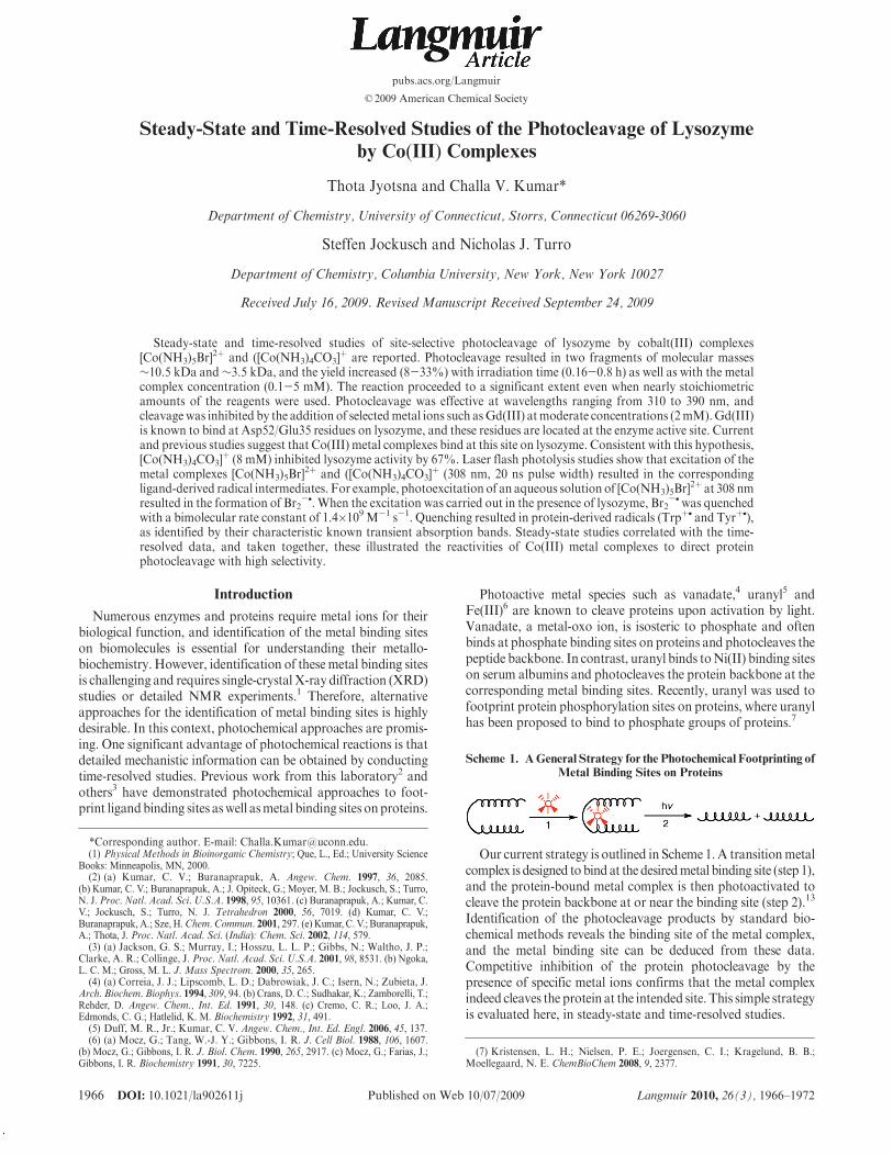
þ (2.5 mM, @350 nm,0.5 h), in the presence of increasing concentrations ofGd(III) andsubsequent analysis by SDS PAGE indicated gradual inhibitionof the photocleavage. The bands in the gel were quantified, andplots of ø0/ø as a function of inhibitor concentration wereobtained (Figure 3), where ø0 is the yield of photoproduct I in theabsence ofquencher, andø is the yield in the presenceof quencher.
Figure 2. Photocleavage of lysozyme (75 !M, 10 mM Tris HCl,pH 7.0, 310 nm irradiation) as a function of metal complexconcentration. Lane 1 contains molecular weight markers (14, 6,and3.5kDa), and theother lanes are asmarked. (A)Photocleavageby [Co(NH3)5Br]
2þ, as a function of metal concentration (0-0.6mM). (B) Photocleavage by [Co(NH3)4CO3]
þ as a function ofmetal concentration (0-5mM).Product formationwith little ornochange in selectivity is demonstrated.
Table 2. A Comparison of Best Yields of Lysozyme Photocleavage byCo(III) Complexes
Co(III)complex
%yield
[Co(III)complex](mM)
irradiationtime (h)
"irr(nm) buffer
[Co(NH3)5Br]2þ 43 0.6 0.5 370 TrisHBr
[Co(NH3)4CO3]þ 40 5 0.67 370 TrisHCl
(35) Pribush, R. A.; Poon, C. K.; Bruce, C. M.; Adamson, A. W. J. Am. Chem.Soc. 1974, 96, 3027.
(36) Adamson, A. W. Discuss. Faraday Soc. 1960, 29, 163.(37) (a) Brasted, R. C.;Hirayama, C. J. Phys. Chem. 1959, 63, 780. (b) Sastri, V. S.
Inorg. Chim. Acta 1972, 6, 264. (c) Endicott, J. F.; Ferraudi, G. J.; Barber, J. R. J. Phys.Chem. 1975, 79, 630–. (d) Endicott, J. F.; Hoffman, M. Z. J. Am. Chem. Soc. 1965, 87,3348. (e) Sastri, V. S.; Langford, C. H. Can. J. Chem. 1969, 47, 4237.
(38) Li, S. J. Biopolymers 2006, 81, 74.
1970 DOI: 10.1021/la902611j Langmuir 2010, 26(3), 1966–1972
Article Jyotsna et al.
The data were analyzed using the Stern-Volmer equation, ø0/ø=1þKsv[Q] whereKsv is the Stern-Volmer constant (Ksv=kq#,where # is the lifetime) and [Q] is the quencher concentration.The best fit to the plot indicated a slope of 600 M-1, and dataclearly show that Gd(III) quenches the photoproduct formationefficiently.
The Gd(III) quenching of photocleavage is in line with ourearlier work where we showed that Co(II) (10 mM) and Ni(II)(2mM) also inhibit the photocleavage of lysozyme and that thereaction is not inhibited by NaCl (up to 300 mM).13 Therefore,mere increase in ionic strength is not responsible for theobserved inhibition. Molecular modeling studies (Rasmol v.1.62) confirm that the space around Glu35/Asp52 is adequateto accommodate these small metal complexes on the proteinand protein-bound metal complex could be responsible forphotocleavage. This hypothesis was further tested in lysozymeactivity studies.Inhibition of Lysozyme Activity. Since Asp52/Glu35 are in
the active site of lysozyme and these residues are essential forlysozyme activity, binding of the metal complexes to theseresidues is expected to inhibit lysozyme activity.Note thatGd(III)inhibits lysozyme activity by binding to these residues.39 There-fore, we tested the above hypothesis that Co(III) complexes alsobind to this site by testing the activity of lysozyme in the presenceof increasing concentrations of Co(III) complexes.
Activity was followed by a colorimetric method where thehydrolysis product from glycol chitin was used to reduce K3Fe-(CN)6 to K4Fe(CN)6, and the extent of metal reduction wasquantified by the color change in a colorimetric assay.25 Hydro-lysis of chitin was monitored with increasing concentrations (2, 4,and 8 mM) of [Co(NH3)4CO3]
þ, and activity was inhibited by 27,45, and 67% at these concentrations, respectively. Therefore, byconsidering the facts that Gd(III) inhibits lysozyme photoclea-vage and that [Co(NH3)4CO3]
þ inhibits lysozyme activity, weconclude that the metal complex and Gd(III) might bind to theprotein at theGlu35/Asp52 site. Allosteric interactions, where themetal complex binds elsewhere on the protein and inhibits its
activity, can not be ruled out. Given this caveat, ligand-derivedradicals photogenerated from the protein-bound metal complexare likely responsible for the cleavage reaction. The photogenerationof ligand-derived radicals and their reactivity with lysozyme wasexamined by laser flash photolysis studies.Laser Flash Photolysis Studies. Photoexcitation of
[Co(NH3)6]3þ, [Co(NH3)5Cl]
2þ, [Co(NH3)5Br]2þ, and [Co(NH3)4-
CO3]þ into their respective LMCT bands are known to produce
corresponding radical ions, NH3•þ, Cl•, Br•, CO3
-•, respec-tively.40 These intermediates have characteristic absorption spec-tra, lifetimes, and reactivities, which can be monitored in flashphotolysis studies. The transients produced in the photocleavagewere investigated in time-resolved studies in the presence orabsence of lysozyme.
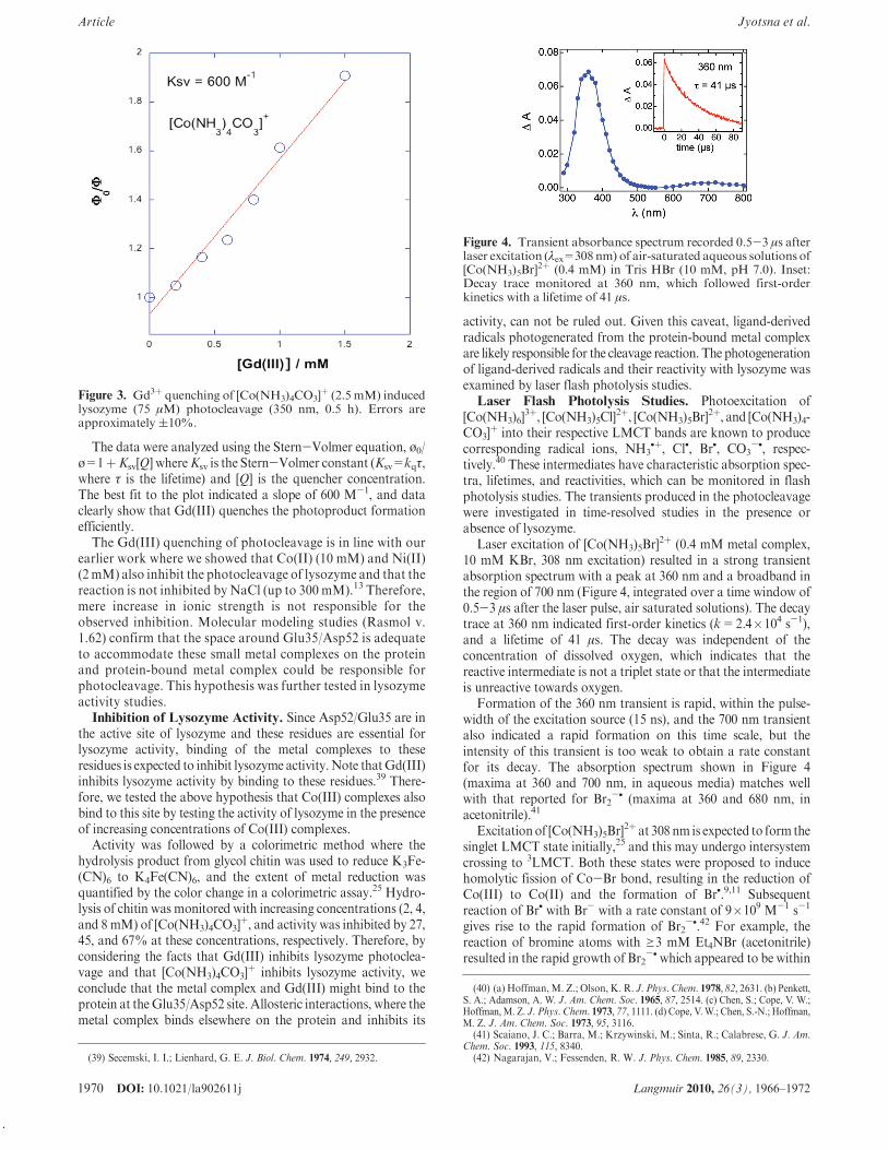
Laser excitation of [Co(NH3)5Br]2þ (0.4 mM metal complex,
10 mM KBr, 308 nm excitation) resulted in a strong transientabsorption spectrum with a peak at 360 nm and a broadband inthe region of 700 nm (Figure 4, integrated over a time window of0.5-3 !s after the laser pulse, air saturated solutions). The decaytrace at 360 nm indicated first-order kinetics (k=2.4"104 s-1),and a lifetime of 41 !s. The decay was independent of theconcentration of dissolved oxygen, which indicates that thereactive intermediate is not a triplet state or that the intermediateis unreactive towards oxygen.
Formation of the 360 nm transient is rapid, within the pulse-width of the excitation source (15 ns), and the 700 nm transientalso indicated a rapid formation on this time scale, but theintensity of this transient is too weak to obtain a rate constantfor its decay. The absorption spectrum shown in Figure 4(maxima at 360 and 700 nm, in aqueous media) matches wellwith that reported for Br2
-• (maxima at 360 and 680 nm, inacetonitrile).41
Excitationof [Co(NH3)5Br]2þ at 308 nm is expected to form the
singlet LMCT state initially,25 and this may undergo intersystemcrossing to 3LMCT. Both these states were proposed to inducehomolytic fission of Co-Br bond, resulting in the reduction ofCo(III) to Co(II) and the formation of Br•.9,11 Subsequentreaction of Br• with Br- with a rate constant of 9"109 M-1 s-1
gives rise to the rapid formation of Br2-•.42 For example, the
reaction of bromine atoms with g3 mM Et4NBr (acetonitrile)resulted in the rapid growth of Br2
-•which appeared to be within
Figure 3. Gd3þ quenching of [Co(NH3)4CO3]þ (2.5 mM) induced
lysozyme (75 !M) photocleavage (350 nm, 0.5 h). Errors areapproximately (10%.
Figure 4. Transient absorbance spectrum recorded 0.5-3 !s afterlaser excitation ("ex=308nm) of air-saturated aqueous solutions of[Co(NH3)5Br]
2þ (0.4 mM) in Tris HBr (10 mM, pH 7.0). Inset:Decay trace monitored at 360 nm, which followed first-orderkinetics with a lifetime of 41 !s.
(39) Secemski, I. I.; Lienhard, G. E. J. Biol. Chem. 1974, 249, 2932.
(40) (a) Hoffman,M. Z.; Olson, K. R. J. Phys. Chem. 1978, 82, 2631. (b) Penkett,S. A.; Adamson, A. W. J. Am. Chem. Soc. 1965, 87, 2514. (c) Chen, S.; Cope, V. W.;Hoffman, M. Z. J. Phys. Chem. 1973, 77, 1111. (d) Cope, V. W.; Chen, S.-N.; Hoffman,M. Z. J. Am. Chem. Soc. 1973, 95, 3116.
(41) Scaiano, J. C.; Barra, M.; Krzywinski, M.; Sinta, R.; Calabrese, G. J. Am.Chem. Soc. 1993, 115, 8340.
(42) Nagarajan, V.; Fessenden, R. W. J. Phys. Chem. 1985, 89, 2330.

DOI: 10.1021/la902611j 1971Langmuir 2010, 26(3), 1966–1972
Jyotsna et al. Article
the excitation pulse. Therefore, under our experimental condi-tions, which contained 10mMbromide, the formation of Br2
-• isalso expected to be complete within the excitation laser pulse, andit is the primary radical intermediate generated in the absence ofthe protein.
Flash photolysis studies of aqueous solutions of[Co(NH3)4CO3]
þ (pH 7.0, 308 nm, air saturated) indicated aweak but longer-lived transient (Figure 5). The absorptionspectrum of the transient collected over the time window of1-17 !s indicated a maximum around 600 nm (Figure 5,Table 3). This transient decayed over a long time with a pseudo-first-order decay of 112 !s, and these featuresmatchwell with thatof carbon trioxide radical ion, which has an absorptionmaximumat 600 nm (acetonitrile) and a pseudo-second-order decay rateconstant of 3.8" 107 s-1.40c,43 Initial photoexcitation was sug-gested to result in the population of 1LMCT state, which, afterintersystem crossing, undergoes rapid homolytic breaking of theCo-O bond. This results in the opening of the metal chelate ring,followed by the release of CO3
-•. At pH values less than 9, thisspecies was suggested to have been protonated to produce[CO3H]•, which can react with a variety of electron-rich substratesto give bicarbonate ion.40d
Photolysis of the metal complexes, under our conditions,generates ligand-derived radicals, and our data are consistentwith literature reports. The characteristics of these transients arecollected in Table 3. Next we examined the reactivities of theseintermediates toward lysozyme, in quenching studies.
Flash photolysis studies of [Co(NH3)5Br]2þ and [Co(NH3)4CO3]
þ
in the presence of increasing concentrations of lysozyme caused adecrease in the lifetimes of the respective transients at 360 nm (Br2
-•)and600nm(CO3
-•), respectively. Initial absorbanceof the transientsatthe end of the laser pulse did not change with lysozyme concentration(data not shown), but the lifetime of the transients gradually decreased.Decay traces ofboth the transients at different lysozyme concentrationsfit well to a pseudo-first-order decay kinetic model (kobs).
The quenching rate constants (kq) were determined from theslopes of the plot of the pseudo-first-order rate constants (kobs)
versus the lysozyme concentration (Q) (Figure 6) according to theequation kobs=ko þ kq [Q], where ko is the rate constant in theabsence of lysozyme. Quenching rate constants of 1.4"109 M-1
s-1 and 6.1"108 M-1 s-1 were obtained for Br2-• and CO3
-•,respectively. The latter value (quenching ofCO3
-• by lysozyme) isin agreement with that reported in the literature (5.5"108 M-1
s-1).44 Note that lysozyme quenches CO3-• (600 nm transient)
more slowly than Br2-• (360 nm transient). Replotting the kinetic
data shown in Figure 6, using the Stern-Volmer equation kobs/ko=1 þ Ksv [lysozyme] indicated Ksv values of∼7"104 M-1 and6"104 M-1 for Br2
-• and CO3-•, respectively. These values are
nearly consistent with the product of the respective lifetimes andquenching constants.
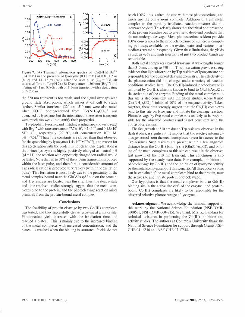
Quenching of these transients by lysozyme resulted in theformation of protein-derived radical intermediates, and theseare probed by recording the time-resolved absorption spectra(Figure7).Laser excitationofamixtureof [Co(NH3)5Br]
2þ (0.4mM)and lysozyme (0.12 mM), resulted in the growth of new transientswith absorbance maxima at 320 and 510 nm. At the end of thelaser pulse, the transient absorption spectrumof Br2
-• is observed(Figure 7A, blue spectrum, 0.8-1.2 !s after the laser pulse).However, the transient spectrum recorded in a time window of14-18 !s (Figure 7A, red spectra) showed that the 360 nmtransient nearly disappeared, but new peaks at 320 and 510 nmwere observable. Consistent with this conclusion, the Trp• spectraformed by pulse radiolysis of tryptophan with Br2
-• indicatedabsorption peaks at 320 and 510 nm, and present data are inagreement with literature values.45
The 360 nm transient decays simultaneously with the growth ofthe 510 nm transient (Figures 7B and C). However, there is asignificant growth of the 510 nm transient (50%) within the laserpulse-width, indicating that there is amuch faster pathway for theproduction of this transient, or static quenching, which is, inaddition to the dynamic quenching mechanism, discussed above.Formation of this portion of the 510 nm intermediate is repro-ducible, very rapid, and it is distinct from the quenching of Br2
-•
by lysozyme, noted earlier.The 510nmtransient decays on a very long time scale (>500!s),
and its lifetime could not be determined on our experimentalsetup. This transient is assigned to radicals generated primarilyfrom Trp, while radicals from other residues such as tyrosine andhistidine may also contribute to this transient.46 Absorbance of
Figure 5. Transient absorbance spectrum of [Co(NH3)4CO3]þ
(0.4mM) in aqueous solutions (TrisHCl 10mM,pH7.0), recorded1-17 !s after laser excitation ("ex=308 nm, air saturated aqueoussolutions). Inset: Transient decay trace at 600 nm, which was fittedto a first-order decay with a lifetime of 112 !s.
Table 3. Properties of Transients Observed in the Laser FlashPhotolysis (308 nm Excimer Laser) of Co(III) Complexes
complex"max of the
transient (nm) ko (s-1)
lifetime(!s)
kq (lysozyme)(108M-1s -1)
[Co(NH3)5Br]2þ 360, 700 2.4 " 104 41 14
[Co(NH3)4CO3]1þ 600 8.9 " 103 112 6
Figure 6. Quenching of transients from [Co(NH3)5Br]2þ (A) and
[Co(NH3)4CO3]þ (B) by lysozyme (10 mM TrisHCl or TrisHBr,
pH7) after laser excitation (308 nm).Dependence of the decay rateconstants at 360 nm (A) and 600 nm (B) on lysozyme concentra-tion.
(43) Behar, D.; Czapski, G.; Duchovny, I. J. Phys. Chem. 1970, 74, 2206.
(44) Chen, S.; Hoffman, M. Z. Radiat. Res. 1973, 56, 40.(45) Adams, G. E.; Aldrich, J. E.; Bisby, R. H.; Cundall, R. B.; Redpath, J. L.;
Willson, R. L. Radiat. Res. 1972, 49, 278.(46) (a) Hsiao, J. S.; Webber, S. E. J. Phys. Chem. 1993, 97, 8289. (b) Bohne, C.;
Abuin, E. B.; Scaiano, J. C. J. Am. Chem. Soc. 1990, 112, 4226. (c) Hirata, Y.; Mataga,N. J. Phys. Chem. 1985, 89, 2439. (d) Vala, M.; Szczepanski, J.; Pauzat, F.; Parisel, O.;Talbi, D.; Ellinger, Y. J. Phys. Chem. 1985, 98, 9187.
1972 DOI: 10.1021/la902611j Langmuir 2010, 26(3), 1966–1972
Article Jyotsna et al.
the 320 nm transient is too weak, and the signal overlaps withground state absorptions, which makes it difficult to studyfurther. Similar transients (320 and 510 nm) were also notedwhen CO3
-• photogenerated from [Co(NH3)4CO3]þ was
quenched by lysozyme, but the intensities of these latter transientswere much too weak to quantify their properties.
Tryptophan, tyrosine, andhistidine residues are known to reactwith Br2
-•with rate constants of 7.7"108, 0.2"108, and 0.15"108
M-1 s-1, respectively (22 !C, salt concentration 10-1 M,pH ∼7.5).45 These rate constants are slower than that observedfor the quenching by lysozyme (1.4"109 M-1s-1), and reason forthis acceleration with the protein is not clear. One explanation isthat, since lysozyme is highly positively charged at neutral pH(pI=11), the reaction with oppositely charged ion radical wouldbe faster. Note that up to 50%of the 510 nm transient is producedwithin the laser pulse, and therefore, a considerable amount ofTrp radical cation is produced very rapidly (within the excitationpulse). This formation is most likely due to the proximity of themetal complex bound near the Glu35/Asp52 site on the protein,and Trp residues are located near this site. Thus, the steady-stateand time-resolved studies strongly suggest that the metal com-plexes bind to the protein, and the photocleavage reaction arisesprimarily from the protein-bound metal complex.
Conclusions
The feasibility of protein cleavage by two Co(III) complexeswas tested, and they successfully cleave lysozyme at a major site.Photoproduct yield increased with the irradiation time andreached a plateau. This is mainly due to the increased bindingof the metal complexes with increased concentration, and theplateau is reached when the binding is saturated. Yields do not
reach 100%; this is often the case with most photoreactions, andrarely are the conversions complete. Addition of fresh metalcomplex to the partially irradiated reaction mixture did notincrease the yield. This clearly shows that the initial photoreactionof the protein branches out to give rise to dead-end products thatdo not undergo cleavage. Most photoreactions seldom provide100% conversions to the products because of numerous compet-ing pathways available for the excited states and various inter-mediates created subsequently. Given these limitations, the yieldsas high as 43% and high selectivity of just two product bands areremarkable.
Both metal complexes cleaved lysozyme at wavelengths longerthan 310 nm, and up to 390 nm. This observation provides strongevidence that light absorption byTrp residues of lysozyme are notresponsible for the observed cleavage chemistry. The selectivity ofthe photoreaction did not change, under a variety of reactionconditions studied here. The Co(III)-induced photocleavage isinhibited by Gd(III), which is known to bind to Glu35/Asp52 atthe active site of the enzyme. Binding of the metal complexes tothis site is also consistent with inhibition studies, where 8 mM[Co(NH3)4CO3]
þ inhibited 70% of the enzyme activity. Takentogether, these data strongly suggest that the Co(III) complexesbind to this site on lysozyme and initiate the cleavage reaction.Photocleavage by free metal complexes is unlikely to be respon-sible for the observed products and is not consistent with theabove observations.
The fast growth at 510 nm due to Trp residues, observed in theflash studies, is significant. It implies that the reactive intermedi-ates generated from the metal complexes have a fast access to theTrp residues. Such residues are present within a few angstromdistance from the Gd(III) binding site (Glu35/Asp52), and bind-ing of the metal complexes to this site can result in the observedfast growth of the 510 nm transient. This conclusion is alsosupported by the steady state data. For example, inhibition ofphotocleavage by Gd(III) and the inhibition of lysozyme activityby themetal complex support this scenario.All three observationscan be explained if the metal complexes bind to the protein, nearthe active site and initiate protein photocleavage.
Our hypothesis is that the metal complexes bind to Gd(III)binding site in the active site cleft of the enzyme, and protein-bound Co(III) complexes are likely to be responsible for theobserved selective photocleavage of lysozyme.
Acknowledgment. We acknowledge the financial support ofthis work by the National Science Foundation (NSF-DMR-0300631, NSF-DMR-0604815). We thank Mrs. K. Bandara fortechnical assistance in performing the Gd(III) inhibition andactivity studies. The authors at Columbia University thank theNational Science Foundation for support through Grants NSF-CHE 04-15516 and NSF-CHE 07-17518.
Figure 7. (A) Transient absorption spectra of [Co(NH3)5Br]2þ
(0.4 mM) in the presence of lysozyme (0.12 mM) at 0.8-1.2 !s(blue) and 14-18 !s (red), after the laser pulse ("ex= 308, airsaturated Tris buffer pH 7). (B) Decay trace at 360 nm (Br2
-•) andlifetime of 41 !s. (C) Growth of 510 nm transient with a decay timeof >200 !s.