stem cell therapy for multiple sclerosis - itkmed.pl · pms progressive ms rrms relapsing-remitting...
TRANSCRIPT

Adv Exp Med Biol – Cell Biology and Translational Medicinehttps://doi.org/10.1007/5584_2018_247# Springer International Publishing AG, part of Springer Nature 2018
Stem Cell Therapy for Multiple Sclerosis
Bilgesu Genc, Hemdem Rodi Bozan, Sermin Genc,and Kursad Genc
Abstract
Multiple sclerosis (MS) is a chronic inflamma-tory, autoimmune, and neurodegenerative dis-ease of the central nervous system (CNS). It ischaracterized by demyelination and neuronalloss that is induced by attack of autoreactive Tcells to the myelin sheath and endogenousremyelination failure, eventually leading tofunctional neurological disability. Althoughrecent evidence suggests that MS relapses areinduced by environmental and exogenoustriggers such as viral infections in a geneticbackground, its very complex pathogenesis isnot completely understood. Therefore, the effi-ciency of current immunosuppression-based
therapies of MS is too low, and emergingdisease-modifying immunomodulatory agentssuch as fingolimod and dimethyl fumarate can-not stop progressive neurodegenerative pro-cess. Thus, the cell replacement therapyapproach that aims to overcome neuronal cellloss and remyelination failure and to increaseendogenous myelin repair capacity is consid-ered as an alternative treatment option. A widevariety of preclinical studies, using experimen-tal autoimmune encephalomyelitis model ofMS, have recently shown that grafted cellswith different origins including mesenchymalstem cells (MSCs), neural precursor and stemcells, and induced-pluripotent stem cells havethe ability to repair CNS lesions and to recoverfunctional neurological deficits. The results ofongoing autologous hematopoietic stem celltherapy studies, with the advantage of periph-eral administration to the patients, havesuggested that cell replacement therapy isalso a feasible option for immunomodulatorytreatment of MS. In this chapter, we overviewcell sources and applications of the stem celltherapy for treatment of MS. We also discusschallenges including those associated withadministration route, immune responses tografted cells, integration of these cells toexisting neural circuits, and risk of tumorgrowth. Finally, future prospects of stem celltherapy for MS are addressed.
B. GencDepartment of Molecular Biology and Genetics, IzmirInstitute of Technology, Izmir, Turkeye-mail: [email protected]
H. R. BozanSchool of Medicine, Dokuz Eylul University HealthCampus, Izmir, Turkeye-mail: [email protected]
S. GencIzmir Biomedicine and Genome Center, Dokuz EylulUniversity Health Campus, Izmir, Turkey
Department of Neuroscience, Institute of Health Sciences,Dokuz Eylul University Health Campus, Izmir, Turkeye-mail: [email protected]
K. Genc (*)Department of Neuroscience, Institute of Health Sciences,Dokuz Eylul University Health Campus, Izmir, Turkeye-mail: [email protected]

Keywords
Experimental autoimmune encephalomyelitis ·Hematopoietic stem cell · Induced pluripotentstem cell · Mesenchymal stem cell · Multiplesclerosis · Neural stem cell · Reprogramming ·Stem cell therapy
AbbreviationsAD-MSCs
Adipose tissue-derived MSCs
AHSCT Autologous hematopoietic stem celltransplantation
APC Antigen-presenting cellsASC Adult stem cellsBBB Blood–brain barrierCNS Central nervous systemCy CyclophosphamideDC Dendritic cellsDMDs Disease-modifying drugsDpi Days of post immunizationEAE Experimental autoimmune
encephalomyelitisEDSS Expanded Disability Status ScaleESC Embryonic stem cellsG-CSF Granulocyte colony-stimulating
factorGWAS Genome-wide association studiesHLA Human leukocyte antigenHSC Hematopoietic stem cellHSCT Hematopoietic stem cell
transplantationIDO Indoleamine 2,3-dioxygenaseIFNɣ Interferon gammaIL-10 Interleukin-10IL-1β Interleukin-1betaiNSC Induced neural stem celliOL Induced oligodendrocyteiOPC Induced oligodendrocyte progenitor
celliPSC Induced pluripotent stem cellMBP Myelin basic proteinMHC Major histocompatibility complexMOG Myelin oligodendrocyte glycoproteinMRI Magnetic resonance imagingMS Multiple sclerosisMSC Mesenchymal stem cell
NK Natural killerNPC Neural progenitor cellsNSC Neural stem cellOPC Oligodendrocyte progenitor cellPBMC Peripheral blood mononuclear cellsPMS Progressive MSRRMS Relapsing-remitting multiple
sclerosisSCT Stem cell transplantationSNP Single nucleotide polymorphismSPMS Secondary progressive multiple
sclerosisSVZ Subventricular zoneTh T helperTNF Tumor necrosis factorTregs T cell regulatoryTRM Transplantation related mortality
1 Multiple Sclerosis
1.1 Overview of Multiple Sclerosis
Multiple sclerosis (MS) is a chronic, inflamma-tory, demyelinating, and autoimmune disease ofthe central nervous system (CNS). Myelin sheathsof neurons are attacked by autoreactive T and Bcells, specific to myelin autoantigens such asmyelin basic protein (MBP). MS was describedin 1868 by Jean-Martin Charcot who observedmultiple lesions and glial scar (plaque) areas inthe white matter of the brain and medulla spinalisGomes Mda and Engelhardt 2013). MS ischaracterized with segmental demyelination, axo-nal injury, and neuron and oligodendrocyte lossleading to neurological dysfunction and disability(Kawachi and Lassmann 2017). MS affectsapproximately 2.5 million people worldwide.High prevalence of MS is seen in northern partsof Europe and North America (Browne et al.2014). MS is a major economical and social bur-den for modern societies, because of its progres-sive, chronic, and debilitating nature, lack of anycure, and its target age group, who are youngadults, the most productive members of thepopulation.
B. Genc et al.

1.2 Etiology
The main cause of MS is not fully understood, butseveral genetic and environmental factors such assmoking contribute to the development of disease(Olsson et al. 2017). MS is not considered ahereditary disease; however genome-wide associ-ation studies (GWAS) have shown that severalsingle nucleotide polymorphisms (SNPs) inimmune system-related genes associate with pre-disposition to MS. Immune-related gene variants,of both adaptive and innate systems, areassociated with MS (Parnell and Booth 2017). Ithas been confirmed by different studies that thehuman leukocyte antigen (HLA) DRB1*1501 isassociated with MS susceptibility in manypopulations (Olsson et al. 2017). Various infec-tious agents, especially Epstein-Barr virus, havebeen suspected in the etiology of MS for over acentury (Mentis et al. 2017).
1.3 Clinical Presentation of MultipleSclerosis
The specific signs and symptoms of MS aredependent on the neuroanatomical localizationof the lesions in the CNS. Typical signs andsymptoms include optic neuritis, diplopia, muscleweakness, sensory deficits, and ataxia (Compstonand Coles 2008). Cognitive, behavioral, and emo-tional problems are also commonly seen in laterstage of the disease. Clinical course of MS iscategorized into four subtypes. “Clinicallyisolated syndrome” is the first episode of neuro-logic symptoms lasting at least 24 h that aresuggestive of MS (Tsang and Macdonell 2011).
Clinically isolated syndrome (CIS) refers to asingle clinical attack of central nervous system(CNS) inflammatory demyelinating symptomsthat are suggestive of multiple sclerosis (MS).
“Relapsing-remitting multiple sclerosis”(RRMS) is the most common form of MS that ischaracterized by recurrent attacks or increasingneurologic symptoms and are followed by remis-sion periods (Lublin et al. 2014). After20–25 years, 90% of patients with RRMS turn
into “secondary progressive multiple sclerosis”(SPMS) which is characterized by progressiveneurological decline without relapses. Approxi-mately 10–15% of patients with MS arediagnosed with “primary progressive multiplesclerosis” (PPMS) which is characterized by thesteady progressive worsening neurologic functionfrom the onset of symptoms, without relapses(Lublin et al. 2014). Some MS patients showrapid progression in a very short time period,and this particular type is called “aggressiveMS” (Rush et al. 2015). To know the course andtype of MS is essential to predict prognosis and tomake treatment decisions both in DMTs and stemcell therapies.
1.4 Diagnosis of MS
The diagnosis of MS is based on neurologicfindings supported by magnetic resonance imag-ing (MRI), cerebrospinal fluid, and evoked poten-tial analyses (Garg and Smith 2015). The mostrecently revised McDonald criteria allowed safeand early diagnosis of MS (Polman et al. 2011).MRI has rapidly become a leading diagnostic toolin MS. Brain MRI shows T2 hyperintense whitematter lesions in periventricular, juxtacortical,and infratentorial regions (Filippi et al. 2017b).Dissemination of MRI lesions in time and spaceare critically important for MS diagnosis.
1.5 Histopathology
Pathological hallmarks of MS are inflammation,gliosis, demyelination, axonal injury, and synap-tic loss (Garg and Smith 2015). The MS plaquesare localized demyelination areas with differentdegrees of inflammatory cell infiltrations predom-inantly located in the white matter of the brain,spinal cord, and optic nerves (Ransohoff et al.2015). Demyelinated plaques can also locate inthe cortical and subcortical gray matter. Theinflammatory infiltrates are composed ofactivated T cells, activated macrophages/microglia, plasma cells, and B cells. While activeplaques are characterized by myelin degradation,
Stem Cell Therapy for Multiple Sclerosis

reactive astrocytes, and perivascular and paren-chymal inflammation, more extensive demyelin-ation, axonal loss, oligodendrocyte injury, andless active inflammation are observed in chronicplaques (Popescu et al. 2013). Blood-brain barrier(BBB) disruption is another pathological featureof MS and allows immune cell infiltration into thebrain. Remyelination following acute inflamma-tory episodes contributes to functional recovery,but it is frequently insufficient to restore neuro-logical functions. The cortical brain atrophy is acharacteristic histopathological feature of MS andis associated with cognitive impairment in pro-gressive MS (PMS) (Filippi et al. 2017b).
1.6 Pathogenesis
Although the pathogenic factors and mechanismswhich lead to MS are largely unknown, the cur-rent view is that environmental triggers such asinfection initiate progress of disease in geneticallypredisposed individuals. A wide variety ofgenetic, clinical, pathological, and epidemiologi-cal studies support this hypothesis. In addition tothe well-known HLA-DRB1 risk gene, GWASstudies showed presence of MS-associated SNPsin non-MHC immune regulatory genes that areinvolved both in innate and adaptive immunity inMS patients (Parnell and Booth 2017). Interest-ingly, MS-associated gene loci show little overlapwith those of primary neurodegenerative diseases(Hemmer et al. 2015). In contrast, one-third ofdisease-associated risk genes are common in MSand other autoimmune diseases (Parnell andBooth 2017; Yadav et al. 2015). Both brainbiopsy and postmortem autopsy studies showimmune cell infiltration in MS brain.Histopathological investigations confirm thisfinding in animal models of MS. Another evi-dence suggesting that immune component playsan important role in MS pathogenesis is therecovery of MS attacks by immunosuppressiveand immunomodulatory therapies (Yadav et al.2015). Finally, immunological analysesperformed with body fluids and peripheral bloodmononuclear cells (PBMC) samples of MSpatients show aberrant immune parameters.
In healthy individuals, the immune systemplays a vital role in self-defense mechanismsagainst bacteria, virus, and other environmentalhazardous factors, discriminating self and non--self-antigens. Following the removal of foreignantigens, immune homeostatic mechanisms pro-vide the resolution of inflammation and fading ofimmune responses. Self-antigen recognizingautoreactive T cells are deleted in thymus. Severalcirculating autoreactive T cells that escaped fromthymic deletion are suppressed by regulatory Tcells (Tregs) and normally do not encounter mye-lin antigens because of BBB (Jones and Hawiger2017). It is thought that Treg activity and prolif-eration decrease and/or autoreactive T cells resistimmunoregulatory mechanisms in MS. Both cel-lular and humoral components of adaptive andinnate immunity are involved in autoimmuneresponses that mediate neurotoxic and gliotoxicinjury.
Either bacterial or viral infections frequentlytrigger MS attacks. Myeloid cells of innate immu-nity (dendritic cells, macrophages, and microglia)become activated and mature when pathogen-associated molecular patterns such as bacteriallipopolysaccharide or viral antigens bind to pat-tern recognition receptors including Toll-likereceptors and NOD-like receptors. Upon activa-tion, these cells secrete interleukin-1beta (IL-1β)and interleukin-18 (IL-18) cytokines that induceexpression of chemokines and their receptorsboth by T cells and antigen-presenting cells(APC), enhancing T cell migration (Lin andEdelson 2017).
Some other cytokines secreted by mature den-dritic cells (DC) polarize CD4+ T helper(Th) subtypes. IL-1β and interleukin-23 stimulateTh17 cells that secrete interleukin-17 (IL-17).Interleukin-12 induces interferon gamma(IFNγ)-secreting proinflammatory Th1 cells(Grigoriadis et al. 2015). DCs are also APCsand using MHC-II receptors recognize self- ornon-self-antigens that show structural similarityto myelin antigens. The second mechanism iscalled molecular mimicry that can initiate autoim-mune responses. CNS antigens can pass to sys-temic circulation through glymphatic drainagesystem and are presented to CD4+ T cells by
B. Genc et al.

DCs in deep cervical lymph nodes (Simon andIliff 2016). Th1 and Th17 cells disrupt the struc-ture of BBB and increase its permeability bycytokines, chemokines, and matrix metallopro-teinases. These soluble factors also mediate therecruitment and infiltration of other immune cells(macrophages, B cells, CD8+ T cells, and naturalkiller (NK) cells) to the CNS (Dargahi et al.2017). All of these cells and their products leadto immune-mediated glial and neuronal injuryand cell death. Upon activation, B lymphocytesconvert to autoantibody-secreting plasma cells.Myelin-reactive autoantibodies result in myelininjury and antibody- and complement-dependentdeath of oligodendrocytes (OLs) and neurons(Compston and Coles 2008; Disanto et al.2012). Recruited CD8+ T cells bind to MHC-Ireceptor-expressing cells including oligoden-drocytes and neurons and secrete cytotoxicgranules containing perforin and granzyme Bthat lyse cells by pore formation (Salou et al.2015). Activated T cells can also kill oligoden-drocytes or neurons through Fas-Fas ligand andtumor necrosis factor (TNF)-α-TNF-α receptorinteractions (Connick et al. 2012). AutoreactiveT cells activate microglia and polarize them toM1 proinflammatory phenotype by TNF-α andIFNɣ (Yadav et al. 2015). Microglia can phago-cytose oligodendrocytes or kill these cells indi-rectly by soluble factors including nitric oxide,reactive oxygen species, reactive nitrogen spe-cies, glutamate, and proinflammatory cytokines(Kawachi and Lassmann 2017). Liberation ofiron from dead oligodendrocytes amplifies oxida-tive injury (Kawachi and Lassmann 2017).Autoregulatory immune mechanisms move in toresolve inflammation in acute MS attacks.Inflammation-resolving lipid mediators such asresolvin D, anti-inflammatory cytokines such asinterleukin-10 (IL-10), Treg cells and microgliawhich are polarized to M2 phenotype, andneurotrophic factors secreted from glial cells con-trol autoimmune responses and repair damagedCNS tissue (Guo 2016; Miron 2017).
In contrast, uncontrolled autoimmuneresponses amplify and increase tissue damage inchronic phase. Activated microglia are predomi-nant in cerebral parenchyma, and B cells
constitute meningeal ectopic foci. Main propertyof chronic neuroinflammation in MS is itscompartmentalization.
1.7 Current Treatment
There is no cure for MS. Currently availabletreatments for MS help improve patient’s overallquality of life and minimize long-term disabilityby preventing the frequency of relapses andseverity of acute MS attacks (Garg and Smith2015). In recent years, many new disease-modifying drug (DMD) options have becomeavailable. These drugs primarily target the under-lying immunologic etiology of the disease. Inter-feron-ß and glatiramer acetate have been used asfirst-line DMDs for RRMS over two decades(Comi et al. 2017). Teriflunomide, fingolimod,and dimethyl fumarate are other moderatelyeffective immunomodulators. Teriflunomide andfingolimod suppress activated T and Blymphocytes through different mechanisms(Grigoriadis et al. 2015). Natalizumab specifi-cally inhibits cell migration via integrin blockage(Delbue et al. 2017). Alemtuzumab, which isnewly introduced in MS treatment, reducesCD52+ T and B cells. While these drugs signifi-cantly reduce the frequency and severity of MSattacks, they have serious side effects includingprogressive multifocal leukoencephalopathy,hypertension, leukemia, viral infections, terato-genesis, and cardiac arrhythmias (Comi et al.2017). These adverse effects hinder their long-term use. They also have limited long-term effec-tiveness, high cost, and inability to reverse dis-ease (Dargahi et al. 2017). Eventually themajority of RRMS patients turn to progressiveMS. DMDs do not prevent progressive neurode-generative processes. Thus, new drugs and treat-ment regimens are required to effectively treatboth primary and secondary PMS (Dargahi et al.2017). As MS enters progressive phase, inflam-mation continues, but it evolves to a moreCNS-restricted pattern without BBB leakage(Grigoriadis et al. 2015). Hence, newCNS-targeted immunomodulatory drugs that can
Stem Cell Therapy for Multiple Sclerosis

cross repaired BBB and reach to ectopic B cellfollicles are still needed.
However, DMDs in multiple sclerosis targetthe immune system and do not have regenerativeeffect. Regenerative treatment approaches pro-moting remyelination are promising for MS treat-ment (Plemel et al. 2017).
2 Basics of Stem Cell Biology
Stem cells are undifferentiated cells that are capa-ble of self-renewal and have the ability to giverise to specialized cells through asymmetric divi-sion (Tabansky and Stern 2016). Stem cells arecategorized into two main parts: embryonic stemcells and adult stem cells.
Embryonic stem cells (ESC) give rise to allcell types and differentiate into tissue types apartfrom extraembryonic tissues (Tabansky and Stern2016). This feature is defined as pluripotency.Furthermore, they are able to pass unlimited andsymmetric mitotic divisions without beingspecialized. ESCs are derived from blastocyst,which is a premature embryo that forms 5 daysafter fertilization. ESCs are obtained from theblastocyst’s inner cell mass that consists of30 cells (Tabansky and Stern 2016). Zygote andthe cells at very early stages of fertilizationare defined as totipotent (Wu et al. 2016). Theyhave ability to constitute all the cells neededto generate a complete organism. Usage of ESCsfor research has started to create ethicalcontroversies because blastocyst is destroyedand loses its ability to generate a human (Kingand Perrin 2014).
Adult stem cells (ASCs) differentiate into lim-ited types of cell of tissue or organ where they arelocated (Mariano et al. 2015). They arecharacterized as multipotent cells because oftheir capacity to produce tissue-specific celltypes. Their main function is to repair or producespecialized cells when wear and tear, injury, ordisease occurs. ASCs have been found in manydifferent tissues or organs and are divided intocategories depending upon their locations ordiversified features such as hematopoietic stemcells (HSCs), mesenchymal stem cells (MSCs),
and neural stem cells (NSCs). HSCs can be foundin the bone marrow (BM), peripheral blood, andumbilical cord blood and generate all types ofblood cells (Ng and Alexander 2017). MSCs arealso located in the BM and develop into differenttypes of cells, including fat cells, cartilage, bone,tendon and ligaments, muscles cells, skin cells,and even nerve cells (Volkman and Offen 2017).NSCs are located in the brain and constituteneurons and glial cells (Nam et al. 2015). ASCs’isolation is very hard and impractical. In 2006,Yamanaka and his co-workers found a solution tothis problem by converting somatic cells intopluripotent stem cells (Takahashi and Yamanaka2006). These reprogrammed pluripotent stemcells are called as induced pluripotent stem cells(iPSCs). iPSCs have similar features to embry-onic stem cells, such as giving rise to many variedtissue types, and are able to divide indefinitely inculture. However, the risk of tumor formation ofiPSCs is very high (Heslop et al. 2015). This riskis reduced by a procedure called transdiffer-entiation (Cieslar-Pobuda et al. 2017), in whicha cell type is converted into another cell type indifferent tissues without going through a pluripo-tent cell state. Progenitor cells such as inducedneural stem cells (iNSC), induced oligodendro-cyte progenitor cells (iOPC), and inducedoligodendrocytes (iOL) can be derived from asomatic cell type by transdifferentiation(An et al. 2016).
3 Hematopoietic Stem CellTherapy
Hematopoietic stem cells are rare cells withcharacteristics of pluripotentiality and self-renewal ability and constitute about 0.01% alltotal nucleated cells in the BM. HSCs are capableof generating all hematopoietic cell lineagesincluding erythrocytes, megakaryocytes,platelets, and innate and adaptive immune systemcells (Ng and Alexander 2017). HSCs undergoself-renewal when transplanted to humans and areable to differentiate into all of the hematopoieticcell types. HSC transplantation (HSCT) has beenused for about half a century in the clinic as an
B. Genc et al.

effective therapeutic approach in cancer. In theearly 1990s, preclinical studies showed thatHSCT is also effective in various experimentalmodels of autoimmunity including experimentalautoimmune encephalomyelitis (EAE) (Karussisand Slavin 2004). During the last two decades,HSCT became an alternative therapy option toimmunosuppressive and immunomodulatorydrugs in autoimmune diseases such asMS. Here, the rational for HSCT is based on theconcept of rebooting the aberrant immune systemthrough the elimination of autoantigen-reactive Tand B lymphocytes, increase of Treg population,and reconstitution of self-tolerance (Swart et al.2017).
3.1 Procedure
HSCs are mobilized from BM by treatment withcyclophosphamide (Cy) and granulocyte colony-stimulating factor (G-CSF). The reason of Cyadministration is to prevent a possible MS relapsedue to G-CSF (Bell et al. 2017). After 4 or 5 days,HSCs are collected by peripheral veinleukapheresis. Following staining by anti-CD34monoclonal antibody, cells are purified usingeither fluorescence-activated cell sorting ormagnetic-activated separation and then are storedfrozen until transplantation. The selection ofCD34+ cells increases the purity excludingautoreactive lymphocytes. The minimum numberrequired for autologous HSCT (AHSCT) is3 � 106 CD34+ cell/kg/body weight. After 4 or5 weeks, the thawed AHSCT are reinfused to thepatient. Generally, 3–5% of all the cells in a graftare HSCs (Atkins and Freedman 2013). To pre-vent the expansion of autoreactive lymphocytesafter transplantation, immune ablative condition-ing regimens that consist of chemotherapeuticsand immunosuppressive drugs are used. Thedoses and combinations of conditioning drugsdetermine the intensity of regimens which arepositively correlated with the outcome ofAHSCT, the frequency of side effects, andtransplantation-related mortality (TRM). TRM ispercentage of mortality in the first 100 days aftertransplantation. The most widely conditioned
scheme is intermediate-intensity regimen whichis called BEAM and includes BCNU, etoposide,AraC, and melphalan. Low-intensity regimens areused to reduce the toxicity related to intenseimmunosuppression. The conditioning regimenis followed by infusion of autologous CD34+stem cells. Most patients are lymphopenic duringseveral months after AHSCT while their immunesystem fully reconstitutes (Sarkar et al. 2017).Prophylactic acyclovir treatment is used for1 year posttransplantation to prevent viralinfections (Bell et al. 2017).
3.2 Clinical Studies
AHSCT has been applied to MS patients since1997 (Fassas et al. 1997). The results of earlyclinical studies with MS patients who underwentAHSCT vary due to small sample sizes, differentcohort characteristics, included populations withdifferent proportions of RRMS and PMS, distinctconditioning regimes for AHSCT, and diversetoxic effects of different drugs, which makecomparisons between studies difficult (Dorr2016). The more recent clinical AHSCT studiesin RRMS have reported beneficial effects basedon Expanded Disability Status Scale (EDSS)score and MRI activity (Burman et al. 2014;Burt et al. 2015; Nash et al. 2015), with noTRM. In a more recent study, AHSCT withoutmaintenance immunosuppressive therapy waseffective for inducing long-term sustained remis-sion of active RRMS at 5 years follow-up(Muraro et al. 2017). The only completed con-trolled randomized clinical study is the Autolo-gous Stem Cell Transplantation InternationalMultiple Sclerosis (ASTIMS) trial (Mancardiet al. 2015). This study, comparing mitoxantroneversus AHSCT, had both aggressive RRMS andSPMS patients. Although no difference wasobserved in EDSS score, results showed thatAHSCT is superior with regard to MRI activityand relapse rate.
AHSCT is not effective in PMS despiteaggressive immune ablation regime resulting ina posttransplant immune reset (Casanova et al.2017). Neurological disability observed in
Stem Cell Therapy for Multiple Sclerosis

SPMS is mainly caused by neurodegenerativeprocesses due to axonal atrophy and not inflam-matory process. As a result, the progressive phasemay not be treatable by neither immunomodula-tory agents nor AHSCT.
3.3 Patient Selection Criteriafor Autologous HematopoieticStem Cell Transplantation
Good candidates for AHSCT are patients in earlyphase of disease. The patient inclusion criteria forAHSCT in MS are as follows: RRMS, agebetween 18 and 45 years, duration of MS notexceeding 5 years, EDSS between 2.5 and 6.5points, clinically active disease, and evidence ofgadolinium enhancement on MRI (Bell et al.2017).
3.4 Follow–Up and Outcome
Long-term follow-up (over years) is mandatory.In a recent long-term outcome study, Muraroet al. have reported that factors associated withneurological progression after AHSCT are olderage, PMS form of MS, and more than two previ-ous DMTs (Muraro et al. 2017) .
“No evidence of disease activity (NEDA)”status has emerged as a composite measure ofRRMS treatment success and is defined as “noevidence of relapse,” “no evidence of disabilityprogression,” and “no MRI activity, namelyabsence of new or enlarging T2 lesions orGd-enhancing lesions” (Matta et al. 2016).NEDA is used as a primary endpoint inAHSCT. A recent meta-analysis study hasreported that pooled proportion of NEDApatients in AHSCT was 83% (70%–92%) at2 years and also was 67% (59%–70%) at 5 years(Sormani et al. 2017). Favorable outcome wasseen in young patients with clinical and radiolog-ically active disease, having short diseaseduration.
3.5 Parameters of TreatmentEffectiveness
The parameters of treatment effectiveness arerelapse-free survival, MRI event-free survival,and progression-free survival (Burman et al.2014). Disease-free survival is determined byabsence of both clinical and radiological diseasesign and symptoms in a particular period.Sustained reduction in disability is defined asthe improvement of 1.0 point in the EDSS scoresustained 6 months.
3.6 Biomarkers
The combination of specific and selectivebiomarkers will contribute to a better and earlierselection of patients for ASHCT and follow-upprocess (Londono and Mora 2016). It is importantto identify these patients using putative predictivemarkers as early as possible and to apply effectivetreatment strategies. Unfortunately, there are nopredictive markers to identify patients who willdevelop aggressive MS.
3.7 Safety Profile and AdverseEffects
Early side effects of ASHCT are fever and viralinfection, while a late adverse effect is the devel-opment of autoimmune thyroiditis (Zeher et al.2017). Following AHSCT, some MS patientsmay show accelerated brain atrophy that is likelyassociated with busulfan neurotoxicity andneurodegeneration of committed tissues (Leeet al. 2017). The rate of determined brain atrophydeclines to that of expected for age-matchedhealthy people. The same group found that busul-fan dose was a significant predictor of white mat-ter and gray matter atrophy. Long-term rates ofgray and white matter atrophy were comparableto those of healthy controls at the same age.Cryopreservation is recommended against infer-tility (Bell et al. 2017).
B. Genc et al.

The overall TRM (1995–2106) for AHSCT is2%, and it has decreased to 0.2% over the last5 years (2012–2016) (Muraro et al. 2017). Nowa-days, TRM reaches 0% in several centers, whichmay result from standardized optimal transplantprocedures being implemented and presence ofeffective collaboration between transplanthematologists and neurologists.
3.8 Immune Mechanisms
The exact mechanisms of AHSCT’s therapeuticeffect in RRMS are not fully understood. Theearly effect is instant and temporary radical deple-tion of autoreactive pathogenic immune cells dueto ablative conditioning regimes. The use of AGTin conditioning regimen specifically may alsoplay a role through complement-mediated lysisof autoantibody-producing plasma cells (Zandet al. 2005). In the late phase, adaptive immunesystem is reconstituted from newly formed naïveT and B cells by increased thymopoietic activity.Naïve B cell reconstitution restores the B cellrepertoire and antibody diversity. FollowingAHSCT, Th1 and Th17 activities apparentlydecrease (Karnell et al. 2017). Clonal diversityof T cell receptor (TCR) improves, and subse-quently new and diverse T cell repertoiredevelops (Muraro et al. 2005). After AHSCT,diverse immune cells are repopulated in a partic-ular order. The immune repopulation process hasvery special dynamics. Firstly, innate immunesystem cells appear just within weeks followingAHSCT. Monocytes are the first cells to engraftwith subsequent population by neutrophils andNK cells. While CD8+ T lymphocytes return tonormal values 3 months after transplantation, Blymphocytes reach a normal value at 6 months(Zeher et al. 2017).
Treg cells erase and suppress proinflammatoryand autoimmune processes driven by Th17 cellsand decrease the development of autoreactive Tcell clones (Zeher et al. 2017). Restoration ofgene expression changes takes longer time afterAHSCT (Muraro et al. 2017).
Not all patients receiving autologous HSCThave achieved long-term clinical responses
(Kelsey et al. 2016). When patients cannotdevelop a diverse repertoire of naive T cellsafter AHSCT, their response to treatment is likelyto be less successful (Muraro et al. 2014). Furtherstudies are necessary to determine which immunemechanisms contribute to the effect of AHSCT inMS (Muraro et al. 2017).
3.9 Cost-Effectiveness and Risk-Benefit Ratio of AHSCT
Risk-benefit profile and cost-effectiveness ofAHSCT are comparable with those of currentMS drugs. These drugs may lead to seriousadverse effects such as PML (natalizumab), sec-ondary autoimmune disease (alemtuzumab), andcardiac arrhythmias (fingolimod) (Bell et al.2017; Burt et al. 2012; Curro and Mancardi2016). Furthermore, these medications are expen-sive and are usually continued infinitely or untilcomplications arise. In comparison, patients’compliance to HSCT is high (Burt et al. 2012).
3.10 Future Studies and Prospects
Well-designed randomized controlled studies thatsystematically investigated the effect and safetyof HSCT compared to other relevant treatmentsare needed in order to clarify treatment benefits.Two randomized controlled phase III trials arebeing conducted to compare AHSCT withFDA-approved DMDs (MIST Study[ClinicalTrials.gov identifier: NCT00273364 andNCT03133403]. iPSCs and transdifferentiatedsomatic cells have been proposed as an alternativesource of HSCs for possible applications thatinclude autologous, autologous and geneticallymodified, or allogeneic cells (van Bekkum andMikkers 2012). Currently, transplantation ofiPSC-derived hematopoietic cells is still limitedto preclinical animal models. Low hematopoieticdifferentiation efficiency and tumor formationrisk must be overcome for clinical application(Hwang et al. 2017). If pathogenic gene variantsare detected in MS patients, iPSC or transdiffer-entiated cells derived from differentiated somatic
Stem Cell Therapy for Multiple Sclerosis

cells can be genetically corrected using the clus-tered regularly interspaced short palindromicrepeats (CRISPR)-Cas9 gene editing andconverted into normal hematopoietic cells thatcan be used in AHSCT.
4 Mesenchymal Stem CellTherapy
4.1 Biology of Mesenchymal StemCells
MSCs are self-replicating cells which were firstdescribed by Friedenstein et al. in 1968(Friedenstein et al. 1974). They were first isolatedfrom BM; besides BM a wide range of adulttissues including adipose tissue, umbilical cordblood, placenta, and dental pulp have been usedas sources of MSCs (Gharibi et al. 2015;Giacoppo et al. 2017). There is no specific markerto discriminate MSCs from other cells. The mini-mum criteria for MSCs determined by the Inter-national Society for Cell and Gene Therapy areplastic adherence; presence of CD105, CD73, andCD90 expression; absence of hematopoietic sur-face markers (CD45, CD34, CD14, CD11b,CD79α, CD19, and HLA-D); and differentiationcapacity to osteoblasts, adipocytes, andchondroblasts in vitro (Dominici et al. 2006;Sarkar et al. 2017).
4.2 In Vivo Studies
In vivo studies investigating the effect of MSCson EAE have been started after demonstration oftheir suppressive effects on T cell proliferationin vitro and in vivo. MSCs derived from severaldifferent adult tissues were used in acute andchronic MS animal models via different routes(Giacoppo et al. 2017; Sarkar et al. 2017). Anearly EAE study showed that administration ofmurine BM-derived MSCs at the early stage ofdisease reduced clinical scores, demyelination,and inflammatory infiltrates in the CNS (Zappiaet al. 2005). MSC transplantation alsoameliorated proteolipid protein (PLP)-induced
EAE symptoms in SJL/J mice through the inhibi-tion of pathogenic T and B cell responses(Gerdoni et al. 2007). Human BM-derivedMSCs promote clinical recovery in chronic andrelapsing-remitting mouse models of MS, possi-bly via reduced Th1 and Th17 cells and increasedIL-4-producing Th2 cells (Bai et al. 2009). Vari-ous studies reported therapeutic effects of adiposetissue-derived MSCs (AD-MSCs) in EAE(Giacoppo et al. 2017; Li et al. 2017; Scruggset al. 2013; Semon et al. 2014; Strong et al.2016). Intraperitoneal administration ofAD-MSCs provides more Treg cells and IL-4production than intravenous route (Yousefi et al.2013). Age and body mass index of donors altertherapeutic effect of MSCs. MSCs derived fromolder donors are less effective than youngerdonors in myelin oligodendrocyte glycoprotein(MOG)-induced EAE (Scruggs et al. 2013).AD-MSCs from obese donors could not suppressclinical signs of EAE and inflammation in thebrain (Strong et al. 2016). As autologousAD-MSCs had no therapeutic effect on the EAEprogression (Semon et al. 2014), allogenicAD-MSC treatment may be preferred in clinicaltrials of MS. Fetal tissues including the placenta,amnion epithelial cells, umbilical cord, umbilicalcord matrix, and decidua have been used for MSCgeneration. These fetal tissue-derived MSCs ame-liorate EAE disease severity and inflammation ofCNS (Giacoppo et al. 2017). Fetal tissues containlarge number genetically stable stem cells, so theyhave become candidate alternative tissue sourcesfor the MSCs. However, these favorable results ofMSC transplantation in EAE may not necessarilyindicate that MSC treatment will be effective inclinical studies. Because, animal models of EAEdo not reflect all aspects of MS and human MSCshave special features that differ them from mousestem cells.
4.3 Clinical Studies
In an initial pilot human study, autologous MSCtreatment was carried out in ten PMS patients viaintrathecal route (Mohyeddin Bonab et al. 2007).Mild improvement in clinical sign of MS was
B. Genc et al.

observed in a 13–26-month follow-up period.Several studies in small patient groups haveprovided evidence on safety and efficacy of asingle-dose MSC treatment (Table 1) (Bonabet al. 2012; Cohen et al. 2017; Connick et al.2012; Karussis et al. 2010; Llufriu et al. 2014;Mohajeri et al. 2011; Mohyeddin Bonab et al.2013; Yamout et al. 2010). Bonab et al. reportedthat improvement or stabilization of clinical orMRI findings was seen in 15 of 25 patients(Bonab et al. 2012; Mohyeddin Bonab et al.2013). Their results support that MSC therapywill be effective in unresponsive MS cases. Intra-thecal administration of MSCs did not alter cyto-kine expression in peripheral blood (MohyeddinBonab et al. 2013), but increased Treg cells andsuppressed the proliferative responses oflymphocytes, and the expression of CD40+,CD83+, CD86+, and HLA-DR+ myeloid DCs(Karussis et al. 2010). Repeated intravenousinfusions of autologous MSCs every month dur-ing 4–8 months resulted in clinical improvementin EDSS scores in six of eight patients (Odinaket al. 2011). BM-derived MSCs were converted toneural progenitor cells (NPCs) and administratedto eight PMS patients via intrathecal route. Twoto five repetitive treatments of MSCs providedimprovement of clinical sign in four of eightpatients. Repeated intrathecal administrations ofMSC-derived NSCs were well-tolerated by sixPMS patients in both short-term and long-termperiods (7.4 years) (Harris et al. 2016). No seriousadverse events were reported during follow-upperiods of clinical MSC transplantation studies.Mild self-limited adverse events, such as head-ache, fever, nausea, vomiting, and weakness inthe lower limbs, have been observed.
Apart from these published studies, several clini-cal trials for MSC treatment in MS are ongoing. Aphase II open-label clinical trial for BM-derivedMSC in PMS will include 80 patients, whose resultsare awaited (ACTiMuS) (ClinicalTrials.gov(NCT01815632)). A phase III MSC transplantationstudy was terminated due to limited number ofparticipation (CMM-EM) (ClinicalTrials.gov(NCT01228266)). There are two AD-MSCtransplantation clinical trials in MS listedin ClinicalTrials.gov (NCT01056471 and
NCT02326935). A phase I/II randomized placebo-controlled study evaluating safety and feasibility oftherapy of autologous MSCs in patients with SPMSwas completed (NCT01056471). The other phase Imulticenter study is currently recruiting patients(NCT02326935). The results of these two clinicaltrials are expected.
4.4 Mechanisms of Action
MSCs have immunomodulatory, immunosup-pressive, neurotrophic, and repair functions(Giacoppo et al. 2017; (Sarkar et al. 2017). Theyinhibit both innate and adaptive immuneresponses. Decreased proliferation and immuneresponses of T cells, B cells, NK cells, and APCare observed (Gharibi et al. 2015). BM-derivedMSCs have transdifferentiation capacity intoneuron-like cells in vitro under certain conditions(Uccelli et al. 2011), but cell replacement effect ofMSCs was not observed in EAE and clinicalstudies. Soluble factors secreted by MSCs andcell-to-cell contact have been the implicatedmechanisms of MSCs’ effects. Inoleamine-2,3-dioxygenase, transforming growth factor-β, hepa-tocyte growth factor (HGF), nitric oxide, andsoluble HLA-G are soluble factors which mediateMSCs therapeutic effects (Bai et al. 2012;Mahfouz et al. 2017; Matysiak et al. 2008).
4.5 Challenges and Future Studies
There are several safety issues with MSC trans-plantation including infusion-related toxicity,infection, malignancy development, and diseaseactivation. Standard procedures about dose, routeof administration, repetition time, and cultureconditions for MSC treatment in MS should bedeveloped. The unsolved problems for MSCtransplantation in MS are to provide migrationof cells to lesion site and homing of cells intodonor tissue. To avoid side effects of cell therapy,administration of MSC-derived exosomes couldbe a noncellular alternative therapy option to stemcell transplantation (SCT) in MS (Jarmalaviciuteand Pivoriunas 2016). Recently, good
Stem Cell Therapy for Multiple Sclerosis

Table
1Clin
icalstud
ieswith
MSCsin
multip
lesclerosis
Autho
rTrial
Folow
-up
MS
type
N (F/M
)Age
EDSS
Transplanted
cell
Rou
teof
administration
Single/
repeated
Outcomes
Sideeffects
Bon
ab20
0713–26
mon
ths
PMS
103.5–
6Autolog
ous
ITSingle
EDSSof
onepatient
improv
edfrom
5to
2.5;
four
patientsno
change;
five
patientsincreased
from
0.5to
2.5
BM-M
SCs
MRI:sevenpatientswith
nodifference,twoextra
plaque,and
onepatient
decrease
inthenu
mber
ofplaques
Karussis
2010
I/II
6–25
mon
ths
N/A
15 (8/7)
35.3
6.7(4–8)
Autolog
ous
ITandIV
Single
EDSSscoreim
prov
edfrom
6.7(1.0)to
5.9(1.6)
Mild
self-lim
ited
febrile
reactio
n,headache
Nonew
orGd+lesion
sAnincrease
inthe
prop
ortio
nof
Tregcells,
decrease
inproliferative
respon
sesof
lymph
ocytes,and
expression
ofCD40
+,
CD83
+,C
D86
+,and
HLADRon
myeloid
DCs
BM-M
SCs
Yam
out
2010
Pilo
t12
mon
ths
SP
7(4/3)
426.42
(4–7.5)
Autolog
ous
ITSingle
EDSSim
prov
ementin
5/7,
stabilizatio
nin
1/7,
andworsening
in1/7
patients.MRI:anew
orenlarginglesion
in5/7
andGd+lesion
sin
3/7
patients.Visionand
low-con
trastsensitivity
testing:
improv
ementin
5/6
Transient
enceph
alop
athy
with
seizurein
one
patients
RR
BM-M
SCs
Moh
ajeri
2011
6mon
ths
RR
7(6/1)
35.5
N/A
Autolog
ous
ITSingle
FOXP3mRNAincreased
BM-M
SCs
B. Genc et al.

Con
nick
2012
IIA
6mon
ths
SP
10 (3/7)
48.8
N/A
(2.0–6.5)
Autolog
ous
IVSingle
Improv
ementafter
treatm
entin
visualacuity
andvisualevok
edrespon
selatency,increase
inop
ticnervearea
Noseriou
sadverse
events
BM-M
SCs
Bon
ab20
12,
2013
12mon
ths
PPSP
25 (19/6)
34.7
(23–
50)
5.5–
7Autolog
ous
ITSingle
MRI(3
T)andEDSS
scores
improv
edor
remainedun
changedin
15(68.18
%)patients,
whereas
7(31.81
%)
patientsshow
ednewT2
orGd+lesion
sor
increasedEDSS.T
hree
(13.63
%)patientsrefused
toun
dergofurtherfollow-
upafter6mon
ths
Transient
low-grade
fever,
nausea/vom
iting
,weakn
essin
the
lower
limbs,and
headache
Nochangesin
gene
expression
andserum
levelof
cytokines
BM-M
SCs
Odinak
2012
4–12
mon
ths
RR
8(3/5)
37.5
(24–
47)
5.56
(3.5–6.5)
Autolog
ous
IVRepeated
permon
th,
4–8mon
ths
Improv
ementson
the
EDSSby
0.5–
1po
int
wereseen
insixof
the
eigh
tpatients,with
stabilizatio
nin
oneand
prog
ressionin
another
patient
Noseriou
sadverse
events.G
eneral
weakn
ess
PPSP
BM-M
SCs
Lilu
fru
2014
II6mon
ths
RR
9(7/2)
36.8
3.5
(3.0–6.0)
5patient
BM-M
SCs;
IVsing
leNoclinicalsign
ificant
change,n
onsign
ificant
redu
ctionin
Gd+MRI
lesion
sandTh1
cells
Noseriou
sadverse
events.F
acial
flushing
,herpes
labialis
4patient
placebo
Harris
2016
Pilo
tstud
y7.4years
SP
6(4/2)
43(6.5–9)
Autolog
ous
ITRepeated
(2–5)
Fou
rof
thesixpatients
show
edmeasureable
clinicalim
prov
ement
Noseriou
sadverse
events.
PP
BM-M
SCs-
NPCs
Coh
en20
17I
6mon
ths
RR
24 (16/8)
46.5
6 (3.0–6.5)
Autolog
ous
IVSingle
Cellinfusionwas
toleratedwellw
ithou
ttreatm
ent-relatedsevere
orseriou
sadverseevents
orevidence
ofdisease
activ
ation
Noseriou
sadverse
events
SP
BM-M
SCs
Stem Cell Therapy for Multiple Sclerosis

manufacturing practices (GMP)-grade standardprotocol for hMSC-derived exosomes have beendeveloped (Pachler et al. 2017).
5 Neural Stem Cell Therapy
5.1 Biology of Neural Stem Cells
Neural stem cells are self-renewing multipotentcells located in the subventricular zone (SVZ) andthe subgranular zone of the dentate gyrus (Xiaoet al. 2017). NSCs can differentiate into bothneurons and glia. Either embryonic or adultbrain tissue can be used as source of NSCs.They can also be generated from ESCs, MSCs,and iPSCs (Volpe et al. 2016). Due to differentia-tion capacity of NSCs to OPCs and oligoden-drocytes, it is conceivable that NSCs can beused for cell replacement in demyelinatingdiseases such as MS.
5.2 In Vivo Studies
The therapeutic effect of NSCs in animal modelsof MS has been shown in several preclinical stud-ies (Table 2). Immunization of SJL/J mice withPLP causes relapsing-remitting-type animalmodel of MS. But, protein immunization withMOG leads to chronic form of EAE in C57/BL6mice. In addition to mice, NSC transplantation inEAE was performed in other species includingLewis rat, SD rat, and common marmoset(Ben-Hur et al. 2003; Einstein et al. 2003; Leeet al. 2015; Pluchino et al. 2009). Mostly embry-onic or fetal cells have been used as the source ofNSCs; however adult NSCs were alsotransplanted into mice (Pluchino et al. 2005; Wuet al. 2013). NSC transplantation was performedat different time points from the first day ofimmunization to 35 days after. NSC transplanta-tion at 10 days of post immunization (dpi)delayed disease onset in addition to suppressingclinical findings of EAE (Yang et al. 2009). NSCtransplantation in EAE was performed via differ-ent routes such as intravenous, subcutaneous,intracerebroventricular, intraspinal, intrathecal,
intracerebral, and intranasal. Direct route viaintracerebral or intraspinal seems more effective,while peripheral route also suppresses clinicalsigns of EAE by reducing peripheral immuneresponses (Einstein et al. 2007). More safe andeffective route for administration of NSCs, suchas intranasal route, may also be used in MS(Wu et al. 2013).
5.3 Mechanisms of Actions
NSCs exert therapeutic effects by severalmechanisms including cell replacement,immunomodulation, trophic support to endoge-nous repair mechanisms, and stimulation of pro-genitor cell differentiation (Volpe et al. 2016;Xiao et al. 2017). The cell replacement effect oftransplanted NSCs has been reported in limitedEAE studies (Ben-Hur et al. 2003). Several stud-ies reported that transplanted NSCs reduce clini-cal signs and inflammatory findings of EAE eventhough they persist in perivascular area and theydo not migrate toward the lesion site (Ben-Huret al. 2003; Einstein et al. 2003, 2006). Thesefindings suggest that NSCs exert beneficialeffects via other mechanisms such as immunemodulation. Local and peripheral immune modu-latory effects of these cells were supported byseveral EAE studies (Einstein et al. 2006, 2003,2007; Pluchino et al. 2009; Yang et al. 2009).Transplantation of NSCs reduces perivascularinfiltrates, CD3+ cells, and ICAM-1 and LFA-1expression and increases Treg cells in the brainand spinal cord (Einstein et al. 2003, 2006). IL-10overexpressing adult NSCs significantly suppressCD45+ cells, CD4+ T cells, CD68+macrophages/microglia, and CD8+ T cells in thespinal cord (Yang et al. 2009). Peripheralimmunosuppressive effects of NSCs also contrib-ute to attenuation of clinical findings. NSC trans-plantation via intravenous route decreased thenumber of CD3+ T cells and Mac3+macrophages infiltrating the spinal cord (Einsteinet al. 2007). Subcutaneous injection of NSCsinhibits generation of effector T cells, DC matu-ration, and cytokine production (Pluchino et al.2009).
B. Genc et al.

Table
2Neuralstem
celltransplantationstud
iesin
EAE
Organism
Mod
elTransplantedcells
Genetic
mod
ificatio
nof
cells
Transplantatio
ntim
eRou
teof
administration
Outcome
References
Lew
israt
SCandCFA
Rat-neon
atalstriatal
spheres
Disease
peak
ICV
orIT
Cellsmigratedinto
thebrainor
spinalcord
(infl
amed
whitematter).C
elldifferentiatio
n(neuronalandglial)
Ben-H
uretal.(20
03)
Lew
israt
SCandCFA
Rat-neon
atalstriatal
spheres
Ondayim
mun
ization
ICV
Decreased
clinicalseverity
ofEAEandthe
braininflam
mation(reductio
nin
perivascular
infiltrates
anddecreased
expression
ofICAM-1
andLFA-1)
Einstein
etal.(20
03)
C57
BL/
6mice
MOG
NPCs
Onday10
,15,
and
22dp
iICV
orIV
Reductio
nof
astrog
liosisandmarked
decrease
intheextent
ofdemyelin
ationand
axon
alloss
Pluchino
etal.(20
03)
C57
B/6
mice
MOG
Neurospheres
Onday6dp
iICV
Dow
nregulated
inflam
matoryprocess,
redu
cing
demyelin
atingprocessandaxon
alinjury
Einstein
etal.(20
06)
C57
B/6
mice
MOG
hESC-derived
NPCs
Onday7dp
iICV
Reductio
nin
clinicalsign
s,axon
aldamage
anddemyelin
ation,
supp
ressionof
enceph
alito
genicTcells
Aharono
wiz
etal.(20
08)
C57
BL/6
mice
MOG
ESC-derived
NPCs
Onthedayof
immun
izationor
onday10
dpi
IVSub
stantialdelayof
thediseaseon
set,
markedredu
ctioninEAEseverity,decreased
inflam
mationanddemyelin
ation
Cao
etal.
(201
1)
C57
BL/6
mice
MOG
mMSC-N
SC
Onday21
,28,
and
35dp
iIT
repeated
Reduced
immun
ecellinfiltration,
area
ofdemyelin
ation,
increasednu
mberof
endo
geno
usnestin+prog
enito
rcells
Harrisetal.
(201
2)
C57
BL/6
mice
MOG
aNSCs
Ondays
14and21
dpi
Intranasal,IV
Reductio
nin
clinicalsign
,noperiph
eral
immun
erespon
ses,decreasedspinal
inflam
mation
Wuetal.
(201
3 )
C57
BL/6
mice
JHMV
hESCs-derivedNSC
transcriptom
icsign
ature-based
selection
Onday14
dpi
Intraspinal
Significant
neurolog
icalrecovery,reduced
neuroinfl
ammation,
decreased
demyelin
ation,
andenhanced
remyelin
ation.
Decreased
accumulationof
CD4+
Tcells,
increase
inTregs
Chenetal.
(201
4)
C57
BL/6
mice
JHMV
Hum
anEB-derived
NSC
Onday14
dpi
Intraspinal
Decreased
accumulationof
CD4+Tcells
intheCNS,reduced
demyelin
ationatthesite
ofinjection,
transientincrease
inTregs
Plaisted
etal.(20
16)
C57
B/6
mice
SJL/J
mice
MOG,
adop
tive
transfer
(PLP)
Mou
seneurosph
eres
Onday8dp
iIV
Sup
pression
ofenceph
alito
genicTcells,
redu
ceddemyelin
atingprocessandaxon
alinjury
Einstein
etal.(20
07)
(con
tinued)
Stem Cell Therapy for Multiple Sclerosis

Table
2(con
tinued)
Organism
Mod
elTransplantedcells
Genetic
mod
ificatio
nof
cells
Transplantatio
ntim
eRou
teof
administration
Outcome
References
SJL
mice
PLP13
9–15
1MiceSVZaN
PCs
Firstdiseaseepisod
eor
firstclinicalrelapse
IVPromoted
brainrepair,ind
uced
apop
tosisof
CNS-infi
ltratingenceph
alito
genicTcells
Pluchino
etal.(20
05)
SJL
mice
PLP13
9–15
1Mou
seNPCs
Onday3and10
dpior
10dp
ion
lySC
Clin
icalim
prov
ement,inhibitio
nof
the
generatio
nof
enceph
alito
genicTcells,
BMP-4-dependent
impairmentof
theDC
maturation
Pluchino
etal.(20
09)
b
Com
mon
marmoset
MOG
1–12
5Hum
anNPCs
Disease
onset
ITor
IVIncrease
oftheCCTvelocitiesatthelower
limb,
decrease
inthenu
mberof
inflam
matoryinfiltrates
Pluchino
etal.(20
09)a
C57
BL/6
mice
MOG
Mou
seaN
SCs
IL-10-
transduced
Onday10
,22,
or30
dpi
IVor
ICV
Sup
pression
ofclinicalsign
,enh
ancedanti-
inflam
matoryeffect,ind
uctio
nof
Tcell
apop
tosis,prom
otionof
remyelin
ation
Yangetal.
(200
9)
C57
BL/6
mice
MOG,
adop
tive
transfer
EAE
Mou
seBM-derived
NPCs
CCR5-
transduced
Onday22
dpi(peak)
IVSup
pressedCNSinflam
matoryinfiltration,
myelin
damage,andclinicalsign
ofEAE
Yangetal.
(201
2)
FVB
mice,
Biozzi
ABH
mice
MOG
Mou
seNPCs
Olig
2-transduced
Disease
onseto
rfirst
relapse
ICV
Reduced
theclinicalsign
sof
acuteand
relapsingdisease
Sheretal.
(201
2)
C57
BL/6
mice
MOG
NSPCs
IL-10prod
ucing
Onday7dp
ioro
nfirst
sign
ofdisease
ICV,IV
Sup
pressclinicalsign
s,inhibitT-cell
activ
ation,
proliferation,
andcytokine
prod
uctio
n
Klose
etal.
(201
3)
SDrats
MOG
Rat/fetalNSCs
IDO
Onday5dp
iIV
Attenu
ated
clinicalscores
andfaster
remission
inlocalimmun
esupp
ressioninthe
cervicallymph
nodesandCNS,reductio
nin
thenu
mberof
activ
ated
Tlymph
ocytes
and
anincrease
inTreg
Lee
etal.
(201
5)
C57
BL/6
mice
MOG
Mice/BM-derived
NSCs
Overexp
ression
IL-10,
NT-3,
andLIN
GO-1-
Fc
Atthe
onset(day
10dp
i)or
chronic
stage(day
60dp
i)of
disease
IVCocktail-NSCssupp
ress
acuteandchronic
stageof
EAE,ind
ucingM2macroph
ages/
microglia,reducingastrog
liosis,and
prom
otingaxon
alintegrity
,rem
yelin
ation,
andendo
geno
usoligod
endrocyte/neuron
differentiatio
n
Lietal.
(201
6)
B. Genc et al.

Human NSCs (hNSCs) inhibit allogeneicimmune cell responses when cocultured with Tcells or DCs. They suppress T cell proliferation,decrease DC differentiation from myeloidprecursors and maturation, suppress antigen-presenting capacity of DCs, and inhibitcostimulatory molecule (CD80, CD86, andMHC-II) expression (Pluchino et al. 2009).
IL-10 overexpressing adult NSCs lead toreduced numbers of CD68+ and CD4+ cells andCD8+ cells, inhibit production of inflammatorycytokines IFNγ and IL-17, and induce apoptosisof encephalitogenic T cells (Yang et al. 2009).Reduced proliferation capacity of autoreactive Tcells in draining lymph nodes was observed inEAE mice treated with IL-10-producing NSCs(Klose et al. 2013). Additionally, they inhibitproliferation and cytokine production of T cells.There are some evidence supporting the notionthat soluble factors can mediate the immunosup-pressive effects of NSCs. NSC supernatantsuppresses differentiation of Th17 cells in vitroand in Th17 cell-driven EAE model with IFNɣ�/� mice, in vivo (Cao et al. 2011). After testingvarious cytokines and neurotrophins, leukemiainhibitory factor (LIF) was found as a solublefactor that is responsible from immunosuppres-sive effects of NSCs (Cao et al. 2011).
5.4 Limitations and Solutions
NSC-based treatment in MS still needs improve-ment. Transplanted cells usually remain in theperivascular area and do not migrate to the lesionsite. To increase the migration capacity, geneti-cally modified NSCs with CCR5 transductionwere generated and used in EAE models (Yanget al. 2012). CCR5 transduction acceleratedmigration of NSCs toward lesion site, evenwhen given intravenously. Several geneticmodifications were performed to increase thera-peutic effects of NSCs such as Olig2, IL-10, andindoleamine 2,3-dioxygenase (IDO) transduction(Burt et al. 2015; Klose et al. 2013; Nam et al.2015; Sher et al. 2012; Yang et al. 2009). Finally,cocktail transduction with IL-10, NT-3, andLingo-1 Fc-engineered cells was used to enhance
immunosuppressive and cell protective effect ofNSCs (Zhang et al. 2016).
Another trouble in NSC therapy in MS isinflammatory microenvironment at lesion sitesduring demyelination. Several soluble factorsand microglial cells affect transplanted NSCs’viability, differentiation, and migration capacityto the lesion site. Nonpermissive microenviron-ment conditions can be reversed by differentmodulations of NSCs. Preconditioning of NSCswith minocycline enhances survival of graftedcells, increases proliferation of NSCs, andinduces release of cytoprotective factors such asNrf2 (Sakata et al. 2012). Treatment of NSCswith IL-10 and IL-4 increases expression of adhe-sion molecules LFA-1 and chemokine receptorsCXCR4 and CCR5 and enhances their migrationcapacity (Guan et al. 2008).
Due to the differentiation capacity to all neu-ronal cell lineages and the presence of anti-inflammatory and trophic effects, NSCs areready for clinical applications. A phase 1 NSCtransplantation study was performed in patientswith Pelizaeus–Merzbacher disease (PMD),which is a leukodystrophy characterized byhypomyelination (Gupta et al. 2012). Resultsdemonstrated that allogeneic NSCs transplanta-tion is safe and effective in PMD. Transplantedcells engrafted to recipient tissue and producedmyelin.
6 Induced Pluripotent Stem Cells
6.1 Generation of InducedPluripotent Stem Cell
Induced pluripotent stem cells (iPSCs) are plurip-otent stem cells produced from adult somatic cellsby reprogramming process using particular tran-scription factors. They were first generated frommouse embryonic and adult fibroblasts by ShinyaYamanaka and his colleagues at Kyoto Univer-sity. Yamanaka’s group used retroviral vectorsOctamer 3/4 (OCT3/4), SRY-box-containinggene 2 (SOX2), cytoplasmic Myc protein(c-MYC), and Krueppel-like factor 4 (KLF4) forreprogramming both mouse and human
Stem Cell Therapy for Multiple Sclerosis

fibroblasts, whereas the group of James Thomsonused lentiviral vectors encoding OCT4, SOX2,NANOG, and Lin28 for reprogramming humanfibroblasts. These iPSCs exhibited similarcharacteristics with ESCs as they can self-renewand differentiate into all cell types in the body.Several factors affect reprogramming efficiencyincluding initial cell type, reprogramming factors,delivery method, and culture conditions. Apartfrom fibroblasts, several types of somatic cellshave been used for iPSC generation, but fibro-blast is still the favorable cell type for iPSC gen-eration. Classical reprogramming factors havetumor-forming capacities. Therefore, new effi-cient reprogramming methods by using chemicalsand microRNAs were developed. Retroviral andlentiviral vectors have been mainly used todeliver these factors into somatic cells. Themajor disadvantage of the original deliverymethod is that viral vectors integrate into genomeof iPSCs and may cause tumor development.New approaches such as the use of nonviral deliv-ery methods or omitting the oncogenic factorsc-MYC and KLF4 could prevent tumorigenicity(Durnaoglu et al. 2011; Harding andMirochnitchenko 2014).
6.2 In Vivo Studies
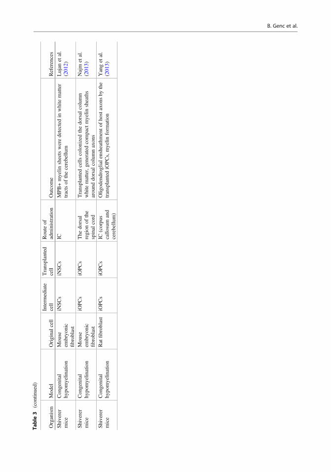
The recent successful improvements in genera-tion of iPSCs and their differentiation into neuralprecursor cells (NPCs) and oligodendrocyte pre-cursor cells (OPCs) initiated autologous iPSCtherapy studies in MS. First, in vivo therapeuticeffect of iPSCs was evaluated in EAE, chemicallyinduced demyelination, and genetichypomyelination models (Table 3). MouseiPSC-derived NPCs (miPSC-NPCs) weretransplanted to C57Bl/6 mice withMOG-induced EAE intrathecally after diseaseonset (Laterza et al. 2013). miPSC-NPC treatmentreduced clinical scores of EAE and decreaseddemyelinated areas and axonal damage in thespinal cord. Transplanted miPSC-NPCs did notdifferentiate to neither neuron nor oligodendro-cyte and did not migrate from perivascular spaceto lesion site. Neuroprotective effects of miPSC-
NPCs were partly through the secretion of LIFthat support resident oligodendrocyte survivaland differentiation. Microarray analysis revealedthat miPSC-NPCs counterbalanced theEAE-associated transcriptional changes in thespinal cord. These cells also limit BBB damageand decrease CNS-infiltrating inflammatory cells(Laterza et al. 2013). Intraventricular transplanta-tion of miPSCs also improved the functionalrecovery of EAE mice and reduced T cell infiltra-tion and white matter damage (Zhang et al. 2016).The effect of hiPSC-derived embryoid bodyintermediate-stage NPCs (EB-NPCs) was exam-ined in neurotropic JHM strain of mouse hepatitisvirus (JHMV)-induced EAE (Plaisted et al.2016). Significant clinical recovery was notobserved in EB-NPCs transplanted mice.EB-NPCs were rapidly eliminated, but theydecreased accumulation of CD4+ T cells in theCNS, reduced demyelination at the site of injec-tion, and increased the number of Treg cells(Plaisted et al. 2016). While healthy hiPSC-derived NPCs decreased inflammation, PPMSpatient-derived NPCs failed to provide any profitin demyelination process (Nicaise et al. 2017).
OPCs derived from iPSCs were alsotransplanted into animal models ofMS. Intracerebrally transplanted OPCs derivedfrom miPSCs and hiPSCs survive and differenti-ate to MBP-expressing oligodendrocytes in bothcuprizone- and lysolecithin-induced models(Czepiel et al. 2011; Nicaise et al. 2017). Therecovery effect of OPC transplantation on demy-elination was also confirmed in congenitalhypomyelination model (Douvaras et al. 2014;Terzic et al. 2016; Wang et al. 2013).Transplanted OPCs differentiate intoMBP-expressing oligodendrocytes (Terzic et al.2016) and contribute to myelination (Douvaraset al. 2014; Terzic et al. 2016; Wang et al.2013). The effect of iPSC-derived OPC transplan-tation was also evaluated in EAE model(Thiruvalluvan et al. 2016). Transplanted OPCsreduced EAE scores, cell infiltration, and demye-lination in the cerebellum. Histological analysisrevealed that transplanted OPCs remained withinthe ventricles; therefore their effect on clinicaland histological features of EAE occurs most
B. Genc et al.

Table
3Indu
cedpluripotentstem
cells
derivedcelltherapiesin
demyelin
ated
anim
almod
els
Organism
Mod
elOriginalcell
Interm
ediate
cell
Transplanted
cell
Rou
teof
administration
Outcome
References
C57
Bl/6
mice
Cup
rizone
indu
ced
Mou
seem
bryo
nic
fibrob
last
miPSCs
OPCs
IC(corpu
scallo
sum)
Differentiateto
MBP-exp
ressingoligod
endrocytes,
contribu
tedto
theremyelin
ation
Czepieletal.
(201
1)
C57
Bl/6
mice
Cup
rizone
indu
ced
Hum
anbloo
dcells
with
PPMS
hiPSCs
NPCs
IVNPCsfrom
PPMSpatientsfailedto
prov
ideany
benefitin
preserving
compactCNSmyelin
ation
during
activ
edemyelin
ation
Nicaise
etal.
(201
7)
Sprague
Daw
ley
rats
Lysolecith
inindu
ced
Hum
anfibrob
last
hiPSCs
OPCs
IC(optic
chiasm
)Recov
eryfrom
symptom
s(m
easuredwith
visual
evok
edpo
tential),transplantedcells
survived
and
integrated
with
inthechiasm
,differentiatedto
PLP
and/or
MBPexpressing
oligod
endrocytes,
contribu
tedto
theremyelin
ation
Pou
yaetal.
(201
1)
C57
Bl/6
mice
MOG
35–55
indu
cedEAE
Mou
seem
bryo
nic
fibrob
lasts
miPSCs
NPCs
ITDecreased
EAEscore,redu
ctionof
demyelin
ated
areasandaxon
aldamage,transplanted
miPSC-N
PCs
coun
terbalancedtheEAE-associatedtranscriptional
changes
Laterza
etal.
(201
3)
C57
BL/6
mice
MOG
35–55
indu
cedEAE
miPSCs
miPSCs
NPCs
Intraventricular
Reduced
Tcellinfiltration,am
elioratedWM
damage
Zhang
etal.
(201
6)C57
BL/6
mice/
marmoset
Hum
anMOG
34–56
indu
cedEAE
Hum
anfibrob
last
hiPSCs
OPCs
ICV
(cisterna
magna),IC
Significant
redu
ctionof
subsequent
EAEscores
and
cellinfiltration;
transplanted
hiPSCsin
ventricule,
migratio
nanddifferentiatio
nto
MBPprod
ucing
cells
inmarmosetmod
el
Thiruvallu
van
etal.(20
16)
C57
BL/6
mice
Intracranially
JHMV
Primaryfetal
human
fibrob
lasts
hiPSCs
NPCs
Intraspinal
EB-N
PCswererapidlyrejected,d
ecreased
accumulationof
CD4+
Tcells
intheCNS,reduced
demyelin
ationatthesiteof
injection,
mod
est
patholog
icalim
prov
ements,n
osign
ificant
clinical
recovery
Plaistedetal.
(201
6)
Shiverer
mice
Con
genital
hypo
myelin
ation
Mou
seem
bryo
nic
fibrob
last
miPSCs
OPCs
IC(corpu
scallo
sum
and
striatum
)
Transplantedcells
expressing
MBP
Terzicetal.
(201
6)
Shiverer
mice
Con
genital
hypo
myelin
ation
Hum
anfibrob
last,
keratin
ocyte
hiPSCs
OPCs
IC(5-site
forebrainand
brainstem)
Increasedthesurvival,m
yelin
ationof
thebrain,
brainstem,and
cerebellu
mWangetal.
(201
3)
Shiverer
mice
Con
genital
hypo
myelin
ation
Hum
anfibrob
last
with
PPMS
hiPSCs
OPCs
IC(forebrain)
Hostm
ouse
axon
swereensheathed,m
aturecompact
myelin
observed
with
electron
microscop
eDou
varasetal.
(201
4) (con
tinued)
Stem Cell Therapy for Multiple Sclerosis
Table
3(con
tinued)
Organism
Mod
elOriginalcell
Interm
ediate
cell
Transplanted
cell
Rou
teof
administration
Outcome
References
Shiverer
mice
Con
genital
hypo
myelin
ation
Mou
seem
bryo
nic
fibrob
last
iNSCs
iNSCs
ICMPB+myelin
sheetsweredetected
inwhitematter
tractsof
thecerebellu
mLujan
etal.
(201
2)
Shiverer
mice
Con
genital
hypo
myelin
ation
Mou
seem
bryo
nic
fibrob
last
iOPCs
iOPCs
The
dorsal
region
ofthe
spinalcord
Transplantedcells
colonizedthedo
rsalcolumn
whitematter,generatedcompactmyelin
sheaths
arou
nddo
rsalcolumnaxon
s
Najm
etal.
(201
3)
Shiverer
mice
Con
genital
hypo
myelin
ation
Ratfibrob
last
iOPCs
iOPCs
IC(corpu
scallo
sum
and
cerebellu
m)
Olig
odendrog
lialensheathm
ento
fho
staxon
sby
the
transplanted
iOPCs,myelin
form
ation
Yangetal.
(201
3)
B. Genc et al.

likely through secreted factors. In contrast to this,intracerebrally transplanted hiPSC-derived OPCsmigrated toward the lesion and differentiated toMBP producing mature oligodendrocytes in mar-moset model of EAE, suggesting that differencesbetween species or route of administration areimportant (Thiruvalluvan et al. 2016). Geneticmodification of transplanted cells can be used toenhance their targeted migration. Polysialylatingenzyme sialyltransferase X (STX) overexpressionin iPSC-derived OPCs increased their migrationalong the axons in cuprizone-induced model(Czepiel et al. 2014).
After the successful reprogramming of somaticcells to iPSCs, more direct neural lineage conver-sion methods have been developed. Functionalneurons were obtained by transdifferentiation offibroblasts using defined factors. Afterward, sim-ilar methods were developed for the generation ofiNSCs and iOPCs using neural lineage-specificsets of TFs. iNSC and iOPC transplantationswere performed in dysmyelinated Shiverer mice(Lujan et al. 2012; Najm et al. 2013; Yang et al.2013). Transplanted iNSCs differentiated intooligodendrocytes capable of integration intodysmyelinated Shiverer brain (Lujan et al.2012). iOPCs when given into the spinal cord,corpus callosum, and cerebellum survived,ensheathed host axons, and produced myelin(Najm et al. 2013; Yang et al. 2013).
6.3 Mechanisms
Because of inadequate endogenousremyelination, cellular therapy is moving forwardin MS treatment. Therapeutic effects of iPSCs arenot limited with cell replacement. iPSCs alsoexhibit immunosuppressive effect and providetrophic support on endogenous repairmechanisms. NPC transplantation decreases Tcell infiltration (Plaisted et al. 2016; Zhang et al.2016). Secreted LIF from transplanted NPCsexerts trophic action on endogenous oligoden-drocytes (Laterza et al. 2013).
Apart from cell replacement, iPSCs have beenused in vitro to model diseases in order to under-stand the underlying mechanisms and for screen-ing drugs that modify the disease process. MSpatient-specific iPSCs were first generated in2011 by using fibroblasts from a 35-year-oldpatient with RRMS (Song et al. 2012). Patient-derived iPSCs were successfully differentiated toneural progenitors and mature neurons. Subse-quently, patient iPSCs were also generated fromPPMS patients (Douvaras et al. 2014; Nicaiseet al. 2017). In the study by Douvaras et al., fouriPSC lines were converted to NSCs and OPCswhich carry normal karyotypes. TransplantedOPCs provided myelination in Shiverer mice.Similar studies should be continued especially inMS patients with high genetic load for diseasemodeling.
6.4 Practical Considerations
There are still some concerns about iPSC-basedtreatment in MS. Firstly, iPSC generationmethods still need to be improved. The otherconcern is preference of initial cell type for trans-plantation. OPCs are superior for cell replace-ment, but anti-inflammatory and trophic effectsof these cells have not been demonstrated yet.NSCs can differentiate into all neuronal celllineages and also have anti-inflammatory and tro-phic effects, but they may differentiate toastrocytes as well and therefore lead to unwantedastrogliosis in MS. Also, they have tumorigenic-ity potential. Another major concern is the routeof administration of cells. Direct intracerebral orintraspinal injection seems more effective, butthey are not practical in clinical setting. Intranasalroute may also be used effectively, and it is a lessinvasive route for administration of iPSCs in MS(Wu et al. 2013). Finally, use of allogeneic orautologous iPSCs should be considered. Autolo-gous iPSCs are preferable, but they may be inef-fective due to intrinsic disease factors (Nicaiseet al. 2017). To avoid immunogenicity, healthy
Stem Cell Therapy for Multiple Sclerosis

allogeneic iPSC therapy needs immunosuppres-sive treatment that may cause MS relapses.
7 Challenges and FuturePerspectives of Stem CellTransplantation for MS
Stem cell transplantation can be regarded as a poten-tial source of treatment for MS. Nevertheless, beforeintroducing stem cell treatment wholesale intoclinics, methodological, ethical, and clinicalchallenges must be overcome in stem cell therapystudies.
7.1 Sources
HSCs and MSCs have been used in clinical MSstem cell trials. In spite of the better availability ofHSCs and MSCs compared to NSCs,remyelination capacity of NSCs makes them thepreferred cell type for MS stem cell clinical trialsparticularly in the progressive stages of MS(Mariano et al. 2015; Sarkar et al. 2017).Although iPSC-derived stem cells have not beenused in clinical trials of MS, they are suitablecandidates for individualized cell replacementtherapy due to their advantage of being easilyobtained from the patient’s own tissue.
The autologous and allogeneic stem cells con-tain different advantages and disadvantages inMS treatment (Cohen 2013). Autologous stemcell is less immunogenic, but generation fromPSCs takes a longer time period, which makes itdisadvantageous particularly in the acute phase ofMS because of the necessity of immediate SCT.Additionally, autologous stem cells may be inef-fective due to intrinsic disease factors (Nicaiseet al. 2017). However, genetic defects can becorrected with several gene-editing methodsincluding zinc finger nucleases, transcriptionactivator-like effector nucleases (TALENs), andthe clustered regularly interspaced short palin-dromic repeats (CRISPR)/Cas9 systems (Maederand Gersbach 2016), which means that
autologous stem cells could be used in MS ther-apy upon genome editing, if necessary. The use ofallogeneic stem cells has advantages such asavoiding the risk by genetic susceptibility of therecipient to develop MS. Since these cells arereadily available from biobanks (Natalwala andKunath 2017), they may be useful in the acute andprogressive phase of MS.
7.2 In Vitro Cell Expansionand Manipulation
Numerous technical factors affect the yield, via-bility, function, and efficacy of SCT (Bang et al.2016). The use of fetal bovine serum in culturemedium raises safety concerns, including possi-ble transmission of zoonoses and infusion-relatedallergic reactions (Cohen 2013). Stem cell pro-duction for clinical trials should be done undercurrent GMP standards (Galvez-Martin et al.2016).
Various approaches for in vitro cell expansionincrease the stem cell proliferation, survival, andtrophic support and reduce senescence of stemcells (Bang et al. 2016). Preclinical studiesshowed that ex vivo treatment of stem cells withtrophic factors or chemical agents enhanced themigration of stem cells and trophic support in thebrain. Lastly, genetic modification of stem cells,such as overexpressing chemokine receptors orIL-10, increased their efficacies and migrationcapacities in animal models of MS (Klose et al.2013; Phillips and Tang 2008; Yang et al. 2012).
7.3 Practical Considerations
7.3.1 StorageThe ability to freeze with xeno-free freezingmedia and storing them in proper conditions areessential steps for clinical use of stem cells(Sarkar et al. 2017). Automated methods ofthawing cells may increase viability of cells(Nishiyama et al. 2016). Other considerations
B. Genc et al.
for storage conditions are cell number, cell den-sity, vial size, and total volume.
7.3.2 Passage NumberThe passage number of transplanted stem cellsaffects their proliferation, differentiation, andtherapeutic capacities (Sisakhtnezhad et al.2017). For example, lower than five passages ofMSCs appear to be optimal for transplantation(Liu et al. 2016).
7.3.3 RouteThe routes of cell administration vary amongstudies. Direct intracerebroventricular or intrathe-cal injections seem more effective, but IV routecould be superior for peripheral immunosuppres-sive effect (Cohen 2013). However, entry to CNSis very low, and most of the injected cells aretrapped by the lung, liver, spleen, and lymphnodes. Effective and less invasive routes foradministration of stem cells such as intranasalroute may also be used in MS (Li et al. 2015).
7.3.4 DoseAs the differences in the number of doses couldbe an important factor to the observed variation inresponses, standardization is required for optimaldoses for each transplanted cell type (Cohen2013). Additionally, repeated administrationsshould be used to increase clinical benefit espe-cially in progressive MS (Harris et al. 2016).
7.4 Tracking of Transplanted Cells
Noninvasive cell-tracking methods allow real-time monitoring of survival, migration, and hom-ing of administered stem cells. MRI, magneticparticle imaging, positron emission tomography,single-photon emission computed tomography,and optical imaging methods can be used forin vivo tracking of stem cells (Filippi et al.2017a; Ngen and Artemov 2017). To date, anoptimal technique for in vivo cell tracking doesnot yet exist in the clinical setting.
7.5 Risks
In general, HSCT andMSC transplantation in MSpatients have been well-tolerated, but severalpotential acute and chronic adverse effects shouldbe considered. Infusion-related toxicity and infec-tion are acute risks of stem cell therapy, andadditive immunosuppressive treatment increasesthe infection risk following transplantation(Sarkar et al. 2017). Ectopic tissue formationand malignant transformation of stem cells aretheoretical concern, although it has not beenreported in MSC therapy studies (Cohen 2013).iPSCs have more tumorigenicity potentialdepending on pluripotency induction method.The use of iOPCs or iNSCs obtained by directreprogramming without pluripotent stage canavoid tumor development risk (Xie et al. 2016).The other main risk in stem cell treatment isallogeneic immune rejection that is primarilymediated by T cell-dependent immune responsesand needs lifelong immunosuppressive treatment.Previous studies supported that SCTs may alsolead to disease activation in MS, presumably byleading to fever (Cohen 2013).
7.6 Cost-Effectiveness
The initial costs of stem cell therapies areextremely high, and cost-effective stem celltreatments must be developed (Sarkar et al.2017). To assess the risk-benefit ratio of stemcell therapies, randomized studies should beperformed comparing the efficacy of stem cellsagainst other conventional therapies.
Stem cell tourism is a ridiculous term used todescribe traveling abroad to undergo medicalstem cell treatments that are not approved oravailable in patients’ home country. Anecdotalevidence suggests that stem cell tourism leads tophysical and financial risk to patients (Marks et al.2017). Clinicians and regulators should worktogether to prevent deregulated cell-basedtherapies.
Stem Cell Therapy for Multiple Sclerosis

8 Conclusion
Stem cell-based therapies are attractivealternatives for the treatment of MS. There arestill some issues which need to be resolved suchas low efficacy of iPSCs, lack of proper differen-tiation protocols, epigenetic alternation in donorcells, and heterogeneity of transplanted cells.Improvement of stem cell technology will con-tribute to overcome these problems. For example,screening and selection of viable, genetically sta-ble, and desired stem cells can prevent tumorige-nicity and immunogenicity side effects of SCT.
Apart from these technical problems, severalclinical difficulties should be considered. One ofmain concerns in SCT is to make a choicebetween autologous versus allogeneic stem cellsfor MS treatment. In older MS patients,HLA-matched young donor cells for transplanta-tion may be more appropriate (Phanthong et al.2013).
Combination therapy with anti-inflammatoryand remyelinating agents (Anti-Lingo1 ab) andalso other stem cells should be considered toincrease the efficacy of stem cell therapy (Harlowet al. 2015). Emerging, innovative treatmentapproaches such as using cell-free stem cellproducts (conditioned media, exosome) shouldbe investigated in animal models of MS (Derkuset al. 2017; Harlow et al. 2015).
Acknowledgment Author thanks Assistant Prof.Dr. Yavuz Oktay for critical reading and editing of thischapter.
References
Aharonowiz, M., Einstein, O., Fainstein, N., Lassmann,H., Reubinoff, B., & Ben-Hur, T. (2008 Sep 5).Neuroprotective effect of transplanted human embry-onic stem cell-derived neural precursors in an animalmodel of multiple sclerosis. PLoS One, 3(9), e3145.https://doi.org/10.1371/journal.pone.0003145 PubMed PMID: 18773082; Pub Med Central PMCID:PMC2522282.
An, N., Xu, H., Gao, W. Q., & Yang, H. (2016). Directconversion of somatic cells into induced neurons.Molecular Neurobiology, 55, 642. https://doi.org/10.1007/s12035-016-0350-0.
Atkins, H. L., & Freedman, M. S. (2013). Hematopoieticstem cell therapy for multiple sclerosis: Top 10 lessonslearned. Neurotherapeutics, 10, 68–76. https://doi.org/10.1007/s13311-012-0162-5.
Bai, L., et al. (2012). Hepatocyte growth factor mediatesmesenchymal stem cell-induced recovery in multiplesclerosis models. Nature Neuroscience, 15, 862–870.https://doi.org/10.1038/nn.3109.
Bai, L., Lennon, D. P., Eaton, V., Maier, K., Caplan, A. I.,Miller, S. D., & Miller, R. H. (2009). Human bonemarrow-derived mesenchymal stem cells induceTh2-polarized immune response and promote endoge-nous repair in animal models of multiple sclerosis.Glia, 57, 1192–1203. https://doi.org/10.1002/glia.20841.
Bang, O. Y., Kim, E. H., Cha, J. M., &Moon, G. J. (2016).Adult stem cell therapy for stroke: Challenges andprogress. Journal of Stroke, 18, 256–266. https://doi.org/10.5853/jos.2016.01263.
Bell, S. M., Sharrack, B., & Snowden, J. A. (2017). Autolo-gous hematopoietic cell transplantation in multiple sclero-sis. Expert Opinion on Biological Therapy, 17, 77–86.https://doi.org/10.1080/14712598.2017.1239706.
Ben-Hur, T., Einstein, O., Mizrachi-Kol, R.,Ben-Menachem, O., Reinhartz, E., Karussis, D., &Abramsky, O. (2003). Transplanted multipotentialneural precursor cells migrate into the inflamed whitematter in response to experimental autoimmuneencephalomyelitis. Glia, 41, 73–80. https://doi.org/10.1002/glia.10159.
Bonab, M. M., et al. (2012). Autologous mesenchymalstem cell therapy in progressive multiple sclerosis:An open label study. Current Stem Cell Research &Therapy, 7, 407–414.
Browne, P., Chandraratna, D., Angood, C., Tremlett, H.,Baker, C., Taylor, B. V., & Thompson, A. J. (2014).Atlas of Multiple Sclerosis 2013: A growing global prob-lem with widespread inequity.Neurology, 83, 1022–1024.https://doi.org/10.1212/WNL.0000000000000768.
Burman, J., et al. (2014). Autologous haematopoietic stemcell transplantation for aggressive multiple sclerosis:The Swedish experience. Journal of Neurology, Neu-rosurgery, and Psychiatry, 85, 1116–1121. https://doi.org/10.1136/jnnp-2013-307207.
Burt, R. K., et al. (2015). Association of nonmyeloablativehematopoietic stem cell transplantation with neurolog-ical disability in patients with relapsing-remitting mul-tiple sclerosis. JAMA, 313, 275–284. https://doi.org/10.1001/jama.2014.17986.
Burt, R. K., Balabanov, R., Voltarelli, J., Barreira, A., &Burman, J. (2012). Autologous hematopoietic stemcell transplantation for multiple sclerosis--if confusedor hesitant, remember: ‘Treat with standard immunesuppressive drugs and if no inflammation, noresponse’. Multiple Sclerosis, 18, 772–775. https://doi.org/10.1177/1352458512442993.
Cao, W., et al. (2011). Leukemia inhibitory factor inhibitsT helper 17 cell differentiation and confers treatmenteffects of neural progenitor cell therapy in autoimmune
B. Genc et al.

disease. Immunity, 35, 273–284. https://doi.org/10.1016/j.immuni.2011.06.011.
Casanova, B., et al. (2017). Autologous hematopoieticstem cell transplantation in relapsing-remitting multi-ple sclerosis: Comparison with secondary progressivemultiple sclerosis. Neurological Sciences, 38,1213–1221. https://doi.org/10.1007/s10072-017-2933-6.
Chen, L., Coleman, R., Leang, R., Tran, H., Kopf, A.,Walsh, C. M., Sears-Kraxberger, I., Steward, O.,Macklin, W. B., Loring, J. F., & Lane, T. E. (2014May 15). Human neural precursor cells promote neu-rologic recovery in a viral model of multiple sclerosis.Stem Cell Reports., 2(6), 825–837. https://doi.org/10.1016/j.stemcr.2014.04.005 eCollection 2014 Jun 3.Pub Med PMID: 24936469; Pub Med CentralPMCID: PMC4050357.
Cieslar-Pobuda, A., et al. (2017). Transdifferentiation andreprogramming: Overview of the processes, theirsimilarities and differences. Biochimica et BiophysicaActa, 1864, 1359–1369. https://doi.org/10.1016/j.bbamcr.2017.04.017.
Cohen, J. A. (2013). Mesenchymal stem cell transplanta-tion in multiple sclerosis. Journal of the NeurologicalSciences, 333, 43–49. https://doi.org/10.1016/j.jns.2012.12.009.
Cohen, J. A., et al. (2017). Pilot trial of intravenous autologousculture-expanded mesenchymal stem cell transplantationin multiple sclerosis. Multiple Sclerosis, 24(4), 501–511.https://doi.org/10.1177/1352458517703802.
Comi, G., Radaelli, M., & Soelberg Sorensen, P. (2017).Evolving concepts in the treatment of relapsing multi-ple sclerosis. Lancet, 389, 1347–1356. https://doi.org/10.1016/S0140-6736(16)32388-1.
Compston, A., & Coles, A. (2008). Multiple sclerosis.Lancet, 372, 1502–1517. https://doi.org/10.1016/S0140-6736(08)61620-7.
Connick, P., et al. (2012). Autologous mesenchymal stemcells for the treatment of secondary progressive multi-ple sclerosis: an open-label phase 2a proof-of-conceptstudy. Lancet Neurology, 11, 150–156. https://doi.org/10.1016/S1474-4422(11)70305-2.
Curro, D., & Mancardi, G. (2016). Autologoushematopoietic stem cell transplantation in multiplesclerosis: 20 years of experience. NeurologicalSciences, 37, 857–865. https://doi.org/10.1007/s10072-016-2564-3.
Czepiel, M., et al. (2011). Differentiation of induced plu-ripotent stem cells into functional oligodendrocytes.Glia, 59, 882–892. https://doi.org/10.1002/glia.21159.
Czepiel, M., Leicher, L., Becker, K., Boddeke, E., &Copray, S. (2014). Overexpression of polysialylatedneural cell adhesion molecule improves the migrationcapacity of induced pluripotent stem cell-derived oli-godendrocyte precursors. Stem Cells TranslationalMedicine, 3, 1100–1109. https://doi.org/10.5966/sctm.2014-0041.
Dargahi, N., Katsara, M., Tselios, T., Androutsou, M. E.,de Courten, M., Matsoukas, J., & Apostolopoulos,
V. (2017). Multiple sclerosis: Immunopathology andtreatment update. Brain Sciences, 7. https://doi.org/10.3390/brainsci7070078.
Delbue, S., Comar, M., & Ferrante, P. (2017).Natalizumab treatment of multiple sclerosis: Newinsights. Immunotherapy, 9, 157–171. https://doi.org/10.2217/imt-2016-0113.
Derkus, B., Emregul, K. C., & Emregul, E. (2017). A newapproach in stem cell research-Exosomes: Their mech-anism of action via cellular pathways. Cell BiologyInternational, 41, 466–475. https://doi.org/10.1002/cbin.10742.
Disanto, G., Morahan, J. M., Barnett, M. H., Giovannoni, G.,& Ramagopalan, S. V. (2012). The evidence for a role ofB cells in multiple sclerosis. Neurology, 78, 823–832.https://doi.org/10.1212/WNL.0b013e318249f6f0.
Dominici, M., et al. (2006). Minimal criteria for definingmultipotent mesenchymal stromal cells. The Interna-tional Society for Cellular Therapy position statement.Cytotherapy, 8, 315–317. https://doi.org/10.1080/14653240600855905.
Dorr, J. (2016). Haemopoietic stem-cell transplantation formultiple sclerosis: What next? Lancet, 388, 536–538.https://doi.org/10.1016/S0140-6736(16)30377-4.
Douvaras, P., et al. (2014). Efficient generation ofmyelinating oligodendrocytes from primary progres-sive multiple sclerosis patients by induced pluripotentstem cells. Stem Cell Reports, 3, 250–259. https://doi.org/10.1016/j.stemcr.2014.06.012.
Durnaoglu, S., Genc, S., & Genc, K. (2011). Patient-specific pluripotent stem cells in neurological diseases.Stem Cells International, 2011, 212487. https://doi.org/10.4061/2011/212487.
Einstein, O., et al. (2007). Neural precursors attenuateautoimmune encephalomyelitis by peripheral immuno-suppression. Annals of Neurology, 61, 209–218.https://doi.org/10.1002/ana.21033.
Einstein, O., et al. (2006). Transplanted neural precursorcells reduce brain inflammation to attenuate chronicexperimental autoimmune encephalomyelitis. Experi-mental Neurology, 198, 275–284. https://doi.org/10.1016/j.expneurol.2005.11.007.
Einstein, O., Karussis, D., Grigoriadis, N., Mizrachi-Kol,R., Reinhartz, E., Abramsky, O., & Ben-Hur,T. (2003). Intraventricular transplantation of neuralprecursor cell spheres attenuates acute experimentalallergic encephalomyelitis. Molecular and CellularNeurosciences, 24, 1074–1082.
Fassas, A., Anagnostopoulos, A., Kazis, A., Kapinas, K.,Sakellari, I., Kimiskidis, V., & Tsompanakou,A. (1997). Peripheral blood stem cell transplantationin the treatment of progressive multiple sclerosis:First results of a pilot study. Bone Marrow Transplan-tation, 20, 631–638. https://doi.org/10.1038/sj.bmt.1700944.
Filippi, M., Boido, M., & Terreno, E. (2017a). Imaging ofMSC transplantation in neuroscience. Oncotarget, 8,10781–10782. https://doi.org/10.18632/oncotarget.14643.
Stem Cell Therapy for Multiple Sclerosis

Filippi, M., Preziosa, P., & Rocca, M. A. (2017b).Microstructural MR imaging techniques in multiplesclerosis. Neuroimaging Clinics of North America,27, 313–333. https://doi.org/10.1016/j.nic.2016.12.004.
Friedenstein, A. J., Chailakhyan, R. K., Latsinik, N. V.,Panasyuk, A. F., & Keiliss-Borok, I. V. (1974). Stro-mal cells responsible for transferring the microenviron-ment of the hemopoietic tissues. Cloning in vitro andretransplantation in vivo. Transplantation, 17,331–340.
Galvez-Martin, P., Sabata, R., Verges, J., Zugaza, J. L.,Ruiz, A., & Clares, B. (2016). Mesenchymal stem cellsas therapeutics agents: Quality and environmental reg-ulatory aspects. Stem Cells International, 2016,1. https://doi.org/10.1155/2016/9783408.
Garg, N., & Smith, T. W. (2015). An update onimmunopathogenesis, diagnosis, and treatment of mul-tiple sclerosis. Brain and Behavior, 5, e00362. https://doi.org/10.1002/brb3.362.
Gerdoni, E., et al. (2007). Mesenchymal stem cells effec-tively modulate pathogenic immune response in exper-imental autoimmune encephalomyelitis. Annals ofNeurology, 61, 219–227. https://doi.org/10.1002/ana.21076.
Gharibi, T., Ahmadi, M., Seyfizadeh, N., Jadidi-Niaragh,F., & Yousefi, M. (2015). Immunomodulatorycharacteristics of mesenchymal stem cells and theirrole in the treatment of multiple sclerosis. CellularImmunology, 293, 113–121. https://doi.org/10.1016/j.cellimm.2015.01.002.
Giacoppo, S., Bramanti, P., & Mazzon, E. (2017). Thetransplantation of mesenchymal stem cells derivedfrom unconventional sources: An innovative approachto multiple sclerosis therapy. Archivum Immunologiaeet Therapiae Experimentalis (Warsz), 65, 363. https://doi.org/10.1007/s00005-017-0460-z.
Gomes Mda, M., & Engelhardt, E. (2013). Jean-MartinCharcot, father of modern neurology: An homage120 years after his death. Arquivos de Neuro-Psiquiatria, 71, 815–817. https://doi.org/10.1590/0004-282X20130128.
Grigoriadis, N., van Pesch, V., & Paradig, M. S. G. (2015).A basic overview of multiple sclerosis immunopathol-ogy. European Journal of Neurology, 22(Suppl 2),3–13. https://doi.org/10.1111/ene.12798.
Guan, Y., Jiang, Z., Ciric, B., Rostami, A. M., & Zhang,G. X. (2008). Upregulation of chemokine receptorexpression by IL-10/IL-4 in adult neural stem cells.Experimental and Molecular Pathology, 85,232–236. https://doi.org/10.1016/j.yexmp.2008.07.003.
Guo, B. (2016). IL-10 modulates Th17 pathogenicity dur-ing autoimmune diseases. Journal of Clinical & Cellu-lar Immunology, 07, 7. https://doi.org/10.4172/2155-9899.1000400.
Gupta, N., et al. (2012). Neural stem cell engraftment andmyelination in the human brain. Science Translational
Medicine, 4, 155ra137. https://doi.org/10.1126/scitranslmed.3004373.
Harding, J., & Mirochnitchenko, O. (2014). Preclinicalstudies for induced pluripotent stem cell-based thera-peutics. The Journal of Biological Chemistry, 289,4585–4593. https://doi.org/10.1074/jbc.R113.463737.
Harlow, D. E., Honce, J. M., & Miravalle, A. A. (2015).Remyelination therapy in multiple sclerosis. Frontiersin Neurology, 6, 257. https://doi.org/10.3389/fneur.2015.00257.
Harris, V. K., Yan, Q. J., Vyshkina, T., Sahabi, S., Liu, X.,& Sadiq, S. A. (2012 Feb 15). Clinical and pathologi-cal effects of intrathecal injection of mesenchymalstem cell-derived neural progenitors in an experimentalmodel of multiple sclerosis. Journal of the Neurologi-cal Sciences, 313(1–2), 167–177. https://doi.org/10.1016/j.jns.2011.08.036 Epub 2011 Oct 1.Pub MedPMID: 21962795.
Harris, V. K., Vyshkina, T., & Sadiq, S. A. (2016). Clini-cal safety of intrathecal administration of mesenchymalstromal cell-derived neural progenitors in multiplesclerosis. Cytotherapy, 18, 1476–1482. https://doi.org/10.1016/j.jcyt.2016.08.007.
Hemmer, B., Kerschensteiner, M., & Korn, T. (2015).Role of the innate and adaptive immune responses inthe course of multiple sclerosis. Lancet Neurology, 14,406–419. https://doi.org/10.1016/S1474-4422(14)70305-9.
Heslop, J. A., Hammond, T. G., Santeramo, I., Tort Piella,A., Hopp, I., Zhou, J., Baty, R., Graziano, E. I., ProtoMarco, B., Caron, A., Sköld, P., Andrews, P. W.,Baxter, M. A., Hay, D. C., Hamdam, J., Sharpe, M.E., Patel, S., Jones, D. R., Reinhardt, J., Danen, E. H.,Ben-David, U., Stacey, G., Björquist, P., Piner, J.,Mills, J., Rowe, C., Pellegrini, G., Sethu, S., Antoine,D. J., Cross, M. J., Murray, P., Williams, D. P.,Kitteringham, N. R., Goldring, C. E., & Park, B. K.(2015 Apr). Concise review: Workshop review:Understanding and assessing the risks of stem cell-based therapies. Stem Cells Translational Medicine,4(4), 389–400. https://doi.org/10.5966/sctm.2014-0110Epub 2015 Feb 26. Review. Pub Med PMID:25722427; Pub Med Central PMCID: PMC4367503.
Hwang, Y., Broxmeyer, H. E., & Lee, M. R. (2017).Generating autologous hematopoietic cells fromhuman-induced pluripotent stem cells through ectopicexpression of transcription factors. Current Opinion inHematology, 24, 283–288. https://doi.org/10.1097/MOH.0000000000000343.
Jarmalaviciute, A., & Pivoriunas, A. (2016). Exosomes asa potential novel therapeutic tools against neurodegen-erative diseases. Pharmacological Research, 113,816–822. https://doi.org/10.1016/j.phrs.2016.02.002.
Jones, A., & Hawiger, D. (2017). Peripherally inducedregulatory T cells: Recruited protectors of the centralnervous system against autoimmuneneuroinflammation. Frontiers in Immunology, 8, 532.https://doi.org/10.3389/fimmu.2017.00532.
B. Genc et al.

Karnell, F. G., et al. (2017). Reconstitution of immune cellpopulations in multiple sclerosis patients after autolo-gous stem cell transplantation. Clinical and Experi-mental Immunology, 189, 268. https://doi.org/10.1111/cei.12985.
Karussis, D., et al. (2010). Safety and immunologicaleffects of mesenchymal stem cell transplantation inpatients with multiple sclerosis and amyotrophic lateralsclerosis. Archives of Neurology, 67, 1187–1194.https://doi.org/10.1001/archneurol.2010.248.
Karussis, D., & Slavin, S. (2004). Hematopoietic stem celltransplantation in multiple sclerosis: Experimental evi-dence to rethink the procedures. Journal of the Neuro-logical Sciences, 223, 59–64. https://doi.org/10.1016/j.jns.2004.04.021.
Kawachi, I., & Lassmann, H. (2017). Neurodegenerationin multiple sclerosis and neuromyelitis optica. Journalof Neurology, Neurosurgery, and Psychiatry, 88,137–145. https://doi.org/10.1136/jnnp-2016-313300.
Kelsey, P. J., Oliveira, M. C., Badoglio, M., Sharrack, B.,Farge, D., & Snowden, J. A. (2016). Haematopoieticstem cell transplantation in autoimmune diseases:From basic science to clinical practice. CurrentResearch in Translational Medicine, 64, 71–82.https://doi.org/10.1016/j.retram.2016.03.003.
King, N. M., & Perrin, J. (2014). Ethical issues in stem cellresearch and therapy. Stem Cell Research & Therapy,5, 85. https://doi.org/10.1186/scrt474.
Klose, J., Schmidt, N. O., Melms, A., Dohi, M., Miyazaki,J., Bischof, F., & Greve, B. (2013). Suppression ofexperimental autoimmune encephalomyelitis byinterleukin-10 transduced neural stem/progenitorcells. Journal of Neuroinflammation, 10, 117. https://doi.org/10.1186/1742-2094-10-117.
Laterza, C., et al. (2013). iPSC-derived neural precursorsexert a neuroprotective role in immune-mediateddemyelination via the secretion of LIF. NatureCommunications, 4, 2597. https://doi.org/10.1038/ncomms3597.
Lee, H., Narayanan, S., Brown, R. A., Chen, J. T., Atkins,H. L., Freedman, M. S., & Arnold, D. L. (2017). Brainatrophy after bone marrow transplantation for treat-ment of multiple sclerosis. Multiple Sclerosis, 23,420–431. https://doi.org/10.1177/1352458516650992.
Lee, Y. E., et al. (2015). The synergistic local immunosup-pressive effects of neural stem cells expressingindoleamine 2,3-Dioxygenase (IDO) in an Experimen-tal Autoimmune Encephalomyelitis (EAE) AnimalModel. PLoS One, 10, e0144298. https://doi.org/10.1371/journal.pone.0144298.
Li, X., Zhang, Y., Yan, Y., Ciric, B., Ma, C. G., Gran, B.,Curtis, M., Rostami, A., & Zhang, G. X. (2016 Aug).Neural stem cells engineered to express three therapeu-tic factors mediate recovery from chronic stage CNSautoimmunity. Molecular Therapy, 24(8), 1456–1469.https://doi.org/10.1038/mt.2016.104 Epub 2016 May16. Pub Med PMID: 27203442; Pub Med CentralPMCID: PMC5023377.
Li, J., et al. (2017). Therapeutic effects of Human AdiposeTissue-Derived Stem Cell (hADSC) transplantation onExperimental Autoimmune Encephalomyelitis (EAE)mice. Scientific Reports, 7, 42695. https://doi.org/10.1038/srep42695.
Li, Y. H., Feng, L., Zhang, G. X., & Ma, C. G. (2015).Intranasal delivery of stem cells as therapy for centralnervous system disease. Experimental and MolecularPathology, 98, 145–151. https://doi.org/10.1016/j.yexmp.2015.01.016.
Lin, C. C., & Edelson, B. T. (2017). New insights into therole of IL-1beta in experimental autoimmune encepha-lomyelitis and multiple sclerosis. Journal of Immunol-ogy, 198, 4553–4560. https://doi.org/10.4049/jimmunol.1700263.
Liu, S., et al. (2016). Strategies to optimize adult stem celltherapy for tissue regeneration. International Journalof Molecular Sciences, 17. https://doi.org/10.3390/ijms17060982.
Llufriu, S., et al. (2014). Randomized placebo-controlledphase II trial of autologous mesenchymal stem cells inmultiple sclerosis. PLoS One, 9, e113936. https://doi.org/10.1371/journal.pone.0113936.
Londono, A. C., & Mora, C. A. (2016). Autologous bonemarrow transplantation in multiple sclerosis: Bio-marker relevance for patient recruitment and followup. Journal of Clinical & Cellular Immunology, 7.https://doi.org/10.4172/2155-9899.1000455.
Lublin, F. D., et al. (2014). Defining the clinical course ofmultiple sclerosis: The 2013 revisions. Neurology, 83,278–286. https://doi.org/10.1212/WNL.0000000000000560.
Lujan, E., Chanda, S., Ahlenius, H., Sudhof, T. C., &Wernig, M. (2012). Direct conversion of mousefibroblasts to self-renewing, tripotent neural precursorcells. Proceedings of the National Academy of Sciencesof the United States of America, 109, 2527–2532.https://doi.org/10.1073/pnas.1121003109.
Maeder, M. L., & Gersbach, C. A. (2016). Genome-editingtechnologies for gene and cell therapy. MolecularTherapy, 24, 430–446. https://doi.org/10.1038/mt.2016.10.
Mahfouz, M. M., Abdelsalam, R. M., Masoud, M. A.,Mansour, H. A., Ahmed-Farid, O. A., & Kenawy,S. A. (2017). The neuroprotective effect of mesenchy-mal stem cells on an experimentally induced model formultiple sclerosis in mice. Journal of Biochemical andMolecular Toxicology, 31. https://doi.org/10.1002/jbt.21936.
Mancardi, G. L., et al. (2015). Autologous hematopoieticstem cell transplantation in multiple sclerosis: A phaseII trial. Neurology, 84, 981–988. https://doi.org/10.1212/WNL.0000000000001329.
Mariano, E. D., Teixeira, M. J., Marie, S. K., & Lepski,G. (2015). Adult stem cells in neural repair: Currentoptions, limitations and perspectives.World Journal ofStem Cells, 7, 477–482. https://doi.org/10.4252/wjsc.v7.i2.477.
Stem Cell Therapy for Multiple Sclerosis

Marks, P. W., Witten, C. M., & Califf, R. M. (2017).Clarifying stem-cell therapy’s benefits and risks. TheNew England Journal of Medicine, 376, 1007–1009.https://doi.org/10.1056/NEJMp1613723.
Matta, A. P., et al. (2016). No evidence of disease activityin multiple sclerosis patients. Expert Review ofNeurotherapeutics, 16, 1279–1284. https://doi.org/10.1080/14737175.2016.1202763.
Matysiak, M., Stasiolek, M., Orlowski, W., Jurewicz, A.,Janczar, S., Raine, C. S., & Selmaj, K. (2008). Stemcells ameliorate EAE via an indoleamine2,3-dioxygenase (IDO) mechanism. Journal ofNeuroimmunology, 193, 12–23. https://doi.org/10.1016/j.jneuroim.2007.07.025.
Mentis, A. A., Dardiotis, E., Grigoriadis, N., Petinaki, E.,& Hadjigeorgiou, G. M. (2017). Viruses and endoge-nous retroviruses in multiple sclerosis: From correla-tion to causation. Acta Neurologica Scandinavica, 136,606. https://doi.org/10.1111/ane.12775.
Miron, V. E. (2017). Beyond immunomodulation: Theregenerative role for regulatory T cells in central ner-vous system remyelination. Journal of Cell Communi-cation and Signaling, 11, 191–192. https://doi.org/10.1007/s12079-017-0392-8.
Mohajeri, M., Farazmand, A., Mohyeddin Bonab, M.,Nikbin, B., & Minagar, A. (2011). FOXP3 geneexpression in multiple sclerosis patients pre-and postmesenchymal stem cell therapy. Iranian Journal ofAllergy, Asthma, and Immunology, 10, 155–161doi:010.03/ijaai.155161.
Mohyeddin Bonab, M., Mohajeri, M., Sahraian, M. A.,Yazdanifar, M., Aghsaie, A., Farazmand, A., &Nikbin, B. (2013). Evaluation of cytokines in multiplesclerosis patients treated with mesenchymal stem cells.Archives of Medical Research, 44, 266–272. https://doi.org/10.1016/j.arcmed.2013.03.007.
Mohyeddin Bonab, M., et al. (2007). Does mesenchymalstem cell therapy help multiple sclerosis patients?Report of a pilot study. Iranian Journal Immunology,4, 50–57 doi:IJIv4i1A7.
Muraro, P. A., et al. (2005). Thymic output generates anew and diverse TCR repertoire after autologous stemcell transplantation in multiple sclerosis patients. TheJournal of Experimental Medicine, 201, 805–816.https://doi.org/10.1084/jem.20041679.
Muraro, P. A., et al. (2017). Long-term outcomes afterautologous hematopoietic stem cell transplantation formultiple sclerosis. JAMA Neurology, 74, 459–469.https://doi.org/10.1001/jamaneurol.2016.5867.
Muraro, P. A., et al. (2014). T cell repertoire followingautologous stem cell transplantation for multiple scle-rosis. The Journal of Clinical Investigation, 124,1168–1172. https://doi.org/10.1172/JCI71691.
Najm, F. J., et al. (2013). Transcription factor-mediatedreprogramming of fibroblasts to expandable,myelinogenic oligodendrocyte progenitor cells. NatureBiotechnology, 31, 426–433. https://doi.org/10.1038/nbt.2561.
Nam, H., Lee, K. H., Nam, D. H., & Joo, K. M. (2015).Adult human neural stem cell therapeutics: Currentdevelopmental status and prospect. World Journal ofStem Cells, 7, 126–136. https://doi.org/10.4252/wjsc.v7.i1.126.
Nash, R. A., et al. (2015). High-dose immunosuppressivetherapy and autologous hematopoietic cell transplanta-tion for relapsing-remitting multiple sclerosis (HALT-MS): A 3-year interim report. JAMA Neurology, 72,159–169. https://doi.org/10.1001/jamaneurol.2014.3780.
Natalwala, A., & Kunath, T. (2017). Preparation, charac-terization, and banking of clinical-grade cells for neuraltransplantation: Scale up, fingerprinting, and genomicstability of stem cell lines. Progress in Brain Research,230, 133–150. https://doi.org/10.1016/bs.pbr.2017.02.007.
Ng, A. P., & Alexander, W. S. (2017). Haematopoieticstem cells: Past, present and future. Cell Death Discov-ery, 3, 17002. https://doi.org/10.1038/cddiscovery.2017.2.
Ngen, E. J., & Artemov, D. (2017). Advances in monitor-ing cell-based therapies with magnetic resonance imag-ing: Future perspectives. International Journal ofMolecular Sciences, 18, 198. https://doi.org/10.3390/ijms18010198.
Nicaise, A. M., et al. (2017). iPS-derived neural progenitorcells from PPMS patients reveal defect in myelin injuryresponse. Experimental Neurology, 288, 114–121.https://doi.org/10.1016/j.expneurol.2016.11.012.
Nishiyama, Y., et al. (2016). Safe and efficient method forcryopreservation of human induced pluripotent stemcell-derived neural stem and progenitor cells by aprogrammed freezer with a magnetic field. Neurosci-ence Research, 107, 20–29. https://doi.org/10.1016/j.neures.2015.11.011.
Odinak, M. M., et al. (2011). Transplantation of mesen-chymal stem cells in multiple sclerosis. ZhurnalNevrologii i Psikhiatrii Imeni S.S. Korsakova, 111,72–76.
Olsson, T., Barcellos, L. F., & Alfredsson, L. (2017).Interactions between genetic, lifestyle and environ-mental risk factors for multiple sclerosis. NatureReviews Neurology, 13, 25–36. https://doi.org/10.1038/nrneurol.2016.187.
Pachler, K., et al. (2017). A Good manufacturing practice-grade standard protocol for exclusively human mesen-chymal stromal cell-derived extracellular vesicles.Cytotherapy, 19, 458–472. https://doi.org/10.1016/j.jcyt.2017.01.001.
Parnell, G. P., & Booth, D. R. (2017). The Multiple Scle-rosis (MS) genetic risk factors indicate both acquiredand innate immune cell subsets contribute to MS path-ogenesis and identify novel therapeutic opportunities.Frontiers in Immunology, 8, 425. https://doi.org/10.3389/fimmu.2017.00425.
Phanthong, P., Raveh-Amit, H., Li, T., Kitiyanant, Y., &Dinnyes, A. (2013). Is aging a barrier to
B. Genc et al.

reprogramming? Lessons from induced pluripotentstem cells. Biogerontology, 14, 591–602. https://doi.org/10.1007/s10522-013-9455-2.
Phillips, M. I., & Tang, Y. L. (2008). Genetic modificationof stem cells for transplantation. Advanced Drug Deliv-ery Reviews, 60, 160–172. https://doi.org/10.1016/j.addr.2007.08.035.
Plaisted, W. C., Zavala, A., Hingco, E., Tran, H.,Coleman, R., Lane, T. E., Loring, J. F., & Walsh, C.M. (2016 Jun 16). Remyelination is correlated withregulatory T cell induction following human Embryoidbody-derived neural precursor cell transplantation in aviral model of multiple sclerosis. PLoS One, 11(6),e0157620. https://doi.org/10.1371/journal.pone.0157620 eCollection 2016. Pub Med PMID:27310015; Pub Med Central PMCID: PMC4911106.
Plemel, J. R., Liu, W. Q., & Yong, V. W. (2017).Remyelination therapies: A new direction and chal-lenge in multiple sclerosis. Nature Reviews Drug Dis-covery, 16, 617. https://doi.org/10.1038/nrd.2017.115.
Pluchino, S., Quattrini, A., Brambilla, E., Gritti, A., Salani,G., Dina, G., Galli, R., Del Carro, U., Amadio, S.,Bergami, A., Furlan, R., Comi, G., Vescovi, A. L., &Martino, G. (2003 Apr 17). Injection of adultneurospheres induces recovery in a chronic model ofmultiple sclerosis. Nature, 422(6933), 688–694 PubMed PMID: 12700753.
Pluchino, S., et al. (2009). Human neural stem cells ame-liorate autoimmune encephalomyelitis in non-humanprimates. Annals of Neurology, 66, 343–354. https://doi.org/10.1002/ana.21745.
Pluchino, S., et al. (2005). Neurosphere-derivedmultipotent precursors promote neuroprotection by animmunomodulatory mechanism. Nature, 436,266–271. https://doi.org/10.1038/nature03889.
Polman, C. H., et al. (2011). Diagnostic criteria for multi-ple sclerosis: 2010 revisions to the McDonald criteria.Annals of Neurology, 69, 292–302. https://doi.org/10.1002/ana.22366.
Popescu, B. F., Pirko, I., & Lucchinetti, C. F. (2013).Pathology of multiple sclerosis: Where do we stand?Continuum (Minneapolis, Minn), 19, 901–921. https://doi.org/10.1212/01.CON.0000433291.23091.65.
Pouya, A., Satarian, L., Kiani, S., Javan, M., & Baharvand,H. (2011). Human induced pluripotent stem cells dif-ferentiation into oligodendrocyte progenitors andtransplantation in a rat model of optic chiasm demye-lination. PLoS One, 6(11), e27925. https://doi.org/10.1371/journal.pone.0027925 Epub 2011 Nov 18. PubMed PMID: 22125639; Pub Med Central PMCID:PMC3220701.
Ransohoff, R. M., Hafler, D. A., & Lucchinetti, C. F.(2015). Multiple sclerosis-a quiet revolution. NatureReviews Neurology, 11, 134–142. https://doi.org/10.1038/nrneurol.2015.14.
Rush, C. A., MacLean, H. J., & Freedman, M. S. (2015).Aggressive multiple sclerosis: Proposed definition and
treatment algorithm. Nature Reviews Neurology, 11,379–389. https://doi.org/10.1038/nrneurol.2015.85.
Sakata, H., et al. (2012). Minocycline-preconditioned neu-ral stem cells enhance neuroprotection after ischemicstroke in rats. The Journal of Neuroscience, 32,3462–3473. https://doi.org/10.1523/JNEUROSCI.5686-11.2012.
Salou, M., Nicol, B., Garcia, A., & Laplaud, D. A. (2015).Involvement of CD8(+) T cells in multiple sclerosis.Frontiers in Immunology, 6, 604. https://doi.org/10.3389/fimmu.2015.00604.
Sarkar, P., Rice, C. M., & Scolding, N. J. (2017). Celltherapy for multiple sclerosis. CNS Drugs, 31,453–469. https://doi.org/10.1007/s40263-017-0429-9.
Scruggs, B. A., et al. (2013). Age of the donor reduces theability of human adipose-derived stem cells to alleviatesymptoms in the experimental autoimmune encephalo-myelitis mouse model. Stem Cells Translational Medi-cine, 2, 797–807. https://doi.org/10.5966/sctm.2013-0026.
Semon, J. A., et al. (2014). Comparison of human adultstem cells from adipose tissue and bone marrow in thetreatment of experimental autoimmune encephalomy-elitis. Stem Cell Research & Therapy, 5, 2. https://doi.org/10.1186/scrt391.
Sher, F., Amor, S., Gerritsen, W., Baker, D., Jackson,S. L., Boddeke, E., & Copray, S. (2012). Intraventric-ularly injected Olig2-NSCs attenuate establishedrelapsing-remitting EAE in mice. Cell Transplanta-tion, 21, 1883–1897. https://doi.org/10.3727/096368911X637443.
Simon, M. J., & Iliff, J. J. (2016). Regulation of cerebro-spinal fluid (CSF) flow in neurodegenerative,neurovascular and neuroinflammatory disease.Biochimica et Biophysica Acta, 1862, 442–451.https://doi.org/10.1016/j.bbadis.2015.10.014.
Sisakhtnezhad, S., Alimoradi, E., & Akrami, H. (2017).External factors influencing mesenchymal stem cellfate in vitro. European Journal of Cell Biology, 96,13–33. https://doi.org/10.1016/j.ejcb.2016.11.003.
Song, B., et al. (2012). Neural differentiation of patientspecific iPS cells as a novel approach to study thepathophysiology of multiple sclerosis. Stem CellResearch, 8, 259–273. https://doi.org/10.1016/j.scr.2011.12.001.
Sormani, M. P., Muraro, P. A., Schiavetti, I., Signori, A.,Laroni, A., Saccardi, R., &Mancardi, G. L. (2017). Autol-ogous hematopoietic stem cell transplantation in multiplesclerosis: A meta-analysis. Neurology, 88, 2115–2122.https://doi.org/10.1212/WNL.0000000000003987.
Strong, A. L., Bowles, A. C., Wise, R. M., Morand, J. P.,Dutreil, M. F., Gimble, J. M., & Bunnell, B. A. (2016).Human adipose stromal/stem cells from obese donorsshow reduced efficacy in halting disease progression inthe experimental autoimmune encephalomyelitismodel of multiple sclerosis. Stem Cells, 34, 614–626.https://doi.org/10.1002/stem.2272.
Stem Cell Therapy for Multiple Sclerosis

Swart, J. F., Delemarre, E. M., van Wijk, F., Boelens, J. J.,Kuball, J., van Laar, J. M., & Wulffraat, N. M. (2017).Haematopoietic stem cell transplantation for autoim-mune diseases. Nature Reviews Rheumatology, 13,244–256. https://doi.org/10.1038/nrrheum.2017.7.
Tabansky, I., & Stern, J. N. H. (2016). Basics of stem cellbiology as applied to the brain. In D. Pfaff &Y. Christen (Eds.), Stem cells in neuroendocrinology.Cham: Springer. https://doi.org/10.1007/978-3-319-41603-8_2.
Takahashi, K., & Yamanaka, S. (2006). Induction of plu-ripotent stem cells from mouse embryonic and adultfibroblast cultures by defined factors. Cell, 126,663–676. https://doi.org/10.1016/j.cell.2006.07.024.
Terzic, D., et al. (2016). Directed differentiation of oligo-dendrocyte progenitor cells from mouse induced plu-ripotent stem cells. Cell Transplantation, 25, 411–424.https://doi.org/10.3727/096368915X688137.
Thiruvalluvan, A., et al. (2016). Survival and functionalityof human induced pluripotent stem cell-derivedoligodendrocytes in a nonhuman primate model formultiple sclerosis. Stem Cells Translational Medicine,5, 1550–1561. https://doi.org/10.5966/sctm.2016-0024.
Tsang, B. K., & Macdonell, R. (2011). Multiple sclerosis-diagnosis, management and prognosis. AustralianFamily Physician, 40, 948–955.
Uccelli, A., Benvenuto, F., Laroni, A., & Giunti,D. (2011). Neuroprotective features of mesenchymalstem cells. Best Practice & Research ClinicalHaematology, 24, 59–64. https://doi.org/10.1016/j.beha.2011.01.004.
van Bekkum, D. W., & Mikkers, H. M. (2012). Prospectsand challenges of induced pluripotent stem cells as asource of hematopoietic stem cells. Annals of theNew York Academy of Sciences, 1266, 179–188.https://doi.org/10.1111/j.1749-6632.2012.06629.x.
Volkman, R., & Offen, D. (2017). Concise review: Mes-enchymal stem cells in neurodegenerative diseases.Stem Cells, 35(8), 1867–1880. https://doi.org/10.1002/stem.2651.
Volpe, G., Bernstock, J. D., Peruzzotti-Jametti, L., &Pluchino, S. (2016). Modulation of host immuneresponses following non-hematopoietic stem cell trans-plantation: Translational implications in progressivemultiple sclerosis. Journal of Neuroimmunology.https://doi.org/10.1016/j.jneuroim.2016.12.005.
Wang, S., et al. (2013). Human iPSC-derived oligodendro-cyte progenitor cells can myelinate and rescue a mousemodel of congenital hypomyelination. Cell Stem Cell,12, 252–264. https://doi.org/10.1016/j.stem.2012.12.002.
Wu, J., Yamauchi, T., & Izpisua Belmonte, J. C. (2016).An overview of mammalian pluripotency. Develop-ment, 143, 1644–1648. https://doi.org/10.1242/dev.132928.
Wu, S., et al. (2013). Intranasal delivery of neural stemcells: A CNS-specific, non-invasive cell-based therapy
for experimental autoimmune encephalomyelitis. Jour-nal of Clinical & Cellular Immunology, 4. https://doi.org/10.4172/2155-9899.1000142.
Xiao, J., et al. (2017). Neural stem cell-based regenerativeapproaches for the treatment of multiple sclerosis.Molecular Neurobiology, 55, 3152. https://doi.org/10.1007/s12035-017-0566-7.
Xie, C., Liu, Y. Q., Guan, Y. T., & Zhang, G. X. (2016).Induced stem cells as a novel multiple sclerosis ther-apy. Current Stem Cell Research & Therapy, 11,313–320.
Yadav, S. K., Mindur, J. E., Ito, K., & Dhib-Jalbut, S. (2015).Advances in the immunopathogenesis of multiple sclero-sis. Current Opinion in Neurology, 28, 206–219. https://doi.org/10.1097/WCO.0000000000000205.
Yamout, B., et al. (2010). Bone marrow mesenchymalstem cell transplantation in patients with multiple scle-rosis: A pilot study. Journal of Neuroimmunology,227, 185–189. https://doi.org/10.1016/j.jneuroim.2010.07.013.
Yang, J., et al. (2009). Adult neural stem cells expressingIL-10 confer potent immunomodulation andremyelination in experimental autoimmune encephali-tis. The Journal of Clinical Investigation, 119,3678–3691. https://doi.org/10.1172/JCI37914.
Yang, J., et al. (2012). Accelerated and enhanced effect ofCCR5-transduced bone marrow neural stem cells on auto-immune encephalomyelitis. Acta Neuropathologica, 124,491–503. https://doi.org/10.1007/s00401-012-0989-1.
Yang, N., et al. (2013). Generation of oligodendroglialcells by direct lineage conversion. Nature Biotechnol-ogy, 31, 434–439. https://doi.org/10.1038/nbt.2564.
Yousefi, F., Ebtekar, M., Soleimani, M., Soudi, S., &Hashemi, S. M. (2013). Comparison of in vivo immu-nomodulatory effects of intravenous and intraperito-neal administration of adipose-tissue mesenchymalstem cells in experimental autoimmune encephalomy-elitis (EAE). International Immunopharmacology, 17,608–616. https://doi.org/10.1016/j.intimp.2013.07.016.
Zand, M. S., et al. (2005). Polyclonal rabbit antithymocyteglobulin triggers B-cell and plasma cell apoptosis bymultiple pathways. Transplantation, 79, 1507–1515.
Zappia, E., et al. (2005). Mesenchymal stem cells amelio-rate experimental autoimmune encephalomyelitisinducing T-cell anergy. Blood, 106, 1755–1761.https://doi.org/10.1182/blood-2005-04-1496.
Zeher, M., Papp, G., Nakken, B., & Szodoray, P. (2017).Hematopoietic stem cell transplantation in autoim-mune disorders: From immune-regulatory processesto clinical implications. Autoimmunity Reviews, 16,817. https://doi.org/10.1016/j.autrev.2017.05.020.
Zhang, C., et al. (2016). Treatment of multiple sclerosis bytransplantation of neural stem cells derived frominduced pluripotent stem cells. Science China LifeSciences, 59, 950–957. https://doi.org/10.1007/s11427-016-0114-9.
B. Genc et al.