stereospecific parp trapping by bmn 673 and comparison...
TRANSCRIPT

Cancer Biology and Signal Transduction
Stereospecific PARP Trapping by BMN 673 and Comparisonwith Olaparib and Rucaparib
Junko Murai1,4, Shar-Yin N. Huang1, Am�elie Renaud1, Yiping Zhang2, Jiuping Ji2, Shunichi Takeda4,Joel Morris3, Beverly Teicher3, James H. Doroshow1,3, and Yves Pommier1,3
AbstractAnti-PARP drugs were initially developed as catalytic inhibitors to block the repair of DNA single-
strand breaks. We recently reported that several PARP inhibitors have an additional cytotoxic mechanism
by trapping PARP–DNA complexes, and that both olaparib and niraparib act as PARP poisons at
pharmacologic concentrations. Therefore, we have proposed that PARP inhibitors should be evaluated
based both on catalytic PARP inhibition and PARP–DNA trapping. Here, we evaluated the novel PARP
inhibitor, BMN 673, and compared its effects on PARP1 and PARP2 with two other clinical PARP
inhibitors, olaparib and rucaparib, using biochemical and cellular assays in genetically modified chicken
DT40 and human cancer cell lines. Although BMN 673, olaparib, and rucaparib are comparable at
inhibiting PARP catalytic activity, BMN 673 is �100-fold more potent at trapping PARP–DNA complexes
and more cytotoxic as single agent than olaparib, whereas olaparib and rucaparib show similar potencies in
trapping PARP–DNA complexes. The high level of resistance of PARP1/2 knockout cells to BMN 673
demonstrates the selectivity of BMN 673 for PARP1/2. Moreover, we show that BMN 673 acts by
stereospecific binding to PARP1 as its enantiomer, LT674, is several orders of magnitude less efficient.
BMN 673 is also approximately 100-fold more cytotoxic than olaparib and rucaparib in combination with
the DNA alkylating agents methyl methane sulfonate (MMS) and temozolomide. Our study demonstrates
that BMN 673 is the most potent clinical PARP inhibitor tested to date with the highest efficiency at
trapping PARP–DNA complexes. Mol Cancer Ther; 13(2); 433–43. �2013 AACR.
IntroductionPoly(ADP-ribose) polymerase 1 (PARP1) and PARP2
detect DNAdamagewith great sensitivity (1–5). PARP1 isan abundant nuclear protein that binds damaged DNAthrough its N-terminal zinc finger motifs, which activatesits catalytic C-terminal domain to hydrolyze NADþ andproduce linear and branched poly(ADP-ribose) (PAR)chains that can extend over hundreds of ADP-ribose units(1–5). The rapid binding of PARP1 and PARP2 to DNA iscritical for the resealingofDNAsingle-strandbreaks (SSB)during base excision repair (BER) and for the repair of
topoisomerase I cleavage complexes (6–11). A large num-ber of SSBs are generated endogenously as well as BERintermediates (12, 13). When replication forks encounterSSBs, they generate double-strand breaks (DSB) that needto be repaired by homologous recombination (7, 13–15).Accordingly, PARP inhibition results in the accumulationof recombinogenic substrates marked by RAD51 andgH2AX nuclear foci (16, 17).
Since the discovery of the synthetic lethality of PARPinhibitors in homologous recombination–deficient cells(15, 18–23), the mechanism by which PARP inhibitorsexert their cytotoxicity has been dominantly interpretedas an accumulation of unrepaired SSBs resulting fromcatalytic PARP inhibition (7, 24). Hence, more highlyefficacious PARP catalytic inhibitors with IC50 (inhibitoryconcentration 50%) values reaching the low nanomolarrange have been developed (4, 21, 24–26).
Recently, we showed that, in addition to catalytic inhi-bition, PARP inhibitors exert their cytotoxicity by trap-ping PARP1 and PARP2 on SSB sites (27). Such PARP–DNA complexes are more effective at killing cancer cellsthan unrepaired SSBs caused by the absence of PARP.Because the cytotoxicity is mediated by the presence ofPARP1 and PARP2, PARP inhibitors have been proposedto act as "PARP poisons". PARP trapping is also notmerely interpreted as resulting from catalytic PARP
Authors' Affiliations: 1Developmental TherapeuticsBranch, Laboratory ofMolecular Pharmacology, Center for Cancer Research; 2National ClinicalTarget ValidationLaboratory; 3Division ofCancer Treatment andDiagnosis,National Cancer Institute, NIH, Bethesda, Maryland; and 4Department ofRadiationGenetics, Graduate School ofMedicine, KyotoUniversity, Yoshi-dakonoe, Sakyo-ku, Kyoto, Japan
Note: Supplementary data for this article are available at Molecular CancerTherapeutics Online (http://mct.aacrjournals.org/).
Corresponding Author: Yves Pommier, Developmental TherapeuticsBranch, Laboratory of Molecular Pharmacology, Center for CancerResearch, National Cancer Institute, 37 Convent Drive, Building 37, Room5068, NIH, Bethesda, MD 20892-4255. Phone: 301-496-5944; Fax: 301-402-0752; E-mail: [email protected]
doi: 10.1158/1535-7163.MCT-13-0803
�2013 American Association for Cancer Research.
MolecularCancer
Therapeutics
www.aacrjournals.org 433
on March 27, 2019. © 2014 American Association for Cancer Research. mct.aacrjournals.org Downloaded from
Published OnlineFirst December 19, 2013; DOI: 10.1158/1535-7163.MCT-13-0803

inhibition, which prevents dissociation of PARP fromDNA and is required for repair completion (28), becausethe potency to trap PARP–DNA complexes varies widelyacross the different PARP inhibitors and is not correlatedwith their PARP catalytic inhibition potency (27). Indeed,veliparib is a highly potent catalytic PARP inhibitor withrelatively limited trapping of PARP–DNA complexes incomparison with niraparib and olaparib (27). Therefore,we have proposed that PARP inhibitors should be cate-gorized according to PARP poisoning capability in addi-tion to catalytic PARP inhibition.
In the present study, we examined the ability of threeclinical inhibitors, olaparib (AstraZeneca), rucaparib
(Clovis), and BMN 673 (BioMarin; Fig. 1A) in terms ofcatalytic PARP inhibition and PARP poisoning. We alsoevaluated potential off-target effects of these drugs. To doso, we took advantage of the fact that avian cells genet-ically lack PARP2 (29). PARP1�/� avian B lymphoblastDT40 cells are equivalent to PARP1 and PARP2 double-knockout cells, and do not have detectable level of poly(ADP-ribosyl)ation (PARylation; refs. 27, 29). We alsocompared the cytotoxicity of the three PARP inhibitorsas single agents in the BRCA-deficient DT40 cells, and inhuman prostate cancer and Ewing’s sarcoma cells, whichhave been reported to be selectively sensitive to PARPinhibitors (30), in the NCI60 cell line panel, and in
N
O NH 2
HN
NO F
N
N
O
O
N
NN
N
NH 2
O
OHOH
O
O
OHOH
O P
O–
O
O
PO
O–F NH
NO
NH
CH3
O NN
NF
N
NN
CH3
F
A
BDT40 (wild-type)
Olaparib Rucaparib BMN 673
0.001
No
drug
C
µmol/L
Olaparib (AZD2281)
Rucaparib (AG-014699)
BMN 673
NAD+
0.01 0.1 1 0.001 0.01 0.1 1 0.001 0.01 0.1 1
0.001 0.01 0.1 1 100
20
40
60
80
100 Olaparib
Rucaparib
BMN 673
% P
AR
DU145
Drug concentration (µmol/L)
00.001 0.01 0.1 1 100
20
40
60
80
100
DT40 (wild-type)
Drug concentration (µmol/L)
Olaparib
Rucaparib
BMN 673
H
H
*
250
148
9864
50
36
16
(kDa)
PAR
PAR
H
Figure 1. Comparative PARPcatalytic inhibition of BMN 673. A,chemical structures of BMN 673,olaparib (AZD2281), rucaparib(AG-014699), and NADþ. Thenicotinamide moiety is outlined indotted lines. Arrows in the BMN673 structure indicate chiralcenters involved in drug activity(see Supplementary Fig. S1). B,catalytic PARP inhibition potencyof BMN 673 in comparison witholaparib and rucaparib. Total PARlevels in wild-type DT40 cells wereexamined by Western blottingagainst PAR 30 minutes after theindicated drug treatments. Theasterisk represents a nonspecificband. C, PAR levels in drug-treatedwild-type DT40 andDU145cells measured by ELISA. Cellswere incubated with the indicatedconcentrations of PARP inhibitorsfor 2 hours. PAR levels withoutdrug treatment were set as 100%in each cell line.
Murai et al.
Mol Cancer Ther; 13(2) February 2014 Molecular Cancer Therapeutics434
on March 27, 2019. © 2014 American Association for Cancer Research. mct.aacrjournals.org Downloaded from
Published OnlineFirst December 19, 2013; DOI: 10.1158/1535-7163.MCT-13-0803

combination with the DNA alkylating agents, methylmethane sulfonate (MMS), and temozolomide.
Materials and MethodsCell lines and drug treatmentsThe DT40 cell lines used in this study were obtained
from the Laboratory of Radiation Genetics GraduateSchool of Medicine in Kyoto University (Kyoto, Japan) in2011–2012. Human prostate cancer cells (DU145) andhuman breast cancer cells (MDA-MB231) were obtainedfrom the National Cancer Institute Developmental Ther-apeutics Program (Frederick, USA) in 2011–2012. Thehuman Ewing’s sarcoma cell line (EW8) was a kind giftfromDr. LeeHelman, PediatricOncologyBranch,Nation-al Cancer Institute (NCI),NIH (Bethesda,MD) obtained in2012.Wedid not authenticate these cells in our laboratory.BMN673andLT674wereprovidedbyDr. LeonardE.Post(BioMarin Pharmaceutical Inc). Olaparib, rucaparib, andtemozolomide were obtained from the Drug Synthesisand Chemistry Branch, Developmental Therapeutics Pro-gram, DCTD, NCI. Drug stock solutions were made indimethyl sulfoxide (DMSO)at 10mmol/Lor 100mmol/L.The stock solutions were stored at �20�C in the dark anddiluted in culture medium immediately before use. Ofnote, 1% or 10%MMSwas prepared fresh each time from99%MMS (129925; Sigma-Aldrich) in PBS, and then dilut-ed in culture medium to final concentration.
ImmunoblottingTo prepare whole-cell lysates, cells were lysed with
CelLytic M lysis reagent (C2978; Sigma-Aldrich). Afterthorough mixing and incubation at 4�C for 30 minutes,lysates were centrifuged at 15,000 � rpm at 4�C for 10minutes, and supernatants were collected. To preparechromatin-bound subcellular fraction, 10 million DT40cells with 10 mL medium in 15 mL tube or semiconfluenthuman cells with 10 mL medium in 10 cm dish weretreated with indicated drugs for 30 minutes or 4 hours,respectively. Cells were collected and fractionated usinga Subcellular Protein Fractionation Kit from ThermoScientific (78840) following the manufacturer’s instruc-tions. Immunoblotting was carried out using standardprocedures.Rabbit polyclonal anti-PARP1 antibody (sc-7150) was
purchased from Santa Cruz Biotechnology. Rabbit poly-clonal anti-histone H3 antibody (07–690) was fromUpstate Biotechnology. Rabbit polyclonal anti-PAR poly-mer antibody (#4336-BPC-100) was fromTrevigen. Rabbitpolyclonal anti-PARP2 antibody (ab93416) was fromAbcam. Secondary antibodies were horseradish peroxi-dase (HRP)–conjugated antibodies to rabbit immunoglob-ulin G (IgG; GE Healthcare).
PAR immunoassayThe validated chemiluminescent immunoassay for
PAR using commercially available reagents has beendescribed previously (31). The detailed laboratory proce-dures can be viewed at the website (32).
Measurement of cellular sensitivity to DNA-damaging agents
To measure the sensitivity of cells to drugs, cells werecontinuously exposed to various concentrations of thedrugs for 72 hours in triplicate. For DT40 cells, 200 cellswere seeded into 384-well white plates (#6007680 Perki-nElmer Life Sciences) in 40 mL of medium per well. Forhuman cells, 1,500 cells were seeded in 96-well whileplates (#6005680 PerkinElmer Life Sciences) in 100 mL ofmedium per well. Cell survival was determined using theATPlite 1step Kits (PerkinElmer). Briefly, 20 or 50 mLATPlite solution for 384- or 96-well plate, respectively,was added to each well. After 5 minutes, luminescencewas measured with an EnVision 2104 Multilabel Reader(PerkinElmer). The ATP concentration in untreated cellswas defined as 100%. Viability (%) of treated cells wasdefined as ATP concentration of treated cells/ATP con-centration of untreated cells � 100.
The cell viability assaysacross theNCI60 cell panelwerecarried out by theNCI/NIHDevelopmental TherapeuticsProgram (33) by using standard procedures (34–36).
Fluorescence anisotropy DNA-binding assayThe fluorescence anisotropy experiments were carried
out with a 30-bp duplex labeled with 50-Alexa Fluor 488.The deoxyoligonucleotide (sequence: 50-Alexa Fluor 488-ACCCTGCTGTGGGCdUGGAGAACAAGGTGAT) wasannealed to its cDNA strand in buffer containing 50mmol/L potassium acetate, 20 mmol/L Tris acetate, 10mmol/L magnesium acetate, and 1 mmol/L dithiothrei-tol, pH 7.9. All oligonucleotides were purchased fromIntegrated DNA Technologies. Uracil-DNA glycosylaseand APE1 (New England Biolabs) were added to theannealed DNA sample and incubated at 37�C for 1 hour.The resulting DNA construct contains a DNA nick and a50-dRP at the nicked site. The completion of digestion isverifiedbydenaturingPAGE. Forfluorescence anisotropymeasurements, 250 nmol/L recombinant PARP1 (a kindgift from Dr. Valerie Schreiber, University of Strasbourg,Strasbourg, France), 1 nmol/L DNA construct, andincreasing concentration of PARP inhibitors were com-bined and incubated for 30 minutes in a buffer containing50 mmol/L Tris–HCl (pH 8.0), 4 mmol/L MgCl2, 100mmol/L NaCl, and 100 ng/mL bovine serum albumin(BSA). Of note, 1 mmol/L NADþ was added to thesamples to initiate the experiment and the fluorescenceanisotropy values were measured at indicated time usingan EnVision 2104 Multilabel Reader equipped with aFITC FP Label (PerkinElmer). The control samples lackNADþ or PARP inhibitor. The fluorescence anisotropyvalues reported here are average of three independentexperiments.
ResultsBMN 673 is a stereospecific PARP catalytic inhibitorat least as potent as olaparib and rucaparib
We first tested the potency of BMN 673 to inhibit totalcellular PARylation in comparison with olaparib and
PARP Trapping by BMN 673
www.aacrjournals.org Mol Cancer Ther; 13(2) February 2014 435
on March 27, 2019. © 2014 American Association for Cancer Research. mct.aacrjournals.org Downloaded from
Published OnlineFirst December 19, 2013; DOI: 10.1158/1535-7163.MCT-13-0803

rucaparib. Western blot analyses against PAR usingtotal cell lysates of drug-treated wild-type DT40 cells(27) showed that all three PARP inhibitors reduced totalcellular PAR levels in a concentration-dependent man-ner at submicromolar concentrations, with BMN 673producing full inhibition at 0.1 mmol/L (Fig. 1B). Figure1C and Table 1 show a quantitative analysis using theclinically validated PAR ELISA assay (27, 37) with IC50
(inhibitory concentration 50%) and IC90 (inhibitory con-centration 90%) for all three drugs in DT40 cells andhuman prostate DU145 cancer cells. In DT40 cells, BMN673 was approximately 2-fold more potent than ola-parib, and rucaparib was approximately 3-fold lesspotent than olaparib. In DU145 cells, the three drugswere comparable. We also compared BMN 673 with itsisomer, LT674 (Supplementary Fig. S1). LT674 showedno detectable cellular PARylation inhibition by Westernblotting and several orders of magnitude less reductionof PAR by ELISA. These results demonstrate that BMN673, olaparib, and rucaparib are highly potent PARPinhibitors at low nanomolar concentrations, and theyare indistinguishable above 0.1 mmol/L as PAR levelsbecome flat, and close to zero under these conditions.
BMN 673 produces higher PARP-mediatedcytotoxicity than olaparib or rucaparib
To determine the selective targeting of PARP1 byBMN 673, we examined its single-agent cytotoxicity inwild-type, PARP1�/�, and BRCA2tr/� [BRCA2 trun-cated mutant that is deficient in homologous recombi-nation (38)] DT40 cells (Fig. 2A; ref. 27). We measuredATP concentration to evaluate cellular viability acrossthis study. Because alteration of NADþ pool by cata-lytic PARP inhibition might affect the ATP pool, wechecked ATP concentration in BMN 673 treated anduntreated cells in the same conditions as Fig. 1B (Sup-plementary Fig. S2). We confirmed that catalytic PARPinhibition did not alter the total ATP concentration.BMN 673 showed single-agent cytotoxicity at nanomo-lar concentrations. Yet, PARP1�/� cells were immuneto BMN 673, indicating a PARP1 requirement for thecytotoxicity of BMN 673, and therefore the selectivetargeting of PARP1 with no off-target effect for BMN673. Similar results were observed for olaparib(ref. 27; Fig. 2A, second panel from top). Rucaparib
also showed PARP-dependent cytotoxicity at lowmicromolar concentrations (Fig. 2A, third panel fromtop).
The additional sensitivity of wild-type compared withPARP1�/� cells is mediated by PARP1 (27, 39). The IC90
of wild-type DT40 cells to BMN 673 was six to 10 timeslower than for the other two PARP inhibitors. We alsoconfirmed by flow cytometry analyses the higher cyto-toxicity of BMN 673 compared with olaparib and ruca-parib (Supplementary Fig. S3A). As expected (20, 27),homologous recombination–deficient BRCA2tr/� cellsshowed greater sensitivity than wild-type cells to allthree drugs. The IC90 of BRCA2tr/� cells to BMN 673was 25 to 33 times lower than for olaparib and rucaparib(Fig. 2A, bottom). Moreover, LT674, the inactive stereo-isomer of BMN 673, was markedly less cytotoxic (�100-fold) even in the BRCA2-deficient cells (SupplementaryFig. S4).
We next examined the cytotoxicity of each drug inhuman cancer cells (Fig. 2B). Ewing’s sarcoma cells haverecently been identified as being selectively sensitive toolaparib (30), andEW8 is an Ewing’s sarcoma cell line thatcarries the EWS-FLI1 translocation (40). DU145, a prostatecancer cell line without TMPRSS2-ERG translocation, isamong the most sensitive NCI60 cell lines to olaparib andBMN 673. Both cell lines showed comparable sensitivitycurveswith the threePARP inhibitors (Fig. 2B). The IC90 ofBMN 673was 10- and 5-fold lower than that of olaparib inDU145 and EW8 cells, respectively (Fig. 2B, bottom).Rucaparib was also markedly less cytotoxic than BMN673 in EW8 and DU145 cells (Fig. 2B). Consistently, theflow cytometry analyses revealed higher cytotoxic effectin BMN 673 compared with olaparib and rucaparib (Sup-plementary Fig. S3B). These results indicate that BMN673produces greater PARP-mediated cytotoxicity than ola-parib and rucaparib.
To test whether the three PARP inhibitors have othercellular target(s) beside PARP1 and PARP2, the drugswere tested at high concentrations in PARP1�/� DT40cells (Fig. 2C). Unlike BMN 673 and olaparib, 50 mmol/Lrucaparib showed marked PARP1/2-independent cyto-toxicity. The off-target effect of rucaparib (with respect toPARP1/2) was also demonstrated in the human MDA-MB231 breast cancer cell line (Fig. 2D), one of the NCI60cell lines (34, 41), that was insensitive to 100 mmol/Lolaparib or BMN 673 (Fig. 3). Rucaparib decreasedMDA-MB231 cells viabilities down to 50% at 50 mmol/L, whereas olaparib and BMN 673 showed no apparenteffect (Fig. 2D). Moreover the sensitivity data acrossNCI60 showed that the cytotoxicity profile of rucaparibis not correlated with olaparib and BMN 673, whereasolaparib and BMN 673 are significantly correlated witheach other, as expected for drugs with similar targets(ref. 41; Fig. 3; Table 2). Notably, the IC50 of BMN 673was overall lower than that of olaparib in the olaparib-sensitive cell lines, which is consistent with the fact thatBMN 673 has higher PARP-mediated cytotoxicity thanolaparib.
Table 1. Summaryof the IC50 and IC90PAR levelinhibitions for each drug in DT40 and DU145cells
IC50, nmol/L IC90, nmol/L
Drug DT40 DU145 DT40 DU145
BMN 673 4 11 30 71Olaparib 6 18 80 120Rucaparib 21 18 100 120
Murai et al.
Mol Cancer Ther; 13(2) February 2014 Molecular Cancer Therapeutics436
on March 27, 2019. © 2014 American Association for Cancer Research. mct.aacrjournals.org Downloaded from
Published OnlineFirst December 19, 2013; DOI: 10.1158/1535-7163.MCT-13-0803

BMN 673 traps PARP–DNA complexesapproximately 100-fold more efficiently thanolaparib and rucaparibTrapping PARP1 and PARP2 on damaged DNA has
recently beenproposed as amechanismaccounting for thecytotoxicity of olaparib, niraparib, and to a lesser extentveliparib (27). Using a cellular assay to measure PARPtrapping ondamagedDNA(27),we examined chromatin-bound PARP1 and PARP2 (Fig. 4). Wild-type DT40 andprostate cancer DU145 cells were treated with different
concentrations ofPARP inhibitors in thepresence of 0.01%MMS to produce base damage that recruits PARP1/2(Fig. 4). DT40 cells only have PARP1 (no PARP2; Fig.4A), whereas DU145 have both PARP1 and PARP2 (Fig.4B; ref. 27). PARP1 and PARP2 were not detectable inchromatin-bound fractions without drug exposure (Fig.4A and B, lanes 1 and 8). Although olaparib and ruca-parib induced similar amounts of PARP–DNA com-plexes (Fig. 4A and B, lanes 2–7), 0.1 mmol/L BMN673 induced equivalent levels of PARP–DNA complexes
0 2 4 6 81
10
100
100
0 2 4 6 8
1
10
A
Olaparib (mmol/L)
Rucaparib (mmol/L)
BMN 673 (mmol/L)
Via
bili
ty (
%)
Via
bili
ty (
%)
Wild-type
Wild-type
PARP1-/-
PARP1-/-
PARP1-/-
BRCA2tr/–
BRCA2tr/–
Drug IC90 (mmol/L)
Wild-type BRCA2tr/-
Olaparib
4.6 0.20
Rucaparib
3.1 0.15
BMN 673 0.5 0.006
B
Olaparib (mmol/L)
Rucaparib (mmol/L)
BMN 673 (mmol/L)
Via
bili
ty (
%)
Via
bili
ty (
%)
DU145
DU145
DU145
EW8
EW8
EW8
0 20 40 601
10
100
0.0 0.5 1.0 1.5 2.0 2.51
10
100
0 20 40 6010
100
Wild-type
BRCA2tr/–
D
0 20 40 600.1
1
10
100
Olaparib
Rucaparib
BMN 673
Via
bili
ty (
%)
Via
bili
ty (
%)
Drug concentration (mmol/L)
C
1
10
100
Via
bili
ty (
%)
0 0.2 0.4 0.6
0 20 40 6010
100
Drug concentration (mmol/L)
Via
bili
ty (
%)
Olaparib
Rucaparib
BMN 673
MDA-MB231DT40 (PARP1-/-)
Drug IC90 (mmol/L)
DU145 EW8
Olaparib
5 5
Rucaparib
N/A N/A
BMN 673 0.5 1.0
Figure 2. BMN 673 is markedlymore cytotoxic than olaparib andrucaparib while requiring PARP1/2for activity. For all experiments,viability curves were derived aftercontinuous treatment for 72 hourswith the indicated PARP inhibitorsin the indicated cell lines. CellularATP concentration was used tomeasure cell viability. The viabilityof untreated cells was set as100%. Error bars represent SD(n � 3). A, viability curves of wild-type, PARP1�/�, and BRCA2tr/�DT40 cells. Drug IC90 values aretabulated at the bottom. B, viabilitycurves of DU145 (human prostatecancer) and EW8 (Ewing'ssarcoma) cells. Drug IC90 valuesare tabulated at the bottom. C,viability curves of PARP1�/� DT40cell line to high concentrations ofthe PARP inhibitors. D, viabilitycurves of MDA-MB231 (humanbreast cancer) cells.
PARP Trapping by BMN 673
www.aacrjournals.org Mol Cancer Ther; 13(2) February 2014 437
on March 27, 2019. © 2014 American Association for Cancer Research. mct.aacrjournals.org Downloaded from
Published OnlineFirst December 19, 2013; DOI: 10.1158/1535-7163.MCT-13-0803

as 10 mmol/L olaparib (Fig. 4A and B, lanes 11 and 12),indicating that BMN 673 is approximately 100-fold morepotent than olaparib and rucaparib at trapping PARP1and PARP2.
To further investigate the differential trapping ofPARP–DNA complexes by BMN 673 at the molecularlevel, we expanded PARP1–DNA binding using fluo-rescence anisotropy (27). A nicked oligonucleotideduplex DNA with a single 50-dRP end at the break sitewas used as a fluorescent substrate (Fig. 5A). Its anisot-ropy was enhanced upon PARP1 binding to the dam-aged DNA site. PARylation following addition ofNADþ reduced the fluorescence anisotropy signal byfreeing the DNA. Figure 5B shows that both PARPinhibitors enhanced the fluorescence anisotropy signal,which reflects the stabilization of PARP1–DNA com-plexes. BMN 673 was approximately 40-fold morepotent than olaparib. Time-course experiments follow-ing NADþ addition also showed that BMN 673 slowedthe dissociation of PARP1–DNA complexes more effi-ciently than olaparib (Fig. 5C). Together, these results
RE:UO-31RE:TK-10RE:SN12CRE:RFX 393RE:CAKI-1RE:ACHNRE:A498RE:786-0PR:DU-145PR:PC-3OV:NCI/ADR-RESOV:SK-OV-3OV:OVCAR-8OV:OVCAR-5OV:OVCAR-4OV:OVCAR-3OV:IGROV-1LC:NCI-H522LC:NCI-H460LC:NCI-H322MLC:NCI-H23LC:NCI-H226LC:HOP-92LC:HOP-62LC:EKVXLC:A549/ATCCME:MDA-NME:MDA-MB-435ME:UACC-62ME:UACC-257ME:SK-MEL-5ME:SK-MEL-28ME:SK-MEL-2ME:M14ME:MALME-3MME:LOX IMVILE:SRLE:RPMI-8226LE:MOLT-4LE:K-562LE:HL-60(TB)LE:CCRF-CEMCO:SW-620CO:KM12CO:HT-29CO:HCT-15CO:HCT-116CO:HCC-2998CO:COLO 205SNC:U251SNC:SNB-75SNC:SNB-19SNC:SF-539SNC:SF-295SNC:SF-268BR:T-47DBR:BT-549BR:HS 578TBR:MDA-MB231BR:MCF7
RE:UO-31RE:TK-10
RE:SN12CRE:RFX 393
RE:CAKI-1RE:ACHNRE:A498RE:786-0
PR:DU-145PR:PC-3
OV:NCI/ADR-RESOV:SK-OV-3
OV:OVCAR-8OV:OVCAR-5OV:OVCAR-4OV:OVCAR-3OV:IGROV-1LC:NCI-H522LC:NCI-H460
LC:NCI-H322MLC:NCI-H23
LC:NCI-H226LC:HOP-92LC:HOP-62
LC:EKVXLC:A549/ATCC
ME:MDA-NME:MDA-MB-435
ME:UACC-62ME:UACC-257ME:SK-MEL-5
ME:SK-MEL-28ME:SK-MEL-2
ME:M14ME:MALME-3M
ME:LOX IMVILE:SR
LE:RPMI-8226LE:MOLT-4
LE:K-562LE:HL-60(TB)
LE:CCRF-CEMCO:SW-620
CO:KM12CO:HT-29
CO:HCT-15CO:HCT-116
CO:HCC-2998CO:COLO 205
SNC:U251SNC:SNB-75SNC:SNB-19SNC:SF-539SNC:SF-295SNC:SF-268
BR:T-47DBR:BT-549
BR:HS 578TBR:MDA-MB231
BR:MCF7
Rucaparib (µmol/L)BMN 673 (µmol/L)
1001011001010.10.01
Olaparib (µmol/L)
100101
N/A
N/A
N/A
N/A
N/A
N/A
N/A
N/A
N/A
N/A
N/A
Figure 3. Comparison of the sensitivity patterns in the three PARP inhibitors across the NCI60 cell lines. The IC50 obtained from the NCI60 databases(refs. 35, 41; http://discover.nci.nih.gov/cellminer) is plotted for each cell line. Cell lines are colored according to tissue of origin (41). NA, data not available.
Table 2. Pearson correlation coefficientanalyses of Fig. 3 between the drugs
BMN 673 Olaparib Rucaparib
BMN 673 1.00Olaparib 0.52 1.00Rucaparib 0.04 0.16 1.00
NOTE: Numbers in italic indicate highly significant correla-tions (P < 0.001).
Murai et al.
Mol Cancer Ther; 13(2) February 2014 Molecular Cancer Therapeutics438
on March 27, 2019. © 2014 American Association for Cancer Research. mct.aacrjournals.org Downloaded from
Published OnlineFirst December 19, 2013; DOI: 10.1158/1535-7163.MCT-13-0803
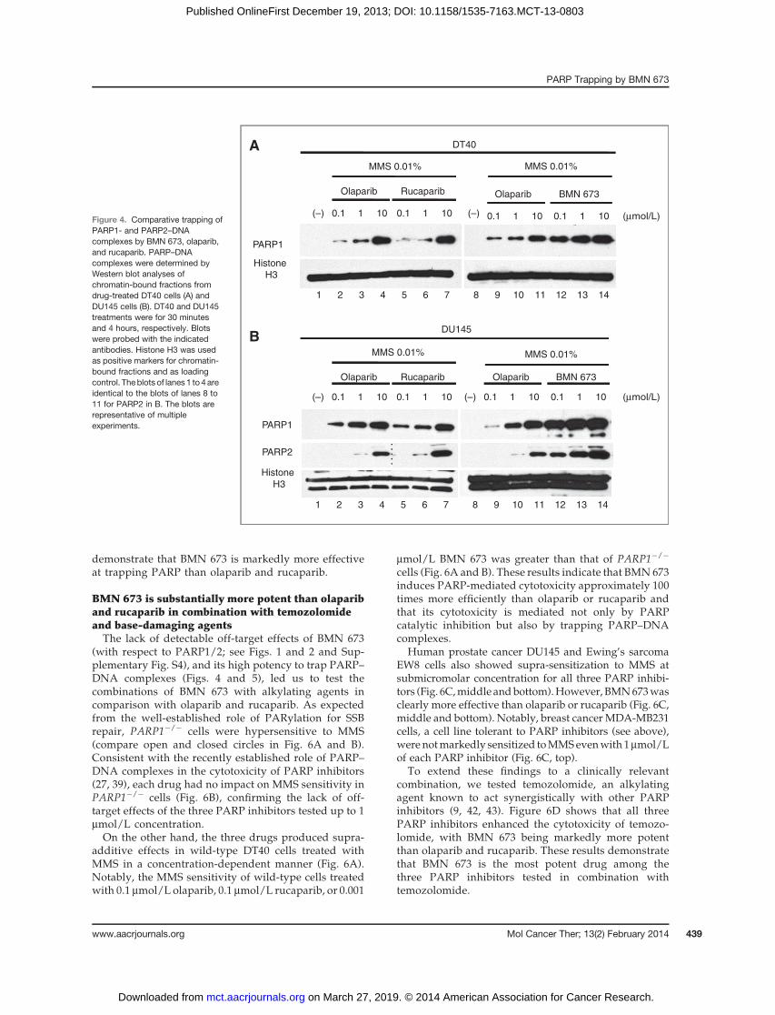
demonstrate that BMN 673 is markedly more effectiveat trapping PARP than olaparib and rucaparib.
BMN 673 is substantially more potent than olapariband rucaparib in combination with temozolomideand base-damaging agentsThe lack of detectable off-target effects of BMN 673
(with respect to PARP1/2; see Figs. 1 and 2 and Sup-plementary Fig. S4), and its high potency to trap PARP–DNA complexes (Figs. 4 and 5), led us to test thecombinations of BMN 673 with alkylating agents incomparison with olaparib and rucaparib. As expectedfrom the well-established role of PARylation for SSBrepair, PARP1�/� cells were hypersensitive to MMS(compare open and closed circles in Fig. 6A and B).Consistent with the recently established role of PARP–DNA complexes in the cytotoxicity of PARP inhibitors(27, 39), each drug had no impact on MMS sensitivity inPARP1�/� cells (Fig. 6B), confirming the lack of off-target effects of the three PARP inhibitors tested up to 1mmol/L concentration.On the other hand, the three drugs produced supra-
additive effects in wild-type DT40 cells treated withMMS in a concentration-dependent manner (Fig. 6A).Notably, the MMS sensitivity of wild-type cells treatedwith 0.1 mmol/L olaparib, 0.1 mmol/L rucaparib, or 0.001
mmol/L BMN 673 was greater than that of PARP1�/�
cells (Fig. 6A and B). These results indicate that BMN 673induces PARP-mediated cytotoxicity approximately 100times more efficiently than olaparib or rucaparib andthat its cytotoxicity is mediated not only by PARPcatalytic inhibition but also by trapping PARP–DNAcomplexes.
Human prostate cancer DU145 and Ewing’s sarcomaEW8 cells also showed supra-sensitization to MMS atsubmicromolar concentration for all three PARP inhibi-tors (Fig. 6C,middle andbottom).However, BMN673wasclearly more effective than olaparib or rucaparib (Fig. 6C,middle and bottom). Notably, breast cancer MDA-MB231cells, a cell line tolerant to PARP inhibitors (see above),were notmarkedly sensitized toMMSevenwith 1mmol/Lof each PARP inhibitor (Fig. 6C, top).
To extend these findings to a clinically relevantcombination, we tested temozolomide, an alkylatingagent known to act synergistically with other PARPinhibitors (9, 42, 43). Figure 6D shows that all threePARP inhibitors enhanced the cytotoxicity of temozo-lomide, with BMN 673 being markedly more potentthan olaparib and rucaparib. These results demonstratethat BMN 673 is the most potent drug among thethree PARP inhibitors tested in combination withtemozolomide.
DT40
(–)
MMS 0.01%
1 100.1
Olaparib Rucaparib
1 100.1
PARP1
Histone
H3
DU145
(–)
MMS 0.01%
1 100.1
Olaparib Rucaparib
1 100.1 1 100.1
BMN 673
(mmol/L)
PARP2
PARP1
Histone
H3
1 100.1
Olaparib
1 100.1
BMN 673
(mmol/L)(–)
MMS 0.01%
(–) 1 100.1
Olaparib
MMS 0.01%
A
B
1 2 3 4 5 6 7 8 9 10 11 12 13 14
1 2 3 4 5 6 7 8 9 10 11 12 13 14
Figure 4. Comparative trapping ofPARP1- and PARP2–DNAcomplexes by BMN 673, olaparib,and rucaparib. PARP–DNAcomplexes were determined byWestern blot analyses ofchromatin-bound fractions fromdrug-treated DT40 cells (A) andDU145 cells (B). DT40 and DU145treatments were for 30 minutesand 4 hours, respectively. Blotswere probed with the indicatedantibodies. Histone H3 was usedas positive markers for chromatin-bound fractions and as loadingcontrol. The blots of lanes 1 to 4areidentical to the blots of lanes 8 to11 for PARP2 in B. The blots arerepresentative of multipleexperiments.
PARP Trapping by BMN 673
www.aacrjournals.org Mol Cancer Ther; 13(2) February 2014 439
on March 27, 2019. © 2014 American Association for Cancer Research. mct.aacrjournals.org Downloaded from
Published OnlineFirst December 19, 2013; DOI: 10.1158/1535-7163.MCT-13-0803

DiscussionIn this study, we report that BMN 673 is the most
potent PARP-trapping drug tested to date. It is approx-imately 2 orders of magnitude more potent than ola-parib both in prostate cancer DU145 and lymphomaDT40 cells for both PARP1 and PARP2 (see Fig. 4). Wealso show that olaparib and rucaparib trap PARP1 andPARP2 with comparable efficiency. The present resultscomplement our recent study (27) revealing PARP–DNA complex trapping, and comparing olaparib withniraparib and veliparib. Veliparib differed from the twoother drugs by its much weaker ability to trap PARP–DNA complexes despite its remarkable activity as aPARP catalytic inhibitor (27).
Our data indicate that BMN 673 is only slightly morepotent than olaparib and rucaparib at inhibiting PARPcatalytic activity. The differential potencies of the drugs attrapping PARP versus inhibiting PARP catalytic activitymay possibly be interpreted as resulting from an allostericeffect of the drugs (27). As shown in Fig. 1A, the chemicalstructure of BMN 673 is rigid, whereas olaparib andrucaparib are flexible. This might explain their weakerimpact on PARP trapping. The binding of PARP inhibitorto the catalytic pocket of PARP1 and PARP2may enhancethebindingbetweenDNAand theDNA-bindingdomainsof PARP, which would be the converse allosteric effectproduced by the binding of PARP toDNA,which inducesconformational distortions that stimulate the catalyticdomain (44). Notably, LT674, the inactive enantiomer ofBMN 673, is markedly less active than BMN 673 both atPARP-mediated cytotoxicity and at inhibiting its catalyticactivity. This difference between the enantiomers reflects
the optimal structure of BMN 673 for PARP binding andthe inability of LT674 to fit in the nicotinamide-bindingpocket (45). We believe that BMN 673 can now be viewednot only as a valuable anticancer agent but also as amolecular tool to elucidate PARP allosteric regulation.For the comprehensive understanding of the mechanismof differential PARP trapping, further studies such as co-crystal structure analysis will be required.
The nanomolar cytotoxicity of BMN 673 is notablygreater than that of rucaparib or olaparib (�10-fold inlymphomaDT40 and prostate cancer DU145 and� 5-foldin Ewing’s sarcoma EW8 cells; see Fig. 2). The potency ofBMN 673 as a cytotoxic agent was just reported indepen-dently (46). However, these studies did not examine themolecular mechanism of action of BMN 673, especiallywith respect to PARP trapping. The potency of BMN 673observed across the NCI60 cell line panel (see Fig. 3)showed significant correlation between BMN 673 andolaparib (Table 2). The greater cytotoxic potency of BMN673 over olaparib and rucaparib can be related to thetrapping of PARP–DNA complexes because knocking outPARP1 in lymphoma DT40 cells, which by itself is well-tolerated despite the fact that DT40 cells also lack PARP2(29), conferred extreme resistance to BMN 673 (see Fig. 2).Moreover, the greater cytotoxicity of BMN 673 comparedwith olaparib is correlated with the greater potency ofBMN 673 at trapping PARP (see Figs. 4 and 5), while bothdrugs are equally effective at inhibiting PARP catalyticactivity (see Fig. 1). The BMN 673 findings reinforce ourproposal (27) that PARP inhibitors should be categorizedand evaluated on the basis of both PARP inhibition andPARP trapping.
A
Drug concentration (µmol/L)
FA
(m
A)
Time (min)
No NAD+
Olaparib 0.1 µmol/L
BMN 673 0.1 µmol/L
No drug
B C
BMN 673
0 50 100 150 200100
120
140
160
BMN 673
0.01 0.1 1 10100
120
140
160
Olaparib
Time = 0
Figure 5. Biochemical trapping of PARP1 by BMN 673. A, scheme of the fluorescence anisotropy (FA) binding assay. The star indicates the site labeled onthe DNA substrate with Alexa Fluor 488. Unbound nicked DNA substrate rotates fast and gives low fluorescence anisotropy. PARP1 binding to thesubstrate slows the rotation and gives high fluorescence anisotropy. Addition of NADþ leads to PARP1 dissociation from DNA due to autoPARylation.B, concentration-dependent PARP1–DNA association in the presence of BMN 673 or olaparib. Fluorescence anisotropy was measured 40 minutesafter addingNADþ. C, time-course of PARP1–DNAdissociation in the presence of BMN673 and olaparib (0.12mmol/L each). Addition of NADþ in the absenceof PARP inhibitor immediately reduces PARP1–DNA complexes (no drug, DMSO control). In the absence of NADþ, PARP1–DNA complexes remain stable forat least 180 minutes (no NADþ). Data are mean � SEM (n ¼ 3).
Murai et al.
Mol Cancer Ther; 13(2) February 2014 Molecular Cancer Therapeutics440
on March 27, 2019. © 2014 American Association for Cancer Research. mct.aacrjournals.org Downloaded from
Published OnlineFirst December 19, 2013; DOI: 10.1158/1535-7163.MCT-13-0803

Our study shows that rucaparib exhibits off-targeteffect with respect to PARP1 and PARP2 (Fig. 2C),which fits with a previous report showing that ruca-parib has more promiscuous inhibitory activity (extend-ing to PARP1-4 and tankyrases) than olaparib (specificto PARP1-4; ref. 47). The NCI60 data also revealed thedifferences between rucaparib and BMN 673 or ola-parib, and the general cytotoxicity of rucaparib irre-spective of cell lines and tissue origin (Fig. 3). Wespeculate that the inhibition of tankyrases may contrib-ute to the broader cytotoxicity of rucaparib as tankyr-ase-1 RNA interference (RNAi) results in mitotic arrest(48).Our results provide relevant information for the clinical
use of PARP inhibitors. As single agent in BRCA-deficientcells, we found that BMN 673 demonstrates �30-fold
greater potency in isogenic BRCA2-deficient lymphomaDT40 cells (see Fig. 2). Consistent results have just beenreported independently using other cellular systems (45).BMN673 is also significantlymore potent than olaparib incombination with temozolomide or MMS (see Fig. 6;ref. 45), which is consistent with the enhanced trappingof PARPbyBMN673 andolaparib in thepresence ofMMS(see Fig. 4). Despite the fact that BMN 673 is a highlypotent drug by inducing PARP–DNA complexes, it issurprising that half of the NCI60 cell lines are resistanteven at 100 mmol/L BMN 673 (see Fig. 3). Further studiesarewarranted to elucidatewhy some cell lines are tolerantor selectively sensitive to PARP trapping. One possibilityis that sensitive cell lines are deficient in postreplicationrepair, Fanconi anemia pathway, ATM, homologousrecombination (27), or PTEN (49). It will also be important
A
0 0.0002 0.0004 0.0006 0.0008 0.0010
1
10
100
MMS (%)
MMS (%)
MMS (%)
DT40 wild-type
+ Olaparib (mmol/L)
0
0.001
0.010.1
1
0
0.001
0.010.1
1
Via
bili
ty (
%)
Via
bili
ty (
%)
Via
bili
ty (
%)
DT40 wild-type
+ Rucaparib (mmol/L)
DT40 wild-type
+ BMN 673 (mmol/L)
0.0002 0.0004 0.0006 0.0008 0.0010
1
10
100
0
0.0010.01
DT40 PARP1-/-
No drug (0)Olaparib 1 mmol/LRucaparib 1 mmol/LBMN 673 1 mmol/L
Via
bili
ty (
%)
MMS (%)
0
0.0002 0.0004 0.0006 0.0008 0.0010
1
10
100
0
C
Via
bili
ty (
%)
DU145
No drug
Olaparib 0.1 mmol/L
Rucaparib 0.1 mmol/L
BMN 673 0.1 mmol/L
MMS (%)
Via
bili
ty (
%)
EW8
No drug
Olaparib 0.1 mmol/L
Rucaparib 0.1 mmol/L
BMN 673 0.1 mmol/L
MMS (%)0
Temozolomide (mmol/L)
No drug
Olaparib 0.1 mmol/L
Rucaparib 0.1 mmol/L
BMN 673 0.1 mmol/L
DU145D
Via
bili
ty (
%)
0 0.0002 0.0004 0.0006 0.0008 0.0010
1
10
100
0.0001 0.0002 0.0003
1
10
100
0.0001 0.0002 0.0003
10
100
0
DU145
EW8
DU145
0 100 200 300 40010
100
0.0001 0.0002 0.000310
100
MDA-MB231
MMS (%)0
Via
bili
ty (
%)
No drugOlaparib 1 mmol/LRucaparib 1 mmol/L
BMN 673 1 mmol/L
B
Figure 6. BMN 673 enhances thecytotoxicity of alkylating agentsmore efficiently than olaparib andrucaparib. A, survival curves ofwild-type DT40 cells treated withMMS alone (upper curves labeled"0") or with the indicatedconcentrations of PARP inhibitors(at the concentration shownbeside each curve in micromolarunits). The viability of untreatedcells was set as 100%. Data aremean � SD (n � 3). B, PARP1�/�
cells are hypersensitive to MMS(compare with upper curves in A)and resistant to the PARPinhibitors. C, viability curves of theindicated human cancer cellstreated with MMS in combinationwith the indicated PARP inhibitors(the concentration of each PARPinhibitor is shown beside eachcurve). The viability of untreatedcells was set as 100%. Data aremean � SD (n � 3). D, same as Cbut using temozolomide instead ofMMS in prostate cancer DU145cells.
PARP Trapping by BMN 673
www.aacrjournals.org Mol Cancer Ther; 13(2) February 2014 441
on March 27, 2019. © 2014 American Association for Cancer Research. mct.aacrjournals.org Downloaded from
Published OnlineFirst December 19, 2013; DOI: 10.1158/1535-7163.MCT-13-0803

to determine whether the resistant cells exhibit prefer-ential homologous recombination by 53BP1 inactivation(50, 51).
Disclosure of Potential Conflicts of InterestNo potential conflicts of interest were disclosed.
Authors' ContributionsConception and design: J. Murai, S. Takeda, Y. PommierDevelopment of methodology: J. Murai, S.-Y.N. Huang, S. Takeda,Y. PommierAcquisition of data (provided animals, acquired and managed patients,provided facilities, etc.): J. Murai, S.-Y.N. Huang, A. Renaud, J. Morris,J.H. Doroshow, Y. PommierAnalysis and interpretation of data (e.g., statistical analysis, biostatis-tics, computational analysis): J. Murai, S.-Y.N. Huang, Y. Zhang, J. Ji,Y. PommierWriting, review, and/or revision of the manuscript: J. Murai, Y. Zhang,J. Ji, B. Teicher, J.H. Doroshow, Y. PommierAdministrative, technical, or material support (i.e., reporting or orga-nizing data, constructing databases): Y. Zhang, Y. PommierStudy supervision: J. Ji, Y. Pommier
AcknowledgmentsThe authors thank the Intramural Program of the National Cancer
Institute, Center for Cancer Research (CCR), by the Developmental Ther-apeutics Program (DTP), Division of Cancer Treatment and Diagnosis(DCTD) for sharing the NCI60 data and drugs. The authors also thankCRADA with BioMarin Pharmaceuticals Inc. for providing BMN 673 andLT674 compounds.
Grant SupportJ. Murai was a recipient of fellowships from the John Mung Program
(KyotoUniversity) and the Kyoto University Foundation. Y. Pommier andJ.Muraiwere supported by the Intramural Programof theNationalCancerInstitute, Center for Cancer Research (Z01 BC 006150). J. Murai andS. Takeda were supported by JSPS Core-to-Core Program. J. Murai wassupported by JSPSKAKENHIGrantNumber 25740016. J. Jiwas supportedby the federal fund from the National Cancer Institute, National Institutesof Health (Contract No. HHSN261200800001E).
Thecostsofpublicationof this articleweredefrayed inpartby thepaymentof page charges. This article must therefore be hereby marked advertisementin accordance with 18 U.S.C. Section 1734 solely to indicate this fact.
Received September 24, 2013; revised December 2, 2013; acceptedDecember 8, 2013; published OnlineFirst December 19, 2013.
References1. Schreiber V, Dantzer F, Ame JC, deMurcia G. Poly(ADP-ribose): novel
functions for an old molecule. Nat Rev Mol Cell Biol 2006;7:517–28.2. Hassa PO, Hottiger MO. The diverse biological roles of mammalian
PARPS, a small but powerful family of poly-ADP-ribose polymerases.Front Biosci 2008;13:3046–82.
3. Krishnakumar R, Kraus WL. The PARP side of the nucleus: molecularactions, physiological outcomes, and clinical targets. Mol Cell 2010;39:8–24.
4. Rouleau M, Patel A, Hendzel MJ, Kaufmann SH, Poirier GG. PARPinhibition: PARP1 and beyond. Nat Rev Cancer 2010;10:293–301.
5. Banerjee S, Kaye SB, Ashworth A. Making the best of PARP inhibitorsin ovarian cancer. Nat Rev Clin Oncol 2010;7:508–19.
6. Helleday T, Petermann E, Lundin C, Hodgson B, Sharma RA. DNArepair pathways as targets for cancer therapy. Nat Rev Cancer 2008;8:193–204.
7. Lord CJ, Ashworth A. The DNA damage response and cancer therapy.Nature 2012;481:287–94.
8. Zhang YW, Regairaz M, Seiler JA, Agama KK, Doroshow JH, PommierY. Poly(ADP-ribose) polymerase and XPF-ERCC1 participate in dis-tinct pathways for the repair of topoisomerase I-induced DNA damagein mammalian cells. Nucleic Acids Res 2011;39:3607–20.
9. Delaney CA, Wang LZ, Kyle S, White AW, Calvert AH, Curtin NJ, et al.Potentiation of temozolomide and topotecan growth inhibition andcytotoxicity by novel poly(adenosine diphosphoribose) polymeraseinhibitors in a panel of human tumor cell lines. Clin Cancer Res2000;6:2860–7.
10. Ray Chaudhuri A, Hashimoto Y, Herrador R, Neelsen KJ, Fachinetti D,Bermejo R, et al. Topoisomerase I poisoning results in PARP-mediatedreplication fork reversal. Nat Struct Mol Biol 2012;19:417–23.
11. Berti M, Chaudhuri AR, Thangavel S, Gomathinayagam S, Kenig S,VujanovicM, et al. HumanRECQ1 promotes restart of replication forksreversed by DNA topoisomerase I inhibition. Nat Struct Mol Biol2013;20:347–54.
12. Lindahl T, Wood RD. Quality control by DNA repair. Science 1999;286:1897–905.
13. Hoeijmakers JH. DNA damage, aging, and cancer. N Engl J Med2009;361:1475–85.
14. Strumberg D, Pilon AA, Smith M, Hickey R, Malkas L, Pommier Y.Conversion of topoisomerase I cleavage complexes on the leadingstrand of ribosomal DNA into 50-phosphorylated DNA double-strandbreaks by replication runoff. Mol Cell Biol 2000;20:3977–87.
15. Helleday T. The underlying mechanism for the PARP and BRCAsynthetic lethality: clearing up the misunderstandings. Mol Oncol2011;5:387–93.
16. Noel G, Godon C, Fernet M, Giocanti N, Megnin-Chanet F, FavaudonV. Radiosensitization by the poly(ADP-ribose) polymerase inhibitor 4-amino-1,8-naphthalimide is specificof theSphaseof the cell cycle andinvolves arrest of DNA synthesis. Mol Cancer Ther 2006;5:564–74.
17. Saleh-Gohari N, BryantHE, Schultz N, Parker KM,Cassel TN,HelledayT. Spontaneous homologous recombination is induced by collapsedreplication forks that are caused by endogenous DNA single-strandbreaks. Mol Cell Biol 2005;25:7158–69.
18. Bryant HE, Schultz N, ThomasHD, Parker KM, Flower D, Lopez E, et al.Specific killing of BRCA2-deficient tumours with inhibitors of poly(ADP-ribose) polymerase. Nature 2005;434:913–7.
19. Farmer H, McCabe N, Lord CJ, Tutt AN, Johnson DA, Richardson TB,et al. Targeting the DNA repair defect in BRCA mutant cells as atherapeutic strategy. Nature 2005;434:917–21.
20. Fong PC, Boss DS, Yap TA, Tutt A, Wu P, Mergui-Roelvink M, et al.Inhibition of poly(ADP-ribose) polymerase in tumors from BRCAmuta-tion carriers. N Engl J Med 2009;361:123–34.
21. Curtin NJ, Szabo C. Therapeutic applications of PARP inhibitors:anticancer therapy and beyond. Mol Aspects Med 2013;34:1217–56
22. McCabe N, Turner NC, Lord CJ, Kluzek K, Bialkowska A, Swift S, et al.Deficiency in the repair of DNA damage by homologous recombinationand sensitivity to poly(ADP-ribose) polymerase inhibition. Cancer Res2006;66:8109–15.
23. Ashworth A. A synthetic lethal therapeutic approach: poly(ADP) ribosepolymerase inhibitors for the treatment of cancers deficient in DNAdouble-strand break repair. J Clin Oncol 2008;26:3785–90.
24. Kummar S, Chen A, Parchment RE, Kinders RJ, Ji J, Tomaszewski JE,et al. Advances in using PARP inhibitors to treat cancer. BMC Med2012;10:25.
25. Chuang HC, Kapuriya N, Kulp SK, Chen CS, Shapiro CL. Differentialanti-proliferative activities of poly(ADP-ribose) polymerase (PARP)inhibitors in triple-negative breast cancer cells. Breast Cancer ResTreat 2012;134:649–59.
26. Patel AG, Flatten KS, Schneider PA, Dai NT, McDonald JS, Poirier GG,et al. Enhanced killing of cancer cells by poly(ADP-ribose) polymeraseinhibitors and topoisomerase I inhibitors reflects poisoning of bothenzymes. J Biol Chem 2012;287:4198–210.
27. Murai J, Huang SY, Das BB, Renaud A, Zhang Y, Doroshow JH, et al.TrappingofPARP1andPARP2byclinical PARP inhibitors.CancerRes2012;72:5588–99.
28. SatohMS, Lindahl T. Role of poly(ADP-ribose) formation inDNA repair.Nature 1992;356:356–8.
29. Hochegger H, Dejsuphong D, Fukushima T, Morrison C, Sonoda E,Schreiber V, et al. Parp-1 protects homologous recombination from
Murai et al.
Mol Cancer Ther; 13(2) February 2014 Molecular Cancer Therapeutics442
on March 27, 2019. © 2014 American Association for Cancer Research. mct.aacrjournals.org Downloaded from
Published OnlineFirst December 19, 2013; DOI: 10.1158/1535-7163.MCT-13-0803

interference by Ku and Ligase IV in vertebrate cells. EMBO J 2006;25:1305–14.
30. Garnett MJ, Edelman EJ, Heidorn SJ, Greenman CD, Dastur A, LauKW, et al. Systematic identification of genomic markers of drugsensitivity in cancer cells. Nature 2012;483:570–5.
31. Ji J, Kinders RJ, Zhang Y, Rubinstein L, Kummar S, Parchment RE,et al. Modeling pharmacodynamic response to the poly(ADP-Ribose)polymerase inhibitor ABT-888 in humanperipheral bloodmononuclearcells. PLoS ONE 2011;6:e26152.
32. Division of Cancer Treatment and Diagnosis [Internet]. Bethesda, MD:National Cancer Institute, NIH (US); [updated 2013 Aug 16; cited 2013Oct 10]. Available from: http://dctd.cancer.gov/ResearchResources/biomarkers/PolyAdenosylRibose.htm.
33. NCI-60 DTP Human Tumor Cell Line Screen [Internet]. Bethesda, MD:National Cancer Institute, NIH (US); [cited 2013Oct 10]. Available from:http://dtp.nci.nih.gov/branches/btb/ivclsp.html.
34. Holbeck SL, Collins JM, Doroshow JH. Analysis of Food andDrug Administration–approved anticancer agents in the NCI60panel of human tumor cell lines. Mol Cancer Ther 2010;9:1451–60.
35. Holbeck S, Chang J, Best AM, Bookout AL, Mangelsdorf DJ, MartinezED. Expression profiling of nuclear receptors in the NCI60 cancer cellpanel reveals receptor-drug and receptor-gene interactions. MolEndocrinol 2010;24:1287–96.
36. Kummar S, Chen HX, Wright J, Holbeck S, Millin MD, Tomaszewski J,et al.Utilizing targeted cancer therapeutic agents in combination: novelapproaches and urgent requirements. Nat Rev Drug Discov 2010;9:843–56.
37. Kummar S, Kinders R, Gutierrez ME, Rubinstein L, Parchment RE,Phillips LR, et al. Phase 0 clinical trial of the poly (ADP-ribose) poly-merase inhibitor ABT-888 in patients with advanced malignancies.J Clin Oncol 2009;27:2705–11.
38. Hatanaka A, Yamazoe M, Sale JE, Takata M, Yamamoto K, Kitao H,et al. Similar effects of Brca2 truncation and Rad51 paralog deficiencyon immunoglobulin V gene diversification in DT40 cells support anearly role for Rad51 paralogs in homologous recombination. Mol CellBiol 2005;25:1124–34.
39. Pettitt SJ, Rehman FL, Bajrami I, Brough R, Wallberg F, Kozarewa I,et al. A genetic screen using the PiggyBac transposon in haploid cellsidentifies Parp1 as a mediator of olaparib toxicity. PLoS ONE 2013;8:e61520.
40. Grohar PJ, Griffin LB, Yeung C, Chen QR, Pommier Y, Khanna C, et al.Ecteinascidin 743 interferes with the activity of EWS-FLI1 in Ewingsarcoma cells. Neoplasia 2011;13:145–53.
41. Reinhold WC, Sunshine M, Liu H, Varma S, Kohn KW, Morris J, et al.CellMiner: a web-based suite of genomic and pharmacologic tools toexplore transcript and drug patterns in the nci-60 cell line set. CancerRes 2012;72:3499–511.
42. Boulton S, Pemberton LC, Porteous JK, Curtin NJ, Griffin RJ, GoldingBT, et al. Potentiation of temozolomide-induced cytotoxicity: a com-parative study of the biological effects of poly(ADP-ribose) polymeraseinhibitors. Br J Cancer 1995;72:849–56.
43. Plummer R, Jones C, Middleton M, Wilson R, Evans J, Olsen A, et al.Phase I study of the poly(ADP-ribose) polymerase inhibitor,AG014699, in combination with temozolomide in patients withadvanced solid tumors. Clin Cancer Res 2008;14:7917–23.
44. Langelier M-F, Planck JL, Roy S, Pascal JM. Structural basis forDNA damage-dependent poly(ADP-ribosyl)ation by human PARP-1.Science 2012;336:728–32.
45. Shen Y, Rehman FL, Feng Y, Boshuizen J, Bajrami I, Elliott R, et al.BMN673, anovel andhighlypotentPARP1/2 inhibitor for the treatmentof human cancers with DNA repair deficiency. Clin Cancer Re 2013;19:5003–15.
46. Postel-Vinay S, Bajrami I, Friboulet L, Elliott R, Fontebasso Y, DorvaultN, et al. A high-throughput screen identifies PARP1/2 inhibitors as apotential therapy for ERCC1-deficient non–small cell lung cancer.Oncogene 2013;32:5377–87
47. Wahlberg E, Karlberg T, Kouznetsova E, Markova N, Macchiarulo A,Thorsell AG, et al. Family-wide chemical profiling and structural anal-ysis of PARP and tankyrase inhibitors. Nat Biotechnol 2012;30:283–8.
48. Chang P, Coughlin M, Mitchison TJ. Tankyrase-1 polymerization ofpoly(ADP-ribose) is required for spindle structure and function. NatCellBiol 2005;7:1133–9.
49. Mendes-Pereira AM,Martin SA, BroughR,McCarthy A, Taylor JR, KimJ-S, et al. Synthetic lethal targeting of PTEN mutant cells with PARPinhibitors. Embo Mol Med 2009;1:315–22.
50. Bunting SF,Callen E,WongN,ChenHT, Polato F, GunnA, et al. 53BP1inhibits homologous recombination in Brca1-deficient cells by block-ing resection of DNA breaks. Cell 2010;141:243–54.
51. Jaspers JE, Kersbergen A, Boon U, Sol W, van Deemter L, Zander SA,et al. Loss of 53BP1 causes PARP inhibitor resistance in Brca1-mutated mouse mammary tumors. Cancer Discov 2013;3:68–81.
PARP Trapping by BMN 673
www.aacrjournals.org Mol Cancer Ther; 13(2) February 2014 443
on March 27, 2019. © 2014 American Association for Cancer Research. mct.aacrjournals.org Downloaded from
Published OnlineFirst December 19, 2013; DOI: 10.1158/1535-7163.MCT-13-0803

2014;13:433-443. Published OnlineFirst December 19, 2013.Mol Cancer Ther Junko Murai, Shar-Yin N. Huang, Amèlie Renaud, et al. Olaparib and RucaparibStereospecific PARP Trapping by BMN 673 and Comparison with
Updated version
10.1158/1535-7163.MCT-13-0803doi:
Access the most recent version of this article at:
Material
Supplementary
http://mct.aacrjournals.org/content/suppl/2013/12/19/1535-7163.MCT-13-0803.DC1
Access the most recent supplemental material at:
Cited articles
http://mct.aacrjournals.org/content/13/2/433.full#ref-list-1
This article cites 49 articles, 17 of which you can access for free at:
Citing articles
http://mct.aacrjournals.org/content/13/2/433.full#related-urls
This article has been cited by 35 HighWire-hosted articles. Access the articles at:
E-mail alerts related to this article or journal.Sign up to receive free email-alerts
Subscriptions
Reprints and
To order reprints of this article or to subscribe to the journal, contact the AACR Publications Department at
Permissions
Rightslink site. Click on "Request Permissions" which will take you to the Copyright Clearance Center's (CCC)
.http://mct.aacrjournals.org/content/13/2/433To request permission to re-use all or part of this article, use this link
on March 27, 2019. © 2014 American Association for Cancer Research. mct.aacrjournals.org Downloaded from
Published OnlineFirst December 19, 2013; DOI: 10.1158/1535-7163.MCT-13-0803