steven j. lester md, facc, frcp(c), fase mayo clinic,...
TRANSCRIPT

5/7/2018
1
Steven J. Lester MD, FACC, FRCP(C), FASE
Mayo Clinic, Arizona
Right Ventricle
a. When there is pulmonary valve stenosis.
b. When there is severe tricuspid regurgitation
c. When there is marked dilation of the right ventricle
d. When you are unable to align the continuous wave Doppler signal parallel to the intercept angle of the axis of flow.
1. In which scenario will applying the simplified Bernoulli equation to the peak
tricuspid regurgitation velocity and adding an estimate of right atrial pressure
likely not result in an under estimation the right ventricular systolic pressure.
5/7/2018
2
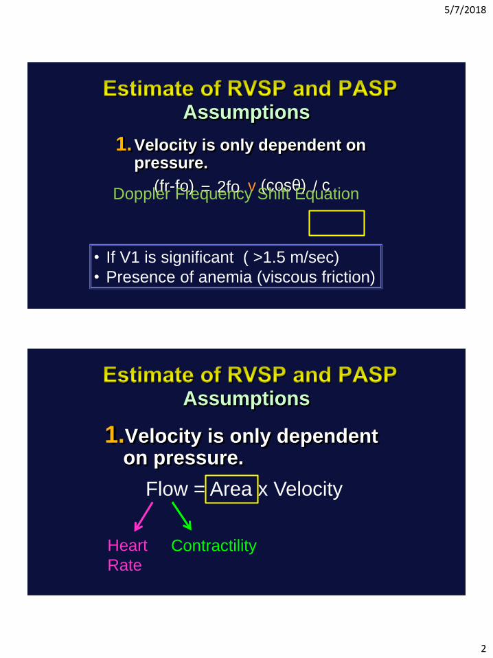
Assumptions
1.Velocity is only dependent on pressure.
(fr-fo) = 2fo v (cosθ) / cDoppler Frequency Shift Equation
• If V1 is significant ( >1.5 m/sec)
• Presence of anemia (viscous friction)
1.Velocity is only dependent on pressure.
Flow = Area x Velocity
Heart
Rate
Contractility
Assumptions

5/7/2018
3
Velocity =Flow
Area
1.Velocity is only dependent on pressure.
2.You can accurately estimate right atrial pressure.
- Non simultaneous
- Peak systole
Assumptions

5/7/2018
4
RA
R
V
Systolic RV → RA pressure equalization

5/7/2018
5
1.Velocity is only dependent on pressure.
2.You can accurately estimate right atrial pressure.
3.Right ventricular systolic pressure = Pulmonary artery systolic pressure
Assumptions
1 = 2.64 cm
2 = 1.96 cm
1 2
TRV = 4.8 m/sec
RVSP = 4 X TRvmax2 + estimated RA pressure
= 4 (4.8)2 + 13 = 105 mmHg

5/7/2018
6
RV-PA Gradient = 36 mmHg
3
SPAP = RVSP – PV gradient = 105 – 36 = 69 mmHg
a) The RVEF would be the same in both the older women and younger men.
b) The RVEF would be higher in older women compared to younger men
c) The RVEF would be lower in older women compared to younger men.
2. Women have smaller right ventricular end-diastolic volumes when
compared to men. Also older age is associated with smaller volumes.
Therefore, you would expect the right ventricular ejection fraction (RVEF) of
older women compared to younger men to be?

5/7/2018
7
a. Higher than prior to treatment with diuretics
b. Lower than prior to treatment with diuretics
c. Unchanged
3. A 47 year old women has a dilated right ventricle with reduced right
ventricular function along with signs and symptoms of right heart failure. The
calculated right ventricular index of myocardial performance (RIMP)
determined by pulsed wave Doppler was 0.64. She was treated with diuretics
and clinically improved as her right atrial pressure decreased. You now re-
calculate the RIMP and you would expect the value to?
arterial
ventricular
atrial
5/7/2018
8
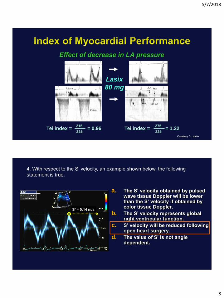
Courtesy Dr. Hatle
Lasix
80 mg
Ao
Ac Mo
Tei index = = 0.96215
225Tei index = = 1.22
275
225
Effect of decrease in LA pressure
a. The S’ velocity obtained by pulsed wave tissue Doppler will be lower than the S’ velocity if obtained by color tissue Doppler.
b. The S’ velocity represents global right ventricular function.
c. S’ velocity will be reduced following open heart surgery.
d. The value of S’ is not angle dependent.
4. With respect to the S’ velocity, an example shown below, the following
statement is true.
S’ = 0.14 m/s
5/7/2018
9
a. 3D methods of calculating RV volumes and ejection fraction are less load dependent than 2D methods.
b. 3D methods of calculating RV volumes and ejection fraction are less dependent on image quality then 2D methods.
c. 3D but not 2D methods with which to calculate RV volumes and ejection fraction correlate with values obtained by MRI.
d. 3D methods include the RV outflow tract contribution to overall function while 2D methods do not.
5. 3D methods with which to calculate right ventricular (RV) volumes and
ejection fraction may be superior to 2D methods for the following reason.