stimulation of subterritories of the subthalamic nucleus … · stimulation of subterritories of...
TRANSCRIPT

Stimulation of subterritories of the subthalamicnucleus reveals its role in the integration of theemotional and motor aspects of behaviorLuc Mallet*†‡, Michael Schupbach†, Karim N�Diaye§, Philippe Remy¶, Eric Bardinet§, Virginie Czernecki�,Marie-Laure Welter†, Antoine Pelissolo**, Merle Ruberg††, Yves Agid†,††, and Jerome Yelnik††
*Institut National de la Sante et de la Recherche Medicale AVENIR Group, Behavior, Emotion, and Basal Ganglia, 75013 Paris, France; †Centre d’InvestigationClinique, Centre Hospitalier Universitaire Pitie-Salpetriere, 75013 Paris, France; §Laboratoire de Neurosciences Cognitive & Imagerie Cerebrale, CentreNational de la Recherche Scientifique–Unite Propre de Recherche 640-LENA, 47 Boulevard de L’hopital, 75651 Paris Cedex 13, France; ¶Unite de RechercheAssociee, Centre National de la Recherche Scientifique-Commissariat a l’Energie Atomique 2210, Orsay et Departement de Neurosciences, Centre HospitalierUniversitaire Henri Mondor et Faculte de Medecine Paris 12, 94000 Creteil, France; �Neuro-Anatomie Fonctionnelle du Comportement et de Ses Troubles,Institut National de la Sante et de la Recherche Medicale-U610, 75013 Paris, France; **Service de Psychiatrie, Assistance Publique–Hopitaux de Paris, GroupePitie-Salpetriere, 75013 Paris, France; and ††Neurologie et Therapeutique Experimentale, Institut National de la Sante et de la Recherche Medicale-U679,Universite Pierre et Marie Curie–Paris 6, Institut Federatif de Recherche de Neurosciences Unite Mixte de Recherche S679, F-75013 Paris, France
Edited by Marcus E. Raichle, Washington University School of Medicine, St. Louis, MO, and approved May 8, 2007 (received for review December 7, 2006)
Two parkinsonian patients who experienced transient hypomanicstates when the subthalamic nucleus (STN) was stimulated duringpostoperative adjustment of the electrical parameters for antipar-kinsonian therapy agreed to have the mood disorder reproduced,in conjunction with motor, cognitive, and behavioral evaluationsand concomitant functional neuroimaging. During the experiment,STN stimulation again induced a hypomanic state concomitantwith activation of cortical and thalamic regions known to processlimbic and associative information. This observation suggests thatthe STN plays a role in the control of a complex behavior thatincludes emotional as well as cognitive and motor components.The localization of the four contacts of the quadripolar electrodewas determined precisely with an interactive brain atlas. Theresults showed that (i) the hypomanic state was caused only bystimulation through one contact localized in the anteromedial STN;(ii) both this contact and the contact immediately dorsal to itimproved the parkinsonian motor state; (iii) the most dorsal andventral contacts, located at the boundaries of the STN, neitherinduced the behavioral disorder nor improved motor performance.Detailed analysis of these data led us to consider a model in whichthe three functional modalities, emotional, cognitive, and motor,are not processed in a segregated manner but can be subtlycombined in the small volume of the STN. This nucleus would thusserve as a nexus that integrates the motor, cognitive, and emo-tional components of behavior and might consequently be aneffective target for the treatment of behavioral disorders thatcombine emotional, cognitive, and motor impairment.
basal ganglia � emotion � deep brain stimulation � neuroimaging
In humans, motor activity consists of complex behavioralsequences that are subtly but strongly influenced by the
cognitive and emotional context in which they are executed.However, how emotions interact with cognition and motoractivity in the brain is still not well known (1). Experimentalstudies in monkeys have identified well individualized corticalareas involved in the processing of motor, cognitive and emo-tional information that is transmitted to separate territories inthe striatum, globus pallidus, and subthalamic nucleus (2, 3),suggesting a segregated model of information processing. Theexistence of a limbic circuit in the basal ganglia of humans hasbeen suggested by the results of studies in volunteers subjectedto emotional stimuli (4) and in patients with psychiatric disordersand focal lesions in the basal ganglia (5), using functional MRIand positron emission tomography (PET) (6, 7). Electricalstimulation of the subthalamic nucleus (STN), used to treatpatients with Parkinson’s disease (8, 9), has recently proved tobe a powerful and accurate tool for testing these functions and
their related circuits (10–12), because the functioning of thestimulated structure can be reversibly altered in a spatially andtemporally controlled manner. Manic episodes have been re-ported after stimulation of the STN (13, 14) or the substantianigra (15, 16). Hypomania induced by high frequency STNstimulation has also been reported (14, 17, 18), but the structurethat was stimulated was not localized with precision. In thisstudy, we used an interactive brain atlas to precisely localize eachcontact of the quadripolar electrodes in the STN of two parkin-sonian patients who had experienced transient hypomanic statesduring postoperative adjustment of the electrical parameters fortherapeutic stimulation of the STN, and who agreed to undergoexperimental STN stimulation to reproduce the mood disorder,in conjunction with motor, cognitive and behavioral evaluationsand concomitant functional neuroimaging.
ResultsPostoperative evaluation of the effects of stimulation througheach of the four contacts (numbered ventrodorsally from 0 to 3)on the implanted electrodes showed that bilateral stimulation(pulse width, 60 �s; frequency, 130 Hz; voltage, 2.1–2.7 V) fromcontact 2 (hereafter called the ‘‘dorsal contact’’) caused maximalimprovement of motor symptoms in both patients [78% and86%; Unified Parkinson’s Disease Rating Scale (UPDRS) PartIII]. Contacts 0 and 3 did not improve motor symptoms in patient1 and provoked aversive effects (dysarthria, diplopia) in patient2. In the absence of any change in medication or voltage,stimulation through contact 1, the ‘‘ventral contact,’’ induced apersistent abnormal expansive mood in both patients, associatedwith clinical features that correspond to hypomania as definedby the Diagnostic and Statistical Manual of Mental Disorders
Author contributions: L.M., M.S., and P.R. designed research; L.M., M.S., P.R., V.C., M.-L.W.,and A.P. performed research; E.B. and J.Y. contributed new reagents/analytic tools; L.M.,M.S., K.N., E.B., V.C., Y.A., and J.Y. analyzed data; and L.M., K.N., M.R., Y.A., and J.Y. wrotethe paper.
The authors declare no conflict of interest.
This article is a PNAS Direct Submission.
Abbreviations: STN, subthalamic nucleus; PET, positron-emission tomography; UPDRS,Unified Parkinson’s Disease Rating Scale; rCBF, regional cerebral blood flow; THVA, ventralanterior nucleus of the thalamus; YMRS, Young Mania Rating Scale.
‡To whom correspondence should be addressed at: Centre d’Investigation Clinique, Sal-petriere Hospital, 47 Boulevard de L’Hopital, 75013 Paris, France. E-mail: [email protected].
This article contains supporting information online at www.pnas.org/cgi/content/full/0610849104/DC1.
© 2007 by The National Academy of Sciences of the USA
www.pnas.org�cgi�doi�10.1073�pnas.0610849104 PNAS � June 19, 2007 � vol. 104 � no. 25 � 10661–10666
NEU
ROSC
IEN
CE

(19). Only a slight worsening of their motor condition wasinduced by stimulation of this ventral contact.
Because stimulation through the dorsal and ventral contacts,which are only 2 mm apart, resulted in completely differentclinical pictures, we undertook to determine the exact anatom-ical localization of each contact, using a method previouslydeveloped in our center (Fig. 1). The placement of the electrodeswithin the STN was assured by intra-operative electrophysiolog-ical recordings to identify the structure. The precise anatomicallocalization of each of the four contacts of each electrode wasdetermined postoperatively by MRI, as described (20, 21). Thepatients’ MRIs were registered by a computerized method to thebrain images of a three-dimensional histological atlas (21) inwhich the three functional territories of the human STN (sen-sorimotor, associative, limbic) were defined by homology withthe same territories in the monkey identified by axonal tracing(22). The ventral and dorsal contacts of the electrodes in bothpatients were found to be localized in the associative territory ofthe STN (Fig. 2a). The ventral contacts, however, bordered onthe limbic territory, whereas the dorsal contacts bordered on themotor territory.
Because the hypomanic state induced by stimulation throughthe ventral contact was reversible, we proposed to determinewhether correlative responses would be observed in brain re-gions associated with this kind of psychic disorder. The patientswere asked to undergo a PET study of regional cerebral blood
flow (rCBF) during stimulation of the dorsal and ventral con-tacts, in parallel with assessments of their motor, cognitive andbehavioral status (Fig. 2b). When stimulation was switched fromthe dorsal (therapeutic) to the ventral contact, a hypomanic statewas again observed, as during the initial evaluation of thestimulation parameters, and was quantified by using the YoungMania Rating Scale (YMRS) (23). The computerized neuropsy-chological assessments [CPT-II (24)] revealed attention impair-ment as reported in mania (25–27). Motor performance, quan-tified by using the Unified Parkinson Disease Rating Scale partIII (UPDRS III) (28), was little affected (Fig. 2c).
During the hypomanic state, rCBF was significantly greaterthan normal (P � 0.00001, uncorrected; cluster size: 100 voxels)in the left thalamus, right middle and inferior temporal gyrus,right inferior parietal gyrus and right inferior frontal gyrus(Table 1, Fig. 3). In both subjects, the subcortical activation peakwas located in the ventral anterior nucleus of the thalamus(THVA) as demonstrated by coregistration of the individualPET and MRI acquisitions with the atlas (Fig. 3). Deactivation
Fig. 1. Method for localizing electrodes implanted in the brain of a patientwith Parkinson’s disease for stimulation of the STN. (a) Postoperative MRI ofa patient showing the electrodes in blue. (b) Identification of the four contacts(blue cylinders) in the electrode MRI artifact. The MRI was reformatted alongthe trajectory of each electrode and a scaled template of the contacts (1.5 �1.27 mm cylinder) was positioned at the center of the artifact (20). (c)Medtronic 3389 electrode. (d and e) The three dimensional histological atlasof the basal ganglia is fitted to the patient MRI as described in ExperimentalProcedures (21). (d) 3D view of caudate, thalamus, and STN after registrationwith the atlas. (e) Sagittal view of the fitted atlas image including sensorimo-tor, associative and limbic territories of caudate and STN. ( f) Electrode con-tacts and STN territories after rotation along STN main axes.
a
b
c
Fig. 2. Motor, cognitive and behavioral status of two parkinsonian patientsundergoing stimulation of the STN through a ventral contact that induceshypomania and a dorsal contact that ameliorates motor performance. (a) 3Dviews of the STN territories and contacts for patients 1 and 2 in sagittal viewswith MRI and after rotation along main STN axes. Therapeutic dorsal contact,yellow; ventral hypomania-inducing contact, red. (b) Schematic representa-tion of the protocol for evaluating cerebral blood flow by PET. Yellow barsrepresent the normal state during dorsal contact stimulation, red bars thehypomanic state induced by ventral contact stimulation. (c) Motor, neuropsy-chological and behavioral scores the day before and during the PET protocol.Motor performance: UPDRS III (28) score indicating the severity of parkinson-ism. Cognitive abilities: CPT-II score of attention (values �11 are indicative ofproblems) (24). Behavior: YMRS score (values �12 are considered pathologi-cal) (23). The baseline score for patient 1 before PET, which was zero, coincideswith the base of the histograms. The CPT-II score returned to baseline valuesa few hours after the second series of PET acquisitions.
10662 � www.pnas.org�cgi�doi�10.1073�pnas.0610849104 Mallet et al.

was observed in left posterior middle temporal and occipitalgyrus, left middle frontal gyrus, bilateral cuneus and rightmedial prefrontal/anterior cingulate gyrus (Table 1).
DiscussionIn this paper we report the case of two parkinsonian patients whoexperienced transient hypomanic states when stimulated froman electrode contact located in the anteromedial STN. Simul-taneously, rCBF was modified in the same cortical areas in bothpatients. We will first discuss the methodological aspects of ourapproach and then the possible physiopathological implicationsof these observations.
Clinical Evaluation. These two fortuitous observations, made dur-ing the standard postoperative phase of electrical tuning, areexperimentally significant because (i) the hypomanic state couldbe reproducibly induced before and during the PET study in eachindividual and (ii) the behavioral picture and PET results werehighly similar in the two patients. Non-motor side effects havealready been observed in patients with STN lesions (29, 30) or
in parkinsonian patients undergoing stimulation of the STN [forreview see (31, 32)] or substantia nigra (15, 16) but the two casesreported here are particularly interesting because they corre-sponded to hypomania not to bipolar mania (13, 14). The fewcases of hypomania that have been reported (17, 33) were notstudied by brain imaging. In this study, we diagnosed thehypomanic state using the DSM-IV criteria, and quantified thesymptoms using a standardized scale [YMRS (23)]. Althoughthis scale is usually used to assess changes which occur overseveral days, the time frame is not restrictive, and the scale canbe used to assess acute mood changes. It takes only 15–30 minto administer the YMRS and most items reflect symptoms thatcan fluctuate over hours or even minutes.
STN Identification and Electrode Localization. Intra-operative mi-croelectrophysiological recordings were performed to identifythe STN (34, 35). The two definitive electrodes were implantedalong the central trajectory in both of our patients (35). Theelectrophysiological recordings, during the operation, did notdetect differences in the response of neurons in different partsof the STN. It was only, postoperatively, that two differentbehavioral responses, motor improvement and hypomania, wereobtained when two different parts of the STN were stimulated.The precise localization of the two different contacts was thendetermined with respect to the three subdivisions of the STN asin the computerized atlas.
Reliability of Atlas/MRI-Based Localization. Our approach is basedon the registration of the atlas with each patient’s postoperativeMRI. The possibility that the MR signals were distorted wasexcluded before MRI was chosen as the standard procedure forDBS targeting and electrode localization (35, 36). As stimulatingelectrodes are known to produce artifacts on MRI (37), wepreviously verified that the electrode itself was localized at thegeometric center of the artifact (20). In addition, the correspon-dence between the electrophysiological identification and theatlas-based identification of the STN was demonstrated to beexcellent (21). The subdivisions of the STN were delimited byatlas/MRI registration, the reliability of which was verified withrespect to specific landmarks (21). Fig. 3 shows that the atlascontour of the putamen fits perfectly with the correspondingMRI hyposignal. The submillimetric resolution of the atlas wasfinally sufficient to show that the therapeutic and hypomaniainducing contacts were both localized in the associative region ofthe STN but bordered respectively on the limbic and motorterritories (Fig. 2). We therefore consider that electrode contactswere accurately localized, although the absence of motor im-provement obtained by stimulation of contact 3 in the dorsalSTN was surprising with regard to current literature (20, 37–40).This discrepancy was primarily due to the fact that this contactproduced aversive effects in one patient whereas contacts 2 and1 did not and were therefore privileged.
Table 1. Significant clusters of regional activation–deactivation detected by PET analysis of cerebral blood flow
Contrast Anatomical structuresBrodmann
areasMNI coordinates
(x y z), mmZ valueat peak
Activation Left thalamus �6 �8 6 6.04Right middle/inferior temporal cortex 20/21 48 �22 �30 5.95Right angular/supramarginal gyrus 40 54 �50 24 5.34Right inferior frontal/sup. temporopolar cortex 45/38 58 16 0 5.12
Deactivation Left superior temporal sulcus 19/37/21/39 �46 �50 10 6.92Left middle frontal cortex 8/9 �36 16 44 6.04Left and right cuneus 18 4 �76 24 5.99Right medial prefrontal/anterior cingular cortex 11/10 10 44 �20 5.15
Fig. 3. Brain regions showing activation (red) or deactivation (green) duringhypomania induced by stimulation of the STN in patients with Parkinson’sdisease. (a) Statistical parametric contrast t-maps showing clusters of voxels inwhich rCBF differed significantly in hypomanic and baseline states. (b and c)Parasagittal sections superimposed on significantly deactivated clusters indeeper brain regions (b, anterior cingulate/subgenual medial prefrontal cor-tex; c, cuneus). (d) Activated cluster in the left thalamus on an axial section.(e) Superimposition of MRI, single-subject SPM{t} map of patient 1 on thepatient-fitted histological atlas showing that the left thalamic activation wascentered on the THVA. Similar data were obtained for patient 2. Pu, putamen;GPe, external globus pallidus; THVA, ventral anterior thalamic nucleus; THVL,ventral lateral thalamic nucleus; THVI, ventral intermediate thalamic nucleus;THVP, ventral posterior thalamic nucleus; THPU, pulvinar; THMD, dorsal me-dial thalamic nucleus.
Mallet et al. PNAS � June 19, 2007 � vol. 104 � no. 25 � 10663
NEU
ROSC
IEN
CE

How the Stimulating Current Induced Hypomania. The distributionof afferent projections from the external globus pallidus (GPe)(22, 41, 42) and the selective distribution of the calcium bindingproteins parvalbumin in neurons in the motor territory of theSTN and calretinin in the associative and limbic territories (43,44) suggest that the STN is divided into separate sensorimotor,associative and limbic territories. How STN simulation induceda hypomanic state therefore depends on the distance over whichthe stimulating current diffuses, which in turn depends on thephysical properties of both the current itself and the localnervous tissue (45–47). Given the small size of the STN (3 � 5 �12 mm) (48), it may be asked whether the stimulating current inthe patients spread over the whole of the STN and beyond, oraffected different specific subregions, or was restricted to a singleterritory. These three possibilities are discussed below.
Four-Millimeter Diffusion Hypothesis. A current diffusing over adistance of 4 mm would have affected about two thirds of theentire STN as well as neurons or axon bundles outside the STN(Fig. 4a). Because the contacts of stimulating electrodes wereonly 2 mm apart, the areas they stimulate would be expected tooverlap and produce the same effect, which was not the case.Only the ventral contact induced hypomania. In addition, stim-ulation of the region medial to the STN was previously shown toproduce aggressive behavior (49), not hypomania. The same typeof argument holds for motricity. Contacts 3 and 0, localized atborders of the STN, did not improve motricity, again suggestingthat the stimulating current did not diffuse over a large area,otherwise these contacts would have been as efficient as contacts1 and 2 localized within the STN. Finally, the changes in rCBFin the cortex elicited by stimulation of the dorsal and ventralcontacts would also have been similar if the current had diffused
over such a large area, but we in fact observed significantdifferences in the limbic and para-limbic cortical networks.Stimulation of structures outside the STN does not, therefore,seem to explain the hypomanic state produced by STN stimu-lation in our patients.
Two-Millimeter Diffusion Hypothesis. If the stimulating currentdiffused over a smaller distance, 2 mm for example, substruc-tures of the STN implicated in different functions might havebeen affected specifically by the two contacts. Indeed, the dorsalcontact that bordered on the motor territory might be expectedto affect motricity, whereas current from the ventral contact thatbordered on the limbic territory might be expected to affectbehavior (Fig. 4b). This was the effect observed, which iscompatible with a model of the basal ganglia based on theparallel processing of information through sharply segregatedchannels (2). However, this interpretation would not explain whymotor improvement was observed when the associative STNterritory was stimulated through both the dorsal and ventralcontacts. Furthermore, this model is not supported by theanatomical data: firstly, axon tracing studies in primates (22) andcalbindin labeling in humans (50) have shown that the subdivi-sions of the STN do not have sharply defined boundaries, but areseparated by functional gradients; secondly, STN neurons havelong dendrites (51, 52) that extend to neighboring territories,suggesting a certain degree of convergence of afferent informa-tion on individual neurons (53). Furthermore, although previousstudies have shown that STN stimulation at rest strongly in-creases rCBF in premotor and motor networks compared withthe unstimulated condition (54–56), the PET images did notshow any significant differences in those motor regions whetherthe motor territory of the STN was stimulated through the dorsalcontact or when a supposedly non-motor territory was stimu-lated through the ventral contact. This pattern is hardly com-patible with the view that moving the stimulation from the dorsalto the ventral site within the STN would correspond to a switchbetween motor and limbic circuits.
One-Millimeter Diffusion Hypothesis. If the stimulating currentdiffused even less, on the order of 1 mm, which would beexpected for a 2.5 V current (45), the dorsal and ventral contactswould then both affect the same associative territory of the STN(Fig. 4c) and thus have the same clinical effect. Because thissimilarity was not observed, a model in which the stimulation oftwo locations in the same territory could have different conse-quences should be considered. We propose a model in which theSTN would serve as a nexus that subtly integrates the motor,cognitive and emotional components of behavior (Fig. 4 d ande) and could combine exaggerated motor activity, cognitivefluidity and emotionality in the hypomanic state. Four obser-vations support this hypothesis. (i) The STN, the smalleststructure in the basal ganglia circuitry (0.16 cm3), receivesconvergent information from much larger structures, the exter-nal globus pallidus (GPe, 0.8 cm3) and the striatum (10 cm3),which itself receives motor, associative and limbic informationfrom the entire cerebral cortex (500 cm3) (48) (Fig. 4d). (ii)Motor, associative and limbic territories of the STN are sepa-rated by functional gradients, not by sharp boundaries (22). (iii)The motor, associative and limbic input to the STN from the GPe(22) overlaps with input from the motor cortices (57), which canexplain the motor improvement obtained in the associativeterritory. (iv) The output of STN neurons to its different targetsis also unsegregated (58). These observations suggest that theSTN does not preserve a clear-cut segregation between differentfunctional modalities. Conversely, it could combine limbic,associative and motor information into an output message whichwould integrate the emotional, cognitive and motor componentsof behavior. It is our hypothesis that limbic, associative and
Fig. 4. Mechanisms by which stimulation of the ventral contact (red cylinder)but not the dorsal contact (yellow cylinder) could induce a hypomanic state.(a–c) The STN is represented along its longest axis with its three functionalterritories, sensorimotor, associative, and limbic, color coded as in Fig. 1. Greencircles are superimposed to indicate different possible zones of diffusion ofthe stimulating current from the dorsal and ventral contacts: in a radius of 4mm to structures outside the STN (a), to different STN territories within aradius of 2 mm (b), to the associative territory of the STN within a radius of 1mm (c). (d) Schematic illustration of the convergence of projections from thecerebral cortex (CX), caudate nucleus (CD), and globus pallidus (GP) on theSTN. (e) Schematic illustration, based on data from refs. 22 and 50–52, showingthe gradient organization of the different territories of the STN and thedisposition of the dendrites of individual neurons which extend over neigh-boring territories.
10664 � www.pnas.org�cgi�doi�10.1073�pnas.0610849104 Mallet et al.
motor information is distributed in a medio-lateral gradient inthe STN of all individuals, but that the precise anatomical limitsof each component are progressive and might probably varyamong individuals. Consequently, only slight variations in theplacement of the stimulating electrode could induce differenteffects in different patients. This model would also explain whystimulation from a contact in the middle of the associative STNwould have effects on both motor and limbic functions. In ourstudy, the behavioral effects were indeed correlated withchanges in rCBF in widely distributed associative regions of thecortex along with clusters in the limbic-related anterior cingulatecortex.
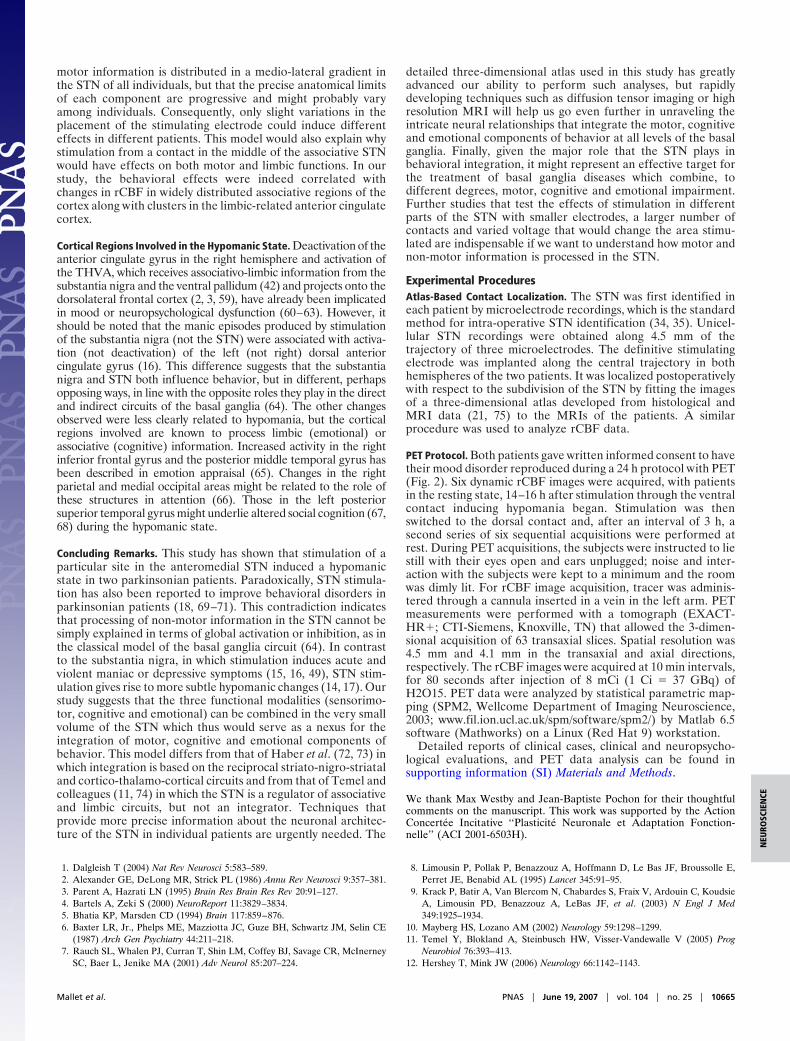
Cortical Regions Involved in the Hypomanic State. Deactivation of theanterior cingulate gyrus in the right hemisphere and activation ofthe THVA, which receives associativo-limbic information from thesubstantia nigra and the ventral pallidum (42) and projects onto thedorsolateral frontal cortex (2, 3, 59), have already been implicatedin mood or neuropsychological dysfunction (60–63). However, itshould be noted that the manic episodes produced by stimulationof the substantia nigra (not the STN) were associated with activa-tion (not deactivation) of the left (not right) dorsal anteriorcingulate gyrus (16). This difference suggests that the substantianigra and STN both influence behavior, but in different, perhapsopposing ways, in line with the opposite roles they play in the directand indirect circuits of the basal ganglia (64). The other changesobserved were less clearly related to hypomania, but the corticalregions involved are known to process limbic (emotional) orassociative (cognitive) information. Increased activity in the rightinferior frontal gyrus and the posterior middle temporal gyrus hasbeen described in emotion appraisal (65). Changes in the rightparietal and medial occipital areas might be related to the role ofthese structures in attention (66). Those in the left posteriorsuperior temporal gyrus might underlie altered social cognition (67,68) during the hypomanic state.
Concluding Remarks. This study has shown that stimulation of aparticular site in the anteromedial STN induced a hypomanicstate in two parkinsonian patients. Paradoxically, STN stimula-tion has also been reported to improve behavioral disorders inparkinsonian patients (18, 69–71). This contradiction indicatesthat processing of non-motor information in the STN cannot besimply explained in terms of global activation or inhibition, as inthe classical model of the basal ganglia circuit (64). In contrastto the substantia nigra, in which stimulation induces acute andviolent maniac or depressive symptoms (15, 16, 49), STN stim-ulation gives rise to more subtle hypomanic changes (14, 17). Ourstudy suggests that the three functional modalities (sensorimo-tor, cognitive and emotional) can be combined in the very smallvolume of the STN which thus would serve as a nexus for theintegration of motor, cognitive and emotional components ofbehavior. This model differs from that of Haber et al. (72, 73) inwhich integration is based on the reciprocal striato-nigro-striataland cortico-thalamo-cortical circuits and from that of Temel andcolleagues (11, 74) in which the STN is a regulator of associativeand limbic circuits, but not an integrator. Techniques thatprovide more precise information about the neuronal architec-ture of the STN in individual patients are urgently needed. The
detailed three-dimensional atlas used in this study has greatlyadvanced our ability to perform such analyses, but rapidlydeveloping techniques such as diffusion tensor imaging or highresolution MRI will help us go even further in unraveling theintricate neural relationships that integrate the motor, cognitiveand emotional components of behavior at all levels of the basalganglia. Finally, given the major role that the STN plays inbehavioral integration, it might represent an effective target forthe treatment of basal ganglia diseases which combine, todifferent degrees, motor, cognitive and emotional impairment.Further studies that test the effects of stimulation in differentparts of the STN with smaller electrodes, a larger number ofcontacts and varied voltage that would change the area stimu-lated are indispensable if we want to understand how motor andnon-motor information is processed in the STN.
Experimental ProceduresAtlas-Based Contact Localization. The STN was first identified ineach patient by microelectrode recordings, which is the standardmethod for intra-operative STN identification (34, 35). Unicel-lular STN recordings were obtained along 4.5 mm of thetrajectory of three microelectrodes. The definitive stimulatingelectrode was implanted along the central trajectory in bothhemispheres of the two patients. It was localized postoperativelywith respect to the subdivision of the STN by fitting the imagesof a three-dimensional atlas developed from histological andMRI data (21, 75) to the MRIs of the patients. A similarprocedure was used to analyze rCBF data.
PET Protocol. Both patients gave written informed consent to havetheir mood disorder reproduced during a 24 h protocol with PET(Fig. 2). Six dynamic rCBF images were acquired, with patientsin the resting state, 14–16 h after stimulation through the ventralcontact inducing hypomania began. Stimulation was thenswitched to the dorsal contact and, after an interval of 3 h, asecond series of six sequential acquisitions were performed atrest. During PET acquisitions, the subjects were instructed to liestill with their eyes open and ears unplugged; noise and inter-action with the subjects were kept to a minimum and the roomwas dimly lit. For rCBF image acquisition, tracer was adminis-tered through a cannula inserted in a vein in the left arm. PETmeasurements were performed with a tomograph (EXACT-HR�; CTI-Siemens, Knoxville, TN) that allowed the 3-dimen-sional acquisition of 63 transaxial slices. Spatial resolution was4.5 mm and 4.1 mm in the transaxial and axial directions,respectively. The rCBF images were acquired at 10 min intervals,for 80 seconds after injection of 8 mCi (1 Ci � 37 GBq) ofH2O15. PET data were analyzed by statistical parametric map-ping (SPM2, Wellcome Department of Imaging Neuroscience,2003; www.fil.ion.ucl.ac.uk/spm/software/spm2/) by Matlab 6.5software (Mathworks) on a Linux (Red Hat 9) workstation.
Detailed reports of clinical cases, clinical and neuropsycho-logical evaluations, and PET data analysis can be found insupporting information (SI) Materials and Methods.
We thank Max Westby and Jean-Baptiste Pochon for their thoughtfulcomments on the manuscript. This work was supported by the ActionConcertee Incitative ‘‘Plasticite Neuronale et Adaptation Fonction-nelle’’ (ACI 2001-6503H).
1. Dalgleish T (2004) Nat Rev Neurosci 5:583–589.2. Alexander GE, DeLong MR, Strick PL (1986) Annu Rev Neurosci 9:357–381.3. Parent A, Hazrati LN (1995) Brain Res Brain Res Rev 20:91–127.4. Bartels A, Zeki S (2000) NeuroReport 11:3829–3834.5. Bhatia KP, Marsden CD (1994) Brain 117:859–876.6. Baxter LR, Jr., Phelps ME, Mazziotta JC, Guze BH, Schwartz JM, Selin CE
(1987) Arch Gen Psychiatry 44:211–218.7. Rauch SL, Whalen PJ, Curran T, Shin LM, Coffey BJ, Savage CR, McInerney
SC, Baer L, Jenike MA (2001) Adv Neurol 85:207–224.
8. Limousin P, Pollak P, Benazzouz A, Hoffmann D, Le Bas JF, Broussolle E,Perret JE, Benabid AL (1995) Lancet 345:91–95.
9. Krack P, Batir A, Van Blercom N, Chabardes S, Fraix V, Ardouin C, KoudsieA, Limousin PD, Benazzouz A, LeBas JF, et al. (2003) N Engl J Med349:1925–1934.
10. Mayberg HS, Lozano AM (2002) Neurology 59:1298–1299.11. Temel Y, Blokland A, Steinbusch HW, Visser-Vandewalle V (2005) Prog
Neurobiol 76:393–413.12. Hershey T, Mink JW (2006) Neurology 66:1142–1143.
Mallet et al. PNAS � June 19, 2007 � vol. 104 � no. 25 � 10665
NEU
ROSC
IEN
CE
13. Romito LM, Raja M, Daniele A, Contarino MF, Bentivoglio AR, Barbier A,Scerrati M, Albanese A (2002) Mov Disord 17:1371–1374.
14. Herzog J, Reiff J, Krack P, Witt K, Schrader B, Muller D, Deuschl G (2003)Mov Disord 18:1382–1384.
15. Kulisevsky J, Berthier ML, Gironell A, Pascual-Sedano B, Molet J, Pares P(2002) Neurology 59:1421–1424.
16. Ulla M, Thobois S, Lemaire JJ, Schmitt A, Derost P, Broussolle E, Llorca PM,Durif F (2006) J Neurol Neurosurg Psychiatry 77:1363–1366.
17. Mandat TS, Hurwitz T, Honey CR (2006) Acta Neurochir 148:895–897.18. Houeto JL, Mallet L, Mesnage V, Tezenas du Montcel S, Behar C, Gargiulo
M, Torny F, Pelissolo A, Welter ML, Agid Y (2006) Arch Neurol 63:1090–1095.19. APA (2000) Diagnostic and Statistical Manual of Mental Disorders (DSM-IV-
TR) (American Psychiatric Publishing, Inc, Arlington, VA), 4th Ed, TextRevision.
20. Yelnik J, Damier P, Demeret S, Gervais D, Bardinet E, Bejjani BP, FrancoisC, Houeto JL, Arnule I, Dormont D, et al. (2003) J Neurosurg 99:89–99.
21. Yelnik J, Bardinet E, Dormont D, Malandain G, Ourselin S, Tande D, KarachiC, Ayache N, Cornu P, Agid Y (2007) NeuroImage 34:618–638.
22. Karachi C, Yelnik J, Tande D, Tremblay L, Hirsch EC, Francois C (2005) MovDisord 20:172–180.
23. Young RC, Biggs JT, Ziegler VE, Meyer DA (1978) Br J Psychiatry 133:429–435.
24. Conners CK (2000) Conners’ Continuous Performance Test II (Multi-HealthSystems Inc, Toronto), Version 5 for Windows.
25. Tavares JV, Drevets WC, Sahakian BJ (2003) Psychol Med 33:959–967.26. Chamberlain SR, Sahakian BJ (2004) Curr Psychiatry Rep 6:451–458.27. Sax KW, Strakowski SM, Zimmerman ME, DelBello MP, Keck PE, Jr,
Hawkins JM (1999) Am J Psychiatry 156:139–141.28. Fahn S, Oakes D, Shoulson I, Kieburtz K, Rudolph A, Lang A, Olanow CW,
Tanner C, Marek K (2004) N Engl J Med 351:2498–2508.29. Lee MS, Marsden CD (1994) Mov Disord 9:493–507.30. Trillet M, Vighetto A, Croisile B, Charles N, Aimard G (1995) Rev Neurol
(Paris) 151:416–419.31. Takeshita S, Kurisu K, Trop L, Arita K, Akimitsu T, Verhoeff NP (2005)
Neurosurg Rev 28:179–186.32. Voon V, Kubu C, Krack P, Houeto JL, Troster AI (2006) Mov Disord
21:S305–S327.33. Herzog J, Volkmann J, Krack P, Kopper F, Potter M, Lorenz D, Steinbach M,
Klebe S, Hamel W, Schrader B, et al. (2003) Mov Disord 18:1332–1337.34. Hutchison WD, Allan RJ, Opitz H, Levy R, Dostrovsky JO, Lang AE, Lozano
AM (1998) Ann Neurol 44:622–628.35. Bejjani BP, Dormont D, Pidoux B, Yelnik J, Damier P, Arnulf I, Bonnet AM,
Marsault C, Agid Y, Philippon J, et al. (2000) J Neurosurg 92:615–625.36. Dormont D, Ricciardi KG, Tande D, Parain K, Menuel C, Galanaud D,
Navarro S, Cornu P, Agid Y, Yelnik J (2004) Am J Neuroradiol 25:1516–1523.37. Saint-Cyr JA, Hoque T, Pereira LC, Dostrovsky JO, Hutchison WD, Mikulis
DJ, Abosch A, Sime E, Lang AE, Lozano AM (2002) J Neurosurg 97:1152–1166.
38. Herzog J, Fietzek U, Hamel W, Morsnowski A, Steigerwald F, Schrader B,Weinert D, Pfister G, Muller D, Mehdorn HM, et al. (2004) Mov Disord19:1050–1054.
39. Lanotte MM, Rizzone M, Bergamasco B, Faccani G, Melcarne A, Lopiano L(2002) J Neurol Neurosurg Psychiatry 72:53–58.
40. Zonenshayn M, Sterio D, Kelly PJ, Rezai AR, Beric A (2004) SurgicalNeurology 62:216–225.
41. Sato F, Lavallee P, Levesque M, Parent A (2000) J Comp Neurol 417:17–31.42. Haber SN, Lynd-Balta E, Mitchell SJ (1993) J Comp Neurol 329:111–128.43. Augood SJ, Waldvogel HJ, MÅnkle MC, Faull RLM, Emson PC (1999)
Neuroscience 88:521–534.44. Levesque JC, Parent A (2005) Mov Disord 20:574–584.45. Butson CR, Cooper SE, Henderson JM, McIntyre CC (2007) NeuroImage
34:661–670.46. McIntyre CC, Mori S, Sherman DL, Thakor NV, Vitek JL (2004) Clin
Neurophysiol 115:589–595.47. Miocinovic S, Parent M, Butson CR, Hahn PJ, Russo GS, Vitek JL, McIntyre
CC (2006) J Neurophysiol 96:1569–1580.48. Yelnik J (2002) Mov Disord 17(Suppl 3):S15–S21.49. Bejjani BP, Houeto JL, Hariz M, Yelnik J, Mesnage V, Bonnet AM, Pidoux
B, Dormont D, Cornu P, Agid Y (2002) Neurology 59:1425–1427.50. Karachi C, Francois C, Parain K, Bardinet E, Tande D, Hirsch E, Yelnik J
(2002) J Comp Neurol 450:122–134.51. Yelnik J, Percheron G (1979) Neuroscience 4:1717–1743.52. Pearson JC, Norris JR, Phelps CH (1985) J Comp Neurol 238:323–339.53. Bevan MD, Clarke NP, Bolam JP (1997) J Neurosci 17:308–324.54. Ceballos-Baumann AO, Boecker H, Bartenstein P, von Falkenhayn I, Riescher
H, Conrad B, Moringlane JR, Alesch F (1999) Arch Neurol 56:997–1003.55. Hershey T, Revilla FJ, Wernle AR, McGee-Minnich L, Antenor JV, Videen
TO, Dowling JL, Mink JW, Perlmutter JS (2003) Neurology 61:816–821.56. Payoux P, Remy P, Damier P, Miloudi M, Loubinoux I, Pidoux B, Gaura V,
Rascol O, Samson Y, Agid Y (2004) Arch Neurol 61:1307–1313.57. Nambu A, Takada M, Inase M, Tokuno H (1996) J Neurosci 16:2671–2683.58. Sato F, Parent M, Levesque M, Parent A (2000) J Comp Neurol 424:142–152.59. Francois C, Tande D, Yelnik J, Hirsch EC (2002) J Comp Neurol 447:249–260.60. Drevets WC, Price JL, Simpson JR, Jr, Todd RD, Reich T, Vannier M, Raichle
ME (1997) Nature 386:824–827.61. Mayberg HS, Liotti M, Brannan SK, McGinnis S, Mahurin RK, Jerabek PA,
Silva JA, Tekell JL, Martin CC, Lancaster JL, et al. (1999) Am J Psychiatry156:675–682.
62. Blumberg HP, Stern E, Martinez D, Ricketts S, de Asis J, White T, Epstein J,McBride PA, Eidelberg D, Kocsis JH, et al. (2000) Biol Psychiatry 48:1045–1052.
63. Strakowski SM, Delbello MP, Adler CM (2005) Mol Psychiatry 10:105–116.64. Albin RL, Young AB, Penney JB (1989) Trends Neurosci 12:366–375.65. Goldin PR, Hutcherson CA, Ochsner KN, Glover GH, Gabrieli JD, Gross JJ
(2005) NeuroImage 27:26–36.66. Behrmann M, Geng JJ, Shomstein S (2004) Curr Opin Neurobiol 14:212–217.67. Adolphs R (2003) Nat Rev Neurosci 4:165–178.68. Moll J, de Oliveira-Souza R, Eslinger PJ, Bramati IE, Mourao-Miranda J,
Andreiuolo PA, Pessoa L (2002) J Neurosci 22:2730–2736.69. Mallet L, Mesnage V, Houeto JL, Pelissolo A, Yelnik J, Behar C, Gargiulo M,
Welter ML, Bonnet AM, Pillon B, et al. (2002) Lancet 360:1302–1304.70. Fontaine D, Mattei V, Borg M, von Langsdorff D, Magnie MN, Chanalet S,
Robert P, Paquis P (2004) J Neurosurg 100:1084–1086.71. Ardouin C, Voon V, Worbe Y, Abouazar N, Czernecki V, Hosseini H, Pelissolo
A, Moro E, Lhommee E, Lang AE, et al. (2006) Mov Disord 21:1941–1946.72. Haber SN (2003) J Chem Neuroanat 26:317–330.73. Haber SN, Kim KS, Mailly P, Calzavara R (2006) J Neurosci 26:8368–8376.74. Tan SK, Temel Y, Blokland A, Steinbusch HW, Visser-Vandewalle V (2006)
J Chem Neuroanat 31:155–161.75. Yelnik J, Bardinet E, Dormont D, Francois C, Tande D, Parain C, Malandain
G, Ayache N, Hirsch E, Agid Y (2003) NeuroImage 99:S48.
10666 � www.pnas.org�cgi�doi�10.1073�pnas.0610849104 Mallet et al.