stimulation of the po-shen and shen-hun scalp-acupuncture bands modifies levels of inhibitory and...
TRANSCRIPT

Neurochemistry International 63 (2013) 275–282
Contents lists available at SciVerse ScienceDirect
Neurochemistry International
journal homepage: www.elsevier .com/locate /nci
Stimulation of the Po-shen and Shen-hun scalp-acupuncture bandsmodifies levels of inhibitory and excitatory amino acids in the immaturerat brain
0197-0186/$ - see front matter � 2013 Elsevier Ltd. All rights reserved.http://dx.doi.org/10.1016/j.neuint.2013.05.004
⇑ Corresponding author. Tel./fax: +52 55 5578 0240.E-mail addresses: [email protected] (L.E. Franco-Santana), sergiotorcas@
yahoo.com.mx (S. Torres-Castillo), [email protected] (M.E. González-Trujano),[email protected], [email protected] (M. González-Ramírez).
Lucia Estrella Franco-Santana a,b, Sergio Torres-Castillo b, María Eva González-Trujano c,Misael González-Ramírez a,⇑a Unidad de Investigación Médica en Enfermedades Neurológicas, Hospital de Especialidades, Centro Médico Nacional Siglo XXI, IMSS, Av. Cuauhtémoc 330, Col. Doctores,C.P. 06720 México D.F., Mexicob Universidad Estatal del Valle de Ecatepec, Av. Central esquina Leona Vicario S/N, Col. Valle de Anáhuac, C.P. 55210 Edo. de México, Mexicoc Dirección de Investigación en Neurociencias del Instituto Nacional de Psiquiatría Ramón de la Fuente Muñiz, Calz. México-Xochimilco 101, Col. Sn Lorenzo Huipulco, C.P. 14370México D.F., Mexico
a r t i c l e i n f o
Article history:Received 15 November 2012Received in revised form 7 May 2013Accepted 12 May 2013Available online 18 May 2013
Keywords:AcupunctureExcitatory amino acidInhibitory amino acidPo-shen bandScalp-acupunctureShen-hun bandPentobarbital
a b s t r a c t
Objective: In the present study, the effect of stimulation of the Po-shen and Shen-hun scalp-acupuncturebands on tissue amino acid concentrations in several brain regions in awake and pentobarbital-sedatedimmature rats was evaluated.Materials and methods: Sprague–Dawley rats (aged 15 days) were organized in four groups of at leasteight animals: control groups received saline solution 0.9% or sodium pentobarbital at 30 mg/kg dosagevia intraperitoneal. Experimental groups received saline solution or sodium pentobarbital plus stimula-tion in Po-shen and Shen-hun scalp-acupuncture bands for one continuous hour during 10 sessions byusing scalp-acupuncture.Results: As compared to rats receiving saline solution, scalp-acupuncture produced significant changes inamino acid concentrations, depending on the analyzed region, as follows: in inhibitory amino acids, aGABA increase was observed in amygdala and hippocampus (491 and 184%, respectively), but a decreasein the substantia nigra (80%); glycine showed decrease in all the analyzed regions, except for an increasein brainstem(78%); glutamine presented an increase in hippocampus and cortex (42 and 149%, respec-tively). In the case of excitatory amino acids, glutamate decreased in all the analyzed regions; whereasaspartate decreased in substantia nigra and brainstem (77.08 and 35%, correspondingly) but increasedin hippocampus and cortex (32 and 54%, respectively). The combined treatment of scalp-acupunctureand a GABAergic depressant drug like pentobarbital resulted in almost all changes induced in amino acidsfor scalp-acupuncture alone being significantly reverted.Conclusion: Stimulation of the Po-shen and Shen-hun scalp-acupuncture bands by using scalp-acupunc-ture alone might produce depressant activity by changes in amino acids, but the combination with a GAB-Aergic tranquilizer like sodium pentobarbital can interfere with this response.
� 2013 Elsevier Ltd. All rights reserved.
1. Introduction
The word acupuncture derives from the Latin ‘‘Acus’’, meaningneedle, and ‘‘Pungue’’, meaning to penetrate. According to theWorld Health Organization (WHO), acupuncture consists of thestrategic penetration and stimulation through the skin of certainpoints in the body, specifically of points that have a low electricalresistance and high conductivity. Stimulation of these points can
also be achieved via moxibustion, magneto-therapy, electro-stimulation, laser, ultrasound, cupping glasses, tacks, small bulletsor acupressure; to choose the most suitable method, one mustconsider a patient’s group of symptoms and her/his diagnosis(WHO, 2002). Scalp-acupuncture was modeled after traditionalacupuncture by Jiao Shiunfa in China in 1970. Shiunfa theorizedthat points of acupuncture could exist in the ‘‘brain’’ (i.e., in thescalp), just as they do in the back, thorax and abdomen. The firstsupporting case for scalp-acupuncture is the case of an elderlywoman who suffered spasmodic pains in her arms and legs causedby arteriosclerosis. The woman inserted needles into her scalp,considering the sensorial area. By the following day, the pain hadsubsided considerably (Gaynor, 2000). Scalp-acupuncture is now

276 L.E. Franco-Santana et al. / Neurochemistry International 63 (2013) 275–282
used extensively in China and in many other countries to treat avariety of pathologies. It has been shown to be particularlyeffective in treating consequences of cerebral events and infantilecerebral paralysis (González, 1995; Marié 1998).
Scalp-acupuncture is a therapeutic procedure in which specificcortical areas, i.e., those involved in motor functions, speech, sen-sation, vision, audition, vertigo, and balance are punctured (Smileyand Falchier, 2009; Zilles and Wree, 1995). These areas are stimu-lated with the insertion of a needle below the hair, where it re-mains for at least 30 minutes. The therapist can additionallystimulate the site by manually turning the needle clockwise orcounterclockwise or by electrically stimulating the handle (coil)of the needle using an electro-stimulating device to regulate thefrequency and intensity of the electric current (Mayer et al., 1977).
Scalp-acupuncture is applied to well-defined ‘‘bands’’ or loca-tions of the scalp to obtain specific therapeutic results (Rapson,1984). A cun is a unit of measure of the width and length of the fin-gers; each cun depends on the physical constitution of each pa-tient. The shen-hun band begins at 5 cun posterior to theimplantation of hair and 1.5 cun outside of the midline the left side(Tongtian BL-7); it finishes at 6.5 cun posterior implantation of hairand 1.5 cun outside the mean line of the right side (Luoque BL-8),crossing through vertex (Baihui GV-20) (Maciocia, 2009; Zhangand Zheng, 2003). Stimulation of the shen-hun acupuncture bandis used as an anxiolytic and sleep inducer to treat attention deficitdisorders, neuroses, and memory impairments that arise fromemotional stress; it is also used to promote long term memory.The Po-shen band begins 2 cun posterior to the implantation of hair(Xinhui GV-22) and 1.5 cun lateral to the median line, starting fromright to left and at 0.1 cun above is from right to left horizontally(Maciocia, 2009; Zhang and Zheng, 2003). The Po-shen band acti-vates the po function on shen: stimulation of the Po-shen bandinhibits mental activity in order to increase attention and focusand is used to treat attention deficit disorders, palpitations, andanxiety (fear of punishment, sensation of failure) (Maciocia,2009; Zhang and Zheng, 2003).
The purpose of the present study was to determine the effects ofscalp-acupuncture stimulation of the Po-shen and Shen-hun bandson GABA, glycine, glutamine, glutamate and aspartate levels inthe cortex, amygdala, hippocampus, substantia nigra, and brain-stem. The effects were measured in immature rats also treatedwith the sedative sodium pentobarbital or with saline solution.
2. Materials and methods
2.1. Animals
All experimental procedures were carried out according to aprotocol approved by the Local Animal Ethics Committee and incompliance with national (NOM-062-ZOO-1999) and internationalrules stated in the National Institutes of Health Guide for Care andUse of Laboratory Animals. The experiment was performed on fif-teen-day-old male and female Sprague–Dawley rats. At birth, eachpup was assigned at random to one of four mothers. Each motherhad 8 rats (4 males and 4 females). All the animals were housed ina temperature and light controlled room under a 12 h light-darkcycle (lights on at 7:00 a.m.) with water and food ad libitum. Thenumber of experimental animals was kept to a minimum. Animalswere sacrificed immediately after the experiment. All of the exper-imental sessions took place between 10:00 h and 12:00 h to avoidcircadian variations.
2.2. Experimental groups
Rats were assigned to either control group (saline solution 0.9%,i.p.) or an experimental group (sodium pentobarbital, 30 mg/kg,
i.p.) without scalp-acupuncture stimulation. Other rats receivedsaline solution or sodium pentobarbital plus one continuous hourstimulation in Po-shen and Shen-hun acupuncture bands for eachof 10 daily sessions starting at age 15 days. Thirty minutes aftersaline solution or pentobarbital, animals were immobilized bybeing wound on a flannel adjusted with tweezers, and then theirPo-shen and Shen-hun scalp-acupuncture bands were stimulatedusing a sterile disposable 0.17 mm diameter needle (CW-SOOJI).For the placement of two needles into the Po-Shen scalp-acupunc-ture band, an imaginary line from the external angle of the eye ofthe rat was taken into account to insert one needle from right toleft and the other from left to right. The band width comprisesthe distance between the inner corners of the eyes of the rats(Fig. 1). For the placement of the needle into the Shen-hun scalp-acupuncture band it is necessary to consider two angles as follows:for the first angle, two imaginary lines are delineated to form a 90�angle, one is directed from the inner angle of the eye to the poster-ior side of the head to converge with other line directed from thebeginning of one ear to the other one (angle 1, Fig. 1A). For angle2, two imaginary lines forming a ‘‘T’’ are delineated: the first isdemarcated from one ear to the other one and the second line isdirected from the tip of the nose to the first line, then the needleis inserted between these two angles (Fig. 1B). Immediately afterthe 10th session, all animals were sacrificed by decapitation andthe brainstem, cortex, amygdala, hippocampus, and substantia ni-gra were obtained from the sacrificed animals. Brain regions werefrozen immediately with liquid nitrogen and stored at �70 �C untilthe amino acid and protein analyses were performed.
2.3. Amino acid analyses
Each brain region was weighed and homogenized in a solutionof perchloric acid (0.1 M), (J.T. Baker, Pittsburgh, New Jersey, USA)and sodium methabisulphyte (4 mM) (J.T. Baker, Pittsburgh, NewJersey, USA) at 30 ll/10 mg of tissue using a Thomas homogenizerwith Teflon emboli at a speed of 1000 rpm for 60 s. The sampleswere centrifuged at 12,000 rpm for 15 min and then the proteinconcentration was determined in the pellet by the Folin poteinmethod, as described by Lowry et al. (1951). Brain tissue was re-suspended in 200 ll double-distilled water and diluted 40 times;100 ll were taken from this dilution. The reaction began when500 ll of solution A were added, solution A contained 500 ll ofcopper sulphate (0.04 M) (Sigma Aldrich) and 500 ll of sodiumand potassium tartrate (0.07 M) (Sigma Aldrich) in 50 ml of sodiumcarbonate (0.19 M) (Sigma Aldrich) diluted in sodium hydroxide(1 M) (Sigma Aldrich). The mixture was vigorously shaken and leftto rest at room temperature. After 10 min, 50 ll of solution B wereadded (Folin diluted 1:2 (Sigma Aldrich) with double-distilledwater). The samples were incubated for 30 min, and the colorimet-ric reaction was analyzed using a spectrophotometer (Perkin ElmerLambda 25 UV/VIS including wavelengths to 700 nm). This proce-dure enabled data obtained from the HPLC analysis to express theamino acid concentrations in ng/mg of protein.
The supernatant was used to analyze GABA and glycine (GLY) asinhibitory amino acids, and glutamate (GLU) and aspartate (ASP) asexcitatory amino acids; as well as glutamine (GLN), in the obtainedtissue. The off-line procedure was performed as described by For-ster and Marsden, (2001). A high performance liquid chromatogra-phy (HPLC) system was used. The entire procedure was performedwith HPLC grade water (Mallinkrodt Baker Pittsburgh, New Jersey,USA), in 4 �C temp and under light protection. The system was inte-grated with Empower software and an electrochemical detector(model 2465; Waters of México) operated at a cell voltage of+540 mV (with a range of 5 nA/V). The detector potential usedwas 0.80 V, the flow-rate was 0.30 ml min�1, and the column tem-perature was 30 �C, with a working electrode surface of carbon

(A)Po-shen acupuncture band
(B) Shen-hun acupuncture band
Fig. 1. A representative photography of the scalp-acupuncture by using needles inserted for the stimulation of the Po-shen (A) and Shen-hun (B) acupuncture bands inimmature rats.
L.E. Franco-Santana et al. / Neurochemistry International 63 (2013) 275–282 277
paste. Separation was achieved using a symmetry C18, with a par-ticle size of 5 lm and 4.6 � 150 mm, column. Solution 1 consistedof 1 M sodium sulfite (Sigma Aldrich) in HPLC grade water (Mal-linkrodt Baker, Pittsburgh, New Jersey, USA); solution 2 consistedof 0.1 M sodium tetraborate (Sigma Aldrich) in HPLC grade water,adjusted to a pH of 10.4 with 5 M sodium hydroxide (SigmaAldrich); solution 3 consisted of 0.5 ml of solution 1 and 0.5 mlof absolute ethanol combined with 9 ml of solution 2. A derivatiza-tion solution consisted of 0.164 mM of o-phtaldialdehyde (OPA)(Sigma Aldrich) combined with 10 ml of solution 3. Mobile phase
A consisted of 0.1 M of monosodium phosphate (Sigma Aldrich),0.5 mM of ethylenediaminetetraacetic acid (EDTA) (Sigma Aldrich),and 25% methanol (Mallinkrodt Baker Pittsburgh, New Jersey,USA), adjusted to pH 4.5, combined with 1 M of phosphoric acid(Sigma Aldrich) and degassed using an In-Line Degasser AF(Waters). The calibration curves were adjusted from chromato-grams with standard solutions (Sigma Aldrich) that contained 20,50, 250 and 500 ng/lL of amino acid. Peak area ratios of the stan-dards concentration were adjusted using a least-squares linearregression analysis, using the Empower system of Waters.
278 L.E. Franco-Santana et al. / Neurochemistry International 63 (2013) 275–282
2.4. Statistical analysis
Differences in the percentage of change of amino-acid levels forthe experimental groups with or without pentobarbital were ana-lyzed by using a chi-squared test. A Kolmogorov–Smirnov test re-vealed that the values were normally distributed for both thecontrol and the experimental groups. The data are presented interms of the mean ± SE. The levels of amino acids in each of theanalyzed tissues are presented as histograms. The increase in ami-no acid content was analyzed using a two-way ANOVA with a posthoc Student–Newman–Keuls test. All statistical analyses were per-formed using Sigma Stat version 3.0 (Jandel Scientific). A test witha probability value of 0.05 or below was defined as showing a sta-tistically significant difference.
3. Results
3.1. Amino acid content after scalp-acupuncture stimulation
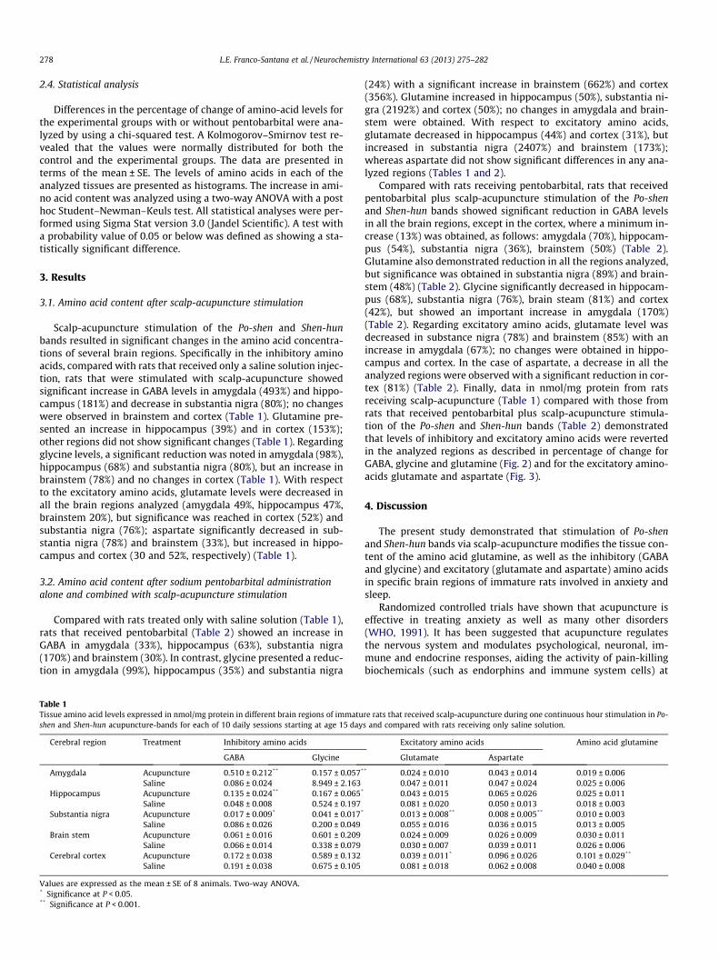
Scalp-acupuncture stimulation of the Po-shen and Shen-hunbands resulted in significant changes in the amino acid concentra-tions of several brain regions. Specifically in the inhibitory aminoacids, compared with rats that received only a saline solution injec-tion, rats that were stimulated with scalp-acupuncture showedsignificant increase in GABA levels in amygdala (493%) and hippo-campus (181%) and decrease in substantia nigra (80%); no changeswere observed in brainstem and cortex (Table 1). Glutamine pre-sented an increase in hippocampus (39%) and in cortex (153%);other regions did not show significant changes (Table 1). Regardingglycine levels, a significant reduction was noted in amygdala (98%),hippocampus (68%) and substantia nigra (80%), but an increase inbrainstem (78%) and no changes in cortex (Table 1). With respectto the excitatory amino acids, glutamate levels were decreased inall the brain regions analyzed (amygdala 49%, hippocampus 47%,brainstem 20%), but significance was reached in cortex (52%) andsubstantia nigra (76%); aspartate significantly decreased in sub-stantia nigra (78%) and brainstem (33%), but increased in hippo-campus and cortex (30 and 52%, respectively) (Table 1).
3.2. Amino acid content after sodium pentobarbital administrationalone and combined with scalp-acupuncture stimulation
Compared with rats treated only with saline solution (Table 1),rats that received pentobarbital (Table 2) showed an increase inGABA in amygdala (33%), hippocampus (63%), substantia nigra(170%) and brainstem (30%). In contrast, glycine presented a reduc-tion in amygdala (99%), hippocampus (35%) and substantia nigra
Table 1Tissue amino acid levels expressed in nmol/mg protein in different brain regions of immatushen and Shen-hun acupuncture-bands for each of 10 daily sessions starting at age 15 day
Cerebral region Treatment Inhibitory amino acids
GABA Glycine
Amygdala Acupuncture 0.510 ± 0.212** 0.157 ± 0.057*
Saline 0.086 ± 0.024 8.949 ± 2.163Hippocampus Acupuncture 0.135 ± 0.024** 0.167 ± 0.065*
Saline 0.048 ± 0.008 0.524 ± 0.197Substantia nigra Acupuncture 0.017 ± 0.009* 0.041 ± 0.017*
Saline 0.086 ± 0.026 0.200 ± 0.049Brain stem Acupuncture 0.061 ± 0.016 0.601 ± 0.209
Saline 0.066 ± 0.014 0.338 ± 0.079Cerebral cortex Acupuncture 0.172 ± 0.038 0.589 ± 0.132
Saline 0.191 ± 0.038 0.675 ± 0.105
Values are expressed as the mean ± SE of 8 animals. Two-way ANOVA.* Significance at P < 0.05.** Significance at P < 0.001.
(24%) with a significant increase in brainstem (662%) and cortex(356%). Glutamine increased in hippocampus (50%), substantia ni-gra (2192%) and cortex (50%); no changes in amygdala and brain-stem were obtained. With respect to excitatory amino acids,glutamate decreased in hippocampus (44%) and cortex (31%), butincreased in substantia nigra (2407%) and brainstem (173%);whereas aspartate did not show significant differences in any ana-lyzed regions (Tables 1 and 2).
Compared with rats receiving pentobarbital, rats that receivedpentobarbital plus scalp-acupuncture stimulation of the Po-shenand Shen-hun bands showed significant reduction in GABA levelsin all the brain regions, except in the cortex, where a minimum in-crease (13%) was obtained, as follows: amygdala (70%), hippocam-pus (54%), substantia nigra (36%), brainstem (50%) (Table 2).Glutamine also demonstrated reduction in all the regions analyzed,but significance was obtained in substantia nigra (89%) and brain-stem (48%) (Table 2). Glycine significantly decreased in hippocam-pus (68%), substantia nigra (76%), brain steam (81%) and cortex(42%), but showed an important increase in amygdala (170%)(Table 2). Regarding excitatory amino acids, glutamate level wasdecreased in substance nigra (78%) and brainstem (85%) with anincrease in amygdala (67%); no changes were obtained in hippo-campus and cortex. In the case of aspartate, a decrease in all theanalyzed regions were observed with a significant reduction in cor-tex (81%) (Table 2). Finally, data in nmol/mg protein from ratsreceiving scalp-acupuncture (Table 1) compared with those fromrats that received pentobarbital plus scalp-acupuncture stimula-tion of the Po-shen and Shen-hun bands (Table 2) demonstratedthat levels of inhibitory and excitatory amino acids were revertedin the analyzed regions as described in percentage of change forGABA, glycine and glutamine (Fig. 2) and for the excitatory amino-acids glutamate and aspartate (Fig. 3).
4. Discussion
The present study demonstrated that stimulation of Po-shenand Shen-hun bands via scalp-acupuncture modifies the tissue con-tent of the amino acid glutamine, as well as the inhibitory (GABAand glycine) and excitatory (glutamate and aspartate) amino acidsin specific brain regions of immature rats involved in anxiety andsleep.
Randomized controlled trials have shown that acupuncture iseffective in treating anxiety as well as many other disorders(WHO, 1991). It has been suggested that acupuncture regulatesthe nervous system and modulates psychological, neuronal, im-mune and endocrine responses, aiding the activity of pain-killingbiochemicals (such as endorphins and immune system cells) at
re rats that received scalp-acupuncture during one continuous hour stimulation in Po-s and compared with rats receiving only saline solution.
Excitatory amino acids Amino acid glutamine
Glutamate Aspartate
* 0.024 ± 0.010 0.043 ± 0.014 0.019 ± 0.0060.047 ± 0.011 0.047 ± 0.024 0.025 ± 0.0060.043 ± 0.015 0.065 ± 0.026 0.025 ± 0.0110.081 ± 0.020 0.050 ± 0.013 0.018 ± 0.0030.013 ± 0.008** 0.008 ± 0.005** 0.010 ± 0.0030.055 ± 0.016 0.036 ± 0.015 0.013 ± 0.0050.024 ± 0.009 0.026 ± 0.009 0.030 ± 0.0110.030 ± 0.007 0.039 ± 0.011 0.026 ± 0.0060.039 ± 0.011* 0.096 ± 0.026 0.101 ± 0.029**
0.081 ± 0.018 0.062 ± 0.008 0.040 ± 0.008

Table 2Tissue amino acid levels expressed in nmol/mg protein in different brain regions of immature rats that received scalp-acupuncture during one continuous hour stimulation inPo-shen and Shen-hun acupuncture-bands for each of 10 daily sessions starting at age 15 days with a 30 mg/kg pentobarbital injection and compared to rats receiving onlypentobarbital administration.
Cerebral region Treatment Inhibitory amino acids Excitatory aminoacids Amino acid glutamine
GABA Glycine Glutamate Aspartate
Amygdala Acupuncture plus pentobarbital 0.035 ± 0.016** 0.273 ± 0.140** 0.087 ± 0.025 0.035 ± 0.013 0.023 ± 0.010Pentobarbital 0.114 ± 0.032 0.101 ± 0.025 0.052 ± 0.014 0.053 ± 0.009 0.027 ± 0.007
Hippocampus Acupuncture plus pentobarbital 0.036 ± 0.008** 0.111 ± 0.040* 0.046 ± 0.012 0.027 ± 0.014 0.012 ± 0.002Pentobarbital 0.078 ± 0.018 0.342 ± 0.092 0.045 ± 0.012 0.036 ± 0.009 0.027 ± 0.010
Substantia nigra Acupuncture plus pentobarbital 0.148 ± 0.024* 0.037 ± 0.009* 0.300 ± 0.076* 0.029 ± 0.005 0.034 ± 0.007*
Pentobarbital 0.232 ± 0.087 0.153 ± 0.091 1.379 ± 0.737 0.039 ± 0.025 0.298 ± 0.169Brain stem Acupuncture plus pentobarbital 0.043 ± 0.009* 0.494 ± 0.175* 0.012 ± 0.004* 0.016 ± 0.013 0.014 ± 0.002*
Pentobarbital 0.086 ± 0.019 2.574 ± 0.741 0.082 ± 0.022 0.032 ± 0.011 0.027 ± 0.008Cerebral cortex Acupuncture plus pentobarbital 0.165 ± 0.043 1.797 ± 0.254 0.059 ± 0.016 0.016 ± 0.004* 0.044 ± 0.011
Pentobarbital 0.144 ± 0.029 3.080 ± 0.813 0.056 ± 0.010 0.086 ± 0.025 0.060 ± 0.014
Values are expressed as the mean ± SE of 8 animals. Two-way ANOVA.* Significance at P < 0.05.** Significance at P < 0.001.
Fig. 2. Percentage of change of inhibitory amino acids levels (GABA, glycine and glutamine) in amygdala, hippocampus, substantia nigra, brainstem and cerebral cortex ofSprague–Dawley rats after a continuous 1 h scalp-acupuncture stimulation in Po-shen and Shen-hun acupuncture-bands for each of 10 daily sessions starting at age 15 days,with or without pentobarbital. v2 test, ⁄P < 0.05.
L.E. Franco-Santana et al. / Neurochemistry International 63 (2013) 275–282 279
specific sites in the body. In addition, studies have shown that acu-puncture may alter brain chemistry by modulating the release ofneurotransmitters and neurohormones, affecting the parts of thecentral nervous system related to sensation and involuntary bodyfunctions (including immune reactions, blood pressure, blood flow,and body temperature) (WHO, 1991). The standard acupuncturenomenclature published by the WHO lists approximately 400 acu-puncture points and 20 meridians connecting these points (WHO,1991).
4.1. Effect of the stimulation in the Po-shen and Shen-hun scalp-acupuncture bands on amino acid concentrations in immature rats
Primarily, the analyses demonstrate that rats who receivedscalp-acupuncture alone significantly increased the content ofthe main inhibitory amino acid GABA in amygdala and hippocam-pus with a significant decrease in the main excitatory amino acidglutamate in all regions in comparison to baseline levels observedin rats receiving saline solution. These results resembled those
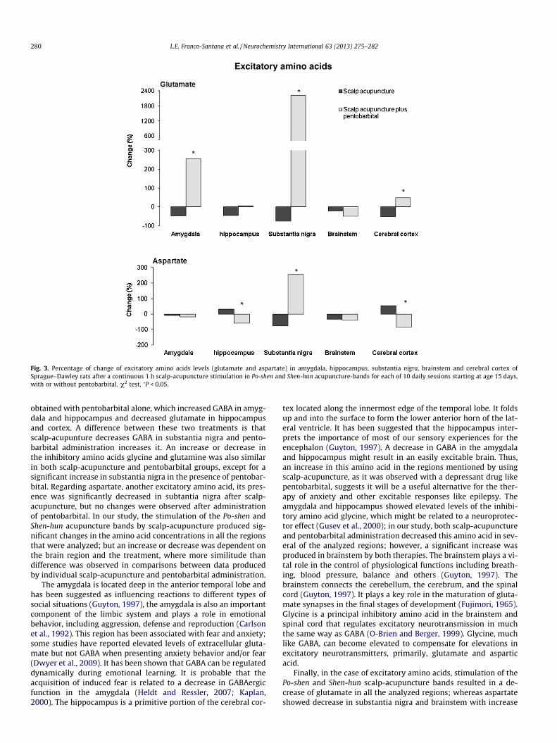
Fig. 3. Percentage of change of excitatory amino acids levels (glutamate and aspartate) in amygdala, hippocampus, substantia nigra, brainstem and cerebral cortex ofSprague–Dawley rats after a continuous 1 h scalp-acupuncture stimulation in Po-shen and Shen-hun acupuncture-bands for each of 10 daily sessions starting at age 15 days,with or without pentobarbital. v2 test, ⁄P < 0.05.
280 L.E. Franco-Santana et al. / Neurochemistry International 63 (2013) 275–282
obtained with pentobarbital alone, which increased GABA in amyg-dala and hippocampus and decreased glutamate in hippocampusand cortex. A difference between these two treatments is thatscalp-acupunture decreases GABA in substantia nigra and pento-barbital administration increases it. An increase or decrease inthe inhibitory amino acids glycine and glutamine was also similarin both scalp-acupuncture and pentobarbital groups, except for asignificant increase in substantia nigra in the presence of pentobar-bital. Regarding aspartate, another excitatory amino acid, its pres-ence was significantly decreased in subtantia nigra after scalp-acupuncture, but no changes were observed after administrationof pentobarbital. In our study, the stimulation of the Po-shen andShen-hun acupuncture bands by scalp-acupuncture produced sig-nificant changes in the amino acid concentrations in all the regionsthat were analyzed; but an increase or decrease was dependent onthe brain region and the treatment, where more similitude thandifference was observed in comparisons between data producedby individual scalp-acupuncture and pentobarbital administration.
The amygdala is located deep in the anterior temporal lobe andhas been suggested as influencing reactions to different types ofsocial situations (Guyton, 1997), the amygdala is also an importantcomponent of the limbic system and plays a role in emotionalbehavior, including aggression, defense and reproduction (Carlsonet al., 1992). This region has been associated with fear and anxiety;some studies have reported elevated levels of extracellular gluta-mate but not GABA when presenting anxiety behavior and/or fear(Dwyer et al., 2009). It has been shown that GABA can be regulateddynamically during emotional learning. It is probable that theacquisition of induced fear is related to a decrease in GABAergicfunction in the amygdala (Heldt and Ressler, 2007; Kaplan,2000). The hippocampus is a primitive portion of the cerebral cor-
tex located along the innermost edge of the temporal lobe. It foldsup and into the surface to form the lower anterior horn of the lat-eral ventricle. It has been suggested that the hippocampus inter-prets the importance of most of our sensory experiences for theencephalon (Guyton, 1997). A decrease in GABA in the amygdalaand hippocampus might result in an easily excitable brain. Thus,an increase in this amino acid in the regions mentioned by usingscalp-acupuncture, as it was observed with a depressant drug likepentobarbital, suggests it will be a useful alternative for the ther-apy of anxiety and other excitable responses like epilepsy. Theamygdala and hippocampus showed elevated levels of the inhibi-tory amino acid glycine, which might be related to a neuroprotec-tor effect (Gusev et al., 2000); in our study, both scalp-acupunctureand pentobarbital administration decreased this amino acid in sev-eral of the analyzed regions; however, a significant increase wasproduced in brainstem by both therapies. The brainstem plays a vi-tal role in the control of physiological functions including breath-ing, blood pressure, balance and others (Guyton, 1997). Thebrainstem connects the cerebellum, the cerebrum, and the spinalcord (Guyton, 1997). It plays a key role in the maturation of gluta-mate synapses in the final stages of development (Fujimori, 1965).Glycine is a principal inhibitory amino acid in the brainstem andspinal cord that regulates excitatory neurotransmission in muchthe same way as GABA (O-Brien and Berger, 1999). Glycine, muchlike GABA, can become elevated to compensate for elevations inexcitatory neurotransmitters, primarily, glutamate and asparticacid.
Finally, in the case of excitatory amino acids, stimulation of thePo-shen and Shen-hun scalp-acupuncture bands resulted in a de-crease of glutamate in all the analyzed regions; whereas aspartateshowed decrease in substantia nigra and brainstem with increase

L.E. Franco-Santana et al. / Neurochemistry International 63 (2013) 275–282 281
in hippocampus and cortex. The substantia nigra is part of the mid-brain, located behind the corticospinal fiber layer and corticopon-tine; the midbrain typically has higher concentrations of GABA(López-Muñoz et al., 2005). The neurons of the substantia nigrafunction as part of the basal ganglia system to control subconsciousmuscle activity (Guyton, 1997). An increase in the excitatory ami-no acids glutamate and aspartate induces neuronal excitability andinduce neurotoxic effects; contrarily, a significant decrease pro-duced with scalp-acupuncture might modulate this neuronal excit-ability and protect from neurotoxicity.
4.2. Effect of stimulation of the Po-shen and Shen-hun scalp-acupuncture bands on amino acid concentrations in immature ratstreated with sodium pentobarbital
Barbiturates are drugs that act as central nervous systemdepressants and have a wide variety of effects that range from mildsedation to total anesthesia. Barbiturates are also effective as anx-iolytics, hypnotics, and anticonvulsants (Kilbaugh et al., 2010;Toyoshima et al., 2009). Pentobarbital is a barbiturate commonlyused as an anesthetic. Even a low dosage of pentobarbital (e.g.,30 mg/kg) can produce sedative and hypnotic activities. Whenstimulation of the Po-shen and Shen-hun scalp-acupuncture bandswas combined with an injection of pentobarbital, the result wasnot a potentiated response on the inhibitory and excitatory aminoacid concentrations, as expected in comparison when only scalp-acupuncture stimulation or pentobarbital alone was administered.Conversely, effects produced by scalp-acupuncture stimulationalone were reversed when it was tested in combination with30 mg/kg of pentobarbital.
It has been shown that barbiturates are ligands that bind withhigh affinity and specificity to the GABAA receptor in certain neuralareas and that there is a strong correlation between the affinity ofthese sites and their potential for producing depressant activity inanimals. GABA produces a change in GABAA receptor that causesthat neuronal membrane to be more permeable to chloride ionsand to subsequent inhibition by hyperpolarization of the postsyn-aptic neuron. Barbiturates potentiate inhibitory GABAA receptors,but also inhibit excitatory AMPA receptors. In fact, pentobarbitalsleeping time is the primary test by which the depressant activityof pharmacological alternatives is measured in the search for newdrugs acting on the central nervous system (González-Trujanoet al., 1998; Sethy et al., 1967). The GABAA receptor has severalbinding sites, one of which binds GABA, other barbiturates, benzo-diazepines, and steroids (among others). The mechanism of actionsof scalp-acupuncture are unknown; to our knowledge this is thefirst experiment to assess the depressant effect of scalp-acupunc-ture stimulation alone and combined with a GABA depressant druglike pentobarbital, but our results suggest that scalp-acupuncturemight produce an interaction with this neurotransmission system.
The effect of acupuncture has been observed in animal behaviorin relation to pain, where the stimulation of electro-acupuncturehas been shown to affect the progression of pain by modulatingthe expression of N-methyl-D-aspartate (NMDA) receptor in pri-mary sensorial neurons, in particular, IB4-positive neurons (Wanget al., 2003). Electro-acupuncture treatment can particularly atten-uate inflammatory and mechanical edema by modulating theexpression of glutamate and NMDA receptors in the dorsal spearof spinal marrow. Changes in amino acid concentrations have alsobeen shown in the hippocampus of the rat in an experimentalmodel of epilepsy induced by kainic acid; these results demon-strate that the levels of glutamate, aspartate, GABA and glycinewere significantly increased in the hippocampus 40 min after theadministration of kainic acid and when electro-acupuncture wasused to inhibit seizures (Liu and Cheng, 1995), suggesting that
the acupuncture technique resulted in an increase in the aminoacids.
In conclusion, scalp-acupuncture of the Shen-hun and Po-shenbands, whether alone or combined with a depressant drug,resulted in the modulation of amino acid levels in the brain; nev-ertheless, combination of scalp-acupunture with GABAergic drugsshould be taken into account carefully in order to avoid antagonis-tic responses instead of a potent therapy. The results of this studysuggest an interaction in the mechanisms of action in the centralnervous system of immature rats and it will be interesting to con-duct future studies to evaluate the utility of these scalp-acupunc-ture induced changes in the treatment of mental disorders suchas anxiety, depression, epilepsy, and other central nervousdiseases.
Acknowledgements
This work was supported by Coordinación de Investigación enSalud del Instituto Mexicano del Seguro Social. We thank Mr.Raúl Calderón and Mr. José Luis for their technical assistance.
References
Carlson, B.X., Mans, A.M., Hawkins, R.A., Baghdoyan, H.A., 1992. Pentobarbital-enhanced [3H]flunitracepam binding throughout the rat brain: anautoradiographic study. J. Pharmacol. Exp. Ther. 263, 1401–1414.
Dwyer, G., Baur, L., Higgs, J., Hardy, L., 2009. Promoting children’s health and well-being broadening the therapy perspective. Phys. Occup. Ther. Pediatr. 29, 27–43.
Forster, C.D., Marsden, C.A., 2001. The analysis of amino acids using precolumnderivatization, HPLC, and electrochemical detection. In: Cooper, C., Packer, N.(Eds.), Amino Acids Analysis Protocols. Human Press, New Jersey, pp. 55–62.
Fujimori, H., 1965. Potentiation of barbital hypnosis as an evaluation method forcentral nervous system depressants. Psychopharmacology 7, 374–378.
Gaynor, J.S., 2000. Acupuncture management of pain. Vet. Clin. North. Am. Small.Anim. Practi. 30, 861–884.
González, G.R., 1995. Craneoacupuntura, AMASA, México D.F.González-Trujano, M.E., Navarrete, A., Reyes, B., Hong, E., 1998. Some
pharmacological effects of the ethanol extract of leaves of Annona diversifoliaon the central nervous system in mice. Phytother Res. 12, 600–602.
Gusev, E.I., Skvortsova, V.I., Dambinova, S.A., Raevskiy, K.S., Alekseev, A.A.,Bashkatova, V.G., Kovalenko, A.V., Kudrin, V.S., Yakovleva, E.V., 2000.Neuroprotective effects of glycine for therapy of acute ischaemic stroke.Cerebrovasc. Dis. 10, 49–60.
Guyton, C.A., 1997. Anatomía y fisiología del sistema nervioso. Neurociencia básica,second ed. Médica Panamericana, Buenos Aires, Argentina.
Heldt, S.A., Ressler, K.J., 2007. Training-induced changes in the expression of GABAA-associated genes in the amygdala after the acquisition and extinction ofPavlovian fear. Eur. J. Neurosci. 26, 3631–3644.
Kaplan, H.I., 2000. Sinopsis de Psiquiatría, Ciencias de la Conducta. PsiquiatríaClínica, eighth ed. Médica Panamericana, Buenos Aires, Argentina.
Kilbaugh, T.J., Friess, S.H., Raghupathi, R., Huh, J.W., 2010. Sedation and analgesia inchildren with developmental disabilities and neurological disorders. Int. J.Pediatr.. http://dx.doi.org/10.1155/2010/189142.
Liu, J., Cheng, J., 1995. Changes of amino acids release in rat’s hippocampus duringkainic acid induced epilepsy and acupuncture. Zhen. Ci. Yan. Jiu. 20, 50–54.
López-Muñoz, F., Ucha-Udabe, R., Alamo, C., 2005. The history of barbiturates acentury after their clinical introduction. Neuropsychiatr. Dis. Treat. 1, 329–343.
Lowry, O.H., Rosenbrough, J., Farr, A.L., Randall, R.J., 1951. Protein measurementwith the follin phenol reagent. J. Biol. Chem. 193, 265–275.
Maciocia, G., 2009. La Practica de la Medicina Tradicional China. Elseiver, Barcelona,España.
Marié, E., 1998. Compendio de Medicina China Fundamentos, Teoría y Práctica.EDAF, S.A., Madrid, España.
Mayer, D.J., Price, D.D., Raffi, A.A., 1977. Antagonism of acupuncture analgesia inman by the narcotic antagonist naloxone. Brain. Res. 121, 368–372.
O-Brien, J.A., Berger, A.J., 1999. Cotransmission of GABA and glycine to brain stemmotoneurons. J. Neurophysiol. 82, 1638–1641.
Rapson, L.M., 1984. Acupuncture: a useful treatment modality. Can. Fam. Physician.30, 109–115.
Sethy, V.H., Mandrekar, S.S., Sheth, U.K., 1967. Potentiation of barbital sodiumhypnosis as a screening method for central nervous system depressants. IndianJ. Med. Sci. 2, 132–137.
Smiley, J.F., Falchier, A., 2009. Multisensory connections of monkey auditorycerebral cortex. Hear. Res. 258, 37–46.
Toyoshima, M., Sakurai, K., Shimazaki, K., Takeda, Y., Nakamoto, M., Serizawa, S.,Shimoda, Y., Watanabe, K., 2009. Preferential localization of neural cellrecognition molecule NB-2 in developing glutamatergic neurons in the ratauditory brainstem. J. Comp. Neurol. 13, 349–362.

282 L.E. Franco-Santana et al. / Neurochemistry International 63 (2013) 275–282
Wang, L., Zhang, Y., Dai, J., Yang, J., Gang, S., 2003. Electroacupuncture (EA)modulates the expression of NMDA receptors in primary sensory neurons inrelation to hyperalgesia in rats. Brain. Res. 1120, 46–53.
World Health Organization., 1991. A proposed standard international acupuncturenomenclature, Report of a WHO scientific group. Geneva, Italy.
World Health Organization., 2002. Acupuncture: review and analysis of reports oncontrolled clinical trials. Geneva: WHO.
Zilles, K., Wree, A., 1995. Cortex: areal and laminar structure. In: Paxinos, G. (Ed.),The Rat Nervous System. Academic Press, San Diego California, pp. 649–685.
Zhang, J., Zheng, J., 2003. Compiled by the Institute of the Traditional Medicine ofBeijing, China; Institute of the Traditional Medicine of Shanghái, China;Institute of the Traditional Medicine of Nanjing, China and the Institute ofResearch in investigation of Acupuncture and Moxibustion from the Academyof the editions in foreign languages, Beijing China. 174, 265–266.