strategies to treat infected root canalsrootcanaldoc.com/wp-content/uploads/2015/04/2001_cda.pdf ·...
TRANSCRIPT

2001 JOURNAL OF THE CALIFORNIA DENTAL ASSOCIATION
Endodontics
Strategies to Treat Infected Root CanalsJosé F. Siqueira, Jr., DDS, MSc, PhD
Copyright 2001 Journal of the California Dental Association.
Periradicular lesions are diseases either primarily or secondarily caused bymicroorganisms and therefore they must be prevented or treated accordingly. If theprofessional is well-versed in both preventing and eliminating the root canalinfection, the success rate of endodontic therapy may exceed 90 percent. Thepresent paper discusses theoretical and practical aspects of effective antimicrobialendodontic therapy and delineates strategies to effectively control root canalinfections.
Periradicular lesions are diseases either primarily or secondarily caused bymicroorganisms.1-3 Microorganisms of probable pathogenic significance in endodonticinfections include Porphyromonas species, Prevotella species, Fusobacterium nucleatum,species of the Streptococcus anginosus group, Bacteroides forsythus, Treponema denticola,Peptostreptococcus species, Eubacterium species, and Actinomyces species.2,4-6 In addition,enterococci, pseudomonas, yeasts, and some enteric rods may be involved in persistent orsecondary root canal infections7-9 (Figure 1).
Because of the critical role played by microorganisms in the pathogenesis of periradicularlesions, endodontic therapy should be considered for the clinical management of a microbialdisease. Thus, it is extremely important that clinicians understand the role of microorganismsin the pathogenesis of periradicular lesions and be aware that they are treating and/orpreventing an infectious disease. Nonsurgical and surgical endodontic techniques are uniquetools to treat and/or prevent root canal infections.
Antimicrobial endodontic therapy is based on the premise that periradicular diseases areinfectious disorders. At a minimum, antimicrobial intracanal procedures must be able toeradicate pathogenic microorganisms effectively. As knowledge of the microorganismsimplicated in the pathogenesis of periradicular diseases and of the structure of the root canal
2001 CDA Journal - Feature Article
file:///C|/Documents and Settings/John Tar/My Docum...ser articles/2001 CDA Journal - Feature Article.htm (1 of 22) [8/27/2003 8:03:37 PM]

microbiota increases, clinicians will be able to incorporate more-effective antimicrobialstrategies as part of their armamentarium for optimum treatment. To date, from a treatmentpoint of view, root canal infections should be considered polymicrobial and treatedaccordingly.
This paper outlines basic and current concepts of and practical approaches to antimicrobialroot canal therapy and attempts to relate current knowledge to clinical protocol.
Root Canal Infection
As are all connective tissues, the dental pulp is a sterile tissue. Contact with oralmicroorganisms is prevented by a barrier that consists of enamel at the crown of the toothand cementum at the root. In certain conditions, such as caries, the pulp may come intocontact with microorganisms from the oral cavity and therefore be injured and becomeinflamed. If pulp necrosis occurs as a consequence of injury, the pulp then loses its defensecapability. As a result, microorganisms colonize the root canal system.
Most pulpal and periradicular pathoses are inflammatory diseases of microbial etiology.Microorganisms and their products play an essential role in the induction, progression, andperpetuation of such diseases.1-3 More than 150 microbial species have been isolated frominfected root canals, usually in mixed infections consisting of four to seven different speciesand with predominance of obligate anaerobic bacteria.10
Whereas most of the endodontic microbiota remains suspended in the fluid phase of the rootcanal,11 dense bacterial aggregates also commonly adhere to the root canal walls, sometimesforming multilayered bacterial condensations (Figure 2). In addition, particularly in teethassociated with periradicular lesions, infection can propagate to dentinal tubules andanatomic variables, which are more common in the apical third of the root canal.
Given the importance of bacteria in the development of periradicular lesions, the eradicationof the root canal infection is paramount in endodontic therapy. Studies have revealed that thesuccess rate of the endodontic treatment is significantly increased when the endodonticinfection is effectively eradicated before filling.12-14 In addition to the eradication of the rootcanal infection, maintenance of the aseptic chain also assumes special importance in rootcanal therapy. Treatment must be undertaken in a sterile environment, thereby precluding thepossibility of new microorganisms entering the root canal system and establishing asecondary infection. A rubber dam must be used, and it should not leak. Efforts should alsobe made to effectively remove plaque and all vestiges of caries, to decontaminate theoperative field, to avoid touching with fingers the parts of the sterilized endodonticinstruments that will enter the root canal, and always to use sterilized or self-sterilizingirrigant solutions.10
Treating Infected Root Canals
Root canal infections possess some peculiarities that differentiate them from infections inother human sites. Once established, a root canal infection cannot be eliminated by the hostdefense mechanisms nor by systemic antibiotic therapy. This is explained by the fact thatmicroorganisms present in root canal infections are in a privileged sanctuary, where theabsence of a blood supply in a necrotic pulp impedes the transport of defense cells and
2001 CDA Journal - Feature Article
file:///C|/Documents and Settings/John Tar/My Docum...ser articles/2001 CDA Journal - Feature Article.htm (2 of 22) [8/27/2003 8:03:37 PM]

molecules as well as systemically administrated antibiotics to the infected site. On the otherhand, although host defense mechanisms and systemic antibiotics are ineffective againstmicroorganisms within the root canal system, if microorganisms gain access to the highlyvascularized periradicular tissues, they are usually effectively eliminated and therebyprevented from spreading to other sites. Due to the anatomical localization of the endodonticinfection, it only can be treated through professional intervention using both chemical andmechanical procedures. Thus, the endodontic treatment involves three important steps tocontrol of the root canal infection: the chemomechanical preparation; the intracanalmedication; and the root canal obturation.10
Role of the Chemomechanical Preparation
The main root canal makes up the largest area of the root canal system. Because most of theintracanal microorganisms and their products are located in the main root canal, thechemomechanical preparation may be considered an essential step in the root canaldisinfection, once significant amounts of irritants are removed during this phase.15-21 Theremoval of irritants from the root canal is carried out through mechanical action ofinstruments and the flow and backflow of the irrigant solution.15-18 In addition, antibacterialirrigants may be of significant help in eliminating bacterial cells from the root canalsystem.19-21
Mechanical Action
Studies in which no antibacterial irrigants were used have reported that the mechanical actionof instrumentation and irrigation was effective in significantly reducing the number ofbacterial cells in the root canal.17,18 However, total elimination of bacteria was not observedin most of the cases. Ingle and Zeldow22 have observed that immediately afterinstrumentation, using sterile water as an irrigant, 80 percent of the initially infected rootcanals yielded positive cultures. At the beginning of the second appointment, 48 hours later,this number increased to 95.4 percent. Byström and Sundqvist,17 using physiologic salinesolution during instrumentation, found that bacteria persisted in about half of the casesdespite treatment on five successive occasions. Infection persisted in those teeth with a highnumber of bacteria in the initial sample. Siqueira et al.18 evaluated the reduction of thebacterial population within root canals experimentally infected with E. faecalis by themechanical action of instrumentation using hand Nitiflex files in alternate rotary motions, GTfiles, and Profile 0.06 taper Series 29 rotary instruments. Irrigation was performed usingsterile saline solution. All the techniques and instruments tested significantly reduced thenumber of bacterial cells in the root canal. Instrumentation with a Nitiflex #30 wassignificantly more effective than GT files. There were no significant differences whencomparing the effects of the Profile instrument #5 with either the GT files or the Nitiflex #30.Enlargement to a Nitiflex #40 was significantly more effective in eliminating bacteria whencompared with the other techniques and instruments tested. The larger the apical preparation,the higher the percentage of bacteria eliminated from the root canal.
In clinical practice, the extent of instrumentation will depend on the root dimension, thepresence of curvatures, and the type of endodontic instruments used. Hand and rotarynickel-titanium instruments can predictably enlarge curved root canals, while maintaining the
2001 CDA Journal - Feature Article
file:///C|/Documents and Settings/John Tar/My Docum...ser articles/2001 CDA Journal - Feature Article.htm (3 of 22) [8/27/2003 8:03:37 PM]

original path, to sizes not routinely attainable with stainless steel files. Sufficient largepreparations can incorporate more anatomic irregularities and allow the removal of asubstantial amount of bacterial cells from the root canal. In addition, instrumentation withlarger file sizes can also result in better irrigant exchange in the apical third of the root canal.Since larger preparations remove more bacterial cells, a higher rate of treatment success canbe expected.
A higher success rate for endodontic treatment has been reported for teeth instrumented withhand NiTi files when compared with teeth prepared with hand stainless steel files.23 Theauthors observed that NiTi file utilization was five times more likely to achieve success thanutilization of stainless-steel files.23 This probably occurred because of the greater capabilityof NiTi files in maintaining the original canal shape during instrumentation.
Thus, it appears that regardless of whether hand or rotary instruments are used, it is moreimportant how much the root canal is enlarged. NiTi instruments allow the attainment oflarger preparations in curved root canals with reduced risks of procedural accidents. Becauseof this, they should be the instruments of choice to prepare curved root canals. One shouldbear in mind that enlargement must be restricted up to 1 mm short of the root terminus.Although the apical foramen ideally should be cleaned, disinfected, and maintained patent, itmust not be enlarged, The clinician should be aware of the risks in using large instruments atthe patency length, as this procedure can result in severe periradicular injury, cause lack of anapical stop, and extrude a large amount of infected debris, which can predispose the tooth topostoperative discomfort and/or jeopardize the outcome of the endodontic therapy.10,13,24,25
Chemical Action
Although considerable bacterial reduction can be achieved by the mechanical action ofinstruments and irrigants, microorganisms are rarely completely eliminated from the rootcanals regardless of the instrumentation technique and file sizes employed. Remainingpathogens may survive in sufficient numbers to jeopardize the outcome of the root canaltreatment.9,14,24,25 Therefore, it becomes evident that antibacterial irrigants must be used tomaximize bacterial elimination from the root canal. Stewart26 and Auerbach,27 in clinicalinvestigations, reported negative cultures in more than 70 percent of the initially infected rootcanals after chemomechanical preparation using antibacterial irrigants. Siqueira et al.21,28
found that irrigation with antibacterial irrigants was significantly more effective than salinesolution in rendering canals free of bacteria.
During World War I, Dakin introduced the widespread use of a 0.5 percent to 0.6 percentsodium hypochlorite solution for antisepsis of open and infected wounds.29 NaOCl wasrecommended as an endodontic irrigant by Coolidge in 1919;30 and, in 1936, Walkerintroduced the use of double-strength chlorinated soda (5 percent NaOCl) solution as a rootcanal irrigant.31 NaOCl use as an irrigant in endodontic practice has continued worldwide,and no study has hitherto definitively shown any other substance to be more effective.NaOCl has tissue-dissolving ability and a broad-spectrum antimicrobial activity; it canrapidly kill vegetative bacteria, spore-forming bacteria, fungi, protozoa, viruses, and bacterialspores.32-35
2001 CDA Journal - Feature Article
file:///C|/Documents and Settings/John Tar/My Docum...ser articles/2001 CDA Journal - Feature Article.htm (4 of 22) [8/27/2003 8:03:37 PM]

Siqueira et al.35 compared the antibacterial activity of several irrigants against fourblack-pigmented anaerobic bacteria and four facultative bacteria through the agar diffusiontest. The antibacterial effectiveness was ranked as follows, in decreasing order: 4 percentNaOCl; 2.5 percent NaOCl; 2 percent chlorhexidine; 0.2 percent chlorhexidine; EDTA; citricacid; and 0.5 percent NaOCl. These laboratory findings also confirmed that the antimicrobialeffectiveness of NaOCl is directly dependent on the concentration of the solution.
In another study, Siqueira et al.28 investigated the ability of a 4 percent NaOCl solution usedin different irrigation methods in eliminating E. faecalis from the root canal. Regardless ofthe irrigation method used, more than half of the teeth yielded negative cultures. Conversely,all specimens irrigated with saline solution yielded positive cultures. Although themechanical effects of irrigation can significantly contribute to the elimination of root canalbacteria, this finding confirmed the need to use antimicrobial substances to maximize the rootcanal disinfection.
Siqueira et al.21 evaluated the in vitro intracanal bacterial reduction produced byinstrumentation and irrigation with 1 percent, 2.5 percent, or 5.25 percent NaOCl or salinesolution. All test solutions significantly reduced the number of bacterial cells in the rootcanal. There was no significant difference between the three NaOCl solutions tested.Nonetheless, all NaOCl solutions were significantly more effective than saline solution inreducing the number of bacterial cells within the root canal. This emphasized the importanceof the chemical effects together with the mechanical effects in eliminating intracanalbacteria. Regular exchange and the use of large amounts of irrigant should maintain theantibacterial effectiveness of the NaOCl solution, compensating for the effects ofconcentration. The same observation was done by Baumgartner and Cuenin36 whenevaluating the tissue-dissolving ability of NaOCl solutions.
Therefore, the use of an antimicrobial irrigant significantly contributes to the elimination ofmicroorganisms from the root canal. NaOCl remains as the irrigant of choice in root canaltherapy. Regardless of the concentration, high volumes and frequent exchange are requiredfor optimum antimicrobial and tissue-dissolving capabilities.
Role of the Intracanal Medication
Although a considerable reduction in bacterial cell numbers within the root canal can beachieved by the chemical and mechanical effects of instrumentation and irrigation, viablebacteria can still be found in at least half of the cases.17,19-21,28 Whilst minor anatomicalirregularities are usually incorporated into preparation, other areas such as isthmuses,culs-de-sac, branches, and dentinal tubules can harbor microorganisms. These areas are notcommonly affected by the chemomechanical preparation because of inherent physicallimitations of instruments and the short time the irrigants are present within the root canal(Figure 3).
In situ investigations have revealed that bacteria can infect dentinal tubules to an extentranging from 10 to 300 ¼m37-38 (Figure 4). Bacterial cells penetrating up to approximately200 to 300 ¼m are unlikely to be eliminated by chemomechanical procedures. In such areasof dentin infection, the root canal should be theoretically enlarged to a diameterapproximately 0.4 to 0.6 mm larger than the initial diameter of the root canal in order to
2001 CDA Journal - Feature Article
file:///C|/Documents and Settings/John Tar/My Docum...ser articles/2001 CDA Journal - Feature Article.htm (5 of 22) [8/27/2003 8:03:37 PM]

remove bacteria inside tubules. This is practically impossible to accomplish in most cases,particularly in the apical third of the root canal. In vitro studies have evaluated the capacityof irrigants in eliminating bacterial cells within tubules during varying periods.39,40
However, depths of disinfected zones in dentin have been rarely reported. It is unknown towhat extent irrigants can reach antimicrobial effectiveness within dentin in in vivoconditions.
In most cases, surviving bacteria within tubules are entombed by the root canal filling andmay have a drastically reduced substrate. In such anatomical regions, bacteria entombed bythe root filling usually die or are prevented from gaining access to the periradicular tissues.Even interred, some bacterial species are likely to survive for relatively long periods,deriving residues of nutrients from tissue remnants and dead cells.25 If the root canal fillingfails in promoting a fluid-tight seal, seepage of tissue fluids into the canal can providesubstrate for bacterial growth. If growing bacteria reach a significant number and gain accessto the periradicular tissues, they can perpetuate inflammation.25 Thus, one might assume thatpersistent dentinal infection has the potential to jeopardize the outcome of the endodontictherapy and ideally should be eradicated before filling.
Histologic studies have shown that some root canal walls remain untouched afterchemomechanical preparation, regardless of the instrument type, the instrumentationtechnique, and the irrigant used.16,41-43 Untouched areas may contain bacteria and necrotictissue substrate even though the root canal filling appears to be radiographically adequate. Ifinfected areas are not effectively isolated from the periradicular tissues by athree-dimensional seal provided by the root canal filling, microorganisms may maintainperiradicular inflammation. The fact that studies have reported the occurrence of viablemicrobial cells in treated teeth with a persistent periradicular lesion indicates thatmicroorganisms derive nutrition from tissue fluid, which can seep into the root canal space.9
Studies have revealed that the success rate of endodontic treatment is increased if the rootcanal is free from microorganisms at the time of obturation.12-14 Since microorganisms arethe major etiological agents of periradicular diseases, their presence in the root canal systemat the time of root canal filling jeopardizes the outcome of the treatment. Therefore, allefforts should be directed toward the thorough elimination of microorganisms.
Inherent physical limitations impede action of the instruments in areas beyond the main rootcanal. Irrigants remain for a short time in the root canal to eliminate microorganisms locatedin such areas, and the faster the instrumentation technique the lesser the time of irrigantpresence within the root canal. Thus, the effects of the chemomechanical preparation arerestricted to the main root canal. By remaining for a longer time in the canal than irrigants,antimicrobial intracanal medicaments have a higher probability to reach microorganismslocated in areas unaffected by the chemomechanical preparation and thereby help indisinfection of the entire root canal system.
One-Visit Versus Two-Visit Treatment
One-visit endodontic treatment offers some potential advantages to both the dentist andpatient. In addition to being faster and well-accepted by patients, it prevents thecontamination or recontamination of the root canal system between appointments. In cases of
2001 CDA Journal - Feature Article
file:///C|/Documents and Settings/John Tar/My Docum...ser articles/2001 CDA Journal - Feature Article.htm (6 of 22) [8/27/2003 8:03:37 PM]

vital pulp, treatment ideally should be finished in one session provided that the timeavailable, operator’s skills and anatomical conditions are all favorable. On the other hand,treatment in one session of necrotic pulps whether associated with a periradicular lesion ornot is still a controversial issue in endodontics.
Despite anecdotal evidence supporting endodontic therapy in a single visit, two factors mustbe taken into account before deciding upon a one-visit treatment of teeth with necrotic pulp:the incidence of postoperative pain and the long-term outcome of the treatment. Studies havefound no difference in the incidence of postoperative pain between one- and multiple-visitendodontics.44-46 As consequence, the outcome of the endodontic treatment should be themajor factor taken into account when deciding the number of therapy sessions.
There is a paucity of studies comparing the success rate of the endodontic therapy performedin one or more sessions. Most of these few studies have been based on poorly defined criteriaof evaluation. The most common flaws include short-term follow-up, no differentiationbetween pathological conditions (vital or necrotic pulps, presence of periradicular bonedestruction, etc.), nonstandardized intracanal procedures, multiple operators with obviousdivergent skills, retrospective evaluation, and loose criteria in determining success andfailure.
Pekruhn46 published one of the largest studies on single-visit treatment results. His studyused a one-year follow-up period, and the inclusion criteria was undefined. Many cases weretreated in two visits. There were significantly fewer failures in the two-visit treatment groupthan in the one-visit treatment group, regardless of the pretreatment diagnosis.
A few studies have presented clearly defined criteria. In a very well-controlled clinical study,Sjögren et al.14 investigated the role of infection in the outcome of one-visit treatment after afollow-up period of five years. All followed-up teeth (n = 53) showed infected pulps beforetreatment. The irrigant solution used was 0.5 percent NaOCl. Although it is considered aweak solution, it has not been demonstrated to be clinically less effective than 5 percentNaOCl in eliminating intracanal microorganisms.19,20 Forty-four cases were successful (83percent). Of the nine failed root canals, seven yielded positive culture before filling. Slightoverfilling appeared to have no influence on the outcome because all 10 overfilled teeth weresuccessful. The remaining 43 cases were obturated within 2 mm of the apex. These findingscan be directly compared to others of the same research group.13 Success was reported for 94percent of the infected root canals associated with periradicular lesions treated in multiplevisits when the root canals were filled within 2 mm from the root apex (the same conditionsof the one-visit study). Thus, a difference of 11 percent could be detected between single-and multivisit treatment.
In another well-controlled clinical study, Trope et al.47 evaluated radiographic healing ofteeth with periradicular lesions treated in one or two visits. All patients were treated by thesame operator. Instrumentation was standardized with 2.5 percent NaOCl used as irrigant.All teeth were obturated with lateral condensation of gutta-percha and Roth 801 sealer. In thetwo-visit group, root canals were medicated with calcium hydroxide for at least one week.After a one-year follow-up evaluation, the additional disinfecting action of calciumhydroxide resulted in a 10 percent increase in healing rates. This difference should be
2001 CDA Journal - Feature Article
file:///C|/Documents and Settings/John Tar/My Docum...ser articles/2001 CDA Journal - Feature Article.htm (7 of 22) [8/27/2003 8:03:37 PM]

considered clinically important.47
Katebzadeh et al.48,49 radiographically and histologically compared periradicular repair afterendodontic treatment of infected root canals of dogs performed in one or two sessions. Theyreported better results for the two-visit treatment in which calcium hydroxide was used as anintracanal disinfecting medicament for one week.
Microorganisms can survive the effects of chemomechanical preparation in 40 percent to 70percent of the cases.17,19-21,28 Most of the surviving microorganisms die either by theantimicrobial action of root canal filling material or by the absence of available nutrients in afilled root canal. Nonetheless, in certain cases, microorganisms can survive even in awell-filled root canal, acquiring nutrients and reaching sufficient numbers to perpetuate aperiradicular lesion.
Perpetuation of a periradicular lesion caused by a persistent root canal infection will dependon (a) the access of remaining microorganisms to the periradicular tissues; (b) the ability ofresidual microorganisms to survive in an environment with low nutrient availability; (c) thevirulence; (d) the number of the surviving microorganisms; and e) the host resistance.10
Therefore, overwhelming scientific evidence indicates that microorganisms can survive theeffects of chemomechanical preparation in at least a half of the cases; and microorganismsare the major causative factors of the endodontic failure, even in well-treated cases. Becauseremaining microorganisms jeopardize the long-term outcome of the endodontic treatment,additional measures should be taken to predictably eradicate the root canal infection. To date,the support of an interappointment antimicrobial dressing is necessary to accomplish such anobjective.
In cases of vital pulp, a single-visit treatment should be used whenever possible. This isbased on the fact that the pulp is only superficially infected and the root canal is free ofbacteria, provided the aseptic chain is maintained during the intracanal procedures.Therefore, there is no apparent reason not to treat vital pulps in a single visit.
On the other hand, if the pulp is necrotic and associated with a periradicular disease, there isample evidence that the root canal system is infected. In these cases, the root canal ideallyshould be cleaned and shaped, an intracanal medication placed, and the canal filled in asecond appointment. These procedures, as previously mentioned, are based on scientificevidence and not merely suppositions.
It is obvious that in the future, the single-visit treatment will become a suitable choice fortreating infected teeth also. Ongoing research has the potential to discover measures that willenable dentists to treat infected root canals in one session predictably. However, the currenttreatment that offers a significantly higher success rate is accomplished in two or moresessions and, for this reason, should be the only choice for the treatment of infected rootcanals at this time.
Intracanal Medicaments
Since its introduction by B.W. Hermann,50 a German dentist, in 1920, calcium hydroxide hasbeen widely used in endodontics. It is a strong alkaline substance with a pH of approximately
2001 CDA Journal - Feature Article
file:///C|/Documents and Settings/John Tar/My Docum...ser articles/2001 CDA Journal - Feature Article.htm (8 of 22) [8/27/2003 8:03:37 PM]

12.5. Currently, this chemical substance is acknowledged as one of the most importantantimicrobial dressings used during endodontic therapy.
Most endodontopathogens are unable to survive in a highly alkaline environment such as thatof calcium hydroxide, therefore several bacterial species commonly found in infected rootcanals are eliminated after a short period when in direct contact with this substance.51
The antimicrobial activity of calcium hydroxide is related to the release of hydroxyl ions inan aqueous environment. Hydroxyl ions are highly oxidant free radicals that show extremereactivity, reacting with several biomolecules. This reactivity is high and indiscriminate, sothis free radical rarely diffuses away from sites of generation. Their lethal effects on bacterialcells are probably due to the following mechanisms:
Damage to the bacterial cytoplasmic membrane. Hydroxyl ions from calcium hydroxidecan induce lipid peroxidation, resulting in the destruction of phospholipids, structuralcomponents of the cellular membrane. Hydroxyl ions remove hydrogen atoms fromunsaturated fatty acids, generating a free lipidic radical. This free lipidic radical reacts withoxygen, resulting in the formation of a lipidic peroxide radical, which removes anotherhydrogen atom from a second fatty acid, generating another lipidic peroxide. Thus, peroxidesthemselves act as free radicals, initiating an autocatalytic chain reaction, and resulting infurther loss of unsaturated fatty acids and extensive membrane damage.52
Protein denaturation. Alkalinization provided by calcium hydroxide can induce thebreakdown of ionic bonds that maintain the tertiary structure of proteins. As a consequence,the enzyme maintains its covalent structure; but the polypeptide chain is randomly unraveledin variable and irregular spatial conformation. These changes frequently result in the loss ofbiological activity of the enzyme and disruption of the cellular metabolism. Structuralproteins may also be damaged by hydroxyl ions.
Damage to the DNA. Hydroxyl ions react with the bacterial DNA and induce the splitting ofthe strands. Genes are then lost.53 Consequently, DNA replication is inhibited, and thecellular activity is disarranged. Free radicals may also induce lethal mutations.
Several studies have demonstrated that calcium hydroxide exerts lethal effects on bacterialcells.51,54,55 Optimum effects were observed when the substance was in direct contact withbacteria in solution. In such conditions, the concentration of hydroxyl ions is very high,reaching incompatible levels to bacterial survival. Clinically, this direct contact is not alwayspossible.
Although hydroxyl ions possess antibacterial effects, rather high pH values are required todestroy microorganisms. Killing of bacteria by calcium hydroxide will depend on theavailability of hydroxyl ions in solution, which is higher where the paste is applied (the mainroot canal). Calcium hydroxide exerts antibacterial effects in the root canal as long as theyretain a very high pH. If calcium hydroxide needs to diffuse to tissues and the hydroxylconcentration is decreased as result of the action of buffering systems (bicarbonate andphosphate), acids, proteins, and carbon dioxide, its antibacterial effectiveness may bereduced or impeded.56
Bacteria inside dentinal tubules may constitute an important reservoir from which root canal
2001 CDA Journal - Feature Article
file:///C|/Documents and Settings/John Tar/My Docum...ser articles/2001 CDA Journal - Feature Article.htm (9 of 22) [8/27/2003 8:03:37 PM]

infection or reinfection may occur during and after endodontic treatment. As previouslymentioned, remaining microorganisms may cause a persistent infection that puts the outcomeof the endodontic therapy at risk. Bacteria inside dentinal tubules are protected from theeffects of host defense cells and molecules, systemically administered antibiotics, andchemomechanical preparation. Therefore, treatment strategies that are directed toward theelimination of tubule infection are necessary and must include medicaments that penetratedentinal tubules and kill microorganisms.
After a short-term intracanal dressing with calcium hydroxide, pH levels reached in dentinemay still allow the survival or growth of some microbial strains. Microorganisms vary intheir pH tolerance ranges, and most human pathogens grow well within a range of 5 to 9pH.57 Some strains of Escherichia coli, Proteus vulgaris, Enterobacter aerogenes andPseudomonas aeruginosa can survive in pH 8 or 9.58 These bacterial species haveoccasionally been isolated from infected root canals, usually causing secondary infections.7Certain bacteria, such as some enterococci, tolerate very high pH values, varying from 9 to11. Fungi generally also exhibit a wide pH range, growing within a range of 5 to 9 pH.58 Ithas been demonstrated that enterococci and fungi are highly resistant to calciumhydroxide.51,59 Since these microorganisms are commonly found in cases of endodonticfailure, the routine use of calcium hydroxide should be questioned.
The ability of a medicament to dissolve and diffuse in the root canal system would seemessential for its successful action. A saturated aqueous suspension of calcium hydroxidepossesses a high pH, which has a great cytotoxic potential. Nevertheless, this substance owesits biocompatibility to its low water solubility and diffusibility. Because of these properties,cytotoxicity is limited to the tissue area in direct contact with calcium hydroxide. On theother hand, the low solubility and diffusibility of calcium hydroxide may make it difficult toreach a rapid and significant increase in the pH to eliminate bacteria within dentinal tubulesand enclosed in anatomical variations. Likewise, the tissue buffering ability controls pHchanges. Because of these factors, calcium hydroxide is a slowly working antiseptic.Prolonged exposure may allow for saturation of the dentine and tissue remnants. Thelong-term use of calcium hydroxide may be necessary to obtain a bacteria-free root canalsystem.56 However, in most instances, the routine use of an intracanal medication for a longperiod does not seem to be an acceptable practice in modern endodontics.
Although clinical studies have revealed that the treatment using calcium hydroxide asintracanal dressing showed higher success rates when compared with single-visit treatment,the search for more-effective medicaments or combinations should not necessarily stop. Thisstatement is based on the following facts: Living microorganisms still remain inapproximately 20 percent of the previously infected canals after one week of medication withcalcium hydroxide;60-62 and some microorganisms associated with endodontic failures areintrinsically resistant to calcium hydroxide. Endodontic infections are polymicrobial, and noknown medicament is effective against all the bacteria found in infected root canals. Inaddition, the medicament should ideally reach microorganisms located in distant areas of theroot canal system in lethal concentrations. Combination of two medicaments may produceadditive or synergistic effects. Recently, renewed interest has been generated regarding theassociation of calcium hydroxide with other antimicrobial substances, such as camphoratedparamonochlorophenol (CPMC), chlorhexidine, or iodine potassium iodide (IPI). Laboratory
2001 CDA Journal - Feature Article
file:///C|/Documents and Settings/John Tar/My Docu...er articles/2001 CDA Journal - Feature Article.htm (10 of 22) [8/27/2003 8:03:37 PM]

studies have shown that these substances significantly increase the antimicrobial spectrum ofcalcium hydroxide.63-66
Evidence suggests that the association of calcium hydroxide with CPMC has a broaderantibacterial spectrum, has a higher radius of antibacterial action, and kill bacteria faster thanmixtures of calcium hydroxide with inert vehicles (water, saline, glycerin).56,63-65,67
Although CPMC has strong cytotoxic activities,68 studies have reported a favorable tissueresponse to calcium hydroxide/CPMC mixture.69,70 This association probably owes itsbiocompatibility to:
* The small concentration of released paramonochlorophenol (PMC). Calcium hydroxideplus CPMC yields calcium paramonochlorophenolate, which is a weak salt that progressivelyreleases PMC and hydroxyl ions to the surrounding medium.71 It is well-known that asubstance may have either beneficial or deleterious effects, depending on its concentration.The low release of PMC from the paste might not be sufficient to have cytotoxic effects;
* The denaturing effect of calcium hydroxide on connective tissue, which may prevent thetissue penetration of PMC, reducing its toxicity;56
* The fact that the effect on periradicular tissues is probably associated with the antimicrobialeffect of the paste, which allows natural healing to occur without persistent infectiousirritation. If the wound area is free of bacteria when the transitory chemical irritation occurs,there is no reason to believe that tissue repair would not take place as the initial chemicalirritant decreases in intensity.56
Therefore, the use of an antimicrobial intracanal dressing can significantly contribute to theeradication of the root canal infection. Logically, not all antimicrobial substances used asintracanal medication exert such desirable effects. Calcium hydroxide has yet to be tried bytime and scientific assessment. It is not a panacea. Besides not being effective against allmicroorganisms present in the root canal infection, after a short exposure calcium hydroxidemay not reach microorganisms located beyond the main root canal in lethal concentrations.Association of calcium hydroxide with other antimicrobial substances, such as CPMC, IPI,and chlorhexidine, has the potential to optimize the antimicrobial effectiveness of theintracanal medication.
Role of the Root Canal Obturation
Most endodontic sealers show antimicrobial activity before setting, but most of them alsolose this ability after setting. Because antimicrobial activity of most sealers is not pronouncedand is usually ephemeral,72-74 it is highly unlikely that sealers will be of significantassistance in killing microorganisms that survived the effects of the chemomechanicalpreparation and the intracanal medicament (if used).
In reality, cleaned and shaped root canals must be three-dimensionally filled, eliminating theempty space, which has the potential to be infected or reinfected. In addition, by creating afluid-tight apical, lateral, and coronal seal, root canal fillings may confine residual irritantswithin the root canal system, impeding their egress to the periradicular tissues. A fluid-tightseal of the root canal system also prevents both the coronal recontamination by saliva and the
2001 CDA Journal - Feature Article
file:///C|/Documents and Settings/John Tar/My Docu...er articles/2001 CDA Journal - Feature Article.htm (11 of 22) [8/27/2003 8:03:37 PM]

seeping of periradicular tissue fluids into the root canal, denying nutrient supply to remainingmicroorganisms. Therefore, the critical function of the root canal obturation is preventive,essentially acting as a barrier to infection or reinfection of both the root canal system and theperiradicular tissues.
The root canal system often possesses a complex anatomy, including fins, culs-de-sac,isthmi, ramifications, and other irregularities. It has been claimed that many of these areasare difficult to fill using conventional techniques, such as the lateral condensation technique.Thermoplasticized gutta-percha techniques have been advocated for root canal obturation asthey can provide a more homogenous mass of obturation and a better filling of root canalintricacies when compared with the traditional lateral condensation technique.75,76
Theoretically, such properties might favor the attainment of an impervious coronal and apicalseal of the root canal system.
Nonetheless, numerous studies have shown that neither contemporary root canal obturationtechniques nor available filling materials can provide an impervious seal to leakage.77-80 Todate, no well-controlled clinical study has demonstrated that thermoplasticized gutta-perchatechniques provide more favorable treatment outcomes than traditional lateral condensationtechnique. Further, one should bear in mind that apparently moving gutta-percha or sealer orboth into all anatomic variations does not necessarily mean that the root canal system wasappropriately cleaned, disinfected, and sealed.
Antibiotics
The purpose of antibiotic therapy is to aid the host defenses in controlling and eliminatingmicroorganisms that have temporarily overwhelmed the host defense mechanisms.81 Themost important decision in antibiotic therapy is not so much which antibiotic should beemployed but whether antibiotics should be used at all.
The vast majority of infections of endodontic origin can be treated without antibiotics. Due tothe absence of blood circulation within a necrotic and infected pulp, antibiotics cannot reachand eliminate microorganisms present in the root canal system. Thus, the source of infectionis unaffected by systemic antibiotic therapy. On the other hand, antibiotics can help impedethe spread of the infection and the development of secondary infections in compromisedpatients. Therefore, antibiotic therapy can be a valuable adjunct for the management of somecases of endodontic infection. The rare occasions in which antibiotics are indicated inendodontics include:
* Acute periradicular abscesses associated with systemic involvement, such as fever, malaise,and lymphadenopathy;
* Spreading infections resulting in cellulitis, progressive diffuse swelling and/or unexplainedtrismus (Figure 5);
* Acute periradicular abscesses (even with localized swelling) in medically compromisedpatients who are at increased risk of a secondary infection at a distant site following abacteremia;
* Prophylaxis for medically compromised patients during routine endodontic therapy;
2001 CDA Journal - Feature Article
file:///C|/Documents and Settings/John Tar/My Docu...er articles/2001 CDA Journal - Feature Article.htm (12 of 22) [8/27/2003 8:03:37 PM]

* Some cases of persistent exudation not resolved after revision of intracanal procedures; and
* Replantation of avulsed teeth.
Acute periradicular abscesses in healthy patients without systemic involvement andcharacterized by localized swelling do not require antibiotic therapy.
Patients under antibiotic therapy must be monitored daily. The best practical guide fordetermining the duration of antibiotic therapy is clinical improvement of the patient. Whenclinical evidence indicates that the infection is certain to resolve or is resolved, antibioticsshould be administrated for no longer than one or two additional days.
Antibiotic treatment of infections of endodontic origins is initiated based on the knowledgeof the most likely pathogens. Amoxicillin, a broad-spectrum semisynthetic penicillin, is theantibiotic of first choice for such infections. Most of the root canal microbiota is susceptibleto amoxicillin.82 In patients allergic to penicillins or in cases resistant to amoxicillin therapy,clindamycin is indicated. The risk/benefit ratio should be always considered beforeadministration of systemic antibiotic therapy.
Laser Irradiation of Infected Root Canals
A laser that transforms light of various frequencies into a chromatic radiation in the visible,infrared, and ultraviolet regions with all the waves in phase capable of mobilizing immenseheat and power when focused at close range.83 Many kinds of laser devices have been usedin dentistry. Among potential applications in endodontics, lasers have been tested for efficacyin disinfecting root canals. All lasers have an antimicrobial effect at high power that varieswith the type of laser. The Nd-YAG laser has been studied the most because its laser energyand laser fiber can be easily controlled. Although promising results have been reported invitro,84,85 root canal disinfection can be problematical in narrow curved canals and becauseof the possible thermal injury to periodontal tissues. In addition, laser devices are stillrelatively costly. Future research will help to define optimal laser parameters for safe andeffective disinfection of root canals.
Clinical Protocol Based on an Antimicrobial Strategy
Diligent antimicrobial therapy should focus upon employing well-tolerated antimicrobialagents exhibiting effectiveness against the most prevalent microorganisms involved inprimary and persistent root canal infections. Moreover, the antimicrobial endodontic therapyshould be able to eliminate microorganisms present not only in the main root canal, but alsoin all variations of the root canal system. The following protocol to routinely treat infectedroot canals is based on both scientific evidence and clinical experience (Figures 6 and 7):
1. The tooth to be treated must be free of plaque and calculus.
2. Preparation of the access cavity can be initiated before the application of a rubber dam butcannot be concluded until after its placement. All carious tissue must be removed.
3. After rubber dam placement, the operative field must be cleaned with hydrogen peroxideand disinfected with iodine solution, chlorhexidine, or sodium hypochlorite solution.
2001 CDA Journal - Feature Article
file:///C|/Documents and Settings/John Tar/My Docu...er articles/2001 CDA Journal - Feature Article.htm (13 of 22) [8/27/2003 8:03:37 PM]

4. After completion of access preparations, the pulp chamber must be copiously irrigatedwith a 2.5 percent NaOCl solution.
5. Chemomechanical preparation should be performed using a crown-down technique, withhand and/or rotary instruments and at least 1 to 2 ml of 2.5 percent NaOCl after each filesize. NiTi instruments should be used in curved root canals. The root canal should beenlarged to 1 mm short of the apex. Overinstrumentation is undesirable as it can predisposethe tooth to both postoperative symptomatology and treatment failure. However, the 1 mmapical segment ideally should be cleaned and maintained free of debris by using small sizepatency files.
6. After smear layer removal, the root canal is medicated with a calciumhydroxide/CPMC/glycerin paste. The paste is prepared on a glass slab, using equalproportions of CPMC and glycerin (1:1, v:v). The two liquids are mixed and then calciumhydroxide is slowly added until a creamy consistency is reached. The paste ideally is appliedin the canal using lentulo spirals.
7. The tooth is radiographed to check the proper placement of the intracanal medication, anda temporarily coronal material is applied.
8. In the second appointment, three to seven days later, the paste is removed using files undercopious irrigation with 2.5 percent NaOCl and the root canal obturated.
Outline of Strategies to Treat Root Canal Infections
1. Periradicular lesions are diseases of infectious origin and therefore must be prevented ortreated accordingly;
2. Maintenance of the aseptic chain is as important as disinfection of the root canal for theoutcome of root canal therapy. In other words, from a microbiological point of view, whatone removes from the root canal is as important as what one places into it.
3. Root canal therapy in vital pulp cases ideally should be concluded in a single visit.
4. It is essential to disrupt the microbial communities within root canals by mechanicalmeans (root canal instrumentation) with sodium hypochlorite irrigation.
5. Antimicrobial dressings are valuable adjuncts to predictably eliminate microorganismsfrom the root canal system. The smear layer should be removed to facilitate diffusion of themedicaments into dentinal tubules.
6. Two-visit endodontic treatment using calcium hydroxide dressing results in a highersuccess rate than a single-visit treatment. The success rate of treatment may even beincreased if an intracanal medicament or a combination of medicaments (such as calciumhydroxide plus CPMC) with a broader antimicrobial spectrum and a higher radius of action isused.
7. Root canal obturation assumes a special relevance in perpetuating the status of root canaldisinfection obtained after both chemomechanical preparation and intracanal medication.
8. Antibiotics are never a substitute for either drainage procedures or proper endodontic
2001 CDA Journal - Feature Article
file:///C|/Documents and Settings/John Tar/My Docu...er articles/2001 CDA Journal - Feature Article.htm (14 of 22) [8/27/2003 8:03:37 PM]

therapy. Thus, antibiotics are not used to treat root canal infections, but mainly to preventtheir spreading. Clinicians should be aware of the risk/benefit ratio before indicatingsystemic antibiotic therapy.
Acknowledgments
The author is very grateful to Drs. Isabela N. Rôças, Hélio P. Lopes and Milton de Uzeda,and Mr. Fernando A. C. Magalhães for their valuable support in the preparation of thismanuscript.
This study was supported in part by grants from CNPq, a Brazilian Governmental Institution.
Author
José F. Siqueira, Jr., DDS, MSc, PhD, is a professor and chairman of the Department ofEndodontics at the School of Dentistry, Estácio de Sá University, Rio de Janeiro, RJ, Brazil.
References
1. Kakehashi S, Stanley HR, Fitzgerald RJ, The effects of surgical exposures of dental pulpsin germ-free and conventional laboratory rats. Oral Surg Oral Med Oral Pathol 20:340-9,1965.
2. Sundqvist G, Bacteriological studies of necrotic dental pulps [Dissertation]. University ofUmea, Umea, Sweden, 1976.
3. Möller AJR, Fabricius L, et al, Influence on periapical tissues of indigenous oral bacteriaand necrotic pulp tissue in monkeys. Scand J Dent Res 89:475-84, 1981.
4. Siqueira JF Jr, Rôças IN, et al, Checkerboard DNA-DNA hybridization analysis ofendodontic infections. Oral Surg Oral Med Oral Pathol 89:744-8, 2000.
5. Siqueira JF Jr, Rôças IN, et al, Detection of putative oral pathogens in acute periradicularabscesses by 16S rDNA directed PCR. J Endod 27:164-7, 2001.
6. Rôças IN, Siqueira JF Jr, et al, Red complex (Bacteroides forsythus, Porphyromonasgingivalis and Treponema denticola) in endodontic infections: a molecular approach. OralSurg Oral Med Oral Pathol 91: 468-71, 2001.
7. Haapasalo M, Ranta H, Ranta KT, Facultative gram-negative enteric rods in persistentperiapical infections. Acta Odont Scand 41:19-22, 1983.
8. Molander A, Reit C, et al, Microbiological status of root-filled teeth with apicalperiodontitis. Int Endod J 31:1-7, 1998.
9. Sundqvist G, Figdor D, et al, Microbiologic analysis of teeth with failed endodontictreatment and the outcome of conservative re-treatment. Oral Surg Oral Med Oral Pathol85:86-93, 1998.
10. Siqueira JF Jr, Tratamento das infecções endodônticas. MEDSI, Rio de Janeiro, 1997.
11. Nair PNR, Light and electron microscopic studies of root canal flora and periapical
2001 CDA Journal - Feature Article
file:///C|/Documents and Settings/John Tar/My Docu...er articles/2001 CDA Journal - Feature Article.htm (15 of 22) [8/27/2003 8:03:37 PM]

lesions. J Endod 13:29-39, 1987.
12. Byström A, Happonen R-P, et al, Healing of periapical lesions of pulpless teeth afterendodontic treatment with controlled asepsis. Endod Dent Traumatol 3:58-63, 1987.
13. Sjögren U, Hägglund B, et al, Factors affecting the long-term results of endodontictreatment. J Endod 16:498-504, 1990.
14. Sjögren U, Figdor D, et al, Influence of infection at the time of root filling on theoutcome of endodontic treatment of teeth with apical periodontitis. Int Endod J 30:297-306,1997.
15. Dalton C, Orstavik D, et al, Bacterial reduction with nickel-titanium rotaryinstrumentation. J Endod 24:763-7, 1998.
16. Siqueira JF Jr, Araújo MCP, et al, Histological evaluation of the effectiveness of fiveinstrumentation techniques for cleaning the apical third of root canals. J Endod 23:499-502,1997.
17. Byström A, Sundqvist G, Bacteriologic evaluation of the efficacy of mechanical rootcanal instrumentation in endodontic therapy. Scand J Dent Res 89:321-8, 1981.
18. Siqueira JF Jr, Lima KC, et al, Mechanical reduction of the bacterial cell number insidethe root canal by three instrumentation techniques. J Endod 25:332-5, 1999.
19. Byström A, Sundqvist G, Bacteriologic evaluation of the effect of 0.5 percent sodiumhypochlorite in endodontic therapy. Oral Surg Oral Med Oral Pathol 55:307-12, 1983.
20. Byström A, Sundqvist G, The antibacterial action of sodium hypochlorite and EDTA in60 cases of endodontic therapy. Int Endod J 18:35-40, 1985.
21. Siqueira JF Jr, Rôças IN, et al, Chemomechanical reduction of the bacterial population inthe root canal after instrumentation and irrigation with 1%, 2.5%, and 5.25% sodiumhypochlorite. J Endod 26:331-4, 2000.
22. Ingle JI, Zeldow BJ, An evaluation of mechanical instrumentation and the negativeculture in endodontic therapy. J Am Dent Assoc 57:471-6, 1958.
23. Pettiette MT, Delano EO, Trope M, Evaluation of success rate of endodontic treatmentperformed by students with stainless-steel K-files and nickel-titanium hand files. J Endod27:124-7, 2001.
24. Sjögren U, Success and failure in endodontics. Umea University OdontologicalDissertations, 1996.
25. Siqueira JF Jr, Aetiology of the endodontic failure: why well-treated teeth can fail. IntEndod J 34:1-10, 2001.
26. Stewart GG, Importance of chemomechanical preparation of the root canal. Oral SurgOral Med Oral Pathol 8:993-7, 1955.
27. Auerbach MB, Antibiotics vs. instrumentation in endodontics. NY State Dent J 19:225-8,
2001 CDA Journal - Feature Article
file:///C|/Documents and Settings/John Tar/My Docu...er articles/2001 CDA Journal - Feature Article.htm (16 of 22) [8/27/2003 8:03:37 PM]

1953.
28. Siqueira JF Jr, Machado AG, et al, Evaluation of the effectiveness of sodiumhypochlorite used with three irrigation methods in the elimination of Enterococcus faecalisfrom the root canal. Int Endod J 30:279-82, 1997.
29. Dakin HD, On the use of certain antiseptics substances in treatment of infected wounds.Brit Med J 28:318-20, 1915.
30. Coolidge ED, The diagnosis and treatment of conditions resulting from diseased dentalpulps. J Nat Dent Assoc 6: 337-49, 1919.
31. Walker A, Definite and dependable therapy for pulpless teeth. J Am Dent Assoc23:1418-24, 1936.
32. Bloomfield SF, Miles GA, The antibacterial properties of sodium dichloroisocyanurateand sodium hypochlorite formulations. J Appl Bacteriol 46:65-73, 1979.
33. Siqueira JF Jr, Silva CHP, et al, Effectiveness of four chemical solutions in eliminatingBacillus subtilis spores on gutta-percha cones. Endod Dent Traumatol 14:124-6, 1998.
34. Rutala WA, Weber DJ, Uses of inorganic hypochlorite (bleach) in health-care facilities.Clin Microbiol Rev 10: 597-610, 1997.
35. Siqueira JF Jr, Batista MMD, et al, Antibacterial effects of endodontic irrigants onblack-pigmented gram-negative anaerobes and facultative bacteria. J Endod 24:414-6, 1998.
36. Baumgartner JC, Cuenin PR, Efficacy of several concentrations of sodium hypochloritefor root canal irrigation. J Endod 18:605-12, 1992.
37. Sen BH, Piskin B, Demirci T, Observation of bacteria and fungi in infected root canalsand dentinal tubules by SEM. Endod Dent Traumatol 11:6-9, 1995.
38. Siqueira JF Jr, Rôças IN, Lopes HP, Patterns of microbial colonization in primary rootcanal infections. Oral Surg Oral Med Oral Pathol (in press).
39. Orstavik D, Haapasalo M, Disinfection by endodontic irrigants and dressings ofexperimentally infected dentinal tubules. Endod Dent Traumatol 6:142-9, 1990.
40. Silva CHP, Siqueira JF Jr, et al, Dentinal tubule disinfection by chlorhexidine solutions:an in vitro study. Braz Endod J 2:55-7, 1997.
41. Langeland K, Liao K, Pascon EA, Work-saving devices in endodontics: Efficacy of sonicand ultrasonic techniques. J Endod 11:499-510, 1985.
42. Walton RE, Histologic evaluation of different methods of enlarging the pulp canal space.J Endod 2:304-11, 1976.
43. Evans GE, Speight PM, Gulabivala K, The influence of preparation technique andsodium hypochlorite on removal of pulp and pre-dentine from root canals of posterior teeth.Int Endod J 34:322-30, 2001.
2001 CDA Journal - Feature Article
file:///C|/Documents and Settings/John Tar/My Docu...er articles/2001 CDA Journal - Feature Article.htm (17 of 22) [8/27/2003 8:03:37 PM]

44. Trope M, Flare-up rate of single-visit endodontics. Int Endod J 24:24-7, 1991.
45. Fava LRG, A comparison of one versus two appointment endodontic therapy in teethwith non-vital pulps. Int Endod J 22:179-83, 1989.
46. Pekruhn RB, The incidence of failure following single-visit endodontic therapy. J Endod12:68-72, 1986.
47. Trope M, Delano EO, Orstavik D, Endodontic treatment of teeth with apicalperiodontitis: single vs. multivisit treatment. J Endod 25:345-50, 1999.
48. Katebzadeh N, Hupp J, Trope M, Histological periapical repair after obturation ofinfected root canals in dogs. J Endod 25:364-8, 1999.
49. Katebzadeh N, Sigurdsson A, Trope M, Radiographic evaluation of periapical healingafter obturation of infected root canals: an in vivo study. Int Endod J 33:60-5, 2000.
50. Hermann BW, Calciumhydroxyd als mittel zum behandel und füllen vonzahnwurzelkanälen. Würzburg, Med. Diss. v., 29: Sept. 1920.
51. Byström A, Claesson R, Sundqvist G, The antibacterial effect of camphoratedparamonochlorophenol, camphorated phenol and calcium hydroxide in the treatment ofinfected root canals. Endod Dent Traumatol 1:170-5, 1985.
52. Halliwell B, Oxidants and human disease: some new concepts. FASEB Journal 1:358-64,1987.
53. Imlay JA, Linn S, DNA damage and oxygen radical toxicity. Science 240:1302-9, 1988.
54. Stuart KG, Miller CH, et al, The comparative antimicrobial effect of calcium hydroxide.Oral Surg Oral Med Oral Pathol 72:101-4, 1991.
55. Georgopoulou M, Kontakiotis E, Nakou M, In vitro evaluation of the effectiveness ofcalcium hydroxide and paramonochlorophenol on anaerobic bacteria from the root canal.Endod Dent Traumatol 9:249-53, 1993.
56. Siqueira JF Jr, Lopes HP, Mechanisms of antimicrobial activity of calcium hydroxide. Acritical review. Int Endod J 32:361-9, 1999.
57. Padan E, Zilberstein D, Schuldiner S, pH homeostasis in bacteria. Biochim Biophys Acta650:151-66, 1981.
58. Atlas RM, Principles of microbiology, 2nd ed. WCB Publishers, Dubuque, Iowa, 1997.
59. Waltimo TMT, Sirén EK, et al, Susceptibility of oral Candida species to calciumhydroxide in vitro. Int Endod J 32:94-8, 1999.
60. Reit C, Dáhlen G, Decision making analysis of endodontic treatment strategies in teethwith apical periodontitis. Int Endod J 21:291-9, 1988.
61. Orstavik D, Kerekes K, Molven O, Effects of extensive apical reaming and calciumhydroxide dressing on bacterial infection during treatment of apical periodontitis: A pilot
2001 CDA Journal - Feature Article
file:///C|/Documents and Settings/John Tar/My Docu...er articles/2001 CDA Journal - Feature Article.htm (18 of 22) [8/27/2003 8:03:37 PM]

study. Int Endod J 24:1-7, 1991.
62. Barbosa CAM, Gonçalves RB, et al, Evaluation of the antibacterial activities of calciumhydroxide, chlorhexidine and camphorated paramonochlorophenol as intracanal medicament.A clinical and laboratory study. J Endod 23:297-300, 1997.
63. Siqueira JF Jr, Uzeda M, Intracanal medicaments: evaluation of the antibacterial effectsof chlorhexidine, metronidazole, and calcium hydroxide associated with three vehicles. JEndod 23:167-9, 1997.
64. Siqueira JF Jr, Uzeda M, Disinfection by calcium hydroxide pastes of dentinal tubulesinfected with two obligate and one facultative anaerobic bacteria. J Endod 22:674-6, 1996.
65. Siqueira JF Jr, Uzeda M, Influence of different vehicles on the antibacterial effects ofcalcium hydroxide. J Endod 24:663-5, 1998.
66. Waltimo TMT, Orstavik D, et al, In vitro susceptibility of Candida albicans to fourdisinfectants and their combinations. Int Endod J 32:421-9, 1999.
67. Difiore PM, Peters DD, et al, The antibacterial effects of calcium hydroxide apexificationpastes on Streptococcus sanguis. Oral Surg Oral Med Oral Pathol 55:91-4, 1983.
68. Spangberg L, Rutberg M, Rydinge E, Biologic effects of endodontic antimicrobialagents. J Endod 5:166-75, 1979.
69. Torneck CD, Smith JS, Grindall P, Biologic effects of endodontic procedures ondeveloping incisor teeth. IV. Effect of debridement procedures and calciumhydroxide-camphorated parachlorophenol paste in the treatment of experimentally inducedpulp and periapical disease. Oral Surg Oral Med Oral Pathol 35:541-54, 1973.
70. Holland R, Souza V, et al, A histological study of the effect of calcium hydroxide in thetreatment of pulpless teeth of dogs. J Brit Endod Soc 12:15-23, 1979.
71. Anthony DR, Gordon TM, Del Rio CE, The effect of three vehicles on the pH of calciumhydroxide. Oral Surg Oral Med Oral Pathol 54:560-5, 1982.
72. Abdulkader A, Duguid R, Saunders EM, The antimicrobial activity of endodontic sealersto anaerobic bacteria. Int Endod J 29:280-3, 1996.
73. Siqueira JF Jr, Gonçalves RB, Antibacterial activities of root canal sealers againstselected anaerobic bacteria. J Endod 22:89-90, 1996.
74. Siqueira JF Jr, Favieri A, et al. Antimicrobial activity and flow rate of newer andestablished root canal sealers. J Endod 26:274-7, 2000.
75. Schilder H, Filling root canals in three dimensions. Dent Clin North Am 11:723-44, 1967.
76. Budd CS, Weller RN, Kulild JC, A comparison of thermoplasticized injectablegutta-percha obturation techniques. J Endod 17:260-4, 1991.
77. Gutmann JL, Clinical, radiographic, and histologic perspectives on success and failure inendodontics. Dent Clin North Am 36:379-81, 1992.
2001 CDA Journal - Feature Article
file:///C|/Documents and Settings/John Tar/My Docu...er articles/2001 CDA Journal - Feature Article.htm (19 of 22) [8/27/2003 8:03:37 PM]

78. Siqueira JF Jr, Rôças IN, et al, Coronal leakage of two root canal sealers containingcalcium hydroxide after exposure to human saliva. J Endod 25:14-6, 1999.
79. Siqueira JF Jr, Rôças IN, et al, Bacterial leakage in coronally unsealed root canalsobturated with three different techniques. Oral Surg Oral Med Oral Pathol 90:587-90, 2000.
80. Siqueira JF Jr, Rôças IN, Valois CRA, Apical sealing ability of five endodontic sealers.Austr Endod J 27:33-5, 2001.
81. Pallasch TJ, Pharmacokinetic principles of antimicrobial therapy. Periodontology 200010:5-11, 1996.
82. Baumgartner JC, Xia T, Antibiotic susceptibility of bacteria associated with endodonticabscesses. J Endod 27:220, 2001.
83. Kimura Y, Wilder-Smith P, Matsumoto K, Lasers in endodontics: a review. Int Endod J33:173-85, 2000.
84. Fegan SE, Steiman R, Comparative evaluation of the antibacterial effects of intracanalNd:YAG laser irradiation: an in vitro study. J Endod 21:415-7, 1995.
85. Moshonov J, Orstavik D, et al, Nd:YAG laser irradiation in root canal disinfection.Endod Dent Traumatol 11:220-4, 1995.
To request a printed copy of this article, please contact/José F. Siqueira, Jr., Rua Herotides deOliveira 61/601, Icaraí, Niterói, RJ, CEP: 24230-230, or [email protected].
Legends
Figure 1. Fungi cells colonizing the dentinal walls in the middle third of the root canal(original magnification 2,100x). Although fungi are occasionally found in primary root canalinfections, they have been associated with several cases of persistent infections.
Figure 2. Dense mixed bacterial population colonizing the root canal walls (originalmagnification 3,300x).
2001 CDA Journal - Feature Article
file:///C|/Documents and Settings/John Tar/My Docu...er articles/2001 CDA Journal - Feature Article.htm (20 of 22) [8/27/2003 8:03:37 PM]
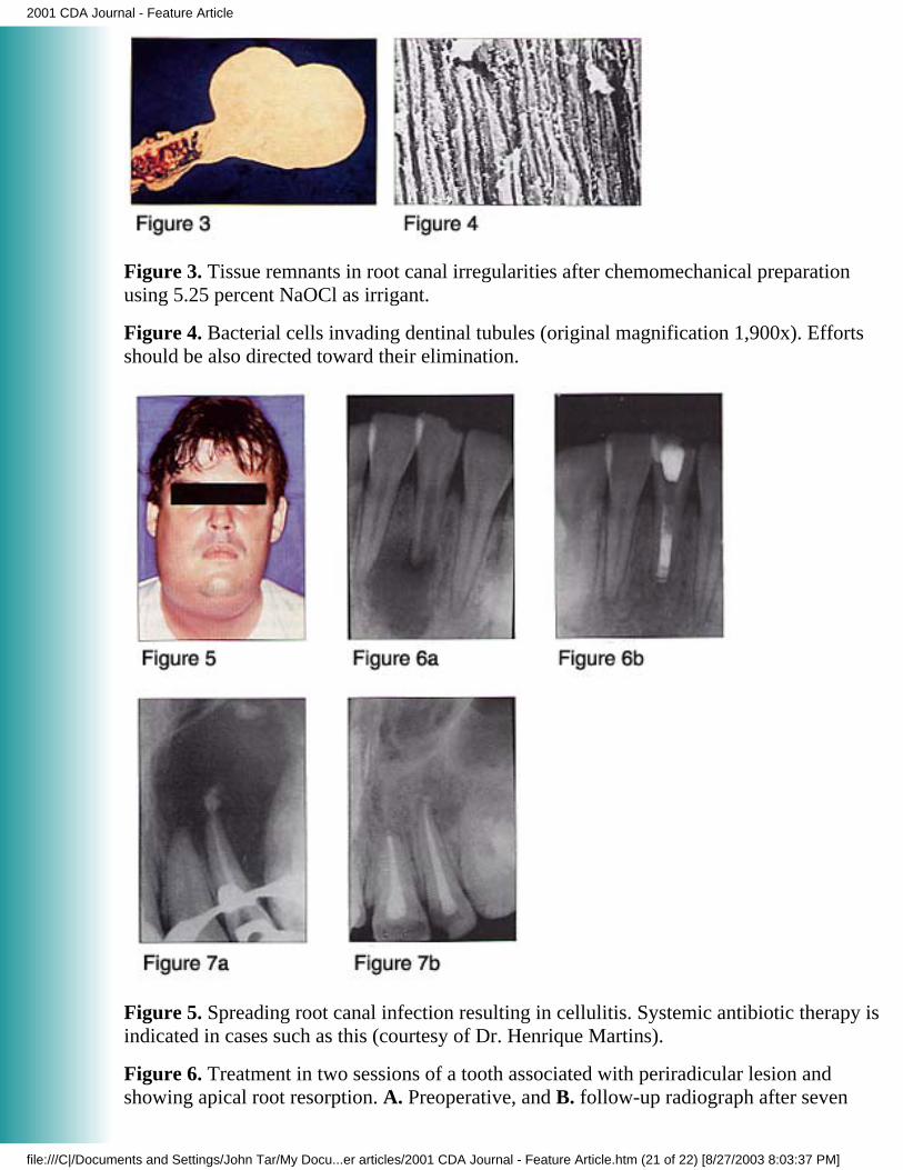
Figure 3. Tissue remnants in root canal irregularities after chemomechanical preparationusing 5.25 percent NaOCl as irrigant.
Figure 4. Bacterial cells invading dentinal tubules (original magnification 1,900x). Effortsshould be also directed toward their elimination.
Figure 5. Spreading root canal infection resulting in cellulitis. Systemic antibiotic therapy isindicated in cases such as this (courtesy of Dr. Henrique Martins).
Figure 6. Treatment in two sessions of a tooth associated with periradicular lesion andshowing apical root resorption. A. Preoperative, and B. follow-up radiograph after seven
2001 CDA Journal - Feature Article
file:///C|/Documents and Settings/John Tar/My Docu...er articles/2001 CDA Journal - Feature Article.htm (21 of 22) [8/27/2003 8:03:37 PM]

months showing repair of the lesion and stopping of the root resorption process.
Figure 7. Treatment in two sessions of a tooth associated with extensive periradicular bonedestruction. A. Preoperative, and B. follow-up radiograph showing bone repair of the lesion(courtesy of Dr. Luis Paulo Mussi).
JOURNAL OF THE CALIFORNIA DENTAL ASSOCIATION© 2001 CALIFORNIA DENTAL ASSOCIATION
2001 CDA Journal - Feature Article
file:///C|/Documents and Settings/John Tar/My Docu...er articles/2001 CDA Journal - Feature Article.htm (22 of 22) [8/27/2003 8:03:37 PM]