structural analysis and investigation of the
TRANSCRIPT

HAL Id: tel-01987004https://tel.archives-ouvertes.fr/tel-01987004
Submitted on 20 Jan 2019
HAL is a multi-disciplinary open accessarchive for the deposit and dissemination of sci-entific research documents, whether they are pub-lished or not. The documents may come fromteaching and research institutions in France orabroad, or from public or private research centers.
L’archive ouverte pluridisciplinaire HAL, estdestinée au dépôt et à la diffusion de documentsscientifiques de niveau recherche, publiés ou non,émanant des établissements d’enseignement et derecherche français ou étrangers, des laboratoirespublics ou privés.
Structural analysis and investigation of theStaphylococcus aureus ribosome and potential
anticancer drugsDaniya Kashinskaya
To cite this version:Daniya Kashinskaya. Structural analysis and investigation of the Staphylococcus aureus ribosome andpotential anticancer drugs. Structural Biology [q-bio.BM]. Université de Strasbourg, 2017. English.�NNT : 2017STRAJ110�. �tel-01987004�

UNIVERSITÉ DE STRASBOURG
ÉCOLE DOCTORALE des Sciences de la Vie et de la San té
[IGBMC - CNRS UMR 7104 - Inserm U 964]
THÈSE présentée par :
[ Daniya KASHINSKAYA ]
soutenue le : 12 Décembre 2017
pour obtenir le grade de : Docteur de l’université de Strasbourg
Discipline/ Spécialité : Biochimie, Biologie Moleculare et Structurale
TITRE de la thèse [Structural analysis and investigation of the
Staphylococcus aureus ribosome and potential anticancer drugs]
[Analyse structurale et étude du ribosome de Staphylococcus aureus et des médicaments
antitumoraux potentiels]
THÈSE dirigée par : [M. YUSUPOV Marat] Directeur de Recherche, CNRS, IGBMC
RAPPORTEURS :
[Mme. SCHMITT Emmanuelle] Directeur de Recherche, CNRS, l'Ecole polytechnique de Paris [M. DIAZ Jean-Jacques] Directeur de Recherche, Inserm, Centre de recherche en cancerologie de Lyon
AUTRES MEMBRES DU JURY : [Mme. ROMBY Pascale] Directeur de Recherche, CNRS, IBMC [M. MASQUIDA Benoît] Directeur de Recherche, CNRS, GMGM [M. KOLB Vyacheslav] Directeur de Recherche, Institut de recherche de protéines, Moscou

1
TABLE OF CONTENTS
LIST OF FIGURES ................................................................................................. 4
LIST OF TABLES ................................................................................................... 6
ACKNOWLEDGMENT .......................................................................................... 7
ABBREVIATIONS ................................................................................................. 8
RÉSUMÉ DE THÈSE ............................................................................................ 10
PREFACE ........................................................................................................... 20
General Introduction ......................................................................................... 23
I RIBOSOME CHARACTERIZATION...................................................................... 24
1. RIBOSOME ORGANIZATION .............................................................................................................. 24
2. SPECIFICATIONS OF BACTERIAL AND EUKARYOTIC RIBOSOME ................................................. 25
3. PROTEIN SYNTHESIS AS A KEY PROCESS OF LIFE ......................................................................... 28
3.1 INITIATION .................................................................................................................................................. 29
3.2 ELONGATION .............................................................................................................................................. 30
3.3 TERMINATION AND RECYCLING .......................................................................................................... 31
4. HISTORICAL ASPECTS OF STRUCTURAL STUDIES OF THE RIBOSOME ....................................... 32
5. X-RAY CRYSTALLOGRAPHY AND CRYO-ELECTRON MICROSCOPY OF THE RIBOSOME ............. 33
6. RIBOSOME – POTENTIAL THERAPEUTIC TARGET ........................................................................ 36
II MAIN CHARACTERISTICS OF STAPHYLOCOCCUS AUREUS ................................ 39
1. REGULATORY MECHANISMS OF VIRULENCE DETERMINANTS IN S. AUREUS ............................ 40
2. SUPER KILLER OF THE XXI CENTURY ............................................................................................. 43
3. ANTIBIOTIC RESISTANCE ................................................................................................................. 45
4. MECHANISMS OF ANTIBIOTICS ACTION AND BACTERIAL RESISTANCE .................................... 46
4.1 MODIFICATIONS OF THE ANTIBIOTIC MOLECULE ........................................................................ 47
4.2 DECREASED ANTIBIOTIC PENETRATION AND EFFLUX ............................................................... 48
4.3 CHANGES OF THE TARGET SITES ........................................................................................................ 49
5. S. AUREUS RIBOSOME AS A TARGET FOR DEVELOPING NEW THERAPEUTIC DRUGS ............... 50
III MAIN CHARACTERISTICS OF CANCER ............................................................. 53
1. CANCER CONTROL SYSTEM .............................................................................................................. 53
2
2. MECHANISMS OF DRUG RESISTANCE ............................................................................................. 54
3. POTENTIAL ANTICANCER DRUGS AGAINST RIBOSOME ASSEMBLY ........................................... 58
Part I - Structural analysis and investigation of the Staphylococcus aureus
ribosome........................................................................................................... 65
IV RIBOSOME PURIFICATION ............................................................................. 66
1. DESCRIPTION OF INITIAL RIBOSOME PURIFICATION PROTOCOL FROM S. AUREUS,
DEVELOPED IN THE YUSUPOV LABORATORY ....................................................................................... 67
1.1 S. AUREUS CELL GROWTH ....................................................................................................................... 67
1.2 LYSIS .............................................................................................................................................................. 68
1.3 PEG PRECIPITATION AND SUCROSE CUSHION ............................................................................... 68
1.4 SUCROSE DENSITY GRADIENT CENTRIFUGATION ........................................................................ 68
1.5 RIBOSOME CONCENTRATION AND STORAGE ................................................................................. 69
2. MODIFICATIONS OF RIBOSOME PURIFICATION PROTOCOL ........................................................ 70
2.1 CELL PREPARATION ................................................................................................................................. 70
2.2 LYSIS .............................................................................................................................................................. 70
2.3 SUCROSE DENSITY GRADIENT CENTRIFUGATION ........................................................................ 72
2.4 RIBOSOME CONCENTRATION AND STORAGE ................................................................................. 74
V RIBOSOME SAMPLE CHARACTERIZATION ....................................................... 74
VI COMPLEX FORMATION OF THE RIBOSOME FROM S. AUREUS WITH tRNAfMet
AND mRNA ....................................................................................................... 78
1. PURIFICATION OF THE INTACT tRNAfMET ....................................................................................... 80
2. tRNAfMet SAMPLE CHARACTERISZATION ........................................................................................ 83
VII CRYSTALLOGRAPHIC STUDIES OF S.AUREUS RIBOSOME ............................... 85
1. MACROMOLECULAR CRYSTALLIZATION IN THEORY ................................................................... 86
1.1 CRYSTALS GROWTH ................................................................................................................................. 87
1.2 MACROMOLECULAR CRYSTALLIZATION IN PRACTICE ............................................................... 87
2. CRYSTALLIZATION OF S.AUREUS RIBOSOME ................................................................................. 91
2.1 CRYSTALLIZATION ................................................................................................................................... 92
3. POST – CRYSTALLIZATION TREATMENT ...................................................................................... 102
3.1 POST – CRYSTALLIZATION TREATMENT OF S.AUREUS RIBOSOME CRYSTALS ................ 103
4. X-RAY DATA COLLECTION .............................................................................................................. 105
PRESPECTIVES ................................................................................................. 108
Part II - Structural analysis and investigation of potential anticancer drugs ..... 109
VIII BACKGROUND AND AIM OF THE PROJECTS ............................................... 110

3
1. CRYSTAL STRUCTURE OF POTENTIAL ANTICANCER DRUG C45 BOUND TO THE 80S
RIBOSOME ................................................................................................................................................ 110
2. CRYSTAL STRUCTURE OF THE CHEMOTHERAPEUTIC DRUG CISPLATIN BOUND TO THE 80S
RIBOSOME ................................................................................................................................................ 112
IX RESULTS AND METHODS ............................................................................. 113
1. PURIFICATION OF 80S RIBOSOME ................................................................................................. 113
2. CO-CRYSTALLIZATION .................................................................................................................... 114
3. POST-CRYSTALLIZATION TREATMENT ........................................................................................ 115
4. X-RAY DATA COLLECTION AND STRUCTURE DETERMINATION ............................................... 117
X STRUCTURAL INVESTIGATION OF POTENTIAL ANTI-CANCER DRUG C45 ........ 120
XI FLUORESCENCE RESONANCE ENERGY TRANSFER ........................................ 123
DISCUSSION AND PERSPECTIVES ..................................................................... 127
INVISIBLE CONNECTION BETWEEN PROJECTS .................................................. 129
GENERAL CONCLUSION ................................................................................... 130
LIST OF PUBLICATIONS AND COMMUNICATIONS ............................................ 131
REFERENCES .................................................................................................... 132

4
LIST OF FIGURES
Figure 1: Structural organization and functional sites of the ribosome (adapted from Melnikov et
al., 2012) ............................................................................................................................. 25
Figure 2: Composition of bacterial and eukaryotic ribosomes and the common core, based on X-
Ray studies(Melnikov et al.,2012) ......................................................................................... 26
Figure 3: Bacteria- and eukaryote-specific proteins and RNA expansions of the small ribosomal
subunit (Melnikov et al., 2012). ............................................................................................ 27
Figure 4: Bacteria- and eukaryote-specific proteins and RNA expansions of the large ribosomal
subunit (Melnikov et al., 2012). ............................................................................................ 28
Figure 5: The translation cycle in bacteria and eukaryotes (Melnikov et al., 2012). ................. 28
Figure 6: Structural evolution of Ribosome from 1983 to 2011. ............................................. 35
Figure 7: Antibiotic binding sites on the 30S ribosomal subunits (Wilson, 2014). ..................... 37
Figure 8: Antibiotic binding sites on 50S ribosomal subunits (adapted from Wilson, 2014). ..... 38
Figure 9: Schematic model of cell wall of gram-positive bacteria
(http://rcvetsblog.blogspot.fr/2014/03/bacterial-cell-wall.html). ........................................... 39
Figure 10: Regulation network of production of virulence factors in S. aureus (adapted from
Chevalier, 2009). .................................................................................................................. 41
Figure 11: Four waves of antibiotic resistance in S. aureus (Chambers and DeLeo, 2009)......... 44
Figure 12: Development of antibiotics and resistance (Clatworthy et al., 2007). ..................... 46
Figure 13: Antibacterial resistance mechanisms (adapted from Wilson, 2014). ....................... 47
Figure 14: Different types of efflux pumps in gram-positive and gram-negative bacteria (Piddock,
2006). ................................................................................................................................. 48
Figure 15: Schematic representation of the mechanism of action and resistance to linezolid
(Munita and Arias, 2016). ..................................................................................................... 49
Figure 16: Mechanisms of drug resistance (Molecular Genetics of Cancer, Figure 12). ............ 55
Figure 17: Structure of the amicoumacin A complex with yeast ribosome (Prokhorova et al., 2016).
........................................................................................................................................... 59
Figure 18: Crystal Structure of AglA-80S Ribosome (McClary et al., 2017). .............................. 60
Figure 19: The X-ray co-crystal structure of CL with the eukaryotic 80S ribosome (adapted from
Könst et al., 2017). ............................................................................................................... 61
Figure 20: Cisplatin-binding sites on the ribosome (Melnikov et al., 2016). ............................. 62
Figure 21: Cisplatin targets the mRNA channel in the ribosome (Melnikov et al., 2016). .......... 63

5
Figure 22: Cisplatin modification site near the GTPase activating center of the ribosome (Melnikov
et al., 2016). ........................................................................................................................ 64
Figure 23: Sucrose gradient profile. ...................................................................................... 69
Figure 24: Growth curves of Staph RN6390 (OD1 and 2) strain in the BHI medium. ................. 70
Figure 25: Sucrose gradient profiles after experiment II. ........................................................ 72
Figure 26: Sucrose gradient profiles after experiment III. ....................................................... 73
Figure 27: Sucrose gradient profiles after experiment IV. ....................................................... 74
Figure 28: Polyacrylamide Gel Electrophoresis of S. aureus ribosome sample. ........................ 76
Figure 29: Electrophoresis of the S. aureus ribosome samples after two steps of sucrose cushion
sedimentation purification protocol (experiment I). .............................................................. 77
Figure 30: Electrophoresis of the S. aureus ribosome samples with decreasing magnesium
concentration after PEG precipitation and in the sucrose cushion step (experiment II)............ 78
Figure 31: Anion-exchange chromatography (Q-Sepharose 25 ml). ........................................ 82
Figure 32: Denaturing 12% 8M urea PAGE of tRNAfMet. .......................................................... 83
Figure 33: High-pressure liquid chromatography: C4-silica column. ........................................ 84
Figure 34: Schematic representation of diffraction experiment. ............................................. 85
Figure 35: Schematic phase diagram of protein crystallization (Krauss et al, 2013) ................. 86
Figure 36: Idealized phase diagram of the trajectories of four different crystallization methods of
reaching nucleation and metastable zones (adapted from Krauss et al, 2013). ........................ 90
Figure 37: Crystals of S. aureus 70S ribosome in the CrysChem plate. ..................................... 96
Figure 38: Crystals of 70S ribosome with tRNA fMet with ratio 1:1. ......................................... 100
Figure 39: Crystals of 70S ribosome with tRNA fMet and mRNA with ratio 1:1:1. ...................... 101
Figure 40: Scheme of post-crystallization treatment. ............................................................ 105
Figure 41: Diffraction patterns of different crystals. ............................................................. 107
Figure 42: Cytotoxicity and translation-inhibitory activities of lissoclimide analogues and CHX
(adapted from Konst et al., 2017). ....................................................................................... 111
Figure 43: Computational modeling study of C45 compound. ............................................... 112
Figure 44: Sucrose gradient profile. ..................................................................................... 114
Figure 45: Diffraction patterns of C45/80S complex crystals. ................................................ 117
Figure 46: Unbiased difference density helped us to fit C45 unambiguously………………………….117
Figure 47:. Diffraction patterns of ciplatin/80S complex crystals. .......................................... 119
Figure 48: Chemical structure of C45 compound .................................................................. 120
Figure 49: C45, chlorolissoclimide and cycloheximide share the same binding pocket in the 60S E-
site. .................................................................................................................................... 120

6
Figure 50: Comparison of binding sites between C45 and CL ................................................. 121
Figure 51: The X-ray crystal structure of C45 compound with the eukaryotic 80S ribosome reveals
the molecular basis of translation inhibition. ............................................................................. 122
Figure 52: Schematic representation of pre-translocation complex. ......................................... 124
Figure 53: Ligand-Induced changes of the Human 80S Pre-translocation Complex visualized by
FRET ............................................................................................................................................. 124
Figure 54: Quantification of the data for CHX, CL and C45. ....................................................... 125
Figure 55: Population histogram of a washout experiment with C45........................................ 126
LIST OF TABLES
Table 1: Published crystal conditions for 70S ribosomes (adapted from Pearson, 2011). ......... 92
Table 2 Crystals of S. aureus 70S ribosome with presence PEG 8000 and NH4SCN in the
crystallization condition. ...................................................................................................... 93
Table 3: Crystals of the S. aureus 70S ribosome after modification of purification protocol. ..... 99
Table 4: Data collection and refinement statistic of C45/80S complex. .................................... 118

7
ACKNOWLEDGMENT
First of all I would like to thank all jury members Dr. Emmanuelle Schmitt, Dr. Jean-Jacques Diaz, Dr.
Vyacheslav Kolb, Dr. Pascale Romby and Dr. Benoît Masquida who kindly accepted my invitation and
found the time to evaluate my PhD work.
I would like to wholeheartedly thank to my advisors Marat and Gula for the support, assistance and
participation that they provided during all three years of my PhD education. My first meeting with
them in the Kazan University, during my master education, fascinated me and their life's course and
researches mainly motivated me to devote myself to science and in particular to the to the occupation
of ribosome in the structural biology field. Thank you very much for the opportunity to realize my
dream, I learned so much from you.
Thanks a lot to all current members of the ribosomal group Justine, Mumin, Alexey and former
members Marie, David, Irina, Yuzuru for the support, advice and friendly atmosphere, you were not
only my colleagues at the work, but became my good friends. I would like to express my special
gratitude to Iskander, Simone and Melanie for their participation in my projects and assistance in their
implementation.
I am thankful to the structural biology platform of IGBMC: Alistair and Pierre, they taught me various
techniques in crystallization and helped me with advices on the cryo-protection of crystals.
Due to my first project, I had the opportunity to meet wonderful people at the IBMC. I express special
gratitude to Pascale Romby and the members of her laboratory Stefano, Anne-Catherine, Isabelle, as
well as to former members Pierre, Alessandra and Mélodie for their help, advices and support in my
work.
During my PhD student time, I met and became friends with different people from different fields of
science. I would like to thank all my friends Xieyang, Karima, Moamen, Firas, Anya, Damola, Vova,
Grigory, Sasha and others for an unforgettably spent time together with you. I would like to say thank
you to Alexey who helped me a lot at the beginning of my PhD in the Strasbourg and showed me this
beautiful city. Thanks to Paul, the member of the SPB, with whom we together organized the career
lunch during two years and had the opportunity to communicate with various people outside the
academy. Thanks to my friends from Kazan: Katya, Nina and Lena for their support and love.
Most importantly, I would like to say thank you to my mother for the support of my aspiration in the
scientific work. I am very grateful to my beloved husband for his understanding, care and constant
support of me at any difficult moment.
Thanks to all of you that during important period of my life I always was together with such wonderful
people like you.
“We can only be said to be alive in those moments when our hearts are conscious of our treasures.” –
Thornton Wilder

8
ABBREVIATIONS
.
Å Angstrom unit (1Å =10-10 m)
aa-tRNA aminoacyl-tRNA
ABC ATP binding cassette
AglA agelastatin A
agr accessory gene regulator
AME aminoglycoside modifying enzymes
anti-SD
DMF
anti-Shine Dalgarno sequence
dimethylformamide
A-site aminoacyl site, (A-tRNA – A-site tRNA)
BHI Brain Heart Infusion broth
BSI blood stream infections
CAMP cationic antimicrobial peptide
CA-MRSA community-associated MRSA
CAT chloramphenicol acetyltransferases
CHX cycloheximide
CL chlorolissoclimide
CML chronic myelogenous leukemia
CNS coagulase-negative staphylococci
Cryo-EM cryo-electron microscopy
DAP daptomycin
DMSO dimethyl solfoxide
EDTA ethylenediaminetetraacetic acid
EG ethyloene glycol
E-site exit site (E-tRNA – E-site tRNA)
GDP guanosine diphosphate
GSH conjugation of glutathione
GTP guanosine triphosphate
HGT horizontal gene transfer
HPF hibernation-promoting factor
ICU intensive care unit
LSU Large Sub-Unit
MGE mobile genetic element
MLST multilocus sequence typing
MPD 3-methyl,1, 5-pentanediol
mRNA messenger ribonucleic acid
MRP1 multidrug resistance-associated protein 1
MRSA methicillin-susceptible S. aureus
MXR mitoxantrone resistance protein
NHNS National Healthcare Safety Network

9
NMR nuclear magnetic resonance
PAGE polyacrylamide gel electrophoresis
PEG polyethylene glycol
P-site peptidyl site, (P-tRNA – P-site tRNA)
PTC peptidyl transferase centre
PVL Panton–Valentine leukocidin
rRNA ribosomal ribonucleic acid
sae staphylococcal accessory element
sarA staphylococcal accessory regulator A
SCCmec staphylococcal chromosomal cassette mec
SD Shine-Dalgarno
SDS sodium dodecyl sulfate
smFRET single-molecule fluorescence resonance energy transfer
SSSS Staphylococcal Scalded Skin Syndrome
SSTI soft tissue infections
SSU Small Sub-Unit
tRNA transfer ribonucleic acid
tRNAfMet N-Formylmethionine-tRNA
TSS toxic shock syndrome
VAP ventilator-associated pneumonia
VISA vancomycin-intermediate S. aureus
VRSA vancomycin-resistant S. aureus
YPD Yeast extract Peptone Dextrose
σA sigma factor A
σB sigma factor B

10
RÉSUMÉ DE THÈSE
La biosynthèse protéique est un processus fondamental, retrouvé au sein de toutes les cellules, qui permet
d'assurer le décodage de l'information génétique. Ce mécanisme de traduction implique de nombreux
partenaires, protéines et acides nucléiques, et nécessite une parfaite coordination de leurs actions afin
d'assurer la fidélité du transfert de l'information génétique. Le ribosome est l’acteur central de ce
processus de traduction. Abondant dans les cellules et conservé au cours de l'évolution, celui-ci est
composé de deux sous-unités, une petite et une grande, combinant des protéines et des acides nucléiques
qui s'associent au cours de la traduction pour former un ribosome fonctionnel. Dans cet état, le ribosome
est capable de catalyser l'addition séquentielle des acides aminés de la chaîne peptidique en cours de
synthèse en utilisant l'ARN messager (ARNm) comme matrice et les ARN de transfert aminoacylés (aa-
ARNt) comme substrats. Les deux sous-unités présentent des fonctions bien distinctes. Le décodage de
l’information génétique contenu dans l’ARNm est assuré par la petite sous-unité tandis que la grande sous-
unité catalyse la réaction de transpeptidation permettant la formation d’une liaison peptidique entre
chaque acide aminé. Une dizaine de facteurs protéiques se lient successivement au ribosome afin de
catalyser les différentes étapes de la traduction, à savoir: l'initiation, l'élongation, la terminaison et le
recyclage. Bien que la synthèse des protéines conservée parmi tout les domaines du vivant, tous les
organismes ont des spécificités. Soit, les bactéries Gram positives et Gram négatives ont leurs propres
spécificités concernant la régulation et l'organisation de la machinerie traductionnelle.
Les structures à haute résolution des ribosomes et sous-unités ribosomales obtenues par cristallographie
aux rayons X et microscopie électronique ont révolutionnées le domaine de la traduction des protéines. La
connaissance des positions précises des résidus dans le ribosome dans divers états a conduit à une
meilleure compréhension des mécanismes complexes de la synthèse des protéines. Les structures de
complexes ribosomaux avec des antibiotiques et des composés antifongiques ont fourni un aperçu sans
précédent de leurs mécanismes d'action et facilitent également la conception de médicaments plus
efficaces (Garreau de Loubresse et al, 2014; revue Wilson, 2014). Il a été découvert que la plupart des
inhibiteurs de la traduction se lient aux régions conservées des ribosomes (centre de peptidyltransférase,
les sites de liaison d'ARNt, tunnel de sortie de la protéine, etc.). Cependant, les effets d’un même
médicament sur des espèces différentes peuvent varier. Un excellent exemple est fourni par les bactéries
résistantes à de nombreux antibiotiques couramment utilisés. Parmi celles-ci, Staphylococcus aureus (S.
aureus), un agent pathogène qui provoque de graves et nombreuses infections chez l’homme. Cette
bactérie est assez unique et donc, requiert une approche individuelle pour le traitement.
Les projets de la présente thèse ont progressé dans deux directions différentes et avec deux organismes
différents: Staphylococcus aureus et Saccharomyces cerevisiae.
S. aureus est responsable des infections nosocomiales et infections sévères et peuvent causer une variété
de maladies potentiellement mortelles chez l’homme. Ces infections comprennent l'endocardite, la
péritonite, pneumonie nécrosante, la bactériémie, la méningite, l'ostéomyélite, l'arthrite septique, et les
infections des os, des articulations et des organes (Fridkin et al, 2005).
S. aureus est un pathogène majeur d'une importance croissante en raison de sa haute résistance aux
antibiotiques (Lowy, 1998). Un rapport par l'Organisation mondiale de la Santé (OMS) révèle que la
résistance aux antimicrobiens, y compris la résistance aux antibiotiques n'est plus une prédiction pour
l'avenir, elle se passe maintenant dans toutes les régions du monde et a le potentiel de toucher tout le
monde, de tout âge, dans tous les pays. La résistance aux antibiotiques est maintenant une menace

11
majeure pour la santé publique. Selon ce rapport, pas moins de 60% des infections à S. aureus sont
déterminées comme résistantes à la méthicilline (SARM) en Europe (World Health Organization, 2015).
Les gènes responsables de la résistance sont souvent transmis par des éléments génétiques mobiles tels
que les plasmides et, par conséquent, populations bactériennes peuvent acquérir une résistance très
rapidement. Divers mécanismes de résistance aux antibiotiques. Les bactéries peuvent empêcher la
pénétration de l'antibiotique dans la cellule, le retirer de la cellule en utilisant une pompe d'efflux , de
dégrader ou modifier l'antibiotique . Alternativement les bactéries peuvent muter , modifier ou protéger
les cibles des antibiotiques. La conception de nouveaux agents antimicrobiens est fortement requise pour
le traitement des maladies causées par S. aureus. Pour comprendre les aspects structurels de la résistance
aux antibiotiques nous avons besoin de connaître la structure de leur cible. Par conséquent nous nous
sommes concentrés sur la structure du ribosome, qui est la principale cible des antibiotiques les plus
couramment utilisés. La résolution de la structure du ribosome de S. aureus sera la première étape vers la
compréhension du mécanisme précis de sa résistance aux antibiotiques et facilitera la conception de
nouveaux composés antistaphylococciques.
Les ribosomes bactéries et eucaryotes partagent une structure minimale conservée et présentent une série
de protéines et d’éléments d’ARNr spécifiques à chaque domaine du vivant. Cette variation de structure se
traduit par un changement de poids moléculaire. Avec une masse totale avoisinant 3.3 MDa, le ribosome
eucaryote est 40% plus volumineux que son homologue bactérien. Bien que les étapes clés de la synthèse
protéique soient conservées chez les bactéries et les eucaryotes, les fonctions endossées par le ribosome
eucaryote présentent de nombreuses spécificités. Chez les bactéries, une dizaine de facteurs protéiques se
lient successivement au ribosome afin de catalyser les différentes étapes de la traduction, à savoir :
l'initiation, l'élongation, la terminaison et le recyclage. En revanche, les mécanismes de la synthèse des
protéines chez les eucaryotes diffèrent à plusieurs niveaux. A titre d’exemple, jusqu’à treize facteurs
distincts sont impliqués dans l’étape d’initiation de la traduction chez les eucaryotes. Par ailleurs, les étapes
de terminaison et de recyclage s’opèrent de manière différente et requièrent la participation de facteurs
non conservés chez les bactéries. L’assemblage du ribosome eucaryote est hautement contrôlé et implique
une centaine de facteurs. Enfin, hormis son rôle prépondérant dans la synthèse protéique, le ribosome
eucaryote et ses constituants participent activement à la régulation de l’expression des gènes.
Projet de recherche 1: Analyse structurale et étude du ribosome de Staphylococcus aureus.
Le projet dédié à de déterminer la structure cristallographique du ribosome de S. aureus a été initié en
2011 dans le laboratoire de Yusupov. Cependant, des difficultés spécifiques pour obtenir une diffraction à
haute résolution des cristaux de ribosomes de S. aureus 70S n'ont pas permis de résoudre sa structure.
On sait que certains aspects de la synthèse des protéines diffèrent entre les bactéries Gram négatif et Gram
positif. En outre, les bactéries pathogènes ont développé des mécanismes complexes de régulation de la
traduction, qui fournissent une efficacité élevée de la pathogenèse et facilitent la survie dans des
conditions stressantes. De plus, de nombreuses bactéries pathogènes (y compris S. aureus) montrent une
résistance extrêmement élevée aux antibiotiques ciblant les ribosomes, qui est souvent médiée par des
modifications du ribosome. La structure cristalline du ribosome des bactéries pathogènes multirésistantes
Gram-positives S. aureus révélerait des caractéristiques structurelles particulières de la machinerie de
traduction, ce qui nous amènera à mieux comprendre les spécifications de la régulation de sa synthèse
protéique et de la survie des agents pathogènes. Plus important encore, il servira de modèle pour
développer de nouveaux médicaments anti-staphylococciques et rendre le traitement médical plus
efficace. De nombreux antibiotiques agissent en inhibant sélectivement la synthèse des protéines dans les

12
bactéries, sans perturber les ribosomes hôtes et, par conséquent, leurs cellules. Les structures cristallines
d'antibiotiques produits naturellement et leurs dérivés semi-synthétiques liés aux particules ribosomiques
ont fourni un aperçu de leurs mécanismes d'action, et ils facilitent également la conception d'antibiotiques
plus efficaces pour cibler les bactéries multirésistantes.
Mon objectif principal de ce projet était d'élucider la structure du ribosome 70S complet de S. aureus en
utilisant l'analyse des rayons X comme outil principal. Ainsi le projet consistait en des tâches suivantes:
• la préparation des cellules: culture, la récolte, lyse ;
• modification du protocole initial de purification du ribosome 70S de S.aureus (issu du laboratoire de
Yusupov) qui fournira des ribosomes purs et stables, convenant à la cristallisation;
• stabilisation du ribosome 70S par deux voies de formation de complexe avec l'ARNt fMet, l'ARNm
et avec le domaine N-terminal du facteur favorisant l'hibernation (HPF);
• traitement de cristallisation et de post-cristallisation: recherche de conditions de cristallisation,
croissance de gros cristaux et préparation pour l'analyse par diffraction;
• développer la stratégie de collecte de données au synchrotron;
• résoudre la structure: traiter les données et construire le modèle.
Initialement, le protocole de purification des ribosomes de S. aureus a été créé et développé dans le
laboratoire Yusupov (Khusainov et al., 2016) sur la base des protocoles utilisés pour les bactéries Thermus
thermophilus (Gogia et al., 1986, Yusupov et al., 2001) et de levures Saccharomyces cerevisiae (Ben-Shem
et al., 2011). Malheureusement, l'échantillon obtenu après ce protocole n'était pas assez homogène et les
cristaux obtenus à partir de cet échantillon donnaient de mauvaises diffractions (19 Å), même après une
optimisation des conditions de cryo-protection. Les derniers, selon l'analyse cryo -EM des ribosomes, nous
avons obtenu un résultat dans lequel environ 10% de ribosomes ont une contamination par l'ARNt (A / P, P
/ p, P / E, E) et presque tous liés au site P du ribosome. Ainsi, l'ARNt peptydil qui a probablement un peptide
à la surface du ribosome pourrait influencer la diffraction. Donc, ma tâche principale était de se débarrasser
des impuretés de l'ARNt. Pour atteindre cet objectif, un certain nombre d'expériences ont été réalisées
pour modifier le protocole de purification des ribosomes.

13
Fig I: Gel de polyacrylamide Electrophorèse de l'échantillon de S. aureus ribosome. A) Dénaturation de
l'ARN PAGE 4%: 1 - après modification du protocole de purification (9μg), 2 - sans modification du
protocole de purification (contrôle) (9μg). B) Dénaturation de l'ARN PAGE 12%: 1 - tRNAfMet (1μg), 2 -
après modification du protocole de purification (9μg). C) SDS PAGE unidimensionnelle: après
modification du protocole de purification.
Selon la Fig IA, nous avons observé trois bandes correspondant à l'ARNr 23S, 16S et à l'ARN 5S pour le
contrôle et pour l'échantillon de ribosome après modification du protocole de purification. Cependant, en
raison de la PAGE 4%, nous ne pouvons pas interpréter les résultats concernant la contamination par
l'ARNt, car la taille du pore du gel était grande pour une petite taille d'ARNt. A cet effet, a été réalisée PAGE
12% (Fig IB) et nous avons pu observer une réduction de l'intensité dans les bandes de contaminations
ARNt comparer avec témoin (pour plus de détails voir le chapitre avec la caractérisation de l'échantillon de
ribosome). Nous avons analysé l'échantillon après modifications du protocole pour éliminer les protéines
non ribosomiques et le gel SDS a démontré la pureté de l'échantillon ribosomique et n'a pas révélé de
contamination par des protéines de haut poids moléculaire comme les composants du complexe pyruvate
déshydrogénase, qui co-purifient parfois avec des bactéries ribosomes (Fig IC).
Particules cristallines initiales de ribosome 70S de S. aureus ont été obtenus en utilisant une recherche
robotique. Après plusieurs étapes d'optimisation de cristallisation, de grands cristaux tridimensionnels ont
été obtenus. Typiquement, les cristaux sont apparus de façon reproductible dans les 7 - 10 jours et ont
atteint leur taille moyenne (100 × 50 × 20 μm) après deux semaines supplémentaires.
~ 250
~ 130
~ 100
~ 70
~ 55
~ 35
1 2 16S/23S
rRNA
5S rRNA
tRNA
1 2 16S/23S rRNA
5S rRNA
A B C

14
Fig II: Les cristaux du ribosome 70S de S. aureus
La taille des cristaux de ribosome de S. aureus est suffisante pour l'analyse par diffraction aux rayons X. Les
conditions de déshydratation et de cryo-refroidissement ont été optimisées pour les cristaux obtenus. Les
premières données de diffraction de cristaux du ribosome 70S de S. aureus ont été recueillies. La diffraction
de ces cristaux atteint au maximum 17.5 Å, le groupe de l'espace a été déterminée comme P42212 et la
taille de l'unité asymétrique était: 450 × 450 × 280 Å. Dans le but d'améliorer la diffraction des cristaux à
partir du ribosome de S.aureus, nous avons essayé de stabiliser notre macromolécule en raison de la
formation complexe avec l'ARNt fMet et l'ARNm et les cristaux obtenus apparaissaient de façon
reproductible dans les 30-35 jours et atteignaient leur taille moyenne (200 × 80 × 20 μm ) après deux
semaines supplémentaires.
~ 100 µm
~ 90 µm
~ 70 µm
2µl + 2µl CrysChem plate 100 mM Tris-Acetate pH 7.0 400 mM KSCN 5.5 % PEG 8000 10% MPD 2.0 mM Spermedine 8 mg/ml Ribosomes
2µl+2µl CrysChem plate 100 mM Tris-Acetate pH 7.0 400 mM KSCN 5.2-6.0 % PEG 8000 10% MPD 10 mM Mg(OAc)2 8 mg/ml Ribosomes
0.2µl+0.2µl MRC 2 drop plate 100 mM Tris-Acetate pH 7.0 9 mM Mg(OAc)2 500 mM NH4SCN 5 % PEG 8000 6% MPD 2% PEG400 10% Sucrose 8 mg/ml Ribosomes

15
Fig III: Les cristaux du ribosome 70S avec ARNt fMet (A) et avec ARNt fMet et l'ARNm (B).
S. aureus facteur de promotion de l'hibernation (HPF) domaine N – terminal (SaHPF-NTD) se lie à la petite
sous-unité est similaire à ses homologues de E. coli HPF, E. coli YfiA, et un YfiA spécifique de plaste
(Khusainov et al, 2017). Et selon la suggestion (Polikanov et al., 2012) que YfiA empêche la tête (30S) de
déplacer du ribosome et stabilise la macromolécule entière. Nous avons décidé de co-cristalliser le SaHPF-
NTD avec le ribosome afin de stabiliser notre assemblage et potentiellement améliorer notre résolution des
ribosomes de S. aureus.
Nous avons obtenu des cristaux qui apparaissent reproductiblement dans les 7 à 15 jours et ont atteint leur
taille moyenne (100 × 50 × 20 μm) après deux semaines supplémentaires. Tous ces cristaux après formation
complexe ont été diffractés à une résolution maximale de 17.5 Å.
Toutes les études obtenues dans ce travail faciliteront les recherches ultérieures dans la direction
cristallographique du ribosome 70S de S. aureus, qui aidera à obtenir une structure à haute résolution dans
un proche avenir. Les résultats obtenus serviront de base pour le développement de nouveaux composés
contre la bactérie pathogène et extrêmement résistante qu’est S. aureus.
Projet de recherche 2: Analyse structurale et étude des médicaments antitumoraux potentiels
Le ribosome est un propulseur central de tout procédé cellulaire. Il est donc considéré comme la cible
privilégiée des petites molécules inhibitrices. Certains inhibiteurs spécifiques du ribosome eucaryote
(naturels ou synthétisés chimiquement) sont maintenant considérés comme de potentiels médicaments
anticancéreux.
Il est fermement établi que la quantité de ribosomes est fortement corrélée avec le taux de synthèse des
protéines et la croissance et la prolifération des cellules. Il est important de prendre en compte dans le cas
des cellules cancéreuses, ce qui montre une augmentation globale de la synthèse des protéines pour
favoriser leur comportement hyperprolifératif. Le ribosome est donc devenu une importante cible
~90 µm
~150 µm
~70 µm ~80 µm
A B
0.2µl+0.2µl MRC 2 drop plate 4.50% PEG8000 550mM KSCN 10% MPD 5mM Mg(OAc)2 100mM Tris-acetate 7.0 pH 2mM Spermedine 10mg/ml Ribosome
0.2µl+0.2µl MRC 2 drop plate 4.50% PEG8000 600mM KSCN 10% MPD 5mM Mg(OAc)2 100mM Tris acetate 7.0 pH 2mM Spermedine 7mg/ml Ribosome

16
druggable. Les inhibiteurs de la synthèse des protéines eucaryotiques ont montré un potentiel
thérapeutique important pour traiter une large gamme de cancers humains. En tant qu'exemple principal,
en 2012, la FDA a approuvé le premier inhibiteur de la traduction, l'alcaloïde d'homoharringtonine naturel,
tel que Synribo pour le traitement de la leucémie myéloïde chronique. Pour étudier le mécanisme d'action
de ces inhibiteurs ciblant le ribosome eucaryote, dans le laboratoire Yusupov a résolu récemment la
structure de 17 molécules petites différentes ciblant le ribosome 80S de S. cerevisiae (Garreau de
Loubresse et al, 2014; Prokhorova et al.,2016; McClary et al.,2017).
Mon objectif principal était de déchiffrer le mécanisme moléculaire de liaison des inhibiteurs spécifiques
antitumoraux, qui par la suite pourraient conduire à définir d'autres stratégies de conception de
médicaments pour obtenir de nouveaux composés avec une puissance plus élevée tout en ayant une
cytotoxicité inférieure. Je m'intéressais particulièrement à l'analyse de la structure du médicament
chimiothérapeutique cisplatine et d'un analogue nouvellement synthétisé du chlorolissoclimide (nommé
C45) avec les ribosomes eucaryotes.
Les médicaments chimio-thérapeutiques peuvent causer une neurotoxicité périphérique (neuropathie) et
une neurotoxicité centrale (déficits cognitifs mineurs, encéphalopathie, démence, coma). Le cisplatine,
utilisé contre diverses tumeurs malignes, induit une neurotoxicité périphérique, souvent accompagnée
d’une aggravation hors thérapie, d’un signe de Lhermitte et de crampes musculaires. La neurotoxicité
périphérique se développe chez environ 50% des patients traités au cisplatine. Une connaissance détaillée
des sites de liaison du cisplatine au niveau de l'ARN ribosomique peut non seulement constituer un outil de
prédiction des sites de modification d'autres ARN cellulaires essentiels, mais aiderait également à concevoir
des études expérimentales sur la nature polyvalente de la cytotoxicité du cisplatine. Par conséquent, cela
aiderait à résoudre les problèmes d’effets secondaires importants.
Notre groupe a récemment publié une étude multidisciplinaire dans laquelle nous démêlons le mécanisme
d'action du chlorolissoclimide (CL), un composé qui partage la similarité chimique avec le cycloheximide
(CHX), mais présentant une cytotoxicité inférieure prometteuse (Könst et al., 2017). CL se lie au site E de la
sous-unité 60S et crée de nouvelles interactions par rapport à CHX. En détails, il crée une interaction
inhabituelle d'empilement d'halogène-π avec le résidu G2794 à travers son atome de chlore. Nous nous
sommes intéressés à ce nouveau type d'interaction avec le ribosome et nous avons donc décidé de
résoudre la structure cristalline du S.cerevisiae 80S en complexe avec un autre composé de lissoclimide,
porteur d'un atome de chlore supplémentaire. Le couplage de structures à haute résolution avec la
conception de médicaments assistée par ordinateur et l'analyse de transfert d'énergie par résonance de
fluorescence à une seule molécule (smFRET) guidera davantage la conception d'inhibiteurs plus sélectifs et
moins cytotoxiques.
Le modèle eucaryote du ribosome que j'ai utilisé était celui de S. cerevisiae qui a été étudié pendant
plusieurs années dans notre laboratoire, ce qui en fait un outil bien connu pour les études structurales. À ce
jour, 70% des cristaux de ribosomes de S. cerevisiae diffractent à des résolutions proches de 3.0 Å. Le
protocole pour obtenir la structure du médicament anticancéreux potentiel et du cisplatine avec le
ribosome eucaryote se composait de 6 étapes principales:
1) la croissance des cellules de levure;
2) purification du ribosome 80S à partir de S. cerevisiae;
3) la co-cristallisation du ribosome avec des médicaments antitumoraux potentiels appropriés à l'analyse
par diffraction des rayons X;

17
4) cryo-protection et déshydratation du cristal (pour remplacer les molécules d'eau dans les cristaux et les
protéger pendant le processus de refroidissement, cette étape est également nécessaire pour augmenter le
tassement des molécules dans les cristaux);
5) la collecte de données à haute résolution et la résolution du problème de phase;
6) le modèle de construction et de raffinement.
Dans le cas du projet C45, grâce à des connaissances issues de la synthèse semi-synthétique et de la
synthèse analogique, nous avons approfondi notre compréhension de la relation structure-activité dans la
famille des inhibiteurs cytostatiques inhibiteurs de la traduction.
La structure cristalline du ribosome 80S de S. cerevisiae dans le complexe avec un échantillon synthétique
de l'inhibiteur C45 a été résolue à une résolution maximale de 3.1 Å. Nous avons découvert la base
structurelle de l'inhibition de la protéine syntèse avec l'étude co-cristallographique de C45 synthétique lié
au ribosome. Le mode de liaison intéressant comprend un nouveau deux face sur l'interaction de chlore-π
du ligand avec deux résidus de guanine. Cette interaction favorable basée sur la dispersion semble
permettre une stabilisation du composé C45 dans le site E du ribosome, et donc des effets apparentés
pourraient être exploités dans la conception d'autres ligands d'acide nucléique.
Fig IV: La structure cristalline aux rayons X du composé C45 avec le ribosome 80S eucaryote révèle la base
moléculaire de l'inhibition de la traduction. A) Détails des interactions qui se produisent entre la molécule
C45 (bleue, représentée par des bâtonnets) et les résidus voisins. Des contacts directs ont lieu avec les
nucléotides G92, C93, U2763, A2802, G2794 et G2793 de l'ARNr 25S. B) Zoom de la poche de liaison de C45,
avec le dichlorure diaxial dans le système d'anneau décalin créer une conformation en bateau torsadé dans
laquelle les chlorures pseudoquatoriaux sont prêts pour deux interactions face-sur la géométrie halogène-π
avec les guanines G2793 et G2794 de l'ARNr 25S.
Selon l'analyse de structure, le site de liaison C45 est situé à l'extrémité CCA de l'ARNt du site E sur les LSU,
comme indiqué précédemment pour le chlorolossoclimide (CL) et les inhibiteurs de glutarimide
A B

18
cycloheximide (CHX) et lactimidomycine (LTM) (Fig V). La comparaison de la liaison de C45 avec celle de
CHX et CL montre un réseau similaire d'interactions de la fraction contenant de l'imide avec un certain
nombre de nucléotides universellement conservés de l'ARNr 25S, à savoir G92, C93 et U2763
Fig V: C45, le chlorolissoclimide et la cycloheximide partagent la même poche de liaison dans le site E du
60S.
En outre, selon l'analyse FRET dans le complexe de pré-translocation ribosome humain, nous observons
que C45 s'équilibre très lentement avec le ribosome sur une échelle de temps de dizaines de minutes,
cependant, il a un très long temps de résidence une fois lié. De plus, nous avons calculé une constante de
dissociation pour C45 et elle est en moyenne de 50 à 150 μM. En comparaison, CHX et CL sont
respectivement d'environ 5.5 μM et 11.5 μM, montrant ainsi des affinités de liaison similaires. Le C45 est
un liant plus faible que le CL, ce qui est bien corrélé avec les données publiées (Konst et al., 2017).
Concernant le projet complexe cisplatine / 80S, nous avons trouvé le rapport optimal du cisplatine (37.5
μM) pour le processus de co-cristallisation avec le ribosome eucaryote. Ceux-ci nous ont permis d'obtenir
une bonne forme et une bonne taille de cristaux pour effectuer d'autres expériences de diffraction. En
outre, nous avons déterminé l'influence du détergent (Deoxy-Big Chap) sur la formation du complexe
cisplatine avec le ribosome. Très probablement dans ce cas, l'incubation du complexe avec le détergent,
avant le processus de cristallisation, a réduit l'affinité de liaison du cisplatine au ribosome 80S. Nous avons
également observé que le cisplatine lui-même influait sur la croissance cristalline et changeait de forme
cristalline, passant du parallélépipède rectangulaire habituel au prisme hexagonal. Nous avons pu améliorer
la diffraction des cristaux jusqu'à 4 Å. Ces données, nous avons obtenu uniquement dans les cristaux qui
ont été trempés en présence de cisplatine (250 µM). Cependant, le rendement de la résolution moyenne
était très faible, seulement 5%, ce qui rend difficile la construction de la structure. Dans le cas du ribosome
de co-cristallisation avec le cisplatine, nous n'avions pas plus de 6-7 Å de résolution. Ainsi, le projet
nécessiterait beaucoup de procédures d'optimisation et aussi, très probablement, l'évitement de l'osmium
hexamine dans les cristaux, pour pouvoir utiliser le signal anormal provenant des quelques atomes de Pt

19
qui devraient se lier étroitement au ribosome. Cependant, jusqu'à présent, le traitement des S.cerevisiae
80S ribosome cristaux sans osmium hexamine donné que la diffraction pauvre.
En dépit du fait que l'osmium hexamine et le platine ont les mêmes sites de liaison dans le ribosome 80S de
S.cerevisiae et appartiennent au groupe des métaux lourds, le remplacement de l'osmium hexamine dans la
procédure de cryo-protection par le platine entraîne une réduction de la diffraction des cristaux.
La tentative d'exclure l'osmium des conditions normales de cryoprotection a échoué, et l'on peut donc
conclure qu'il est nécessaire de changer le concept de processus de cryo-protection des cristaux de
ribosomes 80S dans leur ensemble. A son tour, notre laboratoire est en train de développer cela de
manière plus robuste.

20
PREFACE
The projects of the present PhD thesis have progressed in two different directions and with two
diverse organisms: Staphylococcus aureus and Saccharomyces cerevisiae.
The first project is dedicated to determine the crystal structure of the 70S ribosome from S. aureus and
was initiated in 2011 in the Yusupov’s laboratory. However, specific difficulties to obtain high
resolution diffraction of S. aureus 70S ribosome crystals did not give the opportunity to solve its
structure.
It is known that some aspects of protein synthesis differ between Gram-negative and Gram-positive
bacteria. Additionally, pathogenic bacteria have evolved complex mechanisms of translation
regulation, that provides high efficiency of pathogenesis and facilitate survival under stressful
conditions. Moreover, many pathogenic bacteria (including S. aureus) show extremely high resistance
to ribosome-targeting antibiotics, which is often mediated by modifications of the ribosome. The
crystal structure of the ribosome from Gram-positive multi-resistant pathogenic bacteria S. aureus
would reveal peculiar structural features of translation machinery, thus will lead us to better
understanding the specifications of regulation of its protein synthesis, and pathogen survival. Most
importantly, it will serve as a model system for developing new anti-staphylococcal drugs and make
medical treatment more efficient. Many antibiotics act by selectively inhibiting the protein synthesis in
bacteria, without perturbing the host ribosomes and, therefore, their cells. Crystal structures of
naturally produced antibiotics and their semi-synthetic derivatives bound to ribosomal particles have
provided insight into their mechanisms of action, and they are also facilitating the design of more
effective antibiotics for targeting multidrug-resistant bacteria.
My main goal of this project was to elucidate the structure of full 70S ribosome from S. aureus using X-
ray analysis as a main tool. Thus the project consisted of next tasks:
• preparing the cells: growing, harvesting, breaking;
• modification of initial protocol of purification 70S ribosome from S.aureus (comes from
Yusupov’s laboratory) which will provide pure and stable ribosomes, suitable for crystallization;
• stabilization of 70S ribosome by two way of complex formation with tRNA fMet, mRNA and with
N-terminal domain of hibernation promoting factor (HPF);
• crystallization and post-crystallization treatment: searching for crystallization conditions,
growing big crystals and preparation for diffraction analysis;
• developing the strategy of data collection at the synchrotron;
• solving the structure: processing the data and building the model.
Results of my work on the determination of high resolution structure of the ribosome from S. aureus
will be discussed in the following chapters.
In the second direction, the project aim is to determine the structure of potential anticancer drugs
targeting the eukaryotic ribosome.
Ribosome is a central prop of all cellular process and it is thus considered as the favored target for
small-molecules inhibitors. It is therefore intuitive that some eukaryotic specific inhibitors of the
ribosome, natural product and chemical synthesized, are now considered as potential anticancer drug.

21
My main goal was to decipher the molecular mechanism of binding of specific antitumor inhibitors,
which subsequently might lead to define further drug-design strategies to obtain new compounds with
higher potency while having lower cytotoxicity.
I was particularly interested in the structure analysis of the chemotherapeutic drug cisplatin and a
newly synthesized analogue of chlorolissoclimide (named C45) with the eukaryotic ribosomes.
Chemotherapeutic drugs may cause both peripheral neurotoxicity, consisting mainly of a peripheral
neuropathy, and central neurotoxicity ranging from minor cognitive deficits to encephalopathy with
dementia or even coma. Cisplatin – the platinum analogs, used for various malignancies, is the most
important drug inducing peripheral neurotoxicity, with often off-therapy worsening, Lhermitte's sign,
muscle cramps. Peripheral neurotoxicity develops in approximately 50% of patients receiving cisplatin.
Platinum compounds are active in the treatment of solid tumors, but peripheral neuropathy is the
major non-hematological dose-limiting adverse effect especially for cisplatin. For the moment, there is
no effective strategy for the management of the neurotoxicity induced by platinum agents. Detailed
knowledge of cisplatin binding sites in the ribosomal RNA may not only provide a tool for prediction of
modification sites in other essential cellular RNAs, but will also help design experimental studies of the
multifaceted nature of cisplatin cytotoxicity. Consequently, this structural knowledge might help to
solve problems related to the strong side effects.
To study the mechanism of action of inhibitors targeting the eukaryotic ribosome, our group recently
solved the structure of 17 different small molecules targeting the S. cerevisiae 80S ribosome (Garreau
de Loubresse et al., 2014; Prokhorova et al., 2016). Additionally, our group recently published a
multidisciplinary study in which we unravel the mechanism of action of chlorolissoclimide (CL), a
compound sharing chemical similarity to cycloheximide (CHX), but showing promising lower
cytotoxicity (Könst et al., 2017). CL binds to the E-site of the 60S subunit and creates novel interactions
compared to CHX. In details, it creates an unusual halogen-π stacking interaction with residue G2794
through its chlorine atom. We were interested in this new type of interaction with the ribosome and
therefore we decided to solve crystal structure of the S.cerevisiae 80S in complex with another
lissoclimide compound, bearing an additional chlorine atom. The coupling of high-resolution structures
with computationally-driven drug design and single-molecule fluorescence resonance energy transfer
(smFRET) analysis will further guide the design of more selective and less cytotoxic inhibitors.
The eukaryotic model of the ribosome that I used was the one from S. cerevisiae that has been studied
for several years in our laboratory, making it a well-understood tool for structural studies. To date,
70% of the S. cerevisiae ribosome crystals are diffracting at resolutions close to 3.0 Å. The protocol for
getting the structure of the potential anticancer drug and cisplatin with the eukaryotic ribosome
consisted of 6 main steps:
1) growing of yeast cells;
2) purification of 80S ribosome from S. cerevisiae;
3) co-crystallization of the ribosome with potential antitumor drugs suitable for X-ray diffraction
analysis;
4) cryo-protection and dehydration of the crystal (to replace water molecules in the crystals and
protect them during the cooling process. This step is also necessary to increase the packing of the
molecules in the crystals);

22
5) data collection of high resolution data and, solving of the phase problem;
6) model building and refinement.
The results of my work on the determination of high resolution structures of potential antitumor drugs
with the eukaryotic ribosome will be discussed in the in the following chapters.

23
General Introduction

24
I RIBOSOME CHARACTERIZATION
Ribosomes play a pivotal role in the molecular life of every cell. This assemble is fundamental
macromolecular machines that function at the heart of the translation machinery, allowing the
conversion of information encoded within messenger RNA (mRNA) into proteins. Ribosome itself
becomes the link between the gene and the displayed protein. The genetic information encoded
within DNA is transcribed into the form of mRNA within the cell nucleus, and this is then used as the
template to produce the protein, using the ribosome as the translational machinery to assemble the
full-length protein molecule. Normally, once the full-length protein has been produced, the ribosome
dissociates from the mRNA, but if the signal instructing the ribosome to dissociate is missing, the
ribosome stops at the end of the mRNA, providing a link between the mRNA and the newly produced
protein.
1. RIBOSOME ORGANIZATION
All ribosomes are composed of two subunits, called small ribosomal subunit (SSU) and large ribosomal
subunit (LSU). Ribosomes are composed of about equal amounts of rRNA (ribosomal ribonucleic acid),
and proteins, with a little proportion of lipids and certain metallic ions such as Mg, Ca and Mn. Proteins
and rRNA are the major constituents of ribosomes.
Subunits consist of rRNA(s) and proteins with average ratio of 2:1 RNA to protein (the exceptions are
mitochondrial and chloroplast ribosomes which have ratios 1:2 and 3:2 respectively (see for review
Sharma and Agrawal, 2012). rRNA(s) generally represents more than 80% of the total RNA present in
cells (Warner, 1999). About 60% of total rRNA presents a helical configuration like DNA, but its base
composition is not like that of Watson – Crick Model of DNA.
The rRNA contains specific number of methyl groups. At least a portion of RNA contains intramolecular
hydrogen bonds. Regions of the molecule in the form of hairpin loops forming two stranded helices.
Obviously, the configuration of rRNA in solution may not be the same as its configuration, where it is
associated with ribosomal protein. About 2/3 of the mass of the ribosome consists of RNA and 1/3 of
protein. Ribosomal proteins are among the most highly conserved proteins across all life forms. Many
ribosomal proteins, particularly those of the large subunit, are composed of a globular, surfaced-
exposed domain with long finger-like projections that extend into the rRNA core to stabilise its
structure. Most of the proteins interact with multiple RNA elements, often from different domains. In
the large subunit, about 1/3 of the rRNA nucleotides are at least in van der Waal's contact with
protein.
The ribosome is an asymmetric macromolecular complex and each subunit has particular structural
and functional organization, thus carries out different functions in translation (Figure 1).

25
Figure 1: Structural organization and functional sites of the ribosome (adapted from Melnikov et al.,
2012). The two ribosomal subunits (left and right) assemble together to form the full ribosome
(center). Main functional sites and natural ligands (mRNA, tRNA) are annotated.
The small subunit is responsible for the decoding process where aminoacyl-tRNA (aa-tRNA) is selected
according to the mRNA sequence. Its major functional sites are the mRNA path used to conduct mRNA
during translation, the decoding center responsible for decoding, and the tRNA binding sites (A, P and
E). The A-site serves to bind aminoacyl-tRNA as it enters into the ribosome during protein synthesis,
the P-site holds tRNA carrying the nascent polypeptide chain (peptidyl- tRNA), and the E-site (exit) is
where tRNA dissociates from the ribosome. During translation, tRNAs are translocated from the A to
the P-site and from the P to the E-site. The large subunit catalyzes peptide bond formation. Its major
functional sites are the tRNA binding sites (A, P and E), the peptide exit tunnel that extends through
the body of the large subunit, and the peptidyl transferase centre (PTC). The PTC is responsible for
peptide bond formation and is located at the entrance to the peptide tunnel in a conserved region on
the interface that is mainly composed of rRNA. As a result of peptide bond formation in the PTC, the
nascent polypeptide chain is transferred from the peptidyl-tRNA in the P- site to the aa-tRNA in the A-
site, thus extending the nascent chain by one amino acid.
2. SPECIFICATIONS OF BACTERIAL AND EUKARYOTIC RIBOSOME
Despite the universal conservation of the core, ribosome composition varies between domains of life,
taxonomic subgroups, organelles and even within a single individual, although to a smaller extent.
Ribosomes may contain their own set of specific moieties: specific proteins, insertions and extensions
of conserved proteins and expansion segments of rRNAs.
Ribosomes in the cytoplasm of eukaryotic cells have a sedimentation coefficient of about 80S (MW
about 4.5 x 10⁶) and are composed of 40S and 60S subunits. In prokaryotic cells, ribosomes are
typically about 70S (MW about 2.7 x 10⁶) and are formed from 30S and 50S subunits. Sedimentation
coefficients are used to characterize and to name isolated or associated subunits.
Both the 70S and the 80S ribosomes are asymmetric assemblies of more than 50 different proteins and
three or four RNA chains. Each ribosomal component is present in the ribosome as a single copy
except for stalk proteins (L7 and L12 in bacteria, P proteins in eukaryotes) that are present in four or
six copies. Early genetic data, corroborated by structural studies, revealed that bacterial and

26
eukaryotic ribosomes share a common structural core, comprising 34 conserved proteins and ~4,400
RNA bases, which harbors the major functional centers of the ribosomes, such as the decoding site,
peptidyl transferase center and tRNA-binding sites (Figure 1).
Major differences in ribosome composition are observed between domains of life. Apart from the
core, the bacterial ribosome (E. coli or T. thermophilus) contains 21 bacteria-specific proteins, a few
extensions of the conserved proteins and of ribosomal RNA. The eukaryotic ribosome (S. cerevisiae)
contains 46 eukaryote-specific proteins (800 kDa) and extensions and insertions in most of the
proteins of the core (200 kDa), and the rRNA harbors several extensions in the conserved rRNA chains
(about 800 nucleotides that account for 350 kDa) (Ben-Shem et al., 2011; Melnikov et al., 2012).
Within each domain of life, the ribosomes usually contain the same set of rRNA and protein chains,
and all divergence is achieved via variations of length and sequence of ribosomal components, mainly
rRNA.
Figure 2: Composition of bacterial and eukaryotic ribosomes and the common core, based on X-Ray
studies. The figure is based on X-ray and Cryo-EM structures from Armache et al., 2010, Ben-Shem et
al., 2011, Jenner et al., 2010, Jarasch et al., 2012 (Melnikov et al., 2012).
Small subunits 30S and 40S have similar shapes, including the landmarks known as ‘head’, ‘body’,
‘platform’, ‘beak’ and ‘shoulder’ (Figure 3). The mRNA- and the three tRNA-binding sites are located on
the subunit interface. The mRNA enters through a tunnel located between the head and the shoulder
and wraps around the neck of the 30S subunit. The mRNA exit site is located between the head and
the platform. The decoding center of the small subunit, where the codon and anticodon are paired is
located on the interface surface and is made up of three domains from the head, shoulder and the
penultimate stem. When comparing the overall structures, it is evident that there are extensive
differences between eukaryotes and bacteria on the small ribosomal subunit solvent side (Figure 3C,
D). These differences are directly correlated to the much more complex pathway of translation
initiation known to exist in eukaryotic cells.

27
Figure 3: Bacteria- and eukaryote-specific proteins and RNA expansions of the small ribosomal subunit
(Melnikov et al., 2012).
In case of the large subunits 50S and the 60S, they include the ‘central protuberance’, ‘L1-stalk’ and
the ‘L7/L12-stalk’ (‘P-stalk’ in eukaryotes) (as we discussed before) (Figure 4). On the 60S ribosomal
subunit, 27 eukaryote-specific proteins, multiple insertions and extensions of conserved proteins and
several rRNA expansion segments are concentrated on the periphery of the subunit forming an almost
continuous ring-shaped assembly enveloping the core (Figure 4B, D). This ring-shaped assembly
comprises two clusters of eukaryote-specific moieties, for which little is known in terms of biological
function.
Located on the interface side of the large ribosomal subunit are the three (A, P and E) tRNA-binding
sites and the PTC where the peptide bond formation is catalyzed. This PTC is adjacent to the entrance
of a tunnel along which nascent proteins progress before they emerge from the ribosome on the
solvent side. The overall absence of bacteria- and eukaryote-specific moieties on the central regions of
both the solvent and interface sides of the subunit is consistent with the universally conserved
functions of these areas. This is seen at the PTC on the intersubunit surface that is relatively devoid of
bacteria- and eukaryote-specific moieties as well as around the peptide tunnel on the solvent side,
which is used for ribosome association with membranes during protein synthesis (Figure 4A–D).
However, important structural differences between the 50S and the 60S subunits—are in the
organization of the peptide tunnel and the surrounding area—which can be understood in terms of
functional divergence (Melnikov et al., 2012).
A B
C D

28
Figure 4: Bacteria- and eukaryote-specific proteins and RNA expansions of the large ribosomal subunit
(Melnikov et al., 2012).
3. PROTEIN SYNTHESIS AS A KEY PROCESS OF LIFE
.
Figure 5: The translation cycle in bacteria and eukaryotes (Melnikov et al., 2012).
A B
C D

29
Translation is a four-stage process that includes the steps of initiation, elongation of the polypeptide
chain, termination and recycling of the ribosomes. Each of these steps is assisted by protein factors
termed initiation factors (IFs in bacteria or eIFs in eukaryotes), elongation factors (EFs or eEFs), release
factors (RFs or eRFs) and recycling factors. The elongation step is the most conserved between bacteria
and eukaryotes and is assisted by homologous elongation factors (Rodnina and Wintermeyer 2009).
3.1 INITIATION Initiation of mRNA translation aims at assembling both ribosomal subunits and at positioning the
mRNA start codon (AUG) together with initiator tRNAMet in the P site.
In bacteria, mRNA often exhibits a specific conserved sequence (Shine-Dalgarno sequence, SD)
upstream to the AUG start codon that is complementary to the 3’ end of the 16S rRNA in the small
subunit (anti-SD). Formation of an SD/anti-SD duplex allows the 30S subunit to bind the mRNA and
orient the AUG in the P-site. Initiator tRNAfMet is subsequently recruited and the large subunit joins the
complex. Three initiation factors (IF1, IF2 and IF3) coordinate the process by binding to the 30S and
leave the ribosome once initiation is complete (Figure 5.1-3). The mRNA and initiator N-
formylmethionyl tRNA then join the complex, with IF-2 (which is bound to GTP) specifically recognizing
the initiator tRNA. IF-3 is then released, allowing a 50S ribosomal subunit to associate with the
complex. This association triggers the hydrolysis of GTP bound to IF-2, which leads to the release of IF-
1 and IF-2 (bound to GDP). The result is the formation of a 70S initiation complex (with mRNA and
initiator tRNA bound to the ribosome) that is ready to begin peptide bond formation during the
elongation stage of translation.
Initiation in eukaryotes is more complicated and requires 13 canonical initiation factors composed of
more than 30 polypeptides participate in the process and exhibit singularities at every stage, which are
designated eIFs.
Before being enrolled in translation initiation, the 40S subunit associates with factors to form the 43S
complex (Figure 5.0-3). This initiation-competent particle is composed of five distinct initiation factors:
eIF1, eIF1A, eIF2/GTP/Met-tRNAi Met ternary complex (TC), eIF3 and eIF5A (Jackson et al., 2010;
Hinnebusch, 2011).
The assembly of initiation factors to the 40S subunit is sequential. When mRNA translation is
completed, ribosomes are dissociated into subunits during the stage of recycling. However,
deacetylated tRNA and mRNA remain attached to the small subunit. In this context, eIF3, 1 and 1A
subsequently bind the 40S subunit and participate cooperatively in the removal of mRNA-tRNA ligands
(Pisarev et al., 2007). Thus, initiation factors prepare the subunit for initiation and link the two
opposite stages of translation. In addition, the binding of eIF1 and eIF1A respectively in P and A site
induces an open conformation of the pre-initiation complex required to further accommodate mRNA.
The 43S complex assembly is achieved when all three factors (1, 1A and 3) cooperate together to
recruit and position eIF2 ternary complex and eIF5B on the solvent side. eIF2 is a heterotrimer GTPase
protein which forms a stable complex with initiator Met-tRNAi Met in the presence of GTP while eIF5B is
a GTPase-activating protein (GAP) specific to eIF2 (Aitken and Lorsch, 2012).
The 43S complex is loaded at the 5’ end of the mRNA. However, the distance between the 5’ extremity
and the AUG start codon can reach a thousand nucleotides. To meet its destination, the 43S complex
slides on the mRNA 5’UTR, in a process called mRNA scanning, until it recognizes the start codon
(Figure 5.2). Scanning requires both unwinding of mRNA structure and movement of the 43S complex

30
along the messenger. Evidences indicate that the ATP-dependent RNA helicase eIF4A, the most
abundant initiation factor, participates in the process (von der Haar and McCarthy, 2002). The 43S
complex is in a scanning-active conformation ready to encounter the AUG start codon. In fact, the
mRNA latch is open and the initiator Met- tRNAi Met is positioned in the P-site but not fully
accommodated. When the AUG codon arrives in the P-site, the complete accommodation of Met-
tRNAiMet allows codon-anticodon base-pairing and triggers a cascade of reactions. As a consequence,
eIF1 dissociates from the complex, its absence provokes the closing of the mRNA latch. The 43S
complex switches to a scanning-inactive state. In parallel, eIF1 departure allows eIF5 to be
repositioned in order to stimulate the eIF2 Pi release. Now in a GDP state, eIF2 leaves the initiation
complex accompanied by eIF5. The departure of these factors promotes the binding of eIF5B-GTP, a
GTPase initiation factor mediating subunit joining. Upon 60S subunit joining, eIF5B hydrolyzes its GTP
that induces a conformational rearrangement of both subunits. After hydrolysis, eIF5B-GDP dissociates
from the 80S ribosome as well as eIF1A. eIF3 however may remain associated with the ribosome
during elongation, to some extent. At this step, initiation is over, leaving the ribosome associated with
the mRNA and initiator Met-tRNAiMet in P-site ready to engage the elongation stage (Aitken and Lorsch,
2012).
3.2 ELONGATION After the initiation complex has formed, translation proceeds by elongation of the polypeptide chain.
The mechanism of elongation in bacteria and eukaryotic cells is very similar (Figure 5.4). The initiator
methionyl tRNA is bound at the P site. The first step in elongation is the binding of the next aminoacyl
tRNA to the A site by pairing with the second codon of the mRNA. The aminoacyl tRNA is escorted to
the ribosome by an elongation factor (EF-Tu in bacteria, eEF-1α in eukaryotes), which is complexed to
GTP. The GTP is hydrolyzed to GDP as the correct aminoacyl tRNA is inserted into the A site of the
ribosome and the elongation factor bound to GDP is released. The requirement for hydrolysis of GTP
before EF-Tu or eEF-1α is released from the ribosome is the rate-limiting step in elongation and
provides a time interval during which an incorrect aminoacyl tRNA, which would bind less strongly to
the mRNA codon, can dissociate from the ribosome rather than being used for protein synthesis. Thus,
the expenditure of a high-energy GTP at this step is an important contribution to accurate protein
synthesis; it allows time for proofreading of the codon-anticodon pairing before the peptide bond
forms.
Once EF-Tu (or eEF-1α) has left the ribosome, a peptide bond can be formed between the initiator
methionyl tRNA at the P site and the second aminoacyl tRNA at the A site. This reaction is catalyzed by
the large ribosomal subunit, with the rRNA playing a critical role (as already discussed). The result is
the transfer of methionine to the aminoacyl tRNA at the A site of the ribosome, forming a peptidyl
tRNA at this position and leaving the uncharged initiator tRNA at the P site. The next step in elongation
is translocation, which requires another elongation factor (EF-G in bacteria, eEF-2 in eukaryotes) and is
again coupled to GTP hydrolysis. During translocation, the ribosome moves three nucleotides along the
mRNA, positioning the next codon in an empty A site. This step translocates the peptidyl tRNA from
the A site to the P site, and the uncharged tRNA from the P site to the E site. The ribosome is then left
with a peptidyl tRNA bound at the P site, and an empty A site. The binding of a new aminoacyl tRNA to
the A site then induces the release of the uncharged tRNA from the E site, leaving the ribosome ready
for insertion of the next amino acid in the growing polypeptide chain.

31
As elongation continues, the EF-Tu (or eEF-1α) that is released from the ribosome bound to GDP must
be reconverted to its GTP form (Figure 5.4). This conversion requires a third elongation factor, EF-Ts
(eEF-1βγ in eukaryotes), which binds to the EF-Tu/GDP complex and promotes the exchange of bound
GDP for GTP. This exchange results in the regeneration of EF-Tu/GTP, which is now ready to escort a
new aminoacyl tRNA to the A site of the ribosome, beginning a new cycle of elongation. The regulation
of EF-Tu by GTP binding and hydrolysis illustrates a common means of the regulation of protein
activities. Similar mechanisms control the activities of a wide variety of proteins involved in the
regulation of cell growth and differentiation, as well as in protein transport and secretion (Cooper,
2000).
3.3 TERMINATION AND RECYCLING Translation termination takes place when the end of the coding sequence is reached by the ribosome
and a stop codon (UAA, UGA, or UAG) enters the A site. Termination in eukaryotes is catalyzed by two
protein factors, eRF1 and eRF3, that appear to collaborate in the process (Stansfield et al. 1995;
Zhouravleva et al. 1995; Alkalaeva et al. 2006). The class I factor, eRF1, is responsible for high-fidelity
stop codon recognition and peptidyl-tRNA hydrolysis. The class II factor, eRF3 - a translational GTPase.
Although bacteria also possess both class I (RF1 and RF2) and class II (RF3) release factors with similar
nomenclature, there are striking structural and mechanistic differences between the classes in
eukaryotes and bacteria. The release factors bind to a termination codon at the A site and stimulate
hydrolysis of the bond between the tRNA and the polypeptide chain at the P site, resulting in release of
the completed polypeptide from the ribosome.
After polypeptide release, both ribosomal subunits remain associated and contain a deacetylated tRNA
in P-site, the mRNA and eRF1 (RF1 and RF2 in bacteria) in A-site. The post-termination complex has to
be dissociated to promote the recycling of its components. Until recently, the mechanism responsible
for ribosome recycling in eukaryotes, which is not conserved in bacteria, remains largely unclear. The
first step consists of ribosome dissociation into subunits. The discovery of a new protein Rli1 in yeast
(ABCE1 in human) resolves the situation (Khoshnevis et al., 2010; Pisarev et al., 2010). Rli1 is a
multidomain protein composed of two nucleotide binding domains, a helix-turn-helix motif and an iron
sulfer cluster (FeS) domain. Cryo-EM reconstructions of ribosomal complexes in the presence of
termination factor homologs (Dom34/Hbs1) provide a structural understanding of the process (Becker
et al., 2011; Becker et al., 2012; Franckenberg et al., 2012). When bound to the ribosome, the FeS
domain of Rli1 makes contacts with eRF1. In fact, Rli1 appears to stimulate eRF1-mediated peptide
release thus coupling termination to recycling (Shoemaker and Green, 2011).In addition, eRF1 is
required for Rli1-mediated subunit dissociation. Rli1 binds to the ribosome in ADP-bound state. ATP
binding and/or subsequent hydrolysis is suspected to provide the mechanic movement to split the
ribosome in two subunits (Becker et al., 2012; Franckenberg et al., 2012).Interestingly, although not
conserved, this scenario exhibits similarities with bacterial recycling. To complete recycling, the 40S-
tRNA-mRNA complex has to be dissociated. A subset of initiation factors eIF1-eIF1A-eIF3 binds the 40S
subunit and cooperatively promotes the release of tRNA and mRNA (Pisarev et al., 2007). Initiation
factors remain on the small subunit, ready to engage initiation.
In bacteria, the class II release factor RF3, induces dissociation of the class I (RF1 and RF2) release
factor, bringing the ribosome to the post-termination state containing an mRNA and a deacylated P-
site tRNA (Freistroffer et al. 1997). To allow for a new round of initiation, the post-termination

32
ribosome is split into its large and small subunits (50S and 30S, respectively) in a GTP hydrolysis-
dependent manner (Karimi et al. 1999; Hirokawa et al. 2005; Peske et al. 2005; Zavialov et al. 2005) by
another GTPase, elongation factor G (EF-G), and ribosome recycling factor (RRF) (Hirashima and Kaji
1973; Janosi et al. 1996). After the split, binding of initiation factor 3 (IF3) to the 30S subunit prevents
premature re-formation of a 70S ribosome (Hirokawa et al. 2005; Peske et al. 2005). Whether the
deacylated tRNA and the mRNA dissociate from the 70S ribosome or from the 30S subunit after
splitting and what role IF3 plays in the recycling process have been controversial. The latter model is
based on experiments showing that: (1) RRF together with EF-G can induce splitting of the post-
termination complex; (2) IF3 is required for recycling of the 30S subunit for formation of a new 70S
initiation complex; (3) IF3 can induce dissociation of tRNA from the mRNA-bound 30S subunit (Karimi
et al. 1999).
4. HISTORICAL ASPECTS OF STRUCTURAL STUDIES OF THE RIBOSOME
Ribosomes, which are “the heart of the protein biosynthesis” have been the focus of structural studies
for more than 50 years. The reconstitution of some of the morphological features of the ribosome was
performed many years ago. In the past ten years, high-resolution structures provided molecular details
of different intermediates in ribosome-mediated translation. Together, these studies have
revolutionized our understanding of the mechanism of protein biosynthesis. This success depended
strictly on the advances in biochemical, biophysical and genetic studies and macromolecular
crystallography that have been made during last decades.
At present the methods of structural biology are at the forefront in the understanding the molecular
mechanisms of protein synthesis. Modern structural approaches are bringing fundamental biochemical
knowledge about the ribosome accumulated during last century to an atomic level (when visualization
of interactions between atoms is available). Pioneering works on structural investigation of biological
material (mainly single proteins and nucleic acids) were made in the middle of 20th century using X-ray
crystallography: structures of vitamin B12 (Hodgkin et al., 1956), insulin (Blundell et al., 1971),
penicillin (Crowfoot et al., 1949) by D. Hodgkin and coworkers; helical structure of the DNA (Watson
and Crick, 1953) by R. Franklin, J. Watson, F. Crick, M. Wilkins; structure of protein haemoglobin
(Perutz et al., 1960); and many others. Later, in 1984 the laboratory of K. Wüthrich produced the first
protein structure determined by NMR (nuclear magnetic resonance), a spectroscopy technique
(Williamson et al., 1985). The same year can be considered as an origin of cryo-electron microscopy
(cryo-EM) of biological materials, when the first images of adenovirus embedded in a vitrified layer of
water were shown (Adrian et al., 1984). The importance of all this discoveries and their colossal impact
in biology, chemistry and medicine cannot be overestimated.
Although solving structures of proteins and nucleic acids became possible, structures of big
macromolecular complexes such as ribosome remained enigmatic. Massive, asymmetric, highly flexible
macromolecular complex composed of two types of molecules (RNA and proteins), the ribosome was
always extremely difficult object for structural analysis.
Use of NMR technique is commonly limited by the size of the molecule to analyse, typically 25 – 100
kDa, although sometimes NMR can be used to obtain structural information in large but symmetric
systems as big as 900 kDa (Fiaux et al., 2002). At the same time, electron microscopy is limited for

33
working with small objects but suitable for resolving the big ones. Hence, the first 3D reconstructions
of the ribosome were obtained using electron microscopy. In 1983 A. Spirin with colleagues modelled
the 70S ribosome from E. coli showing the main structural features of both subunits (Vasiliev et al.,
1983; see also Vasiliev, 1974; Lake 1976 and refs therein; Kastner et al., 1981). During next decades, J.
Frank and colleagues worked on ribosome structure determination using cryo-electron microscopy
technique. They have achieved significant progress in that area, by reaching 11.5 Å resolution of 70S
ribosome in 2000 and contributed to the development of cryo-EM method in general (Wagenknecht et
al, 1989; Stark et al., 1995; Gabashvili et al., 2000).
In the middle of 1980s two groups started to work in the direction of X-ray crystallography, the most
developed, the most accurate and the most reliable method. However, this method is one of the most
challenging one, because the researcher need to produce highly ordered radiation tolerant solid
crystals, which requires a lot of stable homogeneous material and specific conditions. Obviously, this
becomes excessively difficult task working with the ribosome which is large, asymmetric, flexible, and
very dynamic RNA-protein complex. However, years of work and the sharing of experience have
enabled researchers to achieve this aim.
5. X-RAY CRYSTALLOGRAPHY AND CRYO-ELECTRON MICROSCOPY OF THE
RIBOSOME
X-ray crystallography is the presently the only one technique that conceivably provide the high-
resolution structural information of the ribosome.
It has been clear for decades that X-ray crystallography can provide high-resolution structures for
macromolecules, but its relevance to the ribosome was uncertain for a long time. The reason for this
was that for many years there were no ribosome’s crystals available, first because of its size, and
second because the noncrystallographic symmetry (proven to be so important in determining the
structures of comparably large assemblies like viruses) does not exits in the ribosome.
The first crystals of the large ribosomal subunit were obtained from the thermophilic bacterium
Bacillus stearothermophilus and extreme halophilic archaeons Haloarcula marismortui. in the Max
Plank Institute (Berlin) by H. Wittmann and A. Yonath in 1982. Their pioneering work provided the
crystals of the large bacterial and archaeal 50S subunits diffracting to 18 Å resolution (Yonath et al.,
1984; Yonath et al., 1986; Shoham et al., 1986).
Then, in 1983 new extreme thermophile Thermus thermophilus was introduced in the field of
ribosome crystallography in the group of Dr. M. Yusupov from the Institute of Protein Research,
Academy of Sciences of USSR (Puschino). In this group purification procedures of the ribosomes were
developed and reported for the first time crystallization procedures of small ribosomal 30S subunit and
full 70S ribosome (Trakhanov et al., 1987; Yusupov et al., 1987). From 1995 to 2000, V. Ramakrishnan
from Cambridge University (UK) and A. Yonath groups published the T. thermophilus 30S structure at
atomic resolution (Schluenzen et al. 2000, Wimberly et al. 2000). In 2000, T. Steitz (Yale University,
USA) with colleagues produced a 2.4 Å electron density map of the H. marismortui 50S subunit (Ban et
al., 2000).

34
The first crystal structure of full 70S ribosome from T. thermophilus containing bound functional
ligands such as a messenger RNA and three transfer RNAs was solved at 7.8 Å resolution in 1999 in
collaboration between M. Yusupov, G. Yusupova and H. Noller (University of California, Santa Cruz)
(Cate et al., 1999). In 2001 the resolution of this crystal form was extended to 5.5 Å, providing for the
first time detailed analysis of the interactions between the subunits and their interactions with
messenger RNA and tRNAs in the A-, P- and E-sites (Yusupov et al., 2001, Yusupova et al, Cell, 2001).
Within the next few years, a similar crystal form pushed their resolution to 3.7 Å (Korostelev et al.,
2006). The group of J. Cate (University of California, Berkeley, USA) developed experimental protocol
for E. coli 70S ribosome and reached 3.5 Å resolutions (Schuwirth et al., 2005; Zhang et al., 2009). The
resolution limits were soon surpassed again with discovery of a new crystal form of T. thermophilus
which diffracted to 2.8 Å (Selmer et al., 2006). Once the procedures of ribosome purification and
crystallization became well established many of functional complexes have been determined and
provided unprecedented insights into process of protein synthesis in bacteria. In 2009 V.
Ramakrishnan, T. Steitz and A. Yonath received the Nobel Prize in chemistry for the studies of the
structure and function of the ribosome.
However, many questions remained unanswered. Genome analysis, biochemical and biophysical
characterization, electron microscopy studies of eukaryotic ribosomes have shown the differences in
protein translation between eukaryotes and bacteria .In addition, in 2010, the first structure of the full
eukaryotic ribosome from Saccharomyces. cerevisiae was solved at 4.2 Å (Ben-Shem et al., 2010) in the
laboratory of M. Yusupov (IGBMC, Strasbourg, France). Only one year later the same group reported
the crystal structure of the 80S ribosome from S. cerevisiae—including nearly all ribosomal RNA bases
and protein side chains as well as an additional protein, Stm1—at a resolution of 3.0 angstroms (Ben-
Shem et al., 2011). This allowed scientist to observe movement between subunits, and also permitted
cryo-EM scientist to use it as a model to fit in their cryo-EM maps. Since then, considerable amounts of
eukaryotic ribosome structure have been solved by this technique. Structures of the large 60S and
small 40S ribosomal subunits of Tetrahymena thermophila at 3.5 Å and 3.9 Å respectively were
obtained by N. Ban (ETH, Zurich, Switzerland) and his colleagues (Klinge et al., 2011; Rabl et al., 2011).
The crystal structure of mammalian ribosome obtained so far is rabbit 43S initiation complex solved at
11 Å resolution in the laboratory of T. Steitz (Lomakin and Steitz, 2013).
Significant advances in deciphering detailed mechanisms, fidelity and stalling of protein synthesis using
X-ray analysis were made by the laboratories of H. Noller, M. Yusupov, J. Cate, T. Steitz, V.
Ramakrishnan, who carried out many unique studies on crystal structures of different intermediate
states of translating or stalled ribosome (references marked by *).
The relatively "young" but extremely ambitious technique of cryo-electron microscopy (cryo-EM) is
currently receiving a lot of attention from structural biologists. Some would even call it "the new era of
cryo-EM" where structures at near-atomic resolution are no longer the prerogative of X-ray
crystallography or NMR spectroscopy (W. Kühlbrandt, 2014a; W. Kühlbrandt, 2014b). Indeed, cryo-
electron microscopy made a huge progress during last several years and due to technological and
computational developments is able to reach near atomic resolution (Fischer et al., 2015; Khatter et
al., 2015; Bartesaghi et al., 2015). Notably, the ribosome played an important role in this development.

35
Cryo-EM technique has been used to visualize different functional states of ribosome with tRNA,
translation cofactors or other natural ligands (Spahn et al., 2004(a, b); Halic et al., 2006(a, b); Becker et
al., 2009).
However, despite these studies, their research and cryo-EM technique improvement, the limited and
the absence of molecular model of the ribosome hindered an expanded analysis, and so the
translation mechanism remained a mystery for ten years.
1 2
3 4
Figure 6: Structural evolution of Ribosome from 1983 to 2011. 1) Escherichia coli, electron microscopy
(Vasiliev et al., 1983). 2) Saccharomyces cerevisiae, electron microscopy (Frank et al., 2000). 3):
Thermus thermophiles, X-ray crystallography (Yusupov et al., 2001); 4): Saccharomyces cerevisiae (Ben-
Shem et al., 2011)
Due to last development in X-ray crystallography, cryo-electron microscopy have shed light on high
resolution structures of the ribosomes from pathogenic organism Trypanosoma brucei (Hashem et al.,
2013), Plasmodium falciparum (Wong et al., 2014), humans (Anger et al., 2013, Khatter et al., 2015).
Also several full ribosomes from gram-positive bacteria has been solved by cryo-EM microscopy such
as Bacillus subtilis at 3.5 Å (Sohmen et al., 2015) and, recent work, human pathogen bacteria
Mycobacterium smegmatis by Ben's group at 3.3-Å resolution (Hentschel et al., 2017).
Furthermore, it revealed the structures of mitochondrial ribosomal subunits and the full mito-
ribosome (Greber et al., 2014 and 2015; Kaushal et al., 2014; Brown et al., 2014; Amunts et al., 2014

36
and 2015), the chloroplast ribosome (Manuell et al., 2007), the membrane bound mammalian
ribosomes (Voorhees et al., 2014) and many functional complexes that were difficult to get by X-ray
crystallography (see references marked by **).
Also, by cryo-electron microscopy, recently in M.Yusupov laboratory has been solved full ribosome
from Staphylococcus aureus (Khusainov et al., 2016). This prospective work gives us an opportunity to
see the differences in the structure of the ribosome of pathogenic bacteria from the others and to
understand their possible mechanisms of resistance. However, cryo–EM study couldn't give all
necessary information to design of more effective drug for targeting multidrug resistant bacteria. In
this case X-ray study will help to complete all those gaps.
Nowadays, the most used techniques aimed at solving structures of biological macromolecules are
cryo-electron microscopy and X-ray crystallography. Method of cryo-electron microscopy very robust
and straightforward, but it didn't reach atomic resolution yet. Whereas, even if it requires more
material and longer investigation for screening of suitable conditions, X-ray crystallography is the only
method able to provide the high resolution structure of ribosome. Thus, crystallography is the most
suitable method to obtain high resolution at ~3 Å and even higher, especially for projects with
antibiotic and small molecule drugs development and their investigation, where necessary to be very
precise.
6. RIBOSOME – POTENTIAL THERAPEUTIC TARGET
rRNA is the most commonly exploited RNA target for small molecules. The bacterial ribosome
comprises 30S and 50S ribonucleoprotein subunits, contains a number of binding sites for known
antibiotics and is an attractive target for novel antibacterial agents. The large difference between
bacteria and eukaryotic rRNA enables rRNA-targeting against a broad spectrum of pathogenic bacteria
(Howard et al., 1996). Recently, the application of X-ray crystallography has elucidated many antibiotic-
binding sites on the ribosomal subunit (Brodersen et al., 2000; Carter et al., 2000) facilitating the
design of novel antibiotic.
The ribosome is one of the main antibiotic targets in the bacterial cell. After decades of biochemical
studies, and more recent structural studies, have revealed the molecular basis by which a large and
diverse array of antibacterial agents specifically inhibit protein synthesis.
The initial structural insights into the mechanism of antibiotic action on the ribosome were obtained
from crystal structures of antibiotics in complex with the 30S subunit of Thermus thermophilus, the 50S
subunit of Deinococcus radiodurans and the 50S subunit of the archaeon Haloarcula marismortui
(Wilson, 2009). However, the ability to determine the structures of the 70S ribosome from T.
thermophilus (Selmer, 2006) and Escherichia coli (Schuwirth et al., 2005) at high resolution has led to a
rapid increase in the number of structures of 70S–antibiotic complexes over the past 10 years. Crystal
structures of almost all of the major ribosome-targeting antibiotic classes in complex with the
ribosome have already obtained.
The majority of known compounds target the elongation cycle, including all of the clinically important
antibiotic classes: the aminoglycosides, chloramphenicols, fusidic acids, lincosamides, macrolides,
oxazolidinones, streptogramins and tetracyclines (Walsh, 2003). Although a number of compounds

37
specifically target the initiation phase, they are limited in terms of clinical application owing to
problems with drug specificity (edeine and pactamycin), toxicity (evernimicin) and solubility
(thiopeptides and thermorubin). Several antibiotics inhibit the termination and recycling phases of
translation, but these compounds usually have a more pronounced effect during the elongation phase.
However, fusidic acid and blasticidin S are exceptions because they inhibit recycling and termination,
respectively, at substantially lower concentrations than the concentration needed to inhibit elongation
(Savelsbergh et al, 2009; Svidritskiy et al., 2013).
Despite the large size of the ribosome, relatively few sites are targeted by our current arsenal of
antibiotics. On the 30S subunit, the antibiotic binding sites are clustered along the path of the mRNA
and tRNAs (Figure 7). Antibiotics that bind to the 30S subunit, such as edeine and kasugamycin, inhibit
translation initiation by preventing a stable interaction between the initiator tRNA and the start codon
at the P‑site. The majority of other 30S‑targeting antibiotics inhibit translation elongation by
interfering with either the delivery of tRNAs to the A‑site (for example, tetracyclines and
streptomycins) or the subsequent translocation of the mRNA–tRNA complex through the ribosome
(such as the aminoglycosides hygromycin B, neomycin, pactamycin and spectinomycin; and the
tuberactinomycins viomycin and capreomycin).
Figure 7: Antibiotic binding sites on the 30S ribosomal subunits (Wilson, 2014). Overview and
enlargement of antibiotic binding sites along the mRNA binding channel of the 30S subunit, including
tetracycline (Tet), spectinomycin (Spt), kasugamycin (Ksg1 and Ksg2), pactamycin (Pct), edeine (Ede),
hygromycin B (HygB), neomycin (Neo), streptomycin (Str), thermorubin (Thb) and tuberactinomycins
(Tub).
On the 50S subunit, most of the antibiotic binding sites cluster at or near the peptidyl-transferase
centre (PTC), where peptide-bond formation occurs (Figure 8). Exceptions include the orthosomycins
(evernimicin and avilamycin), which interact with 23S rRNA helices H89 and H91 and interfere with the
binding of IF2 (Belova et al., 2001; Mikolajka et al., 2011); and the thiostrepton-like thiopeptides,
which interact with H43 and H44 of the 23S rRNA and interfere with the binding of EF‑G, EF‑Tu and
IF2 (Wilson, 2011) (Figure 8). PTC targeting antibiotics inhibit peptide-bond formation by perturbing or
preventing the correct positioning of the aminoacylated ends of tRNAs in the PTC.

38
The binding sites of PTC-targeting antibiotics overlap with the A‑site tRNA (for example,
chloramphenicol, lincosamides (clindamycin), oxazolidinones (linezolid), puromycin and sparsomycin)
or the P‑site tRNA (blasticidin S), or span both the A- and P‑sites (for example, the pleuromutilin and
streptogramin A (SA) classes) (Figure 8.A). The binding sites of the macrolide and streptogramin B (SB)
classes are located adjacent to the PTC within the ribosomal exit tunnel. Most macrolide and SB
members do not inhibit peptide-bond formation per se but rather prevent elongation of most nascent
chains, which leads to peptidyl-tRNA drop off and the abortion of translation. Below I discuss the
structural basis for the inhibition of tRNA delivery, translocation and peptidyl transfer by ribosomal
antibiotics.
A B
Figure 8: Antibiotic binding sites on 50S ribosomal subunits (adapted from Wilson, 2014). A) Overview
of the binding sites of neomycin (Neo), evernimicin (Evn) and thiostrepton (Ths) on the 50S subunit. B)
Antibiotics at the PTC. Enlargement of the binding sites of blasticidin S (Bls1 and Bls2), sparsomycin
(Spr), lincomycin (Lin), linezolid (Lnz), macrolides (Mac), puromycin (Pmn), pleuromutilins (Plu),
chloramphenicol (Cam) and streptogramins A and B (SA and SB) relative to the A‑site and P‑site
tRNAs.

39
II MAIN CHARACTERISTICS OF STAPHYLOCOCCUS AUREUS
The genus Staphylococcus is composed of many species of Gram-positive cocci with diameters of 0.5 –
1.5 μm that are distinguished microscopically by their characteristic organization into clusters of
organisms as compared to the chain-like arrangement of the genus Streptococcus. Biochemically, a
principle difference between the two genera is the ability of staphylococci to produce the enzyme
catalase, which breaks down hydrogen peroxide to water and oxygen. To date, there are at least 40
species and 11 sub-species in the genus Staphylococcus, many of which preferentially colonize the
human body. Of significance to humans Staphylococcus aureus and Staphylococcus epidermidis are the
two most characterized and studied strains due to their medical importance.
Staphylococci are non-motile, non-spore forming facultative anaerobes that grow by aerobic
respiration or by fermentation. Pathogenic species are commonly identified by their ability to produce
coagulase, and thus clot blood. Members of Staphylococcus genus are catalase-positive and oxidase-
negative, distinguishing them from the genus streptococci, which are catalase-negative, and have a
different cell wall composition to staphylococci. This Staphylococcus can nevertheless become
pathogenic under certain circumstances, for example in immunodeficient patients (AIDS patients,
those undergoing radiotherapy or chemotherapy, or newborn babies) or when foreign materials are
inserted into the body (joint prostheses, prosthetic heart valves, probes, catheters, etc.). The
implanted material can then become contaminated by strains from the mucocutaneous flora of the
patient or hospital staff. This type of infection is mainly nosocomial (hospital-acquired) or iatrogenic
(resulting from a medical procedure).
S. aureus is the one species that has the ability to produce the major virulence factor coagulase, an
enzyme that causes plasma to clot and Coagulase-negative staphylococci (CNS) a group of many
species that are usually not distinguished from one another by clinical laboratories.
S. aureus is a major pathogen of increasing importance due to high antibiotic resistance (Lowy, 1998).
The species name aureus refers to the fact that colonies often have a golden colour when grown on
solid media.
Figure 9: Schematic model of cell wall of gram-positive bacteria
(http://rcvetsblog.blogspot.fr/2014/03/bacterial-cell-wall.html).

40
The cell wall of S. aureus is a tough protective coat, about 20 – 40 nm thick (Shockman and Barrett,
1983). About 50% of the cell wall mass is made of peptidoglycan (Waldvogel, 1990). Another cell wall
constituent is a group of phosphate-containing polymers called teichoic acids, which contribute about
40% of cell wall mass (Knox and Wicken, 1973). Teichoic acids contribute a negative charge to the
staphylococcal cell surface and play a role in the acquisition and localization of metal ions, particularly
divalent cations, and the activities of autolytic enzymes (Wilkinson, 1997). Peptidoglycan and teichoic
acid together only account for about 90% of the weight of the cell wall, the rest is composed of surface
proteins, exoproteins and peptidoglycan hydrolases (autolysins). Some of these components are
involved in attaching the bacteria to surfaces and are virulence determinants.
S. aureus is nearly everywhere in nature and 25 to 30% of the population has it on the skin, hair and/or
in the upper respiratory tract. It is found in both animals and humans. This microorganism is
responsible for severe nosocomial and community-acquired infections. It can cause a variety of self-
limiting to life-threatening diseases in humans. Some skin conditions caused by Staphylococcal
exfoliative toxins include blisters, skin loss, pimples, furuncles, impetigo, folliculitis, abscesses, poor
temperature control, fluid loss, and secondary infection (Murray et al., 2002; Le Loir et al., 2003;
Fridkin et al., 2005). Certain strains of S. aureus produce the superantigen TSST-1, which is responsible
for toxic shock syndrome (TSS) (Murray et al., 2002), from which mortality is very high; death can occur
within 2 hours (Chen et al., 2007). Deeply penetrating S. aureus infections can be severe. Such
infections include endocarditis, peritonitis, necrotizing pneumonia, bacteremia, meningitis,
osteomyelitis, septic arthritis, and infections of bones, joints and organs (Murray et al., 2002; Einstein,
2008; Fridkin et al., 2005).
This pathogen can also cause disease by means of elaborating exotoxins. Two rare but well
characterized entities are TSS often, but not always, associated with tampon use and Staphylococcal
Scalded Skin Syndrome (SSSS) a condition of babies in which toxin causes widespread sloughing of skin.
A common, but infrequently definitively diagnosed, toxin mediated disease is “food poisoning” caused
by ingestion of enterotoxin in food contaminated with S. aureus. This is manifest by vomiting occurring
very shortly (1 – 6 hours) after ingestion and lasting less than 24 hours.
S. aureus is a successful pathogen due to a combination of nasal carriage and bacterial immune-
evasive strategies (Kluytmans et al., 1997; Cole et al., 2001). It can grow in a pH range from 4.2 to 9.3
and in salt concentrations of up to 15% (Le Loir et al., 2003). The bacteria can survive for up to 42 days
on carcasses and organs, for up to 7 days on floors, for up to 46 hours on glass, after exposure to 17 or
7 hours of sunlight or UV respectively, for 60 days on meat products, for up to 7 days on coins, or from
30 min to 38 days on skin (Cimolai, 2008). It has evolved a number of regulatory mechanisms to
control the synthesis of its multiple virulence factors in response to the host, stresses and
environmental changes (Lowy, 1998).
1. REGULATORY MECHANISMS OF VIRULENCE DETERMINANTS IN S. AUREUS
The diverse array of cell wall and extracellular components involved in S. aureus virulence implies that
the pathogenicity of this bacterium is a complex process requiring the tightly coordinated expression
of these factors during different stages of infection (colonization, avoidance of host defense, growth
and cell division, and bacterial spread) (Novick and Jiang, 2003; Torres et al, 2010). Indeed, the

41
regulation of the virulence genes in S. aureus appears to follow a strategy that begins with the
establishment of the bacterium in the host, followed by the attack of its defenses. For this, S. aureus
begins by upregulating the expression of genes coding for surface proteins involved in adhesion and
defense against the host immune system; and only late in infection it starts to up-regulate the
production of toxins that facilitate tissue spread (Bien et al., 2011; Novick and Geisinger, 2008; Cheung
et al., 2004). To control the production of the virulence determinants during infection, S. aureus has
several regulatory systems that respond to bacterial cell density (quorum-sensing) and environmental
cues (e.g. nutrient availability, temperature, pH, osmolarity, and oxygen tension) (Torres et al., 2010;
Wu et al., 1996; Novick et al., 1993; Cheung et al., 1992). These systems can be divided into two broad
categories: two component signal transduction systems and global transcriptional regulators (Cheung
et al., 1992; Cheung et al., 2002)
The two-component regulatory systems in S. aureus include the accessory gene regulator (agr) (Peng
et al., 1988) and the staphylococcal accessory element (sae) (Giraudo et al., 1994).
Figure 10: Regulation network of production of virulence factors in S. aureus (adapted from Chevalier,
2009).
The agr locus regulates more than 70 genes, of which 23 are related to virulence (Ziebandt et al.,
2004). It is responsible for upregulating the expression of many exoproteins (e.g. α-hemolysin, serine
proteinase, TSST-1, enterotoxins, and proteases), and down-regulating the synthesis of cell wall-
associated proteins (e.g. FnbpA, FnbpB, and SpA) (Novick et al., 2008; Novick et al., 1993 Morfeldt et
al., 1995; Oscarsson et al., 2006). The agr locus carrying two divergently transcribed transcripts, RNAII
and RNAIII (Kornblum et al., 1990). The RNAII transcript encodes a two-component signal transduction
system which responds to the extracellular concentration of a secreted octapeptide also encoded by
RNAII (Ji et al., 1995; Novick., 1995). The RNAII locus contains four genes, agrB, agrD, agrC and agrA

42
(Kornblum et al., 1990). Induction of this quorum-sensing mechanism results in production of the 514-
nucleotide RNAIII transcript that is the actual effector of virulence gene expression (Janzon et al., 1990;
Novick et al.,1993). RNAIII acts primarily on target gene transcription; however, the molecular details
of how RNAIII stimulates transcription of exoproteins such as α-toxin and represses transcription of
surface proteins like Protein A remain obscure (Novick et al., 2003).
The sae locus codes for another two-component system that regulates the expression of many
virulence factors involved in bacterial adhesion, toxicity and immune evasion (Rogasch et al, 2006).
This includes the up-regulation of α-, β- and γ- hemolysins (Goerke et al., 2005; Liang et al., 2006) and
the down-regulation of SpA (Giraudo et al., 1997).
Several global regulatory systems have been identified in S. aureus, including the staphylococcal
accessory regulator A (sarA) (Cheung et al., 2001) and its several homologues (Cheung and Projan,
1994). The DNA binding protein SarA, is required for maximal expression of RNAIII. The sarA up-
regulates the expression of some virulence factors (e.g. Fnbps, α- and β-hemolysins) and down-
regulates others (e.g. SpA and proteases) (Chan and Foster, 1998). Furthermore, sarA independently of
agr regulates transcription of selected target genes by a mechanism that apparently involves direct
binding of SarA to the promoter region (Chien et al., 1999; Tegmark et al., 2000; Wolz et al., 2000).
Genome sequencing has revealed that the S. aureus genome encodes at least 13 proteins that have
homology to SarA, and presently a regulatory role in virulence gene expression has been verified for 7
of these (SarS, SarT, SarU, SarV, Rot, MgrA, and TcaR) (Cheung et al., 2002). As an example, SarS was
identified as a direct activator of spa transcription, and it was verified that the strong induction of spa
transcription, observed in the absence of the agr locus, was partly due to enhanced transcription of
SarS (Cheung et al., 2001; Tegmark et al., 2000). The agr-mediated down-regulation of sarS
transcription involves another Sar homologue, namely SarT. Apparently SarT functions as a positive
activator of sarS transcription by directly binding to the sarS promoter, thereby stimulating
transcription (Schmidt et al., 2003). Additionally, Rot, originally identified in a transposon mutagenesis
search as a repressor of toxins, was shown to be a positive regulator of spa transcription (McNamara
et al., 2000; Saïd-Salim et al, 2003). Preliminary data, moreover, showed that Rot was required for
transcription of sarS, indicating that the positive effect of Rot on spa transcription is also mediated
through SarS (Saïd-Salim et al, 2003). Finally, the complexity of the regulatory circuit controlling spa
transcription was strengthened by the recent finding that MgrA, yet another Sar homologue, impacts
negatively on spa transcription by independently of SarT, controlling SarS expression (Ingavale et al.,
2005)
The regulation of virulence determinants may also involve sigma factors (σ), which are proteins that
bind to the core RNA polymerase to form the holoenzyme that binds to specific promoters (Moran et
al, 1993). S. aureus have two sigma factors: the primary sigma factor, σA, which is responsible for the
expression of housekeeping genes essential for growth (Deora et al, 1997); and the alternative sigma
factor σB, which regulates the expression of different genes involved in cellular functions (e.g. stress
response) (Deora and Misra et al, 1996) and at least 30 virulence genes (Horsburgh et al, 2002; Bischoff
et al, 2001). It up-regulates capsule, FnbpA and coagulase, and downregulates hemolysins and serine
protease A (Ziebandt et al, 2004; Entenza et al, 2005; Bischoff et al, 2004). All the above mentioned
regulators do not exert their influence singly; instead they form an interactive regulatory network to
ensure that specific virulence genes are expressed only when required.

43
2. SUPER KILLER OF THE XXI CENTURY
A fundamental biological property of S. aureus is its ability to asymptomatically colonize healthy
individuals. Approximately 30% of humans are nasal carriers (Kluytmans et al., 1997), such that, in
these individuals, the microorganism is part of the normal flora. S. aureus carriers are at higher risk of
infections and they are presumed to be an important source of the S. aureus strains that spread among
individuals. Nasal colonization rates by S. aureus among hospitalized patients are comparable to those
of the general population. However, methicillin-resistant S. aureus (MRSA) carriage among the general
population is less than 1%, (Graham et al., 2006) whereas S. aureus (MRSA) prevalence in nosocomial
bacteremia is over 50% in certain countries (The European Antimicrobial Resistance Surveillance
System, 2009). The primary mode of transmission of this organism is by direct contact, usually skin-
toskin contact, and various host factors can predispose individuals to infection, including the loss of
the normal skin barrier, the presence of underlying diseases such as diabetes or AIDS, and defects in
neutrophil function. To these factors, antibiotic pressure, length of stay in an intensive care unit (ICU),
colonization pressure, professional healthcare workload, hand hygiene levels, isolation treatment
measures, and environmental contamination have been proposed as important factors in the
acquisition and transmission of S. aureus in ICUs (Grundmann et al., 2002).
Infections caused by S. aureus, above all by antibiotic-resistant strains, have reached epidemic
proportions globally (Grundmann et al., 2006). The capacity of this microorganism to cause a spectrum
of human disease reflects an incredible ability to adapt to microenvironments in the human body and
suggests that the pathogenesis of S. aureus is a complex, regulated process (Loughman et al., 2009). As
described before, staphylococcal pathogenesis is multifactorial, involving three classes of factors
accounting for approximately 100: secreted proteins, including superantigens, cytotoxin and tissue
degrading enzymes; cell-surface bound proteins (MSCRAMMs, i.e., microbial surface components
recognizing adhesive matrix molecules), including fibrinogen binding protein, other adhesins and anti-
opsonins; and cell surface proteins, including the polysaccharide capsule and components of the cell
wall peptidoglycan.
The classic staphylococcal infection is an abscess: organisms entering the tissue of a host produce a
series of extracellular proteins and other factors, such as cell wall and capsular components, which
enable them to coagulate fibrinogen, adhere to the intercellular matrix, degrade tissue components,
and lyse local cellular elements. This evokes a potent innate immune response that includes
interleukins, opsonins, complement, and phagocytes. Additionally, humans have circulating antibodies
to most staphylococcal antigens, and these will obviously participate in the initial response. These
antibodies, the innate immune response, and fibrin generated by the organism, wall off the lesion
creating a pocket within which a battle between the organism and phagocytes is waged, generating
pus. Although everyone gets superficial skin infections, staphylococcal infections can be initiated
occasionally in deep tissue sites and these result in deep tissue infections that often require surgery. S.
aureus can also alight on the heart valves, more often in intravenous drug abusers or the elderly. Heart
valve lesions, known as vegetations, consist largely of platelets, fibrin organisms, and neutrophils and
their structure is considered akin to biofilm formed on inanimate surfaces. A particularly troublesome,
but non-fatal, condition is osteomyelitis, which again can occasionally occur spontaneously, but much

44
more often follows an open fracture. A special set of pathological conditions, toxinoses, are caused by
single toxins. In many cases, the purified toxin can generate all the symptoms; in some cases, the living
organism must be present, contributing, for example, to the ability to adhere to the extracellular
matrix or to resist eradication by the host. Examples are TSS, scalded skin syndrome, and necrotizing
pneumonia (Panton–Valentine leukocidin (PVL)) (Stefani and Goglio, 2010).
Such a complex and integrated set of weapons of virulence genes suggests that distinct networks of
virulence genes are likely activated in response to host signals; in vitro results have demonstrated
regulation in a population density manner under the control of global regulators such as the agr
system (Cheung et al., 2004). The overall burden of staphylococcal disease caused by antibiotic-
resistant S. aureus, above all by the methicillin-resistant strains, is increasing in many countries, in
both healthcare and community settings (Campanile et al., 2009; Stefani et al., 2009). American update
by the National Healthcare Safety Network (NHNS) (Hidron et al., 2008) performed on antimicrobial-
resistant organisms responsible for healthcare-associated infections, S. aureus was the most prevalent
isolate in skin and soft tissue infections (SSTIs), in blood stream infections (BSI), and in ventilator-
associated pneumonia (VAP); in the case of surgical site infections its prevalence changed as follows:
neurologic < orthopedic < cardiac < vascular < obstetric/ gynecological. The authors found an overall
MRSA prevalence of 56.2% and their data were comparable to data from other surveillance systems in
the USA and Europe (CDC National Nosocomial Infections Surveillance, 1999). It is clear that MRSA is
still the number one cause of hospital associated infections. The mortality rate associated with invasive
infections is approximately 20% (Klevens et al., 2007) and in the USA, but also in some countries in
Europe, these infections are probably the leading cause of death by any single agent; fatalities
resulting from these infections are estimated to surpass those caused by HIV/AIDS
Figure 11: Four waves of antibiotic resistance in S. aureus (Chambers and DeLeo, 2009). Wave 1
(indicated above the graph), which continues today, began shortly after the introduction of penicillin
into clinical practice in the 1940s. The first pandemic antibiotic-resistant strains, from the lineage
known as phage type 80/81, were penicillin-resistant and produced Panton-Valentine leukocidin (PVL).
Wave 2 began almost immediately following the introduction of methicillin into clinical practice with

45
the isolation of the first MRSA strain (an archaic clone), which contained staphylococcal chromosome
cassette mecl (SCCmecl) (indicated on the graph as MRSA-I); this wave extended into the 1970s. Wave
3 began in the mid to late 1970s with the new emergence of MRSA strains that contained the new
SCCmec allotypes, SCCmecll and SCCmeclll (MRSA-II and MRSA-III), marking the ongoing worldwide
pandemic of MRSA in hospitals and health care facilities. The increase in vancomycin use for the
treatment of MRSA infections eventually led to the emergence of vancomycin-intermediate S. auereus
(VISA) strains. Wave 4, which began in the mid to late 1990s, marks the emergence of MRSA strains in
the community. Community-associated MRSA (CA-MRSA) strains were susceptible to most antibiotics
other than β-lactams, were unrelated to hospital strains and contained a new, smaller, more mobile
SCCmec allotype, SCCmecIV (MRSA-IV) and various virulence factors, including PVL. Vancomycin-
resistant S. aureus (VRSA) strains, ten or so of which have been isolated exclusively in health care
settings, were first identified in 2002.
Given the problems caused by the development of antibiotic-resistant S. aureus, vaccination may well
have a significant role to play in controlling this organism in the future. A number of companies are
developing products intended for active or passive immunization against S. aureus infections, including
a capsular polysaccharide vaccine that has been subjected to a clinical trial with hemodialysis patients
(Fattom et al., 2004), a monoclonal antibody (Hall et al., 2003), and human immunoglobulin that is
enriched for antibodies that recognize clumping factor A (Vernachio et al., 2003).
An increased understanding of how S. aureus colonizes the nares could allow improved methods for
controlling nasal and skin carriage. Recent studies of mutant strains defective in wall teichoic acid
(WTA) in a rat model of nasal colonization implicated WTA in colonization (Weidenmaier et al., 2004).
Also, several different surface proteins can promote adherence of S. aureus to squamous epithelial
cells isolated from the nares (Roche et al, 2003) and could act as adhesins involved in nasal
colonization. Another mystery that deserves greater attention is the question of why some members
of the population never carry S. aureus, while others are persistent carriers.
The study by Melles et al., 2004, in combination with MLST analysis (Feil et al., 2003), provides a solid
foundation for analysis of novel hypervirulent or epidemic drug-resistant S. aureus clones that might
arise in the future. One thing seems certain: S. aureus will continue to respond to challenges imposed
by humans’ continued attempts to combat its carriage and development of related disease.
3. ANTIBIOTIC RESISTANCE
In the modern life, the problem of resistance to antibiotics, by the World Health Organization is singled
out as one of the main threats for all mankind. Whereas erlier multidrug resistant bacteria were mainly
found in hospitals that were the main source of infection, in the 21st century multidrug resistance is
increasingly being recorded in various public places, from which it can be concluded that the reservoirs
of antibiotic resistant bacteria are present outside the hospital. The genetic plasticity of bacterial
pathogens, which causes specific reactions leading to mutational changes, is a vivid example of
bacterial adaptation and the top of evolution, which as a consequence leads to the fact that the
strongest survives. The acquisition of genetic material or alteration of gene expression causes

46
resistance to virtually all antibiotics currently available in clinical practice. Therefore, it is very
important to have a full understanding of the causes and mechanisms of the emergence of resistance,
and structural biology in solving these problems is the first and important step for revealing the
general picture and constructing further ways to striving multidrug-resistant bacteria.
Figure 12: Development of antibiotics and resistance (Clatworthy et al., 2007).
4. MECHANISMS OF ANTIBIOTICS ACTION AND BACTERIAL RESISTANCE
Through years of evolution, pathogen bacteria have developed sophisticated mechanisms to resistance
against antibiotics. This provided them survival in the most hostile environments and the most
important in the human body. At the moment have been formulated three main classes of resistance
mechanisms according to biochemical route such as modifications of the antimicrobial molecule,
decreased antibiotic penetration and efflux, changes of the target sites.

47
Figure 13: Antibacterial resistance mechanisms (adapted from Wilson, 2014). a) Impaired influx owing
to low membrane permeability. b) Active efflux of the drug from the cell. c) Target modification. d)
Target alteration, which lowers the affinity of the drug for the target. e) Overproduction of a molecule
that mimics the target, which lowers the effective drug concentration so that the target remains
unbound. f) The recruitment of a specialized protein factor to actively remove the drug from the
target. g) Modification of the drug. h) Degradation of the drug.
4.1 MODIFICATIONS OF THE ANTIBIOTIC MOLECULE Enzymatic alteration. The production of enzymes capable of introducing chemical changes to the
antimicrobial molecule such as acetylation (aminoglycosides, chloramphenicol, streptogramins),
phosphorylation (aminoglycosides, chloramphenicol), and adenylation (aminoglycosides, lincosamides)
is a well-known mechanism of acquired antibiotic resistance in both gram-negative and gram-positive
bacteria. Also, most of the antibiotics affected by these enzymatic modifications induce their
mechanism of action by inhibiting protein synthesis at the ribosome level (Wilson, 2014). One of the
classical examples involves the chemical modification of chloramphenicol (antibiotic that inhibits
protein synthesis by interacting with the PTC of the 50S ribosomal subunit) by the expression of
acetyltransferases known as CATs (chloramphenicol acetyltransferases) (Schwarz et al., 2004).
Degradation of the drug. Another traditional enzymatic alteration, which lead to destroying the amide
bond of the β-lactam ring of the β-lactam antibiotic molecules by the action of β-lactamases, were first
described in the early 1940s, one year before penicillin was introduced to the market, however, there
is evidence of their existence for millions of years (Abraham and Chain, 1940; D’Costa et al, 2011).
Infections caused by penicillin-resistant S. aureus became clinically relevant after penicillin became
widely available and the mechanism of resistance was found to be a plasmid-encoded penicillinase
that was readily transmitted between S. aureus strains, resulting in rapid spreading of the resistance
trait (Bush, 2013). In order to overcome this problem, new β-lactam compounds with wider spectrum
of activity and less susceptibility to penicillinases (such as ampicillin) were manufactured. However,
during the 1960s a new plasmid-encoded β-lactamase capable of hydrolyzing ampicillin was found
among gram negatives. From then on, the development of newer generations of β-lactams has
systematically been followed by the rapid appearance of enzymes capable of destroying any novel
compound that reach the market, in a process that is a prime example of antibiotic driven adaptive
bacterial evolution (www.lahey.org/studies).

48
4.2 DECREASED ANTIBIOTIC PENETRATION AND EFFLUX Many of the antibiotics used in clinical practice must penetrate the outer and/or cytoplasmic
membrane in order to exert its antimicrobial effect. The outer membrane acts as the first-line of
defense against the penetration of multiple toxic compounds, including several antimicrobial agents.
For example porins, known as a water-filled diffusion channels, could help to lead antibiotic resistance
in case of hydrophilic molecules such as β-lactams, tetracyclines and some fluoroquinolones which are
particularly affected by changes in permeability of the outer membrane (Pagès et al., 2008). There are
classical examples of porin-mediated antibiotic resistance such as three major proteins produced by E.
coli (known as OmpF, OmpC and PhoE) and the P. aeruginosa OprD (also known as protein D2) (Munita
and Arias, 2016).
Efflux pumps. The first described system able to pump tetracycline out of the cytoplasm of E. coli dates
from the early 1980s and has been named efflux pumps system (McMurry et al., 1980), thus efflux
pumps can also result in antimicrobial resistance. Since then, many classes of this have been
characterized in both gram-negative and gram-positive pathogens. There are 5 major families of efflux
pumps: the ATP-binding cassette family (ABC), the major facilitator superfamily (MFS), the multidrug
and toxic compound extrusion family (MATE), the small multidrug resistance family (SMR), the
resistance-nodulation-celldivision family (RND) (Figure 14) .The differences in the families could be
found in terms of structural conformation, energy source, range of substrates they are able to extrude
and in the type of bacterial organisms in which they are distributed. These systems may be substrate-
specific (for a particular antibiotic such as tetracycline and macrolides in pneumococci) or with broad
substrate specificity, which are usually found in multiple drug resistance bacteria (Poole, 2005). Also,
this mechanism of resistance influences a wide range of antimicrobial classes including protein
synthesis inhibitors, fluoroquinolones, β-lactams, carbapenems and polymyxins.
Figure 14: Different types of efflux pumps in gram-positive and gram-negative bacteria (Piddock,
2006).

49
4.3 CHANGES OF THE TARGET SITES
Bacteria developed different strategies of antimicrobial resistance in the directions of avoiding the
action of the antibiotics by modifications (that result in decreased affinity for the antibiotic molecule)
or protection (avoiding the antibiotic to reach its binding site) their target site. Almost all of the
clinically relevant genes involved in this mechanism of resistance are carried by MGEs (mobile genetic
elements).
Bacteria developed various ways of target modifications such as point mutations in the genes
encoding the target site, enzymatic modification of the binding site, replacement or bypass of the
original target.
Mutational changes. Antibiotic resistance due to mutational changes was described according to
oxazolidinones actions (linezolid and tedizolid). These drugs inhibits protein synthesis by interfering
with the positioning of the aminoacyl-tRNA at the A site. Linezolid is the most widely used antibiotic of
this class, mechanisms of linezolid resistance include mutations in genes encoding the domain V of the
23S rRNA and/or the ribosomal proteins L3 and L4, and methylation of A2503 (E. coli numbering) in the
23S rRNA mediated by the Cfr enzyme (Figure 15) (Mendes et al., 2014). Regardless of the position and
type of genetic change, these mutations result in decreased affinity of the drug for its ribosomal target
(Munita and Arias, 2016).
Figure 15: Schematic representation of the mechanism of action and resistance to linezolid (Munita
and Arias, 2016).
Enzymatic modification. One of the best characterized examples of resistance through enzymatic
modification of the target site is the methylation of the ribosome catalyzed by an enzyme encoded by
the erm genes (erythromycin ribosomal methylation), which results in macrolide resistance. These

50
enzymes are capable of mono- or dimethylating an adenine residue in position A2058 of the domain V
of the 23rRNA of the 50S ribosomal subunit. Due to the fact that macrolides, lincosamides, and
streptogramin B antibiotics have overlapping binding sites in the 23S rRNA, expression of the erm
genes confers cross-resistance to all of these antibiotics (Leclercq, 2002; Weisblum, 1995). More than
30 different erm genes have been described, for example in staphylococci, the most important erm
genes are ermA (mostly distributed in a transposon in MRSA) and erm(C) (found in plasmids in
methicillin-susceptible S. aureus).
Bacteria are capable of developing of new targets, by using complete replacement strategy that
accomplishes similar biochemical functions of the original target however are not inhibited by the
antimicrobial molecule. The methicillin resistance in S. aureus and vancomycin resistance in
enterococci are the most relevant clinical examples of this strategy.
“Bypass”. One of the route of avoiding the antimicrobial action is overproducing the antibiotic target,
which named “bypass” of the metabolic pathway. One of the example that is associated with S.aureus
resistance to β-lactams results from the acquisition of a foreign gene mecA often located in a large
DNA fragment designated staphylococcal chromosomal cassette mec (SCCmec). The antibacterial
activity of β-lactams relies on their ability to disrupt cell wall synthesis through inhibition of PBPs
(enzymes responsible for the transpeptidation and transglycosylation of peptidoglycan units emerging
from the cytoplasm). In its turn mecA gene encodes PBP2a, a PBP that has low affinity for all β-lactams
(penicillins, cephalosporins and carbapenem) and thus acquisition of mecA renders most β-lactams
useless against MRSA (Munita and Arias, 2016).
Another important example of the replacement and bypass strategy to achieve resistance is related to
vancomycin resistance. Development of high-level vancomycin resistance in S. aureus (vancomycin-
resistant S. aureus, VRSA) was first described in 2002 and was the result of acquisition by an MRSA
strain of the vanA gene cluster from a VRE (E. faecalis) isolate (Sievert et al., 2008).
One of the best studied examples of target protection is tetracycline resistance determinants TetO and
TetM proteins which interact with the ribosome and dislodge the tetracycline from its binding site in a
GTP-dependent manner. Dönhöfer et al. showed that TetM directly dislodges and releases tetracycline
from the ribosome by an interaction between the domain IV of the 16S rRNA and the tetracycline
binding site. Furthermore, this interaction alters the ribosomal conformation, preventing rebinding of
the antibiotic (Dönhöfer et al., 2012). TetO has also been shown to compete with tetracycline for the
same ribosomal space and to alter the geometry of the binding site of the antibiotic, displacing the
molecule from the ribosome and allowing protein synthesis to resume (Li et al., 2013).
5. S. AUREUS RIBOSOME AS A TARGET FOR DEVELOPING NEW THERAPEUTIC DRUGS
The implementation of antimicrobial therapy in clinical practice has been one of the most successful
advances of modern medicine, paving the way for complex and highly sophisticated medical
interventions that has allowed to significantly prolong the living span of the population around the
globe. In order to survive, bacteria, in a process likely pressed by the increase use of antimicrobials in
clinical practice, have developed complex and creative strategies to circumvent the antibiotic attack.
Indeed, infections that are untreatable due to multidrug resistance of the infected organism have

51
become more common in clinical settings. This dire scenario has been worsened by a shortage of
research and development on antibiotics. If we are to tackle this problem, efforts on research and
development need to be heavily increased and supported. A complete understanding of the
mechanisms by which bacteria become resistant to antibiotics is of paramount importance to design
novel strategies to counter the resistance threat. We are in need of developing antibiotics with the
understanding that the microorganism will respond to them and resistance will develop. Therefore,
efforts to develop antibiotics and study mechanisms of resistance should be continuous, resilient and
steady.
One of the main attractive target for development new therapeutic drugs is ribosome. A fortiori there
are already some examples of successfully introduced synthetic antibiotics, linezolid and tedizolid that
were developed for treatment of serious infections caused by Gram-positive bacteria that are resistant
to several other antibiotics. As already mentioned, unrevealing the structure of ribosomes from Gram-
negative bacteria has led to significant progress in understanding antibiotics action. However, this
knowledge cannot be always expanded to Gram-positive bacteria that have differences in translation
apparatus. Thus, solving the structure of S. aureus 70S ribosome will lead us to better understanding of
the regulation of protein synthesis in Gram-positive bacteria, their mechanisms of antibiotics
resistance. Structure of S. aureus ribosome will provide insights for design of new drugs against this
pathogen.
The first work that shed light on the structure of the S. aureus ribosome was solved by A.Yonath’s
group. The crystal structure of large ribosomal subunit from S. aureus with complexes of known
antibiotics (linezolid and telithromycin) and with new potential antibiotic (pleuromutilin derivative)
(Eyal et al., 2015) showed specific structural motifs that may indicate possible features involved in
species-specific responses to antibiotics. This work provided unique structural tools for discriminating
between pathogenic bacteria and the microbiome, namely the useful bacterial species within the
human body, mainly in the gut flora.
Later, thanks to two ribosome groups Yusupov's and Yonath's, structural information about S.aureus
ribosome started to appear more often. Almost all the works were carried out by cryo-electron
microscopy and NMR.
First full 70S ribosome structure from S. aureus has been solved by cryo-electron microscopy (as
already discussed in X-ray and cryo-EM microscopy of the ribosome chapter) at 3.8 Å in 2016.
Comparative analysis with other known bacterial ribosomes pinpointed several unique features
specific to S. aureus around a conserved core, at both the protein and the RNA levels (Khusainov et al.,
2016).
Recently several works has been published, deserving huge attention, in the study of the formation of
100S ribosome dimer in the S. aureus. Formation of 100S ribosome dimer is generally associated with
translation suppression in bacteria. These works are represent one of the first milestones toward
unraveling the mechanism for ribosome hibernation in S. aureus, which should help guide the
development of more selective treatments against this pathogen.
First work under this direction was published by Yusupov’s laboratory and they reported the structures
of hibernating ribosomes from S. aureus containing a long variant of the hibernation-promoting factor
(HPF) that was solved using cryo-electron microscopy. They showed that the N-terminal domain of S.
aureus HPF binds to the 30S subunit, which is the same for shorter variants of HPF in other species. In
case of C-terminal domain of S. aureus HPF, it protrudes out of each ribosome in order to mediate
dimerization. Ribosome association into disomes is stabilized by helix 26 of the 16S ribosomal RNA.

52
Also was confirmed that ribosomes in the 100S particle adopt both rotated and unrotated
conformations (Khusainov et al., 2017).
Another work which was continuing this story was done by Yonath’s group and showed more detail
information about interactions of 100S. This is first structure of the native 100S ribosome from S.
aureus which has been solved by cryo-EM and revealed the molecular mechanism of its formation.
They showed that 100S dimer is formed through interactions between rRNA h26, h40, and protein uS2,
involving conformational changes of the head as well as surface regions that could potentially prevent
RNA polymerase from docking to the ribosome (Matzov et al., 2017).
In addition, studies continued at full speed in the direction of binding S.aureus with already known and
new antimicrobial agents. For example, novel pleuromutilin antibacterial compound (lefamulin), highly
active against multi-resistant pathogens, has been co-crystalized with 50S ribosome from S.aureus.
According the crystal structure, lefamulin is bound at the PTC. They also showed that all pleuromutilins
bind to ribosomes at the same pocket and form three hydrogen bonds between the drug’s acetyl
carbonyl with the NH and NH2 of G2061 and between C11 hydroxyl group with the phosphate group of
G2505 (Eyal et al., 2016). According to cryo-electron microscopy Yonath’s group observed an
unorthodox mechanism of resistance to the antibiotic linezolid in the 70S ribosomes from a clinical
isolate of S.aureus. This structural information demonstrated that a single amino acid deletion in
ribosomal protein uL3 confers linezolid resistance despite being located 24 Å away from the linezolid
binding pocket in the peptidyl-transferase center. The mutation induces a cascade of allosteric
structural rearrangements of the rRNA that ultimately results in the alteration of the antibiotic binding
site (Belousoff et al., 2017).
Despite the fact that large-scale studies are going in the direction of the structural investigation of
the S.aureus ribosome, the crystal structure of 70S ribosome has not yet been solved. In connection
with the lack of information integrity the overall picture remains incomplete. As a result, there is a
problem of a full understanding of the resistance mechanisms and the lack of essential structural
model for testing potential antimicrobial compounds and, consequently, developing new antibiotics
against multidrug-resistant bacteria.

53
III MAIN CHARACTERISTICS OF CANCER
Cancer is a major burden of disease worldwide. Each year, tens of millions of people are diagnosed
with cancer around the world, and more than half of the patients eventually die from it. In many
countries, cancer ranks the second most common cause of death following cardiovascular diseases.
With significant improvement in treatment and prevention of cardiovascular diseases, cancer has or
will soon become the main cause of death worldwide. As elderly people are most susceptible to cancer
and population aging increases in many countries, cancer will remain a major health problem around
the globe (Ma and Yu, 2006).
The word cancer came from the Greek word “karkinos” to describe carcinoma tumors by a physician,
Hippocrates (460–370 B.C), but he was not the first to discover this disease. Some of the earliest
evidence of human bone cancer were found in mummies in ancient Egypt and in ancient manuscripts
dating in 1600 B.C (Brothwell, 1967). The world’s oldest recorded case of breast cancer hails from
ancient Egypt in 1500 BC and it was recorded that, at the time, there was no treatment for the cancer,
only palliative cures. According to inscriptions, surface tumors were surgically removed in a similar
manner as they are removed today.
Cancer is a class of diseases characterized by out-of-control cell growth. There are over 100 different
types of cancer, and each is classified by the type of cell that is initially affected. Some types of cancer
cells often travel to other parts of the body through blood circulation or lymph vessels, where they
begin to grow (metastasis). Generally cancer cells develop from normal cells due to damage of DNA.
Most of the time DNA damages are repaired by complex mechanisms of DNA repair; however, when
the body is not able to deal with them, the cells accumulate damages and, in turn, they can develop
cancers.
A central feature of today's molecular view of cancer is that cancer does not develop all at once, but
across time, as a long and complex succession of genetic changes. Each change enables precancerous
cells to acquire some of the traits that, altogether, give rise to the malignant growth of cancer cells.
1. CANCER CONTROL SYSTEM
There are two types of genes proto-oncogenes and tumor suppressor genes regulate process of
triggering cancer. In the normal cells, these genes control the cell cycle, the sequence of events by
which cells enlarge and divide. However, when proto-oncogenes genes are mutated, they become
oncogenes, which in turn stimulate an excessive division. Mutations in tumor suppressor genes lead to
removing the critical inhibition step of cell division that normally prevents excessive growth. As the
result both of these mutations collectively lead to uncontrolled cell division that occurs in human
cancers.
Besides the proto-oncogenes and tumor suppressor genes which controls the proliferation cells, there
are at least three other systems (DNA repair pathway, p53 protein, telomeres) that can help them to
avoid runaway cell division. The DNA repair pathway operates in virtually every cell of the body,
detecting and correcting errors in DNA. During all lifetime, a person's genes are under constant attack,

54
both by carcinogens imported from the environment and by chemicals produced by the cell itself. Also,
errors may occur during DNA replication and in most cases, such errors are rapidly corrected by the
cell's DNA repair system. However when the system stops working the mutation becomes a
permanent feature in that cell and in all of its descendants. Despite the fact that DNA repair pathway
system is highly effective and many years typically must pass before all the mutations required for
cancer to develop occur together in one cell there is mutations in DNA repair genes can undermine this
repair system and change the cells behavior dramatically.
A second system involves the p53 protein which in the normal cells, not only halts cell division, but
induces apoptosis in abnormal cells. However p53 is mutated in many types of cancer cells and this
contributes to the growth of tumors and it makes cancer cells resistant to chemical treatment.
The telomeres are the DNA segments at the ends of chromosomes and these segments shorten each
time chromosome replicates. In the normal, healthy cells whenever the telomeres are shorter than
some threshold length, they trigger an internal signal that causes the cell to stop dividing. However, if
the cells continue dividing further shortening of the telomeres eventually causes the chromosomes to
break apart or fuse with one another that are inevitably leads to death of the cell. In case of cancer
cells have been discovered that telomerase (enzyme) systematically replaces telomeric segments that
are trimmed away during each round of cell division. Telomerase is present in most cancer cells and
enables the cells to proliferate endlessly.
2. MECHANISMS OF DRUG RESISTANCE
Cancer drug resistance constitutes a major impediment in medical oncology. Clinically, resistance can
arise prior to or as a result of cancer therapy. There are different mechanisms adapted by cancerous
cells to resist to the treatment: alteration in drug transport, metabolism, mutation, amplification of
drug targets, epithelial-mesenchymal transition, epigenetic modifications, as well as genetic rewiring
which can lead to impaired apoptosis (Housman et al., 2014). Tumor heterogeneity may also
contribute to resistance, where small subpopulations of cells may acquire or stochastically already
possess some of the features enabling them to emerge under selective drug pressure. Making the
problem even more challenging, some of these resistance pathways lead to multidrug resistance,
generating an even more difficult clinical problem to overcome.

55
Figure 16: Mechanisms of drug resistance (Molecular Genetics of Cancer, Figure 12).
Both primary and acquired resistance can be caused by alterations to drug metabolism including its
uptake, efflux, and detoxification. The means by which drugs enter cells depend on their chemical
nature, and it mainly necessitates the use of receptors, which they bind to and transmit their effects
without cellular entry, or transporters, which allow their cellular entry (Gottesman, 2002). At this level,
resistance can result from mutations that modify activity or reduce the expression of surface receptors
and transporters. On the other hand, enhanced drug efflux is frequently caused by increased
expression of ATP binding cassette (ABC) membrane transporters (Gottesman et al., 2002). Among the
48 known ABC transporters in humans, the increase of three members, P-gp (MDR1 gene product),
Multidrug resistance-associated protein 1 (MRP1) and mitoxantrone resistance protein (MXR), have
been correlated with cancer resistance to various drugs (Gottesman, 2002; Gottesman et al., 2002).
For instance, P-gp transports a wide variety of hydrophobic anti-cancer drugs such as vinblastine,
doxorubicin, vincristine and taxol (Gottesman et al., 2002). MRP1 on the other hand, transports
negatively charged natural-product drugs in addition to drugs that have been modified by the
conjugation of glutathione (GSH), glucuronic acid or sulfate (Jedlitschky et al., 1996; Hipfner et al.,
1999; Konig et al., 1999; Borst et al., 2000); finally, MXR overexpression has been correlated with
resistance to topoisomerase I inhibitors, anthracyclines, and mitoxantrone (Gottesman, 2002).
To exert their cytotoxic effects, many anti-cancer drugs must undergo metabolic activation. To
circumvent the effects of these drugs, cancer cells develop resistance through decreased drug
activation (Kufe and Spriggs, 1985; Bardenheuer et al., 2005). This occurs via the downregulation or
mutation of enzymes involved in this metabolic pathway, such as deoxycytidine kinase in the case of
cytarabine (Sampath et al., 2006). Drug inactivation can also play a major role in the development of
resistance. These mechanisms include, for example, conjugation of the drug to GSH, a powerful anti-
oxidant that protects the cells against the damaging effects of reactive oxygen species (Wilson et al.,
2006). GSH conjugation to platinum drugs, such as oxaliplatin and cisplatin used in the treatment of
various types of cancers, renders them substrates for ABC transporters which enhances drug efflux
(Meijer et al., 1992; Ishikawa and Ali-Osman, 1993). Furthermore, the topoisomerase I inhibitor,
irinotecan, used for treating colon cancer, have been shown to become inactivated via phase I drug
metabolizing enzymes, CYP450 (Xu and Villalona-Calero, 2002). Finally, binding of platinum drugs,

56
particularly cisplatin, to metallothionein (MT), a small cysteine-rich protein, is another means of drug
inactivation (Kelley et al., 1988; Kasahara et al., 1991).
In case of alteration of drug targets, many cancer cells develop an overreliance or dependency on an
oncogene. This is referred to as oncogene addiction (Arber et al., 1997; Weinstein, 2002; Weinstein
and Joe, 2006; Sharma and Settleman, 2007). Targeting such oncogenes provided a basis for the
development of targeted therapies. There are several examples of such targeted therapies: (a) imatinib
targeting BCR/ABL tyrosine kinase in chronic myelogenous leukemia (CML) (Hughes et al., 2003), (b)
gefitinib and erlotinib targeting the epidermal growth factor receptor (EGFR) tyrosine kinase domain in
non-small cell lung carcinoma (Lynch et al., 2004; Shepherd et al., 2005; Taron et al., 2005), and (c)
trastuzumab targeting human epidermal growth factor receptor-2 (HER-2) receptor in breast
carcinomas (Slamon et al., 2001; Piccart-Gebhart et al., 2005). Unfortunately, the long term
effectiveness of these drugs is hindered by the development of drug resistance due to mutation of the
targeted protein (Gioeli, 2011; Wong and Lee, 2012). In the case of BCR/ABL and EGFR inhibitors,
resistance emerges as a result of mutations occurring at the gatekeeper residues of the kinase domain
which disables drug binding (Gorre et al., 2001; Blencke et al., 2003; Kobayashi et al., 2005; Pao et al.,
2005; Soverini et al., 2005; Balak et al., 2006; Jabbour et al., 2006, 2008; Nicolini et al., 2006; Apperley,
2007; Costa et al., 2007; Bean et al., 2008; Gioeli, 2011). Furthermore, it has been demonstrated that
mutations conferring resistance can be detected prior to treatment in small subpopulations of tumor
cells suggesting that these mutant forms were selected via the targeted therapy used (Hofmann et al.,
2003; Toyooka et al., 2005; Inukai et al., 2006). In conclusion, understanding how mutations in the
target proteins confer resistance enables the development of new therapeutic approaches to
overcome resistance. For instance, the second generation CML inhibitors have been developed based
on mutational studies of patients who have become Gleevec resistant.
Other mechanisms by which cancerous cells circumvent the effects of targeted inhibitors have also
been described, including amplification of alternative oncogenes or inactivation of alternative survival
pathways (le Coutre et al., 2000; Engelman et al., 2007). In some cases, targeting of one protein alone
(to which cells are showing dependency on) can become ineffective because another parallel pathway
supports tumor survival. In this case, the two pathways develop a synthetic lethal relationship
(Hartman et al., 2001; Tucker and Fields, 2003). This way, the loss/inactivation of one of these genes
would be supported by the other pathway and for a more effective treatment, one would need to
target both pathways (Luo et al., 2009; Nijman, 2011).
Additionally, resistance could result from cell death inhibition evasion of apoptotic pathways triggered
by the acquisition of either inactivating mutations in genes coding for apoptotic proteins, such as p53,
or activating mutations in genes coding for anti-apoptotic proteins, such as B cell lymphoma 2 (Teicher,
2006). Indeed p53 mutations have been correlated with de novo resistance to doxorubicin treatment
in patients with advanced breast cancer, as well as resistance to anthracyclines in a mouse sarcoma
tumor model (Aas et al., 1996; Levine, 1997).
The repair of damaged DNA has a clear role in anticancer drug resistance. In response to
chemotherapy drugs that either directly or indirectly damage DNA damage response (DDR)
mechanisms can reverse the drug-induced damage. For example, platinum-containing chemotherapy
drugs such as cisplatin cause harmful DNA crosslinks, which can lead to apoptosis. However, resistance
to platinum-based drugs often arises due to nucleotide excision repair and homologous

57
recombination, the primary DNA repair mechanisms involved in reversing platinum damage (Bonanno
et al., 2014;Olaussen et al., 2006; Selvakumaran et al., 2003). Thus, the efficacy of DNA-damaging
cytotoxic drugs depends on the failure of the cancer cell’s DDR mechanisms. Inhibition of repair
pathways used in conjunction with DNA damaging chemotherapy could sensitize cancer cells and
therefore increase efficacy of the therapy.
Impaired gene regulation lies at the heart of many disorders, including developmental diseases and
cancer. Although all somatic cells have the same genetic information contained within the sequence of
their DNA, only a portion of this information is utilized in each cell, and a large fraction of cellular
transactions is dedicated to regulating its expression at four main levels: transcriptional, co-/post-
transcriptional, translational and post-translational. These regulatory processes rely heavily on the use
of modifications that target the three biopolymer mediators of genetic information, DNA, RNA and
proteins. Interestingly, all three of them can be chemically modified by methylation. This modification
is usually catalyzed by methyltransferase enzymes that use S-adenosylmethionine (SAM) as a methyl
group donor. In general, methylation does not change the sequence of these polymers, but modifies
their biochemical properties and interaction partners. Consequently, methylation is often considered
as an epigenetic modification which is an important set of mechanisms that cause resistance to cancer
treatment and can also influence carcinogenesis. Although methylations of DNA and histones have
been a major research focus since the 1980s and 1990s, respectively, the study of RNA methylations
has lagged mainly due to technical challenges for its detection (Xhemalce, 2013). However, recent
technical advances and the discovery of novel RNA methylases and demethylases have announced the
birth of a new field, termed RNA epigenetics (He, 2010).
Methylation is involved in many steps of RNA biology, with it counting for more than two-thirds of the
>100 chemically distinct known RNA modifications (Cantara et al., 2011). However, despite the
abundance of RNA methylations, little is known about the function of most of these modifications.
RNA methylations are generally catalyzed post-transcriptionally and can be divided into two major
groups depending on their localization on the RNA polymer: they either decorate the RNA extremities:
5′- and 3′-ends or occur on specific residues on the bases or 2′-hydroxyls of the RNA nucleotides
(Xhemalce, 2013).
The recent discovery that N6-methyladenosine (m6A), a modification present in mRNAs and long
noncoding RNAs, can be removed by the activity of RNA demethylases, launched the field of
epitranscriptomics; the study of how RNA function is regulated through the addition or removal of
post-transcriptional modifications, similar to strategies used to regulate gene expression at the DNA
and protein level. The abundance of RNA post-transcriptional modifications is determined by the
activity of writer complexes (methylase) and eraser (RNA demethylase) proteins. Subsequently, the
effects of RNA modifications materialize as changes in RNA structure and/or modulation of
interactions between the modified RNA and RNA binding proteins or regulatory RNAs. Disruption of
these pathways impairs gene expression and cellular function. RNA post-transcriptional modifications,
through their impact on transcription, splicing, mRNA stability, and rate of translation, regulate
essential features of a cell. It is clear that the balance between methylation and demethylation at
specific RNA transcripts plays a critical role in human health and represents an attractive target for
therapy (Batista, 2017).

58
3. POTENTIAL ANTICANCER DRUGS AGAINST RIBOSOME ASSEMBLY
Traditional anticancer chemotherapeutics targeting DNA replication and cell division have severe side
effects, but they have been proved to be highly successful in treating some cancers. Drugs targeting
signalling oncoproteins that have gained tumor-driving functions through mutations or overexpression
were subsequently developed to increase specificity and thus reduce side effects, but have
disadvantages such as the development of resistance. Now, a new wave of small-molecule anticancer
agents is emerging, targeting complex multicomponent cellular machineries — including ribosome,
chromatin modifiers, heat shock protein chaperones — which interfere with those support systems
that are more essential for cancer cells than for normal cells.
In 2015 the Karbstein’s group has confirmed that ribosome assembly process can be a potentially
fertile new target for anti-cancer drugs by detailing the essential function of a key component in the
assembly process (Ghalei et al., 2015).
Translation, along with DNA replication and transcription, is a highly conserved, fundamental cellular
process essential for cell growth and survival (Bhat et al., 2015). In more recent years, a number of
inhibitors of eukaryotic translation have been identified and shown to affect distinct components of
the complicated eukaryotic translation machinery from initiation to elongation, including pateamine A
(PatA), lactimidomycin, and mycalamide B, all of which have been investigated (Dang et al., 2011;
Garreau de Loubresse et al., 2014; Low et al., 2005; Schneider-Poetsch et al., 2010). Due to their higher
proliferation rate and consequently higher demand for newly synthesized proteins, cancer cells in
general appear more sensitive to inhibitors of translation, creating a therapeutic window for these
inhibitors as potential anticancer drugs (Bhat et al., 2015).
To date, homoharringtonine, a translation elongation inhibitor, has been approved by the U.S. Food
and Drug Administration as a treatment of chronic myeloid leukemia (CML), validating inhibition of
translation as a viable strategy to treat cancer (Gandhi et al., 2014).
Recently have been shown that small molecule translation inhibitors, which is considerable needed for
progress in antibacterial as well could be used as anticancer therapy (Prokhorova et al., 2016). Those
knowledge has an extremely impact in the development of anticancer drugs. This work is presented
evidence for amicoumacin A activity toward the eukaryotic ribosome in both mammalian and yeast
systems, and revealed crystal structural details of its interaction with the yeast 80S ribosome. This
study from Yusupova G. laboratory could be used for rational drug design aiming to improve
amicoumacin A therapeutic potential. Amicoumacin A is the universal translation inhibitor since its
binding pocket in the E-site of the small ribosomal subunit is highly conserved (Figure 17). It interacts
either with universally conserved rRNA residues or with a backbone of rRNA and mRNA in a sequence-
independent manner. Comparison of the structures of amicoumacin A complexes with bacterial
(Polikanov et al., 2014) and eukaryotic ribosomes paves the way to the development of derivatives
that may have better selectivity. While the RNA elements of the amicoumacin A binding site are
absolutely identical for bacterial and eukaryotic ribosomes, structures of ribosomal proteins
surrounding amicoumacin A on the ribosome are different (Prokhorova et al., 2016).

59
Figure 17: Structure of the amicoumacin A complex with yeast ribosome (Prokhorova et al., 2016). The
binding pocket of amicoumacin A in small ribosomal subunit E-site. Two orientations of the 80S
ribosome in complex with amicoumacin A are shown in the upper panel. The view from the side of the
40S head is shown on the left, and the view from the subunit interface is shown on the right.
A recently discovered, structurally unique alkaloid, (-)-agelastatin A (AglA), possesses potent antitumor
activity, particularly against brain tumors due to its excellent penetration of the blood-brain barrier.
AglA is a structurally unique brominated alkaloid isolated from the marine sponge Agelas
dendromorpha by Pietra and coworkers in 1993 and belongs to the pyrrole-2-aminoimidazole alkaloid
family along with the structurally related congeners Agelastatins B-F (AglB-AglF) (D’Ambrosio et al.,
1993).
According to recent works, the Yusupov laboratory has contributed the X-ray crystal structure of the
AglA in complex with the Saccharomyces cerevisiae 80S ribosome revealing further details of the
interaction between AglA and the ribosome. AglA binds to the A-site of the peptidyl-transferase center
(PTC) inducing multiple conformational changes upon its binding. The understanding of the molecular
mechanism of binding of this drug to the ribosome will help to drive future design and synthesis of
novel AglA analogs to increase potency and pharmacological properties.

60
Figure 18: Crystal Structure of AglA-80S Ribosome (McClary et al., 2017). A) The 80S/AglA complex was
solved a t a maximal resolution of 3.5 Å. They show the unbiased Fobs – Fcalc (Fo - Fc) map contoured
at 3σ B) AglA forms hydrogen bonds with U2869 and U2873. The pyrimidine base of U2873 in the 25S
rRNA tilts upon AglA binding to establish an additional bond with an ordered Mg2+ ion. In addition, the
C13 bromine atom of AglA forms a halogen-π stacking interaction with U2875 (Reid et al., 2013), a
quite unusual and unique feature for AglA binding to the A site of the PTC that further distinguishes it
from all known elongation inhibitors. C). AglA binds the A-site cleft of the PTC in order to create stable
contacts with the large subunit A site pocket, through the formation of π stacking interactions. D)
Superimposition of the vacant ribosome structure (PDB: 4V88) and ribosome bound to AglA structure
displaying major movements of nucleotides A2404, C2821, and U2875 induced upon binding of AglA to
the A site pocket of the PTC, underscoring how natural products can fit into and modulate the
conformation of the PTC of the eukaryotic ribosome.
Natural product inhibitors of eukaryotic protein synthesis have significant therapeutic potential to
treat a wide range of human cancers. The structurally unusual labdane diterpenoids lissoclimides are
powerful cytotoxins towards murine leukaemia and certain human carcinoma cell lines. Short
semisynthesis and analogue-oriented synthesis approaches provide a series of lissoclimide natural
products and analogues that expand the structure–activity relationships in this family. The
semisynthesis approach yielded significant quantities of chlorolissoclimide (CL) to permit an evaluation
against the National Cancer Institute’s 60-cell line panel.
Recently, the Yusupov laboratory published a multidisciplinary study in which their unravel the
mechanism of action of CL, a compound sharing chemical similarity to cycloheximide (CHX), but
showing promising lower cytotoxicity (Könst et al., 2017). CL binds to the E-site of the 60S subunit and

61
creates novel interactions compared to CHX (Figure 19). In details, it creates an unusual halogen-π
stacking interaction with residue G2794 through its chlorine atom.
A
E-site tRNA
CL
60S
40S
B
CHX
CL
eL42LTM
C-93
C-2764U-2763
25S
G-2793
G-2794A-2801
A-2802
G-94 G-92
C
U-2763C-93
G-92
PRO-56
eL42A-2802
G-2794
CL
Figure 19: The X-ray co-crystal structure of CL with the eukaryotic 80S ribosome (adapted from Könst
et al., 2017). A) CL clashes with the CCA-end of the E-site tRNA on the large ribosomal subunit (LSU). B)
Comparison of the CL binding with that of CHX and lactidomycin (LTM) shows a similar network of
interactions of the imide-containing moiety with a number of universally conserved nucleotides of the
25S rRNA, namely G92, C93 and U2763. C) Detail of the interaction that occur between the CL
molecule and the neighboring residues. The 80S/CL complex has been determined to the resolution of
3.0 Å.
There are several unique features of CL binding to the eukaryotic ribosome:
1) C7-hydroxyl group present on the B ring forms a hydrogen bond with Pro56 on a stretched loop of
the eukaryotic-specific ribosomal protein eL42;
2) Hydroxyl group on the linker between the decalin and the succinimide moieties interacts with the
phosphate–oxygen backbone of nucleotide A2802 of the 25S rRNA;
3) Chlorine positioned on the decalin ring system interacts with G2794 of the 25S rRNA. The chlorine
atom form a halogen–π interaction with the guanine residue with a face-on geometry.
Scientific interest also increases in the direction of already known small molecular inhibitors, which
have been widely used in the treatment of cancerous tumors. The main tasks for researchers in this
direction is to find new targets for binding drugs, to provide understanding in the design of more
efficient drugs, to develop new therapeutic strategies and also to solve problems with different side
effects. One of such known chemotherapeutic drug substances is a platinum-containing complex-
cisplatin.
Platinum complexes are clinically used as adjuvant therapy of cancers aiming to induce tumor cell
death. Depending on cell type and concentration, cisplatin induces cytotoxicity, for instance, by
interference with transcription and/or DNA replication mechanisms. Additionally, cisplatin damages
tumors via induction of apoptosis, mediated by the activation of various signal transduction pathways,
including calcium signaling, death receptor signaling, and the activation of mitochondrial pathways.
Unfortunately, neither cytotoxicity nor apoptosis are exclusively induced in cancer cells, thus, cisplatin

62
might also lead to diverse side-effects such as neuro- and/or renal-toxicity, bone marrow-suppression,
allergic reactions, decrease immunity to infections, gastrointestinal disorders, hemorrhage, and
hearing loss especially in younger patients. Moreover, the binding of cisplatin to proteins and enzymes
may modulate their biochemical mechanism of action. While the chemotherapy with cisplatin is a
cornerstone for the treatment of multiple cancers, but the challenge is that cancer cells could acquire
resistance to cisplatin. Numerous mechanisms of cisplatin resistance were described before, including
changes in cellular uptake, drug efflux, increased detoxification, inhibition of apoptosis and increased
DNA repair.
Recent study demonstrated that cisplatin binds 20-fold more pervasively to RNA than to DNA, making
RNA a potential new drug target for the platinum compound (Hostetter et al., 2012). Under the
conditions of this study cisplatin toxicity was characterized by irreversible inhibition of cell division, but
not apoptotic cell death of S. cerevisiae. This suggests that in comparison with mammalian systems, a
mediator of cisplatin-induced apoptotic pathways, such as p53, is lacking in S. cerevisiae. These data
showed significant accumulation of Pt adducts in eukaryotic RNA following treatment in cellulo with
cisplatin, with significantly larger amounts of irreversible Pt-RNA adducts accumulating in ribosomal
RNA as compared to mRNA, and a demonstrated specificity for particular binding sites in the
eukaryotic ribosome. Later, according to the new information about cisplatin, structure biologists from
Steitz laboratory solved the crystal structure of cisplatin in complex with the Thermus thermophilus
70S ribosome at 2.6 Å resolution. This structure provides the first high-resolution model of a cisplatin–
RNA adduct. In this study, the authors identified nine cisplatin modification sites in the ribosome and
described precise positions and interactions of cisplatin with ribosome components (Melnikov et al.,
2016).Three of the observed moieties are coordinated by the N7-atoms of adenine bases: A790 of the
16S rRNA (Figure 20A and D), and A1848 and A2531 of the 23S rRNA (Figure 20B, E and F). Five
moieties are coordinated by the N7-atoms of guanine bases at positions G1300 of the 16S rRNA and
G27, G425, A1606, G2220, G2221 of the 23S rRNA (Figure 20). One cisplatin moiety is coordinated by
the N-terminus of the ribosomal protein L9 (Figure 20I).
Figure 20: Cisplatin-binding sites on the ribosome (Melnikov et al., 2016).

63
In the small ribosomal subunit cisplatin targets the universally conserved nucleotide A790 of the 16S
rRNA in the mRNA channel (Figure 21A). At this location, cisplatin forms an additional contact between
the ribosome and the mRNA (Figure 21B), suggesting that cisplatin might stabilize mRNA binding to the
ribosomal P site and thereby inhibit protein synthesis by preventing mRNA/tRNA translocation.
Notably, the A790-modification site has a universally conserved structure, suggesting that the mRNA
channel could be modified by cisplatin in the same way in the ribosomes from humans and other
species (Figure 21C).
Figure 21: Cisplatin targets the mRNA channel in the ribosome (Melnikov et al., 2016). A) Overview and
(B) close-up view of the cisplatin modification site in the mRNA channel, where cisplatin modifies the
universally conserved base A790 and stabilizes contacts between the ribosome and the mRNA. C)
Superposition of the 16S rRNA from Thermus thermophilus in the vicinity of the nucleotide A790 with
the homologous region of the 18S rRNA from Homo sapiens (PDB ID: 4V6X (Anger et al., 2013)).
Superposition is based on the alignment of nucleotides 787–795 from helix 24 of T. thermophilus 16S
rRNA with nucleotides 1055–1063 of H.sapiens 18S rRNA.
In the large ribosomal subunit, cisplatin targets residue A2531 of the 23S rRNA, which contacts the
GTPase activating center (A2662 in the sarcin-ricin loop) (Figure 22A). In this position, the cisplatin
moiety lies at the binding interface of elongation factor G (EF-G) and clashes with the C-terminal
portion of ribosomal protein L6, suggesting that cisplatin modification of A2531 could either alter EFG
binding to the ribosome or affect GTP hydrolysis (Figure 22B). Notably, as is the case with the mRNA
channel, the GTPase activating center is highly conserved across all species (Figure 22C), suggesting
similar targeting of human ribosomes by cisplatin.

64
Figure 22: Cisplatin modification site near the GTPase activating center of the ribosome (Melnikov et
al., 2016). A) Overview is from the top after removing the head of the 30S subunit and protuberances
of the 50S subunit. B) Close-up view of the cisplatin position in the vicinity of the ribosome GTPase
activating center. The structure of cisplatin-modified ribosome is superimposed with the structure of
ribosome-bound EF-G (PDB ID: 4V5F (Gao et al., 2009)). Superposition is based on the alignment of the
23S rRNA. C) Superposition of the 23S rRNA from Thermus thermophilus in the vicinity of the GTPase
activating center with the homologous region of the 28S rRNA from Homo sapiens (PDB ID: 4V6X
(Anger et al., 2013)).
Until recently, it was thought that most of the ribosome-targeting drugs inhibit protein synthesis by
altering the conformation of the ribosome or by preventing the proper binding of the ribosomal
functional substrates. This paradigm, however, was challenged by recent structural studies of bacterial
antibiotics blasticidin S (Svidritskiy et al., 2013), amicoumacin A (Polikanov et al., 2014), negamycin
(Polikanov et al., 2014; Olivier et al., 2014) and hygromycin A (Polikanov et al., 2015; Kaminishi et al.,
2015). These antibiotics were shown to tether the ligands (such as mRNA and tRNAs) to the ribosome
or force their non-productive conformations. It appears from the structure of cisplatin modified
ribosome that the inhibitory activity of cisplatin might rely on a similar mechanism, in which cisplatin
stabilizes interactions between the ribosome and the messenger RNA (Melnikov et al., 2016).

65
Part I - Structural analysis and investigation of the Staphylococcus
aureus ribosome
Results, Methods,
Discussions and
Perspectives

66
IV RIBOSOME PURIFICATION
In actively growing cells, ribosomes are relatively heterogeneous and have different conformational
states, activity levels and are bound to a variety of ligands. Certainly, most ribosomes actively
participate in the elongation step of translation, so they have minor conformational divergence and
bind similar ligands involved in translation. However, there are other populations of non-translating
ribosomes, which can be present as dissociated particles, stalled or inactive ribosomes.
When the cells are transferred to unfavourable conditions, all translating ribosomes continue the
synthesis until they reach the stop codon and release newly synthesized protein. These ribosomes do
not prepare for the next cycle and keep associated as 70S particles called run-off ribosomes. They are
relatively homogeneous, although, some still have residual tRNAs, mRNA and translation factors
bound.
Due to their mass, the total ribosome pool is rather easy to separate from other cellular components
simply by several high-speed centrifugation steps. However, a sample for X-ray studies requires very
high level of purity, homogeneity and stability. Therefore, purification of a unique population of
ribosomes, capable of being crystallized, is a delicate procedure that requires a lot of effort, care and
patience.
Two crucial parameters that have to be thoroughly controlled during purification are the
concentrations of Mg2+ ions and monovalent salts. Magnesium stabilizes the ribosome and is
particularly important for the association of small and large ribosomal subunits of bacterial ribosome.
Monovalent salts at concentrations below 200 mM also contribute to the stability of bacterial
ribosome, but at concentrations above 200 mM these salts can have an opposite effect and lead to
destroying of ribosomal proteins (Spirin et al., 1971c; Stahli and Noll, 1977). Variations in the balance
between magnesium and monovalent salts often help to ameliorate the selection of ribosomal
populations.
Initially ribosome purification protocol from S. aureus was created and developed in laboratory
Yusupov (Khusainov et al., 2016) based on the protocols used for bacteria Thermus thermophilus
(Gogia et al., 1986; Yusupov et al., 2001) and yeast Saccharomyces cerevisiae (Ben-Shem et al., 2011).
Unfortunately, sample which obtained after this protocol was not enough homogeneous, and crystals
obtained from this sample gave poor diffractions (~18 Å), even after optimization of cryo-protection
conditions.
Later, according cryo -EM analyzing of ribosomes, we obtained result in which approximately 10%
ribosomes have tRNA contamination (A/P, P/p, P/E, E) and almost all of them bind with P-site of the
ribosome. Thus, peptydil tRNA which probably has peptide on the surface of the ribosome could
influence on diffraction. Also, cryo-EM analysis showed existing of tRNA in the E-site, however this
contamination is not develop low diffraction, and even on the contrary improves it (Jenner et al.,
2007). This suggestion found the proof in different works with T. thermophilus ribosome in the
Yusupova G. laboratory, in which almost all ribosome contained deacylated tRNA in the E-site.
Thus, at this stage, my main task was to get rid of tRNA impurities. To achieve this goal, a number of
experiments were performed to modify the ribosome purification protocol.

67
Ionic conditions, magnesium and polyethylene glycol (PEG) concentrations, sucrose gradients etc. were
optimized during development of purification protocol.
Chemicals, plastic and kits were purchased from Sigma-Aldrich, Fluka, Merck Millipore, Hampton
Research, Macherey Nagel, Qiagen. Ultrapure Milli Q water was used to prepare all buffers. All
solutions were filtered using 0.22 μm filters or autoclaved.
1. DESCRIPTION OF INITIAL RIBOSOME PURIFICATION PROTOCOL FROM S. AUREUS,
DEVELOPED IN THE YUSUPOV LABORATORY
1.1 S. AUREUS CELL GROWTH Strain
Staphylococcus aureus strain RN6390 the single most widely studied strain was used in the study,
which is an 8325-4 strain generated by curing three prophage from NCTC 8325 clinical strain. RN6390
has an 11-bp deletion in rsbU, which encodes a positive regulator essential for the activity of the stress
response sigma factor SigB. Thus the production of RNAIII is increased and the ability to form biofilms
is decreased in this strain (Figure10). Avoiding biofilm formation is important for cells growth and
harvesting.
Cells preparation
All manipulations with S. aureus cells were performed in special laboratory with safety level 2. Pre-
culture inoculated according the ratio 1:2 (10ml of media in the 20ml of flasks). Single colony
transferred in Brain Heart Infusion Broth (BHI) media in 180 rpm at 37oC for 17 h till OD600 ~ 5.0-5.5.
Two litres of S. aureus culture was grown at 37 °C / 180 rpm in Brain Heart Infusion broth (BHI) and
harvested in early logarithmic phase (OD600 = 1.0), when the ribosomes are found in the highest
concentration and are generally considered to be the most active (Gourse et al., 1996). Cells were
washed two times with 10 mM Tris-HCl pH 7.5 and kept frozen at -80 °C. Freezing of the material was
necessary due to time constraints and also it would be helpful for partly destroying cell wall.
Buffers
Numbers above the name indicate Mg2+/salt concentrations, mM.
Buffer A20/100 Buffer B25/500 Buffer E10/100 Buffer G10/60
20 mM Hepes-KOH pH 7.5 100 mM NH4Cl 21 mM Mg(OAc)2 1 mM DTT
10 mM Hepes-KOH pH 7.5 500 mM KCl 25 mM Mg(OAc)2 1.1 M Sucrose 0.5 mM EDTA 1 mM DTT
10 mM Hepes-KOH pH 7.5 100 mM KCl 10.5 mM Mg(OAc)2 0.5 mM EDTA 1 mM DTT
5 mM Hepes-KOH pH 7.5 50 mM KCl 10 mM NH4Cl 10 mM Mg(OAc)2 1 mM DTT

68
1.2 LYSIS Work with Staphylococcus cells have special safety requirements and should be done in a special
laboratory with safety level 2. Usually these type laboratories are limited in space, and therefore they
can accommodate only middle-sized incubators, shakers and centrifuges. This necessitated the
adaption of purification procedure to use smaller amounts of cells initially, but nevertheless to obtain
reasonable yields of ribosomes for crystallization. In the end, replacing mechanical lysis (French press,
microfluidizer) with enzymatic lysis resulted in a very high efficiency of cell disruption. We used
lysostaphin, a specific enzyme that cleaves cross-linking pentaglycin bridges in the cell wall of
Staphylococci.
Frozen cells (5 g) were re-suspended in 30 ml of Buffer A20/100 containing 1 mM EDTA, 30 µl DNAse I,
3.5 mg lysostaphin and protein inhibitors cocktail (Roche) according to recommendations of
manufacturer. Then came step with incubation at 37 °C for 1 hours in which each 5 min was necessary
to mix suspension, otherwise lysis give low yield. After incubation suspension was transferred on ice
and all following steps were performed either at +4 °C or on ice. Cell debris was removed by
centrifugation at 30000×g for 90 min.
1.3 PEG PRECIPITATION AND SUCROSE CUSHION PEG precipitation step was adapted from purification of yeast ribosomes and used to fractionate the
lysate quickly and gently in order to recover the fraction containing ribosomes. Pelleting through the
sucrose cushion containing 0.5 – 1 M salt is classical procedure for purification of vacant bacterial
ribosomes. High concentration of KCl is compensated with increased concentration of Mg2+ ions. It
allows the washing out remaining ligands (tRNAs, mRNA, translation factors) and some other proteins
bound to the ribosome but keep 70S particle stable. At the same time the treatment should not be too
harsh, so that the ribosomal proteins will not dissociate.
The resulting supernatant (often called S30 for supernatant after centrifugation at 30000×g) was
further subjected to a differential precipitation by PEG. PEG 20000 was added from a 30% w/v stock
(Hampton Research) to a final concentration of 2.8% w/v for the first fractionation. The solution was
centrifuged at 20000×g for 5 min. The ribosomes and light cellular components remained in soluble
fraction, while components that are heavier than the ribosome such as residual polysomes, remaining
cell debris, were precipitated. The supernatant was recovered and PEG 20,000 was increased to 4.2%
for the second fractionation. Under these conditions the ribosomes become insoluble while the
smaller components (small complexes, individual proteins) remain soluble. The solution was
centrifuged at 20000×g for 10 min, the ribosome pellet re-suspended to 30 – 35 ml in buffer A20/100
containing 1 mM EDTA, layered on 25 ml of Buffer B25/500 and then centrifuged at 45000 rpm / 15
hours using Beckman Type 45 Ti rotor.
1.4 SUCROSE DENSITY GRADIENT CENTRIFUGATION This step is typical for purification of ribosomes of any kind (full ribosomes or individual subunits), any
origin (cytoplasmic, mitochondrial or chloroplast) and any organism (bacteria, archaea or eukaryotes).
The idea is to separate full ribosomes from ribosomal subunits. Due to the different size, weight and
density, 70S, 50S and 30S particles sediment with different speed through the gradient of viscous
solution such as sucrose. It is important to select the right concentration of sucrose and maximum

69
amount of ribosomes for good resolutions of the particles along the gradient. It is also important to
find the proper speed/time balance of centrifugation. Fractionation of gradients further allows selective
collection of only the particles of interest.
Ribosome pellet after sucrose cushion was re-suspended in Buffer E10/100 till 7 mg/ml and loaded on the
sucrose density gradients. During development of the protocol, using sucrose gradients 10 – 30% (w/v)
we found that our sample contains some particles, bigger than 70S, that were not detectable on 5 –
20% sucrose gradients usually used for purification of bacterial ribosome. Later we proved that these
are ribosomal particles that sediment faster than monosomes. Later we proved that these particles are
ribosomes that sediment faster than monosomes and called this fraction 100S. The presence of 100S
and 50S fractions together with 70S ribosomes made sucrose gradient step difficult to set up. To purify
only 70S ribosomes, the gradient was modified to 7 – 30% (w/v) and the amount of material loaded on
one gradient was decreased to 3.5 mg. Sucrose gradients 7 – 30% were cast in 36 ml Beckman SW28
tubes using the Gradient Master machine (BioComp). Ribosomal sample (0.5 ml) was layered on each
gradient and centrifuged at 17100 rpm / 15.5 hours in swinging rotor Beckman SW28.
Figure 23: Sucrose gradient profile. Fractions (1 ml) were collected and absorbance was measured by
Nanodrop 2000c (Thermo Scientific). The solid red line indicates pooled 70S fractions and red dashed
circle indicates the peak of 100S ribosomes. One of the unique features of translational control in S.
aureus is the presence of so-called 100S ribosomes in actively growing cultures (Ueta et al., 2010).
Ribosomal dimers are referred to as 100S, and do not participate in translation. The phenomenon of
ribosomal dimerization has been observed in many bacteria; however disomes were usually found
during transition of cells to the stationary growth phase when the substrate is limited and the cells
have to alter metabolism. The role of ribosomal dimers in actively growing cells of S. aureus remains
unclear.
1.5 RIBOSOME CONCENTRATION AND STORAGE The appropriate fractions were pooled and Mg(OAc)2 concentration was adjusted to 25 mM. In order
to precipitate ribosomes, PEG 20000 was added to a final concentration of 4.5% w/v. Ribosomes were

70
pelleted by centrifugation (20000×g / 12 min). The white pellet was gently dissolved in buffer G to a
final concentration of 20 – 25 mg/ml; 30 μl aliquots were flash frozen in liquid nitrogen and stored at -
80 °C. Typically 10 – 12 mg of pure ribosomes were obtained from 5 grams of cells.
2. MODIFICATIONS OF RIBOSOME PURIFICATION PROTOCOL
2.1 CELL PREPARATION In this part we decided to change inoculation parameters for pre- culture single colony: 1:5 (10ml of
medium in the 50ml of flasks) (standard ratio in microbiology for inoculation pre- culture) till OD600 ~
4.5-5.0. According to this change accumulation of oxygen was enough for cells growing and getting
more amount of cells for future culture growth process.
Figure 24: Growth curves of Staph RN6390 (OD1 and 2) strain in the BHI medium. According to
measurements optical density, during cell growth, we calculated preferable time (2h) in early
logarithmic phase (OD600 = 1.0) for harvesting cells.
In addition, in the cells washing process we changed 10 mM Tris-HCl pH 7.5 for Buffer A, which was
much preferable due to procedure of lysis in the next step, and also in order not to create additional
stress to cells with changes in the type of solutions, because Tris-HCl is not a component for further
buffers of ribosome purification procedures.
2.2 LYSIS Due to the lack of problems in the lysis procedure, and also because of the inability to use other types
of lysis such as French press or microfluidizer, we left unchanged this step.
Modifications in sucrose cushion step were made according to Guo and Noller article (2012), in which
was described, how they able to got rid of tRNA contamination in ribosome from E.coli. Two of the
four experiments (I and II) were conducted on the basis of this article.
I Experiment: After main sucrose cushion with Buffer B 25/500 step was prepared second sucrose
cushion step in four different salt conditions 100mM, 200mM, 300mM and 500mM of KCl. In order to
log
OD
60
0
Time(h)

71
washing out different contaminations (mRNA, tRNA, non-ribosomal proteins) from ribosome we used
different salt concentration to find optimal condition , which will not destroy the ribosomal proteins.
After fist sucrose cushion step, pellet was re-suspended with Buffer A20/100 containing 1mM EDTA till 60
ml and 15 ml of ribosome sample was layered on the top of each 10 ml of sucrose cushions. Then
tubes centrifuged at 36200 rpm, 17 hours using Beckman Type 50.2 Ti rotor. All the rest steps followed
the initial protocol.
II Experiment: After PEG precipitation step sample was divided for two parts and dissolved in two
buffers separately (Buffer C10/100 and Buffer D10/500).
Buffer C10/100 Buffer D10/500
20 mM Hepes-KOH pH 7.5 100 mM NH4Cl 10 mM Mg(OAc)2 1 mM DTT 0.5 mM EDTA
20 mM Hepes-KOH pH 7.5 500 mM NH4Cl 10 mM Mg(OAc)2 1 mM DTT 0.5 mM EDTA
In this step was decreased Mg2+ concentration from 20 till 10 mM and increased NH4Cl till 500mM.
Also, according to H.Noller protocol, was decreased the Mg2+ concentration in Buffer B (in sucrose
cushion step) from 25 till 10mM. Due to this modification we tried to wash out non-ribosomal protein
and tRNA contamination, which we observed after initial protocol. Then further steps were followed
the initial protocol.
III Experiment: Another modification procedure was introduced for same purpose, which call “light
dissociation of ribosome”. After normal sucrose cushion step with Buffer B25/500 and re-suspension of
pellet in Buffer E10/100 we added step with incubation ribosome sample in dissociation conditions 2.5
mM Mg(OAc)2 and 200mM KCl (this conditions were obtained by I.Khusainov during working under the
initial protocol of ribosome purification). Ribosome sample were incubated in different time (0 min/15
min/30 min/45 min/1 hour) at 0C⁰. Then was prepared "sucrose cushion" (3.5% sucrose) with
dissociation condition (Buffer B12.5/200) and layered 2ml of this cushion on the top of each sucrose
gradients. After incubation, ribosome was loaded on the top of “sucrose cushion”. According this low –
sucrose cushion step we tried to wash- out tRNA contamination from dissociated ribosome. During
sucrose gradient centrifugation step ribosomes subunits will be mixed with associated Buffer E10/100
and subunits should associate again under these conditions.
IV Experiment: One more modification was introduced after sucrose cushion which is puromycin
treatment. Puromycin is a structural analogue of the 3′ end of aminoacyl-transfer RNA. First,
puromycin binds to the A site of the peptidyl-transferase center of the ribosome in the pre-
translocational state, where the P site is occupied by peptidyl-tRNA. Peptidyl-transferase then
covalently links the antibiotic to the peptide residues of the peptidyl-tRNA in the P site. Because the
ribosome cannot cleave the amide bond of puromycin and because peptidyl-puromycin, which lacks
the whole tRNA body, has a lower affinity for the ribosome than peptidyl-tRNA, peptidyl-puromycin
falls off the ribosome. Those help to cleave peptidyl-tRNA from ribosome and peptide as well.

72
-1
0
1
2
3
4
5
6
7
8
0 10 20 30 40 50-1
0
1
2
3
4
5
6
7
0 10 20 30 40 50
-1
0
1
2
3
4
5
6
7
0 10 20 30 40 50
30S
-1
0
1
2
3
4
5
6
7
0 10 20 30 40 50
100S
30S
After sucrose cushion step with Buffer B25/500, we re-suspended pellet in the Buffer E10/100. And then to
respect the salt and buffer concentration we mixed puromycin with Buffer E5X and BufferE10/100 and
added this mix to the sample with ribosome to have 1mM puromycin at the final concentration in the
solution. Then ribosome sample with antibiotic was incubated during 1h at 0C⁰ and layered on sucrose
density gradient. Then further steps were followed the initial protocol.
2.3 SUCROSE DENSITY GRADIENT CENTRIFUGATION I Experiment: The profile after sucrose density gradient centrifugation has been no significant changes
and shown same results as presented in the Figure 23. So, we could conclude that ribosome quite rigid
and it withstand after high salt of second sucrose cushion step.
II Experiment: According to sucrose gradient profiles (Figure 25) we conclude, that decreasing of Mg2+
concentration in both cases give much more amount of 50S subunits compare with control. Thus could
help us to separate ribosome from "weak" and "strong" ribosome and get only rigid ribosome without
loose couples. However, peak in Figure 25A (green) is not preferable, because 70S partly mixed with
50S, which make separation of pure 70S ribosome more difficult.
A B
Figure 25: Sucrose gradient profiles after experiment II. After PEG precipitation step sample dissolved
in buffer D10/500 (high salt) (A) and in buffer D10/100 (low salt) (B), control (orange).
III Experiment: With an increase in the incubation time of the ribosome under dissociation conditions,
we can observe an increasing in the peaks of 50S and 30S and, conversely, a decreasing in the peak of
70S (Figure26).
A B
Fraction number Fraction number
70S
100S
50S
30S
50S
70S
100S
Bottom Bottom Top Top
70S 70S
100S
Fraction number Fraction number
Bottom Bottom Top Top
50S 50S
Ab
sorb
ance
260
nm
, A
U/m
l A
bso
rban
ce 2
60n
m,
AU
/ml

73
-1
0
1
2
3
4
5
6
7
0 10 20 30 40 50
30S
-1
0
1
2
3
4
5
6
7
0 20 40 60
70S
50S100S
30S
-1
0
1
2
3
4
5
6
7
0 10 20 30 40 50
70S
50S100S
30S
Figure 26: Sucrose gradient profiles after experiment III. A) Profile shows ribosome gradient without
incubation time (0’). B) after incubation ribosomes during 15min, C) 30min, D) 45min, E) 1hour in
dissociation condition (2,5mM Mg(OAc)2 and 200mM KCl), control (orange).
Also we tried to check the ribosome behavior, and loaded ribosome after incubation in dissociation
condition directly on the gradient, after 15' incubation, without “sucrose cushion” (3.5%), interestingly,
that profile was almost the same with control. “Sucrose cushion” in dissociation condition influence on
ribosome dissociation, especially if incubation time in dissociation condition was introduce before.
Probably, ribosome subunits after incubation in dissociation condition and after low “sucrose cushion”
step, dissociate completely and only after sucrose gradient step they associated partly. And if time for
incubation is absent, ribosome could dissociate only partly which we can observe in the Figure 26A.
IV Experiment: After puromycin treatment sucrose gradient profile of ribosome shown stable peak of
70S without any specific changes (Figure 27).
100S 50S
70S Bottom
Bottom
Bottom Top Top
Top
E
C D
Fraction number
Fraction number Fraction number
Ab
sorb
ance
260
nm
, A
U/m
l
Ab
sorb
ance
260
nm
, A
U/m
l

74
Figure 27: Sucrose gradient profiles after experiment IV.
2.4 RIBOSOME CONCENTRATION AND STORAGE Typically 10 – 14 mg of pure ribosomes was obtained from 5 grams of cells (see initial protocol for
ribosome concentration and storage).
V RIBOSOME SAMPLE CHARACTERIZATION
Sample preparation for SDS- PAGE
Sample preparation for analyses was performed according to method described by Hardy and co-
workers (Hardy et al., 1969). Ribosomes were digested for 30 min / 0 °C with 66% acetic acid in
presence of 33 mM Mg(OAc)2. rRNA was precipitated by centrifugation at 11573 rpm/ 30 min. The
soluble protein fraction was treated with by 80% acetone for at least 3 hours at -80 °C. Proteins were
precipitated at 11573 rpm / 60 min, pellet was dried by speed-vac for 30 min and dissolved in buffer G.
This preparation allowed the removal of PEG that caused diffusion of bands on 1D-PAGE and to
eliminate rRNA that would occupy all positively charged ampholytes and block any movement of
ribosomal proteins (RP) along the gel in the capillary.
One-dimensional SDS-PAGE
One-dimensional PAGE separates proteins in denaturing conditions according their molecular weight
but not charge. There are no specifications for ribosomal proteins to run this type of gel. Therefore,
the standard Laemmli method (Laemmli, 1970) with slight modifications was used. The gel was stained
in Coomassie Brilliant Blue R-250 according to standard protocols.
Sample preparation for Denaturing PAGE of RNA
0
1
2
3
4
5
6
7
8
0 5 10 15 20 25 30 35 40 45
Fraction number
Bottom Top
100S
70S
50S
Ab
sorb
ance
260
nm
, A
U/m
l

75
For extraction total RNA from ribosome was used phenol: chloroform: isoamylalcohol (PCI) and
ethanol with LiCl precipitation method. For removal of proteins and most of the free nucleotides, PCI
extraction and ethanol with LiCl precipitation of RNA transcripts is the preferred method.
At the first step was prepared mix with ribosome/1% SDS (denaturation agent) /5mM EDTA (for
decreasing Mg2+ concentration in the ribosome) and adjusted this mix till 150µl volume with RNAs free
water. After incubation at 37⁰C 10 min was added PCI (25:24:1 pH 8.0) to our mixture with ratio 1:1
and vortexed 1-2 min. Then was performed centrifugation at 13000rpm 10 min at RT and during this
process phases separated (RNA comes in upper phase and PCI in the down). After centrifugation RNA
was collected and transferred to the new tube for precipitation step with 3 volume of cold (-20⁰C)
ethanol absolute and 1/10 volume of 8M LiCl. Then sample was leaved for at least 8 hours in the -20⁰C
(for the proper RNA precipitation). LiCl precipitation offers major advantages over other RNA
precipitation methods in that it does not efficiently precipitate DNA, protein or carbohydrate (Barlow
et al., 1963). For harvesting total RNA we centrifuged sample at max speed 12000rpm from 20 min-
1hour at +4⁰C. Pellet was dissolved in RNAs free water.
Denaturing PAGE of RNA with Urea
Denatures secondary DNA or RNA structures and is used for their separation in a polyacrylamide gel
matrix based on the molecular weight. Fragments between 2 to 500 bases, with length differences as
small as a single nucleotide, could be separated by protocol (Southern, 2002). The migration of the
sample is dependent on the chosen acrylamide concentration. A higher percentage of polyacrylamide
resolves lower molecular weight fragments. The combination of urea and temperatures of 45-55 °C
during the gel run allows for the separation of unstructured DNA or RNA molecules. The experiment
was based on the protocol E. Southern but with only slightly differences. In the preparation of RNA
denaturing gel instead of Ficoll and Orange G in the loading buffer solution were used 8M urea and
0.025% xylene cyanol. Final polyacrylamide gel concentrations were 12% (to observe tRNA
contamination) and 4% (to check the quality all bacterial rRNAs from 70S ribosome). For detecting
RNAs was used ethidium bromide stain, according to the standard protocols.

76
Figure 28: Polyacrylamide Gel Electrophoresis of S. aureus ribosome sample. A) RNA Denaturing PAGE
4%: 1 - ribosome sample after experiment IV (9µg), 2 - ribosome control (9µg). B) RNA Denaturing
PAGE 12%: 1, 2, 3 - ribosome sample after experiment III (30min, 45min, 1h incubation in dissociation
condition respectively) (9ug), 4 - sample after experiment IV (9 µg), 5 - ribosome control (9µg), 6 -
tRNAfMet (2µg).C) One-dimensional SDS PAGE: 1,2,3 - ribosome sample after experiment III (30min,
45min, 1h incubation in dissociation condition respectively) (30 µg) D) RNA Denaturing PAGE 12%: 1 -
tRNAfMet (1µg), 2 – sample after experiment II (with Buffer D 10/500) (9µg). 3 - sample after experiment II
(with Buffer C10/100) (9µg).
According to the Figure 28A we observed three bands corresponding to 23S, 16S rRNA and 5S RNA for
control and for ribosome sample after puromycin treatment (experiment IV). However, due to PAGE
4% we cannot interpret results concerning to tRNA contamination, because the size of the gel pore
was large for small size of tRNAs. For this purpose, was performed PAGE 12% and concluded that
puromycin treatment didn't influence on removing tRNA contaminations (Figure 28B 2), because the
band with tRNA identical with control (without treatment Figure 28B 3). The protocol with "light
dissociation" (experiment III) decrease amount of tRNA contaminations in the ribosome sample,
according to the results from Figure 28B 1, 3, 2 but didn't remove completely. In case of Figure 28C
1,ribosome sample was contaminated with non-ribosome proteins, but after 45 min and 1h incubation
in dissociation conditions (experiment III) ribosome sample showed much less amount of non-
ribosomal proteins. Interestingly, that in the Figure 28D 2, 3 (experiment II), we could observe
reducing intensity in the bands of tRNA contaminations compare with control and experiment IV.
16S/23S rRNA
16S/23S rRNA
1 2 1 2 3 4 5 6
tRNA
5S rRNA
16S/23S rRNA
1 2 3
5S rRNA
5S rRNA
tRNA
~ 250 ~ 130 ~ 100 ~ 70 ~ 55 ~ 35 ~25 ~15 ~10
1 2 3
A B C D
kDa

77
A B
Figure 29: Electrophoresis of the S. aureus ribosome samples after two steps of sucrose cushion
sedimentation purification protocol (experiment I). A) One-dimensional SDS PAGE: 1,2,3 – second step
with 100mMKCl sucrose cushion sedimentation (15, 30, 60 µg respectively); 4,5,6 - 200mM KCl (15, 30,
60µg respectively); 7,8 - 300 mM KCl (15, 30 µg respectively); 9, 10, 11 – 500mM KCl (15, 30, 60µg
respectively); 12, 13 – control (sample without modification of protocol (15, 30 µg respectively). B)
RNA Denaturing PAGE 12%: 1, 2 - Before first sucrose cushion step (3 and 9 µg respectively); 3,4 - after
first sucrose cushion sedimentation step (3µg and 9 µg); Ribosome sample after 2 step of sucrose
cushion sedimentation purification protocol with (5) 100mMKCl (3µg), (6) 200mMKCl (3µg), (7) 300mM
KCl (3µg), (8) 500mM KCl (3µg); 9 - tRNAfMet (2µg).
Ribosomal proteins are stable after two step of sucrose cushion sedimentation, even after 500 mM KCl
of sucrose cushion conditions. Also, non- ribosomal protein completely washed-out after two steps of
sucrose cushion sedimentation, compare with control (Figure 29A 12, 13). However, according RNA
Denaturing 12% PAGE, we can observe presence of tRNA contaminations in the all samples (Figure 29B
5, 6, 7, 8). Thus, we could conclude that even high concentration salt in second sucrose cushion step
can’t remove tRNA contamination completely, but could remove non-ribosomal proteins.
Summarizing the various modifications of ribosome purification protocol from S. aureus, we settled on
the protocol with decreasing magnesium concentration in the sample after PEG precipitation and in a
sucrose cushion step (experiment II). In the case of increasing salt concentration NH4Cl (500mM) in the
sample and decreasing Mg2+ concentration we didn’t observe of removing tRNA contaminations and
also this step could most likely affect to the 70S ribosome as this is an additional stress factor for the
ribosome, according to the profile after sucrose density gradient centrifugation (Figure 25A). Also we
analyzed sample after experiment II for removing non-ribosomal proteins and the SDS gel
demonstrated the purity of the ribosomal sample and did not reveal contamination by high molecular
weight proteins such as components of the pyruvate dehydrogenase complex, which sometimes co-
purify with bacterial ribosomes (Figure 30B).
Agarose gel electrophoresis
Agarose gel electrophoresis was performed according to the standard protocol (Sambrook et al.,
1989). The gel was prepared in buffer TAE (40 mM Tris, 1 mM EDTA pH 8.0, 5.7% acetic acid) and
~ 250 ~ 130 ~ 100 ~ 70 ~ 55 ~ 35 ~25 ~15 ~10
1 2 3 4 5 6 7 8 9 10 11 12 13
tRNA
5S rRNA
16S/23S rRNA
1 2 3 4 5 6 7 8 9 kDa

78
contained 1% agarose. Ribosome samples were loaded directly on the gel. The gel has demonstrated
the intactness of the rRNA in this condition (Figure 30A), and the size of S. aureus rRNA is comparable
to the size of rRNA of T. thermophilus and correspond to the 3000bp in case of 23S RNA and 1500bp
for the 16S RNA. Ribosome subunits and 100S have been analyzed after sucrose density gradient
centrifugation step in the experiment II.
Figure 30: Electrophoresis of the S. aureus ribosome samples with decreasing magnesium
concentration after PEG precipitation and in the sucrose cushion step (experiment II). A) Agarose gel
1%: 1 and 2 - S. aureus 100S (5µg), 3 and 4 - S. aureus 50S (15µg), 5 and 6 - S. aureus 30S (10µg), 7 and
8 - S. aureus 70S (18 and 15µg respectively), 9 - T. thermophilus 70S (15 μg).The upper and lower bands
correspond to 23S and 16S rRNA respectively. B) One-dimensional SDS PAGE: sample after experiment
II (with Buffer C10/100)
VI COMPLEX FORMATION OF THE RIBOSOME FROM S. AUREUS WITH tRNAfMet AND mRNA
In order to stabilize the obtained ribosome sample has been performed complex formation with
initiator tRNA and mRNA. This complex formation could help for improving crystals diffraction from
S.aureus ribosome.
For the complex formation have been used two types of mRNA. First sequence of mRNA was
constructed based on the data from Cell journal (Yusupova et al., 2001), but with slightly modification
in the space between Shine Dalgarno - Anti-Shine Dalgarno (SD-anti-SD) and start codon. The idea
about modification came from Vellanoweth and Rabinowitz (1992). They made several experiments to
understand the differences between translation initiation in gram-negative and gram-positive bacteria.
One important feature seems to be the length of the spacing between SD-anti-SD and start codon.
Gram-positive bacteria translated messages with shorter spacing with much less efficiently than gram-
negative, thus, instead of forming an interaction, translation initiation at the shorter-spaced messages
could result from a slippage of the Shine Dalgarno (SD) interaction. According to this knowledge the
length of spacing between SD-anti-SD and start codon has been increased for two nucleotides, these
could help to get a strong SD interaction with 16S RNA (Sequence 1).
1 2 3 4 5 6 7 8 9 ~ 250 ~ 130 ~ 100 ~ 70 ~ 55 ~ 35 ~25 ~15
~10
kDa
bp
~ 1000 ~3000 ~1500 ~100
B A

79
3’UCU UUCCUCCA CUAG
Sequence 1
The second construct was unmodified mRNA sequence MK27 from Yusupova et al., 2001, that about
30 nucleotides (Sequence 2).
Sequence 2
The Shine-Dalgarno helix is bound in a large cleft between the head and the back of the platform. At
the interface, only about eight nucleotides (−1 to +7), centered on the juncson between the A and P
codons, are exposed, and bond almost exclusively to 16S rRNA. Binding of mRNA to the 30S subunit
during translational initiation requires opening one or both of the tunnels (which are closed
5’ GGC AAGGAGGU AAAAAA AUG CAC AAAAAAAAA
3’
-10
Shine Dalgarno
sequence P-site A-site
+3
The space between SD-anti-SD and
start codon
Length 32
5’ GGC AAGGAGGU AAAA AUG CAC AAAAAAAAA
3’
-10
Shine Dalgarno
sequence P-site A-site
+1
The space between SD-anti-SD
and start codon Length 30
+10
+10
Anti-Shine Dalgarno
sequence 3’-end of 16S rRNA of S.aureus

80
noncovalently) depending on the length of the upstream leader, since it has been shown by Bretscher
(1968) that the ribosome is able to initiate translation on a circular message. The contact point
between the head and body has been described as a potential “latch,” the closing of which was
proposed to provide a geometry that guarantees processivity, provides directionality, and prevents
dissociation (Schluenzen et al., 2000). The mRNA enters the ribosome around position +13 to +15, the
location of downstream pseudoknots that stimulate −1 translasonal frame shiting (Yusupova et al.,
2001).
Constructs of the mRNA sequences were ordered from Dharmacon Company and prepared according
to the standard deprotection 2’-ACE protected RNA protocol (http://dharmacon.gelifesciences.com).
Obtained water content RNA were aliquoted and frozen in liquid nitrogen, then stored at -20⁰C.
1. PURIFICATION OF THE INTACT tRNAfMET
Initiation tRNA purification was based on the protocol of E. Schmitt et al., 1998 and adapted in the
Yusupov laboratory.
Cells preparation
Maturated tRNAfMet was produced in E. coli JM101 strain using the pBStRNAfMetY2 overproducing
plasmid (Meinnel and Blanquet, 1995). Bacteria were grown in LuriaBertani (LB) medium with agar and
ampicillin antibiotic at concentrations of 50 µg/ml at +370C overnight. Then single colony transferred
to the pre-culture 2xTY medium and incubated at 37C during 7 hours (until OD600=1-1.5).
2xTY Buffer T1
0.16% Bacto-tryptone 0.1% Yeast extract 0.05% NaCl Ampicillin 50 µg/ml
1 mM Tris-HCl pH 7.5 10 mM Mg(OAc)2
Culture were growth at 1L medium (2xTY) in two 5L flasks overnight (~ 13hours) at 370C 200rmp. When
optical density reached 1-1.5, we centrifuged cells at 4000rmp 20 min and dissolved pellets in 50ml of
Buffer T1 and cool down at 0⁰C. Then cells washed by two times centrifugations in 50ml of Buffer T1 at
40C during 10 min at 4000 rpm. After weighting pellets we dissolved 12.7g of cells with 12.54 ml of
buffer T1 (according ratio 1g cells-1.2 ml of Buffer T1).
Before phenol extraction step we had to saturate buffer T1 with phenol in 1:1 ratio and vortexed for 3
min. Then we centrifuged 5 min this mix at 4000rpm at RT and after removing supernatant we
repeated this procedure twice.

81
Phenol extraction
This method separates mixtures of molecules based on the differential solubilities of the individual
molecules in two different immiscible liquids. According to this step we were able to separate cell
proteins from nucleic acids.
After phenol saturation step we added 15 ml saturated phenol to the cells and vortexed intense for 3
min, then centrifuged in the rotor JLA 25.50 at 15000 rpm during 30min at RT. In the presence of
phenol, the hydrophobic cores interact with phenol, causing precipitation of proteins and polymers
(including carbohydrates) to collect at the interface between the two phases or for lipids to dissolve in
the lower organic phase. During centrifugation step black phenol phase with proteins goes to the
bottom and white layer of denatured proteins appears in the middle of tube. Purified phenol has a
density of 1.07 g/cm3 and therefore forms the lower phase when mixed with water (1.00 g/cm3)
(O'Neil, 2006). The overall solvation capacity of a solvent depends primarily on its polarity. Nucleic
acids are polar because of their negatively charged phosphate backbone, and therefore nucleic acids
are soluble in the upper aqueous phase instead of the lower organic phase (water is more polar than
phenol) (McMurry, 2003). So, upper water phase included nucleic acids and we carefully took
supernatant. Then we added NaCl 5M and ethanol absolute with ratio 1/10V and 2.2V respectively to
the supernatant and centrifuged at 15000rpm, 30min at 40C.
High-salt precipitation of long RNAs
This step is accomplished by adding salt and ethanol to a solution containing RNA. In the presence of
NaCl, ethanol efficiently precipitates nucleic acids.
Supernatant after centrifugation was discarded and white pellet ~1cm was obtained. Then pellet was
re-suspended in 1M NaCl with max volume 10ml and briefly vortexed. Re-suspension was centrifuged
at 15000 rpm during 30 min, 4°C. Supernatant was transferred to fresh tube and mixed with 2 volume
of ethanol absolute and centrifuged at 15000rpm, 30 min, 4°C. Then pellet was frozen in liquid
nitrogen and dried in the lyophilizer during ~30 min. According to this procedure, we were able to
isolate RNA from sample containing large amount of polysaccharides.
tRNA deacylation
Pellet was re-suspended in 2 ml of 1.8 M Tris-HCl pH 8.0 and vortexed. After incubation during 1 hours,
30min, 37°C was added (1/10V) 5 M NaCl and (2.2V) cold 100% ethanol absolute. Mix stored at -20°C
overnight, this supply tRNA precipitation.
Anion-exchange chromatography
For collecting tRNA was performed centrifugation by using rotor JA 25.50, at 15000rpm, 20min, 4°C.
Then pellet was mixed and washed by 70% ethanol then centrifuged (JA 25.50) during 5min,
15000rpm, 4°C. Obtained pellet was dried at 0°C during ~30 min (to get rid of ethanol), then dissolved
in 10ml of Buffer T2 and clarified at 15000rpm, 20min, 4°C.

82
0
0,1
0,2
0,3
0,4
0,5
0,6
0,7
0,8
0,9
1
A2 A5 A8 A11 A14 B14 B11 B8 B5 B2 C2 C5 C78 C11 C14 D14
Buffer T2 (Elution) Buffer T3
20 mM Tris-HCl pH 7.5 0.1 mM EDTA 8 mM MgCl2
0.2M NaCl
20 mM Tris-HCl pH 7.5 8 mM MgCl2 0.1 mM EDTA 1 M NaCl
Anion-exchange chromatography is a process that separates substances based on their charges using
an ion-exchange resin containing positively charged groups, such as quaternary ammonium (Q) strong
anion exchange groups. In solution, the resin is coated with positively charged counter-ions (cations).
Anion exchange resins will bind to negatively charged molecules, displacing the counter-ion. So,
nucleic acids are bound strongly to the column and elute at much higher salt concentrations.
After measuring optical density, 10ml of sample was loaded in the Q-Sepharose (25 ml). Then column
washed with 1 column volume (CV) of Buffer T2 (100%). Column washing parameters: Pressure
max=0.33MPa; Flow 2 ml/min. Then has been started gradient program with (20 – 30 %) 8CV of Buffer
T3 during 100 min (50 fractions with 4ml were obtained).
Figure 31: Anion-exchange chromatography (Q-Sepharose 25 ml). Peak appeared at 0.413M NaCl
(26.6% Elution Buffer). Fractions pooled from C6-C14.
Obtained fractions were combine and mixed with 5M NaCl (in order to have 0.5M at final
concentration of salt in the sample) and with 2.2 volume of ethanol absolute. Then tRNA was
precipitated at -20°C, overnight. Sample was centrifuged after precipitation step (JLA 25.50)
15000rpm, 30 min at4°C. Then pellet was washed with 2ml of 70% EtOH and centrifuged during 5 min,
15000 rpm at 4oC. Pellet dried in lyophilizer ~ 20 min, dissolved in water and clarified through filter 3
min at 10000 rpm. Sample was aliquoted, frozen in liquid nitrogen and stored at-20°C. From 12.7 g of
E.coli cell was obtained 1mg of tRNAfMet.
Fractions Number
C6
C14
Ab
sorb
ance
260
nm
, A
U/m
l

83
2. tRNAfMet SAMPLE CHARACTERISZATION
Denaturing PAGE of RNA with Urea
tRNA samples for 12% PAGE denaturing was mixed with Loading buffer (see chapter with ribosome
sample characterization).
Figure 32: Denaturing 12% 8M urea PAGE of tRNAfMet. 1 - Commercial phenylalanine-tRNA (0.5ug), 2 -
Purified intact –tRNA fMet (0.5ug). According to Figure 32 we concluded that formylmethionine-tRNA is
intact and we did not observe any impurities.
High-pressure liquid chromatography: C4-silica column
Purity analysis and impurities determination was performed by reverse phase fast protein liquid
chromatography (FPLS) with High performance liquid chromatography (HPLC) column C4 silica gel
(Butyl phase). HPLC is a technique used to separate the components in a mixture, to identify and to
quantify each component. FPLS involves the separation of molecules on the basis of hydrophobicity.
The separation depends on the hydrophobic binding of the solute molecule from the mobile phase to
the immobilized hydrophobic ligands attached to the stationary phase, i.e., the sorbent. C4 is the most
polar column that has a short butyl groups bonded to the polar polymeric surface. Butyl phase is using
for reversed phase chromatography for separation hydrophobic substances. For the purpose of
removing all liquid from sample and transferring it to Buffer A, tRNA sample (10ul) was dried in
lyophilizer ~ 20 min. Then pellet was re-suspended in Buffer A (to respect the ratio 1 unit of sample for
loading into column).
Buffer A Buffer B
8mM MgCl2 20mM CH3COONH4 pH 5.0 1M NaCOOH
20mM CH3COONH4 pH 5.0 10% Methanol
76bp tRNAfMet
1 2

84
0
10
20
30
40
50
60
70
530
540
550
560
570
580
590
600
610
620
630
0 10 20 30 40 50 60 70
0
10
20
30
40
50
60
70
0
100
200
300
400
500
600
700
800
0 4 8 12 17 21 25 29 33 37
Before sample injection column was equilibrated with Buffer A (3 column volume). After loading the
sample, parameters have been set for chromatography (flow=0,4ml/min, pressure=5mPa (max) (for 10
min)) and in that time sample from loop was mixed with Buffer A and loaded to the column. Then the
column was washed with Buffer A (3CV, 30min) from impurities in the sample. And was started making
the gradient, with loading Buffer B 100% (flow_0,4ml/min, pressure=5mPa), 60min. During elution
step was collected 1ml fractions in 96well Plate tube type and was obtained profile with peak of N-
Formyl-Methionyl-tRNA (Figure 33A).
Figure 33: High-pressure liquid chromatography: C4-silica column. A) Purified formylmethionine-tRNA
profile. B) Commercial phenylalanine-tRNA profile.
Fraction Number
Fraction Number
Co
nd
uctivity m
S/cm
Co
nd
uctivity m
S/cm
B
A
Ab
sorb
ance
260
nm
, A
U/m
l A
bso
rban
ce 2
60n
m,
AU
/ml

85
VII CRYSTALLOGRAPHIC STUDIES OF S.AUREUS RIBOSOME
Why we use x-rays and why we need to growth the crystals?
Information about three-dimensional atomic structures of biological molecules help us to understand
the mechanism of their action and regulation, to visualize intermolecular interactions, and even to
design new molecules with altered activity. Atomic structure can tell us almost everything what we
want to know about the material: its bond length and angles, torsion angles, non-bonded distances.
There are several reasons why we cannot simply use powerful optical microscope and visible light but
we need x-rays to determine the atomic structure of the molecules. First, atoms are too small to
interact with visible light (390 – 700 nm wavelength) whereas the wavelength of x-rays is close to 1 A
(0.1 nm). Second, in contrast to visible light that can give us information about the surface only, x-rays
can penetrate the matter (inorganic or organic). This property of X-rays is extensively used in
radiology, whereas structural scientists use another property – the ability of matter (more precisely
the electrons of the atoms) to scatter X-rays. These scattered rays contain all information about the
position of each atom of the molecule. However, if molecules are randomly spread in solution, they
scatter randomly, in all possible directions. If we detect these randomly distributed signals, we will not
be able to get any information from it. In order to make the information understandable, we need to
force all x-rays to scatter in the same directions. That is why crystallographers cannot use sample in
solution but rather have to crystallize it. Crystals are essentially a solid comprised of highly ordered
identical units. When passing through such an ordered structure, photons are scattered in particular
directions and registered on the detector forming a diffraction pattern. Diffraction pattern is
representation of a structure in reciprocal space that contains the information about the directions of
scattering and the intensity, but the information about the phase is missing. The phase information
can be recovered using different methods such as anomalous scattering, isomorphous replacement,
molecular replacement etc. Diffraction pattern with exact positions of diffraction spots, their
intensities and phases then can be converted to the electron density (real space representation of
protein structure) using Fourier transform method.
Thus, obtaining crystals of the object of interest is crucial in X-ray crystallography and is always
challenging with biological molecules that are asymmetric and dynamic.
Electron acceleration X-ray data collection Data processing Model building
Synchrotron X-ray beam Scattered X-rays Diffraction pattern Data processing
Figure 34: Schematic representation of diffraction experiment.
DNA model fitted
into electron
density

86
Electrons accelerated in the synchrotron ring can be partially transferred to experimental beamlines
where the X-ray beam bombards the crystal that is mounted on goniometer. The most of the x-rays
passing directly through the crystal and absorbed by the beam stop (these signals are not registered).
Only scattered photons are captured on the detector forming a diffraction pattern (representation of
crystal structure in reciprocal space). The data collected at the synchrotron are then processed at
powerful computers and transformed into electron density (representation of crystal structure in real
space) and 3D model of macromolecule is build according to the density thereafter.
1. MACROMOLECULAR CRYSTALLIZATION IN THEORY
The laws of physical chemistry and thermodynamics control the process of crystallization.
Thermodynamically, crystallization of macromolecules (proteins, nucleic acids, macromolecular
complexes, etc.) is not very different from the crystallization of salt. In both cases, we need to bring
the solution into a supersaturated state after which the salt or the protein will start to crystallize.
However, protein crystallization methods are very different and not as simple. Protein solubility
depends on many factors: temperature, pressure, pH, concentration of protein, type and
concentration of precipitant agent, type and concentration of salts, metal ions, co-factors and ligands
interacting with the protein, etc.
The whole crystal growth process can be conveniently visualized in a two-dimensional phase diagram
(Figure 35) representing the stable states (liquid, crystalline, precipitate) as a function of two
crystallization variables. When the concentration of a protein solution is brought above its solubility
limit, the solution becomes supersaturated. Depending on the level of supersaturation this zone of the
diagram can be divided into three regions: very high supersaturation (“precipitation”), where
molecules form amorphous aggregates, intermediate supersaturation (“labile”), where both growth
and nucleation occur, and lower supersaturation (“metastable”), where only growth is supported.
Figure 35: Schematic phase diagram of protein crystallization (Krauss et al, 2013)

87
The best strategy that should be employed is to induce nucleation at the lowest level of
supersaturation just within the labile region. Following nuclei formation, the concentration of protein
in the solution gradually decreases, driving the system into the metastable zone, where growth occurs
slowly. However, it is very difficult to identify these ideal conditions and in order to obtain high-quality
crystals it could be necessary to physically separate nucleation and growth steps. Chemical space in
crystallization experiments is multidimensional, and several zones may correspond to nucleation and
growth of different crystal forms. It is not yet possible to predict the conditions required to crystallize a
protein from its chemico-physical properties. Changes in a single experimental parameter can
simultaneously influence several aspects of a crystallization experiment.
Protein crystallization is generally a matter of searching, as systematically as possible, the ranges of the
individual parameters that influence crystal formation, finding a set, or multiple sets of factors that
yield some kind of crystals, and then optimizing the individual variables to obtain the best possible
crystals. This is usually achieved by carrying out an extensive series of experiments, or establishing a
vast matrix of crystallization trials, evaluating the results, and using the information obtained to
improve conditions in successive rounds of trials. Because the number of variables is so large, and the
ranges so broad, experience and insight into designing and evaluating the individual and collective
trials becomes an important consideration (McPherson, 2004).
1.1 CRYSTALS GROWTH In principle, crystals will form in any protein solution that is supersaturated i.e., when the protein
concentration exceeds the solubility. In practice, considerable supersaturation is required to overcome
the activation energy barrier which exists when forming the crystal. This barrier represents the free
energy required to create the small microscopic cluster of proteins – known as a nucleus – from which
the crystal will eventually grow (Kashchiev, 2000).
Since there is an energy barrier, nucleation (the process of forming a nucleus) takes time. If the
supersaturation is too small, the nucleation rate will be so slow that no crystals form in a reasonable
period of time. The corresponding area of the phase diagram is known as the "metastable zone"
(Figure 35). In the "labile" or "crystallization" zone, the supersaturation is large enough that
spontaneous nucleation is observable. If the supersaturation is too large, then disordered structures,
such as aggregates or precipitates, may form. The "precipitation zone" is unfavorable for crystal
formation, because the aggregates and precipitates form faster than the crystals. Because these zones
are related to kinetic phenomena, the boundaries between the zones are not well defined (this in
contrast to the solubility line which is an unambiguous delimitation of the equilibrium between
solution and crystal). Even though the division in zones is qualitative, the different behaviors serve as
guide when searching for the appropriate conditions to produce crystals (Mikol et al., 1990; Saridakis
et al., 2003).
1.2 MACROMOLECULAR CRYSTALLIZATION IN PRACTICE The kinetics of equilibration, through dehydration of the protein-containing experiment drop or
through liquid diffusion, will determine the rate at which supersaturation is obtained as well as the
trajectory through the phase diagram and can often be passively controlled (Luft and DeTitta, 1997).
There are three main categories of crystallization methods: vapour-diffusion, batch and liquid-
diffusion.

88
Vapour diffusion
Vapour diffusion crystallization techniques such as hanging drop, sitting drop and sandwich drop
methods are the most commonly used techniques for crystallization. This technique utilizes
evaporation and diffusion of water (and other volatile species) between a small droplet (0.5–10 μL),
containing protein, buffer and precipitant, and a reservoir (well), containing a solution with similar
buffer and precipitant, but at higher concentrations with respect to the droplet.
The wells are sealed by creating an interface of vacuum grease between the rim of each well and the
cover slip, or by using, in specific cases, a sealing tape. The droplet is equilibrated over the well
solution as either a hanging, a sitting or a sandwich drop to allow a slow increase of both the protein
and precipitant concentration that could cause supersaturation and crystal growth. In the hanging
method the drop is placed on the underside of a siliconized glass cover slide, while in the sitting
method the drop is placed on a plastic or glass support above the surface of the reservoir. Finally in the
sandwich drop the protein mixed with the precipitant is placed between two cover slips, one of which
closes the well.
Microbatch
In the batch method the supersaturation is achieved directly rather than by diffusion. The sample is
mixed with the precipitant and appropriate additives to create a homogeneous crystallization medium,
then the mixture is left undisturbed.
Batch experiments, in particular microbatch under oil (Chayen et al., 1992) experiments, are
conceptually simple: a protein solution is combined with a crystallization cocktail under oil; the oil is a
barrier to dehydration of the experiment drop, but also acts as an interface that can affect
crystallization.
The dehydration rate can be controlled by making the oil barrier more or less water-permeable, for
example by combining paraffin (less water-permeable) and silicone-based (more water-permeable) oils
(D'Arcy et al., 1996). Microbatch experiments can be especially useful if the temperature is not stable,
as they do not suffer from the condensation in the experiment well that can occur during vapour
diffusion experiments.

89
Dialysis
This technique utilizes diffusion and equilibration of precipitant molecules through a semi-permeable
membrane as a means of slowly approaching the concentration at which the macromolecule
crystallizes. Provided that the precipitant is a small molecule like a salt or an alcohol, it can easily
penetrate the dialysis membrane, and the protein is slowly brought into equilibrium with the
precipitant solution. Dialysis tubes can be used by itself in the case of large amounts of protein being
available.
Microdialysis buttons, offer a convenient way to perform crystallization trials with a small amount of
sample. In the last case, the protein sample is placed inside a small chamber on top of the button,
which is able to accommodate volumes ranging from 5 to 100 μL; the sample is covered with a dialysis
membrane of appropriate molecular weight cut-off and then is placed in a reservoir containing the
precipitant solution. Dialysis has several advantages, including the possibility to change the reservoir
composition accurately any number of times simply by moving the button from one condition to
another. Furthermore, the rate of equilibration can be modulated by varying the differential between
concentration inside and outside the membrane. On the other hand, this method does not work with
concentrated PEG solutions, as they tend to draw all the water out of the button, thus resulting in
protein precipitation, and does not allow to change protein concentration.
Free Interface Diffusion (FID)
This technique relies on carefully layering the precipitant solution on top of the concentrated protein
solution in a capillary, whose ends are then sealed with wax (Salemme, 1972). The narrow diameter of
the capillary minimizes mixing from natural convection in the system. Thus, the precipitant and the
protein slowly inter-diffuse and the system reaches the equilibrium by a phenomenon called counter-
diffusion.
This relatively new technique for crystallization Counter-Diffusion in Gel, that has been developed from
essentially of the old free-interface diffusion method. In practice, a gel plug is used to separate protein
and precipitant solutions, otherwise one of the two solutions can be gelled before it is introduced in a
capillary (Otalora et al., 2009). A subsequent improvement of this procedure is the gel acupuncture
method (Ruiz et al., 1993) that utilizes a small crystallization vessel containing a gelified medium (silica,
agarose). A capillary tube is filled with the protein solution and one of its ends is sealed with wax or
clay, while the other one is fixed in the gel at a penetration length of 6–7 mm. Then, the gelled matrix
is over-layered with the precipitating solution. Finally, the growth box containing one or more capillary
tubes is kept in a closed environment at constant temperature. After the progression of the
precipitating agent into the capillary, a quick increment of high

90
supersaturation is observed and amorphous precipitation or microcrystals can be formed. Then, as the
value of supersaturation decreases, fewer crystals of bigger size and quality are obtained.
Figure 36: Idealized phase diagram of the trajectories of four different crystallization methods of
reaching nucleation and metastable zones (adapted from Krauss et al, 2013).
In the vapour-diffusion method, the initial drop conditions are undersaturated (Figure 36). As the drop
dehydrates, typically through a dynamic equilibrium with the reservoir solution, the relative
concentration of the protein and precipitant will steadily increase until the drop reaches a metastable
state that will kinetically and thermodynamically support spontaneous homogeneous nucleation. The
drop will typically further dehydrate as it equilibrates with the reservoir solution and the crystal will
pass through the metastable zone; here it will grow to a larger size, but the solution will not be
sufficiently supersaturated to support nucleation events. The drop reaches a saturation point when the
drop and reservoir have equilibrated with respect to the vapour pressure of water, and the protein in
the drop is in a dynamic equilibrium between the liquid and solid. (crystalline) phase.
For batch method (Figure 35), a successful experiment involves setting up at labile supersaturation. A
nucleation event takes place and protein in solution undergoes a phase change to the solid (crystalline)
form. Equilibrium is reached when the protein in the surrounding solution reaches a state of saturation
with the solid (crystal) phase.
In case of dialysis method (Figure 36) the protein solution is held at a fixed volume. As precipitant
passes through the semi-permeable dialysis membrane, the concentration of the precipitant will
continue to increase while the protein concentration remains constant. When the solution reaches a
metastable state then the protein will form a solid phase (crystalline). At this point, the concentration
of the protein in the solution will decrease as protein transitions from a liquid to a solid phase.
Saturation is reached when the solid and liquid phases have reached a state of dynamic equilibrium.
Finally in the FID method, when the solutions initially come into contact and diffusive mixing occurs,
the region of the protein solution in the neighborhood of the interface becomes supersaturated and
the ideal conditions for nuclei formation are created. As time proceeds, the two solutions inter-diffuse

91
along the axis of the capillary and dilute each other, thus promoting the dissolution of the smaller
nuclei and the growth of the larger ones.
2. CRYSTALLIZATION OF S.AUREUS RIBOSOME
Difficulties in crystallization of the ribosome
The main challenge in crystallographic studies of any biological molecule is finding well-diffracting
crystals. And it was obvious that macromolecular complex such as ribosome, with molecular weight 2
300 000 Da, is much more challenging to crystallize than single proteins. For today the ribosome is the
largest asymmetric molecular assembly that has been solved crystallographically, and the complexity
of ribosome crystallography continues to increase with the addition of translation factors and other
accessory proteins. The challenge to crystallize the ribosome consist not only in its huge molecular
weight, but also in the fact that the ribosome is an asymmetric complex, thus ordering and packing
into the crystal can be very sophisticated. Moreover, ribosomes are very dynamic in the solution
meaning that they can have high conformational heterogeneity within one crystallization drop.
Heterogeneity may disturb some crystal contacts. Ribosome is composed of two different types of
molecules, RNA and proteins, therefore it is rather tricky to find the conditions where ribosome would
tend to crystallize rather than form an amorphous precipitate. The phase diagram for the ribosome is
far more complex than for individual protein. Basically it is the result of convolution of phase diagrams
of each individual molecule comprising the full ribosome and their intermolecular interactions. Hence,
ribosomes from different organisms require unique new searches of correct crystallization conditions.
Nowadays, there are several tens of commercially available crystallization screens developed to
facilitate a comprehensive search of crystallization conditions for proteins. These screens are based on
the knowledge of thousands of successful crystallization experiments, but unfortunately are suitable
mostly for individual proteins rather than macromolecular complexes (because the better part of
solved structures belongs to individual proteins). However, even if there are no commercial screens
designed for the ribosomes, the compilation and analysis of crystallization conditions published for
bacterial 70S ribosome revealed only a limited amount of variation summarized in Table 1.

92
Table 1: Published crystal conditions for 70S ribosomes (adapted from Pearson, 2011).Summary of the
concentration and identity of salts, precipitants, and critical additives from multiple 70S ribosome
crystallization conditions (Trakhanov et al., 1987; Yusupov et al., 1987; Yusupov et al.,2001; Korostelev
et al., 2006; Korostelev et al., 2008; Petry et al., 2005; Selmer et al., 2006; Gao et al., 2009; Schuwirth
et al., 2005; Vila-Sanjurjo et al., 2003; Zhang et al., 2009; Blaha et al., 2009).
2.1 CRYSTALLIZATION First crystals of 70S S. auereus ribosome were obtained in the Yusupov laboratory from initial protocol
of purification. Crystallization condition and crystal performed below. Typically, crystals appeared
reproducibly within 7 – 10 days and reached their full size after two additional weeks (Table 2A).
Unfortunately, almost all these crystals were attached to the plastic of crystallization plate, preventing
any manipulations with them and diffracted at very low resolution. So, those results forced us for
searching new crystallization condition for 70S ribosome.
After long screening of variety of crystallization conditions (listed in Table 1), crystals appeared and
grown only with presence of PEG 8000 and NH4SCN in the crystallization conditions (see below Table
2). And whenever we tried to replace PEG 8000 or NH4SCN, crystals didn't appear.
First crystal obtained in new conditions has been shown in the Table 2B. We started with optimization
crystallization condition for improving shape of the crystals. Several screening of different variations of
concentration of Glycerol, NH4SCN, Mg(OAc)2 and PEG 8000 did not give positive results. Crystals were
growth only with high concentration of NH4SCN (not less than 400mM) and with high concentration of
PEG 8000. Absence of Glycerol or Mg(OAc)2 did not influenced to the crystal growth. Changing the
temperature for 4⁰C stopped crystals growing.

93
Crystals Crystallization conditions Crystal growth conditions
CrysChem plate 2µl+2µl 100 mM bis Tris propane pH 7.0 10 mM Mg(OAc)2 325 mM NH4SCN 4.8 % PEG 8000 8 mg/ml Ribosomes
Crystals obtained at 24⁰C
MRC 2 drop plate 0.2µl+0.2µl 100 mM Tris-Acetate pH 7.0 400 mM NH4SCN 5.5 % PEG 8000 3% PEG550 mme 10% Glycerol 8 mg/ml Ribosomes
Crystals obtained at 24⁰C and appeared after 4 days
MRC 2 drop plate 0.2µl+0.2µl 100 mM Tris-Acetate pH 7.0 9 mM Mg(OAc)2 500 mM NH4SCN 5 % PEG 8000 6% MPD 2% PEG400 10% Sucrose 8 mg/ml Ribosomes
Crystals obtained at 24⁰C and appeared after 8 days
MRC 2 drop plate 0.2µl+0.2µl 100 mM Tris-Acetate pH 7.0 9 mM Mg(OAc)2 500 mM NH4SCN 5 % PEG 8000 6% MPD 2% PEG400 10% Sucrose 8 mg/ml Ribosomes
Crystals obtained at 4⁰C and appeared after 30 days
MRC 2 drop plate 0.2µl+0.2µl
100 mM Tris-Acetate pH 7.0 9 mM Mg(OAc)2 375 mM NH4SCN 6.25 % PEG 8000 6% MPD 2% PEG400 10% Sucrose 8 mg/ml Ribosomes
Crystals obtained at 20⁰C and appeared after 34 days
~ 150 µm
~ 60µm
~ 60µm
~ 60µm
A
B
C
D
F

94
Table 2 Crystals of S. aureus 70S ribosome with presence PEG 8000 and NH4SCN in the crystallization
condition.
More perspective type of crystals was obtained in the conditions see Table 2C, D, F. According to this
crystallization condition, crystals grown even at 4⁰C (Table 2D), which allowed to have a range of
manipulation with two type of crystals. In additionally, the crystals obtained at 20⁰C (Table 2C), had
already formed hexagonal prism shape, which was reduced amount of optimization with them. Also,
the crystallization conditions included cryo- protectant compounds such as MPD, Sucrose and PEG400,
which facilitates of further cryo-protection of these crystals. Different types of optimization
crystallization was introduced with PEGs, Sucrose, MPD and salts in order to improve size and quality
of crystals obtained at 4⁰C/20⁰C. Screening of Sucrose showed that increasing of Sucrose helps for
getting bigger size crystals and in another hand decreasing of concentration gave small and tiny
crystals. In case of PEG400, increasing of concentration gave melted crystals. Optimum for NH4SCN is
475 mM, less concentration gave small size crystals and less nucleation in the drop. Initial
concentration of Mg(OAc)2and MPD showed best results.
We found optimum condition for crystals growing at 20⁰C, however in case of crystals at 4⁰C changes
was minimum and the quality of crystal was low. These conditions are promising and need additional
optimization in the future.
Finally, after long screening conditions, we were able to replace NH4SCN (which most probably
influenced to attaching crystals to the plastic of crystallization plate) for KSCN (which is quite often
used for crystallization of ribosome). First crystals-like particles of the70S ribosome from S. aureus with
KSCN in crystallization condition were obtained using robotic screening of variety of combination of
conditions listed in Table1. Ribosome sample (0.2µl (8 mg/ml) containing 2.8 mM Deoxy Big Chap) was
mixed with 0.2µl of reservoir solution (100 mM Tris-Acetate pH 7.0; 9mM Mg(OAc)2, 400 mM KSCN;
5% PEG 8000;10% MPD) in MRC 2 drop plates by the Mosquito robot. Plates were stored at 24 °C, and
crystalline particles appeared after 5 days.

95
Optimization of S. aureus ribosome crystallization was carried out at 24 °C and included several global steps:
1. Variation of salt and precipitant concentrations. As the first step, fine titration of KSCN and PEG
8000 and MPD concentrations improved shape of crystals.
2. Scale up. To grow bigger crystals we switched to larger scale crystallization, making drops 2 + 2 μl in
24 - well sitting drops in CrysChem plates (Hampton Research). This led to shift of PEG 8000
concentration a bit higher to 5.5%. KSCN remained 400 mM. As a result crystals grew to approximately
100 × 40 × 10 μm big on average (Figure 36).
3. Variation of Mg2+
and introduction of polyamines increased the size of the crystals to approximately
100 × 50 × 20 μm.
4. Ribosome concentration. In the next step of optimization we wanted to decrease of ribosome
concentration in order to have less nucleation zones. We observed that even with 3mg/ml of
ribosome, crystals appeared after 6 days and had same size with 8mg/ml.
5. Volume of reservoir. Changing volume of reservoir solution (400, 500, 600, 700 or 800 μl) didn't
affect crystallization significantly. Thus, 500 μl was used in all further experiments.
6. Correlation of drop to reservoir. In the purpose to increasing the size of crystal we introduce
different ratio of volume of drop to reservoir, but it didn't change the crystal size dramatically.
The following crystallization protocol was obtained after optimization. The ribosome sample was
prepared as follows. The frozen sample of 70S ribosome was thawed at room temperature for 10 min,
filtered through 0.22 μm filters (Merck, Millipore), diluted to 8 mg/ml ribosome solution in Buffer G
containing 2.8 mM Deoxy Big Chap, incubated at 30 °C for 30 min and then left to cool down to room
temperature before crystallization. Crystallization was performed at 24 °C using CrysChem sitting drop
(Hampton Research) plates by mixing 2 μl of ribosome solution with 2 μl of 500 μl reservoir solution
(100 mM Tris-Acetate pH 7.0; 400 mM KSCN; 5.5 % PEG 8000; 10% MPD). Typically, crystals appeared
reproducibly within 5 – 7 days and reached their full size after two additional weeks.

96
Figure 37: Crystals of S. aureus 70S ribosome in the CrysChem plate.
7. Replacing cryo-protectant compound. In the next step of optimization we wanted to replace MPD
into the crystallization conditions to another cryo-protective compounds such as glycerol, EG (ethylene
glycol), small PEGs (PEG 400, PEG 550 mme, PEG200), ethanol and 1.5 pentandiol. The hypothesis was
that MPD could influence the diffraction of crystals. We obtained crystals only with conditions with
ethanol, 1.5 pentandiol and EG the in the rest cryo-protective compounds conditions crystals didn't
growth. However, the quality and shape of these crystals was the same with MPD.
8. Replacing salts and precipitants. To improve the crystal quality we have decided to replace SCN -
anion in crystallization trials to another more commonly used salts. We tried to introduce KCl, K-
acetate, NH4Cl and NH4-acetate in combination with KSCN or individually. We observed no changes in
the kinetics of crystals growth, when we use combination of salt and in case of individual salts crystals
didn’t appear. Moreover, when we shifted away from using the KSCN • PEG 8000 pair the crystals
stopped to growth.
9. Changing geometry and plastic. Another possible for increasing the size of crystal was to change
geometry of the crystallization plates and the surface where crystallization drop was mixed. To do this,
we used a hanging drop setup in VDX plates (Hampton Research). In this setup the drop is mixed on
the glass cover slide and kept upside down during equilibration. Crystals were same size as crystals
grow in the CrysChem plate, but melted.
10. Changing the temperature. Crystallization at 4 °C did not give expected results in crystals at the
conditions used. Crystals appeared only if we transferred crystallization plate after 10-30 days from 4
°C to 24°C. In this case crystals appeared in 7 days, but they were less sharp and most of them were
melted.
~ 100 µm
2 µl + 2 µl CrysChem plate 100 mM Tris-Acetate pH 7.0 400 mM KSCN 5.5 % PEG 8000 10% MPD 2.0 mM Spermedine
8 mg/ml Ribosomes
2 µl + 2 µl CrysChem plate 100 mM Tris-Acetate pH 7.0 375 mM KSCN 5.5 % PEG 8000 10% MPD 2.0 mM Spermedine 8 mg/ml Ribosomes
~ 30 µm

97
11. Changing detergent concentration. We tried to reduce concentration of Deoxy Big Chap in the
crystallization drop in order to increase crystal size and have less nucleation zones in the drop. For this
purpose we crystalized ribosome with 0.6/1/1,4/1.8/2.2mM of detergent. Crystals with 1.8 and 2.2
mM Deoxy Big Chap didn’t show much differences with 2.8mM detergent. However, the
concentrations from 1mM till 1.4 mM Deoxy Big Chap, have been performed big size crystals with size
250 x 100 x 50 μm. In cases of their form, they still needed optimization.
12. Seeding. Seeding was used as a last resort to change the kinetics of crystals growth. Seeding is a
very powerful crystallization technique that can in some cases drastically improve the quality of
crystals. We start the process of drop equilibration from metastable zone in the phase diagram (see
Figure 35). Together with our ribosome in solution we put the seeds of crystals (usually smashed by
mechanical force) of our crystallized ribosome sample. These seeds serve as nuclei for the formation of
new crystals. Since we unaware of the exact phase diagram for S. aureus ribosome in particular
crystallization conditions, we used different initial concentrations of every compound as initial point.
Seeds were prepared by vortexing or using a seed-bead kit (Hampton Research) or by smashing the
crystal with glass bead (all done in reservoir solution where crystal doesn't dissolve). Seeds were
diluted up to 10000 times in reservoir solution. Crystallization drops were set up in CrysChem sitting
drop at 24 °C: 1.8 μl of ribosomes were mixed with 2 μl of reservoir and 0.2 μl of seeds. Under these
conditions, seeding did not improve crystallization. All crystals were exactly as before. Possibly, the
treatment of the crystals was too harsh, and all the nuclei were destroyed during the preparation of
the seeds.
13. Analysis of influence ribosome sample to the crystals quality/ Comparative analysis of crystals after
modification of the purification protocol. Puromycin treatment of ribosome (experiment IV) didn’t
show any changes in crystallization conditions. However after two step of sucrose cushion
sedimentation (experiment I) we observe less nucleation in the crystallization drop and best crystals
shape showed purification with second sucrose cushion step (25mM Mg(OAc)2and 500 mM KCl). Under
this condition we observe new pyramidal form crystals and most of them were single and sharp (Table
3C).
After “light dissociation” step (experiment III) of ribosome purification, most of the crystals were small
with a lot of nucleation in the drop. Only 30 minutes of incubation ribosome in dissociation conditions
in the experiment III showed pyramidal form crystals, but with relatively small size. And crystals after
40 minutes of incubation were single, but most of them were deformed. Probably, 40 minutes and
more were long time for incubation in dissociation conditions and the time and the procedure should
be optimized.
0.2µl+0.2µl MRC 2 drop plate 100 mM Tris-Acetate pH 7.0 400 mM KSCN 5.0 % PEG 8000 10% MPD 2.0 mM Spermedine 10 mg/ml Ribosomes 1mM Deoxy Big Chap
~ 250 µm ~ 250 µm
~ 250 µm

98
Ribosome crystals after purification of 70S ribosome with decreasing Mg2+ till10 mM and increasing
NH4Cl till 500mM before sucrose cushion step (experiment II) (see modification of ribosome
purification protocol) showed same results as with Mg 10 mM and NH4Cl 100mM. Most of the crystals
were single, sharp pyramidal form and with size 120 x 50 x 20µm. According to these results, we
decided to keep 10mM of Mg(OAc)2 before sucrose cushion step and to keep 100 mM NH4Cl instead of
500 mM in ribosome purification, in order not to drive additional stress to the ribosome during
purification process(as mentioned before in the ribosome sample characterization chapter).
Protocol with two steps of
sucrose cushion
sedimentation
(Experiment I)
Best shapes of
the crystals
Crystallizations conditions
A. Second step of sucrose
cushion with 100 mM
KCl and 25 mM
Mg(OAc)2
PEG8000 5.5%-5.6%
2µl+2µl CrysChem plate
100 mM Tris-Acetate pH 7.0
400 mM KSCN
4.9-6.0 % PEG 8000
10% MPD
10 mM Mg(OAc)2
8 mg/ml Ribosomes
At 24⁰C
Crystals appeared during 6-8 days
B. Second step of sucrose
cushion with 300 mM
KCl and 25 mM
Mg(OAc)2
PEG8000 5.6-5.9%
C. Second step of sucrose
cushion with 500 mM
KCl and 25 mM
Mg(OAc)2
PEG8000 5.2-6.0%
Protocol with “light
dissociation” step
(Experiment III)
~ 70 µm
~ 60 µm
~ 90 µm

99
D. 15 minutes of incubation
(see modification of
ribosome purification
protocol)
PEG8000 5.7-5.8.%
All crystallization condition same with
previous protocol, except concentration of
Ribosome 7.5mg/ml
E. 30 minutes of incubation
(see modification of
ribosome purification
protocol)
PEG8000 5.7-6.0.%
F. 40 minutes of incubation
(see modification of
ribosome purification
protocol)
PEG8000 5.2%
Table 3: Crystals of the S. aureus 70S ribosome after modification of purification protocol.
14. Complex formation with initiation tRNAfMet
, mRNA and 70S ribosome of S.aureus.
Initiation complex formation with S.aureus ribosome was based on the protocol of complex formation
with 70S ribosome from T. thermophilus (Demeshkina et al., 2010).
The ribosome sample was prepared as follows. The frozen sample of 70S ribosome was thawed at 0°C
during 10 min, and then transferred to 37°C for 30 min, filtered through 0.22 μm filters (Merck,
Millipore). The tRNAfMet sample was thawed at 0°C during 20 min, incubated at 55°C for 2min, then
transferred to 20 °C for 20 min. The frozen mRNA transferred at 0°C for 20min. Then Buffer G was
mixed with 5X of Buffer G in accordance with the concentrations of subsequent RNAs and ribosome.
Then mRNA and tRNAfMet were added to these buffers and mixed very carefully by pipetting. At the
end ribosome was added according to final concentration 7 mg/ml, 10 mg/ml and mixed at room
temperature 30 second, incubated 10 min at 37°C for complex formation. After incubation 2.8mM
Deoxy Big Chap was added, mixed by pipetting and spin down 30 second, then left to cool down at
room temperature before crystallization. Crystallization was performed in two temperatures 20 °C and
4°C. However, crystals appeared only at 20°C.
~ 50 µm
~ 40 µm
~ 60 µm

100
Complex formation was performed according to different ratios 70S ribosome: tRNA fMet: mRNA,
however crystals appeared in the ratios:
1:1:1 1:1:0 1:0:1 1:0:2 1:0:3 1:0:4 1:0:5
Crystallization was performed at 20 °C using 96 MRC sitting drop plates (Hampton Research) by robot
mosquito mixing 0.2μl of ribosome solution with 0.2μl of 50 μl reservoir solution (100 mM Tris acetate
pH 7.0, 450/500/550/600 mM KSCN, 5 mM Mg(OAc)2, 3.0 – 8.75% w/v PEG 8000, 10%-MPD, 2 mM
spermidine).
Typically, crystals with 70S ribosome/ tRNA fMet appeared reproducibly after 30-34 days and reached
their full size after two additional weeks (Figure 37A). In case of complex ribosome/tRNAfMet/mRNA,
crystals appeared after 10-14 days and reached their full size after two additional weeks (Figure 38B).
Two of these complex formations showed different type of crystals which was grown only at high
concentration of salt. Whenever we tried to decrease the concentration (less than 550 mM of KSCN)
crystals became melted and deformed (Figure38B; 39A). Good crystals quality we obtained only with
concentration of ribosome not less than 7 mg/ml.
Ribosome crystals with mRNA appeared after 6-8 days and did not show any differences in the crystals
morphology.
A B
Figure 38: Crystals of 70S ribosome with tRNA fMet with ratio 1:1.
~120 µm
~150 µm
~90 µm
~90u~100 µm
~90u
0.2µl+0.2µl MRC 2 drop plate 4.50% PEG8000 550mM KSCN
10% MPD 5mM Mg(OAc)2
100mM Tris-acetate 7.0 pH 2mM Spermedine
10mg/ml Ribosome
0.2µl+0.2µl MRC 2 drop plate 4.50%-4.75% PEG8000
500mM KSCN 10% MPD
5mM Mg(OAc)2 100mM Tris-acetate 7.0 pH
2mM Spermedine 10mg/ml Ribosome

101
Figure 39: Crystals of 70S ribosome with tRNA fMet and mRNA with ratio 1:1:1.
15. Stabilization of 70S ribosome by co-crystallization with N-terminal domain hibernation promoting
factor.
Hibernation promotion factor (HPF) is requiring for dimerization of active 70S ribosomes into 100S
ribosomes under the stress condition. HPF bound in the mRNA channel between the head and the
body of the 30S subunit. The nearly identical binding sites of HPF and YfiA from E.coli, which overlap
with those of the mRNA, transfer RNA, and initiation factors, which prevents translation initiation in
the Thermus thermophilus ribosome complex (Polikanov et al., 2012). Hence, recent studies reported
that the interface is markedly different between 100S from S. aureus and E. coli. Cryo-electron
microscopy structure of 100S S. aureus ribosomes reveal that the S. aureus HPF-N - terminal domain
(SaHPF-NTD) binds to the 30S subunit as observed for shorter variants of HPF in other species and the
C-terminal domain (CTD) protrudes out of each ribosome in order to mediate dimerization. SaHPF-NTD
binds to the small subunit similarly to its homologs E.coli HPF, E.coli YfiA, and a plastid-specific YfiA
(Khusainov et al., 2017). And according to the suggestion (Polikanov et al., 2012) that YfiA prevents
head (30S) moving of ribosome and stabilizes whole macromolecule. We decided to co-crystalize
SaHPF-NTD with ribosome in order to stabilize of our assembly and potentially improve our resolution
of ribosomes from S.aureus.
N-terminal domain of SaHPF (SaHPF-NTD, residues 1-110) protein was expressed in E. coli
BL21star(DE3) cells with a pGS21A plasmid with histidine tag fused protein at its C-terminus.
Purification of SaHPF-NTD was established in Yusupov laboratory and protein was kindly provided by
researcher from the Kazan Federal University.
0.2µl+0.2µl MRC 2 drop plate 4.50% PEG8000 450mM KSCN
10% MPD 5mM Mg(OAc)2
100mM Tris acetate 7.0 pH 2mM Spermedine 7mg/ml Ribosome
0.2µl+0.2µl MRC 2 drop plate 4.50% PEG8000 600mM KSCN
10% MPD 5mM Mg(OAc)2
100mM Tris acetate 7.0 pH 2mM Spermedine 7mg/ml Ribosome
~50 µm
~70 µm ~80 µm
A B

102
The ribosome sample for crystallization was prepared as described before. Fresh (not frozen) SaHPF-
NTD filtered through 0.22 μm filters (Merck, Millipore) during 2 min, 13000rpm, at 4⁰C.Then protein
mixed with Buffer G and spin down 2-3 seconds and stored at 0⁰C. After adding ribosome (10mg/ml) to
the mixture, we incubated sample during 30 minutes at 37C for complex formation. At the last point
was added detergent (2.8mM Deoxy Big Chap) and spin down 30 second and then was left to cool
down at room temperature before crystallization. Crystallization was performed in two temperatures
20 °C and 4°C. However, crystals were grown only at 20°C.
Crystals appeared according different ratios of 70S ribosome: N-terminal domain of HPF:
1:1,05 1:2 1:5 1:10
Crystallization was performed as described before in previous part. Crystals appeared after 7-10 days
and reached their full size after two additional weeks. Morphology and behavior of the crystals didn’t
change and was the same with vacant crystals. Optimum ratio for crystallization was 1:5 and the size of
crystals were 100 x60 x 20µm. The crystals with ratios 1:2 and 1:1.05 were small 50 x 10x 10 µm. The
ratio 1:10 showed same results as 1:5.
3. POST – CRYSTALLIZATION TREATMENT
Dehydration
Crystals of macromolecules and macromolecular complexes are usually composed of approximately
50% solvent on average, so that in many ways it represents an ordered gel with a very few
intermolecular contacts. Since these contacts provide the lattice interactions essential for crystal
maintenance, it is absolutely necessary to decrease the water content by dehydration procedure.
Usually alcohols like EG, MPD, PEGs, Glycerol, DMSO are used as dehydration agents.
Several classic methods exist for protein crystal dehydration which includes: simple air drying, vapour
diffusion using salts, and soaking with dehydrating compounds (Heras and Martin, 2005; Newman,
2006). The advantages of these methods are that the experiment can be performed with very small
humidity steps over long periods of time, and that the number of crystals can be modified at the same
time. Unfortunately, the outcome of the dehydration is unknown until the experiment is finished, and
there is no direct way of assessing the progress of the experiment. Although, it is the most popular
method, it is crystal and time-consuming and can be difficult to reproduce.
Cryo-protection
Special care has to be taken during post-crystallization treatment to avoid damaging the crystals (i.e.,
when transferring cryo-protection) and even for the freezing process itself we only use the most
robust methods of freezing directly in the gaseous N2 stream at 100 K rather than plunging into liquid
N2, ethane, or propane as is common practice in X-ray structural projects. A combination of severe
radiation decay and generally weak diffracting power limits the amount of data that can be collected
from each crystal making it necessary to merge data collected on different crystals to obtain complete

103
datasets which invariantly degrades the data quality. Several different methods for cooling
macromolecular crystals have been developed. An excellent overview is given in the review by Garman
and Schneider (1997). Currently the most common method is to fish the crystal with a polyethylene
loop via surface tension and cool the crystal either by submerging it in a liquid cryogen or placing it
directly into a cold gas stream (Teng, 1990). This usually vitrifies the bulk solvent, slowing diffusion of
damaging free radicals produced by the ionizing radiation.
Another relatively recent development in cryo-cooling techniques is recovery of "lost" diffraction by
cycling crystals between a low temperature and some higher temperature, which call annealing.
3.1 POST – CRYSTALLIZATION TREATMENT OF S.AUREUS RIBOSOME CRYSTALS Several strategies were used to dehydrate crystals (Figure 40)
Plan of action 1
This involved soaking in a reservoir solution with increasing concentrations of dehydration agents (in
future called dehydration solutions) for different periods of time. The dehydration solution was
introduced into the drop stepwise by sequential transfer or directly into the crystallization drop. As
dehydration agents we used different PEGs (PEG 400, PEG 500 mme, PEG 6000, PEG 8000, PEG 20000),
MPD, EG, glycerol, or DMSO.
Scheme 1. Crystals were sequentially treated by reservoir solution containing increasing
concentrations of MPD or another type of cryo-protectants. After different periods of time, crystals
were fished using nylon cryo loops (Hampton Research) and immediately cryo-cooled in a stream of
liquid nitrogen. This type of cry-protection also was used with increasing of Mg(OAc)2till 10/20/25/30
mM and with incorporation of heavy atoms together with cryoprotectant agent.
Scheme 2. Crystals were sequentially treated by reservoir solution containing 5.3 – 10% of PEG 8000
and MPD or another cryo-protected compound with concentration 5-10%, up to 30%. After different
periods of time, crystals were cryo-protected and were subsequently fished using nylon cryo loops and
immediately cryo-cooled in a stream of liquid nitrogen. This type of cry-protection also was used with
incorporation of heavy atom, for example osmium, at the last step.
Scheme 3. Crystals were sequentially treated by reservoir solution containing 5.3 – 30% of PEG 8000 or
10-30% MPD. After different periods of time, crystals were fished using nylon cryo loops and
immediately cryo-cooled in a stream of liquid nitrogen.
Plan of action 2
This involved dehydration under vapour pressure of the reservoir (Scheme 4). The crystallization drop
was diluted with 10 ul of reservoir solution. Then reservoir solution was replaced by 600 ul of
dehydration agent: EG or MPD 50 – 80%; reservoir solution containing 120 mM Mg(OAc)2 or 2 M KSCN
or 30% PEG 8000. After a certain time (4 – 28 h) the drop was treated by reservoir solution containing
30% cryo-protectant. After different periods of time, crystals were fished using nylon cryo loops
(Hampton Research) and immediately were cryo-cooled in a stream of liquid nitrogen.
The result of dehydration procedure is often an internal rearrangement of the molecules that form this
crystal. Notably, if S. aureus ribosome crystals attached to the surface (plastic or glass), after

104
dehydration and cryo-protection, it could easily detached from the surface. It means that some
changes in the internal organization of the crystals took place.
Also, we analyzed crystals before introductions of any treatment, because it is difficult to estimate
whether these changes improved the diffraction limit or the contrary, by checking crystal quality
directly from MRC 2drop plate in the Swiss Light Source (SLS). Sometimes the crystal packing changed
so dramatically that it completely broke the order inside the crystal. If it happens, we could observe
some cracks or other defects on the crystal. Usually, cracked or damaged crystals diffracted very poor.
1. Scheme of cryo-protection
2. Scheme of dehydration and cryo-protection
3. Scheme of dehydration and cryo-protection
Each step +5% of cryo-protectant compound
5% PEG400
EG Glycerol DMSO
PEG200 PEG6000 15%MPD
20/25/30% MPD
PEG400 EG
Glycerol DMSO
PEG200 PEG6000
10-15 min beetwen
each steps
0h/30min/1h/4h/10h/28h before
freezing
PEG8K 5.5% PEG8K 7.5% PEG8K 10% PEG8K 10%
5% PEG400
EG Glycerol DMSO
PEG200 PEG6000 15%MPD
Each steps +5% of cryo-protectant compound
with PEG 8000 10%
PEG8K 10% 20%/25%/30%
MPD PEG400
EG Glycerol DMSO
PEG200 PEG6000
10-15 min beetwen
each steps
0h/1h/3h/4h/10h/28h dehydration step 10-15 min
beetwen each steps
0h/30min/1h/4h/10h/28h before
freezing
PEG8K 5.5% PEG400 5%
PEG8K 7.5% PEG400 7.5%
PEG8K 10% PEG400 10%
PEG8K 20% PEG400 20%
PEG8K 30% PEG400 30%
MPD 10% PEG400 5%
MPD 15% PEG400 10%
MPD 20% PEG400 15%
MPD 25% PEG400 20%
MPD 30% PEG400 25%
0h/30min/1h/4h/10h/28h before
freezing

105
4. Scheme of dehydration and cryo-protection
Figure 40: Scheme of post-crystallization treatment. Dehydration and cryo-protection of S. aureus
ribosome crystals was performed according to the scheme. Treated crystals were further fished into
crystallization loop and cryo-cooled at nitrogen stream at 100K and tested at the synchrotron.
4. X-RAY DATA COLLECTION
S. aureus 70S ribosome vacant crystals, initiator complex with tRNA/70S ribosome/ mRNA crystals and
complex with N-terminal domain of HPF with ribosome crystals were tested at different synchrotrons
and beamlines including Swiss Light Sourse (SLS), Villigen, Switzerland (PXI beamline); Soliel, Massy,
France (Proxima1 and Proxima 2 beamlines).
Proper data collection from each individual crystal is one of the important parameter to obtain high
quality data and get as much information from the crystal as possible. Different synchrotrons and
beamlines require different conditions for data collection. Detector distance, beamstop distance,
beam size and intensity, exposure time, oscillation angle and other parameters were optimized
empirically according to the properties of crystals and specifications of the beamlines.
SLS
X-ray data collection at SLS synchrotron was performed at the the beamline PX1 - X06SA. All data was
collected using single-photon-counting detector PILATUS 6M (Dectris). In average all crystals types
without treating (directly from plate MRC 2) diffracted around 70-40Å resolution, reaching 30 Å.
Crystals after treating process diffracted around 30Å resolution, reaching 19 Å. Depending on post-
crystallization treatment crystals showed different resolution and mosaicity, although, such evaluation
is quite subjective. Nevertheless, the "best" crystals were chosen to collect a full dataset. Dataset was
collected at 100K and processed using XDS. The resolution limit was shown to be 18 - 19 Å. The space
group was determined and belonged to the space group P42212 and contained 1 ribosome per
asymmetric unit with dimensions 450 × 450 × 280 Å (Figure 40 B).
Soleil
Data collection at Soleil synchrotron was performed at Proxima1 and Proxima2 beamlines. All data
were collected at 100K under macro-focused X-ray beam using PILATUS 6M (Dectris Ltd.) detector in
case of Proxima1 and micro-focused X-ray beam using ADSC Q315r detector (Area Detector System
Res+PEG 8000 30% Res+ Mg(OAc)2120mM Res+KSCN 2M MPD 50,60,70,80% EG 50,60,70,80%
0h/1h/3h/4h/10h/28h dehydration step
30% MPD EG PEG400 Glycerol PEG200
0h/30min/1h/3h/4h/10h/28h cryo-protection step

106
Corporation) in case of Proxima2. All dehydrated, cryo-protected and cryo-cooled crystals were tested.
Vacant ribosome crystals, which was treated according to the scheme 1 (Figure 39) with PEG 400 and
EG 25-30% cryo-protectant compounds, showed good quality of spots without high mosaicity in all 4
angles (19-24 Å) and could reach up to 17.5 Å, compare with other cryo-protectant solutions .Only if
they cryo-cooled before than 2h after cryo-protection. Dehydrated crystals according scheme 2 and 4
(Figure40) showed worse results according the diffraction pattern (40-50 Å).
In case of tRNAfMet/ribosome crystals stepwise treating with DMSO till 25% in scheme 1 (Figure 40)
showed best results (24-26 Å) with good quality of spots. Initiation complex with mRNA crystals
diffracted in the range of 26-28 Å only with PEG400 25% stepwise treating according scheme 1 (Figure
40). Crystals of complex with N-terminal domain of HPF with ribosome diffracted at the range 25-30 Å.
All the crystal belonged to the space group P42212 and contained 1 ribosome per asymmetric unit with
dimensions 450 × 450 × 280 Å. Maximum resolution reached was 19Å.
From the crystallographic studies of S. aureus ribosome, we can conclude that the purified sample was
good enough to be crystallized and to give good quality of crystals. All of the compounds used for
crystallization are very well known for ribosome crystallization. Crystals of vacant S. aureus ribosome
can be grown reproducibly until they reach a reasonable size (150 × 50 × 20 μm in average) at 24 °C
and 20°C. They appear in 7 - 10 days and grow till maximum size during next 2 weeks. The space group
was determined as P42212 and contained 1 ribosome per asymmetric unit with dimensions 450 × 450 ×
280 Å. Crystals survived relatively well under radiation beam, but the resolution limit was always
around 25 – 30 Å reaching 18 – 20 Å in rare cases. Complex formation with ribosome didn't change
diffraction quality even if changed crystallization behavior and type of crystals.

107
Figure 41: Diffraction patterns of different crystals. A) Cryo-cooled crystal in the loop. B) Diffraction
pattern of the crystal reached 19 Å resolution limit (Pilatus 6M detector). C) Diffraction pattern of the
crystal reached 21.7 Å resolution limit analysed at micro focused beam (ADSC Q315r detector).
A B
C

108
PRESPECTIVES
Probably, there are several reasons which more or less influence on the diffraction of crystals. We
managed to get rid of all the non-ribosomal proteins in the sample and minimalize the impurities of
tRNA. However, there is still a small probability that the rest of impurities could give a nonsignificant
deterioration in the diffraction. Ribosomes could have some conformational differences that did not
influence crystal contacts. They could therefore be freely incorporated into the crystal without
affecting crystal packing but may strongly diminish the diffraction. Probably for the decision of the
given problem it is necessary to use the method of purification ribosome with "tight couples".
The 30S and 50S subunit associate together largely via RNA to RNA contacts mediated by magnesium
ions. Reducing the concentration of magnesium ions therefore dissociates the 70S into subunits. By
lowering the magnesium ion concentration to 5 mM, about half of the 70S, known as “loose couples”,
dissociate into subunits, whereas the remaining “tight couple” 70S full dissociate at the lower
concentration of 1 mM magnesium ion. In this way, the resulting 70S ribosomes can be dissociated at
low magnesium concentrations (5 mM), which will lead us to obtain a multitude of free subunits “loose
couples”. And the additional dissociation of the collected 70S under the condition of 1 mM magnesium
ions will result in complete dissociation of 70S ribosome into 50S and 30S. Further with the obtained
subunits, it will be possible to produce various process of washing, for example, using high salt
concentrations in the conditions of the sucrose cushion. And most likely these stages will already
produce more efficient purification of the ribosome. After the washing steps, the subunit must be
associated with traditional concentration of magnesium ion and, as a result, we have to obtain pure
“tight couple” 70S ribosomes.
During translation, the small subunit rotates against the large subunit in the 70S couple. This rotation
(or “ratcheting”) is a fundamental mechanism of translation involved in the movement of transfer
RNAs through the ribosome. In case of loose couple 70S probably it correspond to the ratcheted state
and tight couple 70S is un-ratcheted ground state position. Thus, obtained "tight couples” most
probably will be in one un-ratcheted ground state and this will influence to the crystal diffraction.
A second reason could be the post-crystallization treatment. Due to the fact, that most of the
traditional cryo-protectants have been tested, there is small chance that “exotic” cryo-protectant
agents and nontraditional methods of cryo-protection will improve diffraction quality. These crystals
may require unique and specific dehydration and cryo-cooling procedures that were not identified yet.
In addition, despite the fact, that we have tried different variants of the precipitants (PEG) for the
crystallization of the ribosome, I believe that in order to further development of the project it is
necessary to find a suitable PEG, which will allow us to improve the diffraction quality.

109
Part II - Structural analysis and investigation of potential anticancer
drugs
Results, Methods,
Discussions and
Perspectives

110
VIII BACKGROUND AND AIM OF THE PROJECTS
1. CRYSTAL STRUCTURE OF POTENTIAL ANTICANCER DRUG C45 BOUND TO THE
80S RIBOSOME
Development of small molecule translation inhibitors is needed for progress in antibacterial as well as
anticancer therapy. We already discussed in the cancer chapter that the ribosome is the most favored
target for small-molecule inhibitors. Although the core functions of the ribosome are conserved
throughout all kingdoms of life, very tiny differences at structural level make an inhibitor
discriminating between bacteria and eukaryotes (Prokhorova et al., 2016). It has been also shown that
natural product inhibitors of eukaryotic protein synthesis have significant therapeutic potential to treat
a wide range of human cancers. Recently, interest in identifying additional inhibitors of translation has
been renewed as a consequence of trying to repress this process to defeat particular diseases.
Specifically, the translation initiation phase is usurped in many human cancers (Mamane et al., 2004).
Hence, blocking signaling events that affect this process have significant therapeutic potential (Wendel
et al., 2004). Particularly interesting, the lissoclimide family of compounds display cytotoxicity towards
murine leukaemia and certain human carcinoma cell lines. Originally isolated from ascidians (sea
squirts) by Malochet-Grivois et al., in the early 1990s, this small family of secondary metabolites was
greatly expanded by the discovery of nearly 20 closely related compounds, called the haterumaimides,
by the groups of Ueda and Schmitz (see reference with ***). Each of these compounds has been
tested against the P388 murine leukaemia cell line, with some reaching subnanomolar potencies (half-
maximum inhibitory concentration (IC50) values), but some were completely inactive (Konst et al.,
2017).
Chlorolissoclimide (CL) and dichlorolissoclimide were shown by Pelletier and co-workers to be potent
inhibitors of eukaryotic translation (Robert et al., 2006). They each interfere with the elongation step
of protein synthesis and prevent tRNA from exiting the ribosome, which results in polysomal
accumulation and eventual cell death. In this same study, these authors noted the structural homology
between CL and the well-studied translation inhibitor cycloheximide (CHX). Clearly, the glutarimide
moiety of CHX and the succinimide of the lissoclimides are structurally similar. CHX and the glutarimide
lactimidomycin (Schneider-Poetsch et al., 2010; Sugawara et al., 1992; Ju et al., 2005; Micoine et al.,
2013) have each been shown to stop the elongation phase of protein synthesis by inhibiting the
entrance of the CCA-end of tRNA to the large ribosomal subunit (LSU) E-site, and the structural studies
performed in the Yusupov’s laboratory are consistent with this understanding (Garreau de Loubresse
et al., 2014).
In a recent study (Konst et al., 2017), also based on the X-ray structure determination of CL with the
80S ribosome, we observe that the unusual labdane diterpenoids chlorolissoclimide (CL), which was
obtained according to a newly defined semisynthesis and analogue-oriented synthesis approaches,
binds to the E-site on the LSU of the 80S ribosome.

111
Moreover, different compounds obtained via sclareolide-based semisynthesis approache were tested
against P388 murine leukaemia cell lines as well as against the aggressive melanoma (A2058), prostate
cancer (DU145) cell lines and for eukaryotic translation inhibition
By comparison of IC50 on three different cancer cell lines and from the translation inhibition assay of
twelve lissoclimide congeners, the newly derived compound C45 demonstrates to be a potent protein
synthesis inhibitor and be able to kill cancer cells (Figure 42).
Figure 42: Cytotoxicity and translation-inhibitory activities of lissoclimide analogues and CHX (adapted
from Konst et al., 2017).
According to these results, the CL analogue named C45, which bears C2/C3 diaxial chlorides, is the
second most potent compound (Konst et al., 2017). And, since we observed a novel and quite unusual
halogen–π interaction in CL within the 25S rRNA, the presence of a second chloride on the newly
synthesized C45 was relevant to us in order to understand more the effect of this kind of interaction at
the level of drug inhibition. In addition, docking studies predicted the B ring of C45 to adopt a twist-
boat conformation, that bears the trans-decalin substructure, in which the pseudoequatorial chlorides
are poised for two halogen–π interactions on the facing guanines (G2793/4) (Figure 43A).

112
Figure 43: Computational modeling study of C45 compound. A) Docking-based structure of C45 bound
to 80S ribosome; B) Ab initio calculations on the C2/C3 di-epi-dichloride that bears the trans-decalin
substructure showed that energy difference between the chair (axial chlorides) and twist-boat
conformation is less than 1.5 kcal mol–1, presumably because of the alleviation of multiple 1,3-diaxial
interactions (adapted from Konst et al., 2017). After ab initio calculations on the C2/C3 di-epi-
dichloride energy showed that twist-boat conformation is much more favored than chair (axial) (Figure
43B).
Taking in account all these results we decided to obtain more detailed information about the principle
of action on protein synthesis of the new potential anticancer drug C45. For this purpose, a number of
studies and analysis have been carried out such as x-ray crystallography and fluorescence resonance
energy transfer (FRET).
2. CRYSTAL STRUCTURE OF THE CHEMOTHERAPEUTIC DRUG CISPLATIN BOUND TO
THE 80S RIBOSOME
During the last years, scientific interest has been constantly growing for resolving the problems of
resistance and a large number of side effects of already known chemotherapeutic drugs. In this regard,
the search is going in the directions of new mechanisms of action and new potential targets for binding
known anti-cancer drugs. One of them is called cisplatin. In connection with the recent structural study
(Melnikov et al., 2016) of cisplatin binding to the bacterial 70S ribosome, new details of the
mechanism of binding of cisplatin with rRNA became available (see chapter potential anticancer drugs
against ribosome assembly). However, despite this, there is still open question is whether cisplatin also
have specific binding sites on the eukaryotic ribosomes. Knowing the exact location of such binding
sites would be very informative in terms of both biology and medicine and, specifically, for anticancer
therapy and mechanisms of resistance to cisplatin treatment. This caused us a great deal of interest in
focusing our work in this direction.
Moreover, our collaborators from Brigham and Women's Hospital & Harvard Medical School have
recently done a screening of FDA-approved chemotherapy drugs, analysing their ability to promote or
suppress formation of Stress Granules (SGs), pro-survival RNA granules. They found that cisplatin (and
A B

113
related platin-based drugs) causes formation of ribosomal aggregates in cisplatin-treated cancer cells.
These ribosomal aggregates are distinct from SGs (that also contain 40S ribosomal subunits) in many
respects, e.g. they do not contain mRNAs, selected translation initiation factors and they are not
dynamic (meaning that they are not in equilibrium with translating polysomes, which are the source of
40S subunits for SG formation under stress). Importantly, these cisplatin-induced ribosomal aggregates
contain many proteins that regulate SG formation, and pre-treatment of cancer cells with cisplatin
actually blocks SG formation and thus suppresses their pro-survival functions. This could serve as a
novel mechanism of cisplatin-induced cytotoxicity, which was traditionally attributed to the ability of
cisplatin to promote DNA damage.
IX RESULTS AND METHODS
1. PURIFICATION OF 80S RIBOSOME
Purification and crystallization procedures follow the protocols previously developed (Ben-Shem et al.,
2010). The Saccharomyces cerevisiae engineered strain JD1370 was provided by
Jonathan Dinman (University of Maryland, USA). It was engineered to avoid contaminations by the L-A
virus particles in the ribosome preparation. The L-A virus is a double-stranded RNA virus that infects
and replicates in the yeast S. cerevisiae; therefore the L-A virus was depleted from this strain. In
addition, the JD1370 strain harbors deletions of key proteases and RNAses that are liberated upon cell
lysis: PEP4 vacuolar protease and NUC1 mitochondrial nuclease.
Yeast pre-culture is performed in flasks under agitation in standard YPAD media overnight starting
from a single fresh colony. Cells are harvested by centrifugation when OD600 reaches 1.0-1.5 and are
subjected to glucose starvation treatment (Ashe et al., 2000). Glucose starvation is used to
homogenize ribosome population in vivo by rapidly and reversibly inducing translation inhibition. After
few minutes only, all translating ribosomes in polysomes are converted to inactive 80S monosomes by
run-off (Ashe et al., 2000). Practically, the treatment requires to re-suspend the pellet in YPA media
(i.e. without glucose) and to incubate the flasks at 30°C, 250rpm for 10 minutes and 30 seconds. The
pellet is then recovered by centrifugation and all further steps are performed at 0-4⁰C. Cells are
washed with buffer M (30mM Hepes-K pH 7.5, 50 mM KCl, 10 mM MgCl2, 8.5% mannitol, 2mM DTT,
0.5 mM EDTA). Typically, 5-7 grams of cells are obtained from 4L culture.
After the washing steps, cells are prepared for lysis. The pellet is resuspended in 6.5 ml of buffer M and
supplemented with additional 600 μL of protease inhibitor cocktail (PIC, Roche), 100 μL RNasin
(Promega), 120 μL Pefablock at initial concentration of 100 mM and 56 μL of Na-heparin at initial
concentration of 100 mg/ml. Heparin concentrations were found to have a key role in ribosome
solubility, thus affecting the amount of polyethylene glycol (PEG) required at later steps to precipitate
the ribosomes. Cells are disrupted with glass beads by vortexing the tube 5 times for 1 minute with 1
min breaks on ice between each run (adapted from Lang et al., 1977). This approach was originally
used to isolate yeast mitochondria and introduced here to avoid contamination by mitochondrial
ribosome.

114
All further steps require working in ice. Beads are removed by short centrifugation using the JA-25.50
rotor (13000 rpm - 2 min) and the lysate is further clarified by centrifugation (16000 rpm - 9 min)
before being subjected to a differential precipitation by PEG. This step is used to fractionate quickly
the lysate in order to recover the ribosome-containing fraction. Thus, PEG 20,000 is added from a 30%
w/v stock (Hampton Research) to a final concentration of 4.5% w/v for the first fractionation. The
solution is clarified by centrifugation (13000 rpm - 5 min), the supernatant is recovered and the KCl
concentration is adjusted to 130 mM final concentration. PEG 20,000 concentration is increased to
8,5% for the second fractionation. Ribosomes are pelleted (12000 rpm - 10min) and the supernatant is
discarded. Ribosome pellet is re-suspended (6.5-7 mg/ml) in buffer M2, which is composed of buffer M
with KCl concentration adjusted to 150 mM and supplemented with protease inhibitors and heparin.
At this stage, typically 30-35 mg of ribosomes are obtained from 4.5 grams of yeast cells. Ribosomes
are further purified by a 10-30% sucrose gradient in buffer A (20 mM Hepes-K pH 7.5, 120 mM KCl, 8.3
mM MgCl2, 2 mM DTT, 0.3 mM EDTA) using the SW28 rotor (18000 rpm - 15h).
Figure 44: Sucrose gradient profile. Fractions (1 ml) were collected and absorbance was measured by
Nanodrop 2000c (Thermo Scientific).
The appropriate fractions are pooled and both KCl and MgCl2 concentrations are adjusted to 150 mM
and 10 mM, respectively. In order to precipitate ribosomes, PEG 20,000 is added to a final
concentration of 7% w/v. Ribosomes are pelleted by centrifugation (12000 rpm - 10 min). The resulting
white pellet is gently suspended in buffer G (10 mM Hepes-K pH 7.5, 50 mM KOAc, 10 mM NH4Cl, 2
mM. DTT, 5 mM Mg(OAc)2) to a final concentration of 20 mg/mL. The suspension is further clarified by
centrifugation at 12500 rpm for 5 minutes at 4°C Typically 14-19 mg of pure ribosomes are obtained
from 5 grams of cells.
2. CO-CRYSTALLIZATION
C45 compound was synthesized by the group of Christopher D. Vanderwal (Department of Chemistry,
University of California, Irvine, California, USA) and was dissolved in H2O for initial stock concentration
0123456789
1011
0 10 20 30 40 50
OD
600
nm
Fractions
Sucrose gradient 10% - 30%. 5.5 mg of
ribosomes
80S top bottom

115
at 200 μM. Cisplatin was ordered from Sigma and was dissolved in DMF to prepare initial stock
solutions (500 μM).
The C45/80S complex was formed in 5.5 mM Tris-acetate at pH 7.0, 27 mM KOAc at pH 7.2, 5.5 mM
NH4(OAc), 2 mM Mg(OAc)2, 1.3 mM DTT by incubation of 80S ribosomes (1.5 μM) with 30 and 45-fold
molar excess of C45 (45 and 67.5 μM respectively ) for 15 min at 30 °C. Deoxy-Big Chap was
subsequently added to a final concentration of 2.8 mM before incubation step. The final solution was
cooled down at 4°C prior crystallization. The C45 /80 ribosome complex was crystallized using the
hanging drop method at 4oC by mixing 2-2.4 μL of ribosome solution with 1.6 μL of reservoir solution
(100 mM Tris-Acetate pH 7.0, 100 mM KSCN, 3 mM Mg(OAc)2, 20% glycerol, 3.0-5.4% w/v PEG 20,000,
5mM spermidine). Typically, crystals reproducibly appear within 7-10 days and reach their full size
after two additional weeks. Mostly crystals were large and thick with size of about 200 x 150 x 70 μm.
The Cisplatin/80S ribosome complex formation was based on the described protocol, except several
changes:
• Initially for cisplatin/80S complex formation we tried to use high fold excess of cisplatin as
described by Melnikov and co-workers (Melnikov et al., 2016). However, increasing of the excess didn’t
give expected result, even 50- fold excess induced precipitation of the sample soon after equilibration
started. Only at 25- fold excess (37. 5 μM) crystals were sharp and with average size 120 x 100 x 10 μm.
• Detergent. Deoxy-Big Chap was added to a final concentration of 2.8 mM before or after
incubation of Cisplatin/80S ribosome complex. In presence of detergent in the incubation, crystal grew
normally. Incubation without Deoxy-Big Chap showed thin, but sharp crystals with normal size. Crystals
form also changed from usual rectangular parallelepiped (yeast WT crystal form) to hexagonal prism.
3. POST-CRYSTALLIZATION TREATMENT
Crystals of C45/80S complex located in the hanging-drop cover-slip were placed in a small Petri dish
and the mother liquor was first replaced by a solution with slightly higher concentration of PEG 6000
(218 mM Tris-acetate pH 7.0, 191 mM KSCN, 109 mM KOAc, 20.5 mM NH4(OAc), 27.3 mM Mg(OAc)2,
17.8 mM Spermidine, 20% v/v Glycerol, 13,6% PEG 20,000, 1.4 mM Deoxy Big Chap, 2mM DTT, 1% PEG
6000). This solution was then replaced step-wise by solutions with increasing concentrations of PEG
6000 to reach the final concentration of 20% (all the other components maintain the same
concentration as described before except for the absence of detergent and DTT). This solution was
then replaced with the same solution supplemented with 2 mM osmium hexamine and the drop was
kept in the Petri dish sealed with parafilm for1 hour at 4⁰C. Each steps of cryo-protection was with
incorporation of C45 compound for 30 or 45-fold molar excess depending on the co-crystallization
ratio. Treated crystals were further fished into crystallization loop and cryo-cooled on the nitrogen
stream at 100K and tested at the synchrotron.
Crystals of cisplatin/80S complex were treated as described before, but with several changes from the
initial standard protocol. In the first trial, cisplatin was not introduced into each step of treating. In the

116
second trial, cisplatin was added to each step of treating using 25-fold (over ribosome concentration)
excess. Third way, osmium hexamine was not used for the treatment, although cisplatin was
introduced into each step of treating. Also, for checking additional binding sites of cisplatin with the
80S ribosome, we applied several strategies implying soaking of empty 80S ribosome crystals with
cisplatin. Cisplatin was introduced in concentration (100µM, 200µM, 250µM, 500µM), at the different
steps of post-crystallization treatment (from the beginning, from the third step or at the last step) with
osmium hexamine at the last step (2mM and 1mM) or without. All treated crystals were afterwards
fished into crystallization loop and cryo-cooled at nitrogen stream at 100K and tested at the
synchrotron.

117
4. X-RAY DATA COLLECTION AND STRUCTURE DETERMINATION
X-ray data collection for the two complexes was performed at Soleil synchrotron, X-ray beamline
Proxima 1 equipped with a PILATUS 6M (Dectris Ltd.) detector, headed by Leonard Chavas. The crystal
structure of the S. cerevisiae 80S ribosome in the complex with a synthetic sample of the inhibitor C45
has been solved at maximal resolution of 3.1 Å (Figure 45).
Figure 45: Diffraction patterns of C45/80S complex crystals.
Figure 46: Unbiased difference density helped us to fit C45 unambiguously. Fobs – Fcalc density map
obtained after the first run of rigid-body refinement, using the vacant yeast 80S ribosome structure
(PDB ID: 4V88) (Ben-Shem et al., 2011) as reference model, is shown in green, contoured at 3σ and
represented as two views, 90° apart.

118
Diffraction data were integrated and scaled using XDS software suite (Kabsch, 2010). Dataset collection
statistics are presented in Table 4. The crystal structure was obtained by co-crystallization with a molar
ratio C45/80S of 30 times: at this final concentration (45 µM) we did not observe any secondary
binding site on the 80S ribosome.
Table 4: Data collection and refinement statistic of C45/80S complex.
Coordinates and restraints of the ligand were generated by submitting the 3D coordinates previously
generated to the GradeWebServer (http://grade.globalphasing.org). Ligand fitting and remodelling of
the large subunit E-site binding pocket was performed manually using Coot (Emsley and Cowtan,
2004). Further cycles of coordinates, TLS restraints and individual isotropic B-factor refinement were
performed using phenix.refine. Structure validation was performed using Molprobity. Figures of the
structure were prepared using PyMOL 1.4 (Schrödinger, http://pymol.org/).
C45/80S complex Data collection Space group P21 Cell dimensions a, b, c (Å) 303.77, 287.95, 435.45 α, β, γ (°) 90.00, 98.96, 90.00 Resolution (Å) 100.00 – 3.10 (3.20 – 3.10) * Rmeas (%) 41.8 (277.6) I/σI 7.68 (1.20) CC1/2 (%) 99.2 (50.2) Completeness (%) 100 (100) Redundancy 16.43 (15.69) Refinement Resolution (Å) 98.58 – 3.10 No. reflections 1332816 Rwork/ Rfree
† 0.2193 / 0.2620 No. atoms Protein 178255 RNA 222470 Ions/ligands 8982 B-factors Protein 61.76 RNA 58.85 Ions/ligands 83.37 R.m.s deviations Bond lengths (Å) Bond angles (º)
0.008 1.185
Number of crystals used: 11
a Values in parentheses are for highest-resolution shell. † Rfree flags (2% of the total number of reflections) were taken from the CL/80S complex dataset (PDB ID: 5TBW)

119
In case of cisplatin/80S ribosome complex, we collected data at 3.3 Å from the crystals grown in
presence of cisplatin at 25 - fold excess and incubation with detergent. However, cisplatin was not
detected in the structure. We tried to locate the cisplatin moieties based on the superposition of the
bacterial structure (T. thermophilus 70S) obtained in presence of cisplatin (Figure 21, 22), by soaking
and also co-crystallization. In the case of incubation without detergent crystals did not grow as usual,
meaning that cisplatin has some negative impact in crystallogenesis, confirmed also by poor diffraction
quality patterns (around 6-7 Å). According, different soaking strategies enabled us to collect few data
sets at around 4-4.5 Å. However, we encountered the problem that the yield of medium resolution
crystals is very low (5% maximum).
Figure 47: Diffraction patterns of ciplatin/80S complex crystals.
We also tried to collect anomalous data at the Pt edge but since in our condition we have osmium
hexamine (which contributes a lot at the energy range tested), it is very difficult to extract the clean
anomalous signal for Pt. We performed an X-ray fluorescence scan on the co-crystallized crystals with
cisplatin and we didn’t observe any Pt peak, probably because the concentration of cisplatin is too low.
Soaked crystal with 500µM of cisplatin showed a peak of Pt in the crystal and in mother liquid, but this
is not sufficient to tell us if the Pt is structured or not.

120
X STRUCTURAL INVESTIGATION OF POTENTIAL ANTI-CANCER DRUG C45
Figure 48: Chemical structure of C45 compound
C45 binding site is located at the E-site tRNA CCA-end on the LSU, as previously shown for the
chlorolossoclimide (CL) and the glutarimide inhibitors cycloheximide (CHX) and lactimidomycin (LTM)
(Figure 49).
Figure 49: C45, chlorolissoclimide and cycloheximide share the same binding pocket in the 60S E-site.

121
Comparison of the C45 binding with that of CHX and CL shows a similar network of interactions of the
imide-containing moiety with a number of universally conserved nucleotides of the 25S rRNA, namely
G92, C93 and U2763 (Figure 49). Moreover, the hydroxyl group on the linker between the decalin and
the succinimide moieties in the C45 and CL interacts with the phosphate–oxygen backbone of
nucleotide A2802 of the 25S rRNA through another hydrogen bond, to complete a tetrad of hydrogen
bonds that appears to ‘anchor’ CL and C45 into the binding site (Figure 50). Due to the absence of C7-
hydroxyl group on the B ring in C45 we didn’t observe a hydrogen bond with the peptide backbone of
Pro56 on a stretched loop of the eukaryotic-specific ribosomal protein eL42, and thus distinguished
from the CL. However, this behaviour is similar with most other known inhibitors, which exclusively
bind rRNA (Garreau de Loubresse et al., 2014).
Interestingly, the two chlorine atoms positioned on the decalin ring of C45 interact with G2794 and
G2793 of the 25S rRNA. In case of CL, the decalin ring system bears only one chloride, which binds to
the G2794 of the 25S rRNA (Figure 50).
Figure 50: Comparison of binding sites between C45 and CL

122
Figure 51: The X-ray crystal structure of C45 compound with the eukaryotic 80S ribosome reveals the
molecular basis of translation inhibition. A) Details of the interactions that occur between the C45
molecule (blue, represented as sticks) and the neighbouring residues. Direct contacts take place with
nucleotides G92, C93, U2763, A2802, G2794 and G2793 of the 25S rRNA. B) Zoom in of the binding
pocket of C45, with the diaxial dichloride in the decalin ring system create twist-boat conformation in
which pseudoequatorial chlorides are poised for two face-on geometry halogen–π interactions with
guanines G2793 and G2794 of the 25S rRNA.
The chlorine atoms are positioned 3.2 Å away from the center of the six-membered ring of the purine
heterocycle (Figure 51B), and they appear to form two halogen–π interactions with the guanine
residues with a face-on geometry. Thus, the crystal structure we obtained confirms the prediction
made by the previously conducted computational study with respect to the B-ring of C45 (Figure 43)
The examples of face-on halogen–π attractive forces have been documented in protein–ligand
interactions with the aromatic side chains of Phe, Tyr, Trp and His29, to the best of our knowledge
they have not been reported between halogens and nucleotide bases (according to a search of the
Relibase database v3.2.1, Cambridge Crystallographic Data Centre) (Konst et al., 2017).
A B

123
XI FLUORESCENCE RESONANCE ENERGY TRANSFER
This part of the project has been done by our collaborators: Mikael Holm and Scott Blanchard from
Department of Physiology and Biophysics, Weill Cornell Medical College, USA.
Single molecule fluorescence imaging methods are used to quantify the nature and timing of structural
processes in human ribosomes during single-turnover and processive translation reactions. These
measurements reveal that functional complexes exhibit dynamic behaviors and thermodynamic
stabilities distinct from those observed for bacterial systems. Structurally defined sub-states of pre-
translocation complex have been shown to be sensitive to specific inhibitors of the eukaryotic
ribosome, demonstrating the utility of this platform to probe drug mechanism. The application of
three-color single-molecule fluorescence resonance energy transfer (smFRET) methods further reveal a
long-distance allosteric coupling between distal tRNA binding sites within ribosomes bearing tRNAs,
which contribute to the rate of processive translation (Ferguson et al., 2015).
The experiment that was carried out was to form a human ribosomal pre-translocation complex and
observe its dynamics at equilibrium in the presence of varying concentration of the drugs C45, CHX and
CL.
Subsequent to aa-tRNA accommodation at the A site, the PRE complex, imaged under steady-state
conditions, exhibited predominantly non-classical (lower) FRET states, suggesting that the energy of
aa-tRNA binding and/or peptide bond formation efficiently drives restructuring of the ribosome such
that classical tRNA positions become disfavored (Marshall et al., 2008). Individual FRET traces revealed
that the PRE complex spontaneously transits between low (~0.19), intermediate- (~0.37), and high-
(0.69) FRET states. These observations suggested that tRNAs within the A and P sites of the human
ribosome undergo largescale (ca. 15–25 Å) rearrangements in relative and absolute positions as a
function of time. In the bacterial ribosome, such transitions reflect global remodeling events at the
interface of the small and large subunits that are directly implicated in ribosome function. These
movements also include a 6–9⁰ subunit rotation that directs both coupled and independent
movements of A and P-site tRNAs between classical (A/A and P/P) and hybrid (A/A and P/E; A/P and
P/E) positions (Cornish et al., 2008; Dunkle et al., 2011; Feldman et al., 2010; Frank and Agrawal, 2000;
Moazed and Noller, 1989; Munro et al., 2007, 2009; Wang et al., 2011).
Our main idea was to measure the effect of the drugs on translating human ribosome in vitro, to
determine the Kd of binding of C45, CL and CHX at a single molecule level. For this purpose, the
ribosomes carry Cy3-tRNAMet in the P site and Met-Phe-cy5-tRNAPhe in the A site.
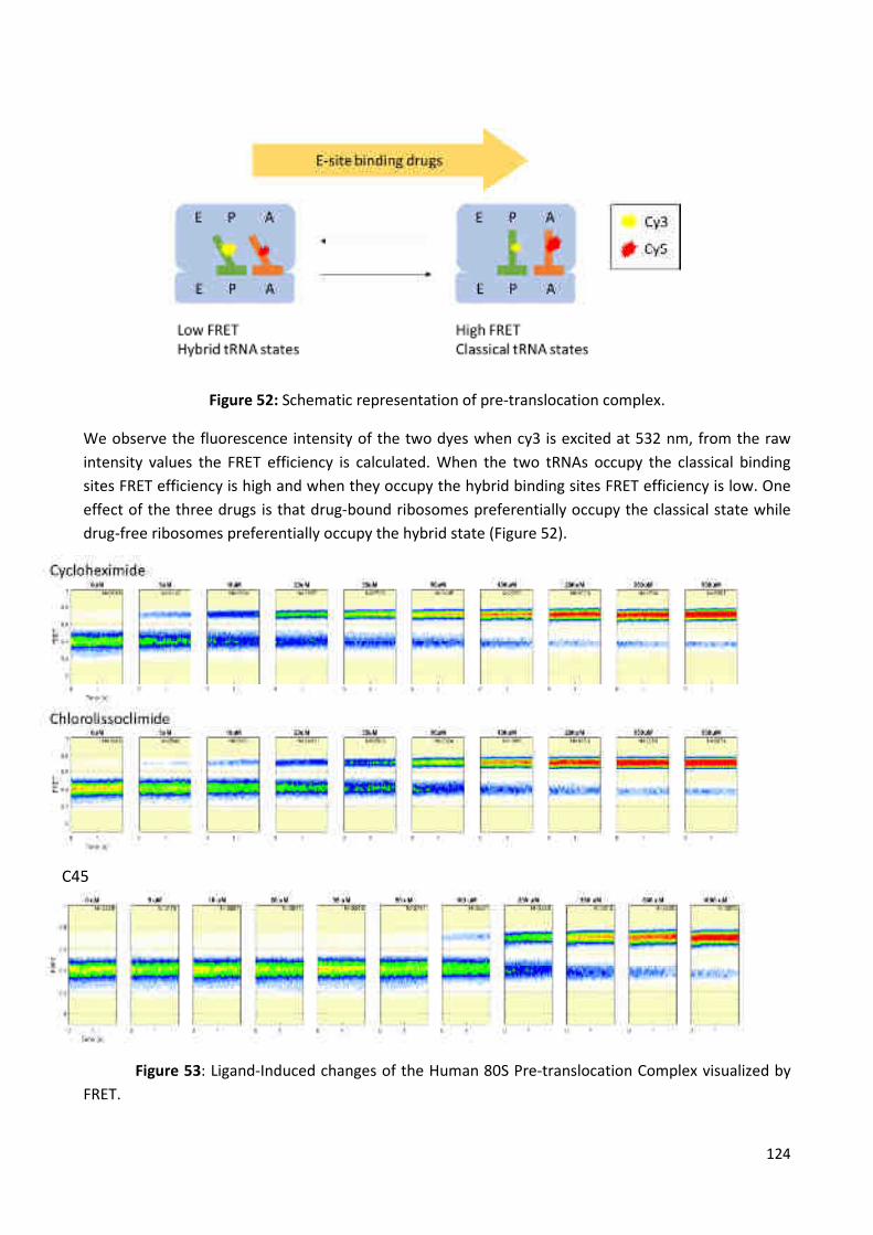
124
Figure 52: Schematic representation of pre-translocation complex.
We observe the fluorescence intensity of the two dyes when cy3 is excited at 532 nm, from the raw
intensity values the FRET efficiency is calculated. When the two tRNAs occupy the classical binding
sites FRET efficiency is high and when they occupy the hybrid binding sites FRET efficiency is low. One
effect of the three drugs is that drug-bound ribosomes preferentially occupy the classical state while
drug-free ribosomes preferentially occupy the hybrid state (Figure 52).
Figure 53: Ligand-Induced changes of the Human 80S Pre-translocation Complex visualized by
FRET.
C45

125
According to Figure 53, the histograms present the distribution of FRET efficiencies over the time after
equilibration at the indicated drug concentration. Two distinct populations are observed, one ‘high
FRET’, indicative of ribosomes where the two tRNA molecules occupy their classical binding state, and
one ‘low FRET’, indicative of ribosomes where the two tRNA molecules occupy the hybrid binding
state. It can be seen that for CL and CHXthe population of 'high FRET' ribosomes, corresponding to the
classical tRNA state, increases with increasing drug. In case of C45 drug, the effect is not as
pronounced since the high FRET state is equilibrating very slowly with the ribosome (on a timescale of
tens of minutes at the lower concentrations).
Figure 54: Quantification of the data for CHX, CL and C45. The error bars represent standard error of
the mean (SEM).
Three graphics in Figure 54 show how the fraction of time that the tRNAs spend in the classical state
changes with increasing drug concentration. In this way we can estimate apparent dissociation
constants for CHX and CL and for C45. In the case of C45 measurements it is not certain that these
values represent true equilibrium distributions, especially for the lower concentrations. We can see
KD = 5.5 ± 2 µM K
D = 11.5 ± 4 µM
(µM) (µM)
KD = 85 ± 40 µM

126
from the graphic that the binding affinity for C45 appears to be lower than for CHX and CL. The poor fit
of this curve compared to the others is likely due to an issue of very slow equilibration which causes
the accuracy of the data to correlate with the concentration of the drug.
Moreover, we observed that ribosome is still dynamic even at saturating drug concentration in all
cases. This is somewhat unexpected based on structural evidence and suggests that the P-site tRNA
body can enter the E site even if the CCA-end of the tRNA is prevented from doing so by the presence
of the drug.
Figure 55: Population histogram of a washout experiment with C45.
After equilibration (1000µM) with the 80S ribosome, C45 has been washed away with imaging buffer
(Figure 55). We observed that C45 had a long residence time on the ribosome and even after several
minutes no dissociation is apparent, i.e. no ribosomes have returned to the FRET distribution observed
in the absence of drug.

127
DISCUSSION AND PERSPECTIVES
Due to the lack of information on a new synthesized analog of lyssoclimide C45, any studies that have
been or will be conducted in the CL direction could help us to formulate our knowledge about the
principles of the action of a potential anti-cancer drug C45.
In summary, the high-resolution crystal structure of newly synthesized C45 compound reveals that this
inhibitor binds the E-site of the large subunit using the same network of interactions as CL. C45 fully
occupies and occludes the tRNA binding pocket. The fascinating binding mode includes two novel face-
on chlorine–π interactions of the ligand with guanine residues (G2794 and G2793). To estimate the
importance of this type of interaction, energy benefit was calculated for the interaction of the chlorine
with both G2794 and G2793 and it is equal to 1.8 kcal mol–1 (Konst et al., 2017). This favorable
dispersion-based interaction appears to afford stabilization to this arrangement of CL and C45 in the E-
site of the ribosome, and related effects might be leveraged in the design of other nucleic acid ligands.
Another structural difference of C45 compound with CL is the absence of C7-hydroxyl group on the B
ring which formed, in CL cause, a hydrogen bond with Pro56 on a stretched loop of the eukaryotic-
specific ribosomal protein eL42. This difference relates C45 with most other known inhibitors which
exclusively bind rRNA (Garreau de Loubresse et al., 2014).
According to FRET analysis, we observed that ribosome is still dynamic even at saturating drug
concentration. On the basis of this, two mechanistic models can be proposed. First possibility is that
CHX, C45 and CL blocks the tRNA in hybrid-like conformation, meaning that tRNA CCA-end is trapped in
the vicinity of E-site on the LSU while the tRNA anticodon stem-loop stays in P-site on the small
subunit. The second possibility is that CHX, C45 and CL alters tRNA dynamics on the ribosome and
displaces the equilibrium in favor of classical states. This was done by measuring FRET signals between
fluorescently-labeled tRNA, with which we can follow tRNA conformation on the ribosome.
Moreover, thanks to the FRET measurements we were able to determine Kd of binding for the newly
synthesized inhibitor C45, CL and for CHX at a single molecule level. We can conclude that the
dissociation constants for CHX and CL are similar, about two fold different (see before), thus showing
similar binding affinities. C45 showed lower binding affinity compared with CL. Two possible reasons
can explain this behavior: first, it could be due to the issue of very slow equilibration which causes the
accuracy of the data to correlate with the concentration of the drug. The second is that C45
demonstrate lower eukaryotic translation inhibition activity, according to translation assay data as
shown by Konst and coworkers in 2017, in comparison with CL.
Also we observed that C45 had a long residence time on the ribosome, after washing out the drug,
where, even after several minutes, no dissociation was occuring. The ribosomes, once bound to C45,
are unable to return to the FRET state observed in the absence of drug. According to this result, we can
conclude that the potential anti-cancer drug C45 could show its effectiveness due to the long-term
retention of the binding with the ribosome and its presence after washing out experiments is the
confirmation of this hypothesis.
In accordance with comparison of cytotoxicity test of CL with homoharringtonine (the US Food and
Drug Administration (FDA) approved the first translation inhibitor) and CHX, Konst and coworkers

128
concluded that CL is as cytotoxic as homoharringtonine in the three cell lines examined, and also the
translation-inhibitory activity is comparable. However, CHX was significantly less cytotoxic than many
observed synthetic lissoclimides, but its translation-inhibitory activity was equivalent to those of CL
and homoharringtonine.
In the close future our laboratory plans to analyze other inhibitors, which could be congeners of CL or
new molecules on which our collaborators are working on. These studies help us in order to
understand how to improve potency of this scaffold molecule.
Concerning the cisplatin/80S complex project we found the optimal ratio of cisplatin (37.5 μM) for co-
crystalization process with eukaryotic ribosome. These allowed us to obtain good shape and size of
crystals to perform further diffraction experiments. In addition, we determined the influence of the
detergent (Deoxy-Big Chap) on the cisplatin complex formation with the ribosome. Most probably in
this case, incubation of complex together with detergent, before crystallization process, reduced
binding affinity of cisplatin to 80S ribosome. Also we observed that cisplatin itself influenced on the
crystal growth and changed crystal form from usual rectangular parallelepiped to hexagonal prism.
We were able to improve of the crystals diffraction till 4 Å. This data we obtained only in crystals which
were soaked in presence of cisplatin (250 μM). However the yield of medium resolution was very low
only 5% and this makes it difficult for us to build the structure. In case of co-crystallization ribosome
with cisplatin we were got no more than 6-7 Å resolution.
So, the project would require a lot of optimization procedures and also, most probably, the avoidance
of osmium hexamine in the crystals, to be able to use the anomalous signal coming from the few Pt
atoms that should tightly bind to the ribosome. However, so far, the treatment of the S.cerevisiae 80S
ribosome crystals without osmium hexamine gave only poor diffraction.
In spite of the fact that osmium hexamine and platinum have the same binding sites in the 80S
ribosome of S.cerevisiae and belongs to the group of heavy metals, the replacement of osmium
hexamine in cryo-protection procedure by platinum leads to a reducing of the crystals diffraction.
The attempt to exclude osmium from standard cryo-protection conditions was failed, and therefore
one can conclude that it is necessary to change the concept of cryo-protection process of 80S
ribosome crystals as a whole. In its turn, our laboratory is on the way of developing this in a more
robust way.

129
INVISIBLE CONNECTION BETWEEN PROJECTS
Often, different directions in science for the target audience can be closely intertwined. On the
example of my two completely different studies, one can trace a very close relationship for the end
user, namely, cancer patients and patients with MRSA. Patients with cancer tumors are at unusually
high risk for developing bloodstream infections, which are a major cause of in-hospital morbidity and
mortality. According to statistics numbers 20% of them suffer from both illnesses simultaneously and
this figure is growing steadily each year. In this case, the attending doctor has to make an uneasy
choice in favor of treating one of the diseases, due to high toxicity and a wide range of side effects of
both types (antibacterial and anticancer drugs) of medicinal substances.
In most cases, the attending physicians had to select treatment of infection (S.aureus) at first, due to
the fact that the infection could lead to mortality in the immunocompromised patients much faster
than the cancerous tumor itself. As well known, success in the cancer treatment very often depends on
the timeliness of the treatment started and this delay associated with the inability to treat both
diseases could lead to serious consequences and possible mortality of the patients. All this leads us to
one of the main problems of modern medicines, to a large numbers of strong side effects of medicines.
My research was aimed at helping to solve this problem.
In the science, a lot of researches have been done that proves the connection of a cancerous tumor
with infectious diseases. For instance, more than 15% of malignancies worldwide are established to
have an infectious cause (Pisani et al., 1997). The main mechanisms by which infections can cause
cancer are (a) transformation of cells by insertion of oncogenes and/or inhibition of tumor suppressors
(e.g., oncogenic HPV types, cervical cancer (Scheffner et al., 1992); (b) carcinogenesis induced by
immunosuppression (e.g., in HIV-related cancers (Beral and Newton, 1998), and (c) infection induced
chronic inflammation can produce nitric oxide and cytokines, which contribute to carcinogenesis
(Ohshima and Bartsch, 1994; Aggarwal et al., 2006) (e.g., Helicobacter pylori (IARC Monographs, 1994)
causes a chronic inflammation in the gastric mucosa, which is likely to be responsible for the
development of cancer (Parsonnet, 1995).
Therefore in my opinion, it is not worth to separate the research aimed at the treatment of cancer and
infectious diseases. All these once again show that different areas of scientific activity should closely
cooperate among themselves to solve the global problems of mankind.

130
GENERAL CONCLUSION
Structure determination of Staphylococcus aureus 70S ribosome project showed certain amount of
results. The growth of S. aureus cells and their harvesting were adapted for purification of ribosomes
for structural studies. After variety of modifications of ribosome purification, the optimal protocol was
obtained, which allowed us (1) to get rid of unwanted non- ribosome proteins, (2) to reduce the
amount of contaminating tRNA, and (3) to improve the shape and the size of crystals. Also amount of
obtained ribosomes was sufficient for broad search conditions of crystallization.
Different crystals in different conditions were obtained from the S. aureus 70S ribosomes, complexes
with tRNAfMet/70S ribosome/mRNA and 70S/N-terminal domain of HPF. Crystals were reproducible and
reached appropriate size to perform diffraction experiments. The conditions of dehydration and cryo-
cooling were optimized for obtained crystals. The diffraction data of S. aureus 70S ribosome and its
complexes crystals were collected. However, the resolution obtained was too low (~17-19 Å) for high-
resolution structure determination.
In case of small-molecular inhibitor project, thanks to knowledge which came from combination of
semisynthesis and analogue-oriented synthesis, supported by docking studies we had opportunity to
expand our understanding of structural-activity relationship in the lissoclimide family of translation
inhibition cytostatic agents. We uncovered the structural basis for inhibition of protein syntesis with
co-crystallographic study of synthetic C45 bound to the ribosome. The interesting binding mode
includes a two novel face-on chlorine-π interaction of the ligand with two guanine residues. This
favorable dispersion-based interaction appears to afford a stabilization of C45 compound in the E-site
of the ribosome, and thus related effects might be leveraged in the design of other nucleic acid
ligands. Also, according FRET analysis in the human ribosome pre-translocation complex we observe
that C45 equilibrates very slowly with the ribosome on a timescale of tens of minutes, however, it has
a very long residence time once bound. In addition we calculated dissociation constant for C45 and it is
in average 50 – 150 µM. In comparison, CHX and CL approximately 5.5 µM and 11.5 µM respectively,
therefore showing similar binding affinities. The C45 is a weaker binder than CL, which is well
correlated with the published data (Konst et al., 2017).
Crystallographic studies of cisplatin drug with 80S ribosome showed a certain degree of success. We
found optimal ratio of cisplatin for co-crystalization process with eukaryotic ribosome. Also we
determined the influence of the detergent (Deoxy-Big Chap) on the cisplatin complex formation with
the ribosome and on the crystals growth. And we improved crystal diffraction till 4 Å.
However we encountered several problems with it. First is the yield of medium resolution is very low
(5% maximum), thus we have a difficulties to get a structure of this complex. Second, are overlapping
platinum binding sites with an osmium hexamine binding sites in the 80S ribosome. This forced us not
to use osmium hexamine for the usual cryo-protection of the crystals with Pt. But, so far S.cerevisiae
80S ribosome crystals treatment without osmium hexamine gave only poor diffraction.

131
LIST OF PUBLICATIONS AND COMMUNICATIONS
List of publications
Article in preparation: “Analysis of halogen-π interactions in inhibitors binding to the
eukaryotic ribosome” Daniya Kashinskaya1, Simone Pellegrino1, Melanie Meyer1, Zef A.
Könst2, Sharon E. Michalak2, Mikael Holm3, Scott Blanchard3, Vamsee K. Voora4 , Gulnara
Yusupova1, Marat Yusupov1 et Christopher D. Vanderwal2. (1)Institut de Génétique et de
Biologie Moléculaire et Cellulaire (IGBMC), INSERM U964, CNRS UMR7104, Université de
Strasbourg, 67404 Illkirch, France. (2)Department of Chemistry, University of California,
1102 Natural Sciences II,Irvine, California 92697-2025, USA. (3) Department of Physiology
and Biophysics, Weill Cornell Medical College, New York 10065, USA. (4) Irvine Department
of Chemistry, University of California, 1102 Natural Sciences II Irvine, CA 92697-2025, USA.
List of communications: talk - poster
Poster: “Structure analysis of potential anticancer drugs” Daniya Kashinskaya1, Simone
Pellegrino1, Melanie Meyer1, Zef A. Könst2, Sharon E. Michalak2, Mikael Holm3, Scott
Blanchard3, Vamsee K. Voora4 , Gulnara Yusupova1, Marat Yusupov1 et Christopher D.
Vanderwal2. (1)Institut de Génétique et de Biologie Moléculaire et Cellulaire (IGBMC), INSERM
U964, CNRS UMR7104, Université de Strasbourg, 67404 Illkirch, France. (2) Department of
Chemistry, University of California, 1102 Natural Sciences II,Irvine, California 92697-2025, USA.
(3) Department of Physiology and Biophysics, Weill Cornell Medical College, New York 10065,
USA. (4) Irvine Department of Chemistry, University of California, 1102 Natural Sciences II
Irvine, CA 92697-2025, USA.
EMBO Conference: “Protein Synthesis and Translational Control”.
6-7 September 2017/EMBL Advanced Training Centre/ Heidelberg/Germany.

132
REFERENCES
Aas T, Borresen AL, Geisler S, Smith-Sorensen B, Johnsen H, Varhaug JE, et al. Specific P53 mutations
are associated with de novo resistance to doxorubicin in breast cancer patients. Nature medicine.
1996;2(7):811-4. PubMed PMID: 8673929.
Abraham EP, Adler E. The specificity of diaphorase (coenzyme factor). The Biochemical journal.
1940;34(1):119-27. PubMed PMID: 16747129; PubMed Central PMCID: PMC1265256.
Abraham EP, Chain E. An enzyme from bacteria able to destroy penicillin. Nature. 1940; 146:837.
Adrian M, Dubochet J, Lepault J, McDowall AW. Cryo-electron microscopy of viruses. Nature.
1984;308(5954):32-6. PubMed PMID: 6322001.
Aggarwal BB, Shishodia S, Sandur SK, Pandey MK, Sethi G. Inflammation and cancer: how hot is the
link? Biochemical pharmacology. 2006;72(11):1605-21. doi: 10.1016/j.bcp.2006.06.029. PubMed
PMID: 16889756.
Aggarwal BB, Shishodia S, Sandur SK, Pandey MK, Sethi G. Inflammation and cancer: how hot is the
link? Biochemical pharmacology. 2006;72(11):1605-21. doi: 10.1016/j.bcp.2006.06.029. PubMed
PMID: 16889756.
**Agirrezabala, X., Lei, J., Brunelle, J.L., Ortiz-Meoz, R.F., Green, R., and Frank, J. (2008). Visualization
of the Hybrid State of tRNA Binding Promoted by Spontaneous Ratcheting of the Ribosome. Mol. Cell
32, 190–197.
**Agirrezabala, X., Liao, H.Y., Schreiner, E., Fu, J., Ortiz-Meoz, R.F., Schulten, K., Green, R., and Frank, J.
(2012). Structural characterization of mRNA-tRNA translocation intermediates. Proc. Natl. Acad. Sci.
109, 6094–6099.
Agrawal RK, Sharma MR. Structural aspects of mitochondrial translational apparatus. Current opinion
in structural biology. 2012;22(6):797-803. doi: 10.1016/j.sbi.2012.08.003. PubMed PMID: 22959417;
PubMed Central PMCID: PMC3513651.
Aitken CE, Lorsch JR. A mechanistic overview of translation initiation in eukaryotes. Nature structural &
molecular biology. 2012;19(6):568-76. doi: 10.1038/nsmb.2303. PubMed PMID: 22664984.
Alkalaeva EZ, Pisarev AV, Frolova LY, Kisselev LL, Pestova TV. In vitro reconstitution of eukaryotic
translation reveals cooperativity between release factors eRF1 and eRF3. Cell. 2006;125(6):1125-36.
doi: 10.1016/j.cell.2006.04.035. PubMed PMID: 16777602.
**Amunts A, Brown A, Bai XC, Llacer JL, Hussain T, Emsley P, et al. Structure of the yeast mitochondrial
large ribosomal subunit. Science. 2014;343(6178):1485-9. doi: 10.1126/science.1249410. PubMed
PMID: 24675956; PubMed Central PMCID: PMC4046073.

133
**Amunts A, Brown A, Toots J, Scheres SHW, Ramakrishnan V. Ribosome. The structure of the human
mitochondrial ribosome. Science. 2015;348(6230):95-8. doi: 10.1126/science.aaa1193. PubMed PMID:
25838379; PubMed Central PMCID: PMC4501431.
Anger AM, Armache JP, Berninghausen O, Habeck M, Subklewe M, Wilson DN, et al. Structures of the
human and Drosophila 80S ribosome. Nature. 2013;497(7447):80-5. doi: 10.1038/nature12104.
PubMed PMID: 23636399.
Apperley JF. Part I: mechanisms of resistance to imatinib in chronic myeloid leukaemia. The Lancet
Oncology. 2007;8(11):1018-29. doi: 10.1016/S1470-2045(07)70342-X. PubMed PMID: 17976612.
Arber N, Doki Y, Han EK, Sgambato A, Zhou P, Kim NH, et al. Antisense to cyclin D1 inhibits the growth
and tumorigenicity of human colon cancer cells. Cancer research. 1997;57(8):1569-74. PubMed PMID:
9108461.
Arias CA, Panesso D, McGrath DM, Qin X, Mojica MF, Miller C, et al. Genetic basis for in vivo
daptomycin resistance in enterococci. The New England journal of medicine. 2011;365(10):892-900.
doi: 10.1056/NEJMoa1011138. PubMed PMID: 21899450; PubMed Central PMCID: PMC3205971.
Ashe MP, De Long SK, Sachs AB. Glucose depletion rapidly inhibits translation initiation in yeast.
Molecular biology of the cell. 2000;11(3):833-48. PubMed PMID: 10712503; PubMed Central PMCID:
PMC14814.
Balak MN, Gong Y, Riely GJ, Somwar R, Li AR, Zakowski MF, et al. Novel D761Y and common secondary
T790M mutations in epidermal growth factor receptor-mutant lung adenocarcinomas with acquired
resistance to kinase inhibitors. Clinical cancer research : an official journal of the American Association
for Cancer Research. 2006;12(21):6494-501. doi: 10.1158/1078-0432.CCR-06-1570. PubMed PMID:
17085664.
Ban N, Nissen P, Hansen J, Moore PB, Steitz TA. The complete atomic structure of the large ribosomal
subunit at 2.4 A resolution. Science. 2000;289(5481):905-20. PubMed PMID: 10937989.
Bardenheuer W, Lehmberg K, Rattmann I, Brueckner A, Schneider A, Sorg UR, et al. Resistance to
cytarabine and gemcitabine and in vitro selection of transduced cells after retroviral expression of
cytidine deaminase in human hematopoietic progenitor cells. Leukemia. 2005;19(12):2281-8. doi:
10.1038/sj.leu.2403977. PubMed PMID: 16304576.
Barlow JJ, Mathias AP, Williamson R, Gammack DB. A Simple Method for the Quantitative Isolation of
Undegraded High Molecular Weight Ribonucleic Acid. Biochemical and biophysical research
communications. 1963;13:61-6. PubMed PMID: 14069514.
Bartesaghi A, Merk A, Banerjee S, Matthies D, Wu X, Milne JL, et al. 2.2 A resolution cryo-EM structure
of beta-galactosidase in complex with a cell-permeant inhibitor. Science. 2015;348(6239):1147-51. doi:
10.1126/science.aab1576. PubMed PMID: 25953817.
Batista PJ. The RNA Modification N6-methyladenosine and Its Implications in Human Disease.
Genomics, proteomics & bioinformatics. 2017;15(3):154-63. doi: 10.1016/j.gpb.2017.03.002. PubMed
PMID: 28533023; PubMed Central PMCID: PMC5487527.

134
Bayer AS, Schneider T, Sahl HG. Mechanisms of daptomycin resistance in Staphylococcus aureus: role
of the cell membrane and cell wall. Annals of the New York Academy of Sciences. 2013;1277:139-58.
doi: 10.1111/j.1749-6632.2012.06819.x. PubMed PMID: 23215859; PubMed Central PMCID:
PMC3556211.
Bean J, Riely GJ, Balak M, Marks JL, Ladanyi M, Miller VA, et al. Acquired resistance to epidermal
growth factor receptor kinase inhibitors associated with a novel T854A mutation in a patient with
EGFR-mutant lung adenocarcinoma. Clinical cancer research : an official journal of the American
Association for Cancer Research. 2008;14(22):7519-25. doi: 10.1158/1078-0432.CCR-08-0151. PubMed
PMID: 19010870; PubMed Central PMCID: PMC2596620.
Becker T, Armache JP, Jarasch A, Anger AM, Villa E, Sieber H, et al. Structure of the no-go mRNA decay
complex Dom34-Hbs1 bound to a stalled 80S ribosome. Nature structural & molecular biology.
2011;18(6):715-20. doi: 10.1038/nsmb.2057. PubMed PMID: 21623367.
Becker T, Bhushan S, Jarasch A, Armache JP, Funes S, Jossinet F, et al. Structure of monomeric yeast
and mammalian Sec61 complexes interacting with the translating ribosome. Science.
2009;326(5958):1369-73. doi: 10.1126/science.1178535. PubMed PMID: 19933108; PubMed Central
PMCID: PMC2920595.
Becker T, Franckenberg S, Wickles S, Shoemaker CJ, Anger AM, Armache JP, et al. Structural basis of
highly conserved ribosome recycling in eukaryotes and archaea. Nature. 2012;482(7386):501-6. doi:
10.1038/nature10829. PubMed PMID: 22358840.
**Behrmann, E., Loerke, J., Budkevich, T.V., Yamamoto, K., Schmidt, A., Penczek, P.A., Vos, M.R.,
Bürger, J., Mielke, T., Scheerer, P., et al. (2015). structural snapshots of actively translating human
ribosomes. Cell 161, 845–857.
***Belle A, Tanay A, Bitincka L, Shamir R, O'Shea EK. Quantification of protein half-lives in the budding
yeast proteome. Proceedings of the National Academy of Sciences of the United States of America.
2006;103(35):13004-9. doi: 10.1073/pnas.0605420103. PubMed PMID: 16916930; PubMed Central
PMCID: PMC1550773.
Belousoff MJ, Eyal Z, Radjainia M, Ahmed T, Bamert RS, Matzov D, et al. Structural Basis for Linezolid
Binding Site Rearrangement in the Staphylococcus aureus Ribosome. mBio. 2017;8(3). doi:
10.1128/mBio.00395-17. PubMed PMID: 28487427; PubMed Central PMCID: PMC5424203.
Belova L, Tenson T, Xiong L, McNicholas PM, Mankin AS. A novel site of antibiotic action in the
ribosome: interaction of evernimicin with the large ribosomal subunit. Proceedings of the National
Academy of Sciences of the United States of America. 2001;98(7):3726-31. doi:
10.1073/pnas.071527498. PubMed PMID: 11259679; PubMed Central PMCID: PMC31120.
***Ben-Shem A, Garreau de Loubresse N, Melnikov S, Jenner L, Yusupova G, Yusupov M. The structure
of the eukaryotic ribosome at 3.0 A resolution. Science. 2011;334(6062):1524-9. doi:
10.1126/science.1212642. PubMed PMID: 22096102.
Ben-Shem A, Jenner L, Yusupova G, Yusupov M. Crystal structure of the eukaryotic ribosome. Science.
2010;330(6008):1203-9. doi: 10.1126/science.1194294. PubMed PMID: 21109664.

135
Beral V, Newton R. Overview of the epidemiology of immunodeficiency-associated cancers. Journal of
the National Cancer Institute Monographs. 1998(23):1-6. PubMed PMID: 9709294.
Bhat M, Robichaud N, Hulea L, Sonenberg N, Pelletier J, Topisirovic I. Targeting the translation
machinery in cancer. Nature reviews Drug discovery. 2015;14(4):261-78. doi: 10.1038/nrd4505.
PubMed PMID: 25743081.
Bien J, Sokolova O, Bozko P. Characterization of Virulence Factors of Staphylococcus aureus: Novel
Function of Known Virulence Factors That Are Implicated in Activation of Airway Epithelial
Proinflammatory Response. Journal of pathogens. 2011;2011:601905. doi: 10.4061/2011/601905.
PubMed PMID: 22567334; PubMed Central PMCID: PMC3335658.
Bischoff M, Dunman P, Kormanec J, Macapagal D, Murphy E, Mounts W, et al. Microarray-based
analysis of the Staphylococcus aureus sigmaB regulon. Journal of bacteriology. 2004;186(13):4085-99.
doi: 10.1128/JB.186.13.4085-4099.2004. PubMed PMID: 15205410; PubMed Central PMCID:
PMC421609.
Bischoff M, Entenza JM, Giachino P. Influence of a functional sigB operon on the global regulators sar
and agr in Staphylococcus aureus. Journal of bacteriology. 2001;183(17):5171-9. PubMed PMID:
11489871; PubMed Central PMCID: PMC95394.
*Blaha G, Stanley RE, Steitz TA. Formation of the first peptide bond: the structure of EF-P bound to the
70S ribosome. Science. 2009;325(5943):966-70. doi: 10.1126/science.1175800. PubMed PMID:
19696344; PubMed Central PMCID: PMC3296453.
Blencke S, Ullrich A, Daub H. Mutation of threonine 766 in the epidermal growth factor receptor
reveals a hotspot for resistance formation against selective tyrosine kinase inhibitors. The Journal of
biological chemistry. 2003;278(17):15435-40. doi: 10.1074/jbc.M211158200. PubMed PMID:
12594213.
Blundell TL, Cutfield JF, Cutfield SM, Dodson EJ, Dodson GG, Hodgkin DC, et al. Atomic positions in
rhombohedral 2-zinc insulin crystals. Nature. 1971;231(5304):506-11. PubMed PMID: 4932997.
Bonanno L, Favaretto A, Rosell R. Platinum drugs and DNA repair mechanisms in lung cancer.
Anticancer research. 2014;34(1):493-501. PubMed PMID: 24403507.
Bretscher MS. Direct translation of a circular messenger DNA. Nature. 1968;220(5172):1088-91.
PubMed PMID: 5723604.
**Brilot, A.F., Korostelev, A.A., Ermolenko, D.N., and Grigorieff, N. (2013). Structure of the ribosome
with elongation factor G trapped in the pretranslocation state. Proc. Natl. Acad. Sci. U. S. A. 110,
20994–20999.
Brodersen DE, Clemons WM, Jr., Carter AP, Morgan-Warren RJ, Wimberly BT, Ramakrishnan V. The
structural basis for the action of the antibiotics tetracycline, pactamycin, and hygromycin B on the 30S
ribosomal subunit. Cell. 2000;103(7):1143-54. PubMed PMID: 11163189.

136
**Brown A, Amunts A, Bai XC, Sugimoto Y, Edwards PC, Murshudov G, et al. Structure of the large
ribosomal subunit from human mitochondria. Science. 2014;346(6210):718-22. doi:
10.1126/science.1258026. PubMed PMID: 25278503; PubMed Central PMCID: PMC4246062.
**Budkevich, T. V., Giesebrecht, J., Behrmann, E., Loerke, J., Ramrath, D.J.F., Mielke, T., Ismer, J.,
Hildebrand, P.W., Tung, C.S., Nierhaus, K.H., et al. (2014). Regulation of the mammalian elongation
cycle by subunit rolling: A eukaryotic-specific ribosome rearrangement. Cell 158, 121–131.
**Budkevich, T., Giesebrecht, J., Altman, R.B., Munro, J.B., Mielke, T., Nierhaus, K.H., Blanchard, S.C.,
and Spahn, C.M.T. (2011). Structure and dynamics of the mammalian ribosomal pretranslocation
complex. Mol. Cell 44, 214–224.
Bush K. Proliferation and significance of clinically relevant beta-lactamases. Annals of the New York
Academy of Sciences. 2013;1277:84-90. doi: 10.1111/nyas.12023. PubMed PMID: 23346859.
Campanile F, Bongiorno D, Borbone S, Stefani S. Hospital-associated methicillin-resistant
Staphylococcus aureus (HA-MRSA) in Italy. Annals of clinical microbiology and antimicrobials.
2009;8:22. doi: 10.1186/1476-0711-8-22. PubMed PMID: 19552801; PubMed Central PMCID:
PMC2708121.
Carter AP, Clemons WM, Brodersen DE, Morgan-Warren RJ, Wimberly BT, Ramakrishnan V. Functional
insights from the structure of the 30S ribosomal subunit and its interactions with antibiotics. Nature.
2000;407(6802):340-8. doi: 10.1038/35030019. PubMed PMID: 11014183.
Cate JH, Yusupov MM, Yusupova GZ, Earnest TN, Noller HF. X-ray crystal structures of 70S ribosome
functional complexes. Science. 1999;285(5436):2095-104. PubMed PMID: 10497122.
CDC National Nosocomial Infections Surveillance (NNIS) System Report, data summary from January
1990–May 1999, issued June 1999. Am J Infect Control 1999; 27:520–32.
Chambers HF, Deleo FR. Waves of resistance: Staphylococcus aureus in the antibiotic era. Nature
reviews Microbiology. 2009;7(9):629-41. doi: 10.1038/nrmicro2200. PubMed PMID: 19680247;
PubMed Central PMCID: PMC2871281.
Chan PF, Foster SJ. Role of SarA in virulence determinant production and environmental signal
transduction in Staphylococcus aureus. Journal of bacteriology. 1998;180(23):6232-41. PubMed PMID:
9829932; PubMed Central PMCID: PMC107708.
Chayen SD, Gross D, Makhoul O, Glaser B. TSH producing pituitary tumor: biochemical diagnosis and
long-term medical management with octreotide. Hormone and metabolic research = Hormon- und
Stoffwechselforschung = Hormones et metabolisme. 1992;24(1):34-8. doi: 10.1055/s-2007-1003247.
PubMed PMID: 1612557.
Chen C, Tang J, Dong W, Wang C, Feng Y, Wang J, et al. A glimpse of streptococcal toxic shock
syndrome from comparative genomics of S. suis 2 Chinese isolates. PloS one. 2007;2(3):e315. doi:
10.1371/journal.pone.0000315. PubMed PMID: 17375201; PubMed Central PMCID: PMC1820848.

137
Cheung AL, Bayer AS, Zhang G, Gresham H, Xiong YQ. Regulation of virulence determinants in vitro and
in vivo in Staphylococcus aureus. FEMS immunology and medical microbiology. 2004;40(1):1-9.
PubMed PMID: 14734180.
Cheung AL, Koomey JM, Butler CA, Projan SJ, Fischetti VA. Regulation of exoprotein expression in
Staphylococcus aureus by a locus (sar) distinct from agr. Proceedings of the National Academy of
Sciences of the United States of America. 1992;89(14):6462-6. PubMed PMID: 1321441; PubMed
Central PMCID: PMC49521.
Cheung AL, Projan SJ, Gresham H. The Genomic Aspect of Virulence, Sepsis, and Resistance to Killing
Mechanisms in Staphylococcus aureus. Current infectious disease reports. 2002;4(5):400-10. PubMed
PMID: 12228026.
Cheung AL, Projan SJ. Cloning and sequencing of sarA of Staphylococcus aureus, a gene required for
the expression of agr. Journal of bacteriology. 1994;176(13):4168-72. PubMed PMID: 8021198;
PubMed Central PMCID: PMC205618.
Cheung AL, Schmidt K, Bateman B, Manna AC. SarS, a SarA homolog repressible by agr, is an activator
of protein A synthesis in Staphylococcus aureus. Infection and immunity. 2001;69(4):2448-55. doi:
10.1128/IAI.69.4.2448-2455.2001. PubMed PMID: 11254606; PubMed Central PMCID: PMC98178.
Chevalier, C. (2009). Function and mechanism of action of RNAIII and new ncRNAs in Staphylococcus
aureus (Doctoral thesis, University of Strasbourg, Strasbourg, France). Retrieved from: http://scd-
theses.u-strasbg.fr/1714/
Chien Y, Manna AC, Projan SJ, Cheung AL. SarA, a global regulator of virulence determinants in
Staphylococcus aureus, binds to a conserved motif essential for sar-dependent gene regulation. The
Journal of biological chemistry. 1999;274(52):37169-76. PubMed PMID: 10601279.
Cimolai N. MRSA and the environment: implications for comprehensive control measures. European
journal of clinical microbiology & infectious diseases : official publication of the European Society of
Clinical Microbiology. 2008;27(7):481-93. doi: 10.1007/s10096-008-0471-0. PubMed PMID: 18273652.
Clatworthy AE, Pierson E, Hung DT. Targeting virulence: a new paradigm for antimicrobial therapy.
Nature chemical biology. 2007;3(9):541-8. doi: 10.1038/nchembio.2007.24. PubMed PMID: 17710100.
Cole AM, Tahk S, Oren A, Yoshioka D, Kim YH, Park A, et al. Determinants of Staphylococcus aureus
nasal carriage. Clinical and diagnostic laboratory immunology. 2001;8(6):1064-9. doi:
10.1128/CDLI.8.6.1064-1069.2001. PubMed PMID: 11687441; PubMed Central PMCID: PMC96227.
*Colussi, T.M., Costantino, D.A., Zhu, J., Donohue, J.P., Korostelev, A.A., Jaafar, Z.A., Plank, T.-D.M.,
Noller, H.F., and Kieft, J.S. (2015). Initiation of translation in bacteria by a structured eukaryotic IRES
RNA. Nature 519, 110–113.
Connell SR, Tracz DM, Nierhaus KH, Taylor DE. Ribosomal protection proteins and their mechanism of
tetracycline resistance. Antimicrobial agents and chemotherapy. 2003;47(12):3675-81. PubMed PMID:
14638464; PubMed Central PMCID: PMC296194.

138
Cornish PV, Ermolenko DN, Noller HF, Ha T. Spontaneous intersubunit rotation in single ribosomes.
Molecular cell. 2008;30(5):578-88. doi: 10.1016/j.molcel.2008.05.004. PubMed PMID: 18538656;
PubMed Central PMCID: PMC2491453.
Costa DB, Halmos B, Kumar A, Schumer ST, Huberman MS, Boggon TJ, et al. BIM mediates EGFR
tyrosine kinase inhibitor-induced apoptosis in lung cancers with oncogenic EGFR mutations. PLoS
medicine. 2007;4(10):1669-79; discussion 80. doi: 10.1371/journal.pmed.0040315. PubMed PMID:
17973572; PubMed Central PMCID: PMC2043012.
Crowfoot, D., Bunn, C.W., Rogers-Low, B.W., and Turner-Jones, A. (1949). The chemistry of penicillin.
Princet. Univ. Press, New Jersey.
D’Ambrosio, M., Guerriero, A., Debitus, C.c., Ribes, O., Pusset, J., Leroy, S.,and Pietra, F. (1993).
Agelastatin a, a new skeleton cytotoxic alkaloid of the oroidin family. Isolation from the axinellid
sponge Agelas dendromorpha of the coral sea. J. Chem. Soc. Chem. Comm, 1305.
Dang Y, Schneider-Poetsch T, Eyler DE, Jewett JC, Bhat S, Rawal VH, et al. Inhibition of eukaryotic
translation elongation by the antitumor natural product Mycalamide B. Rna. 2011;17(8):1578-88. doi:
10.1261/rna.2624511. PubMed PMID: 21693620; PubMed Central PMCID: PMC3153980.
D'Arcy TJ, Hughes SW, Chiu WS, Clark T, Milner AD, Saunders J, et al. Estimation of fetal lung volume
using enhanced 3-dimensional ultrasound: a new method and first result. British journal of obstetrics
and gynaecology. 1996;103(10):1015-20. PubMed PMID: 8863701.
D'Costa VM, King CE, Kalan L, Morar M, Sung WW, Schwarz C, et al. Antibiotic resistance is ancient.
Nature. 2011;477(7365):457-61. doi: 10.1038/nature10388. PubMed PMID: 21881561.
Demeshkina N, Jenner L, Yusupova G, Yusupov M. Interactions of the ribosome with mRNA and tRNA.
Current opinion in structural biology. 2010;20(3):325-32. doi: 10.1016/j.sbi.2010.03.002. PubMed
PMID: 20392630.
*Demeshkina, N., Jenner, L., Westhof, E., Yusupov, M., and Yusupova, G. (2012). A new understanding
of the decoding principle on the ribosome. Nature 484, 256–259.
Deora R, Misra TK. Characterization of the primary sigma factor of Staphylococcus aureus. The Journal
of biological chemistry. 1996;271(36):21828-34. PubMed PMID: 8702982.
Deora R, Tseng T, Misra TK. Alternative transcription factor sigmaSB of Staphylococcus aureus:
characterization and role in transcription of the global regulatory locus sar. Journal of bacteriology.
1997;179(20):6355-9. PubMed PMID: 9335283; PubMed Central PMCID: PMC179550.
**des Georges, A., Hashem, Y., Unbehaun, A., Grassucci, R.A., Taylor, D., Hellen, C.U.T., Pestova, T. V.,
and Frank, J. (2014). Structure of the mammalian ribosomal pre-termination complex associated with
eRF1•eRF3•GDPNP. Nucleic Acids Res. 42, 3409–3418.
Dunkle JA, Wang L, Feldman MB, Pulk A, Chen VB, Kapral GJ, et al. Structures of the bacterial ribosome
in classical and hybrid states of tRNA binding. Science. 2011;332(6032):981-4. doi:
10.1126/science.1202692. PubMed PMID: 21596992; PubMed Central PMCID: PMC3176341.

139
Emsley P, Cowtan K. Coot: model-building tools for molecular graphics. Acta crystallographica Section
D, Biological crystallography. 2004;60(Pt 12 Pt 1):2126-32. doi: 10.1107/S0907444904019158. PubMed
PMID: 15572765.
Engelman JA, Zejnullahu K, Mitsudomi T, Song Y, Hyland C, Park JO, et al. MET amplification leads to
gefitinib resistance in lung cancer by activating ERBB3 signaling. Science. 2007;316(5827):1039-43. doi:
10.1126/science.1141478. PubMed PMID: 17463250.
Entenza JM, Moreillon P, Senn MM, Kormanec J, Dunman PM, Berger-Bachi B, et al. Role of sigmaB in
the expression of Staphylococcus aureus cell wall adhesins ClfA and FnbA and contribution to
infectivity in a rat model of experimental endocarditis. Infection and immunity. 2005;73(2):990-8. doi:
10.1128/IAI.73.2.990-998.2005. PubMed PMID: 15664942; PubMed Central PMCID: PMC547034.
Eyal Z, Matzov D, Krupkin M, Paukner S, Riedl R, Rozenberg H, et al. A novel pleuromutilin antibacterial
compound, its binding mode and selectivity mechanism. Scientific reports. 2016;6:39004. doi:
10.1038/srep39004. PubMed PMID: 27958389; PubMed Central PMCID: PMC5154188 Austria and King
of Prussia, PA, United States). Dr. Paukner and Dr. Riedl are employees of Nabriva Therapeutics and
own Nabriva stocks.
Eyal Z, Matzov D, Krupkin M, Wekselman I, Paukner S, Zimmerman E, et al. Structural insights into
species-specific features of the ribosome from the pathogen Staphylococcus aureus. Proceedings of
the National Academy of Sciences of the United States of America. 2015;112(43):E5805-14. doi:
10.1073/pnas.1517952112. PubMed PMID: 26464510; PubMed Central PMCID: PMC4629319.
Fattom A, Fuller S, Propst M, Winston S, Muenz L, He D, et al. Safety and immunogenicity of a booster
dose of Staphylococcus aureus types 5 and 8 capsular polysaccharide conjugate vaccine (StaphVAX) in
hemodialysis patients. Vaccine. 2004;23(5):656-63. doi: 10.1016/j.vaccine.2004.06.043. PubMed PMID:
15542186.
Feil EJ, Cooper JE, Grundmann H, Robinson DA, Enright MC, Berendt T, et al. How clonal is
Staphylococcus aureus? Journal of bacteriology. 2003;185(11):3307-16. PubMed PMID: 12754228;
PubMed Central PMCID: PMC155367.
Feldman MB, Terry DS, Altman RB, Blanchard SC. Aminoglycoside activity observed on single pre-
translocation ribosome complexes. Nature chemical biology. 2010;6(1):54-62. doi:
10.1038/nchembio.274. PubMed PMID: 19946275; PubMed Central PMCID: PMC2914512.
Ferguson A, Wang L, Altman RB, Terry DS, Juette MF, Burnett BJ, et al. Functional Dynamics within the
Human Ribosome Regulate the Rate of Active Protein Synthesis. Molecular cell. 2015;60(3):475-86.
doi: 10.1016/j.molcel.2015.09.013. PubMed PMID: 26593721; PubMed Central PMCID: PMC4660248.
**Fernández, I.S., Bai, X.-C., Hussain, T., Kelley, A.C., Lorsch, J.R., Ramakrishnan, V., and Scheres,
S.H.W. (2013). Molecular architecture of a eukaryotic translational initiation complex. Science 342,
1240585.
**Fernández, I.S., Bai, X.C., Murshudov, G., Scheres, S.H.W., and Ramakrishnan, V. (2014). Initiation of
translation by cricket paralysis virus IRES requires its translocation in the ribosome. Cell 157, 823–831.

140
Fischer N, Neumann P, Konevega AL, Bock LV, Ficner R, Rodnina MV, et al. Structure of the E. coli
ribosome-EF-Tu complex at <3 A resolution by Cs-corrected cryo-EM. Nature. 2015;520(7548):567-70.
doi: 10.1038/nature14275. PubMed PMID: 25707802.
Franckenberg S, Becker T, Beckmann R. Structural view on recycling of archaeal and eukaryotic
ribosomes after canonical termination and ribosome rescue. Current opinion in structural biology.
2012;22(6):786-96. doi: 10.1016/j.sbi.2012.08.002. PubMed PMID: 23031510.
Frank J, Agrawal RK. A ratchet-like inter-subunit reorganization of the ribosome during translocation.
Nature. 2000;406(6793):318-22. doi: 10.1038/35018597. PubMed PMID: 10917535.
Freistroffer DV, Pavlov MY, MacDougall J, Buckingham RH, Ehrenberg M. Release factor RF3 in E.coli
accelerates the dissociation of release factors RF1 and RF2 from the ribosome in a GTP-dependent
manner. The EMBO journal. 1997;16(13):4126-33. doi: 10.1093/emboj/16.13.4126. PubMed PMID:
9233821; PubMed Central PMCID: PMC1170035.
Fridkin SK, Hageman JC, Morrison M, Sanza LT, Como-Sabetti K, Jernigan JA, et al. Methicillin-resistant
Staphylococcus aureus disease in three communities. The New England journal of medicine.
2005;352(14):1436-44. doi: 10.1056/NEJMoa043252. PubMed PMID: 15814879.
Gabashvili IS, Agrawal RK, Spahn CM, Grassucci RA, Svergun DI, Frank J, et al. Solution structure of the
E. coli 70S ribosome at 11.5 A resolution. Cell. 2000;100(5):537-49. PubMed PMID: 10721991.
*Gagnon, M.G., Lin, J., Bulkley, D., and Steitz, T. a. (2014). Crystal structure of elongation factor 4
bound to a clockwise ratcheted ribosome. Science (80-. ). 345, 684–687.
*Gagnon, M.G., Seetharaman, S. V., Bulkley, D., and Steitz, T.A. (2012). structural basis for the rescue
of stalled ribosomes: structure of YaeJ bound to the ribosome. Science (80-. ). 335, 1370–1372.
***Gandhi V, Plunkett W, Cortes JE. Omacetaxine: a protein translation inhibitor for treatment of
chronic myelogenous leukemia. Clinical cancer research : an official journal of the American
Association for Cancer Research. 2014;20(7):1735-40. doi: 10.1158/1078-0432.CCR-13-1283. PubMed
PMID: 24501394; PubMed Central PMCID: PMC4048124.
Gao YG, Selmer M, Dunham CM, Weixlbaumer A, Kelley AC, Ramakrishnan V. The structure of the
ribosome with elongation factor G trapped in the posttranslocational state. Science.
2009;326(5953):694-9. doi: 10.1126/science.1179709. PubMed PMID: 19833919; PubMed Central
PMCID: PMC3763468.
Garcia Ruiz, J.M.; Moreno, A.; Viedma, C.; Coll, M. Crystal quality of lysozyme single crystals grown by
the gel acupuncture method. Mater. Res. Bull. 1993, 28, 541–546.
Garman, E.F., and Schneider, T.R. (1997). Macromolecular Cryocrystallography. J. Appl. Crystallogr. 30,
211–237.
***Garreau de Loubresse N, Prokhorova I, Holtkamp W, Rodnina MV, Yusupova G, Yusupov M.
Structural basis for the inhibition of the eukaryotic ribosome. Nature. 2014;513(7519):517-22. doi:
10.1038/nature13737. PubMed PMID: 25209664.

141
Geoffrey M Cooper. The Cell, 2nd edition Sunderland (MA): Sinauer Associates; 2000.ISBN-
10: 0-87893-106-6
Ghalei H, Schaub FX, Doherty JR, Noguchi Y, Roush WR, Cleveland JL, et al. Hrr25/CK1delta-directed
release of Ltv1 from pre-40S ribosomes is necessary for ribosome assembly and cell growth. The
Journal of cell biology. 2015;208(6):745-59. doi: 10.1083/jcb.201409056. PubMed PMID: 25778921;
PubMed Central PMCID: PMC4362465.
Gioeli, D.(2011). TargetedTherapies MechanismsofResistance. NewYork: HumanaPress
Giraudo AT, Cheung AL, Nagel R. The sae locus of Staphylococcus aureus controls exoprotein synthesis
at the transcriptional level. Archives of microbiology. 1997;168(1):53-8. PubMed PMID: 9211714.
Giraudo AT, Raspanti CG, Calzolari A, Nagel R. Characterization of a Tn551-mutant of Staphylococcus
aureus defective in the production of several exoproteins. Canadian journal of microbiology.
1994;40(8):677-81. PubMed PMID: 7922890.
Goerke C, Fluckiger U, Steinhuber A, Bisanzio V, Ulrich M, Bischoff M, et al. Role of Staphylococcus
aureus global regulators sae and sigmaB in virulence gene expression during device-related infection.
Infection and immunity. 2005;73(6):3415-21. doi: 10.1128/IAI.73.6.3415-3421.2005. PubMed PMID:
15908369; PubMed Central PMCID: PMC1111833.
Gogia PP, Braatz JH. Validity and reliability of leg length measurements. The Journal of orthopaedic and
sports physical therapy. 1986;8(4):185-8. PubMed PMID: 18802226.
Gogia, Z. V, Yusupov, M.M., and Spirina, T.N. (1986). Structure of Thermus thermophilus ribosomes. I.
Method of isolation and purification of ribosomes. MOL. BIOL.(MOSCOW). 20, 519–526.
Gorre ME, Mohammed M, Ellwood K, Hsu N, Paquette R, Rao PN, et al. Clinical resistance to STI-571
cancer therapy caused by BCR-ABL gene mutation or amplification. Science. 2001;293(5531):876-80.
doi: 10.1126/science.1062538. PubMed PMID: 11423618.
Gottesman MM, Fojo T, Bates SE. Multidrug resistance in cancer: role of ATP-dependent transporters.
Nature reviews Cancer. 2002;2(1):48-58. doi: 10.1038/nrc706. PubMed PMID: 11902585.
Gottesman MM. Mechanisms of cancer drug resistance. Annual review of medicine. 2002;53:615-27.
doi: 10.1146/annurev.med.53.082901.103929. PubMed PMID: 11818492.
Gourse RL, Gaal T, Bartlett MS, Appleman JA, Ross W. rRNA transcription and growth rate-dependent
regulation of ribosome synthesis in Escherichia coli. Annual review of microbiology. 1996;50:645-77.
doi: 10.1146/annurev.micro.50.1.645. PubMed PMID: 8905094.
Graham PL, 3rd, Lin SX, Larson EL. A U.S. population-based survey of Staphylococcus aureus
colonization. Annals of internal medicine. 2006;144(5):318-25. PubMed PMID: 16520472.
Greber BJ, Bieri P, Leibundgut M, Leitner A, Aebersold R, Boehringer D, et al. Ribosome. The complete
structure of the 55S mammalian mitochondrial ribosome. Science. 2015;348(6232):303-8. doi:
10.1126/science.aaa3872. PubMed PMID: 25837512.

142
Greber BJ, Boehringer D, Leibundgut M, Bieri P, Leitner A, Schmitz N, et al. The complete structure of
the large subunit of the mammalian mitochondrial ribosome. Nature. 2014;515(7526):283-6. doi:
10.1038/nature13895. PubMed PMID: 25271403.
Grundmann H, Aires-de-Sousa M, Boyce J, Tiemersma E. Emergence and resurgence of meticillin-
resistant Staphylococcus aureus as a public-health threat. Lancet. 2006;368(9538):874-85. doi:
10.1016/S0140-6736(06)68853-3. PubMed PMID: 16950365.
Grundmann H, Hori S, Winter B, Tami A, Austin DJ. Risk factors for the transmission of methicillin-
resistant Staphylococcus aureus in an adult intensive care unit: fitting a model to the data. The Journal
of infectious diseases. 2002;185(4):481-8. doi: 10.1086/338568. PubMed PMID: 11865400.
*Guo Z, Noller HF. Rotation of the head of the 30S ribosomal subunit during mRNA translocation.
Proceedings of the National Academy of Sciences of the United States of America.
2012;109(50):20391-4. doi: 10.1073/pnas.1218999109. PubMed PMID: 23188795; PubMed Central
PMCID: PMC3528506.
Halic M, Blau M, Becker T, Mielke T, Pool MR, Wild K, et al. Following the signal sequence from
ribosomal tunnel exit to signal recognition particle. Nature. 2006;444(7118):507-11. doi:
10.1038/nature05326. PubMed PMID: 17086193.
Halic M, Gartmann M, Schlenker O, Mielke T, Pool MR, Sinning I, et al. Signal recognition particle
receptor exposes the ribosomal translocon binding site. Science. 2006;312(5774):745-7. doi:
10.1126/science.1124864. PubMed PMID: 16675701.
Hall AE, Domanski PJ, Patel PR, Vernachio JH, Syribeys PJ, Gorovits EL, et al. Characterization of a
protective monoclonal antibody recognizing Staphylococcus aureus MSCRAMM protein clumping
factor A. Infection and immunity. 2003;71(12):6864-70. PubMed PMID: 14638774; PubMed Central
PMCID: PMC308922.
Hardy SJ, Kurland CG, Voynow P, Mora G. The ribosomal proteins of Escherichia coli. I. Purification of
the 30S ribosomal proteins. Biochemistry. 1969;8(7):2897-905. PubMed PMID: 4897206.
Hartman JLt, Garvik B, Hartwell L. Principles for the buffering of genetic variation. Science.
2001;291(5506):1001-4. PubMed PMID: 11232561.
**Hashem, Y., Des Georges, A., Dhote, V., Langlois, R., Liao, H.Y., Grassucci, R.A., Hellen, C.U.T.,
Pestova, T. V., and Frank, J. (2013a). XStructure of the mammalian ribosomal 43S preinitiation complex
bound to the scanning factor DHX29. Cell 153.
**Hashem, Y., des Georges, A., Dhote, V., Langlois, R., Liao, H.Y., Grassucci, R. a, Pestova, T. V, Hellen,
C.U.T., and Frank, J. (2013b). Hepatitis-C-virus-like internal ribosome entry sites displace eIF3 to gain
access to the 40S subunit. Nature 503, 539–543.
**Hashem, Y., des Georges, A., Fu, J., Buss, S.N., Jossinet, F., Jobe, A., Zhang, Q., Liao, H.Y., Grassucci,
R. a, Bajaj, C., et al. (2013c). High-resolution cryo-electron microscopy structure of the Trypanosoma
brucei ribosome. Nature 494, 385–389.

143
He C. Grand challenge commentary: RNA epigenetics? Nature chemical biology. 2010;6(12):863-5. doi:
10.1038/nchembio.482. PubMed PMID: 21079590.
Hentschel J, Burnside C, Mignot I, Leibundgut M, Boehringer D, Ban N. The Complete Structure of the
Mycobacterium smegmatis 70S Ribosome. Cell reports. 2017;20(1):149-60. doi:
10.1016/j.celrep.2017.06.029. PubMed PMID: 28683309.
Heras B, Martin JL. Post-crystallization treatments for improving diffraction quality of protein crystals.
Acta crystallographica Section D, Biological crystallography. 2005;61(Pt 9):1173-80. doi:
10.1107/S0907444905019451. PubMed PMID: 16131749.
Hidron AI, Edwards JR, Patel J, Horan TC, Sievert DM, Pollock DA, et al. NHSN annual update:
antimicrobial-resistant pathogens associated with healthcare-associated infections: annual summary
of data reported to the National Healthcare Safety Network at the Centers for Disease Control and
Prevention, 2006-2007. Infection control and hospital epidemiology. 2008;29(11):996-1011. doi:
10.1086/591861. PubMed PMID: 18947320.
Hinnebusch AG. Molecular mechanism of scanning and start codon selection in eukaryotes.
Microbiology and molecular biology reviews : MMBR. 2011;75(3):434-67, first page of table of
contents. doi: 10.1128/MMBR.00008-11. PubMed PMID: 21885680; PubMed Central PMCID:
PMC3165540.
Hirashima A, Kaji A. Role of elongation factor G and a protein factor on the release of ribosomes from
messenger ribonucleic acid. The Journal of biological chemistry. 1973;248(21):7580-7. PubMed PMID:
4583357.
Hirokawa G, Nijman RM, Raj VS, Kaji H, Igarashi K, Kaji A. The role of ribosome recycling factor in
dissociation of 70S ribosomes into subunits. Rna. 2005;11(8):1317-28. doi: 10.1261/rna.2520405.
PubMed PMID: 16043510; PubMed Central PMCID: PMC1370814.
Hirokawa G, Nijman RM, Raj VS, Kaji H, Igarashi K, Kaji A. The role of ribosome recycling factor in
dissociation of 70S ribosomes into subunits. Rna. 2005;11(8):1317-28. doi: 10.1261/rna.2520405.
PubMed PMID: 16043510; PubMed Central PMCID: PMC1370814.
Hodgkin DC, Kamper J, Mackay M, Pickworth J, Trueblood KN, White JG. Structure of vitamin B12.
Nature. 1956;178(4524):64-6. PubMed PMID: 13348621.
Hofmann WK, Komor M, Wassmann B, Jones LC, Gschaidmeier H, Hoelzer D, et al. Presence of the BCR-
ABL mutation Glu255Lys prior to STI571 (imatinib) treatment in patients with Ph+ acute lymphoblastic
leukemia. Blood. 2003;102(2):659-61. doi: 10.1182/blood-2002-06-1756. PubMed PMID: 12663457.
Hollenbeck BL, Rice LB. Intrinsic and acquired resistance mechanisms in enterococcus. Virulence.
2012;3(5):421-33. doi: 10.4161/viru.21282. PubMed PMID: 23076243; PubMed Central PMCID:
PMC3485979.
Horsburgh MJ, Aish JL, White IJ, Shaw L, Lithgow JK, Foster SJ. sigmaB modulates virulence
determinant expression and stress resistance: characterization of a functional rsbU strain derived from
Staphylococcus aureus 8325-4. Journal of bacteriology. 2002;184(19):5457-67. PubMed PMID:
12218034; PubMed Central PMCID: PMC135357.

144
Hostetter AA, Osborn MF, DeRose VJ. RNA-Pt adducts following cisplatin treatment of Saccharomyces
cerevisiae. ACS chemical biology. 2012;7(1):218-25. doi: 10.1021/cb200279p. PubMed PMID:
22004017; PubMed Central PMCID: PMC3262962.
Housman G, Byler S, Heerboth S, Lapinska K, Longacre M, Snyder N, et al. Drug resistance in cancer: an
overview. Cancers. 2014;6(3):1769-92. doi: 10.3390/cancers6031769. PubMed PMID: 25198391;
PubMed Central PMCID: PMC4190567.
Hughes TP, Kaeda J, Branford S, Rudzki Z, Hochhaus A, Hensley ML, et al. Frequency of major molecular
responses to imatinib or interferon alfa plus cytarabine in newly diagnosed chronic myeloid leukemia.
The New England journal of medicine. 2003;349(15):1423-32. doi: 10.1056/NEJMoa030513. PubMed
PMID: 14534335.
**Hussain, T., Llácer, J.L., Fernández, I.S., Munoz, A., Martin-Marcos, P., Savva, C.G., Lorsch, J.R.,
Hinnebusch, A.G., and Ramakrishnan, V. (2015). Structural changes enable start codon recognition by
the eukaryotic translation initiation complex. Cell 159, 597–607.
Ingavale S, van Wamel W, Luong TT, Lee CY, Cheung AL. Rat/MgrA, a regulator of autolysis, is a
regulator of virulence genes in Staphylococcus aureus. Infection and immunity. 2005;73(3):1423-31.
doi: 10.1128/IAI.73.3.1423-1431.2005. PubMed PMID: 15731040; PubMed Central PMCID:
PMC1064946.
***Ingolia NT, Lareau LF, Weissman JS. Ribosome profiling of mouse embryonic stem cells reveals the
complexity and dynamics of mammalian proteomes. Cell. 2011;147(4):789-802. doi:
10.1016/j.cell.2011.10.002. PubMed PMID: 22056041; PubMed Central PMCID: PMC3225288.
Inukai M, Toyooka S, Ito S, Asano H, Ichihara S, Soh J, et al. Presence of epidermal growth factor
receptor gene T790M mutation as a minor clone in non-small cell lung cancer. Cancer research.
2006;66(16):7854-8. doi: 10.1158/0008-5472.CAN-06-1951. PubMed PMID: 16912157.
Ishikawa T, Ali-Osman F. Glutathione-associated cis-diamminedichloroplatinum(II) metabolism and
ATP-dependent efflux from leukemia cells. Molecular characterization of glutathione-platinum
complex and its biological significance. The Journal of biological chemistry. 1993;268(27):20116-25.
PubMed PMID: 8376370.
Jabbour E, Kantarjian H, Jones D, Breeden M, Garcia-Manero G, O'Brien S, et al. Characteristics and
outcomes of patients with chronic myeloid leukemia and T315I mutation following failure of imatinib
mesylate therapy. Blood. 2008;112(1):53-5. doi: 10.1182/blood-2007-11-123950. PubMed PMID:
18403620; PubMed Central PMCID: PMC4081375.
Jabbour E, Kantarjian H, Jones D, Talpaz M, Bekele N, O'Brien S, et al. Frequency and clinical
significance of BCR-ABL mutations in patients with chronic myeloid leukemia treated with imatinib
mesylate. Leukemia. 2006;20(10):1767-73. doi: 10.1038/sj.leu.2404318. PubMed PMID: 16855631.
Jackson RJ, Hellen CU, Pestova TV. The mechanism of eukaryotic translation initiation and principles of
its regulation. Nature reviews Molecular cell biology. 2010;11(2):113-27. doi: 10.1038/nrm2838.
PubMed PMID: 20094052; PubMed Central PMCID: PMC4461372.

145
Janosi L, Hara H, Zhang S, Kaji A. Ribosome recycling by ribosome recycling factor (RRF)--an important
but overlooked step of protein biosynthesis. Advances in biophysics. 1996;32:121-201. PubMed PMID:
8781287.
Janzon L, Bergqvist D, Boberg J, Boberg M, Eriksson I, Lindgarde F, et al. Prevention of myocardial
infarction and stroke in patients with intermittent claudication; effects of ticlopidine. Results from
STIMS, the Swedish Ticlopidine Multicentre Study. Journal of internal medicine. 1990;227(5):301-8.
PubMed PMID: 2187948.
Jenner L, Rees B, Yusupov M, Yusupova G. Messenger RNA conformations in the ribosomal E site
revealed by X-ray crystallography. EMBO reports. 2007;8(9):846-50. doi: 10.1038/sj.embor.7401044.
PubMed PMID: 17721443; PubMed Central PMCID: PMC1973951.
*Jenner, L., Demeshkina, N., Yusupova, G., and Yusupov, M. (2010a). Structural aspects of messenger
RNA reading frame maintenance by the ribosome. Nat. Struct. Mol. Biol. 17, 555–560.
*Jenner, L., Demeshkina, N., Yusupova, G., and Yusupov, M. (2010b). Structural rearrangements of the
ribosome at the tRNA proofreading step. Nat. Struct. Mol. Biol. 17, 1072–1078.
Ji G, Beavis RC, Novick RP. Cell density control of staphylococcal virulence mediated by an octapeptide
pheromone. Proceedings of the National Academy of Sciences of the United States of America.
1995;92(26):12055-9. PubMed PMID: 8618843; PubMed Central PMCID: PMC40295.
Ju J, Lim SK, Jiang H, Seo JW, Shen B. Iso-migrastatin congeners from Streptomyces platensis and
generation of a glutarimide polyketide library featuring the dorrigocin, lactimidomycin, migrastatin,
and NK30424 scaffolds. Journal of the American Chemical Society. 2005;127(34):11930-1. doi:
10.1021/ja053118u. PubMed PMID: 16117518.
Kabsch W. Integration, scaling, space-group assignment and post-refinement. Acta crystallographica
Section D, Biological crystallography. 2010;66(Pt 2):133-44. doi: 10.1107/S0907444909047374.
PubMed PMID: 20124693; PubMed Central PMCID: PMC2815666.
Kaminishi T, Schedlbauer A, Fabbretti A, Brandi L, Ochoa-Lizarralde B, He CG, et al. Crystallographic
characterization of the ribosomal binding site and molecular mechanism of action of Hygromycin A.
Nucleic acids research. 2015;43(20):10015-25. doi: 10.1093/nar/gkv975. PubMed PMID: 26464437;
PubMed Central PMCID: PMC4787777.
Karimi R, Pavlov MY, Buckingham RH, Ehrenberg M. Novel roles for classical factors at the interface
between translation termination and initiation. Molecular cell. 1999;3(5):601-9. PubMed PMID:
10360176.
Kasahara K, Fujiwara Y, Nishio K, Ohmori T, Sugimoto Y, Komiya K, et al. Metallothionein content
correlates with the sensitivity of human small cell lung cancer cell lines to cisplatin. Cancer research.
1991;51(12):3237-42. PubMed PMID: 1645616.
Kashchiev, D. (2000). Nucleation (Butterworth-Heinemann).
Kastner B, Stoffler-Meilicke M, Stoffler G. Arrangement of the subunits in the ribosome of Escherichia
coli: demonstration by immunoelectron microscopy. Proceedings of the National Academy of Sciences

146
of the United States of America. 1981;78(11):6652-6. PubMed PMID: 7031657; PubMed Central
PMCID: PMC349107.
Kaushal PS, Sharma MR, Booth TM, Haque EM, Tung CS, Sanbonmatsu KY, et al. Cryo-EM structure of
the small subunit of the mammalian mitochondrial ribosome. Proceedings of the National Academy of
Sciences of the United States of America. 2014;111(20):7284-9. doi: 10.1073/pnas.1401657111.
PubMed PMID: 24799711; PubMed Central PMCID: PMC4034187.
Kelley SL, Basu A, Teicher BA, Hacker MP, Hamer DH, Lazo JS. Overexpression of metallothionein
confers resistance to anticancer drugs. Science. 1988;241(4874):1813-5. PubMed PMID: 3175622.
Khatter H, Myasnikov AG, Natchiar SK, Klaholz BP. Structure of the human 80S ribosome. Nature.
2015;520(7549):640-5. doi: 10.1038/nature14427. PubMed PMID: 25901680.
Khoshnevis S, Gross T, Rotte C, Baierlein C, Ficner R, Krebber H. The iron-sulphur protein RNase L
inhibitor functions in translation termination. EMBO reports. 2010;11(3):214-9. doi:
10.1038/embor.2009.272. PubMed PMID: 20062004; PubMed Central PMCID: PMC2838684.
Khusainov I, Vicens Q, Ayupov R, Usachev K, Myasnikov A, Simonetti A, et al. Structures and dynamics
of hibernating ribosomes from Staphylococcus aureus mediated by intermolecular interactions of HPF.
The EMBO journal. 2017;36(14):2073-87. doi: 10.15252/embj.201696105. PubMed PMID: 28645916;
PubMed Central PMCID: PMC5510003.
Khusainov I, Vicens Q, Bochler A, Grosse F, Myasnikov A, Menetret JF, et al. Structure of the 70S
ribosome from human pathogen Staphylococcus aureus. Nucleic acids research. 2016;44(21):10491-
504. doi: 10.1093/nar/gkw933. PubMed PMID: 27906650; PubMed Central PMCID: PMC5137454.
Klevens RM, Morrison MA, Nadle J, Petit S, Gershman K, Ray S, et al. Invasive methicillin-resistant
Staphylococcus aureus infections in the United States. Jama. 2007;298(15):1763-71. doi:
10.1001/jama.298.15.1763. PubMed PMID: 17940231.
Klinge S, Voigts-Hoffmann F, Leibundgut M, Arpagaus S, Ban N. Crystal structure of the eukaryotic 60S
ribosomal subunit in complex with initiation factor 6. Science. 2011;334(6058):941-8. doi:
10.1126/science.1211204. PubMed PMID: 22052974.
Kluytmans J, van Belkum A, Verbrugh H. Nasal carriage of Staphylococcus aureus: epidemiology,
underlying mechanisms, and associated risks. Clinical microbiology reviews. 1997;10(3):505-20.
PubMed PMID: 9227864; PubMed Central PMCID: PMC172932.
Knox KW, Wicken AJ. Immunological properties of teichoic acids. Bacteriological reviews.
1973;37(2):215-57. PubMed PMID: 4578758; PubMed Central PMCID: PMC413812.
Kobayashi S, Boggon TJ, Dayaram T, Janne PA, Kocher O, Meyerson M, et al. EGFR mutation and
resistance of non-small-cell lung cancer to gefitinib. The New England journal of medicine.
2005;352(8):786-92. doi: 10.1056/NEJMoa044238. PubMed PMID: 15728811.
Konst ZA, Szklarski AR, Pellegrino S, Michalak SE, Meyer M, Zanette C, et al. Synthesis facilitates an
understanding of the structural basis for translation inhibition by the lissoclimides. Nature chemistry.
2017;9(11):1140-9. doi: 10.1038/nchem.2800. PubMed PMID: 29064494.

147
Kornblum, J., B. Kreiswirth, S. J. Projan, H. Ross, and R. P. Novick. 1990. agr: a polycistronic locus
regulating exoprotein synthesis in Staphylococcus aureus, p. 373–402. In R. P. Novick (ed.), Molecular
biology of the staphylococci. VCH Publishers, New York, N.Y.
Korostelev A, Asahara H, Lancaster L, Laurberg M, Hirschi A, Zhu J, et al. Crystal structure of a
translation termination complex formed with release factor RF2. Proceedings of the National Academy
of Sciences of the United States of America. 2008;105(50):19684-9. doi: 10.1073/pnas.0810953105.
PubMed PMID: 19064930; PubMed Central PMCID: PMC2604991.
Korostelev A, Trakhanov S, Laurberg M, Noller HF. Crystal structure of a 70S ribosome-tRNA complex
reveals functional interactions and rearrangements. Cell. 2006;126(6):1065-77. doi:
10.1016/j.cell.2006.08.032. PubMed PMID: 16962654.
*Korostelev, A.A. (2011). Structural aspects of translation termination on the ribosome. RNA 17, 1409–
1421.
Krauss IR, Merlino A, Vergara A and Sica F. An Overview of Biological Macromolecule Crystallization.
Int. J. Mol. Sci. 2013, 14, 11643-11691; doi:10.3390/ijms140611643
Kufe DW, Spriggs DR. Biochemical and cellular pharmacology of cytosine arabinoside. Seminars in
oncology. 1985;12(2 Suppl 3):34-48. PubMed PMID: 3925560.
Laemmli UK. Cleavage of structural proteins during the assembly of the head of bacteriophage T4.
Nature. 1970;227(5259):680-5. PubMed PMID: 5432063.
Lake JA. Ribosome structure determined by electron microscopy of Escherichia coli small subunits,
large subunits and monomeric ribosomes. Journal of molecular biology. 1976;105(1):131-9. PubMed
PMID: 792456.
*Laurberg, M., Asahara, H., Korostelev, A., Zhu, J., Trakhanov, S., and Noller, H.F. (2008). Structural
basis for translation termination on the 70S ribosome. Nature 454, 852–857.
le Coutre,P.,Tassi,E.,Varella-Garcia, M., Barni,R.,Mologni,L.,Cabrita, G., etal.(2000).Inductionofresis-
tancetotheAbelsoninhibitorSTI571 in humanleukemiccellsthrough gene amplification. Blood 95, 1758–
1766.
Le Loir, Y., Baron, F., and Gautier, M. (2003). Staphylococcus aureus and food poisoning. Genet. Mol.
Res. 2, 63–76.
Leclercq R. Mechanisms of resistance to macrolides and lincosamides: nature of the resistance
elements and their clinical implications. Clinical infectious diseases : an official publication of the
Infectious Diseases Society of America. 2002;34(4):482-92. doi: 10.1086/324626. PubMed PMID:
11797175.
Levine AJ. p53, the cellular gatekeeper for growth and division. Cell. 1997;88(3):323-31. PubMed PMID:
9039259.
Li X, Mooney P, Zheng S, Booth CR, Braunfeld MB, Gubbens S, et al. Electron counting and beam-
induced motion correction enable near-atomic-resolution single-particle cryo-EM. Nature methods.

148
2013;10(6):584-90. doi: 10.1038/nmeth.2472. PubMed PMID: 23644547; PubMed Central PMCID:
PMC3684049.
Liang X, Yu C, Sun J, Liu H, Landwehr C, Holmes D, et al. Inactivation of a two-component signal
transduction system, SaeRS, eliminates adherence and attenuates virulence of Staphylococcus aureus.
Infection and immunity. 2006;74(8):4655-65. doi: 10.1128/IAI.00322-06. PubMed PMID: 16861653;
PubMed Central PMCID: PMC1539584.
*Lin, J., Gagnon, M.G., Bulkley, D., and Steitz, T.A. (2015). Conformational changes of elongation factor
G on the ribosome during tRNA translocation. Cell 160, 219–227.
Locke JB, Zurenko GE, Shaw KJ, Bartizal K. Tedizolid for the management of human infections: in vitro
characteristics. Clinical infectious diseases : an official publication of the Infectious Diseases Society of
America. 2014;58 Suppl 1:S35-42. doi: 10.1093/cid/cit616. PubMed PMID: 24343830.
Loughman JA, Fritz SA, Storch GA, Hunstad DA. Virulence gene expression in human community-
acquired Staphylococcus aureus infection. The Journal of infectious diseases. 2009;199(3):294-301.
doi: 10.1086/595982. PubMed PMID: 19115951; PubMed Central PMCID: PMC2843142.
Low WK, Dang Y, Schneider-Poetsch T, Shi Z, Choi NS, Merrick WC, et al. Inhibition of eukaryotic
translation initiation by the marine natural product pateamine A. Molecular cell. 2005;20(5):709-22.
doi: 10.1016/j.molcel.2005.10.008. PubMed PMID: 16337595.
Lowy FD. Staphylococcus aureus infections. The New England journal of medicine. 1998;339(8):520-32.
doi: 10.1056/NEJM199808203390806. PubMed PMID: 9709046.
Luft JR, Detitta GT. [7] Kinetic aspects of macromolecular crystallization. Methods in enzymology.
1997;276:110-31. doi: 10.1016/S0076-6879(97)76053-1. PubMed PMID: 27799087.
Luo J, Solimini NL, Elledge SJ. Principles of cancer therapy: oncogene and non-oncogene addiction. Cell.
2009;136(5):823-37. doi: 10.1016/j.cell.2009.02.024. PubMed PMID: 19269363; PubMed Central
PMCID: PMC2894612.
Lynch TJ, Bell DW, Sordella R, Gurubhagavatula S, Okimoto RA, Brannigan BW, et al. Activating
mutations in the epidermal growth factor receptor underlying responsiveness of non-small-cell lung
cancer to gefitinib. The New England journal of medicine. 2004;350(21):2129-39. doi:
10.1056/NEJMoa040938. PubMed PMID: 15118073.
Ma X, Yu H. Global burden of cancer. The Yale journal of biology and medicine. 2006;79(3-4):85-94.
PubMed PMID: 17940618; PubMed Central PMCID: PMC1994799.
***Malochet-Grivois, C.-F. et al. Dichlorolissoclimide, a new cytotoxic labdane derivative from
Lissoclinum voeltzkowi Michaelson (Urochordata). TetrahedronLett. 32, 6701–6702 (1991).
Mamane Y, Petroulakis E, Rong L, Yoshida K, Ler LW, Sonenberg N. eIF4E--from translation to
transformation. Oncogene. 2004;23(18):3172-9. doi: 10.1038/sj.onc.1207549. PubMed PMID:
15094766.

149
Manuell AL, Quispe J, Mayfield SP. Structure of the chloroplast ribosome: novel domains for translation
regulation. PLoS biology. 2007;5(8):e209. doi: 10.1371/journal.pbio.0050209. PubMed PMID:
17683199; PubMed Central PMCID: PMC1939882.
Marshall RA, Dorywalska M, Puglisi JD. Irreversible chemical steps control intersubunit dynamics
during translation. Proceedings of the National Academy of Sciences of the United States of America.
2008;105(40):15364-9. doi: 10.1073/pnas.0805299105. PubMed PMID: 18824686; PubMed Central
PMCID: PMC2563075.
Matzov D, Aibara S, Basu A, Zimmerman E, Bashan A, Yap MF, et al. The cryo-EM structure of
hibernating 100S ribosome dimer from pathogenic Staphylococcus aureus. Nature communications.
2017;8(1):723. doi: 10.1038/s41467-017-00753-8. PubMed PMID: 28959035; PubMed Central PMCID:
PMC5620080.
McClary B, Zinshteyn B, Meyer M, Jouanneau M, Pellegrino S, Yusupova G, et al. Inhibition of
Eukaryotic Translation by the Antitumor Natural Product Agelastatin A. Cell chemical biology.
2017;24(5):605-13 e5. doi: 10.1016/j.chembiol.2017.04.006. PubMed PMID: 28457705; PubMed
Central PMCID: PMC5562292.
McMurry J. Organic Chemistry. 6th edition. Brooks Cole; 2003.
McMurry L, Petrucci RE, Jr., Levy SB. Active efflux of tetracycline encoded by four genetically different
tetracycline resistance determinants in Escherichia coli. Proceedings of the National Academy of
Sciences of the United States of America. 1980;77(7):3974-7. PubMed PMID: 7001450; PubMed
Central PMCID: PMC349750.
McNamara PJ, Milligan-Monroe KC, Khalili S, Proctor RA. Identification, cloning, and initial
characterization of rot, a locus encoding a regulator of virulence factor expression in Staphylococcus
aureus. Journal of bacteriology. 2000;182(11):3197-203. PubMed PMID: 10809700; PubMed Central
PMCID: PMC94507.
McPherson A. Protein crystallization in the structural genomics era. Journal of structural and functional
genomics. 2004;5(1-2):3-12. doi: 10.1023/B:JSFG.0000029199.43875.92. PubMed PMID: 15263838.
Meijer C, Mulder NH, Timmer-Bosscha H, Sluiter WJ, Meersma GJ, de Vries EG. Relationship of cellular
glutathione to the cytotoxicity and resistance of seven platinum compounds. Cancer research.
1992;52(24):6885-9. PubMed PMID: 1458477.
Meinnel T, Blanquet S. Maturation of pre-tRNA(fMet) by Escherichia coli RNase P is specified by a
guanosine of the 5'-flanking sequence. The Journal of biological chemistry. 1995;270(26):15908-14.
PubMed PMID: 7797595.
Melles DC, Gorkink RF, Boelens HA, Snijders SV, Peeters JK, Moorhouse MJ, et al. Natural population
dynamics and expansion of pathogenic clones of Staphylococcus aureus. The Journal of clinical
investigation. 2004;114(12):1732-40. doi: 10.1172/JCI23083. PubMed PMID: 15599398; PubMed
Central PMCID: PMC535072.

150
Melnikov S, Ben-Shem A, Garreau de Loubresse N, Jenner L, Yusupova G, Yusupov M. One core, two
shells: bacterial and eukaryotic ribosomes. Nature structural & molecular biology. 2012;19(6):560-7.
doi: 10.1038/nsmb.2313. PubMed PMID: 22664983.
Melnikov SV, Soll D, Steitz TA, Polikanov YS. Insights into RNA binding by the anticancer drug cisplatin
from the crystal structure of cisplatin-modified ribosome. Nucleic acids research. 2016;44(10):4978-87.
doi: 10.1093/nar/gkw246. PubMed PMID: 27079977; PubMed Central PMCID: PMC4889946.
Mendes RE, Deshpande LM, Jones RN. Linezolid update: stable in vitro activity following more than a
decade of clinical use and summary of associated resistance mechanisms. Drug resistance updates :
reviews and commentaries in antimicrobial and anticancer chemotherapy. 2014;17(1-2):1-12. doi:
10.1016/j.drup.2014.04.002. PubMed PMID: 24880801.
Micoine K, Persich P, Llaveria J, Lam MH, Maderna A, Loganzo F, et al. Total syntheses and biological
reassessment of lactimidomycin, isomigrastatin and congener glutarimide antibiotics. Chemistry.
2013;19(23):7370-83. doi: 10.1002/chem.201300393. PubMed PMID: 23595541.
Mikol V, Hirsch E, Giege R. Diagnostic of precipitant for biomacromolecule crystallization by quasi-
elastic light-scattering. Journal of molecular biology. 1990;213(1):187-95. doi: 10.1016/S0022-
2836(05)80130-5. PubMed PMID: 2338713.
Mikolajka A, Liu H, Chen Y, Starosta AL, Marquez V, Ivanova M, et al. Differential effects of thiopeptide
and orthosomycin antibiotics on translational GTPases. Chemistry & biology. 2011;18(5):589-600. doi:
10.1016/j.chembiol.2011.03.010. PubMed PMID: 21609840; PubMed Central PMCID: PMC3102230.
Moazed D, Noller HF. Intermediate states in the movement of transfer RNA in the ribosome. Nature.
1989;342(6246):142-8. doi: 10.1038/342142a0. PubMed PMID: 2682263.
Moran Jr, C.P. (1993). RNA polymerase and transcription factors. Bacillus Subtilis Other Gram-Positive
Bact. Biochem. Physiol. Mol. Genet. Am. Soc. Microbiol. Washington, DC 653–667.
Morfeldt E, Taylor D, von Gabain A, Arvidson S. Activation of alpha-toxin translation in Staphylococcus
aureus by the trans-encoded antisense RNA, RNAIII. The EMBO journal. 1995;14(18):4569-77. PubMed
PMID: 7556100; PubMed Central PMCID: PMC394549.
**Muhs, M., Hilal, T., Mielke, T., Skabkin, M.A., Sanbonmatsu, K.Y., Pestova, T. V, and Spahn, C.M.T.
(2015). Cryo-EM of ribosomal 80S complexes with termination factors reveals the translocated cricket
paralysis virus IRES. Mol. Cell 57, 422–432.
Munita JM, Arias CA. Mechanisms of Antibiotic Resistance. Microbiology spectrum. 2016;4(2). doi:
10.1128/microbiolspec.VMBF-0016-2015. PubMed PMID: 27227291; PubMed Central PMCID:
PMC4888801.
Munro JB, Altman RB, O'Connor N, Blanchard SC. Identification of two distinct hybrid state
intermediates on the ribosome. Molecular cell. 2007;25(4):505-17. doi: 10.1016/j.molcel.2007.01.022.
PubMed PMID: 17317624; PubMed Central PMCID: PMC2244649.

151
Munro JB, Sanbonmatsu KY, Spahn CM, Blanchard SC. Navigating the ribosome's metastable energy
landscape. Trends in biochemical sciences. 2009;34(8):390-400. doi: 10.1016/j.tibs.2009.04.004.
PubMed PMID: 19647434; PubMed Central PMCID: PMC2914510.
Newman J. A review of techniques for maximizing diffraction from a protein crystal in stilla. Acta
crystallographica Section D, Biological crystallography. 2006;62(Pt 1):27-31. doi:
10.1107/S0907444905032130. PubMed PMID: 16369091.
Nicolini FE, Corm S, Le QH, Sorel N, Hayette S, Bories D, et al. Mutation status and clinical outcome of
89 imatinib mesylate-resistant chronic myelogenous leukemia patients: a retrospective analysis from
the French intergroup of CML (Fi(phi)-LMC GROUP). Leukemia. 2006;20(6):1061-6. doi:
10.1038/sj.leu.2404236. PubMed PMID: 16642048.
Nijman SM. Synthetic lethality: general principles, utility and detection using genetic screens in human
cells. FEBS letters. 2011;585(1):1-6. doi: 10.1016/j.febslet.2010.11.024. PubMed PMID: 21094158;
PubMed Central PMCID: PMC3018572.
Novick RP, Geisinger E. Quorum sensing in staphylococci. Annual review of genetics. 2008;42:541-64.
doi: 10.1146/annurev.genet.42.110807.091640. PubMed PMID: 18713030.
Novick RP, Jiang D. The staphylococcal saeRS system coordinates environmental signals with agr
quorum sensing. Microbiology. 2003;149(Pt 10):2709-17. doi: 10.1099/mic.0.26575-0. PubMed PMID:
14523104.
Novick RP, Projan SJ, Kornblum J, Ross HF, Ji G, Kreiswirth B, et al. The agr P2 operon: an autocatalytic
sensory transduction system in Staphylococcus aureus. Molecular & general genetics : MGG.
1995;248(4):446-58. PubMed PMID: 7565609.
Novick RP, Ross HF, Projan SJ, Kornblum J, Kreiswirth B, Moghazeh S. Synthesis of staphylococcal
virulence factors is controlled by a regulatory RNA molecule. The EMBO journal. 1993;12(10):3967-75.
PubMed PMID: 7691599; PubMed Central PMCID: PMC413679.
Ohshima H, Bartsch H. Chronic infections and inflammatory processes as cancer risk factors: possible
role of nitric oxide in carcinogenesis. Mutation research. 1994;305(2):253-64. PubMed PMID: 7510036.
Olaussen KA, Dunant A, Fouret P, Brambilla E, Andre F, Haddad V, et al. DNA repair by ERCC1 in non-
small-cell lung cancer and cisplatin-based adjuvant chemotherapy. The New England journal of
medicine. 2006;355(10):983-91. doi: 10.1056/NEJMoa060570. PubMed PMID: 16957145.
Olivier NB, Altman RB, Noeske J, Basarab GS, Code E, Ferguson AD, et al. Negamycin induces
translational stalling and miscoding by binding to the small subunit head domain of the Escherichia coli
ribosome. Proceedings of the National Academy of Sciences of the United States of America.
2014;111(46):16274-9. doi: 10.1073/pnas.1414401111. PubMed PMID: 25368144; PubMed Central
PMCID: PMC4246262.
O'Neil M. The Merck Index. 14th edition. New Jersey: Merck & Co.; 2006.
Oscarsson J, Tegmark-Wisell K, Arvidson S. Coordinated and differential control of aureolysin (aur) and
serine protease (sspA) transcription in Staphylococcus aureus by sarA, rot and agr (RNAIII).

152
International journal of medical microbiology : IJMM. 2006;296(6):365-80. doi:
10.1016/j.ijmm.2006.02.019. PubMed PMID: 16782403.
Otalora F, Gavira JA, Ng JD, Garcia-Ruiz JM. Counterdiffusion methods applied to protein
crystallization. Progress in biophysics and molecular biology. 2009;101(1-3):26-37. doi:
10.1016/j.pbiomolbio.2009.12.004. PubMed PMID: 20018206.
Pages JM, James CE, Winterhalter M. The porin and the permeating antibiotic: a selective diffusion
barrier in Gram-negative bacteria. Nature reviews Microbiology. 2008;6(12):893-903. doi:
10.1038/nrmicro1994. PubMed PMID: 18997824.
**Pallesen, J., Hashem, Y., Korkmaz, G., Koripella, R.K., Huang, C., Ehrenberg, M., Sanyal, S., and Frank,
J. (2013). Cryo-EM visualization of the ribosome in termination complex with apo-RF3 and RF1. Elife
2013.
Pao W, Miller VA, Politi KA, Riely GJ, Somwar R, Zakowski MF, et al. Acquired resistance of lung
adenocarcinomas to gefitinib or erlotinib is associated with a second mutation in the EGFR kinase
domain. PLoS medicine. 2005;2(3):e73. doi: 10.1371/journal.pmed.0020073. PubMed PMID:
15737014; PubMed Central PMCID: PMC549606.
Parsonnet J. Bacterial infection as a cause of cancer. Environmental health perspectives. 1995;103
Suppl 8:263-8. PubMed PMID: 8741796; PubMed Central PMCID: PMC1518971.
Pearson, M. (2011) Crystal structures of naturally inhibited ribosomes (Doctoral thesis, University of
California, Santa Cruz). Retrieved from: http://gradworks.umi.com /34/52/3452518.html
Peng HL, Novick RP, Kreiswirth B, Kornblum J, Schlievert P. Cloning, characterization, and sequencing of
an accessory gene regulator (agr) in Staphylococcus aureus. Journal of bacteriology. 1988;170(9):4365-
72. PubMed PMID: 2457579; PubMed Central PMCID: PMC211451.
Perutz MF, Rossmann MG, Cullis AF, Muirhead H, Will G, North AC. Structure of haemoglobin: a three-
dimensional Fourier synthesis at 5.5-A. resolution, obtained by X-ray analysis. Nature.
1960;185(4711):416-22. PubMed PMID: 18990801.
Peske F, Rodnina MV, Wintermeyer W. Sequence of steps in ribosome recycling as defined by kinetic
analysis. Molecular cell. 2005;18(4):403-12. doi: 10.1016/j.molcel.2005.04.009. PubMed PMID:
15893724.
*Petry S, Brodersen DE, Murphy FVt, Dunham CM, Selmer M, Tarry MJ, et al. Crystal structures of the
ribosome in complex with release factors RF1 and RF2 bound to a cognate stop codon. Cell.
2005;123(7):1255-66. doi: 10.1016/j.cell.2005.09.039. PubMed PMID: 16377566.
Piccart-Gebhart MJ, Procter M, Leyland-Jones B, Goldhirsch A, Untch M, Smith I, et al. Trastuzumab
after adjuvant chemotherapy in HER2-positive breast cancer. The New England journal of medicine.
2005;353(16):1659-72. doi: 10.1056/NEJMoa052306. PubMed PMID: 16236737.
Piddock LJ. Clinically relevant chromosomally encoded multidrug resistance efflux pumps in bacteria.
Clinical microbiology reviews. 2006;19(2):382-402. doi: 10.1128/CMR.19.2.382-402.2006. PubMed
PMID: 16614254; PubMed Central PMCID: PMC1471989.

153
Pisani P, Parkin DM, Munoz N, Ferlay J. Cancer and infection: estimates of the attributable fraction in
1990. Cancer epidemiology, biomarkers & prevention : a publication of the American Association for
Cancer Research, cosponsored by the American Society of Preventive Oncology. 1997;6(6):387-400.
PubMed PMID: 9184771.
Pisarev AV, Hellen CU, Pestova TV. Recycling of eukaryotic posttermination ribosomal complexes. Cell.
2007;131(2):286-99. doi: 10.1016/j.cell.2007.08.041. PubMed PMID: 17956730; PubMed Central
PMCID: PMC2651563.
Pisarev MA. Interrelationships between the pancreas and the thyroid. Current opinion in
endocrinology, diabetes, and obesity. 2010;17(5):437-9. doi: 10.1097/MED.0b013e32833e0750.
PubMed PMID: 20717019.
*Polikanov YS, Blaha GM, Steitz TA. How hibernation factors RMF, HPF, and YfiA turn off protein
synthesis. Science. 2012;336(6083):915-8. doi: 10.1126/science.1218538. PubMed PMID: 22605777;
PubMed Central PMCID: PMC3377384.
Polikanov YS, Osterman IA, Szal T, Tashlitsky VN, Serebryakova MV, Kusochek P, et al. Amicoumacin a
inhibits translation by stabilizing mRNA interaction with the ribosome. Molecular cell. 2014;56(4):531-
40. doi: 10.1016/j.molcel.2014.09.020. PubMed PMID: 25306919; PubMed Central PMCID:
PMC4253140.
Polikanov YS, Starosta AL, Juette MF, Altman RB, Terry DS, Lu W, et al. Distinct tRNA Accommodation
Intermediates Observed on the Ribosome with the Antibiotics Hygromycin A and A201A. Molecular
cell. 2015;58(5):832-44. doi: 10.1016/j.molcel.2015.04.014. PubMed PMID: 26028538; PubMed Central
PMCID: PMC4458074.
Polikanov YS, Szal T, Jiang F, Gupta P, Matsuda R, Shiozuka M, et al. Negamycin interferes with
decoding and translocation by simultaneous interaction with rRNA and tRNA. Molecular cell.
2014;56(4):541-50. doi: 10.1016/j.molcel.2014.09.021. PubMed PMID: 25306922; PubMed Central
PMCID: PMC4334386.
Poole K. Efflux-mediated antimicrobial resistance. The Journal of antimicrobial chemotherapy.
2005;56(1):20-51. doi: 10.1093/jac/dki171. PubMed PMID: 15914491.
Prokhorova IV, Akulich KA, Makeeva DS, Osterman IA, Skvortsov DA, Sergiev PV, et al. Amicoumacin A
induces cancer cell death by targeting the eukaryotic ribosome. Scientific reports. 2016;6:27720. doi:
10.1038/srep27720. PubMed PMID: 27296282; PubMed Central PMCID: PMC4906347.
*Pulk, A., and Cate, J.H.D. (2013). Control of ribosomal subunit rotation by elongation factor G. Science
340, 1235970.
*Qu, X., Wen, J.-D., Lancaster, L., Noller, H.F., Bustamante, C., and Tinoco, I. (2011). The ribosome uses
two active mechanisms to unwind messenger RNA during translation. Nature 475, 118–121.
Rabl J, Leibundgut M, Ataide SF, Haag A, Ban N. Crystal structure of the eukaryotic 40S ribosomal
subunit in complex with initiation factor 1. Science. 2011;331(6018):730-6. doi:
10.1126/science.1198308. PubMed PMID: 21205638.

154
**Ramrath, D.J.F., Lancaster, L., Sprink, T., Mielke, T., Loerke, J., Noller, H.F., and Spahn, C.M.T. (2013).
Visualization of two transfer RNAs trapped in transit during elongation factor G-mediated
translocation. Proc. Natl. Acad. Sci. U. S. A. 110, 20964–20969.
Reid SA, Nyambo S, Muzangwa L, Uhler B. pi-Stacking, C-H/pi, and halogen bonding interactions in
bromobenzene and mixed bromobenzene-benzene clusters. The journal of physical chemistry A.
2013;117(50):13556-63. doi: 10.1021/jp407544c. PubMed PMID: 23978255.
Reynolds PE. Structure, biochemistry and mechanism of action of glycopeptide antibiotics. European
journal of clinical microbiology & infectious diseases : official publication of the European Society of
Clinical Microbiology. 1989;8(11):943-50. PubMed PMID: 2532132.
Robert F, Gao HQ, Donia M, Merrick WC, Hamann MT, Pelletier J. Chlorolissoclimides: new inhibitors of
eukaryotic protein synthesis. Rna. 2006;12(5):717-25. doi: 10.1261/rna.2346806. PubMed PMID:
16540697; PubMed Central PMCID: PMC1440909.
Roche FM, Meehan M, Foster TJ. The Staphylococcus aureus surface protein SasG and its homologues
promote bacterial adherence to human desquamated nasal epithelial cells. Microbiology. 2003;149(Pt
10):2759-67. doi: 10.1099/mic.0.26412-0. PubMed PMID: 14523109.
Rodnina MV, Wintermeyer W. Recent mechanistic insights into eukaryotic ribosomes. Current opinion
in cell biology. 2009;21(3):435-43. doi: 10.1016/j.ceb.2009.01.023. PubMed PMID: 19243929.
Rossi F, Diaz L, Wollam A, Panesso D, Zhou Y, Rincon S, et al. Transferable vancomycin resistance in a
community-associated MRSA lineage. The New England journal of medicine. 2014;370(16):1524-31.
doi: 10.1056/NEJMoa1303359. PubMed PMID: 24738669; PubMed Central PMCID: PMC4112484.
*Rozov, A., Demeshkina, N., Westhof, E., Yusupov, M., and Yusupova, G. (2015). Structural insights into
the translational infidelity mechanism. Nat Commun 6.
Said-Salim B, Dunman PM, McAleese FM, Macapagal D, Murphy E, McNamara PJ, et al. Global
regulation of Staphylococcus aureus genes by Rot. Journal of bacteriology. 2003;185(2):610-9. PubMed
PMID: 12511508; PubMed Central PMCID: PMC145333.
Said-Salim B, Mathema B, Kreiswirth BN. Community-acquired methicillin-resistant Staphylococcus
aureus: an emerging pathogen. Infection control and hospital epidemiology. 2003;24(6):451-5. doi:
10.1086/502231. PubMed PMID: 12828324.
Salemme FR. A free interface diffusion technique for the crystallization of proteins for x-ray
crystallography. Archives of biochemistry and biophysics. 1972;151(2):533-9. PubMed PMID: 4625692.
Sambrook, J; Frisch, E.F.M.T. (1989). Molecular Cloning: A laboratory manual. In Cold Spring Harbour
Sampath D, Cortes J, Estrov Z, Du M, Shi Z, Andreeff M, et al. Pharmacodynamics of cytarabine alone
and in combination with 7-hydroxystaurosporine (UCN-01) in AML blasts in vitro and during a clinical
trial. Blood. 2006;107(6):2517-24. doi: 10.1182/blood-2005-08-3351. PubMed PMID: 16293603;
PubMed Central PMCID: PMC1895741.

155
Savelsbergh A, Rodnina MV, Wintermeyer W. Distinct functions of elongation factor G in ribosome
recycling and translocation. Rna. 2009;15(5):772-80. doi: 10.1261/rna.1592509. PubMed PMID:
19324963; PubMed Central PMCID: PMC2673078.
Scheffner M, Takahashi T, Huibregtse JM, Minna JD, Howley PM. Interaction of the human
papillomavirus type 16 E6 oncoprotein with wild-type and mutant human p53 proteins. Journal of
virology. 1992;66(8):5100-5. PubMed PMID: 1321290; PubMed Central PMCID: PMC241378.
Schistosomes, liver flukes and Helicobacter pylori. IARC Working Group on the Evaluation of
Carcinogenic Risks to Humans. Lyon, 7-14 June 1994. IARC monographs on the evaluation of
carcinogenic risks to humans. 1994;61:1-241. PubMed PMID: 7715068.
Schluenzen F, Tocilj A, Zarivach R, Harms J, Gluehmann M, Janell D, et al. Structure of functionally
activated small ribosomal subunit at 3.3 angstroms resolution. Cell. 2000;102(5):615-23. PubMed
PMID: 11007480.
Schmidt KA, Manna AC, Cheung AL. SarT influences sarS expression in Staphylococcus aureus. Infection
and immunity. 2003;71(9):5139-48. PubMed PMID: 12933857; PubMed Central PMCID: PMC187355.
Schneider-Poetsch T, Ju J, Eyler DE, Dang Y, Bhat S, Merrick WC, et al. Inhibition of eukaryotic
translation elongation by cycloheximide and lactimidomycin. Nature chemical biology. 2010;6(3):209-
17. doi: 10.1038/nchembio.304. PubMed PMID: 20118940; PubMed Central PMCID: PMC2831214.
Schneider-Poetsch T, Ju J, Eyler DE, Dang Y, Bhat S, Merrick WC, et al. Inhibition of eukaryotic
translation elongation by cycloheximide and lactimidomycin. Nature chemical biology. 2010;6(3):209-
17. doi: 10.1038/nchembio.304. PubMed PMID: 20118940; PubMed Central PMCID: PMC2831214.
Schuwirth BS, Borovinskaya MA, Hau CW, Zhang W, Vila-Sanjurjo A, Holton JM, et al. Structures of the
bacterial ribosome at 3.5 A resolution. Science. 2005;310(5749):827-34. doi: 10.1126/science.1117230.
PubMed PMID: 16272117.
Schwarz S, Kehrenberg C, Doublet B, Cloeckaert A. Molecular basis of bacterial resistance to
chloramphenicol and florfenicol. FEMS microbiology reviews. 2004;28(5):519-42. doi:
10.1016/j.femsre.2004.04.001. PubMed PMID: 15539072.
Selmer M, Dunham CM, Murphy FVt, Weixlbaumer A, Petry S, Kelley AC, et al. Structure of the 70S
ribosome complexed with mRNA and tRNA. Science. 2006;313(5795):1935-42. doi:
10.1126/science.1131127. PubMed PMID: 16959973.
Selvakumaran M, Pisarcik DA, Bao R, Yeung AT, Hamilton TC. Enhanced cisplatin cytotoxicity by
disturbing the nucleotide excision repair pathway in ovarian cancer cell lines. Cancer research.
2003;63(6):1311-6. PubMed PMID: 12649192.
Sharma SV, Settleman J. Oncogene addiction: setting the stage for molecularly targeted cancer
therapy. Genes & development. 2007;21(24):3214-31. doi: 10.1101/gad.1609907. PubMed PMID:
18079171.

156
Shepherd FA, Rodrigues Pereira J, Ciuleanu T, Tan EH, Hirsh V, Thongprasert S, et al. Erlotinib in
previously treated non-small-cell lung cancer. The New England journal of medicine. 2005;353(2):123-
32. doi: 10.1056/NEJMoa050753. PubMed PMID: 16014882.
Shockman GD, Barrett JF. Structure, function, and assembly of cell walls of gram-positive bacteria.
Annual review of microbiology. 1983;37:501-27. doi: 10.1146/annurev.mi.37.100183.002441. PubMed
PMID: 6139058.
Shoemaker CJ, Green R. Kinetic analysis reveals the ordered coupling of translation termination and
ribosome recycling in yeast. Proceedings of the National Academy of Sciences of the United States of
America. 2011;108(51):E1392-8. doi: 10.1073/pnas.1113956108. PubMed PMID: 22143755; PubMed
Central PMCID: PMC3251084.
Shoham M, Dijk J, Reinhardt R, Wittmann-Liebold B. Purification and characterization of ribosomal
proteins from the 30 S subunit of the extreme halophile Halobacterium marismortui. FEBS letters.
1986;204(2):323-30. PubMed PMID: 22937542.
Sievert DM, Rudrik JT, Patel JB, McDonald LC, Wilkins MJ, Hageman JC. Vancomycin-resistant
Staphylococcus aureus in the United States, 2002-2006. Clinical infectious diseases : an official
publication of the Infectious Diseases Society of America. 2008;46(5):668-74. doi: 10.1086/527392.
PubMed PMID: 18257700.
Slamon DJ, Leyland-Jones B, Shak S, Fuchs H, Paton V, Bajamonde A, et al. Use of chemotherapy plus a
monoclonal antibody against HER2 for metastatic breast cancer that overexpresses HER2. The New
England journal of medicine. 2001;344(11):783-92. doi: 10.1056/NEJM200103153441101. PubMed
PMID: 11248153.
Sohmen D, Chiba S, Shimokawa-Chiba N, Innis CA, Berninghausen O, Beckmann R, et al. Structure of
the Bacillus subtilis 70S ribosome reveals the basis for species-specific stalling. Nature
communications. 2015;6:6941. doi: 10.1038/ncomms7941. PubMed PMID: 25903689; PubMed Central
PMCID: PMC4423224.
Southern EM, Denaturing Gel Electrophoresis of RNA and DNA Using Urea–Polyacrylamide Gels.
Citable reviewes in the life sciences, Wiley online library, 2002, doi: 10.1038/npg.els.0003778
Soverini S, Martinelli G, Rosti G, Bassi S, Amabile M, Poerio A, et al. ABL mutations in late chronic phase
chronic myeloid leukemia patients with up-front cytogenetic resistance to imatinib are associated with
a greater likelihood of progression to blast crisis and shorter survival: a study by the GIMEMA Working
Party on Chronic Myeloid Leukemia. Journal of clinical oncology : official journal of the American
Society of Clinical Oncology. 2005;23(18):4100-9. doi: 10.1200/JCO.2005.05.531. PubMed PMID:
15867198.
**Spahn, C.M.T., Gomez-Lorenzo, M.G., Grassucci, R.A., Jørgensen, R., Andersen, G.R., Beckmann, R.,
Penczek, P.A., Ballesta, J.P.G., and Frank, J. (2004a). Domain movements of elongation factor eEF2 and
the eukaryotic 80S ribosome facilitate tRNA translocation. EMBO J. 23, 1008–1019.

157
**Spahn, C.M.T., Jan, E., Mulder, A., Grassucci, R.A., Sarnow, P., and Frank, J. (2004b). Cryo-EM
visualization of a viral internal ribosome entry site bound to human ribosomes: The IRES functions as
an RNA-based translation factor. Cell 118, 465–475.
Spirin AS, Sabo B, Kovalenko VA. Dependence of dissociation-association of uncharged ribosomes of
Escherichia coli on the Mg(2+) concentration, ionic strength, pH and temperature. FEBS letters.
1971;15(3):197-200. PubMed PMID: 11945844.
Stahli C, Noll H. Structural dynamics of bacterial ribosomes. VI. Denaturation of active ribosomes by
Mg2+ dependent conformational transitions. Molecular & general genetics : MGG. 1977;153(2):159-
68. PubMed PMID: 329113.
Stansfield I, Jones KM, Tuite MF. The end in sight: terminating translation in eukaryotes. Trends in
biochemical sciences. 1995;20(12):489-91. PubMed PMID: 8571447.
Stark H, Mueller F, Orlova EV, Schatz M, Dube P, Erdemir T, et al. The 70S Escherichia coli ribosome at
23 A resolution: fitting the ribosomal RNA. Structure. 1995;3(8):815-21. PubMed PMID: 7582898.
Stefani S, Goglio A. Methicillin-resistant Staphylococcus aureus: related infections and antibiotic
resistance. International journal of infectious diseases : IJID : official publication of the International
Society for Infectious Diseases. 2010;14 Suppl 4:S19-22. doi: 10.1016/j.ijid.2010.05.009. PubMed
PMID: 20843722.
Stefani S, Monaco M, Campanile F, Cafiso V, Sanchini A, Marone P, et al. Characterization of Panton–
Valentine leukocidin positive methicillin-resistant Staphylococcus aureus in Italy. European Congress of
Clinical Microbiology and Infectious Diseases, Helsinki, 2009; P1573.
***Stern-Ginossar N, Weisburd B, Michalski A, Le VT, Hein MY, Huang SX, et al. Decoding human
cytomegalovirus. Science. 2012;338(6110):1088-93. doi: 10.1126/science.1227919. PubMed PMID:
23180859; PubMed Central PMCID: PMC3817102.
Sugawara K, Nishiyama Y, Toda S, Komiyama N, Hatori M, Moriyama T, et al. Lactimidomycin, a new
glutarimide group antibiotic. Production, isolation, structure and biological activity. The Journal of
antibiotics. 1992;45(9):1433-41. PubMed PMID: 1429229.
Svidritskiy E, Ling C, Ermolenko DN, Korostelev AA. Blasticidin S inhibits translation by trapping
deformed tRNA on the ribosome. Proceedings of the National Academy of Sciences of the United
States of America. 2013;110(30):12283-8. doi: 10.1073/pnas.1304922110. PubMed PMID: 23824292;
PubMed Central PMCID: PMC3725078.
**Svidritskiy, E., Brilot, A.F., Koh, C.S., Grigorieff, N., and Korostelev, A.A. (2014). Structures of yeast
80S ribosome-tRNA complexes in the rotated and nonrotated conformations. Structure 22, 1210–
1218.
Taron M, Ichinose Y, Rosell R, Mok T, Massuti B, Zamora L, et al. Activating mutations in the tyrosine
kinase domain of the epidermal growth factor receptor are associated with improved survival in
gefitinib-treated chemorefractory lung adenocarcinomas. Clinical cancer research : an official journal
of the American Association for Cancer Research. 2005;11(16):5878-85. doi: 10.1158/1078-0432.CCR-
04-2618. PubMed PMID: 16115929.

158
**Taylor, D., Unbehaun, A., Li, W., Das, S., Lei, J., Liao, H.Y., Grassucci, R.A., Pestova, T. V., and Frank, J.
(2012). Cryo-EM structure of the mammalian eukaryotic release factor eRF1-eRF3-associated
termination complex. Proc. Natl. Acad. Sci.
Tegmark K, Karlsson A, Arvidson S. Identification and characterization of SarH1, a new global regulator
of virulence gene expression in Staphylococcus aureus. Molecular microbiology. 2000;37(2):398-409.
PubMed PMID: 10931334.
Teicher,B.A.(2006). Cancer Drug Resistance. Totowa:HumanaPress.
Teng, T.Y. (1990). Mounting of crystals for macromolecular crystallography in a free-standing thin film.
J. Appl. Crystallogr. 23, 387–391.
Toh SM, Xiong L, Arias CA, Villegas MV, Lolans K, Quinn J, et al. Acquisition of a natural resistance gene
renders a clinical strain of methicillin-resistant Staphylococcus aureus resistant to the synthetic
antibiotic linezolid. Molecular microbiology. 2007;64(6):1506-14. doi: 10.1111/j.1365-
2958.2007.05744.x. PubMed PMID: 17555436; PubMed Central PMCID: PMC2711439.
Torres VJ, Attia AS, Mason WJ, Hood MI, Corbin BD, Beasley FC, et al. Staphylococcus aureus fur
regulates the expression of virulence factors that contribute to the pathogenesis of pneumonia.
Infection and immunity. 2010;78(4):1618-28. doi: 10.1128/IAI.01423-09. PubMed PMID: 20100857;
PubMed Central PMCID: PMC2849423.
***Toupet, L., Biard, J.-F. & Verbist, J.-F. Dichlorolissoclimide from Lissoclinum voeltzkowi Michaelson
(Urochordata): crystal structure and absolutestereochemistry. J. Nat. Prod. 59, 1203–1204 (1996).
*Tourigny, D.S., Fernández, I.S., Kelley, A.C., and Ramakrishnan, V. (2013). Elongation factor G bound
to the ribosome in an intermediate state of translocation. Science 340, 1235490.
Toyooka S, Kiura K, Mitsudomi T. EGFR mutation and response of lung cancer to gefitinib. The New
England journal of medicine. 2005;352(20):2136; author reply doi: 10.1056/NEJM200505193522019.
PubMed PMID: 15901872.
Trakhanov, S.D., Yusupov, M.M., Agalarov, S.C., Garber, M.B., Ryazantsev, S.N., Tischenko, S.V., and
Shirokov, V.A. (1987). Crystallization of 70 S ribosomes and 30 S ribosomal subunits from Thermus
thermophilus. FEBS Lett. 220, 319–322.
Tran TT, Panesso D, Mishra NN, Mileykovskaya E, Guan Z, Munita JM, et al. Daptomycin-resistant
Enterococcus faecalis diverts the antibiotic molecule from the division septum and remodels cell
membrane phospholipids. mBio. 2013;4(4). doi: 10.1128/mBio.00281-13. PubMed PMID: 23882013;
PubMed Central PMCID: PMC3735187.
Tucker CL, Fields S. Lethal combinations. Nature genetics. 2003;35(3):204-5. doi: 10.1038/ng1103-204.
PubMed PMID: 14593402.
Ueta M, Wada C, Wada A. Formation of 100S ribosomes in Staphylococcus aureus by the hibernation
promoting factor homolog SaHPF. Genes to cells : devoted to molecular & cellular mechanisms.
2010;15(1):43-58. doi: 10.1111/j.1365-2443.2009.01364.x. PubMed PMID: 20015224.

159
Vasiliev VD, Selivanova OM, Baranov VI, Spirin AS. Structural study of translating 70 S ribosomes from
Escherichia coli. I. Electron microscopy. FEBS letters. 1983;155(1):167-72. PubMed PMID: 6341084.
Vasiliev VD. Morphology of the ribosomal 30S subparticle according to electron microscopic data. Acta
biologica et medica Germanica. 1974;33(5-6):779-93. PubMed PMID: 4143298.
Vellanoweth RL, Rabinowitz JC. The influence of ribosome-binding-site elements on translational
efficiency in Bacillus subtilis and Escherichia coli in vivo. Molecular microbiology. 1992;6(9):1105-14.
PubMed PMID: 1375309.
Vernachio J, Bayer AS, Le T, Chai YL, Prater B, Schneider A, et al. Anti-clumping factor A
immunoglobulin reduces the duration of methicillin-resistant Staphylococcus aureus bacteremia in an
experimental model of infective endocarditis. Antimicrobial agents and chemotherapy.
2003;47(11):3400-6. PubMed PMID: 14576094; PubMed Central PMCID: PMC253806.
Vila-Sanjurjo, A., Ridgeway, W.K., Seymaner, V., Zhang, W., Santoso, S., Yu, K., and Cate, J.H.D. (2003).
X-ray crystal structures of the WT and a hyper-accurate ribosome from Escherichia coli. Proc. Natl.
Acad. Sci. U. S. A. 100, 8682–8687.
von der Haar T, McCarthy JE. Intracellular translation initiation factor levels in Saccharomyces
cerevisiae and their role in cap-complex function. Molecular microbiology. 2002;46(2):531-44. PubMed
PMID: 12406227.
Voorhees RM, Fernandez IS, Scheres SH, Hegde RS. Structure of the mammalian ribosome-Sec61
complex to 3.4 A resolution. Cell. 2014;157(7):1632-43. doi: 10.1016/j.cell.2014.05.024. PubMed
PMID: 24930395; PubMed Central PMCID: PMC4081569.
Wagenknecht T, Carazo JM, Radermacher M, Frank J. Three-dimensional reconstruction of the
ribosome from Escherichia coli. Biophysical journal. 1989;55(3):455-64. doi: 10.1016/S0006-
3495(89)82839-5. PubMed PMID: 2649163; PubMed Central PMCID: PMC1330499.
Waldvogel A. [Karl Kraus and psychoanalysis. A historical documentary study]. Psyche. 1990;44(5):412-
44. PubMed PMID: 2193317.
Walsh C. Where will new antibiotics come from? Nature reviews Microbiology. 2003;1(1):65-70. doi:
10.1038/nrmicro727. PubMed PMID: 15040181.
Wang L, Altman RB, Blanchard SC. Insights into the molecular determinants of EF-G catalyzed
translocation. Rna. 2011;17(12):2189-200. doi: 10.1261/rna.029033.111. PubMed PMID: 22033333;
PubMed Central PMCID: PMC3222131.
Warner JR. The economics of ribosome biosynthesis in yeast. Trends in biochemical sciences.
1999;24(11):437-40. PubMed PMID: 10542411.
Watson JD, Crick FH. Molecular structure of nucleic acids; a structure for deoxyribose nucleic acid.
Nature. 1953;171(4356):737-8. PubMed PMID: 13054692.

160
Weidenmaier C, Kokai-Kun JF, Kristian SA, Chanturiya T, Kalbacher H, Gross M, et al. Role of teichoic
acids in Staphylococcus aureus nasal colonization, a major risk factor in nosocomial infections. Nature
medicine. 2004;10(3):243-5. doi: 10.1038/nm991. PubMed PMID: 14758355.
Weinstein IB, Joe AK. Mechanisms of disease: Oncogene addiction--a rationale for molecular targeting
in cancer therapy. Nature clinical practice Oncology. 2006;3(8):448-57. doi: 10.1038/ncponc0558.
PubMed PMID: 16894390.
Weinstein IB. Cancer. Addiction to oncogenes--the Achilles heal of cancer. Science. 2002;297(5578):63-
4. doi: 10.1126/science.1073096. PubMed PMID: 12098689.
Weisblum B. Erythromycin resistance by ribosome modification. Antimicrobial agents and
chemotherapy. 1995;39(3):577-85. PubMed PMID: 7793855; PubMed Central PMCID: PMC162587.
*Weixlbaumer, A., Jin, H., Neubauer, C., Voorhees, R.M., Petry, S., Kelley, A.C., and Ramakrishnan, V.
(2008). Insights into translational termination from the structure of RF2 bound to the ribosome.
Science 322, 953–956.
Wendel HG, De Stanchina E, Fridman JS, Malina A, Ray S, Kogan S, et al. Survival signalling by Akt and
eIF4E in oncogenesis and cancer therapy. Nature. 2004;428(6980):332-7. doi: 10.1038/nature02369.
PubMed PMID: 15029198.
Wilkinson, B.J. (1997). The staphylococci in human disease. Biology (Basel)
Williamson MP, Havel TF, Wuthrich K. Solution conformation of proteinase inhibitor IIA from bull
seminal plasma by 1H nuclear magnetic resonance and distance geometry. Journal of molecular
biology. 1985;182(2):295-315. PubMed PMID: 3839023.
Wilson DN. On the specificity of antibiotics targeting the large ribosomal subunit. Annals of the New
York Academy of Sciences. 2011;1241:1-16. doi: 10.1111/j.1749-6632.2011.06192.x. PubMed PMID:
22191523.
Wilson DN. Ribosome-targeting antibiotics and mechanisms of bacterial resistance. Nature reviews
Microbiology. 2014;12(1):35-48. doi: 10.1038/nrmicro3155. PubMed PMID: 24336183.
Wilson DN. The A-Z of bacterial translation inhibitors. Critical reviews in biochemistry and molecular
biology. 2009;44(6):393-433. doi: 10.3109/10409230903307311. PubMed PMID: 19929179.
Wilson TR, Longley DB, Johnston PG. Chemoresistance in solid tumours. Annals of oncology : official
journal of the European Society for Medical Oncology. 2006;17 Suppl 10:x315-24. doi:
10.1093/annonc/mdl280. PubMed PMID: 17018746.
Wimberly BT, Brodersen DE, Clemons WM, Jr., Morgan-Warren RJ, Carter AP, Vonrhein C, et al.
Structure of the 30S ribosomal subunit. Nature. 2000;407(6802):327-39. doi: 10.1038/35030006.
PubMed PMID: 11014182.
Wolz, C., C. Goerke, R. Landmann, W. Zimmerli, and U. Fluckiger. 2000.Transcription of clumping factor
A in attached and unattached Staphylococcusaureus in vitro and during device-related infection.
Infect. Immun. 70:2758–2762.

161
Wong AL, Lee SC. Mechanisms of Resistance to Trastuzumab and Novel Therapeutic Strategies in
HER2-Positive Breast Cancer. International journal of breast cancer. 2012;2012:415170. doi:
10.1155/2012/415170. PubMed PMID: 22649737; PubMed Central PMCID: PMC3357513.
**Wong W, Bai XC, Brown A, Fernandez IS, Hanssen E, Condron M, et al. Cryo-EM structure of the
Plasmodium falciparum 80S ribosome bound to the anti-protozoan drug emetine. eLife. 2014;3. doi:
10.7554/eLife.03080. PubMed PMID: 24913268; PubMed Central PMCID: PMC4086275.
Wu S, de Lencastre H, Tomasz A. Sigma-B, a putative operon encoding alternate sigma factor of
Staphylococcus aureus RNA polymerase: molecular cloning and DNA sequencing. Journal of
bacteriology. 1996;178(20):6036-42. PubMed PMID: 8830703; PubMed Central PMCID: PMC178463.
Xhemalce B. From histones to RNA: role of methylation in cancer. Briefings in functional genomics.
2013;12(3):244-53. doi: 10.1093/bfgp/els064. PubMed PMID: 23313959.
Xu Y, Villalona-Calero MA. Irinotecan: mechanisms of tumor resistance and novel strategies for
modulating its activity. Annals of oncology : official journal of the European Society for Medical
Oncology. 2002;13(12):1841-51. PubMed PMID: 12453851.
Yonath A, Bartunik HD, Bartels KS, Wittmann HG. Some x-ray diffraction patterns from single crystals of
the large ribosomal subunit from Bacillus stearothermophilus. Journal of molecular biology.
1984;177(1):201-6. PubMed PMID: 6748083.
Yonath A, Saper MA, Makowski I, Mussig J, Piefke J, Bartunik HD, et al. Characterization of single
crystals of the large ribosomal particles from Bacillus stearothermophilus. Journal of molecular biology.
1986;187(4):633-6. PubMed PMID: 3712439.
Yusupov MM, Yusupova GZ, Baucom A, Lieberman K, Earnest TN, Cate JH, et al. Crystal structure of the
ribosome at 5.5 A resolution. Science. 2001;292(5518):883-96. doi: 10.1126/science.1060089. PubMed
PMID: 11283358.
Yusupov, M.M., Trakhanov, S.D., Barynin, V. V, Boroviagin, V.L., and Garber, M.B. (1987). Crystallization
of 30S subparticles from Thermus thermophilus ribosomes(Kristallizatsiia 30 S subchastits ribosom
Thermus thermophilus). In Doklady Akad Nauk (USSR), pp. 1271–1274.
Yusupova GZ, Yusupov MM, Cate JH, Noller HF. The path of messenger RNA through the ribosome.
Cell. 2001;106(2):233-41. PubMed PMID: 11511350.
Zavialov AV, Hauryliuk VV, Ehrenberg M. Splitting of the posttermination ribosome into subunits by the
concerted action of RRF and EF-G. Molecular cell. 2005;18(6):675-86. doi:
10.1016/j.molcel.2005.05.016. PubMed PMID: 15949442.
Zhang W, Dunkle JA, Cate JH. Structures of the ribosome in intermediate states of ratcheting. Science.
2009;325(5943):1014-7. doi: 10.1126/science.1175275. PubMed PMID: 19696352; PubMed Central
PMCID: PMC2919209.
*Zhou, J., Korostelev, A., Lancaster, L., and Noller, H.F. (2012). Crystal structures of 70S ribosomes
bound to release factors RF1, RF2 and RF3. Curr. Opin. Struct. Biol. 22, 733–742.

162
*Zhou, J., Lancaster, L., Donohue, J.P., and Noller, H.F. (2013). Crystal structures of EF-G-ribosome
complexes trapped in intermediate states of translocation. Science 340, 1236086.
*Zhou, J., Lancaster, L., Donohue, J.P., and Noller, H.F. (2014). How the ribosome hands the A-site
tRNA to the P site during EF-G–catalyzed translocation. Sci. 345 , 1188–1191.
Zhouravleva G, Frolova L, Le Goff X, Le Guellec R, Inge-Vechtomov S, Kisselev L, et al. Termination of
translation in eukaryotes is governed by two interacting polypeptide chain release factors, eRF1 and
eRF3. The EMBO journal. 1995;14(16):4065-72. PubMed PMID: 7664746; PubMed Central PMCID:
PMC394485.
Ziebandt AK, Becher D, Ohlsen K, Hacker J, Hecker M, Engelmann S. The influence of agr and sigmaB in
growth phase dependent regulation of virulence factors in Staphylococcus aureus. Proteomics.
2004;4(10):3034-47. doi: 10.1002/pmic.200400937. PubMed PMID: 15378746.

163
Structural analysis and investigation of the Staphylococcus
aureus ribosome and potential anticancer drugs
The ribosome – protein synthesis machinery, is essential to all living cells, and it is one of the
major targets for clinical treatment of bacterial infections. Many antibiotics act by selectively
inhibiting the protein synthesis in bacteria, without perturbing the host ribosomes and,
therefore, their cells. Staphylococcus aureus is one of the major human pathogens, which
causes numerous community-associated and hospital-acquired infections. The crystal
structures of complexes of antibiotics with ribosomes from Gram-negative non-pathogenic
non-resistant bacteria have provided unparalleled insight into mechanisms of antibiotics
action and promoted the development of new semi-synthetic and synthetic antibiotics.
However, the crystal structure of the full ribosome from Gram-positive pathogenic and highly
resistant bacteria such as S. aureus was still unidentified. In this study we performed
optimizations of the purification protocol of S. aureus ribosome and showed different
crystallization conditions for S. aureus ribosome. In addition, we performed complex formation
of S. aureus ribosome with tRNA fMet/mRNA and N-terminal domain of HPF. All obtained results
in this work will help to further researchers systematize their studies in the direction of
S.aureus ribosome crystal structure determination. And also concentrate their efforts in those
aspects that are disclosed in this project and exclude the possibility of further investigation in
unpromising directions.
Ribosomes are ribo-nucleoprotein complexes that read mRNA to synthesize proteins. It is
well known that the amount of ribosomes correlates with the rate of protein synthesis and
with cell growth and proliferation. This is important to consider in the case of cancer cells,
which show a global increase in protein synthesis to support their hyper-proliferative
behaviour. The ribosome has therefore become an important druggable target. Inhibitors
of eukaryotic protein synthesis have been shown to have significant therapeutic potential
to treat a wide range of human cancers. Recently was published a multidisciplinary study in
which was unravel the mechanism of action of chlorolissoclimide (CL), a compound sharing
chemical similarity to cycloheximide (CHX), but showing promising lower cytotoxicity. CL
binds to the E-site of the 60S subunit and creates novel interactions compared to CHX. In
details, it creates an unusual halogen-π stacking interaction with residue G2794 through its
chlorine atom. We were interested in this new type of interaction with the ribosome and
therefore we solved a new crystal structure of the S.cerevisiae 80S in complex with another
lissoclimide compound, bearing an additional chlorine atom, at 3.1 Å resolution. The
coupling of high-resolution structures with computationally-driven drug design and FRET
analysis will further guide the design of more selective and less cytotoxic inhibitors.

164
Daniya KASHINSKAYA Analyse structurale et étude du
ribosome de Staphylococcus aureus et des médicaments antitumoraux
potentiels
Résumé
Les ribosomes sont des complexes ribo-nucléoprotéiques qui lisent l'ARNm pour synthétiser les protéines. Il a été démontré que les inhibiteurs de la synthèse des protéines eucaryotes ont un potentiel thérapeutique important pour traiter un large éventail de cancers humains. Récemment, nous avons publié une étude multidisciplinaire dans laquelle nous démêlons le mécanisme d'action du chlorolissoclimide (CL), un composé qui partage la similarité chimique avec le cycloheximide (CHX), mais qui présente une cytotoxicité inférieure prometteuse. Nous nous sommes intéressés aux nouveaux types d'interactions de CL avec le ribosome et nous avons donc résolu une nouvelle structure cristalline du complexe S.cerevisiae 80S en complexe avec un autre composé de lissoclimide à une résolution de 3.1 Å. Le couplage de structures à haute résolution avec la conception de médicaments axée sur le calcul et l'analyse FRET guidera davantage la conception d'inhibiteurs plus sélectifs et moins cytotoxiques.
Mots-clés: Ribosome, inhibiteurs, cancer, lissoclimide, structure cristalline, haute résolution.
Résumé en anglais
Ribosomes are ribo-nucleoprotein complexes that read mRNA to synthesize proteins. This is important to consider in the case of cancer cells, which show a global increase in protein synthesis to support their hyper-proliferative behaviour. Inhibitors of eukaryotic protein synthesis have been shown to have significant therapeutic potential to treat a wide range of human cancers. Recently we published a multidisciplinary study in which we unravel the mechanism of action of chlorolissoclimide (CL), a compound sharing chemical similarity to cycloheximide (CHX), but showing promising lower cytotoxicity. We were interested in the new types of interactions of CL with the ribosome and therefore we solved a new crystal structure of the S.cerevisiae 80S in complex with another lissoclimide compound at 3.1 Å resolution. The coupling of high-resolution structures with computationally-driven drug design and FRET analysis will further guide the design of more selective and less cytotoxic inhibitors.
Keywords: Ribosome, inhibitors, cancer, lissoclimide, crystal structure, high-resolution.