structural and biochemical studies of the and rna oligonucleotides were ordered from sigma-aldrich...
TRANSCRIPT

Dissertation zur Erlangung des Doktorgrades
der Fakultät für Chemie und Pharmazie
der Ludwig-Maximilians-Universität München
Structural and biochemical studies of the
S. cerevisiae DNA/RNA helicase Sen1
Bronislava Leonaitė-Pittelkov
aus
Utena, Litauen
2018


3
Erklärung
Diese Dissertation wurde im Sinne von §7 der Promotionsordnung vom 1. September 2014 von Frau Prof. Dr. Elena Conti betreut.
Eidesstattliche Versicherung
Diese Dissertation wurde eigenständig und ohne unerlaubte Hilfe erarbeitet.
München, den 26.02.2018
……………………………… Bronislava Leonaitė-Pittelkov
Dissertation eingereicht am 27.02.2018
1. Gutachterin Prof. Dr. Elena Conti
2. Gutachter Prof. Dr. Karl-Peter Hopfner
Mündliche Prüfung am 10.04.2018


5
PREFACE
Parts of this thesis have been published:
Leonaitė B, Han Z, Basquin J, Bonneau F, Libri D, Porrua O & Conti E (2017) Sen1 has unique structural features grafted on the architecture of the Upf1-like helicase family. EMBO J. 36: 1590–1604
I presented parts of this thesis in an international conference: EMBO Conference “Gene transcription in yeast: from chromatin to RNA and back”, Sant Feliu de Guixols, Spain, 2016 “Structural and biochemical studies of the RNA helicase Sen1” The atomic coordinates and structure factors for Sen1 were deposited at the Protein Data Bank (PDB) with the accession code 5MZN


7
Summary
The RNA polymerase II (Pol II) is known to play a central role in transcribing all protein coding genes and non-coding RNAs (ncRNAs) in eukaryotic cells. Intriguingly, the majority of short ncRNAs are immediately degraded in the nucleus and therefore referred to as cryptic unstable transcripts (CUTs). Studies in S. cerevisiae have revealed that the Nab3-Nrd1-Sen1 (NNS) complex couples the short ncRNA transcription termination and RNA degradation by the nuclear exosome. Sen1 (252 kDa) is a well-conserved 5'→3' RNA helicase and a key player in transcription termination.
In order to understand better the mechanism of termination, the helicase core domain of Sen1 (94 kDa) was expressed, purified and crystallized, and the crystal structure was solved. As shown in this work, Sen1 helicase domain has a very similar overall structure to that of Upf1-like helicases. Surprisingly, the structure reveals a unique feature, the “brace”, which fastens the accessory subdomains to RecA1 and frames the helicase in a favorable conformation for RNA binding. Moreover, structure based biochemical studies reveal that the “prong” is an essential element for 5'→3' unwinding and releasing the transcription complex from the template. Finally, I discuss the mechanism of RNA helicase translocation in the 5'→3' direction and propose a structure based model for Pol II elongation complex dissociation.

9
Zusammenfassung
Die RNA Polymerase II (Pol II) spielt bekanntermaßen eine zentrale Rolle in der Transkription aller proteinkodierenden Gene und nicht-kodierender RNAs (ncRNAs, von English: non-coding RNAs) in Eukaryoten. Interessanterweise wird ein Großteil der kleinen ncRNAs noch im Zellkern umgehend abgebaut und deshalb als kryptische instabile Tranksripte (CUTs, von English: cryptic unstable transcripts) bezeichnet. Studien in S. cerevisiae haben gezeigt, dass der Nab3-Nrd1-Sen1 (NNS) Komplex die Transkriptionstermination kleiner ncRNAs mit dem exosomalen RNA Abbau im Zellkern verbindet. Sen1 (252 kDa) ist eine hochkonservierte 5'→3' RNA-Helikase und nimmt eine Schlüsselrolle in der Transkriptionstermination ein.
Um den Terminationsmechanismus besser zu verstehen wurde die zentrale Helikasedomäne von Sen1 (94 kDa) exprimiert, aufgereinigt und kristallisiert, sowie deren Kristallstruktur gelöst. Wie in dieser Arbeit beschrieben, besitzt Sen1 eine Helikasedomäne mit einer ähnlichen Gesamtstruktur wie Upf1-artige Helikasen. Überraschenderweise weist die Struktur als einzigartiges Merkmal den sogenannten „brace“ (Deutsch: Klammer) auf, der die zusätzlichen Unterdomänen an RecA1 fixiert und die Helikase in einer günstigen Konformation für die RNA-Bindung hält. Desweiteren zeigen strukturbasierte biochemische Analysen, dass der sogenannte „prong“ (Deutsch: Zinke) eine wichtige Komponente für die 5'→3' Entwindung und die Freisetzung des Transkriptionskomplexes vom Matrizenstrang ist. Abschließend wird der Mechanismus der RNA-Helikasetranslokation in 5'→3' Richtung diskutiert und ein Modell für die Dissoziation des Pol II Elongationskomplexes vorgeschlagen, basierend auf den Proteinstrukturen.


11
Acknowledgements
This journey has been empowered by many people that I have met along the way to where I am today.
I am whole-heartedly grateful to my supervisor, Elena Conti, for her guidance, support, kindness and enthusiasm. Thank you, Elena, for your trust and for giving me the full responsibility for my projects. The way of how you have been leading your team over these years taught me a lot about great leadership, it was my pleasure working with you.
The work that I have presented in this thesis has been accomplished working shoulder to shoulder with our collaborators. Odil, thank you for the close communication and coordination, we really made it straightforward! Domenico, it was a great pleasure to discuss with you every time we met. Thank you for coming to my TAC meeting and for your enthusiasm and encouragement that you have shared. And thank you, Zhong, I really appreciate all the work that you have done and wish you all the best in the future.
I also owe mountains of gratitude to all the people in our department; I could not have ended up in a better place to develop as a scientist and a person. Judith, thank you for sharing your lab experience and, more importantly, your patience and wisdom of life: at times when it got too hectic you helped me to ground. Thank you, Jérôme, for sharing your expertise in crystallography, exciting trips to SLS or in other times collecting data for me, and the squash sessions with Steffi, of course. And, Steffi, thank you for always being on my side and listening to my complaints about “luxury problems”. My days were definitely brighter in your company!
Also, I would like to say many thanks to Peter and Walter for making sure that all machines in the lab run smoothly, and the cleaning kitchen ladies, Monika, Christel and Sylvia, for making sure that we have all lab’s glassware ready to use. Thomas, thank you for preparing plates, media and buffers for us. Fabien, it was so nice to have been working with you, thank you for teaching me safe and precise work in the hotlab and also for cheering me up (at the end I did not need to do quantitative studies of my crystals). My special thanks go to the MPIB crystallization facility. Karina and Sabine, thank you for the set-up of lots and lots of crystallization trials. Ariane, I additionally thank you for test-expressions and factorial buffer screens. The same holds for the MPIB core facility.

12
Many thanks extend to Claire for the biophysics measurements and for being a great lunch companion. Thank you, Rajan, for the scripts that you have written for us. Elfriede and Marc, thank you for taking care of our insect cell cultures. Petra and Tatjana, thank you for your help in cloning and for being nice desk neighbors. Lisa, thank you for your optimism and organizing our Christmas parties! Steffen, I appreciate our small talks a lot. Petra and Ulrike, thank you for your help with administrative issues. Jörg, thank you for taking care of our finances and safe work.
For the critical reading of my thesis I want to thank Christian, Piotr and Yair. I thank Sebastian and Ingmar for their suggestions and sharing extensive knowledge throughout these years. Also, I would like to say thank you to Mahesh, Michaela, Alex, Achim, Jana, Ksenia and Jan. Moreover, I appreciate a lot to have met the people from Lorentzen, Mizuno and Biertümpfel labs. And the people that have already left the lab: Felix, Shoots, Masami, Debora, Varun, Humayun, Ajla, Ben, Katharina, and Sevim. I hope one day our paths will cross again. Eva, thank you for supervising me during my Master’s thesis, you did a great job warming me up in the lab.
My special thanks go to QBM Graduate Program, especially to Markus, Filiz, Maren, Michael and Julia. My special thanks go to Ulrike for the childcare support, which was a huge help. And of course to all you, Qubies, it was amazing to connect with you!
Last but not least, I deeply appreciate my family support. Ačiū jums, mielieji, kad mane išleidot taip toli vieną, kad jaudinotės, palaikėt ir tikėjot. Didžiausias ačiū ir jums, Justina ir Emili, jūs esat pats nuostabiausias turtas ir laimėjimas turėti jus šiandien.

13
Abbreviations
Å Ångstrom (=10-10 m) ADP adenosine diphosphates ALS4 amyotrophic lateral sclerosis type 4 Amp ampicillin AMPPNP 5’-adenylyl-imido-triphosphate AOA2 ataxia ocular apraxia type 2 ATP adenosine triphosphates bp base pair BSA bovine serum albumin CF cleavage factor CPD cysteine protease domain CPF cleavage polyadenylation factor CTD C-terminal domain of Rpb1 (Pol II) CUT cryptic unstable transcripts dd double distilled DMSO dimethyl sulfoxide DNA deoxyribonucleic acid dNTP deoxynucleotide triphosphate ds double stranded DTT dithiothreitol E. coli Escherichia coli EDTA ethylenediaminetetraacetic acid GST glutathione S-transferase HEPES 4-(2-hydroxylethyl)-1-piperazineethanesulfonic acid H. sapiens Homo sapiens IP6 inositol hexakisphosphate IPTG isopropyl β-D-1thiogalactopyranoside MPI Max Planck Institute KD dissociation constant kDa kilodalton LB Luria-Bertani M molar mRNA messenger RNA MW molecular weight MWCO molecular weight cutoff Nab3 nuclear polyadenylated RNA-binding protein 3
14
NEXT nuclear exosome targeting complex ncRNA non-coding RNA NLS nuclear localization sequence NNS Nab3-Nrd1-Sen1 Nrd1 nuclear pre-mRNA down-regulation protein 1 nt nucleotide PAGE polyacrylamide gel electrophoresis PCR polymerase chain reaction PDB Protein Data Bank PEG polyethylene glycol PEI polyethylenimine PSI Paul Scherrer Institute Pol II RNA polymerase II PROMPT promoter upstream transcript PVDF polyvinylidene fluoride r.m.s.d root mean square deviation RNA ribonucleic acid S. cerevisiae Saccharomyces cerevisiae S. pombe Schizosaccharamyces pombe S200 Superdex 200 SAD single-wavelength anomalous diffraction SDS sodium dodecyl sulphate Sen1 splicing endonuclease gene 1 SETX senataxin SF superfamily snRNA small nuclear RNA snRNP small nuclear ribonucleo proteins snoRNA small nucleolar RNA ss single stranded TBE Tris base, boric acid and EDTA containing buffer TFIIH transcription factor II H Tm melting temperature TRAMP Trf4-Air2-Mtr4 polyadenylation tRNA transfer RNA Trx thioredoxin tag UTR untranslated region UV ultraviolet

15
TABLEOFCONTENT
SUMMARY.....................................................................................................................7
ZUSAMMENFASSUNG....................................................................................................9
ACKNOWLEDGEMENTS.................................................................................................11
ABBREVIATIONS...........................................................................................................13
1 INTRODUCTION......................................................................................................21
1.1 Two main Pol II transcription termination pathways in S.cerevisiae ................................................. 21
1.2 The NNS complex recruitment for transcription termination .......................................................... 24
1.3 The NNS complex links short ncRNA to TRAMP and the nuclear exosome ................................... 26
1.4 DNA/RNA helicase classification .................................................................................................. 27
1.5 RNA helicase Sen1 in S. cerevisiae .................................................................................................. 29
2 AIMS......................................................................................................................33
3 RESULTS.................................................................................................................35
3.1 Characterization of the active helicase core ..................................................................................... 353.1.1 Identification of soluble Sen1 constructs ...................................................................................... 353.1.2 Purification of the Sen1Hel ............................................................................................................ 373.1.3 Biochemical characterization of Sen1Hel ....................................................................................... 39
3.2 Crystal structure determination of Sen1Hel ...................................................................................... 413.2.1 Sen1Hel crystallization ................................................................................................................... 413.2.2 X-ray data collection ..................................................................................................................... 413.2.3 Crystal structure determination and evaluation ............................................................................ 443.2.4 Trials to change crystal-packing in order to obtain protein-RNA structure ................................. 47
3.3 The structure of Sen1Hel ................................................................................................................. 493.3.1 Architecture of Sen1Hel ................................................................................................................. 50
3.3.1.1 Two central RecA domains .................................................................................................. 503.3.1.2 ADP binding ........................................................................................................................ 523.3.1.3 Accessory subdomains: the “stalk” and the “barrel” (1B) ...................................................... 533.3.1.4 Accessory subdomains: the “brace” is unique to Sen1 .......................................................... 543.3.1.5 Accessory subdomains: the “prong” (1C) ............................................................................. 56
3.3.2 RNA-binding ............................................................................................................................... 56
16
3.3.3 The “brace” pre-positions the “barrel” for RNA binding .............................................................. 593.3.4 The “prong” is critical for RNA unwinding and transcription termination .................................. 62
3.4 Sen1Hel as a model for AOA2-associate mutations in SETX ............................................................ 63
4 DISCUSSIONANDCONCLUSIONS............................................................................67
4.1 Sen1Hel structure is silimilar to other Upf1-like RNA helicases of SF1 .............................................. 67
4.2 The “brace” is unique to Sen1 ......................................................................................................... 68
4.3 Structural basis of 5'-3' translocation of Sen1 and related RNA helicases ......................................... 69
4.4 Proposed mechanism of transcription termination by Sen1 ............................................................. 70
4.5 SETX ............................................................................................................................................ 71
4.6 Conclusions ................................................................................................................................... 72
5 OUTLOOK...............................................................................................................73
6 MATERIALSANDMETHODS...................................................................................75
6.1 Materials ....................................................................................................................................... 756.1.1 Chemicals and reagents ................................................................................................................. 756.1.2 Enzymes ........................................................................................................................................ 756.1.3 DNA and RNA oligonucleotides .................................................................................................. 756.1.4 Constructs ..................................................................................................................................... 776.1.5 Cloning kits .................................................................................................................................. 776.1.6 E. coli strains and insect cell lines .................................................................................................. 786.1.7 Media and buffers ......................................................................................................................... 786.1.8 Equipment .................................................................................................................................... 816.1.9 X-ray sources and synchrotron facility .......................................................................................... 816.1.10 Software and web servers ............................................................................................................. 82
6.2 Methods ........................................................................................................................................ 836.2.1 Cloning for expression in E. coli .................................................................................................... 83
6.2.1.1 PCR ...................................................................................................................................... 836.2.1.2 Agarose gel electrophoresis ................................................................................................... 846.2.1.3 DNA fragment purification .................................................................................................. 846.2.1.4 Transformation ..................................................................................................................... 846.2.1.5 Plasmid amplification and isolation ...................................................................................... 846.2.1.6 DNA Sequencing .................................................................................................................. 85
17
6.2.2 Recombinant protein expression in E. coli .................................................................................... 856.2.3 Cloning and expression in insect cells .......................................................................................... 85
6.2.3.1 Donor plasmid cloning and bacmid generation .................................................................... 866.2.3.2 Transfection of Sf21 insect cells and virus generation .......................................................... 886.2.3.3 Recombinant protein test-expression in HighFive insect cells ............................................. 89
6.2.4 Protein purification ...................................................................................................................... 896.2.4.1 Cell lysis ............................................................................................................................... 896.2.4.2 Ni2+-NTA affinity chromatography ..................................................................................... 896.2.4.3 Ion exchange chromatography .............................................................................................. 906.2.4.4 Size exclusion chromatography ............................................................................................ 90
6.2.5 SDS-PAGE ................................................................................................................................. 906.2.6 Denaturing PAGE ....................................................................................................................... 906.2.7 Western-blot ................................................................................................................................ 916.2.8 Measurements of protein concentration ....................................................................................... 916.2.9 Protein storage .............................................................................................................................. 916.2.10 Mass spectrometry ...................................................................................................................... 916.2.11 Edman sequencing ..................................................................................................................... 926.2.12 ATP hydrolysis assay .................................................................................................................. 926.2.13 Fluorescence anisotropy .............................................................................................................. 926.2.14 RNase protection assay ............................................................................................................... 936.2.15 In vitro transcription termination assay ...................................................................................... 936.2.16 RNA:DNA duplex unwinding assay .......................................................................................... 94
6.3 X-ray crystallography ..................................................................................................................... 956.3.1 Protein crystallization ................................................................................................................... 956.3.2 Data collection ............................................................................................................................. 956.3.3 Data processing and structure solution ......................................................................................... 966.3.4 Model building and refinement .................................................................................................... 96
APPENDIX....................................................................................................................97
REFERENCES...............................................................................................................101

18
FIGURESANDTABLES
Figure 1-1. Two major pathways of RNA polymerase II transcription termination. ......... 23Figure 1-2. The Nrd1-Nab3-Sen1 (NNS) complex. ......................................................... 25Figure 1-3. Nrd1 CID domain binding to phosphorylated CTD and Trf4. ..................... 26Figure 1-4. Conserved motifs of the RNA helicases in eukaryotes. ................................... 28Figure 1-5. Comparison of domain organization in yeast Sen1 and human SETX. ......... 30Figure 3-1. Tested constructs of Sen1. .............................................................................. 35Figure 3-2. Test-purification of Sen1 helicase domain (SDS-PAGE). ............................. 36Figure 3-3. Purification of Sen1Hel. .................................................................................... 37Figure 3-4. RNA binding by Sen1Hel. ................................................................................ 40Figure 3-5. Crystal shape changes upon optimization of crystallization conditions. ......... 43Figure 3-7. Sen1Hel model building .................................................................................... 46Figure 3-8. Sen1Hel model validation. ............................................................................... 46Figure 3-9. Attempts to disrupt protein-protein crystal-packing contacts. ........................ 48Figure 3-10. Crystal structure of yeast Sen1Hel. .................................................................. 49Figure 3-11. Conserved motifs for nucleic acid and ATP binding mapped on Sen1Hel
structure. .................................................................................................................... 50Figure 3-12. Sen1Hel ADP binding plot. ........................................................................... 52Figure 3-13. Comparison of nucleotide binding by Sen1, Upf1 (SF1) and Ski2 (SF2). .... 53Figure 3-14. Structural comparison of Sen1Hel with related helicases. ............................... 54Figure 3-15. Evolutionary conserved interactions of the "brace". ...................................... 55Figure 3-16. RNA binding by Motifs Ia and III. .............................................................. 58Figure 3-17. Comparison of the RNA-binding sites of Sen1 and Upf1. ........................... 59Figure 3-18. Sen1Hel double T1289A, R1293A mutant has lost RNA binding. ................ 59Figure 3-19. Comparison of the "barrel" positioning in Sen1 and Upf1. .......................... 60Figure 3-20. Deletions reveal contribution of the “prong” to RNA binding. .................... 61Figure 3-21. "Prong" in transcription termination. ............................................................ 63Figure 3-22. AOA2-associated missense mutations. ......................................................... 64Figure 3-23. Characterization of the Sen1Hel harboring AOA2-associated mutations. ..... 65Figure 4-1. Sen1 in transcription termination. .................................................................. 71

19
Figure 5-1. Interaction site on the surface of Sen1Hel and Pol II. ....................................... 73Figure 6-1. Schematic presentation one-step ligation-independent cloning. ..................... 83Figure 6-2. Generation of recombinant baculovirus and protein expression. ..................... 86Figure 6-3. pFLΔSpeI plasmid map ...................................................................................... 87Figure 6-4. Virus generation in Sf21 insect cells. ............................................................... 89Figure 6-5. Schematic representation of in vitro transcription termination assay .............. 94
Table 3-1. Complex screen II. ........................................................................................... 42Table 3-2. Statistics of data processing. ............................................................................. 44Table 3-3. Crystallographic data collection and refinement statistics. ............................... 47Table 6-1. DNA oligonucleotides used as primers for cloning. ......................................... 75Table 6-2. ssDNA/RNA used in assays. ............................................................................ 77Table 6-3. E. coli strains used for cloning and expression .................................................. 78Table 6-4. Media used for cloning and protein expression. ............................................... 78Table 6-5. Protein purification buffers. .............................................................................. 79Table 6-6. Sample freezing buffer. ..................................................................................... 79Table 6-7. Buffers used in assays ........................................................................................ 80Table 6-8. PAGE gels. ...................................................................................................... 80Table 6-9. List of equipment ............................................................................................. 81


Introduction
21
1 Introduction
Transcription is the first decoding step towards functional expression of the genomic information in the cell. About 85% of the Saccharomyces cerevisiae genome is transcribed (David et al, 2006) and the RNA polymerase II (Pol II) plays a central role in transcribing all protein coding genes as well as many non-coding RNAs (ncRNAs), such as cryptic unstable transcripts (CUTs), small nuclear RNAs (snRNAs), and small nucleolar RNAs (snoRNAs). In fact, the eukaryotic genome is pervasively transcribed to a large extent because Pol II binds DNA within the nucleosome-free regions and forms two adjacent preinitiation complexes, resulting in bidirectional transcription of coding and antisense strands of DNA (reviewed in Berretta & Morillon, 2009). Transcription is essential for a flow of life, however it is equally essential to terminate transcription in order to maintain a balance of RNA concentrations within a cell. Therefore, eukaryotic cells have developed several mechanisms which restrict the extent of pervasive transcription, e.g. by limiting transcription initiation of ncRNAs via gene looping or chromatin remodeling (Terzi et al, 2011; Tan-Wong et al, 2012; Whitehouse et al, 2007). But the main transcriptome surveillance mechanism mostly relies on early termination of unwanted transcription and immediate degradation of transcripts (reviewed in Jensen et al, 2013).
1.1 Two main Pol II transcription termination pathways in S.cerevisiae
Eukaryotic cells maintain tight control over every transcription step through a large number of specific factors that bind to the C-terminal domain (CTD) of Rpb1, the largest subunit of the Pol II core complex. The CTD contains a conserved heptapetide Tyr1-Ser2-Pro3-Thr4-Ser5-Pro6-Ser7 (YSPTSPS, repeated 26 times in yeast and 52 times in humans) and is differentially phosphorylated at Tyr1, Ser2, Ser5, and Ser7 throughout the transcription cycle. In S. cerevisiae, the mechanism of initiation of all transcripts by Pol II appears to be similar, but, dependent on the phosphorylation pattern of the CTD and the sequence of new transcripts, two distinct pathways, the canonical and the NNS-dependent pathways, are utilized for transcription termination (Figure 1-1).
When Pol II binds to a promoter, the CTD is dephosphorylated. Once Pol II has escaped from the promoter, the CTD is phosphorylated on Ser5 by transcription factor TFIIH, resulting in a conformational change of the CTD (Komarnitsky et al, 2000; Buratowski,

Introduction
22
2009; Kim et al, 2009; Heidemann et al, 2013). In S. cerevisiae, this modification is recognized by the Nab3-Nrd1-Sen1 (NNS) complex (Kubicek et al, 2012; Vasiljeva et al, 2008), which then binds to Pol II and scans the emerging nascent RNA for specific sequence motifs to terminate the transcription at early elongation (Wlotzka et al, 2011; Mischo & Proudfoot, 2013; Grzechnik et al, 2015). If these motifs are not detected, Pol II continues to transcribe RNA and the conformation of the CTD is altered by a decrease of Ser5 and increase of Tyr1 and Ser2 phosphorylation levels, which in turn leads to the dissociation of the NNS complex. Phosphorylation level of Tyr1 and Ser2 increases over the gene length, but Tyr1 phosphorylation level sharply decreases at the 3'-end of the gene enabling binding of the cleavage and polyadenylation CPF-CF complex to the CTD for canonical termination of Pol II transcription (Grzechnik et al, 2015; Heidemann et al, 2013; Mayer et al, 2012).
Notably, there is no clear, pronounced separation between the two termination pathways but rather a gradual decrease in efficiency with an overlap that might function as a termination fail-safe mechanism (Gudipati et al, 2008; Grzechnik et al, 2015; Rondón et
al, 2009). The efficiency of early termination can be modulated in response to environmental changes (e.g. nutrient availability), suggesting that ncRNA termination could work in concert with regulatory mechanisms like the Ras pathway (Darby et al, 2012). Moreover, for certain genes transcription termination may occur by both pathways alternatively and the choice of the pathway is autoregulated by a stem-loop near the polyA site in the 3'-UTR, or by certain RNA-binding proteins that might cover premature termination sequence motifs (Gudipati et al, 2012a; Kim & Levin, 2011). Thus, the NNS complex can function in transcriptional attenuation (Kim & Levin, 2011; Chen et al, 2017a).

Introduction
23
Figure 1-1. Two major pathways of RNA polymerase II transcription termination. Phosphorylation levels of the CTD of Pol II throughout the transcription cycle coordinate the recruitment of transcription factors and termination complexes. The Nab3-Nrd1-Sen1 (NNS) complex terminates transcription of short ncRNA and promotes degradation of CUTs or processing of sn/snoRNA by the nuclear exosome, whereas the canonical transcription termination of Pol II by the cleavage and polyadenylation CPF-CF complex leads to mRNA export to the cytoplasm. Both complexes bind to the transcription machinery and scan the emerging nascent RNA for conserved termination motifs; however, the NNS complex is recruited much earlier and binds transcript within a few hundreds nucleotides after transcription start site whereas in the CPF-CF pathway the complex is recruited at 3'-UTR of the RNA. This timing advantage allows the NNS complex to detect and control pervasive transcription early during elongation in order to control pervasive transcription. Modified from Porrua & Libri, 2015b.
Termination factors involved in the two pathways bind to the CTD of all Pol II complexes regardless of the template DNA (Heo et al, 2013; Lenstra et al, 2013), indicating that the termination pathway is determined by the distance of the Pol II from the transcription start site (i.e., the phosphorylation ratio of Ser5 to Ser2), rather than the template sequence. Thus, short ncRNAs are terminated by the NNS pathway within 1kb downstream of the transcription start site, whereas mRNAs are terminated via the CPF-CF pathway (Richard & Manley, 2009; Kuehner et al, 2011; Marquardt et al, 2011).
Generally, the termination pathway determines the fate of transcripts. In the CPF-CF pathway, the nascent mRNAs are cleaved and polyadenylated at the 3'-end and the stable

Introduction
24
mature transcripts are then exported to the cytoplasm (reviewed in Mischo & Proudfoot 2013). In the NNS pathway, the transcripts are linked to the nuclear RNA exosome for rapid degradation of CUTs or processing of sn/snoRNA (reviewed in Porrua & Libri, 2015b). Therefore, the NNS complex has a pivotal role in transcriptome surveillance by selective termination and degradation of cryptic transcripts (Jensen et al. 2013).
It is important to note that the NNS pathway is not evolutionary conserved. Nrd1 and Nab3 are yeast-specific proteins, however, even within various yeast species the function of Nrd1 and Nab3 orthologs differ. For example, in S. pombe, Nrd1 ortholog Seb1 is involved in canonical mRNA transcription termination rather than forming a typical NNS-like complex (Lemay et al, 2016). By contrast, human transcription termination-coupled decay of ncRNAs (PROMPTs) requires the nuclear cap binding complex and the nuclear exosome targeting complex NEXT (Andersen et al, 2013).
1.2 The NNS complex recruitment for transcription termination
The current transcription termination model postulates that the NNS complex is recruited first to CTD of Pol II and in the presence of termination signal – to the nascent RNA. Nrd1 and Nab3 contain RNA-recognition motif (RRM) domains (Figure 1-2) (Hobor et
al, 2011; Franco-Echevarría et al, 2017) with sequence specificity for consensus binding sites GUAA/G and UCUU(G), respectively (Wlotzka et al, 2011; Mischo & Proudfoot, 2013; Carroll et al, 2004; 2007). Importantly, these sequence motifs occur at high frequencies in antisense direction but are depleted in sense direction of protein coding regions, thus distinguishing ncRNAs from protein coding mRNAs (Cakiroglu et al, 2016). In vitro data suggests that Nrd1 and Nab3 form a heterodimer to have a cooperative binding to RNA (Carroll et al, 2004; Creamer et al, 2011; Porrua et al, 2012).
Upon Nrd1-Nab3 binding, Sen1 is brought to the nascent RNA via direct interaction with Nab3 (Chinchilla et al, 2012; Porrua & Libri, 2015b), however the mechanism of Sen1 recruitment is not understood. In vitro studies show that Sen1 alone is sufficient for termination of a stalled polymerase (Porrua & Libri, 2013; Han et al, 2017). It has also been shown that Sen1 can bind to the CTD phosphorylated at Ser2 (i.e. during productive elongation) independently of Nrd1-Nab3 (Chinchilla et al, 2012). Thus, it is possible that

Introduction
25
Nrd1-Nab3 is needed to increase transcription termination efficiency rather than Sen1 recruitment alone. Supporting this possibility is the finding that Nrd1-Nab3 dimer binds to both the extending RNA and Pol II and contributes to polymerase pausing (Schaughency et al, 2014).
Figure 1-2. The Nrd1-Nab3-Sen1 (NNS) complex. (a) Schematic presentation of the domain composition of S. cerevisiae proteins. Nrd1 contains a CTD-interacting domain (CID) that binds to CTD phosphorylated at Ser5. Nrd1 also contains two RNA-recognizing motif (RRM) domains that are fused together. Nab3 harbors only one RRM domain and forms a heterodimer with Nrd1 to bind RNA cooperatively. Sen1 binds RNA in a sequence independent manner; however, it can be recruited to RNA via interaction with Nab3 or CTD phosphorylated at Ser2. The bars indicate the regions of Sen1 interaction with other proteins. Sen1 contains a nuclear localization sequence (NLS). (b) RRM domain structures. Nrd1 is bound UUAGUAAUCC (PDB: 2LO6) and Nab3 is bound UCUU (PDB: 2L41).
Interestingly, Nrd1 and Nab3 have an unusual sequence stretch of 8 and 16 glutamines, respectively, at the C-termini and therefore Nrd1-Nab3 heterodimers can polymerize onto nascent RNA to assemble a large ribonucleoprotein complex (Carroll et al, 2007; Loya et
al, 2013a; 2013b). Moreover, Sen1 also has a stretch of polar residues at the N-terminus. All together, this hints to a possibility of all three proteins forming nuclear foci that may enhance the control of pervasive transcription at highly expressed Pol II genes (Loya et al, 2013b; Bacikova et al, 2014; O'Rourke et al, 2015).
Nab3 RRMNrd1 RRM1- RRM2
1 565 1907 2092CTD Nab3
N-terminal domain Helicase domain 223111147 1869 NLS
CID RRM1 - RRM2 5741290 47162 135
RRM 8021328 401
Nrd1
Nab3
Sen1

Introduction
26
1.3 The NNS complex links short ncRNA to TRAMP and the nuclear exosome
Contrary to the CPF-CF pathway, termination by the NNS-mediated termination is coupled to degradation of CUTs or trimming of the precursors of sn(o)RNAs by the nuclear exosome (Kubicek et al, 2012). Transcriptomic studies of yeast strains with a catalytically inactivated nuclear exosome have revealed up to 1600 CUTs that are otherwise immediately degraded and not detectable in wild type cells (Wyers et al, 2005; Xu et al, 2009; Gudipati et al, 2008; 2012b).
It has been suggested that the NNS complex links transcription with nuclear RNA surveillance after Nrd1 was shown to associate with Rrp6, the catalytic subunit of the nuclear exosome (Vasiljeva & Buratowski, 2006). Follow-up studies have revealed that the CID of Nrd1, the domain that contributes to the NNS complex recruitment to the early elongating Pol II, is also required for direct interaction with Trf4 subunit of the TRAMP complex (Tudek et al, 2014) or another nuclear exosome cofactor Mpp6 (Kim et al, 2016). Structural studies have confirmed that CID interaction with CTD and Trf4 (Figure 1-3) is mutually exclusive (Kubicek et al, 2012). Thus, Nrd1 can interact with the nuclear exosome cofactors TRAMP and Mpp6 or directly with the nuclear exosome, however this direct interaction is possible only when Nrd1 is not bound to Pol II.
Figure 1-3. Nrd1 CID domain binding to phosphorylated CTD and Trf4. Structure of Nrd1CID bound with CTD (green) phosphorylated at Ser5 (orange) is shown in the left panel (PDB: 2LO6). Structure of Nrd1CID bound with Nrd1-interacting motif (NIM) of Trf4, a subunit of TRAMP is shown in the right panel (PDB: 2MOW). Nrd1 interaction with CTD and Trf4 is mutually exclusive.
Nrd1
Met126Met126
Ile130Ile130
Arg74
Leu20
Lys21
Ser25
Ser25Arg28Arg28
Lys30 Ile29Ile29
Ala75Ala75
Asp70
Leu127Leu127
Tyr67 Tyr67
C-term
N-term
N-term
C-term
CID - CTD Nrd1CID NIM - Trf4

Introduction
27
The nuclear exosome is a barrel-shape complex, which has two catalytic subunits, Rrp44 and Rrp6, associated at the bottom and on the top of the barrel, respectively. RNA can be either threaded through the central channel to reach Rrp44 or trimmed by Rrp6 on the top of the complex. Both enzymes are 3'→5' exonucleases, however, Rrp44 possesses an additional endonuclease activity (reviewed in Butler & Mitchell, 2011; Kilchert et al, 2016). According to the current model, the nuclear exosome can be in a “closed” (bound Rrp6 only) or “open” (bound Rrp6 and cofactor Mtr4/Mpp6) conformation (Makino et al, 2015; Schuch et al, 2014). The exosome is active in both conformations, however the conformational change determines whether the RNA will pass through the central channel of the exosome for degradation by Rrp44 or whether it will be trimmed at the surface of the exosome. Exo- and endonuclease activities of the exosome are coordinate by TRAMP and Mpp6 cofactors (Makino et al, 2015). In addition, fate of the RNA is largely dependent on its secondary structure (single-stranded vs. bulky RNA).
Interestingly, Nrd1 also can influence the choice between degradation and 3'-end trimming of ncRNA (Vasiljeva & Buratowski, 2006), most likely because of mutually exclusive interactions with Rrp6, TRAMP and Mpp6 and multiple ways by which the NNS complex binds to the exosome (Kim et al, 2016). Moreover, Nab3 can also interact with Rrp6 and enhance its catalytic activity independently of Nrd1 (Fasken et al, 2015). No direct interaction of Sen1 and exosome or its cofactors has been detected yet, thus the main function of Sen1 is most likely limited to transcription machinery dissociation.
Surprisingly, Nrd1-Nab3 is not limited to transcripts of Pol II but has also been shown to act in the nuclear surveillance of aberrant transcripts of RNA polymerase III, e.g. pre-tRNAs and pre-RPR1, by recognizing consensus binding motifs or structural abnormalities in the RNA and recruiting TRAMP complex (Wlotzka et al, 2011).
1.4 DNA/RNA helicase classification
DNA and RNA helicases are essential for every step of nucleic acid metabolism, from chromatin remodeling and DNA replication to mRNA transcription and protein translation. Depending on their structure and function, helicases are classified into six superfamilies (SFs) (reviewed in Singleton et al, 2007). The helicases that form a

Introduction
28
hexameric ring-shape structure (SF3 to SF6) have been found only in viruses and certain bacteria, but not in eukaryotes. In contrast, all eukaryotic DNA/RNA helicases have a core composed of two structurally similar domains, RecA1 and RecA2, which resemble the fold of the recombination protein RecA. Depending on the characteristic sequence motifs within and the accessory subdomains on the surface of RecA domains, the helicases are separated to SF1 and SF2 (Pyle, 2008; Fairman-Williams et al, 2010) (Figure 1-4). These motifs are either involve in ATP binding and hydrolysis (motifs Q, I, II and VI) or mediate DNA/RNA binding (motifs Ia, Ib, Ic, IIIa, IV and IVa). Additionally, the accessory subdomains can interact with RecA domains and/or nucleic acid to stabilize the helicase binding and therefore influence its catalytic activity. A defined coordination of nucleic acid binding and ATP hydrolysis enables the helicase to move along the nucleic acid chain, which may result in removal of secondary structure or associated proteins. In yeast, there are only three families of processive helicases that can translocate along RNA.
Figure 1-4. Conserved motifs of the RNA helicases in eukaryotes. A schematic representation of conserved sequence motifs in the helicase core across eukaryotic RNA helicases of SF1 and SF2. Grey rectangles represent two RecA domains; a black line indicates a linker between the domains. Motifs Q, I, II, and VI (magenta rectangles) are involved to ATP binding and hydrolysis. Motif IIIa is found only in SF1, and together with Ib it contributes to adenine ring binding to the cleft, whereas this interaction in SF2 is made by motif IVa (magenta rectangles). Motifs Ia, Ic, IV, and V (turquoise rectangles) are involved in RNA binding. Motifs III and Va (dark yellow rectangles) coordinate the nucleotide and RNA binding. Modified from Jankowsky & Fairman, 2007.
DEAH/RHA family helicases (e.g. Prp43, Sub2, Brr2) and Ski2-like family helicases (e.g. Ski2, Mtr4, Suv3) belong to SF2 and can translocate only in the 3'→5' direction. These helicases are required for proper splicing, RNA degradation or processing by the exosome. Conversely, Upf1-like family helicases (e.g. Upf1 and Sen1) of SF1 translocate in an
Introduction
29
opposite 5'→3' direction, although they bind the RNA in the same polarity as SF2 helicases. Upf1-like helicases have a function in nonsense-mediated decay (NMD) or transcription termination (Pyle, 2008; Fairman-Williams et al, 2010).
1.5 RNA helicase Sen1 in S. cerevisiae
Sen1 is the key enzyme in facilitating the termination of ncRNAs (Chinchilla et al, 2012); however, the helicase has a broader function beyond pervasive transcription control. Initially, Sen1 was identified as a splicing endonuclease gene 1 (SEN1) in a screen for mutations that inhibit pre-tRNA splicing (DeMarini et al, 1992). It was later discovered that mutations in SEN1 result in a broad range of phenotypes, including disruption of nucleolar organization, defects in transcription, transcription-coupled DNA repair, RNA processing, and snRNP assembly (Ursic et al, 1995; Ursic, 1997; Steinmetz et al, 2006). Sen1 also binds to the transcription machinery to resolve DNA:RNA hybrid (R-loops) and protects the genome from R-loop mediated genome damage, particularly in higher eukaryotes (Mischo et al, 2011; Hamperl & Cimprich, 2014). Finally, it has been suggested that Sen1 binds to replicating forks as well to counteract the DNA:RNA hybrid formation at collision sites between transcription and replication machineries, thus, preventing DNA damage checkpoint activation (Alzu et al, 2012).
Interestingly, Sen1 is a low-abundance nuclear protein (125 copies/cell) (Ghaemmaghami et al, 2003; Ursic et al, 1995) in comparison to roughly 14,000 transcribing Pol II in the cell (Borggrefe et al, 2001), 19,000 and 5,800 molecules of Nrd1 and Nab3, respectively (Ghaemmaghami et al, 2003). Overexpression of SEN1 does not lead to a significant increase of Sen1 concentration and the excess amount of the protein appears to be degraded by the ubiquitin-dependent 26S proteosome (DeMarini et al, 1995). Also, Sen1 is a low-processivity enzyme that disengages the RNA soon after its binding (Han et al, 2017). Together, this helps avoid spurious termination.
The broad spectrum of Sen1 functions is made possible by multiple protein-protein interactions (Ursic et al, 2004; Singh et al, 2015). For example, the N-terminal domain of Sen1 interacts with Pol II, the endonuclease Rad2 (required for nucleotide excision repair), and the RNase III endonuclease Rnt1 (involved in RNA processing) (Ursic et al, 2004;

Introduction
30
Chinchilla et al, 2012; Li et al, 2016). Mutations that disrupt these interactions cause defects in transcription termination, transcription-coupled DNA repair and RNA processing. Furthermore, the C-terminal domain of Sen1 is required for interactions with Nab3 and phosphatase Glc7 (a subunit of CPF factor) (Jamonnak et al, 2011; Nedea et al, 2008; Ursic et al, 2004).
Figure 1-5. Comparison of domain organization in yeast Sen1 and human SETX. Schematic presentation of a full-length S. cerevisae Sen1 and its human ortholog SETX. The numbers indicate the residue numbers. The bars indicate the regions of helicase interaction with other proteins. Both Sen1 and SETX contain a nuclear localization sequence (NLS) (shown in green boxes). The shortest known viable fragment of Sen1 comprises residues 1089-1929. SETX is not essential in human.
Surprisingly, the N- and C-terminal domains are dispensable for viability unless both are deleted at the same time (DeMarini et al, 1992; Chen et al, 2014; 2017b; Steinmetz et al, 2006). Moreover, Sen1 mutant strains that have abolished interaction with Pol II or Nab3 show an increase of certain yet different ncRNAs as a result of termination readthrough (Jenks et al, 2008; Chen et al, 2017b; Schaughency et al, 2014). This phenotype implies the presence of at least two alternative pathways for these transcripts. Since Sen1 can associated with Pol II, either by directly binding to Ser2-phosphorylated CTD (with the N-terminus) or through indirect interactions with the Ser5-phosphorylated CTD as part of the NNS complex (with the C-terminus) (Jamonnak et al, 2011; Chinchilla et al, 2012), it is possible that the deletion of one of the flanking domains is compensated by another domain. Nevertheless, Sen1 does not require the interaction neither with the CTD of Pol II nor Nab3 for termination reaction itself but rather for earlier steps of commitment to

Introduction
31
termination, the helicase alone is sufficient to dissociate the paused polymerase in vitro
(Porrua & Libri, 2013). However, Sen1 must be recruited to the RNA in close proximity to the transcription machinery and the polymerase should be relatively slow or stalled (Hazelbaker et al, 2013; Han et al, 2017).
Sen1 (252.5 kDa) is a DNA/RNA-dependent ATPase that translocates in the 5'→3' direction and is capable of unwinding RNA:DNA duplexes (Martin-Tumasz & Brow, 2015; Han et al, 2017; Kim et al, 1999; Hamperl & Cimprich, 2014). To date, the shortest known region (1089-1929) of Sen1 that is essential for yeast viability comprises a characteristic SF1 helicase core and a flanking region containing a nucleus localization signal (Chen et al, 2014)(Figure 1-5).
The human ortholog, Senataxin (SETX), has also been implicated to have similar functions to that of yeast Sen1. Additionally, SETX is required for the efficient transcription termination of protein-coding RNA, and is involved in regulation of the circadian rhythm and microRNA biogenesis (Bennett et al, 2013; Skourti-Stathaki et al, 2011). However, SETX seems to be non-essential in mammals; loss-of-function of the helicase leads to downregulation of mitochondrial biogenesis and oxidative stress (Bennett et al, 2013; Sariki et al, 2016). Over 40 missense mutations at the N-terminus and helicase core have been reported to cause progressive neurological diseases, e.g., amyotrophic lateral sclerosis (ALS4) or ataxia ocular apraxia type 2 (AOA2) (reviewed in Bennett & La Spada, 2015). The mechanisms underlying these diseases are not understood, but it is suggested that the mutations cause SETX dysfunction either directly through helicase inactivation or by disrupting protein–protein interactions.


Aims
33
2 Aims
The ultimate goal of this thesis work is to gain understanding of the mechanism of how the helicase Sen1 disrupts the elongation complex of Pol II. An approach combining biochemical and structural methods needs to be taken to define the molecular mechanism of Sen1 binding to RNA, translocation in 5'→3' direction and RNA:DNA duplex unwinding properties. A high-resolution crystal structure of the Sen1 helicase domain and its comparison to available structures of other SF1 RNA helicases would help to reveal why Sen1 is the only RNA helicase that can terminate the transcription of Pol II. The insights and the conclusions of this work should provide strategies for further studies of transcription termination.


Results
35
3 Results 3.1 Characterization of the active helicase core 3.1.1 Identification of soluble Sen1 constructs
Prior to the initiation of this project there was no successful recombinant expression of Sen1 reported. Also here, expression of full-length Sen1 (1-2231) was not successful either in E. coli or in insect cells. Thus, a combination of structure prediction (PSIPRED, Phyre2) and sequence alignment (ClustalW2) was applied to design various constructs of Sen1 (Figure 3-1) (structure prediction, multiple alignment and a full list of constructs see in Appendix).
Figure 3-1. Tested constructs of Sen1. Initially, a full-length Sen1 and the shorter constructs containing both the N-terminal and the helicase domains were tried to express. Also, the domains were expressed separately. The N-terminal domain expression levels were too low for the crystallization trials or not expressed at all and further only expression and purification of the helicase domain alone was continued.
Results
36
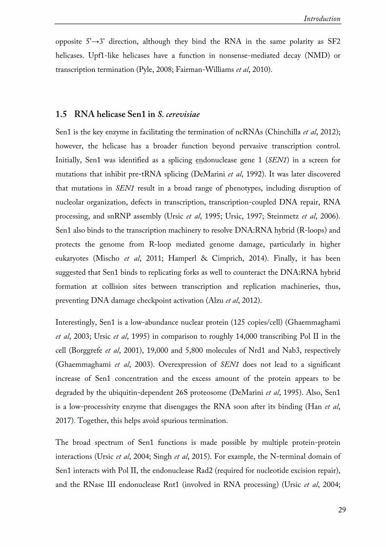
The expression of the N-terminal domain was not suitable for crystallization, thus the main focus was on obtaining the helicase domain alone. At first, a construct 976-1880 that lacks both the N-terminus domain and the low-complexity C-terminus end was designed. The fragment was amplified from S. cerevisiae genomic DNA and cloned into a vector with a CPD-His8 tag at the C-terminus. The construct was well expressed in E. coli, however, during purification it appeared that the protein was about 20 kDa smaller than it was expected (Figure 3-2, a). Mass-spectrometry and Edman sequencing confirmed the endogenous protein degradation of 130 amino acids at the N-terminus.
Figure 3-2. Test-purification of Sen1 helicase domain (SDS-PAGE). (a) Sen1 helicase core [976-1880]. Expected molecular weight of the construct with the tag was 126 kDa but the protein ran just above 100 kDa. (b) Ni2+-NTA elution samples of different Sen1 helicase core constructs.
Secondary structure prediction (see Appendix 1) suggested that residues 1095-1106 form
an α-helix, therefore the following constructs starting at residue 1095 were designed. Moreover, for some of the constructs the C-terminal region was extended up to the nucleus localization signal (NLS) (residue 1910) with the idea to obtain longer fragments of the protein and do limited proteolysis. The constructs were designed for the expression in E. coli and tested for solubility. Additionally, T4 lysozyme (T4L) and thioredoxin (Trx) tags were tested to check whether a tag changes the expression levels of the proteins. It appeared that all the constructs were similarly well expressed (Figure 3-2, b). Some of the constructs were purified in a large scale and set-up for crystallization. A fragment 1095-1904 (hereafter referred to as Sen1Hel) yielded to crystals and, therefore, the further studies were continued only on it.

Results
37
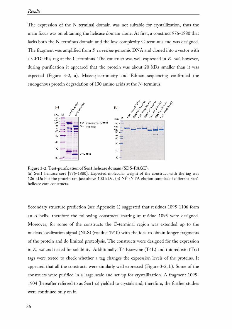
3.1.2 Purification of the Sen1Hel
Several rounds of expression tests and purification buffer screens were performed in order to determine the conditions for the highest yield of Sen1Hel (see Methods 6.2.2 and Methods 6.2.4). The best purity was achieved when Lysis buffer (20 mM sodium
phosphate pH 8.0, 500 mM NaCl, 2 mM MgCl2, 1 mM β-mercaptoethanol) was supplemented with 10 % (v/v) glycerol, 30 mM imidazole, benzonase, and protease inhibitors and the CPD-His8 -tag was cleaved ‘on column’ by adding HRV 3C protease to Ni2+-affinity beads and incubating at 4°C overnight (see Methods 6.2.4.2). A combination of Ni2+-affinity and heparin ion exchange chromatography steps led to almost pure protein (Figure 3-3, a).
Figure 3-3. Purification of Sen1Hel. (a) A chromatogram of the elution from heparin sepharose column with a linear NaCl gradient. Red and blue lines correspond to the absorbance at 260 nm and 280 nm, respectively. Brown line is the conductivity and green line is percentage of Buffer B. A corresponding SDS-PAGE gel, stained with Coomassie blue, shown at the bottom. After elution from heparin sepharose column, Sen1Hel was concentrated and loaded onto a Superdex 200 [16/600] gel filtration column. (b) A chromatogram of SEC and a corresponding SDS-PAGE gel, stained with Coomassie blue, shown at the bottom. Red and blue lines correspond to the absorbance at 260 nm and 280 nm, respectively. In the main peak (green dots), the ratio of 260 nm and 280 nm is below 0.5, indicating that the sample is not contaminated with nucleic acids. A small fraction of the protein eluted in void-volume or as aggregate (first two peaks, violet dots).
Results
38
The collected elution fractions from the heparin column were concentrated to ~12 mg/mL and then further purified by size exclusion chromatography (SEC)(Figure 3-3, b). In this step the aggregates were separated (void volume and peak 1) and the protein eluted in a peak at the expected elution volume for a globular protein of 90 kDa (14 ml on Superdex 200 [16/600] column). Every purification step was monitored by SDS-PAGE.
Usually, 3 L of bacterial culture yielded up to 20 mg of purified protein, which could be concentrated to 10 mg/mL or higher. In order to avoid aggregates forming upon freezing, 50 % (w/v) glycerol was added to the buffer. When kept on ice, Sen1Hel was generally very stable and could be stored for weeks.
Results
39
3.1.3 Biochemical characterization of Sen1Hel
To test whether Sen1Hel is a functionally relevant construct, Dr. Zhong Han and Dr. Odil Porrua from collaboration group of Dr. Domenico Libri (Institut Jacques Monod, Paris) tested the ATPase and helicase activities and did in vitro transcription termination assays (Porrua & Libri, 2013). It appeared that the helicase core domain alone has similar biochemical properties to those of endogenous full-length Sen1 (the results are discussed more in detail later). In parallel, the collaborators studied the endogenous Sen1 proteins. Their helicase domain contains 976-1880 fragment, the same that I could purify from E.
coli, however, endogenously expressed protein did not degrade. The results showed that, indeed, the helicase domain alone is sufficient for transcription termination in vitro and that the N- and C-terminal domains are most relevant for processes in vivo (Han et al, 2017). Very similar conclusions were made by Brow’s group, who studied Sen11095-1876 in
vitro (Martin-Tumasz & Brow, 2015). Whether Sen1 is regulated through posttranscriptional modifications is still not clear, but the fact that the recombinant protein retains the capability of endogenous protein suggests that the modifications are not crucial, at least for the tested truncations and activities.
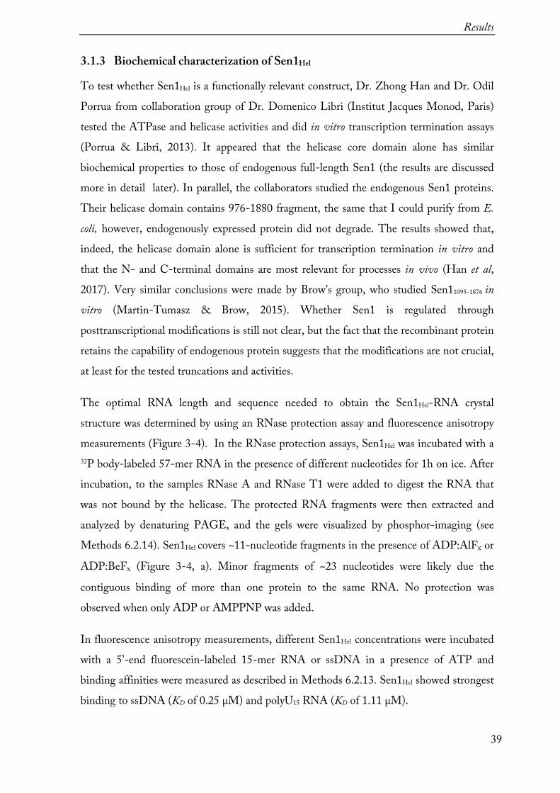
The optimal RNA length and sequence needed to obtain the Sen1Hel-RNA crystal structure was determined by using an RNase protection assay and fluorescence anisotropy measurements (Figure 3-4). In the RNase protection assays, Sen1Hel was incubated with a 32P body-labeled 57-mer RNA in the presence of different nucleotides for 1h on ice. After incubation, to the samples RNase A and RNase T1 were added to digest the RNA that was not bound by the helicase. The protected RNA fragments were then extracted and analyzed by denaturing PAGE, and the gels were visualized by phosphor-imaging (see Methods 6.2.14). Sen1Hel covers ~11-nucleotide fragments in the presence of ADP:AlFx or
ADP:BeFx (Figure 3-4, a). Minor fragments of ~23 nucleotides were likely due the
contiguous binding of more than one protein to the same RNA. No protection was observed when only ADP or AMPPNP was added.
In fluorescence anisotropy measurements, different Sen1Hel concentrations were incubated with a 5’-end fluorescein-labeled 15-mer RNA or ssDNA in a presence of ATP and binding affinities were measured as described in Methods 6.2.13. Sen1Hel showed strongest binding to ssDNA (KD of 0.25 µM) and polyU15 RNA (KD of 1.11 µM).

Results
40
Figure 3-4. RNA binding by Sen1Hel. (a) RNase protection assay in the presence of different nucleotides. RNA fragments were obtained by digesting 57-mer RNA. Sen1Hel binds RNA only in a presence of ADP:BeFx or ADP:AlFx but not in a presence of ADP or AMPPNP. Sen1Hel protects 11-nucleotide fragments. For comparison, the footprints of helicase Dbp5 are shown on the right side of the gel. (b) Fluorescence anisotropy measurements. Sen1Hel can bind to both RNA and ssDNA, with the higher affinity for the latter.

Results
41
3.2 Crystal structure determination of Sen1Hel 3.2.1 Sen1Hel crystallization
Purified Sen1Hel was concentrated to 3-4 mg/mL (30-35 µM), mixed with a 1.2 molar excess of polyU15 RNA and a 10-fold molar excess of freshly prepared nucleotides to set up for automated crystallization screening in the Crystallization facility of our department. The initial crystallization screening was done using several multicomponent screens by sitting-drop vapor diffusion method in 96-well plates. Most of crystallization hits were observed in crystallization conditions from the in-house Complex screen II solution (Table 3-1) at 4°C (Figure 3-5, a-d). The crystals were forming clusters of needles (a), plates (b-c) or three-dimensional rods (c-d) within 6-10 days. Adjusting precipitant pH and PEG concentrations as well as drop size helped to optimize the crystallization conditions further (Figure 3-5, e-h). Optimized crystals formed larger rod clusters (e), which could be broken apart, or single triangular prisms (f-h), and were mounted to nylon loops for data collection. Prior to flash freezing in liquid nitrogen, the crystals were briefly soaked in mother liquor supplemented with 25-28 % (w/v) ethylene glycol for cryo-protection. In some cases, crystals were soaked in mother liquor additionally supplemented with polyU15 RNA.
3.2.2 X-ray data collection
Crystals were first pre-screened using in-house D8 VENTURE (Bruker) X-ray diffractometer to select well diffracting (up to 4 Å) and efficiently cryo-protected ones (no ice ring observed in diffraction pattern). X-ray data collection of data sets used for structure determination was performed at the super-bending magnet beamline X06DA (PXIII) at the Swiss Light Source (Villigen, Switzerland). A native data was collected at 1.0 Å wavelength and a single-wavelength anomalous diffraction (SAD) data set was collected at 2.095 Å.

Results
42
12
34
56
78
910
1112
20%
PEG
400
20%
PEG
400
20%
PEG
400
20%
PEG
400
20%
PEG
400
18%
PEG
2000
18
% PE
G 20
00
18%
PEG
2000
18
% PE
G 20
00
18%
PEG
2000
18
% PE
G 20
00
18%
PEG
2000
A
0,1M
Na a
cetate
0,1
M tri
-Na c
itrate
0,1
M N
a caco
dylat
e 0,1
M H
EPES
0,1
M T
ris0,1
M N
a acet
ate0,1
M tri
-Na c
itrate
0,1
M Bi
s - T
ris
0,1M
Na K
phosp
hate
0,1M
Na c
acody
late
0,1M
MES
0,1
M M
ESpH
4,5
pH 5,
5pH
6,5
pH 7,
5pH
8,5
pH 5,
0pH
5,6
pH 6,
2pH
6,5
pH 6,
0pH
6,0
pH 6,
5
18%
PEG
2000
18
% PE
G 20
00
18%
PEG
2000
18
% PE
G 20
00
18%
PEG
2000
18
% PE
G 20
00
18%
PEG
4000
18
% PE
G 40
00
18%
PEG
4000
18
% PE
G 40
00
18%
PEG
4000
18
% PE
G 40
00
B0,1
M A
DA
0,1M
HEP
ES
0,1M
HEP
ES
0,1M
MOP
S 0,1
M T
ris
0,1M
Tris
0,1
M N
a acet
ate
0,1M
tri-N
a citr
ate
0,1M
Bis-T
ris
0,1M
Na K
phosp
hate
0,1M
Na c
acody
late
0,1M
MES
pH
6,6
pH 7,
0pH
7,5
pH 7,
2pH
8,0
pH 8,
5pH
5,0
pH 5,
6pH
6,2
pH 6,
5pH
6,0
pH 6,
0
18%
PEG
4000
18
% PE
G 40
00
18%
PEG
4000
18
% PE
G 40
00
18%
PEG
4000
18
% PE
G 40
00
18%
PEG
4000
18
% M
PEG
5000
18
% M
PEG
5000
18
% M
PEG
5000
18
% M
PEG
5000
18
% M
PEG
5000
C
0,1M
MES
0,1
M A
DA
0,1M
HEP
ES0,1
M H
EPES
0,1M
MOP
S 0,1
M T
ris
0,1M
Tris
0,1
M N
a acet
ate
0,1M
tri-N
a citr
ate
0,1M
Bis -
Tris
0,1
M N
a K ph
ospha
te 0,
1M N
a caco
dylat
e pH
6,5
pH 6,
6pH
7,0
pH 7,
5pH
7,2
pH 8,
0pH
8,5
pH 5,
0pH
5,6
pH 6,
2pH
6,5
pH 6,
0
18%
MPE
G 50
00
18%
MPE
G 50
00
18%
MPE
G 50
00
18%
MPE
G 50
00
18%
MPE
G 50
0018
% M
PEG
5000
18
% M
PEG
5000
18
% M
PEG
5000
15
% PE
G 60
00
15%
PEG
6000
15
% PE
G 60
00
15%
PEG
6000
D
0,1M
MES
0,1
M M
ES
0,1M
ADA
0,1
M H
EPES
0,1
M H
EPES
0,1M
MOP
S 0,1
M T
ris
0,1M
Tris
0,1
M N
a acet
ate
0,1M
tri-N
a citr
ate
0,1M
Bis -
Tris
0,1
M N
a K ph
ospha
te pH
6,0
pH 6,
5pH
6,6
pH 7,
0pH
7,5
pH 7,
2pH
8,0
pH 8,
5pH
5,0
pH 5,
6pH
6,2
pH 6,
5
15%
PEG
6000
15
% PE
G 60
00
15%
PEG
6000
15
% PE
G 60
00
15%
PEG
6000
15
% PE
G 60
00
15%
PEG
6000
15
% PE
G 60
00
15%
PEG
6000
15
% PE
G 80
00
15%
PEG
8000
15
% PE
G 80
00
E 0,
1M N
a caco
dylat
e 0,1
M M
ES
0,1M
MES
0,1
M A
DA
0,1M
HEP
ES0,1
M H
EPES
0,1M
MOP
S 0,1
M T
ris
0,1M
Tris
0,1
M N
a acet
ate
0,1M
tri-N
a citr
ate
0,1M
Bis -
Tris
pH
6,0
pH 6,
0pH
6,5
pH 6,
6pH
7,0
pH 7,
5pH
7,2
pH 8,
0pH
8,5
pH 5,
0pH
5,6
pH 6,
2
15%
PEG
8000
15
% PE
G 80
00
15%
PEG
8000
15
% PE
G 80
00
15%
PEG
8000
15
% PE
G 80
00
15%
PEG
8000
15
% PE
G 80
00
15%
PEG
8000
15
% PE
G 80
00
12%
PEG
20 00
0 12
% PE
G 20
000
F0,1
M N
a K ph
ospha
te 0,1
M N
a caco
dylat
e 0,1
M M
ES
0,1M
MES
0,1M
ADA
0,1
M H
EPES
0,1
M H
EPES
0,1M
MOP
S 0,1
M T
ris
0,1M
Tris
0,1
M N
a acet
ate
0,1M
tri-N
a citr
ate
pH 6,
5pH
6,0
pH 6,
0pH
6,5
pH 6,
6pH
7,0
pH 7,
5pH
7,2
pH 8,
0pH
8,5
pH 5,
0pH
5,6
12%
PEG
20 00
0 12
% PE
G 20
000
12%
PEG
20 00
0 12
% PE
G 20
000
12%
PEG
20 00
0 12
% PE
G 20
000
12%
PEG
20 00
0 12
% PE
G 20
000
12%
PEG
20 00
0 12
% PE
G 20
000
12%
PEG
20 00
0 1,8
M A
mm. su
lfate
G0,1
M Bi
s - T
ris
0,1M
Na K
phosp
hate
0,1M
Na c
acody
late
0,1M
MES
0,1
M M
ES
0,1M
ADA
0,1
M H
EPES
0,1M
HEP
ES0,1
M M
OPS
0,1M
Tris
0,1
M T
ris
0,1M
Na a
cetate
pH
6,2
pH 6,
5pH
6,0
pH 6,
0pH
6,5
pH 6,
6pH
7,0
pH 7,
5pH
7,2
pH 8,
0pH
8,5
pH 5,
0
1,8M
Amm
. sulfa
te1,8
M A
mm. su
lfate
1,8M
Amm
. sulfa
te1,8
M A
mm. su
lfate
1,8M
Amm
. sulfa
te1,8
M A
mm. su
lfate
1,8M
Amm
. sulfa
te1,8
M A
mm. su
lfate
1,8M
Amm
. sulfa
te1,8
M A
mm. su
lfate
1,8M
Amm
. sulfa
te1,8
M A
mm. su
lfate
H0,1
M tri
-Na c
itrate
0,1
M Bi
s - T
ris
0,1M
Na K
phosp
hate
0,1M
Na c
acody
late
0,1M
MES
0,1
M M
ES0,1
M A
DA
0,1M
HEP
ES
0,1M
HEP
ES
0,1M
MOP
S 0,1
M T
ris
0,1M
Tris
pH
5,6
pH 6,
2pH
6,5
pH 6,
0pH
6,0
pH 6,
5pH
6,6
pH 7,
0pH
7,5
pH 7,
2pH
8,0
pH 8,
5
Tab
le 3-
1. C
ompl
ex sc
reen
II.
In b
rown
are h
ighl
ight
ed p
recip
itant
solu
tions
in w
hich
crys
tals
of S
en1 H
el wer
e for
med
.
Results
43
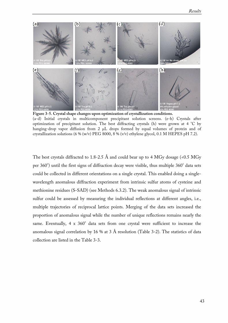
Figure 3-5. Crystal shape changes upon optimization of crystallization conditions. (a-d) Initial crystals in multicomponent precipitant solution screens. (e-h) Crystals after optimization of precipitant solution. The best diffracting crystals (h) were grown at 4 °C by hanging-drop vapor diffusion from 2 µL drops formed by equal volumes of protein and of crystallization solutions (6 % (w/v) PEG 8000, 8 % (v/v) ethylene glycol, 0.1 M HEPES pH 7.2).
The best crystals diffracted to 1.8-2.5 Å and could bear up to 4 MGy dosage (<0.5 MGy per 360°) until the first signs of diffraction decay were visible, thus multiple 360° data sets could be collected in different orientations on a single crystal. This enabled doing a single-wavelength anomalous diffraction experiment from intrinsic sulfur atoms of cysteine and methionine residues (S-SAD) (see Methods 6.3.2). The weak anomalous signal of intrinsic sulfur could be assessed by measuring the individual reflections at different angles, i.e., multiple trajectories of reciprocal lattice points. Merging of the data sets increased the proportion of anomalous signal while the number of unique reflections remains nearly the same. Eventually, 4 x 360° data sets from one crystal were sufficient to increase the anomalous signal correlation by 16 % at 3 Å resolution (Table 3-2). The statistics of data collection are listed in the Table 3-3.

Results
44
Table 3-2. Statistics of data processing.
3.2.3 Crystal structure determination and evaluation
The data could be indexed and processed in orthorhombic space group P21212 with one molecule per symmetric unit. The data was processed using XDS and scaled and merged with XSCALE (Kabsch, 2010). The high-resolution data cutoff was based on the statistical indicators CC1/2 and CC* (Karplus & Diederichs, 2012). Substructure determination and phasing were performed with SHELXC/D/E (Sheldrick, 2010) using HKL2MAP (Pape & Schneider, 2004). The successful SHELXD substructure solution, in a search for 25 sulfur sites, had a CCall and a CCweak of 36.9 and 18.2, respectively. Density modification resulted in a clear separation of hands. Three cycles of chain tracing resulted in the automatic building of 275 amino acids with SHELXE.
Results
45
The initial model was built automatically using BUCCANEER (Cowtan, 2006) and corrected and completed manually using COOT (Emsley & Cowtan, 2004) and the experimental electron density map in. Further model refinements were done against native data with PHENIX.refine (Adams et al, 2010). When building in ADP:AlFx into the model, it appeared that only ADP molecule was bound to Sen1Hel and no electron density for the AlFx moiety could be obtained (Figure 3-6). Finally, also ligands (glycerol and ethylene glycol) and water molecules were built in into the model.
The final model was refined at 1.8-Å resolution with an Rfree of 18.4 % and Rfactor of 15.3 % and good stereochemistry (Table 3-3). In particular, 98 % of residues are in the most favored regions of the Ramachandran plot and the model has no outliers (Figure 3-7, b). Based on the wwPDB evaluation tool, which compares all available structures of the same resolution, Sen1Hel model has relatively good percentile ranks also for key global quality indicators like Clashscore and outliers of Sidechain or Real-space R-value Z-score (RSRZ) (Figure 3-7, a).

Results
46
Figure 3-6. Sen1Hel model building (a) ADP molecule and magnesium ion are built into the Fo-Fc electron density map (green). (b) A hexahydrated octahedral architecture of magnesium dication and six oxygen atoms, four from water molecules and two from Thr1364 residue and β-phosphate of ADP. The 2Fo-Fc electron density map is contoured at 1.7σ. (c) Magnesium (green sphere) is coordinated at the active site of Sen1 by amino acid residues (Thr1364, Asp1590, Glu1591, Glu1418, Arg1422 and Lys1363), ADP and is surrounded with six waters. (d) The overall view of final Sen1Hel model (blue). Water molecules and ligands are displayed in red and grey, respectively.
Figure 3-7. Sen1Hel model validation. (a) Overall X-ray structure quality validation with the respect to all structures in the Protein Data Bank (http://rcsb.org). (b) Ramachandran plot of Φ and Ψ torsion angles of the α-chain of the protein.
Results
47
Table 3-3. Crystallographic data collection and refinement statistics.
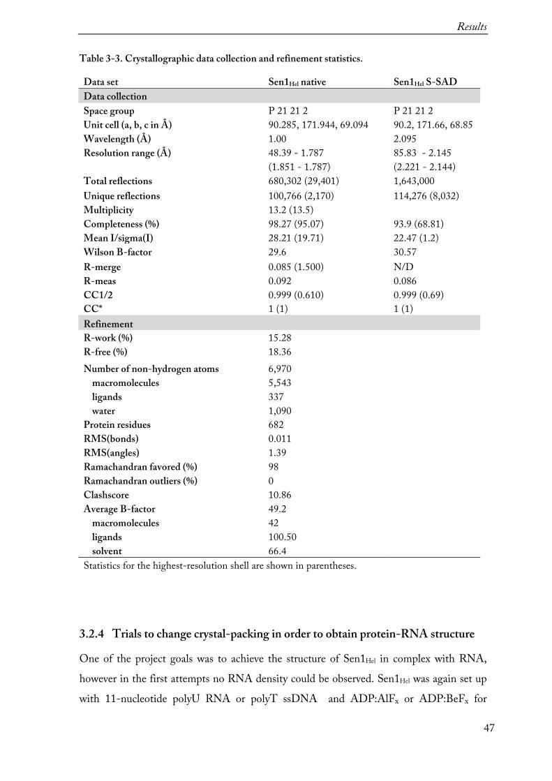
Data set Sen1Hel native Sen1Hel S-SAD Data collection Space group P 21 21 2 P 21 21 2 Unit cell (a, b, c in Å) 90.285, 171.944, 69.094 90.2, 171.66, 68.85 Wavelength (Å) 1.00 2.095 Resolution range (Å) 48.39 - 1.787 85.83 - 2.145 (1.851 - 1.787) (2.221 - 2.144) Total reflections 680,302 (29,401) 1,643,000 Unique reflections 100,766 (2,170) 114,276 (8,032) Multiplicity 13.2 (13.5) Completeness (%) 98.27 (95.07) 93.9 (68.81) Mean I/sigma(I) 28.21 (19.71) 22.47 (1.2) Wilson B-factor 29.6 30.57 R-merge 0.085 (1.500) N/D R-meas 0.092 0.086 CC1/2 0.999 (0.610) 0.999 (0.69) CC* 1 (1) 1 (1) Refinement R-work (%) 15.28 R-free (%) 18.36 Number of non-hydrogen atoms 6,970 macromolecules 5,543 ligands 337 water 1,090 Protein residues 682 RMS(bonds) 0.011 RMS(angles) 1.39 Ramachandran favored (%) 98 Ramachandran outliers (%) 0 Clashscore 10.86 Average B-factor 49.2 macromolecules 42 ligands 100.50 solvent 66.4 Statistics for the highest-resolution shell are shown in parentheses.
3.2.4 Trials to change crystal-packing in order to obtain protein-RNA structure
One of the project goals was to achieve the structure of Sen1Hel in complex with RNA, however in the first attempts no RNA density could be observed. Sen1Hel was again set up with 11-nucleotide polyU RNA or polyT ssDNA and ADP:AlFx or ADP:BeFx for
Results
48
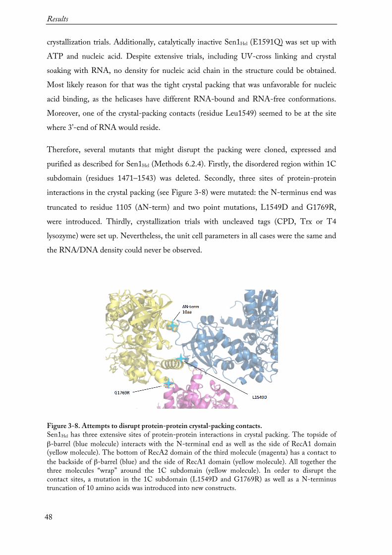
crystallization trials. Additionally, catalytically inactive Sen1Hel (E1591Q) was set up with ATP and nucleic acid. Despite extensive trials, including UV-cross linking and crystal soaking with RNA, no density for nucleic acid chain in the structure could be obtained. Most likely reason for that was the tight crystal packing that was unfavorable for nucleic acid binding, as the helicases have different RNA-bound and RNA-free conformations. Moreover, one of the crystal-packing contacts (residue Leu1549) seemed to be at the site where 3’-end of RNA would reside.
Therefore, several mutants that might disrupt the packing were cloned, expressed and purified as described for Sen1Hel (Methods 6.2.4). Firstly, the disordered region within 1C subdomain (residues 1471–1543) was deleted. Secondly, three sites of protein-protein interactions in the crystal packing (see Figure 3-8) were mutated: the N-terminus end was
truncated to residue 1105 (ΔN-term) and two point mutations, L1549D and G1769R, were introduced. Thirdly, crystallization trials with uncleaved tags (CPD, Trx or T4 lysozyme) were set up. Nevertheless, the unit cell parameters in all cases were the same and the RNA/DNA density could never be observed.
Figure 3-8. Attempts to disrupt protein-protein crystal-packing contacts. Sen1Hel has three extensive sites of protein-protein interactions in crystal packing. The topside of β-barrel (blue molecule) interacts with the N-terminal end as well as the side of RecA1 domain (yellow molecule). The bottom of RecA2 domain of the third molecule (magenta) has a contact to the backside of β-barrel (blue) and the side of RecA1 domain (yellow molecule). All together the three molecules “wrap” around the 1C subdomain (yellow molecule). In order to disrupt the contact sites, a mutation in the 1C subdomain (L1549D and G1769R) as well as a N-terminus truncation of 10 amino acids was introduced into new constructs.

Results
49
3.3 The structure of Sen1Hel
The model of Sen1Hel has only one molecule in the asymmetric unit. The majority of the cloned construct could be modeled with exception of a disordered region in the subdomain 1C (residues 1471–1543), the C-terminus (residues 1876–1904) and a loop region in the RecA1 domain (residues 1382–1395). Also, the regions encompassing residues 1705–1713 and 1799–1801 have very poor electron density and could not be modeled. The model includes one ADP and one magnesium ion (Figure 3-9). Overall, Sen1Hel has a domain organization similar to that of the related helicases Upf1 (Upf1Hel, also known as Upf1-
ΔCH) (Cheng et al, 2007; Clerici et al, 2009; Chakrabarti et al, 2011) and IGHMBP2 (IGHMBP2Hel) (Lim et al, 2012).
Figure 3-9. Crystal structure of yeast Sen1Hel. Top: schematic representation of the domain organization of the SenHel. Bottom: the front and the side views of the SenHel structure. The core domain is formed of two RecA domains (yellow). The “prong” (red) and the “stalk” with the “barrel” on top of it are protruding from RecA1. The “brace” is wrapping around the “stalk” and is sandwiched between the “stalk” and the “barrel”.

Results
50
3.3.1 Architecture of Sen1Hel 3.3.1.1 Two central RecA domains
The helicase core of Sen1 is composed of two small globular RecA domains (residues 1314-1651 and 1657-1861) that pack against each other forming a 10 Å-wide wedge-like cleft, and are connected with a short linker of five residues. RecA2 is rotated of about 30° from the position that is typical of helicases in an active RNA-ATP-bound conformation
(Figure 3-14) (Pyle, 2008). Both domains have a similar topology of a central parallel β-
sheet surrounded by eight α-helices. The loops between β-strands contain characteristic motifs for DNA/RNA and ATP binding (Figure 3-10). Tight coordination of the motif binding to nucleotides and RecA domain rotation upon ATP hydrolysis is a key for translocation on nucleic acid (Saikrishnan et al, 2009).
Figure 3-10. Conserved motifs for nucleic acid and ATP binding mapped on Sen1Hel structure. The characteristic sequence motifs are involved in ATP binding and hydrolysis (Q, I, Ib, II, IIIa, and VI, shown in magenta), in DNA/RNA binding (Ia, Ic, IV, V, and Vb, shown in turquoise) and coordination between nucleic acid and ATP binding (III and Va, shown in dark yellow). Importantly, Motif IIIa is a part of the linker between two RecA domains and is found only in SF1 helicases.
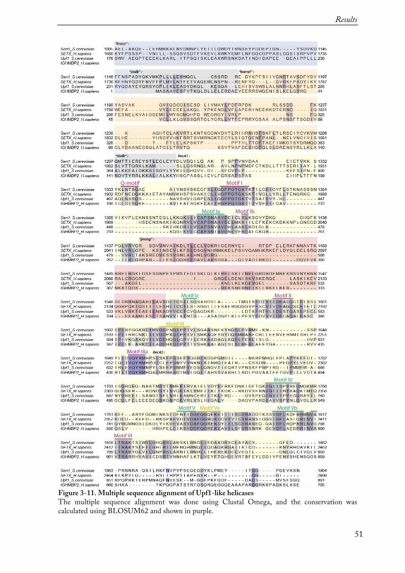
Results
51
Figure 3-11. Multiple sequence alignment of Upf1-like helicases The multiple sequence alignment was done using Clustal Omega, and the conservation was calculated using BLOSUM62 and shown in purple.

Results
52
3.3.1.2 ADP binding
ADP is bound to the RecA1 domain inside the cleft between RecA1 and RecA2 (Figure 3-9 and Figure 3-10). The adenine ring is sandwiched between an apolar surface of Lys1365 of RecA1 and Tyr1655 of the linker. This tyrosine is conserved among Upf1-like helicases, it corresponds to Tyr638Upf1 and Tyr422IGHMBP2 (see alignment Figure 3-11), and is a part of the Motif IIIa (Gln-Tyr-Arg-Met), which is present only in SF1 helicases but not in SF2 (Fairman-Williams et al, 2010). In SF2 helicases, e.g. in yeast Ski2 (Halbach et
al, 2012), the adenine ring is also sandwiched between the two RecA domains by Phe328 and Arg767 (part of Motif IVa that is found only in SF2 helicases), however the linker to the nucleotide binding is not involved (Figure 3-13). In addition, the adenine amino group is recognized by conserved Gln1339 side chain (corresponding to Gln413Upf1, Gln196IMGMBP2 and Gln334Ski2) of the Q-motif. This Gln-based specificity is common to all SF1 and SF2 helicases that have substrate preference to ATP over other nucleotides
(Cordin et al, 2004). Gly1362, Lys1363, Thr1364 and Arg1422 coordinate α- and β-phosphates of the ADP (Figure 3-13).
Figure 3-12. Sen1Hel ADP binding plot. A two-dimensional representation of the residues involved in ADP binding by Sen1Hel. ADP is shown in black, the residues are shown in yellow. The spheres in red represent oxygen, in blue – nitrogen, in green – magnesium ion. Blue dashed lines indicate hydrogen bonds, and numbers indicate bond length in Å. The adenine ring is sandwiched between an apolar surface of Lys1356 and an aromatic ring of Tyr1655. The ADP binding plot was calculated using LigPlot.
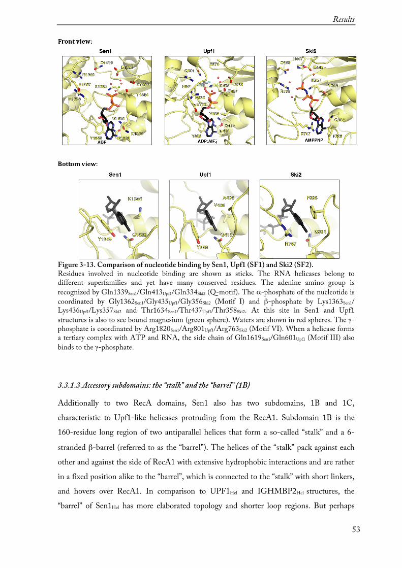
Results
53
Figure 3-13. Comparison of nucleotide binding by Sen1, Upf1 (SF1) and Ski2 (SF2). Residues involved in nucleotide binding are shown as sticks. The RNA helicases belong to different superfamilies and yet have many conserved residues. The adenine amino group is recognized by Gln1339Sen1/Gln413Upf1/Gln334Ski2 (Q-motif). The α-phosphate of the nucleotide is coordinated by Gly1362Sen1/Gly435Upf1/Gly356Ski2 (Motif I) and β-phosphate by Lys1363Sen1/ Lys436Upf1/Lys357Ski2 and Thr1634Sen1/Thr437Upf1/Thr358Ski2. At this site in Sen1 and Upf1 structures is also to see bound magnesium (green sphere). Waters are shown in red spheres. The γ-phosphate is coordinated by Arg1820Sen1/Arg801Upf1/Arg763Ski2 (Motif VI). When a helicase forms a tertiary complex with ATP and RNA, the side chain of Gln1619Sen1/Gln601Upf1 (Motif III) also binds to the γ-phosphate. 3.3.1.3 Accessory subdomains: the “stalk” and the “barrel” (1B)
Additionally to two RecA domains, Sen1 also has two subdomains, 1B and 1C, characteristic to Upf1-like helicases protruding from the RecA1. Subdomain 1B is the 160-residue long region of two antiparallel helices that form a so-called “stalk” and a 6-
stranded β-barrel (referred to as the “barrel”). The helices of the “stalk” pack against each other and against the side of RecA1 with extensive hydrophobic interactions and are rather in a fixed position alike to the “barrel”, which is connected to the “stalk” with short linkers, and hovers over RecA1. In comparison to UPF1Hel and IGHMBP2Hel structures, the “barrel” of Sen1Hel has more elaborated topology and shorter loop regions. But perhaps

Results
54
more importantly, the “barrel” in apo-structure of Sen1Hel is fixed to the top of the “stalk” whereas in the other helicases the “barrel” is positioned on the top of RecA domains where the RNA would reside (Figure 3-14).
Figure 3-14. Structural comparison of Sen1Hel with related helicases. The apo-structures of the helicase core of yeast Sen1, human Upf1 (PDB: 2GJK) and IGHMBP2 (PDB: 4B3F). The structures are shown in the orientation of superposed RecA1 domains. Dashed lines indicate missing parts of the structure. The schematic representations of full-length helicase domain organization are shown on top, with structured domains shown as rectangles and unstructured regions as a black line. Domains in scheme and the structure are highlighted in the same colors. Among Upf1-like helicases only Sen1 has an additional fragment to the helicase core, which braces the “barrel” and the “stalk” together, and therefore is referred to as the “brace”. 3.3.1.4 Accessory subdomains: the “brace” is unique to Sen1
Structural comparison of Sen1Hel with Upf1Hel and IGHMBP2Hel revealed key differences in subdomain 1B. First of all, the “barrel” has a short 3-residue linker to the ascending helix of the “stalk” in contrast to a 10-residue linker observed in Upf1Hel or to a helix in IGHMBP2Hel. Similarly, the “barrel” is directly connected to the descending helix of the “stalk” in Sen1Hel, whereas it has a short linker in case of Upf1Hel and IGHMBP2Hel. Thus, in contrast to other Upf1-like helicases, the “barrel” of Sen1 seems to be spatially restricted. In addition, Sen1 has a unique N-terminal segment (residues 1096-1149) that we refer to as the “brace” (Figure 3-14). The “brace” fastens together the RecA1, the “stalk” and the “barrel” with extensive hydrophobic interactions.
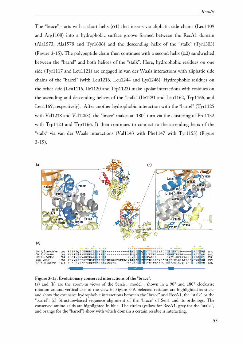
Results
55
The “brace” starts with a short helix (α1) that inserts via aliphatic side chains (Leu1109 and Arg1108) into a hydrophobic surface groove formed between the RecA1 domain (Ala1573, Ala1578 and Tyr1606) and the descending helix of the “stalk” (Tyr1303)
(Figure 3-15). The polypeptide chain then continues with a second helix (α2) sandwiched
between the “barrel” and both helices of the “stalk”. Here, hydrophobic residues on one side (Tyr1117 and Leu1121) are engaged in van der Waals interactions with aliphatic side chains of the “barrel” (with Leu1216, Leu1244 and Lys1246). Hydrophobic residues on the other side (Leu1116, Ile1120 and Trp1123) make apolar interactions with residues on the ascending and descending helices of the “stalk” (Ile1291 and Leu1162, Trp1166, and Leu1169, respectively). After another hydrophobic interaction with the “barrel” (Tyr1125
with Val1218 and Val1283), the “brace” makes an 180° turn via the clustering of Pro1132 with Trp1123 and Trp1166. It then continues to connect to the ascending helix of the “stalk“ via van der Waals interactions (Val1143 with Phe1147 with Tyr1153) (Figure 3-15).
Figure 3-15. Evolutionary conserved interactions of the "brace". (a) and (b) are the zoom-in views of the Sen1Hel model , shown in a 90° and 180° clockwise rotation around vertical axis of the view in Figure 3-9. Selected residues are highlighted as sticks and show the extensive hydrophobic interactions between the “brace” and RecA1, the “stalk” or the “barrel”. (c) Structure-based sequence alignment of the “brace” of Sen1 and its orthologs. The conserved amino acids are highlighted in blue. The circles (yellow for RecA1, grey for the “stalk”’, and orange for the “barrel”) show with which domain a certain residue is interacting.

Results
56
The deletions of the “brace” (constructs 1128-1904, 1136-1094, 1149-1904, and 1178-1880) resulted in insoluble protein, most likely because upon removal of the “brace” the above-mentioned hydrophobic residues become exposed to the solvent. Consistently, also the proteins of point mutants where interaction between the “brace” and the “barrel” were disrupted (L1121R, W1123R, and Y1117R) were insoluble. Thus, it seems that the “brace” is a distinct structural feature that stabilizes the overall fold of Sen1Hel and enables to promote transcription termination.
3.3.1.5 Accessory subdomains: the “prong” (1C)
The subdomain 1C is the second structural feature characteristic to Upf1-like helicases (Figure 3-14). In Sen1Hel this 120-residue prong-like protrusion from the RecA1 domain
(residues 1443-1563) is composed of an α-helix almost parallel to the “stalk”, two short
α-helices at the base and a 75-residue region in between that could not be modeled (Figure
3-9), although this region is predicted (PSIPRED) to form α-helices as well. In contrast, the “prong” subdomains in Upf1 and IGHMBP2 are 65 and 80-residue long, respectively. Although the overall structures of the three “prong” subdomains are very similar, their sequences are poorly conserved with the exception of a few residues that are important for RNA binding (see multiple sequence alignment Figure 3-11).
3.3.2 RNA-binding
Attempts to get a structure of Sen1Hel in complex with RNA failed and the following studies were based on the comparison of the available structures of Sen1Hel-ADP with Upf1CH-Hel-ADP:AlF4--RNA (Chakrabarti et al, 2011) and IGHMBP2Hel-ADP-RNA (Lim et al, 2012). All three proteins share conserved RNA binding motifs that enable the helicase to bind or translocate along the RNA. Particularly interesting is the comparison of RNA binding by Motif Ia and Motif III in “open” and “close” conformations of the helicases (Figure 3-16).
Motif III (GDPNQLPPTSen1, GDHQQLGPVUpf1, GDHKQLPPTIGHMBP2) together with Motif Vb coordinates RNA binding and ATPase activity (Jankowsky & Fairman, 2007). In Sen1, the GDPNQ fragment (residues 1615-1619) stacks on top of Motif I with

Results
57
Gln1619 (corresponding to Gln597Upf1 and Gln399IGHMBP2) at the position where γ-phosphate of ATP would reside. At the other end of Motif III, the hydrophobic Leu1620 and non-polar Thr1624 side chains stack with the interaction site between Motif Va, Motif V and the RNA backbone.
Motif Ia (CAPSNAAVSen1, CAPSNVAVUpf1, CAPSNIAVIGHMBP2) is particularly characteristic to both DNA and RNA helicases of SF1 that translocate in 3'→5' direction. As there are more structures of DNA helicases available, the mechanism of how the helicases translocate on DNA is better understood. Here, structural studies show, that during translocation the base at position 5 flips in and out of Motif Ia pocket and stacks to Val/Thr of Motif III (Saikrishnan et al, 2009) (Figure 3-16, bottom right panel). In the available structures of RNA helicases such base flipping has not been observed although the same conserved residues are involve in RNA binding. However, in an “open” conformation there is enough space between Motif Ia and Motif III to fit two nucleotides whereas in a “close” conformation - only one nucleotide could fit. Probably, the main structural difference between the structures of DNA and RNA helicase of SF1 is the presence of accessory subdomains on RecA1. In DNA helicases, the N-terminal domain is pushing ssDNA to topside of RecA domains, whereas in RNA helicases the “stalk” and the “barrel” are pushing the RNA towards the “prong”.

Results
58
Figure 3-16. RNA binding by Motifs Ia and III. Comparison of top views of nucleic acid binding sites of Sen1, IGHMBP2 (PDB: 4B3G) and Upf1 (PDB: 2XZL) and DNA helicase RecD2 (PDB: 3GP8). Motif Ia is shown in blue and Motif III is shown in orange. The nucleotides bound between the two motifs are highlighted in magenta. The tertiary helicase structure changes upon DNA/RNA-dependent ATP hydrolysis, the helicase is in an “open” conformation when bound ADP or a “close” conformation when bound to an ADP:AlF4 (right top panel).
Sen1Hel shares the conserved residues that interact with RNA (Figure 3-17). RecA2 residues (Tyr1752Sen1 and Arg1813Sen1 corresponding to yeast Tyr732Upf1 and Arg794Upf1) interact with nucleotides 1 and 2 at the 5'-end of the RNA, whereas RecA1 residues (Pro1622Sen1, Thr1623Sen1, Asn1413Sen1, and Thr1289Sen1 corresponding to Pro604Upf1, Val605Upf1, Asn462Upf1, and Thr356Upf1) approach the central portion of the RNA. Finally, Sen1Hel also shares conserved residues in the “stalk” (Thr1289Sen1, Arg1293Sen1 corresponding to Thr356Upf1, Arg360Upf1) and in the “prong” (Arg1552Sen1 corresponding to Arg537Upf1) that interact with ribonucleotides 6 and 7, respectively, at the 3'-end of the RNA. Thus unsurprisingly, a double mutant T1289A, R1293A of Sen1Hel was impaired in RNA binding, ATPase activity and transcription termination in vitro (Figure 3-18).
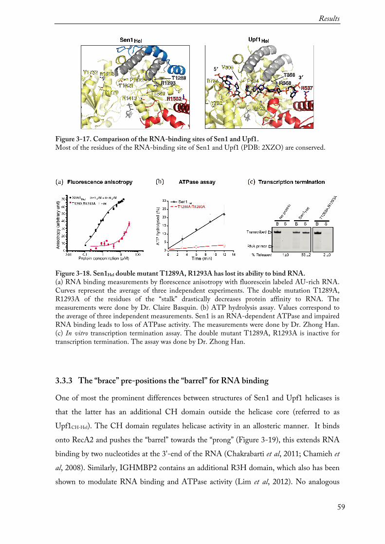
Results
59
Figure 3-17. Comparison of the RNA-binding sites of Sen1 and Upf1. Most of the residues of the RNA-binding site of Sen1 and Upf1 (PDB: 2XZO) are conserved.
Figure 3-18. Sen1Hel double mutant T1289A, R1293A has lost its ability to bind RNA. (a) RNA binding measurements by florescence anisotropy with fluorescein labeled AU-rich RNA. Curves represent the average of three independent experiments. The double mutation T1289A, R1293A of the residues of the “stalk” drastically decreases protein affinity to RNA. The measurements were done by Dr. Claire Basquin. (b) ATP hydrolysis assay. Values correspond to the average of three independent measurements. Sen1 is an RNA-dependent ATPase and impaired RNA binding leads to loss of ATPase activity. The measurements were done by Dr. Zhong Han. (c) In vitro transcription termination assay. The double mutant T1289A, R1293A is inactive for transcription termination. The assay was done by Dr. Zhong Han.
3.3.3 The “brace” pre-positions the “barrel” for RNA binding
One of most the prominent differences between structures of Sen1 and Upf1 helicases is that the latter has an additional CH domain outside the helicase core (referred to as Upf1CH-Hel). The CH domain regulates helicase activity in an allosteric manner. It binds onto RecA2 and pushes the “barrel” towards the “prong” (Figure 3-19), this extends RNA binding by two nucleotides at the 3'-end of the RNA (Chakrabarti et al, 2011; Chamieh et
al, 2008). Similarly, IGHMBP2 contains an additional R3H domain, which also has been shown to modulate RNA binding and ATPase activity (Lim et al, 2012). No analogous

Results
60
allosteric regulation domain is present in Sen1 but, surprisingly, in RNase protection assays (see Methods 6.2.14) Sen1Hel protected the 11-ribonucleotide long fragments, more similar to Upf1CH-Hel than to Upf1Hel (Figure 3-19). In fact, Sen1Hel has an intrinsic feature, the “brace”, which might mimic the effect of the CH domain of Upf1CH-Hel on the RNA-binding footprint by pre-positioning the “barrel” of Sen1Hel for RNA binding.
Figure 3-19. Comparison of the "barrel" positioning in Sen1 and Upf1. (a) Comparison of the structures of yeast Sen1Hel, human UPF1Hel (PDB: 2GJK) and UPF1Hel-RNA (PDB: 2XZO), and yeast Upf1CH-Hel-RNA (PDB: 2XZL). The molecules are shown in a front view (top) and their 90° clockwise rotations around a vertical axis (bottom). (b) RNase protection assay with yeast Sen1Hel, Upf1Hel, and Upf1CH-Hel in the presence of ADP:BeFx or ADP:AlFx. The assay was done by Fabien Bonneau. The asterisk (*) marks fragments protected by helicases continuously. On the left panel, a Coomassie stained SDS-PAGE gel of the proteins used in the assay.

Results
61
In order to test this hypothesis, two “prong” mutants in Sen1Hel were engineered, cloned,
expressed and purified (see Figure 3-20). In the first mutant, the part of the sequence in the “prong” that could not be modeled (residues 1471–1538) was replaced with a linker
Gly-Ser-Gly-Ser (Sen1HelΔUP for upper “prong” deletion). In the second mutant, the
entire solvent-exposed portion (residues 1461-1554) was replaced with a linker Ser-Gly-
Gly (Sen1HelΔLP for lower “prong” deletion). In RNase protection assays, Sen1HelΔUP had
an RNA footprint similar to that of the wild-type protein, but Sen1HelΔLP resulted in a
shorter protection of ~8-9-ribonucleotide fragments (Figure 3-20).
Figure 3-20. Deletions reveal contribution of the “prong” to RNA binding. (a) The structures show the deletions of the “prong”. The dashed line represents a region of the upper part of the prong that was disordered in the crystal structure. The two structures depicted below, represent models of the two deletion constructs, Sen1HelΔUP and Sen1HelΔUP, used in the assay. (b) RNase protection assay with yeast Sen1Hel, Sen1HelΔUP, and Sen1HelΔLP in the presence of ADP:AlFx. RNA fragments were obtained by digesting 32P body-labeled 57-mer (CU)28C RNA with RNases A and T1. The assay was done by Fabien Bonneau. (c) A Coomassie stained SDS-PAGE gel of the proteins used in the assay.

Results
62
3.3.4 The “prong” is critical for RNA unwinding and transcription termination
One function of Sen1 in the cell is processive RNA unwinding. The helicase translocates on ssRNA in 3'→5' direction and removes secondary structure of RNA, DNA from RNA:DNA duplex, or RNA-bound proteins (Jankowsky & Fairman, 2007). It is reasonable to expect the unwinding element to be located at the RNA entrance to the helicase, i.e., RecA1 domain. A close inspection of the “prong” of Sen1 and related helicases points out two residues.
Firstly, Sen1Hel shares a conserved residue Arg1552Sen1 (corresponding to Arg537Upf1 and Lys331IGHMBP2), which in the Upf1CH-Hel structure is located near the last nucleotide of the RNA (Chakrabarti et al, 2011). Secondly, Sen1Hel has a hydrophobic residue, Leu1549, which is involved in protein-protein interaction in crystal packing as mentioned above (see Chapter 3.2.4). The residue is not only exposed to the surface, but also is in a close proximity to where the RNA is entering and the helicase might have a protein-protein interaction with encountered RNA-bound protein (i.e., RNA polymerase). Therefore, in collaboration with Dr. Zhong Han and Dr. Odil Porrua, we checked the effect of the
“prong” deletions, Sen1HelΔUP and Sen1HelΔLP, as well as L1549D mutation for the
helicase activity.
All three mutants retained RNA-dependent ATPase activity (Figure 3-21, a), however,
the full “prong” deletion in Sen1HelΔLP abolished the unwinding activity (Figure 3-21, b).
This was not due to a decrease in the affinity for the RNA, since we observed similar RNA
binding by Sen1HelΔLP compared to Sen1Hel (Figure 3-20). In contrast, the deletion of the
disordered part of the “prong” in Sen1HelΔUP had the lowest ATPase activity but showed
the highest capacity to dissociate the duplex. Interestingly, the higher the ATPase activity that was measured, the less active was the mutant in RNA:DNA unwinding, suggesting that the processivity of the helicase is not directly dependent on ATP hydrolysis rate.
More intriguingly, Sen1HelΔUP mutant, which was the most active in unwinding assays,
exhibited a moderate decrease in termination efficiency in vitro. (Figure 3-21, c). In
addition, the double Sen1HelΔUP,L1549D mutant exhibited levels of duplex unwinding activity similar to the wild-type protein but was strongly affected in transcription
termination. In contrast, Sen1HelΔLP mutant was inactive for both duplex unwinding and

Results
63
transcription termination. Consistently, the full “prong” deletion leads to lethality and transcription termination defects in vivo (Leonaitė et al, 2017). Thus, for the first time we could show that both translocation and RNA unwinding are required for Sen1 to terminate Pol II transcription.
Figure 3-21. "Prong" in transcription termination. (a) ATP hydrolysis assay. Values correspond to the average of three independent measurements. (b) RNA:DNA duplex unwinding assay. (c) In vitro transcription termination assay. The assays were done by Dr. Zhong Han.
3.4 Sen1Hel as a model for AOA2-associate mutations in SETX
Based on secondary structure prediction and sequence alignment, several constructs of the helicase domain (fragments 1671-2478, 1671-2488, 1694-2478, and 1694-2488) of human SETX were cloned for expression in E. coli, as described in Methods 6.2.1. The designed constructs were similar length to crystallized Sen1Hel, however the proteins were not expressed.

Results
64
The helicase domain of SETX shares about 30 % sequence identity with Sen1Hel, including the key residues of the “brace” that are absent in other SF1 helicases. Thus, the disease-associated mutations in SETX gene were tested using Sen1Hel as a surrogate. In total, 26 missense AOA2-associated mutations (reported in LOVD Database) could be mapped onto Sen1Hel structure (Figure 3-22). The majority of the mutated residues are buried inside the helicase core, meaning that the substitution disrupt the protein fold and hinders the helicase activity. Other mutations affect SETX catalytic activity directly, as the mutated side chains are in places important for RNA recognition or ATP hydrolysis. Only a few residues were mapped at the surface of the helicase, however, those residues were not conserved.
Figure 3-22. AOA2-associated missense mutations. Conserved AOA2-associated mutations mapped onto the Sen1Hel structure. The residues that represent SETX missense mutations (black labels) are shown as sticks in magenta. All known SETX mutations are reported in LOVD Database.
A subset of AOA2-associated substitutions was cloned into Sen1Hel, the helicase was purified and tested for its enzymatic activity (Figure 3-23). A D1616V mutation of the residue buried at the interface between the two RecA domains resulted in an insoluble protein. The N1413S, P1622L and T1779P mutations targeted the residues that were expected to have a direct contact with RNA. These mutants were impaired in RNA

Results
65
binding (FA measurements, Figure 3-23), and, consequently, did not elicit duplex unwinding and in vitro transcription termination. Finally, the R1820Q mutant that
supposedly has a direct contact to γ-phosphate of the ATP and the substitution of which would impair ATP hydrolysis, was inactive in all assays.
Figure 3-23. Characterization of the Sen1Hel harboring AOA2-associated mutations. (a) Selected Sen1Hel point mutations that mimic the disease mutations of SETX mapped in Sen1Hel structure. (b) Comparison of RNA binding affinity of the mutants measured by fluorescence anisotropy using fluorescently labeled AU-rich RNA as the substrate. (c) ATPase activity analysis of the R1820Q mutant. Arg1820 is highly conserved and coordinates γ-phosphate of the ATP. The point mutation was predicted to affect nucleotide binding. (d) The mutant activity in RNA:DNA duplex unwinding assays. The efficiency of the unwinding measured at 30 min. (e) Analysis of mutation impact on efficiency of transcription termination. All values correspond to the average and SD of three independent measurements. The assays were done by Dr. Zhong Han.


Discussion and conclusions
67
4 Discussion and conclusions 4.1 Sen1Hel structure is silimilar to other Upf1-like RNA helicases of SF1
The RNA binding and unwinding activities of Sen1 require the RecA1-RecA2 core domain as well as two distinct accessory subdomains that protrude from RecA1: the “stalk” with the “barrel” and the “prong”. Overall, the domain organization of Sen1Hel is very similar to the helicase core of other RNA helicases of the SF1. Consistently, Sen1Hel-ADP
is in an “open” conformation, in which the RecA2 domain is rotated 30° outwards from the position observed in a “close” conformation of Upf1Hel-ATP bound structure. This domain movement upon ATP binding and hydrolysis is important for helicases translocation on a nucleic acid chain.
In the Sen1Hel apo structure, ADP is bound to RecA1 inside the cleft. The adenine ring is sandwiched between RecA1 and Motif IIIa (see Figure 3-13). Motif IIIa is characteristic for Upf1-like helicases and resides in the linker that connects the two RecA domains. In SF2 helicases the adenine ring is sandwiched directly between RecA1 and RecA2 (Motif IVa) domains. In addition, the adenine amino group is recognized by a conserved Q-motif, common to all SF1 and SF2 helicases that have substrate preference for ATP over other nucleotides (Cordin et al, 2004). Sen1Hel also harbors Motif I and Motif II (also known as Walker A and Walker B) that are crucial components for ATP hydrolysis.
Importantly, Sen1Hel binds RNA in an ATP-dependent manner. Although in RNase assays ADP:AlFx and ADP:BeFx were bound to Sen1Hel, no electron density for AlFx or BeFx moiety but for ADP was obtained. Moreover, the protein has crystallized in an “open” conformation that most likely leads to the reduced affinity for RNA binding. The structure of Sen1Hel bound to RNA was not obtained.
However, the RNA binding motifs are well conserved and superposition with the structure of Upf1Hel-RNA suggests the key residues for RNA binding (see Figure 3-17). Most of the RNA contacts are made with sugar phosphate backbone at the top of RecA domains; therefore the helicases do not discriminate between specific nucleotides or “read” the sequence of RNA and can also bind ssDNA. Additionally, Upf1-like helicases have an extended RNA binding site on the RecA1 domain. The conserved residues of Upf1-like helicases at nearly identical positions on surfaces of the “stalk” and the “prong” bind

Discussion and conclusions
68
additional three to four nucleotides. The “barrel” positioning is very important for RNA binding at this site and.
4.2 The “brace” is unique to Sen1
Specialized domains gained during evolution serve helicases by regulating RNA binding. Additional to the accessory domains (“stalk”, “barrel”, and “prong”), Upf1 and IGHMBP2 have domains outside the helicase core that coordinate the “barrel” positioning to tune RNA-binding affinity and ATPase activity. When this project was started there was no indication that Sen1 had a similar allosteric regulation domain to modulate the positioning of the “barrel”. However, the crystal structure revealed that Sen1 has a unique feature, the “brace”, that has not been predicted as a part of the helicase domain before. It is a ~50-residue fragment (residues 1096-1149) located upstream of the helicase domain and is well conserved across species. From the structure prediction or sequence alignment it was not possible to predict the importance of the “brace”. Although some residues, e.g. Leu1121 and W1123, are conserved in all organisms, homology-based structural modelling has not predicted these two residues to interact with the barrel. However, studies in vivo had demonstrated that the region 1089-1135 is important for yeast viability (Chen et al., 2014), but the authors suggested that this region serves as a secondary nuclear localization sequence (NLS). The sequence motif MRKRL could indeed be assigned to Class 3 of NLS for the helicase import into nucleus (Kosugi et al, 2009).
In Sen1Hel structure, the “brace” fastens the RecA1 domain, the “stalk” and the “barrel” together, thus the “barrel” is in a rather fixed position. Also, in comparison to other helicases, the linkers between the “barrel” and the “stalk” of Sen1 are the shortest and the “barrel” movement would be limited even if there were no “brace” present. Moreover, results of the RNase protection assays confirm that in Sen1Hel the “barrel” is in a position similar to that of the Upf1CH-Hel structure (see Figure 3-19). All together, this suggests that the N-terminal domain is not regulating the helicase in a similar manner to the CH or R3H domains of Upf1 and IGHMBP2, respectively.
It is possible that the “barrel” conformation in the crystal structure of Sen1Hel is partially stabilized by crystal lattice contacts. However, the “brace” buries 2400 Å2 of the Sen1Hel

Discussion and conclusions
69
surface and is absolutely required. It has been shown previously that a single point mutant within the “brace”, W1166S, is defective in vivo (Chen et al, 2014). Moreover, the deletion of N-terminal residues 1-1134 in Sen1 is lethal, whereas the deletion of residues 1-1088 does not have such a drastic effect (Chen et al, 2014).
4.3 Structural basis of 5'-3' translocation of Sen1 and related RNA helicases
Translocation and unwinding are one of the fundamental properties that define helicases. Structural studies of DNA helicases of SF1 revealed that the 5'→3' translocation directionality is achieved via alteration of the grip to DNA between RecA1 and RecA2 domains (Saikrishnan et al, 2009). During ATP binding and closing of the cleft, the helicase grips the ssDNA most tightly with RecA1 domain. During ATP hydrolysis and opening of the cleft, the tightest grip is with RecA2 domain and as the consequence the “relaxed” RecA1 domain is sliding towards 3'-end of the ssDNA.
The mechanism of how RNA helicases of SF1 translocate in 5'→3' direction is not known. However, Sen1 and the related helicases share conserved residues on the accessory subdomains that contribute to RNA binding. As it has previously been reported for Upf1 (Chakrabarti et al, 2011), Sen1 also achieves an extended RNA-binding through conserved residues on the accessory domains, particularly on the “prong”. Another interesting aspect is that in transition between the “open” and “closed” conformations, the RecA2 domain
rotates about 30°. In IGHMBP2Hel-ADP-RNA (PDB: 4B3G), the cleft is “open” and the RNA is clearly twisted but relaxed on RecA2. In the case of Upf1CH-Hel-ADP:AlF4-RNA (PDB: 2XZL), the cleft is “closed” and the RNA is “squeezed” (Figure 3-16). Here, similarly to DNA helicases (Saikrishnan et al, 2009), the fifth nucleotide of the RNA chain is switching between Motif III and Ia. Unfortunately, the structure of both “open” and “close” conformations are not available, neither for Sen1, Upf1, nor IGHMBP2, and only a comparison between the helicases is possible. Moreover, in RNase protection assays, the footprint of ground and transition states of Sen1 and Upf1 is different (Figure 3-17). Nevertheless, Sen1Hel shares all the conserved residues of Motifs Ia, III and V, thus the mechanism of how Sen1 translocates in 5'→3' direction most plausibly is similar to that of DNA helicases of SF1.

Discussion and conclusions
70
4.4 Proposed mechanism of transcription termination by Sen1
In the NNS transcription termination pathway, the recognition of Nrd1 and Nab3 consensus motifs is a critical first step but Sen1 is a key enzyme in facilitating the elongation complex dissociation. It has continuously been demonstrated that Sen1 alone is sufficient for termination of the elongation complex. Sen1Hel is the smallest enzyme variant that has been studied in transcription termination in vitro. The N- and C-terminal domains of the helicase are limiting Sen1 functions only in vivo (Han et al, 2017), as they are needed for recruitment to termination sites. As mentioned, the CTD of Pol II directly or through Nab3-Nrd1 mediates timely recruitment of Sen1. However, it has not been shown whether a direct contact of Sen1 with surface-exposed regions of Pol II could help to disassemble the elongation complex. There are though examples of other transcription factors, e.g. CF IA, that have been shown to bind to the evolutionary conserved flap loop above the RNA exit channel and to induce an allosteric signal to terminate (Pearson & Moore, 2014). The current model suggests that, similarly to bacterial Rho-factor, Sen1 translocates on the nascent RNA up to the transcribing Pol II and displaces it. However, Rho-factor is incapable to disassemble yeast elongation complex (Porrua & Libri, 2013). Consistently, it has been shown that the termination efficiency increases if the polymerase decelerates, thus the kinetic competition between Sen1 and the Pol II plays a role.
The results of this thesis reveal that not only translocation but also unwinding properties are required for Pol II termination. Sen1Hel has residues on the surface of the “prong” that do not have a direct effect to RNA binding or ATP hydrolysis, however are important for
the elongation complex dissociation. For example, Sen1HelΔUP mutant, which was the
most active in unwinding assays, exhibited a moderate decrease in termination efficiency in
vitro (Figure 3-21). But probably more importantly, point mutation L1549D acerbated the effect of deletion of the upper part of the “prong”. The double mutant retained unwinding activity similar to wild-type protein, however termination efficiency dropped by 5-fold.

Discussion and conclusions
71
Figure 4-1. Sen1 in transcription termination. A schematic step-wise representation of Sen1 translocation along newly transcribed RNA in 5'→3' direction. Sen1 is a strong RNA-dependent ATPase, however it has a transient state after ATP hydrolysis in which the helicase is weakly bound to RNA, thus Sen1 is a low processivity enzyme. For efficient transcription termination it is crucial that Sen1 is either recruited in a close proximity to Pol II and/or that Pol II is stalled. In vivo, Sen1 is recruited to Ser2-phosphorylated Pol II via interaction with Nrd1-Nab3 heterodimer but also Sen1 can bind to Ser5-phospborylated Pol II directly. It is still not clear whether the N-terminal domain of Sen1 is binding to the RNA and whether the domain is in front or behind of the helicase core during the translocation. However, Sen1 translocates on the transcript RNA until collides with Pol II, leading to disruption of the elongation complex. Most likely the close contact causes allosteric changes in Pol II, it also could be that Sen1 destabilizes the transcription bubble by pulling out the RNA from active site.
4.5 SETX
The majority of ataxia ocular apraxia type 2 (AOA2)-associated mutations are recessive missense, nonsense and deletion mutations that result in loss of helicase function. We have mapped the missense mutations onto the structure of Sen1Hel and tested 6 disease mimicking Sen1Hel variants in ATPase, unwinding and transcription termination assays. D1616VSen1 was insoluble, and all other proteins have partially or completely lost their enzymatic activity. Other AO2-mutations, for example E65K, W305C and P413L abolish
binding of Rrp45 (Richard & Manley, 2014), which indicates that the N-terminal domain
Discussion and conclusions
72
of SETX functions as the exosome cofactor. Interestingly, none of ALS mutations cause
direct inactivation of the helicase. For example, H1956R (Q1356Sen1) is at the back site of
RecA2 and K2018E (L1421Sen1) is at the front site of RecA1 domain, and R2136C/H
(K1539Sen1) is in the disordered upper part of the sidewall. The exact role of ALS4-related
mutations remains unclear, but these mutations are dominant missense mutations that most
likely have a gain-of-function phenotype. It could be that ALS4-related mutations impair
SETX interaction with other proteins and ALS4 phenotype is not directly related to
transcription termination. For instance, the most common ALS4 mutation, L389S, results
in increased binding of SUMOylation proteins or to a peptide encoded by BCYRN1-
reverse complement (Bennet et al, 2013).
4.6 Conclusions
In this thesis the identified ~94 kDa helicase core of S. cerevisiae Sen1 has biochemical properties similar to those of the endogenous full-length protein in in vitro experiments. Sen1Hel has a very similar overall structure to that of Upf1-like helicases. Although the helicases share the same conserved sequence motifs for RNA binding and ATP hydrolysis, Upf1 is not capable to terminate stalled Pol II in vitro. The most prominent difference in structures of Sen1 and Upf1 is the “brace”, which fastens the RecA1, the “stalk” and the “barrel” into a rigid domain. Sen1 has a relatively high ATPase activity and is capable to disrupt a tiny fraction of Pol II even when the “prong” is fully deleted. Interestingly, the processivity of the helicase seems not to be directly dependent on ATP hydrolysis but rather on affinity to RNA (Han et al, 2017). It could be, that relatively low processivity and a low copy number of Sen1 molecules per cell prevent spurious termination events.
All together the results of this thesis support the view that Sen1 uses the nascent RNA as a guide to catch up with the elongation complex of Pol II moving in the same direction. Most likely, at direct contact, Sen1 inserts the “prong” into the RNA exit channel of Pol II to open the cleft of the polymerase. Additionally, surface interaction between the two proteins might provoke a conformational change in the elongation complex. It is also plausible that the elongation complex is destabilized using a “ratchet” mechanism for pulling the nascent RNA out of transcription burble.

Outlook
73
5 Outlook
To date it is not clear at which side of Sen1 structure the N-terminal domain resides. It also is not known whether the N-terminal domain can bind to RNA directly. Unfortunately, we could not test it in RNase assays because of the limited amount of the protein. And although the presence of the N-terminal domain decreases the termination efficiency in vitro (Han et al, 2017), it is not clear whether the domain has any regulatory role or the decrease is due to the large size. It is possible that the N-terminal domain partially interferes in interaction between the helicase domain and Pol II. These should be the main questions trying to solve the structures of the pre-termination complex of Sen1 and Pol II in the future.
Nevertheless, in in vitro experiments the helicase domain alone is capable to disassemble the elongation complex of Pol II. A closer inspection of the structures suggested at which surface of Sen1Hel and the elongation complex (PDB: 3HOV (Sydow et al, 2009)) could make a contact (Figure 5-1). At the collision of Sen1Hel and Pol II, the “brace”, the “barrel”, the “prong” together with the part of the RecA1 domain would bed in to the side of Pol II where the transcript is exiting.
Figure 5-1. Interaction site on the surface of Sen1Hel and Pol II. At the collision of Sen1Hel with the Pol II elongation complex (PDB: 3HOV), Sen1Hel would bed in closely to the RNA exit channel.
At the side of the RecA1 domain, Sen1Hel has a 20-residue loop (residues 1382-1402) that protrudes from the domain (Figure 5-1). PSIPRED predicted this region to be disordered
Outlook
74
and only a part of the loop could be built into the model. Deletion of the loop had no effect on RNA binding or ATPase activity but, surprisingly, the mutant has increased transcription termination activity (60 % termination compared to 47 % of the wild-type Sen1Hel). We have not studied the mutant further, however this loop together with other conserved residues on the Sen1 surface should be investigated in the future. For example, we have also found that a double mutation on the top of the “barrel” (R1188D, Y1195F) has a decreased activity in transcription termination assays (40 % termination).
On the other side, it is not known which regions of Pol II are mediating the interaction with Sen1 at the collision. Pearson and Moore have demonstrated that the evolutionary conserved flap loop of Rpb2 interacts with Pcf11 (essential for canonical transcription termination after polyA site), and the deletion of that loop leads to termination defects even at NNS-dependent noncoding genes (Pearson & Moore, 2014). Furthermore, a few point mutations of Rpb3 and Rpb11 at the bottom at the Pol II provoke significant transcription termination defects in vivo (Steinmetz et al, 2006). These residues seem to be too far for the helicase domain of Sen1 to reach but it could be that this is the site where the N-terminal domain would make the contact.
Finally, it is not known at which distance from Pol II the helicase should be loaded on the transcript RNA. Currently it is suggested that Sen1 processivity is ~20-40 nt (Han et al., 2017). However, the RNA length during Sen1 recruitment via Nrd1-Nab3 or direct interaction with CTD is not known and should be addressed in the future.

Materials and methods
75
6 Materials and methods 6.1 Materials 6.1.1 Chemicals and reagents
All common chemicals and reagents were ordered from Sigma-Aldrich or Fluka unless otherwise stated. Chemicals fot crystallization were additionally order from Hampton.
6.1.2 Enzymes
For DNA amplification by PCR, Phusion polymerase from Thermo Fisher Scientific was used. For RNase protection assays RNase A and RNase T1 (Fermentas) were used. Rhinovirus 3C protease was expressed and purified in-house.
6.1.3 DNA and RNA oligonucleotides
DNA and RNA oligonucleotides were ordered from Sigma-Aldrich and Purimex, respectively. All oligonucleotides were ordered desalted and lyophilized, RNA was additionally purified by PAGE. DNA was dissolved in ddH2O and RNA – in 100 mM
HEPES pH 7.5 and stored at -20°C.
Table 6-1. DNA oligonucleotides used as primers for cloning.
Name Sequence (5' to 3')1_F cacccgggatctcgagcATGAATTCCAACAATCCTGOneStrep_R cccccatctcccggtaccgCTATTTCTCGAACTGCGGGTGGC2231_R cccccatctcccggtaccgTCATGATCTAGGCTTTCSM_1105_F aactttaagaaggagatatacatATGAGAAAGAGATTAAATGTTGSM_1105_R CAACATTTAATCTCTTTCTCATatgtatatctccttcttaaagttins3C_F gtggttctgttccaggggcccCTCGCGGGCGGTAAAATACTCCATAATCins3C_1881_R ctggaacagaaccaccagTGATGGTACGGGTACATTGAACTTTCins3C_1904_R ctggaacagaaccaccagATTGGATTTTACCTCATCGGGGCCTTGCGins3C_1910_R ctggaacagaaccaccagTCTCTTCTTTGTGTCCTTATTGGATTTTACCT4L_1_R GGTGGTGGTGGTGGTGGTGGTGtccCAGGTTCTTGTAAGCGTCCCT4L_2_F GGGACGCTTACAAGAACCTGggaCACCACCACCACCACCACCACCTrx_1_R GGTGGTGGTGGTGGTGGTGGTGtccCAGGTTAGCGTCGAGGAACTCTrx_2_F GAGTTCCTCGACGCTAACCTGggaCACCACCACCACCACCACCACC43_1_F GAAAGTTCAATGTACCCGTACCATCACTGGAAGTTCTGTTCCAGGG
GCCCATGAACATCTTCGAGATGCTGCG43_2_R CGCAGCATCTCGAAGATGTTCATGGGCCCCTGGAACAGAACTTCCA
GTGATGGTACGGGTACATTGAACTTTC44_1_F GGCCCCGATGAGGTAAAATCCAATCTGGAAGTTCTGTTCCAGGGGC
CCATGAACATCTTCGAGATGCTGCG

Materials and methods
76
44_2_R CGCAGCATCTCGAAGATGTTCATGGGCCCCTGGAACAGAACTTCCAGATTGGATTTTACCTCATCGGGGCC
45_1_F ATCCAATAAGGACACAAAGAAGAGACTGGAAGTTCTGTTCCAGGGGCCCATGAACATCTTCGAGATGCTGCG
45_2_R CGCAGCATCTCGAAGATGTTCATGGGCCCCTGGAACAGAACTTCCAGTCTCTTCTTTGTGTCCTTATTGGAT
46_1_F AAGTTCAATGTACCCGTACCATCACTGGAAGTTCTGTTCCAGGGGCCCATGAGCGATAAAATTATTCACCTG
46_2_R CAGGTGAATAATTTTATCGCTCATGGGCCCCTGGAACAGAACTTCCAGTGATGGTACGGGTACATTGAACTT
47_1_F GGCCCCGATGAGGTAAAATCCAATCTGGAAGTTCTGTTCCAGGGGCCCATGAGCGATAAAATTATTCACCTG
47_2_R CAGGTGAATAATTTTATCGCTCATGGGCCCCTGGAACAGAACTTCCAGATTGGATTTTACCTCATCGGGGCC
48_1_F GTAAAATCCAATAAGGACACAAAGAAGAGACTGGAAGTTCTGTTCCAGGGGCCCATGAGCGATAAAATTATTCACCTG
48_2_R CAGGTGAATAATTTTATCGCTCATGGGCCCCTGGAACAGAACTTCCAGTCTCTTCTTTGTGTCCTTATTGGATTTTAC
IA2_41_R2 CAGTCTGAGGCAGATTTCGTCCACAGCGGCATTACTGGGGGCIA4_41_R2 ccttcttaaagttaaacaaaattatttctagaggggIA6_41_R2 TACACGGACCAATTGAGGTTTAAATTGATGCCCCIA7_41_R2 CATTACAATTAAGTCAGATTCAGAAATACCACIA1_41_F GGTTACTTTTTGTCTACAAAGAATGCCTCAggtggtAAGCAAAAAATTC
TAATCTGTGCCCCCAGIA1_41_R AATAATACCCAGTATAGTCTTTGIA2_41_F GAAATCTGCCTCAGACTGAAGAGTGGAggtggtggtTTGGTCCGTGTAG
GTAGGTCIA2_41_R GTCCACAGCGGCATTACTGGIA3_41_F GGACCTTACTTTAGAAGAACTTagcggtggtCAGGCCCATATCTTGGCG
GTTAGTGIA3_41_R TTAATTGCAACGTTTACAACGTCIA4_41_F gtttaactttaagaaggagatatacatATGAATAGTGAATATCCTGATGATGAGCCTA
TTGGIA4_41_R aaaattatttctagaggggIA5_4_IA4_F CATGAAGCCTTTACTACTTCTAGAATCTTcGCAAGGTaacTGTTCTTC
ACGTGATAGAGAGGACIA5_4_IA4_R ACTTTTTGGTAGTCAGCAGGAGIA6_41_F CCTCAATTGGTCCGTGTAGGTggcggcggcATTAAGGACCTTACTTTAGIA6_41_R TTTAAATTGATGCCCCTGIA7_41_F GACTTAATTGTAATGGCTTATggcggcCATACATGTTTAGCAAAGGTGIA7_41_R AGATTCAGAAATACCACAATCCIA8_41_F CAACATACATGTTTAGCAgAGGTGAGAACATTGAAAAATACCIA8_41_R TGCTTTCTTGAAATCATCACTGGACIA9_41_F CCTCTATATGAAATCATAagGCAATGGGATTACACCAGIA9_41_R GTTCATATCAACATTTAATCTCTTTCTCATATGTTCIA10_41_F CCTCTATATGAAATCATAagGCAAtgGGATTACACCAGAAATAGTG
Materials and methods
77
Small letters show overhangs of vector sequence or introduced mutations. Capital letters correspond to the sequence of the gene of interest.
Table 6-2. ssDNA/RNA used in assays.
6.1.4 Constructs
All S. cerevisiae variants were cloned into pCB-A-bax10 plasmid (kindly shared by Dr. Christian Biertümpfel) for expression in E. coli and into pFLΔSpeI donor plasmid (Invitrogen) for expression in insect cells. See list of all cloned mutants in Appendix 3.
6.1.5 Cloning kits
Wizard Gel and PCR Clean-up (Promega) and Qiaquick Spin Miniprep (Qiagen) kits were used.
IA11_41_F GTTGATATGAACCCTCTAaggGAAATCATATTGCAATGGGIA11_41_R ATTTAATCTCTTTCTCATATGTTCIA12_41_F GTTGATATGAACCCTCTAaggGAAATCATAagGCAATGGGATTACAC
CAGIA13_41_F CACAACATACATGTTTAGCAAAGGTGgaAACATTGAAAAATACCAA
AGGTGGIA13_41_R CTTTCTTGAAATCATCACTGGACIA14_41_F GGTAATGTTGATGTCACATTAgaaATTCATAGAAATCATTCTTTCAG
TAAATTTTTGACIA14_41_R ACCTTTGGTATTTTTCAATGTTCTCACCIA15_41_F gtttaactttaagaaggagatatacatATGTCTCCTGCTGACTACCAAAAAGTCATGA
AGCCIA16_41_F gtttaactttaagaaggagatatacatATGCCTATTGGTAATTATTCTGACGTAAAG
GATTTCTTCIA10_41_F CCTCTATATGAAATCATAagGCAAagGGATTACACCAGAAATAGTGIA19_41_F GAGATTAAATGTTGATATGAACCCTCTAgctGAAATCATAgcgCAAgcg
GATTACACCAGAAATAGTGAATATCCTG
Name Sequence16-mer RNA oligonucleotide used for UGCAUUUCGACCAGGC assembly of ECs. 5'-end FAM labeledTemplate DNA strand used for assembly GGCCGGGTAACCCCCGTGTGGAGATGGGTGAGAGATGTTGof ECs. Labeled with biotin at the 5'-end AGGGCCTGGTCGTTTCCTATAGTTTGTTTCCT Non-template strand usedfor assembly CTAGAGGAAACAAACTATAGGAAACGACCAGGCCCTCAACof Ecs. Labeled with biotin at the 5'-end ATCTCTCACCCATCTCCACACGGGGGTTACCCGGCCTGCA ssDNA/RNA for ATPase assay UUUUUUUUUUUUUUU5'-end FL labeled CCCCCCCCCCCCCCC
AAAAAAAAAAAAAAACCCTAACCCTAAC
RNA for FA measurements UUUCUAUUUAUUUUG
Materials and methods
78
6.1.6 E. coli strains and insect cell lines Table 6-3. E. coli strains used for cloning and expression.
For protein expression in insect cells the Autographa californica cell lines Sf21 and HighFive (Invitrogen) were used.
6.1.7 Media and buffers Table 6-4. Media used for cloning and protein expression.
E. coli strains GenotypeOne Shot™ OmniMAX™ 2 T1 F′ {proAB+ lacIq lacZ∆M15 Tn10(TetR) ∆(ccdAB)} mcrA (Thermo Fisher Scientific) ∆(mrr-hsdRMS-mcrBC)∆M15φ80(lacZ) ∆(lacZYA-argF)
U169endA1 recA1 supE44 thi-1 gyrA96 relA1 tonA panDDH10MultiBacY F– mcrA Δ(mrr-hsdRMS-mcrBC) φ80lacZΔM15 ΔlacX74(Invitrogen) recA1 endA1 araD139 Δ(ara, leu)7697galUgalKλ–
rpsLnupG/bMON14272/pMON7124BL21 (DE3) STAR pRARE F- ompT hsdSB (rB -mB -) gal dcm rne131 (DE3) (pRARE (Stratagene) araW argU glyT ileX leuW proL metT thrT tyrU thrU CamR)
Media ComponentsS.O.C.Medium 2%(w/v)bactotryptone
0.5%(w/v)bactoyeastextract10mMNaCl1mMMgCl22.5mMKCl10mMMgSO4
0.4%(w/v)glucoseadjustpHto7.2
Luria-Bertani(LB) 1%(w/v)bactotryptone(Miller,1972) 0.5%(w/v)bactoyeastextract
170mMNaCladjustpHto7.6
LBagaroseplates 1.5%(w/v)bactoagarinLB100µg/mLampicillin
TerrificBroth(TB) 1.2%(w/v)bactotryptone(Sambrook&Russell,2001) 2.4%(w/v)bactoyeastextract
0.4%(v/v)glycerolddH2Oto900ml0.017MKH2PO4
0.072MK2HPO4
Sf-900TMSFM PurchasedfromInvitrogen(insectcellculturemedium)ESF921 PurchasedfromExpressionSystems(insectcellculturemedium)

Materials and methods
79
Table 6-5. Protein purification buffers.
Table 6-6. Sample freezing buffer.
Buffer ComponentsLysis buffer 20 mM sodium phosphate pH 8.0
500 mM Nacl2 mM MgCl2
30 mM imidazole10 % (v/v) glycerol1 mM β-mercaptoethanol
Salt wash buffer 20 mM sodium phosphate pH 8.01000 mM Nacl2 mM MgCl2 30 mM imidazole1 mM β-mercaptoethanol
ATP wash buffer 20 mM sodium phosphate pH 8.0250 mM Nacl2 mM MgCl2 50 mM KCl10 mM MgSO4
2 mM ATP1 mM β-mercaptoethanol
Tag cleavage buffer 20 mM Tris-HCl pH 7.5250 mM Nacl2 mM MgCl2
10 % (v/v) glycerol1 mM β-mercaptoethanol
Buffer A 20 mM Tris-HCl pH 7.5250 mM Nacl2 mM MgCl2
1 mM DTTBuffer B 20 mM Tris-HCl pH 7.5
1000 mM Nacl2 mM MgCl2
1 mM DTTSEC buffer 20 mM HEPES pH 7.5
250 mM NaCl2 mM MgCl2
1 mM DTT
Buffer ComponentsSamplefreezingbuffer(2x) 40mMHEPESpH7.5
250mMNaCl2mMMgCl299%(v/v)glycerol1mMDTT
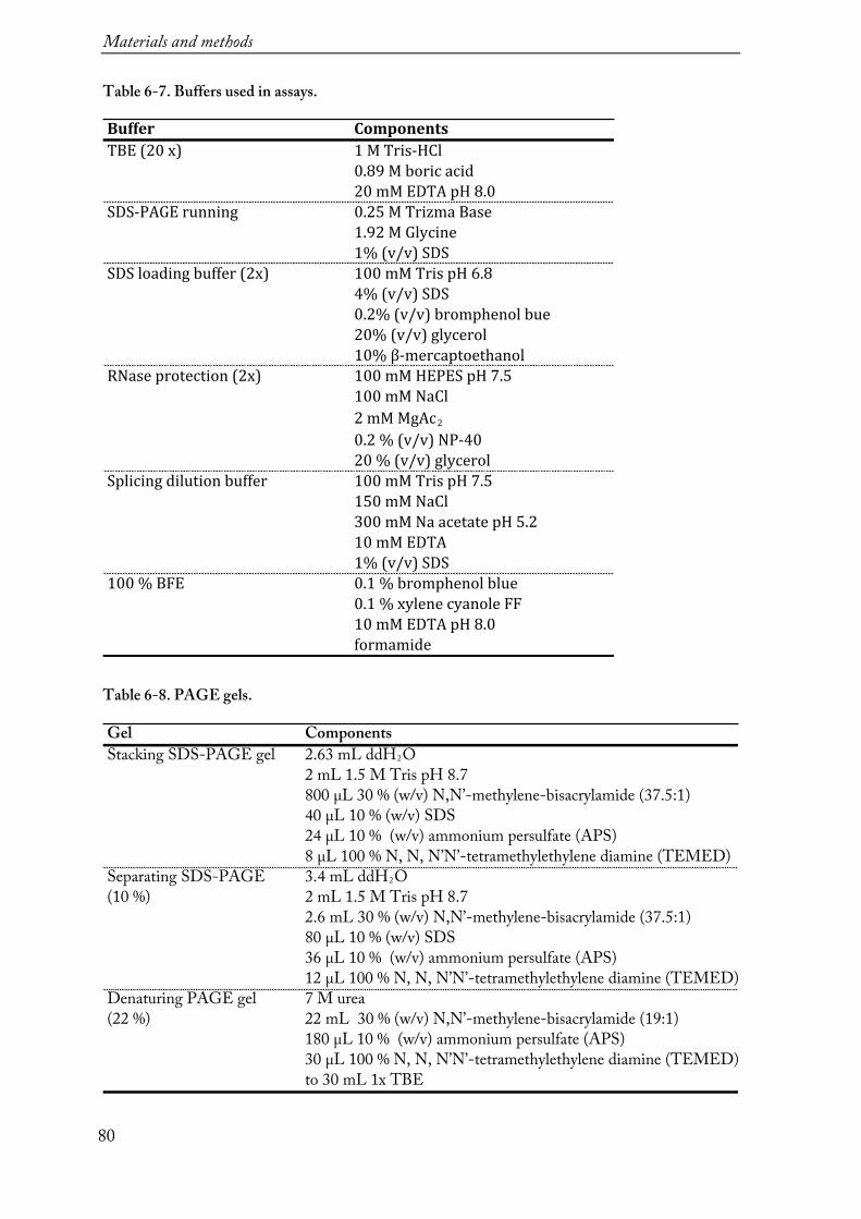
Materials and methods
80
Table 6-7. Buffers used in assays.
Table 6-8. PAGE gels.
Buffer ComponentsTBE(20x) 1MTris-HCl
0.89Mboricacid20mMEDTApH8.0
SDS-PAGErunning 0.25MTrizmaBase1.92MGlycine1%(v/v)SDS
SDSloadingbuffer(2x) 100mMTrispH6.84%(v/v)SDS0.2%(v/v)bromphenolbue20%(v/v)glycerol10%β-mercaptoethanol
RNaseprotection(2x) 100mMHEPESpH7.5100mMNaCl2mMMgAc20.2%(v/v)NP-4020%(v/v)glycerol
Splicingdilutionbuffer 100mMTrispH7.5150mMNaCl300mMNaacetatepH5.210mMEDTA1%(v/v)SDS
100%BFE 0.1%bromphenolblue0.1%xylenecyanoleFF10mMEDTApH8.0formamide
Gel ComponentsStacking SDS-PAGE gel 2.63 mL ddH2O
2 mL 1.5 M Tris pH 8.7800 µL 30 % (w/v) N,N’-methylene-bisacrylamide (37.5:1)40 µL 10 % (w/v) SDS24 µL 10 % (w/v) ammonium persulfate (APS)8 µL 100 % N, N, N’N’-tetramethylethylene diamine (TEMED)
Separating SDS-PAGE 3.4 mL ddH2O(10 %) 2 mL 1.5 M Tris pH 8.7
2.6 mL 30 % (w/v) N,N’-methylene-bisacrylamide (37.5:1)80 µL 10 % (w/v) SDS36 µL 10 % (w/v) ammonium persulfate (APS)12 µL 100 % N, N, N’N’-tetramethylethylene diamine (TEMED)
Denaturing PAGE gel 7 M urea(22 %) 22 mL 30 % (w/v) N,N’-methylene-bisacrylamide (19:1)
180 µL 10 % (w/v) ammonium persulfate (APS)30 µL 100 % N, N, N’N’-tetramethylethylene diamine (TEMED)to 30 mL 1x TBE

Materials and methods
81
6.1.8 Equipment Table 6-9. List of equipment.
6.1.9 X-ray sources and synchrotron facility
Crystals were tested with in-house X-ray diffractometers PX-scanner (Rigaku) and D8 venture (Bruker). Some of the diffraction data sets were also collected on in-house D8 venture, however the higher resolution data sets were obtained at Swiss Light Source (SLS) synchrotron at the Paul Scherrer Institute (PSI), Villingen, Switzerland.
Equipment Model ManufacturerPipettes Eppendorf Research EppendorfScales LA1200S & TE1502S SartoriuspH meter Lab860 SchottPCR machine Mastercycle EppendorfUV transilluminator Safe Imager InvitrogenVortex mixer Vortex-Genie Scientific IndustriesElectroporator Gene Pulser/Micro Pulser Bio-RadElectro-cuvette Gene pulser 0.1 cm electrode gap Bio-RadBacteria shaker KS-15/Climo-shaker ISF1X Kühner Thermo shaker Thermomixer Comfort EppendorfCell lysis sonicator Sonifyer VS70T Bandelin ElectronicsInsect cell shaker Kühner shaker Kühner Laminal flow hood Holten LaminAir Thermo electron corporationCell counter Vi-Cell-XR Beckman CoulterDounce Homogenizer Borosilicate Glass Thermo Fisher ScientificCentrifuge Avanti J-20 XP Beckman Coulter
Micro centrifuge 5417C & 5810 EppendorfColumns Ni2+-NTA beads & HisTrap FF GE Healthcare
HiTrap Heparin HP Superdex 200 [16/600]
Peristaltic pump Ismatec 78001-40 Cole-ParmerChromatography FPLC ÄKTA Prime GE Healthcare
ÄKTA PurifierProtein concentrator Amicon Ultra Centrifugal filters UltracelSpectrophotometer Nanodrop PeqLabGel imaging Gel visualization RothCrystallization plates 96-well MRC96T SwissCl
24-well vdx Hampton ResearchCrystallization pipetting robot Phoenix Art Robbins InstrumentsCrystallization visualization system Xtal-Focus ExploraNova La RochelleCryo loops CryoLoop 20 micro Hampton ResearchCaps and vials Magnetic caps and vials Molecular DimensionsX-ray diffractometer PX scanner Rigaku
D8 venture BrukerFluorescence spectrometer Infinite M1000 Pro TecanPhosphorimager Typhoon FLA 9500 Fuji

Materials and methods
82
6.1.10 Software and web servers
The following software was used to write this thesis, analyze data and generate figures:
Adobe Illustrator (www.adobe.com/products/illustrator) ApE (http://biologylabs.utah.edu/jorgensen/wayned/ape/) BIOEQS (http://abcis.cbs.cnrs.fr/BIOEQS/) Buccaneer (http://www.ccp4.ac.uk/html/cbuccaneer.html) Clustal Omega (http://www.ebi.ac.uk/Tools/msa/clustalo/) CodonCode Aligner (http://www.codoncode.com/aligner/) Coot (http://www2.mrc-lmb.cam.ac.uk/Personal/pemsley/coot/) EndNote X7 (http://endnote.com) Hampton Research Make-tray tool (http://hamptonresearch.com/make_tray.aspx) Jalview (http://www.jalview.org/) Microsoft Office (www.microsoft.com) LigPlot (https://www.ebi.ac.uk/thornton-srv/software/LIGPLOT/) LOVD Database (https://databases.lovd.nl/shared/variants/SETX/unique) Phenix (http://www.phenix-online.org) Phyre2 (http://www.sbg.bio.ic.ac.uk/phyre2) ProtParam (http://web.expasy.org/cgi-bin/protparam) PSIPRED (http://bioinf.cs.ucl.ac.uk/psipred/) PyMOL (http://pymol.org) SHELX (http://shelx.uni-ac.gwdg.de/SHELX/) UNIPROT (http://www.uniprot.org) wwPDB (https://www.wwpdb.org) XDS (http://xds.mpimf-heidelberg.mpg.de)

Materials and methods
83
6.2 Methods 6.2.1 Cloning for expression in E. coli
For cloning a one-step ligation-independent method was used. The method relies on DNA repair machinery of E. coli cells. For each transformation a set of a linearized vector and an insert with the >25 bp extensions were designed (Figure 6-1, a). In case of cloning a point mutation, a vector was linearized using primers that harbored the mutation (Figure 6-1, b). After PCR, purified unprocessed DNA was directly transformed into cells. All the constructs were cloned into a pCB-A-bax10 plasmid (kindly shared by Dr. Christian Biertümpfel) that contained a cleavable C-terminal His tag coupled to Vibrio cholerae MARTX toxin cysteine protease domain (CPD-His8) (Shen et al, 2009).
Figure 6-1. Schematic presentation one-step ligation-independent cloning. (a) Cloning with a linearized vector and an insert. The insert (red) encodes Sen1 sequence and the >25 bp extensions at both ends. These extensions are identical to the sequence of the vector (navy). After transformation, under selective ampicillin pressure, the cell is forced to repair the plasmid and recombines the insert with the DNA containing the Ampicillin-resistance cassette (yellow). (b) Cloning of a point mutation. The vector is linearized using primers that encode a point mutation (light blue). Both ends if linearized vector are identical. After transformation the cells repairs the plasmid to gain ampicillin resistance.
6.2.1.1 PCR
The PCR reactions for vector linearization and the inserts were set up in a total volume of 50 µL:
5 µL forward primer (10 µM) 5 µL reverse primer (10 µM) 20 ng template DNA 2.5 µL DMSO 25 µM 2x Master Mix buffer (kit) 0.5 µM Phusion Polymerase (1 U/µL)

Materials and methods
84
The following thermocycling conditions were used:
1-3 min - 98°C initial denaturation 10 s - 98°C denaturation* 10 s - Tm-5°C annealing* 30 s per kb - 72°C extension* 10 min - 72°C final extension
* Steps were repeated in 30-35 cycles. The annealing temperature varied depending on the
melting temperature (Tm) of the primers. In some cases touch-down PCR was applied with the
decrease of the annealing temperature of 1°C per cycle.
6.2.1.2 Agarose gel electrophoresis
DNA fragments of PCR products were separated on 0.75 % (w/v) agarose gels prepared in 1x TBE, supplemented with SYBR Safe (Invitrogen) stock 1:10000 dilution as a dye for visualization. Prior to loading samples were mixed with 6x loading buffer (Orange Loading Dye, Fermentas). 0.5 µg of the 1Kb Plus DNA ladder (Fermentas) was used as a marker in a separate lane. Gel electrophoresis was performed at 6 V/cm in 1x TBE buffer. DNA fragments were visualized and excited from the gel using a blue light transilluminator (Invitrogen) emitting light at a wavelength of ~470 nm.
6.2.1.3 DNA fragment purification
The separated DNA bands from an agarose gel were purified with the Wizard SV Gel and PCR Clean-up system (Promega) according the manufacturer’s protocol. All DNA fragments were eluted in 30 µL ddH2O.
6.2.1.4 Transformation
The purified linearized vector and the insert (1.5 µL and 3.5 µL each) were added to 50 µL aliquots of OmniMAX chemically supercompetent E. coli cells. Cells were then gently mixed and incubated on ice for 5 min. A heat shock was performed in order to transfer the vectors into bacteria cells. Therefore, the mixture was incubated 45 s at 42°C and then chilled on ice for 2 min followed by addition of 600 µL of S.O.C. medium and incubation at 37°C, 600 rpm for 30 min. Next, 100-300 µL of the cells were plated on LB agar plates, supplemented with 100 µg/mL ampicillin, and incubated over night at 37°C.
6.2.1.5 Plasmid amplification and isolation
Materials and methods
85
For plasmid amplification a single-cell-derived colony from the E.coli agar plate was picked with a pipette tip and added to 5 mL LB medium supplemented with 100 µg/mL ampicillin. The cell culture was incubated over night 160 rpm at 37°C. The cells were harvested by centrifugation (4000 rpm / 10 min / 4°C). Plasmid DNA was purified with Wizard Gel and PCR Clean-up System kit (Promega) and eluted in 50 µL ddH2O. The concentration of plasmid was measured with the Nanodrop spectrophotometer.
6.2.1.6 DNA Sequencing
All plasmid DNA was sequenced by the core facility of MPI Biochemistry or Eurofins. Custom sequence primers were ordered with the aim for a GC content of at least 45% and Tm ~65°C. The received nucleotide sequence was analyzed and compared to the nucleotide sequence of the original gene using CodonCode Aligner and ApE programs.
6.2.2 Recombinant protein expression in E. coli For the recombinant protein expression in E. coli a plasmid was transformed to BL21 (DE3) STAR pRARE cells as described before. The successful transformation was selected by incubating 100 µL of the cells on LB agar plates containing 100 µg/mL chloramphenicol and 100 µg/mL ampicillin. Next day a small amount of cells was scraped from a single colony into 70 mL LB media supplemented with respective antibiotics and incubated at 37°C and 200 rpm for 2-3 hours. 10 mL of pre-culture was added to pre-warmed 500 mL TB media supplemented with respective antibiotics, and incubated further at 37°C and 220 rpm until OD600 ~1.5 (3-4 hours). Then the temperature was decreased to 18°C and 0.5 mM IPTG was added to induce protein expression. After over night expression (~16 hours), cells were harvested by centrifugation (9 000 x g, 10 min). The cell pellet was either used immediately or flash frozen in liquid nitrogen and stored at -80°C.
6.2.3 Cloning and expression in insect cells
For recombinant protein expression in cultured insect cells a recombinant baculovirus has to be generated in two steps (Figure 5-1). First, a donor plasmid is created and then the plasmid is transformed into competent E. coli cells that contain the bacmid (baculovirus shuttle vector) for site-specific transposition. Afterwards, the recombinant bacmid DNA is
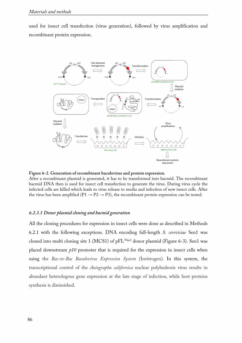
Materials and methods
86
used for insect cell transfection (virus generation), followed by virus amplification and recombinant protein expression.
Figure 6-2. Generation of recombinant baculovirus and protein expression. After a recombinant plasmid is generated, it has to be transformed into bacmid. The recombinant bacmid DNA then is used for insect cell transfection to generate the virus. During virus cycle the infected cells are killed which leads to virus release to media and infection of new insect cells. After the virus has been amplified (P1 → P2 → P3), the recombinant protein expression can be tested. 6.2.3.1 Donor plasmid cloning and bacmid generation
All the cloning procedures for expression in insect cells were done as described in Methods 6.2.1 with the following exceptions. DNA encoding full-length S. cerevisiae Sen1 was cloned into multi cloning site 1 (MCS1) of pFLΔSpeI donor plasmid (Figure 6-3). Sen1 was placed downstream p10 promoter that is required for the expression in insect cells when using the Bac-to-Bac Baculovirus Expression System (Invitrogen). In this system, the transcriptional control of the Autographa californica nuclear polyhedrosis virus results in abundant heterologous gene expression at the late stage of infection, while host proteins synthesis is diminished.

Materials and methods
87
Figure 6-3. pFLΔSpeI plasmid map. pFLΔSpeI is a donor plasmid used to generate recombinant viruses. The plasmid contains one origin of replication and can be propagated in E. coli. The multiple cloning sites (MCS1 and MCS2) contain several restriction sites for gene insertion and are flanked by Tn7 elements (Tn7L and Tn7R) for transposition to the attTn7 attachment sites on the bacmid. Additionally, these elements contain an expression cassette consisting of a Gmr and Apr genes and an SV40 poly(A) signals.
The newly cloned donor plasmid has to be then transposed into a bacmid. The transposition is based on site-specific transposition of an expression cassette into a bacmid DNA propagated in E. coli. The donor plasmid contains mini-Tn7 element that attaches to mini-attTn7 on the bacmid. The required Tn7 transposase is provided by helper plasmid that is hosted by competent cell. The lac operator controls the successful
transposition as the bacmid contains a segment of DNA encoding the lacZα peptide. Insertion of the mini-Tn7 into the mini-attTn7 attachment site on the bacmid disrupts peptide’s expression and the colonies containing the recombinant bacmid are white in background of blue non-transposed colonies. Additionally, the bacmid and the helper plasmid confer resistance to kanamycin and tetracycline, respectively.
Here, for the transposition the verified pFLΔSpeI plasmid was transformed into DH10EmBacY E. coli strain by electroporation. Therefore, 2 µL of plasmid DNA were added to 50 µL competent cells and incubated for 5 min on ice. Then the cells were transferred into electrophoresis cuvette. Immediately after the electroporation at 2.5 kV 900 µL S.O.C. medium was added and the culture was incubated at 37°C and 600 rpm for over night. The successful transformation was selected by blue-white screen. Therefore,

Materials and methods
88
100-300 µL of the cells were plated on LB containing 50 µg/mL kanamycin, 7 µg/mL gentamicin, 10 µg/mL tetracyclin, 100 µg/mL Bluo-gal (dissolved in DMSO) and 80 µg/mL IPTG. The bacmid was amplified by growing 2 mL bacteria culture in LB from an isolated white colony as described previously. The selective pressure was maintained by adding 50 µg/mL kanamycin, 7 µg/mL gentamicin, and 10 µg/mL tetracyclin. Finally, the bacmid was isolated by alkaline lysis, using the Qiaprep Spin Miniprep kit, and DNA precipitation in isopropanol and dissolved in 30 µL ddH2O.
6.2.3.2 Transfection of Sf21 insect cells and virus generation
During the transfection, Sf21 cells are infected with the recombinant bacmid DNA. To significantly increase the efficiency, the DNA has to be incubated with transfection agent such as polyethylenimine (PEI) that acts as an attachment factor for cell adherence. Both PEI are polycationic polymers and condense DNA into positively charged particles, which bind to anionic cell surface residues and are brought into the cell via endocytosis. Once the vesicle enters the cell, it undergoes osmotic swelling and bursts releasing the polymer-DNA complex into the cytoplasm. Then the polymer is replaced by other cations leading to unpacked bacmid DNA. The bacmid then can enter the nucleus where viral transcription and replication occurs. The baculovirus particles get budded outwards the cell by 24-72 h post-infection to spread the infection. In this time the recombinant protein is also expressed.
The initial virus titer and volume is too low for sufficient recombinant protein expression and thus several cycles (P1 → P2 → P3) of virus harvesting and new infection are required (Figure 6-4). Therefore, the infected insect cells were centrifuged and the supernatant with virus P1 was used to infect the larger amount of Sf21 insect cells. This procedure was repeated two more times and the final P3 virus was used further for protein expression in HighFive insect cell line. The cell growth and lysis was monitored throughout the virus generation using cell counter Vi-Cell-XR (Beckman Coulter).
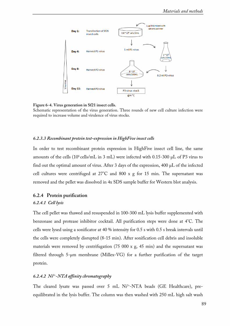
Materials and methods
89
Figure 6-4. Virus generation in Sf21 insect cells. Schematic representation of the virus generation. Three rounds of new cell culture infection were required to increase volume and virulence of virus stocks.
6.2.3.3 Recombinant protein test-expression in HighFive insect cells
In order to test recombinant protein expression in HighFive insect cell line, the same amounts of the cells (106 cells/mL in 3 mL) were infected with 0.15-300 µL of P3 virus to find out the optimal amount of virus. After 3 days of the expression, 400 µL of the infected cell cultures were centrifuged at 27°C and 800 x g for 15 min. The supernatant was removed and the pellet was dissolved in 4x SDS sample buffer for Western blot analysis.
6.2.4 Protein purification 6.2.4.1 Cell lysis
The cell pellet was thawed and resuspended in 100-300 mL lysis buffer supplemented with benzonase and protease inhibitor cocktail. All purification steps were done at 4°C. The cells were lysed using a sonificator at 40 % intensity for 0.5 s with 0.5 s break intervals until the cells were completely disrupted (8-15 min). After sonification cell debris and insoluble materials were removed by centrifugation (75 000 x g, 45 min) and the supernatant was filtered through 5-µm membrane (Millex-VG) for a further purification of the target protein.
6.2.4.2 Ni2+-NTA affinity chromatography
The cleared lysate was passed over 5 mL Ni2+-NTA beads (GE Healthcare), pre-equilibrated in the lysis buffer. The column was then washed with 250 mL high salt wash

Materials and methods
90
buffer to remove contamination. An additional wash with 250 mL ATP wash buffer was performed to remove persistent chaperon contamination. Finally, 100 mL Ni Buffer A was passed through the column. The sample was eluted by ‘on-column’ tag cleavage with recombinant HRV 3C protease over night.
6.2.4.3 Ion exchange chromatography
To increase the purity of the protein, the elution fraction of Ni2+-affinity was loaded to pre-equilibrated HiTrap Heparin HP column (GE Healthcare) using Buffer A for binding. A stepwise 20 % wash with Buffer B removed protein still containing nucleic acid contamination. Finally, Sen1 was eluted with a gradient over 4-6 CV to 100 % Buffer B. The purest fractions were pooled and used for further purification by size exclusion chromatography. Alternatively, the sample was centrifuged at 4000 g using 15 mL Amicon Ultra Centrifugal filters (Ultracel) concentrators with a molecular weight cut off (MWCO) of 50 kDa and flash frozen in liquid nitrogen and stored at -80°C.
6.2.4.4 Size exclusion chromatography
Shortly before injecting onto Superdex 200 [16/600] (GE Healthcare) the sample was again centrifuged in an eppendorf tube at 13.000 x g for 5 min to pellet insoluble components. The chromatography flow rate was set up at 0.5 mL/min, fractions of 0.5-1 mL were pooled and concentrated to 10-15 mg/mL.
6.2.5 SDS-PAGE
Every step of protein purification was analyzed on Sodium dodecyl sulphate polyacrylamide gel electrophoresis (SDS-PAGE) gels with 10% (w/v) acrylamide (according to Laemmli, 1970). The respective samples were mixed with 2 x SDS loading dye and denatured at 95°C for 2 min. 10 µL of each sample was loaded. As a marker 3 µL of PageRuler unstained protein ladder (Thermo Fisher Scientific) was also loaded on the gel. Electrophoresis was performed in tanks with 1x SDS running buffer at 200 V until the running front reached the bottom of the gel. Thereafter, the gel was stained with Coomassie Brilliant Blue.
6.2.6 Denaturing PAGE
Denaturing PAGE was used to analyze RNA or DNA samples (according to (Summer et
al, 2009). For RNase protection assays the samples were subjected to 22 % (w/v)

Materials and methods
91
denaturing PAGE with 5 M urea. For RNA:DNA duplex unwinding and in vitro transcription termination assays the samples were subjected to 15 % (w/v) denaturing PAGE (8 M urea) and 10% (w/v) denaturing PAGE (8 M urea), respectively.
6.2.7 Western-blot
For Western-blot analysis, 15 µL of samples were loaded on 10 % SDS-PAGE gel to separate proteins by their size. After electrophoresis, the gels were incubated in Transfer buffer (20 mM trizma base, 150 mM glycine, 0.1 % SDS, 20 % methanol) for 15 min and the proteins were transferred onto a polyvinylidene difluoride (PVDF) membrane (Merck Millipore) for 80 min at 200 mA using cold Transfer buffer. To fix the transferred proteins, the membrane was incubated in 10 % HOAc for 15 min and air-dried. Afterwards rehydration in 1x Tris-buffered saline (TBS, 50 mM Tris pH 7.5, 150 mM NaCl) the membrane was blocked with 5 % non-fat dry milk in 1x TBS to prevent non-specific antibodies binding to the membrane by incubating for 1 h at room temperature, washed two times with TBS containing 0.05 % Tween 20, and once with TBS. Then the proteins were detected with the mouse anti-HA-tag antibodies followed by incubation with horseradish peroxidase (HRP) conjugated anti-Mouse secondary antibodies. Alternatively, the membranes were stained with Ponceau S solution (Sigma-Aldrich).
6.2.8 Measurements of protein concentration
Proteins concentrations were determined using a spectrophotometer (Nanodrop) to measure the UV absorbance of proteins at 280 nm. Extinction coefficient was calculated with ProtParam. The protein concentration (mg/mL) was calculated equal to Absorbance280 (mg/mL)/extinction coefficient.
6.2.9 Protein storage
For short storage proteins were kept at 4°C or on ice. If needed to store for longer period, the proteins were mix with Sample freezing buffer (40 mM HEPES pH 7.5, 300 mM NaCl, 2 mM MgCl2, 99% (w/v) glycerol) and stored at -80°C or at -20°C.
6.2.10 Mass spectrometry
For mass spectrometry analysis protein sample bands from SDS-gels were cut out or, alternatively, a liquid protein sample was analyzed. The peptide fingerprinting was used to determine the region that was cleaved endogenously during Sen1976-1880 purification. The

Materials and methods
92
samples were digested with trypsin; the peptides were analyzed by Orbitrap mass spectrometry, and identified using the Max Quant software. Peptide finger printing was performed by Dr. Nagarjuna Nagaraj, PhD (MPI Biochemistry Core Facility). The total mass spectrometry was used to assess the molecular mass of a protein in solution. ESI-TOF MS was performed by Elisabeth Weyher-Stingl (MPI Biochemistry Core Facility).
6.2.11 Edman sequencing
SDS-PAGE gel with a sample of Sen1976-1880 was blotted onto PVDF membrane. The membrane was stained with Amido Black. N-terminal Sequencing of the first four residues was carried out by Dr. Josef Kellermann (MPI Microchemistry Core Facility)
6.2.12 ATP hydrolysis assay
ATPase assays were performed with 5 nM Sen1 proteins at 28°C in 10-µL reactions containing 10 mM Tris–HCl pH 7.5, 75 mM NaCl, 1 mM MgCl2, 1 mM DTT, 25%
glycerol, and 50 ng/µL polyU. The reaction started with the addition of a 250 µM ATP
solution containing 0.25 µM of 800 Ci/mmol a 32P-ATP (final concentrations). Aliquots were taken at various times, mixed with one volume of quench buffer (10 mM EDTA, 0.5% SDS), and subjected to thin-layer chromatography on PEI cellulose plates (Merck) in 0.35 M potassium phosphate (pH 7.5). Hydrolysis products were analyzed by phosphor-imaging with Typhoon scanner (Fuji).
6.2.13 Fluorescence anisotropy
Fluorescence anisotropy measurements were performed by Dr. Claire Basquin, MPI Biochemistry. The 5’-end fluorescein-labeled 15-mer RNA was dissolved to a
concentration of 10 nM and incubated with Sen1Hel variants at different concentrations
in a buffer containing 20 mM Hepes pH 7.5, 300 mM NaCl, 2 mM MgCl2, and 1 mM
DTT. The excitation and emission wavelengths (485 nm and 535 nm, respectively) were measured at 20°C in 50-µL reactions on Infinite M1000 Pro (Tecan). Each titration point was measured three times using ten reads. The data were analyzed by nonlinear regression fitting using the BIOEQS software (Royer, 1993).
Materials and methods
93
6.2.14 RNase protection assay
Proteins (10 pmol each) were mixed with 5 pmol 32
P body-labeled RNA (kindly provided by Fabien Bonneau) to a final 20 µL reaction volume in 50 mM HEPES pH 6.5, 150 mM NaCl, 1 mM magnesium diacetate, 10% (v/v) glycerol, 0.1% (w/v) NP-40, and 1 mM DTT. After incubation at 4°C for 1 h, the reaction mixtures were digested with 1 µg RNase A/T1 mix and 2.5 U RNase T1 (Fermentas) at 20°C for 20 min. Protected RNA fragments were then extracted twice with 400 µL phenol:chloroform:isoamyl alcohol (25:24:1 (v/v), Invitrogen), precipitated with 1 mL 100% ethanol. The RNA pellet was dried out in speed-vac at 45°C for 10min and then resuspended in 10 µL 10% bromphenol blue buffer. The samples were subjected to 22% (w/v) denaturing PAGE (4 M urea), and gels were visualized by phosphor-imaging (Fuji).
6.2.15 In vitro transcription termination assay
The termination assays were performed by Dr. Zhong Han (Institut Jacque Monod in Paris) as described in (Porrua & Libri, 2015a) (Figure 6-5). A ternary Pol II elongation complexes were assembled in a promoter-independent manner by incubating the RNA:DNA hybrid with purified RNA Pol II. Next, the non-template DNA strand and recombinant Rpb4/7 heterodimer were sequentially added to the mixture to form a artificial transcription bubble and complete elongation complex. Used RNA is FAM labeled and DNA is biotin labeled at the 5’-ends. The ternary Pol II elongation complexes were then immobilized on streptavidin beads (Dynabeads MyOne Streptavidin T1 from Invitrogen) and washed with transcription buffer (TB) containing 20 mM Tris–HCl pH
7.5, 100 mM NaCl, 8 mM MgCl2, 10 µM ZnCl2, 10% glycerol, and 2 mM DTT. The
experiment reactions were performed at 28°C in TB in a final volume of 20 µL in the absence or in the presence of 20–80 nM of Sen1 proteins. Transcription was initiated after addition of a 1 mM mixture of ATP, UTP, and CTP to allow transcription through the G-less cassette up to a G-stretch in the non-template strand. After 15 min the reactions were stopped with 1 µL of 0.5 M EDTA and the supernatant was separated from the beads. The beads fractions were resuspended in 8 µL of loading buffer (1× Tris-borate-EDTA, 8 M urea) and boiled for 5 min at 95°C, while RNAs in the supernatant fractions were ethanol precipitated and resuspended in 8 µL of loading buffer. Transcripts were

Materials and methods
94
subjected to 10% (w/v) denaturing PAGE (8 M urea), and gels were scanned with a Typhoon scanner (Fuji).
Figure 6-5. Schematic representation of in vitro transcription termination assay The ternary Pol II elongation complex contains a biotinylated DNA that has a G-rich site to pause the transcription in the absence of GTP in the reaction. Biotinylated DNA allows immobilizing the complex on the streptavidin beads. If the helicase Sen1 is added to the reaction, it dissociates the elongation complex and the 5’-end FAM labeled transcripts are found in supernatant fraction. The transcription termination efficiency is calculated as percentage of released. FAM labeled RNA is shown in a dark red bar line and the Pol II shown in a rectangular box.
6.2.16 RNA:DNA duplex unwinding assay
Duplex unwinding assays were performed by Dr Zhong Han (Institut Jacque Monod in Paris). The RNA:DNA annealed using 44-mer RNA oligonucleotide with a 20-mer DNA to form a 5’-overhang of RNA. The unwinding reactions were mixed to a final 20 µL volume in 10 mM Tris–HCl pH 7.5, 50 mM NaCl, 7.5 µM ZnCl2, 0.5 mM DTT, 10% (w/v) glycerol, and 0.1 mg/ml BSA at 28°C. Sen1 proteins were preincubated with the corresponding duplex substrate, and the reaction was initiated by adding a 2 mM ATP, 2
mM MgCl2, and 0.1 µM of unlabeled DNA oligonucleotide to trap the unwound RNA.
Aliquots were taken at the indicated time-points and mixed with 1 volume of stop/loading buffer containing 50 mM EDTA, 1% (w/v) SDS, and 20% (w/v) glycerol. Samples were subjected to electrophoresis on a 15% (w/v) native PAGE, and gels were directly scanned using Typhoon scanner (Fuji).
+Sen1
GGGG GGGG
Beads
Supernatant
Materials and methods
6.3 X-ray crystallography 6.3.1 Protein crystallization
Purified Sen1Hel was diluted to 3-4 mg/mL (30-35 µM) concentration in SEC buffer containing 20 mM HEPES pH 7.5, 300 mM NaCl, 2 mM MgCl2, and 0.5 mM TCEP. Protein was then mixed with a 10-fold molar excess of freshly prepared ATP analogues and a 1.2 molar excess of RNA. Initial screening was carried out using both commercially available screens and in-house screens. The 100 - 500 nL drops were set up 96-well plates by mixing to a 1:1 volume ration of protein and crystallization solution using a Phoenix pipetting robot. The plates were stored in the incubators at 4°C, 10°C, or 18°C. The crystal grow was monitored using an XtalFocus robot (ExploraNova, La Rochelle, France) or under a microscope. Once a crystallization hit was observed, the grid screening of initial precipitant solution (salt, pH, PEGs, additives) was pursued. The Crystallization facility of the MPI Biochemistry carried all automated screenings and drop visualization.
The best crystals were obtained at 4°C by hanging-drop vapor diffusion from 2-µL drops formed by equal volumes of protein and of crystallization solution (6% (w/v) PEG 8000, 8% (v/v) ethylene glycol, 0.1 M HEPES pH 7.5). The drops formed a film on a surface and many of the crystals were attached to it, hence for crystal mounting the drops had to be opened on the side and the crystals were scooped with a cryo loop (Hampton Research), equilibrated in a range of cryoprotectants and flash frozen in liquid nitrogen. Screening on the in-house D8 VENTURE (Bruker) crystallography system showed 25-28% (w/v) ethylene glycol to be the most effective cryoprotectant.
6.3.2 Data collection
All diffraction data was collected at the super-bending magnet beamline X06DA (PXIII) at the Swiss Light Source (Villigen, Switzerland). Native data was collected at 1.0 Å wavelength using 0.1° oscillations, exposure time of 0.1sec/frame.
The S-SAD data collection was performed at 2.095 Å as a compromise to maximize the anomalous signal while minimizing absorption. However, collecting many low-dose data sets (<0.5 MGy per 360°) from a single crystal and merging the data sets leads to the enhanced anomalous signal-to-noise ratio. The beamline is set up with a multi-axis goniometer PRIGo, which has an inversed head that is mounted on an air-bearing stage
and can rotate with a high precision at two angles (χ and ϕ) (Waltersperger et al, 2015). By

Materials and methods
96
collecting data in different orientations the systematic errors are reduced as the same reflections are measured in different diffraction geometries on different areas of the detector. Multi-orientation data collection was carried out on a single crystal. The 4 x 360°
ω scans were collected at 5° χ increments from 0 to 15° while keeping the ϕ orientation
constant, using 0.1° ω oscillations, 0.1sec/frame exposure and using a noise-free pixel-array
PILATUS 2M-F detector (Henrich et al, 2009) at a sample-to-detector distance of 120 mm.
6.3.3 Data processing and structure solution
The data was processed using XDS and scaled and merged with XSCALE (Kabsch, 2010). The high-resolution data cutoff was based on the statistical indicators CC1/2 and CC* (Karplus & Diederichs, 2012). Substructure determination and phasing were performed with SHELXC/D/E (Sheldrick, 2010) using the HKL2MAP interface (Pape & Schneider, 2004). The successful SHELXD substructure solution, in a search for 25 sulfur sites, had a CCall and a CCweak of 36.9 and 18.2, respectively. Density modification resulted in a clear separation of hands. Three cycles of chain tracing resulted in the automatic building of 275 amino acids with SHELXE.
6.3.4 Model building and refinement
An initial model was built automatically with BUCCANEER (Cowtan, 2006) and completed with iterative rounds of manual in the experimental electron density in COOT (Emsley & Cowtan, 2004) and refinement against the native data with PHENIX.refine (Adams et al, 2010). The model was validated using PHENIX or in wwPDB online tools.
Figures were generated with PyMOL (Schrödinger). Structural superpositions were performed with the structure matching function of COOT or alignment function of PyMOL.
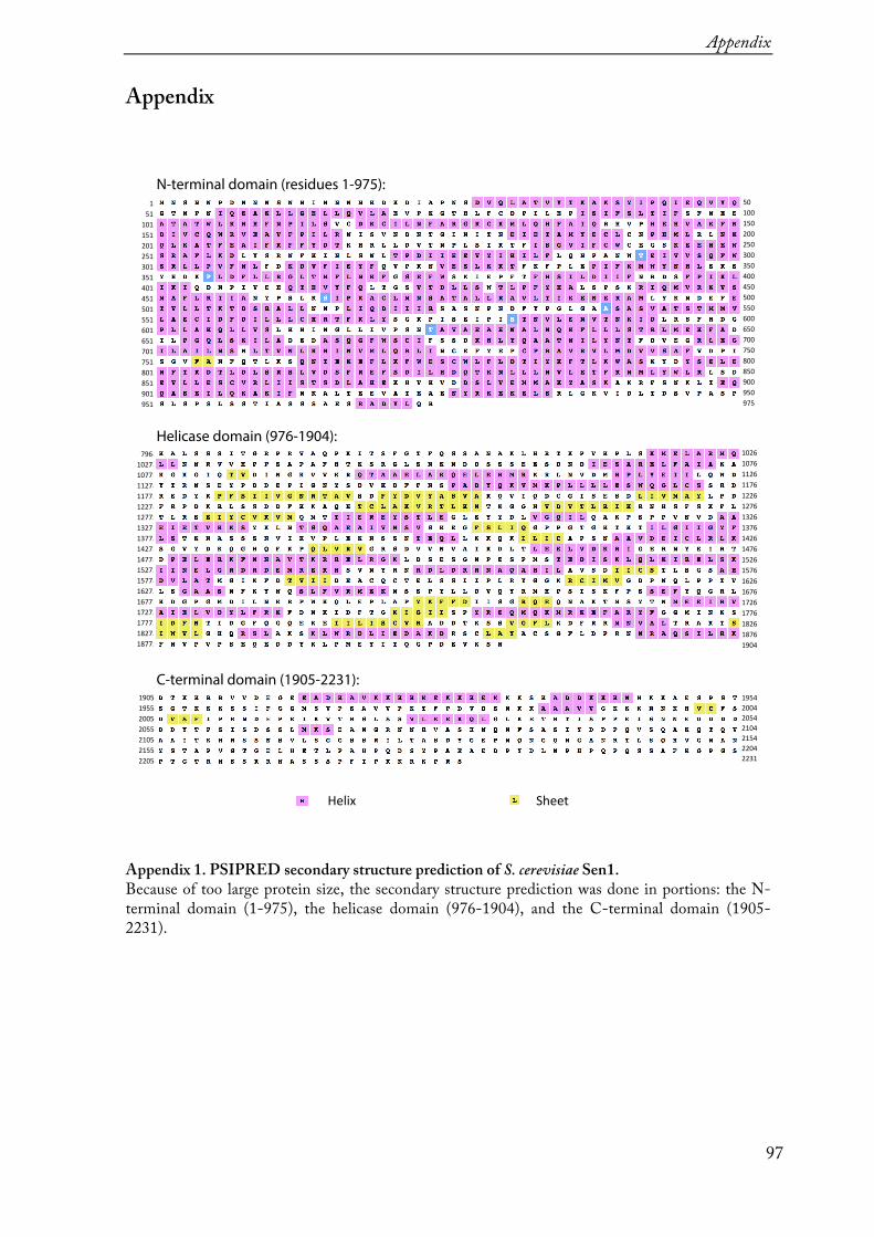
Appendix
97
Appendix
Appendix 1. PSIPRED secondary structure prediction of S. cerevisiae Sen1. Because of too large protein size, the secondary structure prediction was done in portions: the N-terminal domain (1-975), the helicase domain (976-1904), and the C-terminal domain (1905-2231).
N-terminal domain (residues 1-975):
Helicase domain (976-1904):
C-terminal domain (1905-2231):
796 1026
1027
1077
1127
1177
1227
1277
1327
1377
1427
1477
1527
1577
1627
1677
1727
1777
1827
1877
1076
1126
1176
1226
1326
1376
1426
1526
1576
1626
1676
1726
1776
1826
1876
1904
1954
2004
2054
2104
2154
2204
2231
1905
1955
2005
2055
2105
2155
2205
1476
1276
50
100
150
200
250
350
400
450
550
600
650
700
750
800
850
900
950
975
500
300
1
51
101
151
201
301
351
401
501
551
601
651
701
751
801
851
901
951
451
251
Helix Sheet
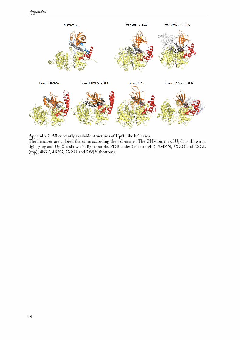
Appendix
98
Appendix 2. All currently available structures of Upf1-like helicases. The helicases are colored the same according their domains. The CH-domain of Upf1 is shown in light grey and Upf2 is shown in light purple. PDB codes (left to right): 5MZN, 2XZO and 2XZL (top), 4B3F, 4B3G, 2XZO and 2WJV (bottom).
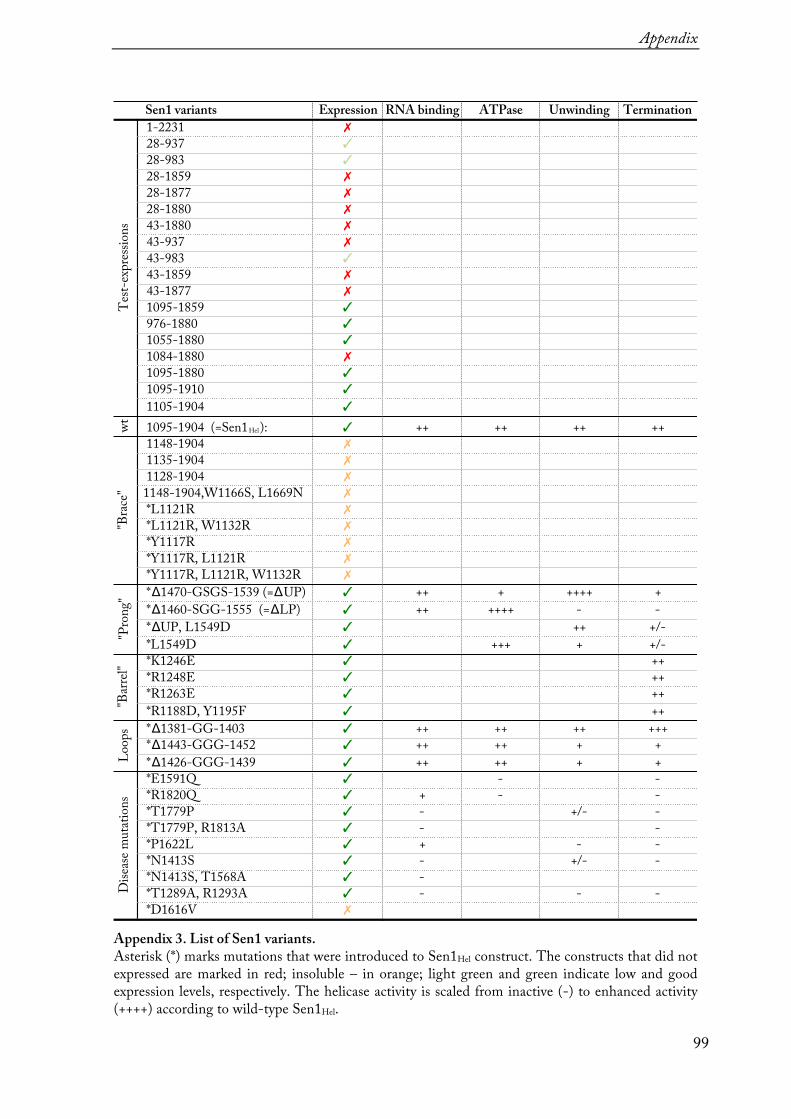
Appendix
99
Appendix 3. List of Sen1 variants. Asterisk (*) marks mutations that were introduced to Sen1Hel construct. The constructs that did not expressed are marked in red; insoluble – in orange; light green and green indicate low and good expression levels, respectively. The helicase activity is scaled from inactive (-) to enhanced activity (++++) according to wild-type Sen1Hel.
Test-
expres
sions
Sen1 variants Expression 1-2231 ✗
28-937 ✓
28-983 ✓
28-1859 ✗
28-1877 ✗
28-1880 ✗
43-1880 ✗
43-937 ✗
43-983 ✓
43-1859 ✗
43-1877 ✗
RNA binding ATPase Unwinding TerminationTe
st-exp
ressio
ns
1095-1859 ✓
wtLo
ops
"Brac
e" "B
arrel"
Disea
se mu
tation
s"P
rong"
Test-
expres
sions
976-1880 ✓
1055-1880 ✓
1084-1880 ✗
1095-1880 ✓
1095-1910 ✓
1105-1904 ✓
1095-1904 (=Sen1Hel): ✓
1148-1904 ✗
1135-1904 ✗
1128-1904 ✗
1148-1904,W1166S, L1669N ✗
*L1121R ✗
*L1121R, W1132R ✗
*Y1117R ✗
*Y1117R, L1121R ✗
*Y1117R, L1121R, W1132R ✗
*Δ1470-GSGS-1539 (=ΔUP) ✓
*Δ1460-SGG-1555 (=ΔLP) ✓
*ΔUP, L1549D ✓
*L1549D ✓
*K1246E ✓
*R1248E ✓
*R1263E ✓
*R1188D, Y1195F ✓
*Δ1381-GG-1403 ✓
*Δ1443-GGG-1452 ✓
*Δ1426-GGG-1439 ✓
*E1591Q ✓
*R1820Q ✓
*T1779P ✓
*T1779P, R1813A ✓
*P1622L ✓
*N1413S ✓
*N1413S, T1568A ✓
*T1289A, R1293A ✓
*D1616V ✗
++ ++ ++ ++
++ + ++++ +++ ++++ - -
++ +/-+++ + +/-
++++++++
++ ++ ++ +++++ ++ + +++ ++ + +
- -+ - -- +/- -- -+ - -- +/- -- - - -

100

References
101
References Adams PD, Afonine PV, Bunkóczi G, Chen VB, Davis IW, Echols N, Headd JJ, Hung
L-W, Kapral GJ, Grosse-Kunstleve RW, McCoy AJ, Moriarty NW, Oeffner R, Read RJ, Richardson DC, Richardson JS, Terwilliger TC & Zwart PH (2010) PHENIX: a comprehensive Python-based system for macromolecular structure solution. Acta Crystallogr. D Biol. Crystallogr. 66: 213–221
Alzu A, Bermejo R, Begnis M, Lucca C, Piccini D, Carotenuto W, Saponaro M, Brambati A, Cocito A, Foiani M & Liberi G (2012) Senataxin associates with replication forks to protect fork integrity across RNA-polymerase-II-transcribed genes. Cell 151: 835–846
Andersen PR, Domanski M, Kristiansen MS, Storvall H, Ntini E, Verheggen C, Schein A, Bunkenborg J, Poser I, Hallais M, Sandberg R, Hyman A, LaCava J, Rout MP, Andersen JS, Bertrand E & Jensen TH (2013) The human cap-binding complex is functionally connected to the nuclear RNA exosome. Nat. Struct. Mol. Biol. 20: 1367–1376
Bacikova V, Pasulka J, Kubicek K & Stefl R (2014) Structure and semi-sequence-specific RNA binding of Nrd1. Nucleic Acids Res. 42: 8024–8038
Bennett CL & La Spada AR (2015) Unwinding the role of senataxin in neurodegeneration. Discov Med 19: 127–136
Bennett CL, Chen Y, Vignali M, Lo RS, Mason AG, Unal A, Huq Saifee NP, Fields S & La Spada AR (2013) Protein interaction analysis of senataxin and the ALS4 L389S mutant yields insights into senataxin post-translational modification and uncovers mutant-specific binding with a brain cytoplasmic RNA-encoded peptide. PLoS ONE 8: e78837
Berretta J & Morillon A (2009) Pervasive transcription constitutes a new level of eukaryotic genome regulation. EMBO Rep. 10: 973–982
Borggrefe T, Davis R, Bareket-Samish A & Kornberg RD (2001) Quantitation of the RNA polymerase II transcription machinery in yeast. J. Biol. Chem. 276: 47150–47153
Buratowski S (2009) Progression through the RNA polymerase II CTD cycle. Mol. Cell 36: 541–546
Butler JS & Mitchell P (2011) Rrp6, rrp47 and cofactors of the nuclear exosome. Adv. Exp. Med. Biol. 702: 91–104
Cakiroglu SA, Zaugg JB & Luscombe NM (2016) Backmasking in the yeast genome: encoding overlapping information for protein-coding and RNA degradation. Nucleic
References
102
Acids Res. 44: 8065–8072
Carroll KL, Ghirlando R, Ames JM & Corden JL (2007) Interaction of yeast RNA-binding proteins Nrd1 and Nab3 with RNA polymerase II terminator elements. RNA 13: 361–373
Carroll KL, Pradhan DA, Granek JA, Clarke ND & Corden JL (2004) Identification of cis elements directing termination of yeast nonpolyadenylated snoRNA transcripts. Molecular and Cellular Biology 24: 6241–6252
Chakrabarti S, Jayachandran U, Bonneau F, Fiorini F, Basquin C, Domcke S, Le Hir H & Conti E (2011) Molecular mechanisms for the RNA-dependent ATPase activity of Upf1 and its regulation by Upf2. Mol. Cell 41: 693–703
Chamieh H, Ballut L, Bonneau F & Le Hir H (2008) NMD factors UPF2 and UPF3 bridge UPF1 to the exon junction complex and stimulate its RNA helicase activity. Nat. Struct. Mol. Biol. 15: 85–93
Chen X, Müller U, Sundling KE & Brow DA (2014) Saccharomyces cerevisiae Sen1 as a model for the study of mutations in human Senataxin that elicit cerebellar ataxia. Genetics 198: 577–590
Chen X, Poorey K, Carver MN, Müller U, Bekiranov S, Auble DT & Brow DA (2017a) Transcriptomes of six mutants in the Sen1 pathway reveal combinatorial control of transcription termination across the Saccharomyces cerevisiae genome. PLoS Genet. 13: e1006863
Chen X, Poorey K, Carver MN, Müller U, Bekiranov S, Auble DT & Brow DA (2017b) Transcriptomes of six mutants in the Sen1 pathway reveal combinatorial control of transcription termination across the Saccharomyces cerevisiae genome. PLoS Genet. 13: e1006863
Cheng Z, Muhlrad D, Lim MK, Parker R & Song H (2007) Structural and functional insights into the human Upf1 helicase core. EMBO J. 26: 253–264
Chinchilla K, Rodriguez-Molina JB, Ursic D, Finkel JS, Ansari AZ & Culbertson MR (2012) Interactions of Sen1, Nrd1, and Nab3 with multiple phosphorylated forms of the Rpb1 C-terminal domain in Saccharomyces cerevisiae. Eukaryotic Cell 11: 417–429
Clerici M, Mourao A, Gutsche I, Gehring NH, Hentze MW, Kulozik A, Kadlec J, Sattler M & Cusack S (2009) Crystal structure of the complex between human nonsense mediated decay factors UPF1 and UPF2 Orthorhombic form.
Cordin O, Tanner NK, Doère M, Linder P & Banroques J (2004) The newly discovered Q motif of DEAD-box RNA helicases regulates RNA-binding and helicase activity. EMBO J. 23: 2478–2487

References
103
Cowtan K (2006) The Buccaneersoftware for automated model building. 1. Tracing protein chains. Acta Crystallogr. D Biol. Crystallogr. 62: 1002–1011
Creamer TJ, Darby MM, Jamonnak N, Schaughency P, Hao H, Wheelan SJ & Corden JL (2011) Transcriptome-Wide Binding Sites for Components of the Saccharomyces cerevisiae Non-Poly(A) Termination Pathway: Nrd1, Nab3, and Sen1. PLoS Genet. 7: e1002329
Darby MM, Serebreni L, Pan X, Boeke JD & Corden JL (2012) The Saccharomyces cerevisiae Nrd1-Nab3 transcription termination pathway acts in opposition to Ras signaling and mediates response to nutrient depletion. Molecular and Cellular Biology 32: 1762–1775
David L, Huber W, Granovskaia M, Toedling J, Palm CJ, Bofkin L, Jones T, Davis RW & Steinmetz LM (2006) A high-resolution map of transcription in the yeast genome. Proc. Natl. Acad. Sci. U.S.A. 103: 5320–5325
DeMarini DJ, Papa FR, Swaminathan S, Ursic D, Rasmussen TP, Culbertson MR & Hochstrasser M (1995) The yeast SEN3 gene encodes a regulatory subunit of the 26S proteasome complex required for ubiquitin-dependent protein degradation in vivo. Molecular and Cellular Biology 15: 6311–6321
DeMarini DJ, Winey M, Ursic D, Webb F & Culbertson MR (1992) SEN1, a positive effector of tRNA-splicing endonuclease in Saccharomyces cerevisiae. Molecular and Cellular Biology 12: 2154–2164
Emsley P & Cowtan K (2004) Coot: model-building tools for molecular graphics. Acta Crystallogr. D Biol. Crystallogr. 60: 2126–2132
Fairman-Williams ME, Guenther U-P & Jankowsky E (2010) SF1 and SF2 helicases: family matters. Curr. Opin. Struct. Biol. 20: 313–324
Fasken MB, Laribee RN & Corbett AH (2015) Nab3 facilitates the function of the TRAMP complex in RNA processing via recruitment of Rrp6 independent of Nrd1. PLoS Genet. 11: e1005044
Franco-Echevarría E, González-Polo N, Zorrilla S, Martínez-Lumbreras S, Santiveri CM, Campos-Olivas R, Sánchez M, Calvo O, González B & Pérez-Cañadillas JM (2017) The structure of transcription termination factor Nrd1 reveals an original mode for GUAA recognition. Nucleic Acids Res. 45: 10293–10305
Ghaemmaghami S, Huh W-K, Bower K, Howson RW, Belle A, Dephoure N, O'Shea EK & Weissman JS (2003) Global analysis of protein expression in yeast. Nature 425: 737–741
Grzechnik P, Gdula MR & Proudfoot NJ (2015) Pcf11 orchestrates transcription

References
104
termination pathways in yeast. Genes Dev. 29: 849–861
Gudipati RK, Neil H, Feuerbach F, Malabat C & Jacquier A (2012a) The yeast RPL9B gene is regulated by modulation between two modes of transcription termination. EMBO J. 31: 2427–2437
Gudipati RK, Villa T, Boulay J & Libri D (2008) Phosphorylation of the RNA polymerase II C-terminal domain dictates transcription termination choice. Nat. Struct. Mol. Biol. 15: 786–794
Gudipati RK, Xu Z, Lebreton A, Séraphin B, Steinmetz LM, Jacquier A & Libri D (2012b) Extensive degradation of RNA precursors by the exosome in wild-type cells. Mol. Cell 48: 409–421
Halbach F, Rode M & Conti E (2012) The crystal structure of S. cerevisiae Ski2, a DExH helicase associated with the cytoplasmic functions of the exosome. RNA 18: 124–134
Hamperl S & Cimprich KA (2014) The contribution of co-transcriptional RNA:DNA hybrid structures to DNA damage and genome instability. DNA Repair (Amst.) 19: 84–94
Han Z, Libri D & Porrua O (2017) Biochemical characterization of the helicase Sen1 provides new insights into the mechanisms of non-coding transcription termination. Nucleic Acids Res. 45: 1355–1370
Hazelbaker DZ, Marquardt S, Wlotzka W & Buratowski S (2013) Kinetic Competition between RNA Polymerase II and Sen1-Dependent Transcription Termination. Mol. Cell 49: 55–66
Heidemann M, Hintermair C, Voß K & Eick D (2013) Dynamic phosphorylation patterns of RNA polymerase II CTD during transcription. Biochim. Biophys. Acta 1829: 55–62
Henrich B, Bergamaschi A, Broennimann C, Dinapoli R, Eikenberry EF, Johnson I, Kobas M, Kraft P, Mozzanica A & Schmitt B (2009) PILATUS: A single photon counting pixel detector for X-ray applications. Nuclear Instruments and Methods in Physics Research Section A: Accelerators, Spectrometers, Detectors and Associated Equipment 607: 247–249
Heo D-H, Yoo I, Kong J, Lidschreiber M, Mayer A, Choi B-Y, Hahn Y, Cramer P, Buratowski S & Kim M (2013) The RNA polymerase II C-terminal domain-interacting domain of yeast Nrd1 contributes to the choice of termination pathway and couples to RNA processing by the nuclear exosome. J. Biol. Chem. 288: 36676–36690
Hobor F, Pergoli R, Kubicek K, Hrossova D, Bacikova V, Zimmermann M, Pasulka J, Hofr C, Vanacova S & Stefl R (2011) Recognition of transcription termination signal
References
105
by the nuclear polyadenylated RNA-binding (NAB) 3 protein. J. Biol. Chem. 286: 3645–3657
Jamonnak N, Creamer TJ, Darby MM, Schaughency P, Wheelan SJ & Corden JL (2011) Yeast Nrd1, Nab3, and Sen1 transcriptome-wide binding maps suggest multiple roles in post-transcriptional RNA processing. RNA 17: 2011–2025
Jankowsky E & Fairman ME (2007) RNA helicases--one fold for many functions. Curr. Opin. Struct. Biol. 17: 316–324
Jenks MH, O'Rourke TW & Reines D (2008) Properties of an intergenic terminator and start site switch that regulate IMD2 transcription in yeast. Molecular and Cellular Biology 28: 3883–3893
Jensen TH, Jacquier A & Libri D (2013) Dealing with pervasive transcription. Mol. Cell 52: 473–484
Kabsch W (2010) Integration, scaling, space-group assignment and post-refinement. Acta Crystallogr. D Biol. Crystallogr. 66: 133–144
Karplus PA & Diederichs K (2012) Linking crystallographic model and data quality. Science 336: 1030–1033
Kilchert C, Wittmann S & Vasiljeva L (2016) The regulation and functions of the nuclear RNA exosome complex. Nat. Rev. Mol. Cell Biol. 17: 227–239
Kim HD, Choe J & Seo YS (1999) The sen1(+) gene of Schizosaccharomyces pombe, a homologue of budding yeast SEN1, encodes an RNA and DNA helicase. Biochemistry 38: 14697–14710
Kim K, Heo D-H, Kim I, Suh J-Y & Kim M (2016) Exosome Cofactors Connect Transcription Termination to RNA Processing by Guiding Terminated Transcripts to the Appropriate Exonuclease within the Nuclear Exosome. J. Biol. Chem. 291: 13229–13242
Kim K-Y & Levin DE (2011) Mpk1 MAPK association with the Paf1 complex blocks Sen1-mediated premature transcription termination. Cell 144: 745–756
Kim M, Suh H, Cho E-J & Buratowski S (2009) Phosphorylation of the yeast Rpb1 C-terminal domain at serines 2, 5, and 7. J. Biol. Chem. 284: 26421–26426
Komarnitsky P, Cho EJ & Buratowski S (2000) Different phosphorylated forms of RNA polymerase II and associated mRNA processing factors during transcription. Genes Dev. 14: 2452–2460
Kosugi S, Hasebe M, Matsumura N, Takashima H, Miyamoto-Sato E, Tomita M &

References
106
Yanagawa H (2009) Six classes of nuclear localization signals specific to different binding grooves of importin alpha. J. Biol. Chem. 284: 478–485
Kubicek K, Cerna H, Holub P, Pasulka J, Hrossova D, Loehr F, Hofr C, Vanacova S & Stefl R (2012) Serine phosphorylation and proline isomerization in RNAP II CTD control recruitment of Nrd1. Genes Dev. 26: 1891–1896
Kuehner JN, Pearson EL & Moore C (2011) Unravelling the means to an end: RNA polymerase II transcription termination. Nat. Rev. Mol. Cell Biol. 12: 283–294
Lemay J-F, Marguerat S, Larochelle M, Liu X, van Nues R, Hunyadkürti J, Hoque M, Tian B, Granneman S, Bähler J & Bachand F (2016) The Nrd1-like protein Seb1 coordinates cotranscriptional 3' end processing and polyadenylation site selection. Genes Dev. 30: 1558–1572
Lenstra TL, Tudek A, Clauder S, Xu Z, Pachis ST, van Leenen D, Kemmeren P, Steinmetz LM, Libri D & Holstege FCP (2013) The role of Ctk1 kinase in termination of small non-coding RNAs. PLoS ONE 8: e80495
Leonaitė B, Han Z, Basquin J, Bonneau F, Libri D, Porrua O & Conti E (2017) Sen1 has unique structural features grafted on the architecture of the Upf1-like helicase family. EMBO J. 36: 1590–1604
Li W, Selvam K, Rahman SA & Li S (2016) Sen1, the yeast homolog of human senataxin, plays a more direct role than Rad26 in transcription coupled DNA repair. Nucleic Acids Res. 44: 6794–6802
Lim SC, Bowler MW, Lai TF & Song H (2012) The Ighmbp2 helicase structure reveals the molecular basis for disease-causing mutations in DMSA1. Nucleic Acids Res. 40: 11009–11022
Loya TJ, O'Rourke TW & Reines D (2013a) Yeast Nab3 protein contains a self-assembly domain found in human heterogeneous nuclear ribonucleoprotein-C (hnRNP-C) that is necessary for transcription termination. J. Biol. Chem. 288: 2111–2117
Loya TJ, O'Rourke TW, Degtyareva N & Reines D (2013b) A network of interdependent molecular interactions describes a higher order Nrd1-Nab3 complex involved in yeast transcription termination. J. Biol. Chem. 288: 34158–34167
Makino DL, Schuch B, Stegmann E, Baumgärtner M, Basquin C & Conti E (2015) RNA degradation paths in a 12-subunit nuclear exosome complex. Nature 524: 54–58
Marquardt S, Hazelbaker DZ & Buratowski S (2011) Distinct RNA degradation pathways and 3' extensions of yeast non-coding RNA species. Transcription 2: 145–154
References
107
Martin-Tumasz S & Brow DA (2015) Saccharomyces cerevisiae Sen1 Helicase Domain Exhibits 5‘- to 3’-Helicase Activity with a Preference for Translocation on DNA Rather than RNA. J. Biol. Chem. 290: 22880–22889
Mayer A, Heidemann M, Lidschreiber M, Schreieck A, Sun M, Hintermair C, Kremmer E, Eick D & Cramer P (2012) CTD tyrosine phosphorylation impairs termination factor recruitment to RNA polymerase II. Science 336: 1723–1725
Mischo HE & Proudfoot NJ (2013) Disengaging polymerase: terminating RNA polymerase II transcription in budding yeast. Biochim. Biophys. Acta 1829: 174–185
Mischo HE, Gómez-González B, Grzechnik P, Rondón AG, Wei W, Steinmetz L, Aguilera A & Proudfoot NJ (2011) Yeast Sen1 helicase protects the genome from transcription-associated instability. Mol. Cell 41: 21–32
Nedea E, Nalbant D, Xia D, Theoharis NT, Suter B, Richardson CJ, Tatchell K, Kislinger T, Greenblatt JF & Nagy PL (2008) The Glc7 phosphatase subunit of the cleavage and polyadenylation factor is essential for transcription termination on snoRNA genes. Mol. Cell 29: 577–587
O'Rourke TW, Loya TJ, Head PE, Horton JR & Reines D (2015) Amyloid-like assembly of the low complexity domain of yeast Nab3. Prion 9: 34–47
Pape T & Schneider TR (2004) HKL2MAP: a graphical user interface for macromolecular phasing with SHELXprograms. Journal of Applied Crystallography 37: 843–844
Pearson E & Moore C (2014) The evolutionarily conserved Pol II flap loop contributes to proper transcription termination on short yeast genes. Cell Rep 9: 821–828
Porrua O & Libri D (2013) A bacterial-like mechanism for transcription termination by the Sen1p helicase in budding yeast. Nat. Struct. Mol. Biol. 20: 884–891
Porrua O & Libri D (2015a) Characterization of the mechanisms of transcription termination by the helicase Sen1. Methods Mol. Biol. 1259: 313–331
Porrua O & Libri D (2015b) Transcription termination and the control of the transcriptome: why, where and how to stop. Nat. Rev. Mol. Cell Biol. 16: 190–202
Porrua O, Hobor F, Boulay J, Kubicek K, D'Aubenton-Carafa Y, Gudipati RK, Stefl R & Libri D (2012) In vivo SELEX reveals novel sequence and structural determinants of Nrd1-Nab3-Sen1-dependent transcription termination. EMBO J. 31: 3935–3948
Pyle AM (2008) Translocation and Unwinding Mechanisms of RNA and DNA Helicases. Annual Review of Biophysics 37: 317–336

References
108
Richard P & Manley JL (2009) Transcription termination by nuclear RNA polymerases. Genes Dev. 23: 1247–1269
Rondón AG, Mischo HE, Kawauchi J & Proudfoot NJ (2009) Fail-safe transcriptional termination for protein-coding genes in S. cerevisiae. Mol. Cell 36: 88–98
Saikrishnan K, Powell B, Cook NJ, Webb MR & Wigley DB (2009) Mechanistic basis of 5‘-3’ translocation in SF1B helicases. Cell 137: 849–859
Sariki SK, Sahu PK, Golla U, Singh V, Azad GK & Tomar RS (2016) Sen1, the homolog of human Senataxin, is critical for cell survival through regulation of redox homeostasis, mitochondrial function, and the TOR pathway in Saccharomyces cerevisiae. FEBS J. 283: 4056–4083
Schaughency P, Merran J & Corden JL (2014) Genome-wide mapping of yeast RNA polymerase II termination. PLoS Genet. 10: e1004632
Schuch B, Feigenbutz M, Makino DL, Falk S, Basquin C, Mitchell P & Conti E (2014) The exosome‐binding factors Rrp6 and Rrp47 form a composite surface for recruiting the Mtr4 helicase. EMBO J. 33: 2829–2846
Sheldrick GM (2010) Experimental phasing with SHELXC/ D/ E: combining chain tracing with density modification. Acta Crystallogr. D Biol. Crystallogr. 66: 479–485
Shen A, Lupardus PJ, Morell M, Ponder EL, Sadaghiani AM, Garcia KC & Bogyo M (2009) Simplified, enhanced protein purification using an inducible, autoprocessing enzyme tag. PLoS ONE 4: e8119
Singh V, Azad GK, Sariki SK & Tomar RS (2015) Flocculation in Saccharomyces cerevisiae is regulated by RNA/DNA helicase Sen1p. FEBS Lett. 589: 3165–3174
Singleton MR, Dillingham MS & Wigley DB (2007) Structure and mechanism of helicases and nucleic acid translocases. Annu. Rev. Biochem. 76: 23–50
Skourti-Stathaki K, Proudfoot NJ & Gromak N (2011) Human senataxin resolves RNA/DNA hybrids formed at transcriptional pause sites to promote Xrn2-dependent termination. Mol. Cell 42: 794–805
Steinmetz EJ, Warren CL, Kuehner JN, Panbehi B, Ansari AZ & Brow DA (2006) Genome-wide distribution of yeast RNA polymerase II and its control by Sen1 helicase. Mol. Cell 24: 735–746
Summer H, Grämer R & Dröge P (2009) Denaturing urea polyacrylamide gel electrophoresis (Urea PAGE). J Vis Exp: e1485–e1485
Sydow JF, Brueckner F, Cheung ACM, Damsma GE, Dengl S, Lehmann E, Vassylyev D

References
109
& Cramer P (2009) Structural Basis of Transcription: Mismatch-Specific Fidelity Mechanisms and Paused RNA Polymerase II with Frayed RNA. Mol. Cell 34: 710–721
Tan-Wong SM, Zaugg JB, Camblong J, Xu Z, Zhang DW, Mischo HE, Ansari AZ, Luscombe NM, Steinmetz LM & Proudfoot NJ (2012) Gene loops enhance transcriptional directionality. Science 338: 671–675
Terzi N, Churchman LS, Vasiljeva L, Weissman J & Buratowski S (2011) H3K4 Trimethylation by Set1 Promotes Efficient Termination by the Nrd1-Nab3-Sen1 Pathway. Molecular and Cellular Biology 31: 3569–3583
Tudek A, Porrua O, Kabzinski T, Lidschreiber M, Kubicek K, Fortova A, Lacroute F, Vanacova S, Cramer P, Stefl R & Libri D (2014) Molecular basis for coordinating transcription termination with noncoding RNA degradation. Mol. Cell 55: 467–481
Ursic D (1997) The yeast SEN1 gene is required for the processing of diverse RNA classes. Nucleic Acids Res. 25: 4778–4785
Ursic D, Chinchilla K, Finkel JS & Culbertson MR (2004) Multiple protein/protein and protein/RNA interactions suggest roles for yeast DNA/RNA helicase Sen1p in transcription, transcription-coupled DNA repair and RNA processing. Nucleic Acids Res. 32: 2441–2452
Ursic D, DeMarini DJ & Culbertson MR (1995) Inactivation of the yeast Sen1 protein affects the localization of nucleolar proteins. MGG Molecular & General Genetics 249: 571–584
Vasiljeva L & Buratowski S (2006) Nrd1 interacts with the nuclear exosome for 3' processing of RNA polymerase II transcripts. Mol. Cell 21: 239–248
Vasiljeva L, Kim M, Mutschler H, Buratowski S & Meinhart A (2008) The Nrd1-Nab3-Sen1 termination complex interacts with the Ser5-phosphorylated RNA polymerase II C-terminal domain. Nat. Struct. Mol. Biol. 15: 795–804
Waltersperger S, Olieric V, Pradervand C, Glettig W, Salathe M, Fuchs MR, Curtin A, Wang X, Ebner S, Panepucci E, Weinert T, Schulze-Briese C & Wang M (2015) PRIGo: a new multi-axis goniometer for macromolecular crystallography. J Synchrotron Radiat 22: 895–900
Whitehouse I, Rando OJ, Delrow J & Tsukiyama T (2007) Chromatin remodelling at promoters suppresses antisense transcription. Nature 450: 1031–1035
Wlotzka W, Kudla G, Granneman S & Tollervey D (2011) The nuclear RNA polymerase II surveillance system targets polymerase III transcripts. EMBO J. 30: 1790–1803

References
110
Wyers F, Rougemaille M, Badis G, Rousselle J-C, Dufour M-E, Boulay J, Régnault B, Devaux F, Namane A, Séraphin B, Libri D & Jacquier A (2005) Cryptic pol II transcripts are degraded by a nuclear quality control pathway involving a new poly(A) polymerase. Cell 121: 725–737
Xu Z, Wei W, Gagneur J, Perocchi F, Clauder-Münster S, Camblong J, Guffanti E, Stutz F, Huber W & Steinmetz LM (2009) Bidirectional promoters generate pervasive transcription in yeast. Nature 457: 1033–1037