structural basis for control by...
TRANSCRIPT

Structural Basis for Control by Phosphorylation
Louise N. Johnson* and Richard J. Lewis
Laboratory of Molecular Biophysics, Department of Biochemistry, University of Oxford, South Parks Road, Oxford OX1 3QU
Received December 6, 2000
ContentsI. Introduction 2209II. Eukaryotic Protein Phosphorylation 2211
A. Eukaryotic Protein Kinases 22111. Serine/Threonine Kinases 22112. Tyrosine Kinases 2216
B. Phospho-Signaling and Protein/ProteinAssociation
2219
1. STAT Proteins 22192. KID Domain of CREB Bound to the KIX
Domain of CBP2220
C. Phosphorylation Signals Order/DisorderTransitions and Protein/Protein Dissociation
2221
1. Inactivation Gate of a K+ Channel 22212. Retinoblastoma Tumor Suppressor 22213. Neuronal-Sec1/Syntaxin1a Complex 2222
D. Phosphorylase: The Evolution of Control byPhosphorylation
2222
1. Human Liver Glycogen Phosphorylase 22222. Yeast Phosphorylase 22243. E. coli Maltodextrin Phosphorylase 2225
III. Prokaryotic Signal Transduction 2225A. Two-Component Signaling 2226
1. Response Regulators 22262. Histidine Kinases 22303. Histidine-Containing Phosphotransfer
Domains2230
B. The Haloacid Dehalogenase Superfamily 2233C. The Sugar Phosphotransferase System 2234D. Anti-Sigma Factor Antagonist 2236E. Phosphatases 2237
IV. Summary and Conclusions 2237V. Abbreviations 2239VI. Acknowledgments 2240VII. References 2240
I. Introduction
Posttranslational modification by phosphorylationis a ubiquitous regulatory mechanism in both eu-karyotes and prokaryotes. Intracellular phosphory-lation by protein kinases, triggered in response toextracellular signals, provides a mechanism for thecell to switch on or switch off many diverse processes.These processes include metabolic pathways, kinase
cascade activation, membrane transport, gene tran-scription, and motor mechanisms. The reverse reac-tion of dephosphorylation is catalyzed by proteinphosphatases that are controlled by response todifferent stimuli so that phosphorylation and dephos-phorylation are separately controlled events. In eu-karyotes, the protein kinase domain responsible forphosphorylation on serine, threonine, or tyrosineresidues is the first, second, and third most commondomain in the genome sequences of yeast (S. cerevi-siae), the worm (C. elegans), and the fly (D. melano-gaster), respectively, indicating the importance ofphospho-signaling in higher organisms. The humangenome contains 575 eukaryotic protein kinase do-mains, representing 2% of the total genome and thethird most populous domain.1 In prokaryotes, signal-ing by phosphorylation is equally important. In E.coli there are 62 genes that encode proteins involvedin dual histidine kinase/response regulator systems,representing approximately 1.5% of the entire ge-nome.
In this review we ask what are the structuralconsequences of phosphorylation and how is thisstructural response correlated with signal? We limitthe examples for the most part to those where thestructures of both the phospho- and non-phosphoprotein are known and coordinates available in theProtein Data Bank. We also limit our examples tophosphorylation of intact proteins and exclude thenumerous structural examples of phospho-peptidebinding to cognate proteins such as phospho-tyrosinepeptide binding to SH2 (e.g., ref 2) and PTB domains(e.g., ref 3) (reviewed in ref 4) and phospho-serinepeptide binding to 14-3-3 domains.5 The phospho-peptide interactions give insights into the recognitionby other proteins of the phospho-amino acid, animportant result especially for the design of ligandsthat might interfere with this association, but thesestructures do not illuminate how the recognition siteis engineered in the target protein as a consequenceof phosphorylation nor the response of the phospho-protein on recognition. We also exclude results wherea phospho-protein is an intermediate in a reactionpathway such as in the phosphatases,6,7 topoisomer-ases,8 and Cre recombinase.9 In these examples,phosphorylation is part of the catalytic pathway anddoes not result in a response in the target proteinthat signals to other proteins. Finally, phenylalaninehydroxylase is an allosteric protein regulated byphenylalanine, tetrahydrobiopterin, and phosphor-ylation on serine 16. The non-phospho and phospho
* To whom correspondence should be addressed. Phone: 01865275365. Fax: 01865 510454. E-mail: [email protected].
2209Chem. Rev. 2001, 101, 2209−2242
10.1021/cr000225s CCC: $36.00 © 2001 American Chemical SocietyPublished on Web 07/11/2001

structures of phenylalanine hydroxylase showed nodifferences, and the region containing the phospho-serine was disordered, suggesting that other allo-steric effectors need to be present to produce theactivation changes.10 We omit a discussion of “silent”phosphorylation.
Addition of a phosphoryl group to serine, threonine,or tyrosine (in eukaryotes and occasionally in prokary-otes) or to histidine or aspartic acid (in prokaryotes)residues confers properties that can have profoundeffects on protein conformation and function. Thephosphoryl group with pKa ∼ 6.7 is likely to bedianionic at physiological pH. The property of adouble-negative charge and the capacity for formingextensive hydrogen-bond networks with the four
phosphoryl oxygens confers special characteristics.Analysis of protein/phosphate interactions in proteinstructures has shown that two types of interactionsare dominant. In one, the phosphoryl group interactswith main-chain nitrogens at the start of an R-helixand utilizes the positive charge of the helix dipolefor charge neutralization. In the other the phosphategroup interacts with the side chain of one or morearginine residues. The guanidinium group of anarginine residue is well suited for interactions witha phosphate group by virtue of its planar structureand its ability to form multiple hydrogen bonds.Because of its resonance stabilization, the guani-dinium group is a poor protein donor (pKa > 12) andcannot function as a general acid catalyst in thehydrolysis of phosphorylated amino acids. Electro-static interactions between arginine and phosphorylgroups provide tight binding sites that play a domi-nant role in recognition and conformational response.In addition, a number of other residues are alsoinvolved at phospho-recognition sites, and theseinclude lysine, tyrosine, serine, threonine, aspar-agine, histidine, and, additionally in prokaryotes,metal ions.
The first phospho-protein structures, namely, rab-bit muscle glycogen phosphorylase (reviewed in ref11) and E. coli isocitrate dehydrogenase (reviewed inref 12), revealed two different mechanisms for thestructural response to phosphorylation. Glycogenphosphorylase is a dimer and is allosterically acti-vated by phosphorylation. Phosphorylation on aserine, Ser14, results in a conformational change ofthe N-terminal residues such that Ser14 shifts 50 Åto change contacts from intrasubunit to intersubunit.The intersubunit contacts of the phosphoryl-serine,pSer14, to two arginines (one in its own subunit andthe other in the other subunit) and other contacts ofthe residues that surround Ser14 are correlated witha conformational response that results in a changedorientation of the two subunits of the dimer andchanges in tertiary structure at the catalytic siteleading to activation. Isocitrate dehydrogenase isinhibited by phosphorylation. The mechanism forcontrol by phosphorylation is simple. In this enzymethere is no change in conformation on phosphoryla-tion. Instead, inhibition is achieved by phosphoryla-tion of a serine residue, Ser113, at the start of anR-helix, which blocks access to the catalytic site ofthe negatively charged isocitrate substrate, throughboth steric and electrostatic effects.
Our understanding of the regulatory properties ofphosphorylation has increased most notably withstudies on the protein kinases, the serine/threoninekinases (namely, cyclic AMP-dependent protein ki-nase (cAPK), cyclin-dependent protein kinase 2(CDK2), and extracellular regulated kinase (ERK2or MAPK)), the tyrosine kinases (namely, insulinreceptor tyrosine kinase (IRK)), and the family of Srckinases. Structural studies have shown the role ofthe phospho group in the activation segment of thekinase as an organizing center that localizes andorganizes the activation segment for substrate rec-ognition. Additional structural information aboutinhibitory phosphorylation includes inhibitory phos-
Louise Johnson obtained her Ph.D. degree in 1965 at the Royal Institution,London, where she worked on the binding of inhibitors to lysozyme, thefirst enzyme structure to be solved. After a postdoctoral year at YaleUniversity, she has held academic positions in the University of Oxfordsince 1967. She is currently David Phillips Professor of MolecularBiophysics at the University of Oxford, a position that she has held since1990. Her main research interests are in structural biology with the aimof seeking an explanation for biological function in terms of three-dimensional structures of macromolecules. She has made contributionsin the fields of control by phosphorylation, carbohydrate recognition andcatalytic mechanisms, time-resolved crystallography, and regulatoryproteins of the cell cycle. She is a fellow of the Royal Society and Memberof EMBO.
Richard J. Lewis received his B.A. and D.Phil. degrees in Biochemistryfrom the University of Oxford in 1991 and 1994, respectively, beforecommencing postdoctoral studies in the Chemistry Department of theUniversity of York until October, 2000. He is currently a Wellcome TrustResearch Career Development Fellow in the Laboratory of MolecularBiophysics, University of Oxford. His research interests include themolecular basis of control in phosphorylation-regulated biological pro-cesses, prokaryotic signal transduction, and protein/protein recognitionwith an emphasis on Bacillus species.
2210 Chemical Reviews, 2001, Vol. 101, No. 8 Johnson and Lewis

phorylation on Tyr15 in CDK2 and recognition of thephospho-tyrosine located in the C-terminal regionby the SH2 domain of Src. More recent structuralexamples have shown how phosphorylation maycause disorder (e.g., K+ channel inactivation domain),how phosphorylation may cause order, structuralchanges, and protein association (e.g., KIX/pKID fromthe CBP co-activator protein/CREB transactivationdomain system and some prokaryotic response regu-lator signaling proteins such as CheY), how phos-phorylation can promote dimerization (e.g., ERK2,STAT3B, and STAT1 and other prokaryotic responseregulators such as FixJ and Spo0A), and how phos-phorylation can inhibit protein/protein association(e.g., the prokaryotic HPr/enzyme I association in thesugar phosphotransferase system) and also causeprotein/protein dissociation in the anti-anti-σF/anti-σF (SpoIIAA/SpoIIAB) interaction. Thus, we begin tohave a portfolio of structural examples that demon-strates the most frequently observed aspects ofphosphorylation, namely, enzyme activation, enzymeinhibition, protein association, protein dissociation,and order/disorder phenomena.
II. Eukaryotic Protein Phosphorylation
A. Eukaryotic Protein KinasesThe 28 eukaryotic protein kinase structures that
are known to date have shown that they all have avery similar fold, as anticipated from conservationof certain residues throughout the kinase domain.The fold comprises an N-terminal lobe of mostlyâ-sheet with one R-helix termed the C-helix and aC-terminal domain that is mostly R-helical with asmall amount of sheet. The catalytic site for the ATPmoiety of the substrate is located at the lobe inter-face. Many protein kinases, but not all, requirephosphorylation on a threonine (Thr160 in humanCDK2) or tyrosine residue (e.g., Tyr416 in Src) thatis located in a region termed the activation segmentrunning from the conserved DFG motif to the con-served APE motif (residues 145-172 in CDK2). (Thisregion is sometimes called the ‘T loop’, but we preferthe term ‘activation segment’ because the regioncontains some secondary structure, because it in-cludes both phospho-threonine and phospho-ty-rosine residues in different kinases, and because itplays a key role in kinase activation). We use thesymbol “-” to denote covalent modification and thesymbol “/” to denote noncovalent association. Phosphoresidues or phospho proteins are prefixed by “p” (e.g.,pThr160 or pCDK2).
The first observation of phosphorylation on theactivation segment was reported for cyclic AMP-dependent protein kinase (cAPK) in 1979,13 but it wasnot until 1990 that mutagenesis studies showed thatphosphorylation of Thr197 was important for recog-nition of the inhibitory regulatory subunit14 and notuntil 1993 that it was recognized that threonineautophosphorylation was essential for kinase activ-ity.15 The crystal structure of cAPK, the first proteinkinase structure to be solved in 1991, demonstratedthe key contacts made by the phosphoryl threonine(pThr197) and provided ideas for its role as an
organizing center critical for activation.16,17 Struc-tural studies with an inhibitory peptide showedinteractions of the peptide with part of the activationsegment, but the side chains of the peptide also madekey specific interactions with side chains of cAPK inother regions.17-19 For tyrosine kinases, autophos-phorylation of v-Src at position Tyr416 had beenshown in the early 1980s and its significance for theactivation of the cellular counterpart c-Src kinaseestablished in 1987 (reviewed in ref 20).
As more and more kinases were discovered and thepresence of kinase cascades established in whichactivation of one kinase led to the activation of afollowing kinase, it became recognized that controlby phosphorylation in the activation segment is aproperty of most, but not all, kinases.21 All proteinkinases contain an aspartic acid residue (Asp166 incAPK; Asp127 in CDK2) that participates in cataly-sis. In kinases regulated by phosphorylation in theiractivation segment, an arginine residue precedes thecatalytic aspartate. These are the so-called RD ki-nases. The arginine contacts the phosphoryl groupof the phosphorylated residue in the activation seg-ment. The contact does not change the position of thecatalytic aspartate, but it assists in the correctalignment of the activation segment for interactionwith the substrate. The structural studies on thosekinases that are not regulated by phosphorylationhave shown that these kinases have other mecha-nisms for localization of their activation segments.In phosphorylase kinase domain, a glutamate takesthe place of the phosphoryl group.22 The kinasedomain is constitutively active without the require-ment for posttranslational modification, and theactivation segment is held in the correct position toaccept the peptide substrate.23 In twitchin kinase, acluster of nonpolar groups is likely to take the placeof the ionic interactions.24 In casein kinase I25,26 andin the cell cycle checkpoint kinase, Chk1,27 a dianion(sulfate) can occupy the positively charged pocketthat accommodates the phosphoryl group in phospho-regulated kinases, but studies have shown thatremoval of the dianion does not change the structure.In these examples the activation segment is stabi-lized by secondary structural elements and by side-chain interactions. Titin kinase represents an un-usual example. The kinase is activated by phosphor-ylation of a tyrosine residue (Tyr170) that is outsidethe activation segment (two residues beyond the Eof the APE motif). It is not an RD kinase. In theinactive non-phosphorylated state, the tyrosine isdirected into the catalytic site.28 It is proposed thaton phosphorylation, the pTyr is displaced and thatit may make a contact to the basic residue preferredin the P + 1 position for substrates of titin kinase.Evidently there are several different ways of stabiliz-ing the activation segment. Those kinases controlledby activation segment phosphorylation representsome of the most important enzymes in signalingcascades.
1. Serine/Threonine Kinases
CDK2. Cyclin-dependent kinases (CDKs) play acentral role in the coordination of the eukaryotic cell
Structural Basis for Control by Phosphorylation Chemical Reviews, 2001, Vol. 101, No. 8 2211
cycle (reviewed in ref 29). Their activation is a two-step process that requires cyclin binding and phos-phorylation by the CDK-activating kinase (CAK) ona threonine (Thr160 in CDK2) in the activationsegment. pCDK2 in complex with cyclin A becomesactive as cells enter the S phase. The complexphosphorylates downstream targets that include thetumor-suppressor proteins, pRb and the related p107,and other proteins that regulate transcription andreplication. CDK2 has become the most intenselystudied and the best understood structurally of theCDKs.
The crystal structure of the free inactive form ofCDK2 was solved in 1993.30 This revealed that theactivation segment was located in a region thatpartially blocked the ATP binding site (Figure 1a).The two lobes of the kinase structure were in a closedconformation, and the C-helix was in the wrongorientation to promote crucial contacts with thetriphosphate moiety of ATP, contacts that wereknown from the structure of active cAPK. Structuralstudies have shown that phospho-CDK2 (pCDK2)phosphorylated on Thr160 is similar in structure tofree inactive CDK2, except that the part of theglycine-rich loop (residues 10-20) and the activationsegment from residues 153-164 have become disor-dered (Figure 1b).31 A partial rationalization for thedisorder is provided by the observation that theactivation segment in free inactive CDK2 placesThr160 close to a glutamate (Glu12), and hence, onphosphorylation there would be electrostatic repul-sion. Phospho-CDK2 exhibits only 0.3% of the activityof fully active pCDK2/cyclin A. Evidently phospho-rylation and the electrostatic repulsion are not suf-ficient to trigger the essential conformational changesrequired to activate the kinase, but some pliabilityis induced in the enzyme that allows a basal level ofactivity. The crystal structure of CDK2/cyclin A32
showed that association of cyclin A with CDK2 ledto significant changes in conformation that seeminglyallowed a near active conformation, but the complexalso exhibits less than 0.3% of the activity exhibitedby the fully active phospho-CDK2/cyclin A complex.The major conformational changes involve openingof the bilobal structure, rearrangement of secondarystructural elements, and reorganization of the activa-tion segment (Figure 1c). The C helix (which containsthe motif PSTAIRE) shifts and rotates by 90°, result-ing in tight packing of the C-helix against theN-terminal lobe with extensive nonpolar interactions.The movement brings the side chain of Glu51 intothe catalytic site where it hydrogen bonds withLys33, an important contact residue to the R phos-phate of ATP bound at the catalytic site. In freeinactive CDK2, residues from the activation segmentGly147-Gly153 form a short helix (L12 helix) thatpacks against the C helix (Figure 1a). On bindingcyclin A, this short helix melts to allow movement ofthe C helix and the L12 residues form a reverse turnfollowed by a â sheet (residues 150-152) with theâ6 strand (residues 122-124) of the C-terminal lobe.The conformational changes observed on binding ofcyclin A to CDK2 result in the displacement of theactivation segment from the catalytic site and expo-
sure of Thr160. A nearby glutamate, Glu162, occupiesthe phospho-recognition site (Figure 1c). There is nochange in the conformation of cyclin A in bindingCDK2.33
In the structure of the fully active pCDK2/cyclinA34 complex, the pThr160 turns into the CDK2molecule and the phosphate group contacts threearginines, Arg50 from the PSTAIRE C helix, whoseposition is adjusted by the movement of the C-helixon binding cyclin A, Arg126 that precedes the cata-lytic aspartate, and Arg150 from the activationsegment whose position is adjusted by the meltingof the L12 helix on association with cyclin A34,35
(Figure 1d). The major change in conformation onforming the active pCDK2/cyclin A complex comparedwith the partially active (<0.3%) CDK2/cyclin Acomplex is a reorganization of the activation segmentin the region between residues 152-163. The phos-pho-threonine group acts as an organizing center,reminiscent of the seryl-phosphate in glycogen phos-phorylase. Its position appears to be determined bythe need to neutralize the positive charge cluster ofthe three arginine residues (Figure 1e). The geometryof coordination is close to the ideal arrangement seenin model compounds. Arg50 and Arg150 also hydro-gen bond to cyclin A main-chain carbonyls, andArg126 hydrogen bonds to CDK2 atoms. The phos-phate group is buried in an environment that is likelyto strengthen the phospho-arginine interactionsstabilizing the conformation in the vicinity.
The change in conformation engineered by thephospho-threonine in the CDK2/cyclin A complex iscrucial for the recognition of the peptide substrate(Figure 1f).36 CDK2 is specific for a proline residue(P + 1 site) following the phosphorylatable serine (P0site) and a basic residue 3 residues C-terminal to theserine (P + 3 site). The peptide used in the crystal-lographic binding studies was derived from an opti-mal peptide substrate with sequence HHASPRK,using the single amino acid code. The peptide isbound in an extended conformation across the cata-lytic site on the surface of the kinase contacting onlythe C-terminal lobe and the activation segment. Thepocket to accommodate the proline residue in theposition P + 1 is created by a special conformationof the activation segment in which a valine residue(Val164) adopts a left-handed conformation stabilizedby contact from its carbonyl oxygen to an arginine(Arg169). Binding of any residue except proline wouldbe unfavorable because of an uncompensated hydro-gen bond from the substrate’s main-chain nitrogen.In the non-phosphorylated CDK2/cyclin A complex,Val163 adopts a different conformation and blocksthe peptide binding site, thus explaining how theconformational changes produced by phosphorylationon the activation segment lead to activation throughcreating the peptide substrate recognition site. Thephospho-threonine also plays a direct role in sub-strate recognition. The lysine in the P + 3 positioncontacts pThr160 (Figure 1f).
Recent kinetic results with a peptide substratehave shown an increase in kcat of 843-fold and adecrease in Km of 137-fold, resulting in an overallincrease in catalytic efficiency (kcat/Km) on phospho-
2212 Chemical Reviews, 2001, Vol. 101, No. 8 Johnson and Lewis
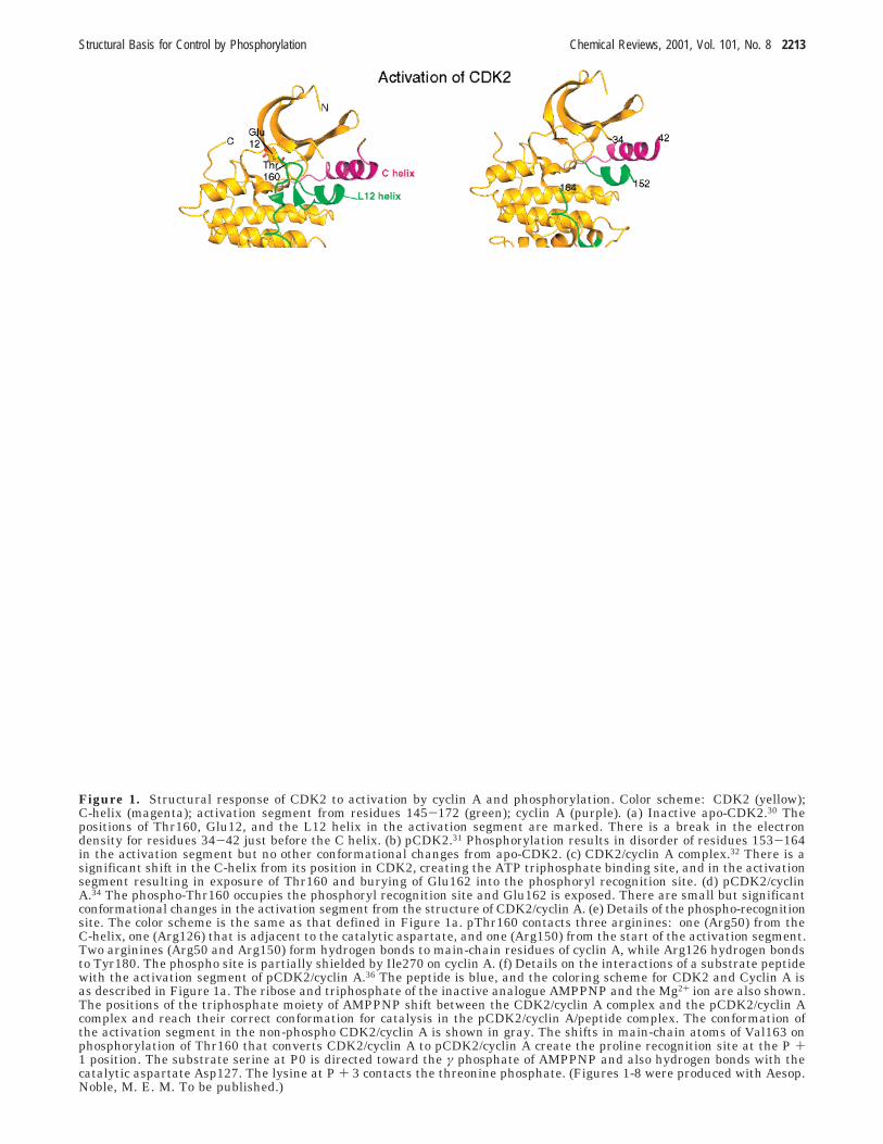
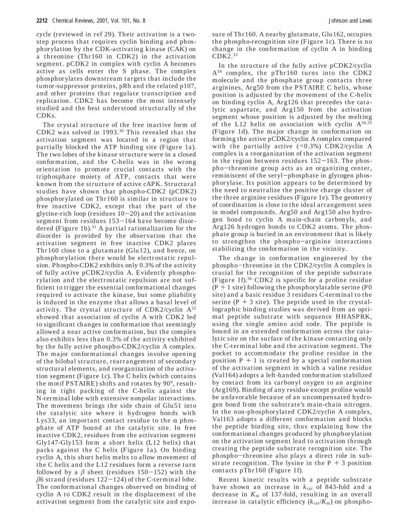
Figure 1. Structural response of CDK2 to activation by cyclin A and phosphorylation. Color scheme: CDK2 (yellow);C-helix (magenta); activation segment from residues 145-172 (green); cyclin A (purple). (a) Inactive apo-CDK2.30 Thepositions of Thr160, Glu12, and the L12 helix in the activation segment are marked. There is a break in the electrondensity for residues 34-42 just before the C helix. (b) pCDK2.31 Phosphorylation results in disorder of residues 153-164in the activation segment but no other conformational changes from apo-CDK2. (c) CDK2/cyclin A complex.32 There is asignificant shift in the C-helix from its position in CDK2, creating the ATP triphosphate binding site, and in the activationsegment resulting in exposure of Thr160 and burying of Glu162 into the phosphoryl recognition site. (d) pCDK2/cyclinA.34 The phospho-Thr160 occupies the phosphoryl recognition site and Glu162 is exposed. There are small but significantconformational changes in the activation segment from the structure of CDK2/cyclin A. (e) Details of the phospho-recognitionsite. The color scheme is the same as that defined in Figure 1a. pThr160 contacts three arginines: one (Arg50) from theC-helix, one (Arg126) that is adjacent to the catalytic aspartate, and one (Arg150) from the start of the activation segment.Two arginines (Arg50 and Arg150) form hydrogen bonds to main-chain residues of cyclin A, while Arg126 hydrogen bondsto Tyr180. The phospho site is partially shielded by Ile270 on cyclin A. (f) Details on the interactions of a substrate peptidewith the activation segment of pCDK2/cyclin A.36 The peptide is blue, and the coloring scheme for CDK2 and Cyclin A isas described in Figure 1a. The ribose and triphosphate of the inactive analogue AMPPNP and the Mg2+ ion are also shown.The positions of the triphosphate moiety of AMPPNP shift between the CDK2/cyclin A complex and the pCDK2/cyclin Acomplex and reach their correct conformation for catalysis in the pCDK2/cyclin A/peptide complex. The conformation ofthe activation segment in the non-phospho CDK2/cyclin A is shown in gray. The shifts in main-chain atoms of Val163 onphosphorylation of Thr160 that converts CDK2/cyclin A to pCDK2/cyclin A create the proline recognition site at the P +1 position. The substrate serine at P0 is directed toward the γ phosphate of AMPPNP and also hydrogen bonds with thecatalytic aspartate Asp127. The lysine at P + 3 contacts the threonine phosphate. (Figures 1-8 were produced with Aesop.Noble, M. E. M. To be published.)
Structural Basis for Control by Phosphorylation Chemical Reviews, 2001, Vol. 101, No. 8 2213

rylation of CDK2 in the CDK2/cyclin A complex of100 000-fold.37 The phospho and non-phospho formsshowed equivalent rates of ATPase activity. Combi-nation of the structures and the kinetic analysissuggests that the role of cyclin A in the CDK2/cyclinA complex is to align residues that form the ATPrecognition site, while the role of Thr160 phospho-rylation is to align the activation segment to createthe correct peptide or protein recognition site toenable phosphoryl transfer. Phosphorylation en-hances the rate of phosphoryl transfer (by about3000) and increases the affinity for substrate binding.In pCDK2/cyclin A, the rate of dissociation of prod-ucts is rate limiting but the differences in ratesbetween the chemical step and product dissociationare not as extreme as those observed for cAPK38-40
and phosphorylase kinase (PhK).41 In pCDK2/cyclinA, the phosphoryl transfer step (k3) is 22 s-1, whichis slower than that observed for cAPK and PhK (k3) 500 s-1 and >360 s-1, respectively).
Inactivation of pCDK2/cyclin A involves ubiquitin-mediated destruction of cyclin A by the proteasomeand dephosphorylation of pThr160. Genetic andbiochemical studies implicate kinase-associated phos-phatase (KAP) as one of the protein phosphatasesresponsible for dephosphorylating the activatingpThr residue of human CDK2.42 The enzyme, discov-ered using a yeast two-hybrid genetic screen as aCDK2-interacting protein, is a member of the dual-specificity phosphatase family and is capable ofdephosphorylating artificial protein substrates con-taining both pThr and pTyr residues. KAP associateswith CDK2 in vivo, and dephosphorylation of pThr160is dependent upon the tertiary structure of CDK2, afeature that distinguishes KAP from the nonspecific
PP2A catalytic subunit. The crystal structure of KAPin association with pThr160-CDK2 has recently beensolved43 and provides the first example of a proteinphosphatase in complex with its intact protein sub-strate (Figure 2).
The C-terminal lobe of CDK2 and the C-terminalhelix of KAP form the major protein interface be-tween the two molecules, regions remote from thekinase activation segment and the KAP catalytic site.The kinase activation segment interacts with thecatalytic site of KAP almost entirely via the phos-phate group of pThr160 (Figure 2). The pThr160phosphate group contacts the main-chain nitrogensof residues Gly142 and Arg146 and the side-chain OGof Ser140, contacts that are similar to those madeby phospho substrates in tyrosine phosphatases suchas PTB1B.7 (The KAP catalytic cysteine, Cys140, hadbeen mutated to serine in order to allow a stablecomplex to be formed.) This interaction requires thatthe activation segment is drawn away from thekinase molecule, thereby inducing a conformation ofpCDK2 to one similar to the activated state observedin the CDK2/cyclin A complex (except for the activa-tion segment conformation). The contact sites onCDK2 for cyclin A and for KAP are nonoverlapping,suggesting that cyclin A and KAP could bind CDK2simultaneously. However, KAP is unable to dephos-phorylate pCDK2/cyclin A and is only effective on freepCDK2.42 The explanation lies in conformationaldifferences. In the pCDK2/cyclin A complex thepThr160 is firmly bound by the three arginines(Arg50, Arg126, and Arg150) and is not accessible torecognition by KAP. The sequestration of pThr160,the defined conformation of the activation segment,and the localization of the arginine residues in the
Figure 2. Complex of pCDK2 with KAP.43 CDK2 is in gold with the activation segment in green and pThr160 marked.KAP is in cyan with the acid/base loop of the phosphatase carrying Asp110 in red and the loop that carries the catalyticcysteine (here mutated to serine, Ser140) in blue. The orientation of CDK2 is rotated with respect to Figure 1 in order toshow the interactions with KAP more clearly. The pCDK2/KAP interaction positions the catalytic site of KAP to recognizepThr160. The activation segment is drawn away from the main body of the kinase, and the remainder of the kinase moleculecollapses to a conformation that is nearer to the conformation of CDK2 seen in CDK2/cyclin A complexes than that observedfor pCDK2. There is a break in the electron density in pCDK2 between residues 34-43 just before the C helix.
2214 Chemical Reviews, 2001, Vol. 101, No. 8 Johnson and Lewis

pCDK2/cyclin A complex prevent access of KAP toits substrate. In free pCDK2, the activation segmentis mobile and pThr160 is accessible for binding toKAP. The second recognition site on CDK2 for KAP,which involves the GDSEID and DYK motifs, is notchanged between the pCDK2/KAP and pCDK2/cyclinA structures. It is accessible to KAP in the pCDK2/cyclin A complex, suggesting that it should be pos-sible for KAP to bind to the pCDK2/cyclin A complexbut unable to exert phosphatase activity. The struc-ture of the pCDK2/KAP complex reveals how theconformation of a phosphoprotein may be alteredfollowing its interaction with a binding partner whichrecognizes the phosphorylated residue. It will beinteresting to see if this is a common feature in otherprotein/phospho-protein complexes.
CDK2 is inhibited by phosphorylation of Tyr15 inthe glycine-rich loop. The phosphorylation by Wee1kinase and its dephosphorylation by CDC25 phos-phatase form important checkpoints in the cell andplay a role in response to DNA damage. The crystalstructure of pTyr15,pThr160-CDK2 in complex withcyclin A has been determined (Tucker, J.; Endicott,J. A.; Noble, M. E. M. To be published). Tyr15phosphorylation does not significantly alter the over-all structure of the complex nor does it prevent ATPbinding. It appears that phosphorylation of Tyr15may perturb the binding of protein substrate at thecatalytic site through steric hindrance, but this hasto be explored through further experiments.
MAP Kinases. Members of the MAP kinase familytransduce extracellular signals. They phosphorylateand activate transcription factors leading to trans-formation, proliferation, and other changes in thecell. Multiple parallel MAP kinase pathways aredifferentially responsive to growth factors and envi-ronmental stresses. Each member is activated by aspecific MAP/ERK kinase (MEK) and a MAP/ERKkinase kinase (MEKK) cascade, and each phospho-rylates a distinct spectrum of cellular substrates.44,45
The MAP kinase ERK2 is activated in response togrowth factors and mitogens by tyrosine kinasereceptors, G-protein-linked receptors, and proteinkinase C pathways. ERK2 achieves maximum activ-ity only when both Thr183 and Tyr185 from theactivation segment are phosphorylated by the up-stream kinase MEK. Thr183 is equivalent to Thr160in CDK2 and Thr197 in cAPK. Singly phosphorylatedforms of ERK2 have less than 1% of the activity ofthe fully phosphorylated enzyme.
The crystal structure of non-phosphorylated ERK2showed a typical kinase domain in which the twolobes exhibited an open conformation and the peptidebinding site was blocked by Tyr185, one of the tworesidues that are phosphorylated on the activationsegment.46 The activation segment was well orderedwith a different conformation than that seen in non-phosphorylated CDK2. ERK2 contains a C-terminalextension in which the chain spans both N-terminaland C-terminal lobes and terminates as an R-helix-labeled L16 that wraps around the C-helix. Thestructure of the dual-phosphorylated ERK2 (Figure3) showed a rearrangement of the activation segmentbringing the phospho-threonine and the phospho-
tyrosine into alignment with surface arginine resi-dues.47 Upon phosphorylation, pThr183 turns into theprotein, shifting 9 Å to make ionic contacts withN-terminal lobe residues, thereby promoting domainclosure. The contacts include Arg68 from the C-helixand Arg146 (preceding the catalytic aspartate) andArg170 from the activation segment. These contactsare equivalent to those made by pThr160 in CDK2,and like pThr160, pThr183 in ERK2 acts as anorganizing center through an extensive network ofhydrogen bonds that link it to the C-helix and theC-terminal helix L16 (Figure 3). On phosphorylation,pTyr185 moves from its buried site to a surface sitewhere it interacts with Arg189 and Arg192. Toaccommodate these changes, the activation segmentrefolds between residues Gly167 (the Gly of the DFGmotif) and Ala187. Ala187 adopts a left-handedconformation (similar to the conformation of Val164in CDK2) and hydrogen bonds through its main-chaincarbonyl to the side chain of Arg192. ERK2, likeCDK2, is specific for a proline in the P + 1 position.Modeling the peptide substrate from CDK2 into thecatalytic site of pERK2 shows that the same mech-
Figure 3. Comparison of non-phospho and phospho-ERK2.47 ERK2 is in gold with the activation segment(residues 165-195) in green and the C-terminal region(residues 320-359 including the L16 helix) in purple. TheL16 helix docks against the C-helix in a similar interactionto that seen with the interaction of cyclin A with CDK2(Figure 1). The positions of pThr183 and pTyr185 aremarked together with the arginine residues with whichthey interact (Arg68, Arg146, and Arg170 and Arg189 andArg192, respectively). Note the similarity in conformationof the activation segment to that of pCDK2/cyclin A (Figure1d). The position of the activation segment in non-phospho-ERK2 is shown in gray. The 9 Å shift in Thr183 isapparent. Also shown are residues Leu333, Leu336, andLeu344 in the loop preceding the L16 helix that form partof the dimerization interface in pERK2 dimers.
Structural Basis for Control by Phosphorylation Chemical Reviews, 2001, Vol. 101, No. 8 2215

anism operates for creation of a proline recognitionpocket with the same specificity determinant engi-neered by the left-handed conformation of Ala187.
Thus, in ERK2, the dual phosphorylation promotesdomain closure, allowing the catalytic and ATPbinding groups to take up their correct conformationfor catalysis, and phosphorylation reorganizes theactivation segment to create the correct substraterecognition site for the protein substrate. It is inter-esting that, like CDK2, single phosphorylation is notsufficient to promote these changes; CDK2 requirescyclin A, and ERK2 requires dual phosphorylation.
The dual phosphorylation also promotes changesin the L16 helix. Shifts in L16 are correlated withthe movement of the C-helix to its correct conforma-tion. Curiously, the L16 helix appears to form asimilar function to cyclin A in CDK2, packing againstthe C-helix (Figures 1c and 3). The L16 helix is alsoinvolved in dimerization. Three leucine residues(Leu333, Leu336, and Leu344) just prior to the L16helix become more exposed in the activated enzyme.In the crystal, these residues contact their equivalentresidues in a symmetry-related molecule forming ahydrophobic leucine zipper. On activation by phos-phorylation, ERK2 dimerizes and translocates to thenucleus. The dissociation constant (Kd) for dimerschanges from 20 µM for ERK2 to 7.5 nM for 2pERK2.The structural results combined with mutagenesisexperiments provide a satisfying explanation for thecreation of the dimerization site.48 They show thatsuch a site is engineered as a direct consequence ofthe structural changes following phosphorylation,although the phospho residues are not directly in-volved in the dimerization site.
Structures of three other members of the MAPkinase family have also been solved: P38R in non-phospho state,49,50 P38γ in dual-phosphorylated state,51
and JNK3 (a neuronal specific form of JNK) in non-phospho state.52 The P38 kinases are activated inresponse to proinflammatory cytokines and by cel-lular stress. P38R and P38γ exhibit 63% sequenceidentity. The non-phospho forms of P38R showed astructure similar to ERK2 but with a greater degreeof lobe opening and some differences in the confor-mation of the activation segment, which is sixresidues shorter in p38 than in ERK2. In p38, Tyr185is exposed to solvent while it is buried in the non-phospho form of ERK2. The non-phospho form ofJNK3 also shows an open conformation leading tomisalignment of catalytic residues. The activationsegment adopts a different conformation from ERK2,and the equivalent phosphorylatable threonine andtyrosine residues are 16 and 12 Å from their corre-sponding positions on non-phospho-ERK2. The struc-ture of the dual-phosphorylated p38γ shows a dra-matic closure of domains compared with p38R by 20°to bring catalytic residues into their correct orienta-tion. The activation segment, despite its differencein length, adopts a similar conformation to that seenin 2pERK2 with identical contacts for the residuescorresponding to pThr183 and pTyr185. These resultsillustrate a general theme in protein kinase struc-tures. In their inactive state, protein kinase struc-tures are different. In their active state, protein
kinase structures converge to a common frameworkwith regard to the correct orientation of catalyticgroups and the activation segment. In P38γ, thesequence of amino acid residues in the L16 helix hassignificant differences from that of ERK2. The threeleucine residues of ERK2 become aspartate, arginine,and valine, and the L16 helix has regions of disorder.P38γ does not dimerize on activation by phosphory-lation, most likely because of noncomplementarychanges in the surface residues in this region.
2. Tyrosine Kinases
Insulin Receptor Tyrosine Kinase. The cyto-plasmic region of the insulin receptor contains thetyrosine kinase domain. In response to insulin bind-ing to the extracellular domain, autophosphorylationon three tyrosines (Tyr1158, Tyr1162, and Tyr1163)in the activation segment of the kinase domainstimulates kinase activity toward exogenous sub-strates such as the insulin receptor substrate (IRS).A number of mutations in the tyrosine kinase regionof the gene encoding the insulin receptor have beenidentified in patients with noninsulin-dependentdiabetes.
The structure of the non-phosphorylated insulinreceptor tyrosine kinase (IRK)53 revealed the basickinase fold in which the two lobes were held open bysteric interactions between the glycine-rich loop andthe start of the activation segment in the region ofthe DFG motif. The activation segment traverses thecleft between the N- and C-terminal lobes such thatboth protein substrate and ATP binding sites areblocked. One of the tyrosines from the activationsegment, Tyr1162, is directed into the catalytic siteand is an example of active-site-directed intrastericautoregulation.54 It is hydrogen bonded to the cata-lytic aspartate and seemingly poised for cis-auto-phosphorylation. However, catalysis is prevented bythe blocking interactions of residues from the activa-tion segment (e.g., Phe1151) that inhibit ATP bind-ing.
The structure of the triply phosphorylated IRK internary complex with an inactive ATP analogue,AMPPNP, and a substrate peptide (Figure 4) re-vealed the significant conformational changes in-duced by the triple phosphorylation to create afunctionally active kinase.55 On phosphorylation,Tyr1158 shifts 30 Å and occupies a surface position,making no contacts with the protein but possiblyoffering a phospho-recognition site for other proteins.pTyr1162 is displaced from the catalytic site andhydrogen bonds to Arg1164 on the surface. Thisarginine is conserved in other tandem-phosphory-lated receptor tyrosine kinases. pTyr1163 is the lastresidue to be phosphorylated but is the most impor-tant for conferring activity. It turns in and fills thepocket equivalent to that observed for phosphoresidues in other protein kinases (e.g., pThr197 incAPK and pThr160 in CDK2), although the contactsto the phospho residue are less than those observedin the serine/threonine protein kinases. pTyr1163hydrogen bonds to Arg1155 (from the activationsegment equivalent to Arg150 in CDK2), and thereare stacking interactions between the aromatic ring
2216 Chemical Reviews, 2001, Vol. 101, No. 8 Johnson and Lewis

of the tyrosine and the aliphatic carbons of thearginine. There is no direct interaction to Arg1131,the arginine that precedes the catalytic aspartate,although this arginine is in the vicinity. There areno contacts to residues from the C-helix, and theresidue equivalent to Arg50 in CDK2 or His87 incAPK is a glutamate. Nevertheless, phosphorylationstimulates significant domain closure by 21° and theC helix rotates by 35° so that the glutamate (E1047)becomes closer to the ATP binding lysine (Lys1030).
The peptide substrate (sequence of GDYMNM)binds with the tyrosine directed into the catalytic siteclose to but not in contact with the γ phosphate ofAMPPNP (Figure 4). The substrate aspartate makesa contact through water to a lysine, while the twomethionine residues C-terminal to the tyrosine oc-cupy nonpolar pockets. Crucially the peptide is heldin place by hydrogen bonds characteristic of anantiparallel â sheet between the P + 1 and P + 3main-chain groups and activation segment residuesLeu1171 and Gly1169, respectively, thus demonstrat-ing the crucial role for phosphorylation in facilitatingthe correct conformation of the activation segmentto create the peptide substrate recognition site.Likewise the correct orientation of the activationsegment is critical for phosphorylase kinase peptidesubstrate recognition, where there is also antiparallelâ sheet interactions between the substrate and theactivation segment,23 and in CDK2 where the activa-tion segment creates the substrate prolyl and basicresidue recognition sites.36
The fact that the pTyr residue is anchored ratherless strongly than the corresponding phosphorylgroups in CDK2 or in cAPK may facilitate access toprotein phosphatases that switch off the insulinsignal. Recent structural studies56 on the tyrosineprotein phosphatases (PTP1B) in complex with triplyand doubly phosphorylated peptides from the insulinreceptor indicate that the major specificity is forpTyr1162 but that the specificity is enhanced by the
tandem phosphorylation. The pTyr1163 makes spe-cific contacts away from the catalytic site. The resultsindicate a hierarchical dephosphorylation process.
The structure of the kinase domain of the vascularendothelial growth factor receptor 2 (VEGFR2) hasbeen solved in the phosphorylated state57 and com-pared with the structure of the homologous (55%sequence identity) platelet-derived growth factorreceptor (PDGFR) kinase domain.58 Curiously, theVEGFR2 kinase domain, although phosphorylated onTyr1059 (equivalent to Tyr1163 in IRK or Thr160 inCDK2), exhibits a similar conformation to the non-phosphorylated inactive PDGFR kinase domain. Mostof the activation segment is disordered, and a portionof it occupies a position inhibitory to substratebinding. The VEGFR2 kinase domain has kinaseactivity, but the enhancement of activity for thephospho versus the non-phospho forms of the enzymeis only about 10-fold, compared with the 1000-foldenhancement of activity observed on phosphorylationof CDK2 in the CDK2/cyclin A complex. VEGFR2kinase domain can additionally be phosphorylated onTyr1054 (equivalent to Tyr1158 in IRK), and it maybe that VEGFR2 kinase requires this phosphoryla-tion or other factors for full activation or it may bethat VEGFR kinase domain exists in a dynamicequilibrium involving several conformations thatinclude the inactive conformation observed in thecrystal. In this example, single phosphorylation hasnot been sufficient to establish the definite activeconformation, just as single phosphorylation of freeCDK2 is insufficient to activate.
Src Family Kinases (Src, Hck, Lck). The Srcfamily of tyrosine kinases are a closely related groupof nonreceptor kinases which are involved in signal-ing pathways that control growth and differentiationin cells in response to the activation of cell surfacereceptors by growth factors and cytokines. Src familymembers differ in cellular expression and localiza-tion. Src is expressed in a wide range of tissues:
Figure 4. Tyrosine kinase domain of the insulin receptor (IRK)55 showing interactions of the peptide substrate (in pink)with the activation segment (residues 1150-1179 in green). The catalytic loop (residues 1130-1137) that carries the catalyticaspartate (Asp1132) is shown in gold. The three phospho-tyrosine residues, pTyr1158 that is external, pTyr1162 thatcontacts Arg1164, and pTyr1163 that contacts Arg1155, are shown. The main chain of the peptide substrate forms a shortantiparallel â sheet with the activation segment main-chain atoms of residues 1169-1171. The substrate tyrosine is directedinto the catalytic site, and the phenolic OH group is within hydrogen-bonding distance of Asp1132, the catalytic aspartate,and directed toward the γ phosphate of AMPPNP, the inactive analogue of ATP.
Structural Basis for Control by Phosphorylation Chemical Reviews, 2001, Vol. 101, No. 8 2217

haematopoietic cell kinase (Hck) is expressed inlymphoid and myeloid cells and lymphoid cell kinase(Lck) plays a role in T cell signaling. The viralhomologue of Src, v-Src, is the gene product respon-sible for the cell-transforming ability of the Roussarcoma virus and was the first tyrosine kinase tobe identified. Src family members comprise fivedomains: an N-terminal domain involved in mem-brane binding and subcellular localization, an SH3domain, an SH2 domain, a kinase domain (SH1), anda C-terminal tail that includes a tyrosine, Tyr527,whose phosphorylation by another kinase, Csk, leadsto down regulation of Src-kinase activity. The im-portance of the C-terminal tail tyrosine phosphory-lation for cellular regulation is demonstrated byv-Src, which lacks this site of phosphorylation andexhibits unregulated activity in cells. Src familymembers are regulated by autophosphorylation of atyrosine in the activation segment (Tyr416 in Src).
The crystal structures of the down-regulated formsof human and chicken Src59,60 and human Hck61 andthe active phospho form of the kinase domain ofhuman Lck62 have been solved. These structures haverevealed a rich variety of control mechanisms forprotein kinases both by phosphorylation and byprotein/protein interactions. The structures of theSH3-SH2-kinase-C-terminal construct of down-regulated Src and Hck (Figure 5) show a compactstructure in which the SH2 and SH3 domains arelocated on the opposite surface of the kinase domainto the catalytic site, indicating immediately that theexplanation for down regulation of kinase activity bythese domains is through indirect interactions and
not a steric blocking mechanism. The C-terminalpTyr527 interacts with the SH2 domain at a site thatis over 40 Å from the catalytic site (Figure 5). Insimple terms, the down regulation appears to beeffected by two factors: a constraint on the kinaseC-helix so that it is displaced from its active confor-mation and, second, the engagement of the SH2 andSH3 domains at either ends of the two lobes of thekinase in such a way as to impair the correct lobedisposition needed for catalysis. The details havebeen well summarized in reviews.63-65 Here we focuson the effects of phosphorylation (inhibitory phos-phorylation at Tyr527 and activatory phosphorylationat Tyr416) and show how these phosphorylationcontrol events are correlated with the protein/proteininteractions mediated by the domains.
The SH3 domain adopts a similar structure to thatobserved in the crystal structures of the singledomain, as does the SH2 domain. The linker betweenthe SH2 and the kinase domain interacts withresidues at the C-terminal end of the kinase C-helixand part of the region of a turn between the â strandsâ2 and â3 of the N-terminal lobe of the kinase. Thelinker adopts a conformation similar to a polyprolinehelix (although it contains only one proline in Src)and forms a docking site for the SH3 domain (Figure5). These interactions form the first of the compo-nents that restrain the kinase in the inactive con-formation. The kinase domain has a structure thathas many similarities to the inactive conformationof CDK2. The contacts of the SH2 domain to thekinase are mediated by the docking of the C-terminalregion pTyr527 to the phospho-tyrosine site of theSH2 domain. The phosphoryl group of pTyr527 inHck and in chicken Src makes six hydrogen bondsin total to Arg 155, Arg175, Glu178 (main-chain NH),and the OG atoms of Ser177 and Thr179. Arg155 alsocontributes a positive charge interaction with the πelectrons of the phenolic ring of pTyr527. Theseinteractions are similar to the contacts observed instudies of model phospho-peptides bound to SH2domains and suggest a relatively high-affinity phos-phoryl-binding site. However, the sequence sur-rounding the pTyr527 in both Src and Hck is anonoptimal sequence for SH2 recognition, and themanner of binding of the surrounding residues inboth structures is suggestive of a low-affinity mode.Consistent with this is the ability of high-affinityphospho-tyrosine peptides to displace pTyr527, lead-ing to an increase in kinase activity without phos-phorylation on Tyr416.66
The contact of the C-terminal tail pTyr527 localizesthe SH2 domain, reinforcing the contacts of the SH2-kinase linker region and the contacts of this regionto the SH3 domain (Figure 5). Displacement of theSH3 domain by the SH3-binding protein HIV-1 Nefprovides maximal activation of Hck,67 indicating thatthe restraining interactions mediated by the SH3/linker/kinase N-terminal lobe are critical for main-taining the kinase in the inactive conformation. Oneinteraction, that of Leu255 from the linker region tothe N-terminal lobe of the kinase, appears to be animportant interaction in the restraint of the kinase.68
The leucine docks into nonpolar pocket formed by
Figure 5. Structure of Src kinase (coordinates taken fromref 69). The N- and C-termini are marked. The SH3 domainis in blue, the SH2 domain in dark green, the linker incyan, the kinase domain in gold with the C-helix (residues304-317) in magenta and the activation segment (residues404-432) in green, and the C-terminal tail in red. Alsoshown are the positions of Leu255, which is important forthe linker to kinase interaction, Tyr416 in the activationsegment that is phosphorylated for activation, and pTyr527that interacts with the SH2 domain and holds Src in theinactive conformation.
2218 Chemical Reviews, 2001, Vol. 101, No. 8 Johnson and Lewis

Trp286 and Tyr326 of the kinase â2/â3 region andâ4 strands, respectively (Figure 5). Mutational stud-ies showed that when replaced by a smaller residue(e.g., Leu255Val), the mutant protein had kinaseactivity and this activity was not down regulated byphosphorylation of Tyr527. Occupancy of this non-polar pocket by a bulky hydrophobic group thereforeappears to be a key factor in the down regulation,and oblation of this interaction cannot be compen-sated for by phosphorylation of the C-terminal tailtyrosine.
In the original publications of Src and Hck struc-tures, it was reported that the activation segmentwas mobile. Later work with different crystal forms,with less glycerol as cryo-protectant, in the presenceand absence of ATP analogues, and at higher resolu-tion has shown a defined conformation for the activa-tion segment that provides further explanation forregulation by autoinhibition.69,70 In the Src number-ing, the activation segment running from the DFGto the APE motifs corresponds to residues 404-432.Residues 404-411 adopt a 310 helix similar to the L12helix in CDK2 (residues 145-152) (Figure 5). Hy-drophobic residues from the helix in Src interact withthe kinase C-helix, stabilizing the C-helix in itsinactive conformation, in a rotated conformationsimilar to that observed in inactive CDK2. Furtheron in the activation segment, residues 413-418 forman R-helix such that Tyr416 is directed into to anonpolar pocket and forms water-mediated hydrogenbonds to residues from the catalytic loop of thekinase. This interaction blocks the catalytic site, andcomparison with the substrate-bound active form ofIRK shows that the conformation of the activationsegment is not in an appropriate conformation torecognize substrate.
Although the Src and Hck structures represent theinactive conformations, much can be learned aboutthe transitions to an active kinase through compari-son with the active kinase domain structure of Lck.62
The construct used for Lck lacks the SH3, SH2 andC-terminal tail. It is comprises only the autophos-phorylated (pTyr394) form of the kinase domain. Thekinase domain has a structure that is similar to theactive forms of other kinases (e.g., pIRK) with thecorrect disposition of the C-helix and lobe orientationsto allow participation of the key binding and catalyticresidues in substrate recognition and catalysis. Theactivation segment (residues 381-402) contains someregions of mobility, but pTyr394 is well localized withthe phosphoryl group occupying a similar location tothat seen in cAPK, CDK2, and IRK. The phosphorylgroup contacts Arg363 (the arginine before thecatalytic aspartate corresponding to Arg126 in CDK2)and Arg387 from the activation segment (correspond-ing to Arg150 in CDK2), but this interaction ismediated by two water molecules. The binding of thephosphoryl group therefore appears to be less strongthan in cAPK and CDK2 but is nevertheless suf-ficient to generate and stabilize an active conforma-tion of the kinase domain.
It has been remarked that Src and CDK2 sharesimilar features in their disposition of the C-helix,which is important for maintenance of the inactive
conformation. However, the mechanisms of activationare different. Src is restrained by interactions at theC-terminal end of the C-helix that are relieved bydisplacement of the SH3 and SH2 domains by avariety of mechanisms, while CDK achieves basalactivity through association with cyclin A and theinteraction of the cyclin at the N-terminal end of theC-helix (Figures 1c and 5). Once the SH3 and SH2restraints are relieved in Src, full activation can beachieved by the trans-autophosphorylation of thetyrosine Tyr416 in the activation segment increasingactivity by at most 50-fold. In contrast, CDK2/cyclinA has only basal activity and requires another kinaseto phosphorylate Thr160, thus conferring a 1000-foldincrease in activity.
The double SH2-regulated tyrosine phosphataseSHP-2 exhibits a different mechanism for SH2 regu-lation. In the crystal structure of the inactive formof SHP-2, the N-terminal SH2 domain directly blocksthe catalytic site of the phosphatase domain.71 Theinteraction appears to alter the phospho-tyrosinepeptide binding site on the N-SH2 domain, disrupt-ing its phospho-peptide recognition. It is proposedthat when tyrosine-phosphopeptides bind to the twotandem SH2 domains with the C-terminal SH2domain enhancing the poor affinity of the N-SH2domain, conformational changes are also inducedthat release the N-SH2 from the phosphatase cata-lytic site and allows activation.
B. Phospho-Signaling and Protein/ProteinAssociation
1. STAT ProteinsFurther insights into the role of phosphotyrosine/
SH2 interactions in regulation have been elucidatedwith structure determinations of STAT-172 andSTAT3â73. STAT (signal transducers and activatorsof transcription) proteins are activated in responseto extracellular signals. Binding of growth factors orcytokines to their cognate receptors results in intra-cellular tyrosine phosphorylation. STAT proteinscontain an SH2 domain. They are recruited to thereceptor intracellular region and then phosphorylatedeither directly by the receptor kinase or by thereceptor-associated JAK kinase. Phosphorylationcauses the STAT proteins to dimerize and translocateto the nucleus where they bind to specific promotersequences in target genes. The JAK/STAT pathwayprovides one of the most direct signaling pathwaysfrom the receptor to the nucleus. The crystal struc-tures of both STATs comprised constructs that lackthe N-terminal DNA cooperative binding domain andthe C-terminal transcriptional activator domain butincluded four domains: a four-helix coiled-coil do-main, the DNA binding domain that has an immu-noglobulin-like fold similar to that of NFκB and p53,a linker region composed of R-helices, and the SH2domain followed by the C-terminal region containingthe phospho-tyrosine. The STAT proteins crystalstructures were solved in complex with an 18-merand a 17-mer duplex of DNA. The structures im-mediately provided an explanation for the dimeriza-tion and DNA binding promoted by the interactionof the phospho-tyrosine with the SH2 domain.
Structural Basis for Control by Phosphorylation Chemical Reviews, 2001, Vol. 101, No. 8 2219
The four domains form a stable tandem structurein which each domain interacts with its neighborsby significant hydrophobic interactions (Figure 6).Two STAT monomers form a dimer in which themonomers are related by a 2-fold crystallographicsymmetry axis. The two monomers bind DNA, grip-ping it like a pair of pliers, in such a way that thetwo DNA binding domains interact with DNA but notwith each other. The phospho-tyrosine of one mono-mer crosses over and docks into the SH2 recognitionsite of the other monomer. The C-terminal regionthen crosses back into its own monomer. The contactsto the phosphoryl group are highly conserved (withthe exception that one arginine is a lysine) with thoseobserved for other SH2 interactions, although thesequences of the STAT SH2 domains diverge consid-erably from those of other SH2 domains. The lengthof the linker region from the end of the SH2 domainto the phospho-tyrosine is too short to allow thephospho-tyrosine to reach the recognition site of itsown monomer. The phosphotyrosine/SH2 interactionprovides the major contact in promoting dimerization.
The SH2 domain is in contact with the DNAbinding domain through interactions with an R helixof the linker region. Thus, phospho-tyrosine recogni-tion by the SH2 domain promotes dimerization andsignals to the dimeric DNA binding site. There aresimilarities with the way in which the C-terminal tailphospho-tyrosine of Src signals. Both proteins, onethrough intramolecular (Src) and the other throughintermolecular (STAT) recognition, utilize the local-
ization of the SH2 domain by the phospho-tyrosineto signal to other structural elements of the protein.In Src protein/protein interactions mediated by theSH2 hold the kinase in an inactive conformation, andin STAT, protein/protein interactions localize theDNA binding domain.
2. KID Domain of CREB Bound to the KIX Domain ofCBP
The cAMP-regulated transcription factor CREB isstimulated for target gene expression by associationwith the co-activator, the CREB binding protein,CBP. Complex formation between CREB and CBPrequires cAPK phosphorylation of Ser133 on CREB.CREB transactivation domain is composed of thekinase-inducible domain (KID) that carries Ser133and a Q2 domain that stimulates transcriptionthrough association with a subunit of TFIID. CBPcontains a domain known as KIX with which its bindsto the phospho-KID domain of CREB. The NMR-determined structures74 of pKID (residues 101-160)from CREB in association with KIX (residues 586-666) from CBP have provided a basis for understand-ing the role of phosphorylation in association. FreepKID exhibits a NMR spectrum indicative of anunstructured peptide. Upon binding to KIX, the pKIDpeptide folds into two mutually perpendicular helicesbetween the regions 119-145, although outside theseregions the peptide remains unstructured. The KIXdomain has a well-defined structure of three anti-parallel helices with a hydrophobic groove running
Figure 6. Stat3â homodimer DNA complex.73 The N-terminal helix bundle (residues 130-320) is shown in blue, theDNA binding domain (residues 320-465) in magenta, the linker domain (residues 465-585) in yellow, and the SH2 domainand phospho-tyrosine-containing region (residues 585-722) in green in the right-hand molecule and red in the left-handmolecule. The disordered regions that link the R1 and R2 helices of the N-terminal helix bundle and the region from 689to 701 in the phospho-tyrosine-containing loop are shown by dotted lines. The 17 base pair DNA duplex is shown with allatoms drawn. The pTyr705 from one molecule reaches into the SH2 binding site on the other molecule of the dimer. Theview is approximately along the DNA dyad axis with the dyad axis of the complex running vertically.
2220 Chemical Reviews, 2001, Vol. 101, No. 8 Johnson and Lewis

across helices 1 and 3 into which the second helix(RB) of pKID binds (Figure 7). In addition to thenonpolar interactions between pKID and KIX, thereare three charge/charge interactions and the overallchange in solvent accessible area (1200 Å2) indicatesa reasonably tight interaction. However, there is noassociation without phosphorylation of Ser133.
The phosphoryl-serine residue is situated at thestart of the second helix (RB) of pKID (Figure 7). Thecontacts to KIX vary in the family of NMR structures,but the essential interaction, as demonstrated bymutagenesis, is that between the phosphoryl groupand Tyr658. A lysine, Lys662, is in the vicinity, butits interaction does not appear to be essential. Thereis likely to be stabilization of the RB helix throughinteraction of the phosphoryl group with the helixdipole. Free phosphoryl serine does not bind to KIX(in contrast, phosphoryl tyrosine will bind to SH2domains). Thus, the mechanism by which phospho-rylation confers structure and association of the KIDdomain appears to utilize mutually cooperative in-teractions. The folding of pKID through associationwith KIX is dependent upon the phosphoryl groupat the N-terminal end of the developing R-helix andone key interaction of the phosphoryl group to atyrosine residue.
Phosphoryl binding sites at the start of R-helicesare common in enzymes that recognize phosphosubstrate or cofactors (e.g., triose phosphate isomeraseand lactate dehydrogenase). There are now threeexamples where a phosphoryl amino acid occurs atthe start of a 310 or R-helix: glycogen phosphorylase,isocitrate dehydrogenase, and now pKID. To berecognized by their respective kinases (phosphorylasekinase for glycogen phosphorylase and cAPK forKID), the crystal structures of these kinases haveshown that the region in the vicinity of the phospho-rylatable residue must adopt an extended conforma-tion.23 Hence, on release from the kinase, a confor-mational change must take place (extended to helix)and indeed the incentive for this conformational
change may well encourage product release from thekinase.
C. Phosphorylation Signals Order/DisorderTransitions and Protein/Protein Dissociation
There are many instances in vivo where phospho-rylation on multiple sites causes protein/proteindissociation or prevents protein/protein association.The structures of non-phospho and phospho forms ofthe inactivation domain of a K+ channel have pro-vided insight into the basis of these events based onelectrostatic repulsion, while two further proteinswhose structures have been determined in the non-phospho form (the retinoblastoma tumor-suppressorprotein Rb and the neuronal Sec1 protein) haveindicated that similar mechanisms may be possiblewith these systems.
1. Inactivation Gate of a K+ Channel
In common with many ion channels, the voltage-gated K+ channel Kv.4 is regulated by phosphoryla-tion. Inactivation of the channel is achieved throughthe interaction of residues 1-30 (termed the inacti-vation domain (ID)) which block the pore on thecytoplasmic side. Phosphorylation of the ID by pro-tein kinase C (PKC) on three potential serine resi-dues (Ser8, Ser15, and Ser21) results in reduction ofinactivation. The NMR analysis of the non-phospho-rylated ID showed a compact structure that had ahigh affinity for the receptor. Phosphorylation at Ser8resulted in an increase in the rate of dissociation fromthe receptor, and phosphorylation of Ser15 and Ser21resulted in a decrease in the rate of association. TheNMR structures of pSer8-ID and pSer15, pSer21-IDshowed a loss of overall structural stability.75 InpSer8-ID, residues 5-15 were ordered (i.e., thosearound the phospho site) but residues N- and C-terminal were disordered. In pSer15, Ser21-ID, resi-dues 15-25 were ordered (again those regions aroundthe phospho sites) and the remainder of the structuredisordered. The phospho sites are close to twoglutamate residues, Glu27 and Glu28, and it is likelythat the disorder arises from steric and electrostaticrepulsion. Whereas in free pCDK2 it is the regioncarrying the phosphoryl-threonine that becomesdisordered because of electrostatic repulsion, in theID domain of Kv3.4 it is the other parts of thestructure.
2. Retinoblastoma Tumor Suppressor
The growth-suppressive action of the tumor-sup-pressor protein Rb is coupled to the cell cycle throughthe actions of the CDKs. Inactivation of Rb is acommon event in many cancers. In its active state,Rb binds to the transcription factor complex E2F andthereby inhibits expression of genes that are requiredfor the transition from the G1 into the S phase andDNA replication. Rb phosphorylation on multiplesites, first by CDK4/cyclin D and then by CDK2/cyclinE followed by CDK2/cyclin A, leads to dissociation ofRb from E2F and release of transactivation compe-tent E2F. Sixteen potential Ser/Thr-Pro phosphory-lation sites have been identified, and at least 10 such
Figure 7. Structure of the KIX domain of CBP bound tothe phosphorylated KID transactivation domain of CREB.KIX is in purple and pKID in magenta.74 The interactionof pSer133(KID) with Tyr658(KIX) is shown. There areextensive interactions between the C-terminal helix of KIDand the R1 and R3 helices of KIX.
Structural Basis for Control by Phosphorylation Chemical Reviews, 2001, Vol. 101, No. 8 2221

sites have been shown to be phosphorylated in vivo(Ser249, Thr252, Thr373, Ser567, Ser608, Ser780,Ser807, Ser811, Thr821, and Thr826). Some of theseare specific for individual CDK/cyclin combinations.Rb also forms complexes with several other proteinsincluding viral oncoproteins. Phosphorylation differ-entially effects the binding of other proteins.76
The crystal structure of the so-called pocket regionof Rb comprising the A and B boxes (residues 372-589 and 635-787) has been solved in complex witha peptide from the human papilloma virus E7 onco-protein.77 The oncoprotein binds to Rb through arecognition sequence of LxCxE and inactivates Rbcausing release of E2F. The A and B boxes areresponsible for binding many of the cellular and viralproteins that mediate transcriptional repression.They contain three of the phosphorylation sites(Thr373, Ser567, and Ser780). The other phospho-rylation sites are contained in the N-terminal region,the linker between the A and B boxes, and the 15KC-terminal domain. The A and B boxes exhibit thefive-helix assembly characteristic of the cyclin fold,and this fold is augmented by eight other helices.Serine567, one of the phosphorylatable serines, isnear the interface between the A and B boxes, andits phosphorylation would result in unfavorablecontacts. Another phosphorylatable serine, Ser780,is in the middle of a 13 residue carboxyl tail of the Bbox. It packs against a hydrophobic region but doesnot make any significant interactions with the restof the protein. Phosphorylation of Ser780 could resultin some restructuring, although the consequences arehard to predict. Two other phosphorylatable residues,Thr821 and Thr826, are outside the construct usedfor the crystal structure determination. However,they are thought to influence the recognition andbinding of proteins containing the LxCxE motif. TheRb fragment structure bound with the E7 LxCxEpeptide reveals a six-lysine basic patch on the rim ofthe LxCxE binding site that may provide a recogni-tion site for the two phosphorylated threonines andallow the phosphorylated segment to bind to and toblock the site.77
3. Neuronal-Sec1/Syntaxin1a Complex
Syntaxin1a and neuronal Sec1 are two proteinsthat are involved in vesicle trafficking and membranefusion. Neuronal Sec1 binds with nanomolar affinityto syntaxin 1a and forms a complex that is essentialfor vesicle trafficking and membrane fusion andwhich prevents syntaxin complex formation withother vesicle membrane proteins. The crystal struc-ture of the nSec1/syntaxin1a complex revealed con-formational changes in the four-helix syntaxin pro-tein relative to the free syntaxin and that complexedwith a SNARE protein.78 Protein kinase C modulatesexocytosis in several different cell types, possibly byregulation of the nSec1/syntaxin interaction. PKCphosphorylates Ser306 and Ser313 of nSec1, whichinhibits the formation of the Sec1/syntaxin complex.These sites can no longer be phosphorylated in thecomplex. Ser313 is near the syntaxin recognition sitein a region that caries a negative charge. In thenSec1/syntaxin complex, the negative charge is neu-
tralized by a complementary positive charge regionfrom Sec1. Although a phospho-serine could beaccommodated at this site without disruption of thestructure, the introduction of a further negativecharge by the phosphoryl group could provide elec-trostatic repulsion and promote conformationalchanges in nSec1. Thus, phosphorylation of Ser313is likely to prevent nSec1/syntaxin association. Ser306however is further away from the nSec1/syntaxininterface, and the consequences of phosphorylationat this site, if any, are not clear.
These examples indicate that electrostatic effects,either electrostatic repulsion (as seen in the inactiva-tion domain of the K+ channel or that hypothesisedfor nSec1/syntaxin interactions) or electrostatic at-traction (as proposed for Rb pThr820 and pThr826interactions), are likely to be significant. Both effectsare seen in glycogen phosphorylase, where in the non-phospho state the basic peptide region surroundingthe phosphorylatable serine, Ser14, docks against anacidic region on the protein surface while on phos-phorylation pSer14 shifts to contact a newly createdbasic site comprising two arginines.79 The examplesalso demonstrate the difficulty of predicting theconsequences of phosphorylation in the absence ofstructures of both the phospho and non-phosphoforms.
D. Phosphorylase: The Evolution of Control byPhosphorylation
Muscle glycogen phosphorylase, discovered by Carland Gerty Cori in the late 1930s, was the firstenzyme for which control by reversible phosphory-lation was established.80 The enzyme has been thesubject of intense biochemical and crystallographicstudies. Through structural studies on the non-phospho (rmGPb) and phospho (rmGPa) forms of therabbit muscle glycogen phosphorylase, the structuralbasis of control by phosphorylation is well understood(reviewed in ref 11). Some of the features of theresponse are illustrated in Figure 8a. More recently,the structures of the phosphorylated human liverisozyme (hlGPa) in the inactive and active states,81
the phospho and non-phospho forms of the yeastphosphorylase (yGPb and yGPa),82-84 and the E. colimaltodextrin phosphorylase85 have been solved, lead-ing to further insights into the structural basis ofcontrol by phosphorylation.
1. Human Liver Glycogen Phosphorylase
Phosphorylases catalyze the phosphorylysis of anR-1,4-glycosidic bond in glycogen or maltodextrins toyield glucose-1-phosphate. In muscle, glucose-1-phosphate is fed into the glycolytic pathway to meetthe energy needs of the cell, while in the liver,glucose-1-phosphate is converted to glucose andglucose is output for the benefit of other tissues. Thehuman muscle and liver isozymes exhibit 79% iden-tity in sequence but have slightly different controlproperties. Both are regulated by phosphorylation ona single serine, Ser14, leading to activation, but themuscle enzyme may be activated by AMP; thisactivity was inhibited by ATP and glucose-6-phos-phate, while the liver enzyme is not sensitive to these
2222 Chemical Reviews, 2001, Vol. 101, No. 8 Johnson and Lewis

Structural Basis for Control by Phosphorylation Chemical Reviews, 2001, Vol. 101, No. 8 2223

effectors. Both isozymes are dimers formed from twomonomers of 842 residues in muscle and 846 residuesin liver. The human liver phosphorylase structurehas been solved in its phosphorylated state in aninactive and active conformation.81 The inactive formwas achieved by co-crystallizing in the presence of apotent glucose analogue inhibitor (N-acetyl-â-D-glu-copyranosylamine).86,87 The active form was crystal-lized in the presence of AMP. Thus, although thestructures do not allow a direct comparison betweenphospho and non-phospho forms, they do allowcomparison between inactive and active states andfurther elaborate on the allosteric connection betweenthe phosphoryl-serine site and the catalytic site thatare about 45 Å apart.
In the inactive hlGPa, the N-terminal region sur-rounding Ser14 is disordered, consistent with obser-vations that binding of glucose or glucose-analogueinhibitors at the catalytic site promotes the transitionform the R to T state and allows greater accessibilityof the phosphoryl-serine to the protein phosphatasePP1. In the active hlGPa, the N-terminal peptide iswell ordered, making contacts at the intersubunitinterface. The phosphoryl-serine contacts Arg43′(from the other subunit) and Arg69 from its ownsubunit, as in rmGPa (Figure 8a). The allosterictransitions are mediated through these intersubunitcontacts. In the N-terminal region on one side of thedimer, the phosphoryl-serine contacts tighten theinterface and bring the cap region from one subunit(residues 42′-45′, so-called because it caps the AMPrecognition site) closer to the R2 helix of the othersubunit. At the other side of the dimer, two helices,the tower helices (residues 267-274 and 267′-274′),form intersubunit contacts through helix-helix′ pack-ing. In the inactive hlGPa, the catalytic site is blockedby the 280s loop (residues 280-286) that follows thetower helix, and the 280s loop is held in place bycontacts to the glucose analogue and to the 380s loop,as seen in the inactive form of rmGPb. In the activehlGPa, the tower helices are shorter by two turns andthe 280s loop folds up in an ordered conformation andcontributes an extra turn to a helix, R8 helix. Accessto the catalytic site is open, and the 380s loopbecomes more disordered. In rmGPa, the 280s loopitself becomes disordered. The structure at the cata-lytic site and the 280s loop observed with hlGPa issimilar to that observed for E. coli maltodextrinphosphorylase as described below. The changes ob-served on the transition from inactive to active hlGPastructures not only allow access to the catalytic site,
but also most crucially create the recognition site forthe inorganic phosphate substrate. This site is withinhydrogen-bonding distance of the 5′-phosphate groupof the essential cofactor pyridoxal phosphate (PLP)and is created through the displacement of an acidicresidue (Asp283) from the 280s loop and the replace-ment with a basic residue (Arg569), as observed withthe rmGPb and rmGPa comparisons.
As in rmGP, there is a rotation of the hlGPasubunits with respect to each other on activation byabout 7°, resulting in hlGPa in a change of surfaceinterface buried from 4200 to 7300 Å2, a rather largerchange than that observed with rmGP. At the AMPrecognition site, there is no change in sequencebetween hl and rmGP but many of the interactionsobserved for AMP binding to rmGPa are absent inhlGPa as a result of the small differences in subunitorientation and different disposition of side chains.The adenine-recognition loop (residues 315-325)adopts a different conformation. These changes resultin the loss of cooperative binding of AMP. Despitesequence conservation between the muscle and liverisozymes, the changes at the subunit interface andnot the changes in residues that contact the ligandsresult in a muscle enzyme that can be allostericallyregulated by effectors as well as by phosphorylationand a liver enzyme that can only be activated byphosphorylation. Both can be down regulated byglucose and glucose analogues, a phenomenon thatis important in the liver but is not relevant for musclephosphorylase. In muscle, the effects of the allostericeffectors, AMP, Glc6P, and ATP, can be overriddenby phosphorylation.
2. Yeast PhosphorylaseThe structures of the non-phospho and phospho
forms of the yeast (S. cerevisiae) glycogen phospho-rylase revealed a subtle modification of the mecha-nism established for the mammalian phosphorylases.In yeast, phosphorylase is activated during theapproach to the stationary phase to enable cells toutilize glycogen when other substrates may be de-pleted. Yeast glycogen phosphorylase (yGP) has anextra 39 residues at the N-terminal region (numbered-1 to -39) compared with rmGP, and the site ofphosphorylation is at Thr-10. Although overall rmGPand yGP exhibit about 46% identity in sequence,there are no similarities in sequence surrounding thesites of phosphorylation. Further, yeast phosphory-lase kinase and rabbit muscle phosphorylase kinaseare different enzymes and do not cross phosphorylate
Figure 8. Comparison of the structures of the phospho active forms of rabbit muscle glycogen phosphorylase and yeastglycogen phosphorylase. (a) Rabbit muscle phosphorylase a (GPa).177 The dimeric enzyme is viewed down the 2-fold axisof symmetry. One subunit is in cyan and the other in blue. The N-terminal tail residues 10-23 are in red in both subunits.The N-terminal residues 10-23 in GPb are in yellow in both subunits. The position of Ser14 is marked in the non-phosphoform and in the phospho form. The black curved arrow in the top subunit highlights the 50 Å shift in Ser14 onphosphorylation. The other conformational changes on the conversion of GPb to GPa are not shown. The catalytic site isindicated by the position of the pyridoxal phosphate. Entrance to the catalytic site is on the far side of the molecule. Thesite of interaction of the pSer14 is over 40 Å from the catalytic site. The inset shows the details of the interaction of thepSer14 with Arg69 from the R2 helix and Arg43′ form the cap′ region of the other subunit. (b) Yeast glycogen phosphorylase.83
The dimeric enzyme is viewed down the 2-fold axis of symmetry. The coloring scheme is similar to that described for parta. The N-terminal residues from -14 to 23 are shown in red for both subunits. The details of the interaction of the pThr-10 with two arginine residues (Arg309 and Arg310) are shown in the inset. Lys313 is in the vicinity but is not in contact.In the non-phospho form of yeast phosphorylase, the N-terminal residues wrap around the other subunit and block entranceto the catalytic site and the pThr-10 site is occupied by glucose-6-phosphate.
2224 Chemical Reviews, 2001, Vol. 101, No. 8 Johnson and Lewis

rabbit and yeast phosphorylases, respectively. Thecontrol sites appear to have evolved through differentevolutionary mechanisms in which a phosphorylat-able peptide was spliced to a common core of aphosphorylase.
The non-phospho form of yGP was crystallized inthe presence of the inhibitor, Glc-6-P. The core of thestructure is similar to that of rmGP, but there aresome differences in the relative disposition of the twosubunits of the dimer.82,83 The structure of thecatalytic site of yGP is similar to that of the activatedform rmGPa. The 280s loop is folded up so as to allowaccess, and the residues that form the inorganicphosphate substrate recognition site are in theiractive conformation for substrate binding. However,despite this apparent active structure, the non-phospho form of yGP is held in an inhibited state bya blocking mechanism from residues of the extendedN-terminal tail. From residue 22 to -39, the tail firstwraps around its own subunit, partly mimicking theconformation and position adopted by the N-terminaltail in rmGP. It then continues with residues towardthe N-terminus to wrap around the other subunit sothat residues -23 to -38 contact the catalytic siteentrance and block access. In the non-phospho state,Glc-6-P is bound at the AMP/Glc-6-P recognition siteof rmGP and utilizes the same arginine residues forrecognition of the phosphate (Arg309 and Arg310) butthe contacts of the glucosyl moiety are different thanthose observed for Glc-6-P binding to rmGPb becauseof changes in sequence.88
In the non-phospho yGP, Thr-10 is surrounded bynonpolar residues. In the phosphoform of yGP, theN-terminal tail undergoes displacement and Thr-10shifts about 36 Å.84 Phospho-Thr-10 displaces Glc-6-P and itself occupies the phospho recognition sitethrough interactions with Arg309 and Arg310 (Figure8b). There are also nonpolar interactions between thenonpolar residues surrounding Thr-10 and those onthe core surface of the phosphorylase. These contactsare different than those made by the glucosyl moietyof Glc-6-P, so that there are distinct regulatoryresponses to the inhibitor (Glc-6-P) and the activator(pThr-10). There are accompanying changes at thesubunit interface as in rmGP that include a tighten-ing of contacts at the allosteric effector interface siteand opening around the interface leading to thecatalytic site. Thus, activation by phosphorylation inyGP involves competition between the phosphoaminoacid and an allosteric inhibitor for a phospho-recogni-tion site, relief of steric blocking of the catalytic site,and some reorganization of the two subunits.
3. E. coli Maltodextrin Phosphorylase
E. coli maltodextrin phosphorylase (MalP) is a 796amino acid dimeric enzyme. Unlike rmGP and yGP,MalP is not regulated by phosphorylation or byallosteric effectors and is constitutively active. Levelsof MalP in E. coli cells are regulated by geneexpression in response to changes in nutritionalstatus. Under conditions of low glucose and highmaltose or maltodextrins, the Mal genome pathwayis switched on in response to elevated cAMP levelsthat enable the maltodextrins to be transported into
the cell and utilized. Overall MalP is 46% identicalin sequence to rmGP, but it lacks 17 residues at theN-terminus and 13 residues at the C-terminus.Residues 18-80 (using the rabbit muscle GP num-bering) display minimal sequence similarity betweenrmGP and MalP. These residues correspond to thefirst exon in human muscle GP, and it has beensuggested that control by phosphorylation in eukary-otic phosphorylases could have been achieved bysplicing a phosphorylatable peptide to a commonancestral phosphorylase core.
The structure of the core of MalP is very similarto that of rmGP.85 At the catalytic site surroundingthe cofactor PLP, residues are 100% conserved.Outside the core, the MalP and rmGP structuresdiffer in the subunit/subunit contacts in the regionof the cap′/R2 interface and the tower/tower′ inter-face. Despite the lack of sequence similarity, residues18-80 in MalP show superficial similarity in struc-ture to rmGP and occupy the same space. The regionfrom 246 to 286 adopts a different conformation. Asa result of sequence changes, there is a differentorganization of residues 254-260 and the towerhelices. The 280s loop folds up and adds two turnsof R helix to the R-8 helix, allowing access to thecatalytic site where the residues are in the correctconformation to promote catalysis.
The structure of E. coli MalP has been solved inthe ternary complex with inorganic phosphate andpentasaccharide substrate analogue in which theglycosyl oxygen linkage was replaced with a thiolinkage.89 The structure has revealed not only thedetails of the catalytic residues when poised forcatalysis, but also those residues important for therecognition of the oligosaccharide. Tyr280, a residueconserved in all phosphorylases, from the 280s loopplays a crucial role.
Thus, the comparative studies reveal a commonstructural core for the different phosphorylases, buteach exhibits different regulatory properties. In theprokaryotic MalP, there are no effector ligand bindingsites and the core exhibits constitutive activitywithout the need for activation by phosphorylation.In the eukaryotic yeast and mammalian musclephosphorylases, control by phosphorylation has oc-curred twice with the splicing of different peptidescontaining a phosphorylatable group, resulting indifferent solutions to the same evolutionary chal-lenge.
III. Prokaryotic Signal TransductionFor many years it was generally held that revers-
ible protein phosphorylation was a regulatory mech-anism that had developed late in evolution and wasthus unique to the demands of signaling withinmulticellular organisms. Intercellular communicationwas not understood to be a function of simple,unicellular organisms, since it was assumed thatthese had evolved with no requirement for sophisti-cated means of control. It is now well established thatregulation by protein phosphorylation/dephosphory-lation also occurs in microorganisms and that bac-teria do communicate within colonies. Eukaryotesutilize most frequently the hydroxyl side chains of
Structural Basis for Control by Phosphorylation Chemical Reviews, 2001, Vol. 101, No. 8 2225

serine, threonine, and tyrosine residues in kinasecascade signal transduction pathways, and oftenmultiple phosphorylation sites are present in thesame protein. Prokaryotes instead favor histidine andaspartic acid, although no cogent argument has beenmade that explains this difference, and their proteinsare most frequently phosphorylated only on singlecenters. Sequencing of microbial genomes has shownthat prokaryotes may encode eukaryotic-like signal-ing domains within their genomes.90 For instance,seven putative Ser/Thr kinases and seven hypotheti-cal Ser/Thr and Tyr phosphatases from three distinctphosphatase families have been identified in thegenome of the cyanobacterium Synechocystis sp. PCC6803.91 This is in addition to the ∼80 open readingframes (some 2.5% of the genome) encoding proteinsof the two-component signal transduction system,92
the most commonly found signal transduction mech-anism in bacteria (for recent reviews of the two-component system, see refs 93-96).
A. Two-Component SignalingTwo-component signaling systems comprise a his-
tidine protein kinase (HK) and a cognate responseregulator (RR). RRs are most frequently composedof an N-terminal, conserved, regulatory domain of∼120 amino acids and a C-terminal effector domain,which exhibits a diversity of sequences that relatesto the range of biological processes that these mol-ecules regulate: in prokaryotes, for instance, two-component systems mediate metabolic pathways97
and adaptive responses such as chemotaxis98 sporu-lation,99 virulence,100 and differentiation.101 Whilethese systems are prevalent in bacteria and archea,they are found rarely in eukaryotes. While two-component systems regulate the ethylene responseof Arabidopsis thaliana102 and osmoregulation inSaccharomyces cerevisiae,103 no two-component sys-tems have been reported in the completed genomesequences of the worm, C. elegans, the fruit fly, D.melanogaster, or in the available sequence to date(some 1 billion bases) of man. (For a review in greaterdetail of eukaryotic two-component systems, see thearticle by Saito in this issue.) The number of two-component proteins found within bacteria can varysignificantly; where B. subtilis104 and E. coli92 eachhave more than 60, none have been found in Myco-plasma genitalium.105 Similarly, there are 0 and 20in the archaeons Methanococcus jannaschii and Ha-lobacterium sp. HRC-1, respectively.106 The presenceof two-component systems in pathogenic bacteriasuch as S. pneumonia and M. tuberculosis and theirabsence in man places two-component systems cen-trally in the future development of novel anti-infective therapies.107
In a general reaction, the HK uses ATP to auto-phosphorylate the Nε2 atom of a conserved histidineresidue within the kinase. The phosphoryl group isthen transferred to a conserved aspartic acid residuein the RR, via a trigonal bipyramid pentavalentphosphorus transition state,108 a probable intermedi-ate also in the mechanism of dephosphorylation.109
Divalent cations are essential for these (de)phospho-rylation reactions and serve to shield the negative
charge on the anions, polarize the P-O bond, andform a template for the transition state. Phosphory-lation induces conformational changes in the RR,which can cause both the dissociation (CheB110) andassociation (FixJ111) of intramolecular contacts inmultidomain RRs and the establishment of newintermolecular interactions in single domain RRs(CheY112). The inherent activity of either the RR orits downstream target is thus stimulated to elicit theappropriate biological response. The free energy ofhydrolysis of the acyl phosphate of RRs (and proteinsof the haloacid dehalogenase superfamily that utilizean acyl phosphate enzyme intermediate113) can varysignificantly from that measured from the hydrolysisof small molecule model acyl phosphate compounds.114
Therefore, the energy stored within the acyl phos-phate bond may drive the conformational changes inRRs required for activation. Furthermore, the high-energy acyl phosphate bond, created on phosphory-lation of aspartic acid, is readily hydrolyzed inaqueous solutions, unlike the stable phosphoesterlinkages of phospho-serine, -threonine, and -ty-rosine, and has precluded structural analysis ofproteins phosphorylated on aspartate. As a result,while much has been gleaned from the intense studyof activated eukaryotic kinases, it is only recentlythat the structural consequence of phosphorylationin prokaryotic signal transduction systems has beendetailed.
1. Response Regulators
The first structure of a response regulator to bedetermined (Figure 9) was that of the single-domainchemotaxis factor CheY more than a decade ago.115
While several other RR domain structures havesubsequently been determined, all were similarly inthe non-phosphorylated, resting state. The overallfold of all RR domains is a doubly wound (â-R)5barrel, comprising a central core of a five-strandedparallel â-sheet, in the order â2-â1-â3-â4-â5. Onone side, this core is flanked by R-helices 1 and 5,whereas R-helices 2, 3, and 4 are found on theopposite face. The lengths of the elements of second-ary structure and two important loops are quitesimilar in all structures determined to date, such thatto a first approximation all RR domains are virtuallyindistinguishable. Despite this conservation in struc-ture, crucial signaling fidelity is maintained even inbacteria that encode over 30 such proteins and over30 histidine kinases (see below).
The molecular basis of RR activation has remainedelusive. RRs spontaneously dephosphorylate, withhalf-lives of the phosphorylated forms that vary fromjust a few seconds to several hours. In fact, the half-life of phosphorylated RRs is frequently shorter thanthat of free phospho-aspartic acid, and it has thusbeen concluded that RRs harbor an inherent auto-phosphatase activity. Accordingly, the lability of thephosphorylated form of RRs has made structuralanalysis inaccessible, until recently. At the time ofwriting, six structures of activated response regulatordomains have been published, those of the sporula-tion factor Spo0A,116 FixJ111 and NtrC117 (both in-volved in nitrogen metabolism), and three structures
2226 Chemical Reviews, 2001, Vol. 101, No. 8 Johnson and Lewis

of CheY.118-120 Interestingly, while stable RR activa-tion has been achieved in a variety of different ways,the structures are largely supportive of a generalmechanism of two-component RR activation.
For FixJ and NtrC, phosphorylation was achievedby the action of the small molecule phosphodonoracetyl phosphate. Magnesium depletion of FixJ withEDTA stabilized the acyl phosphate prior to crystal-lization,111 whereas for NtrC, the sample was main-tained in a steady state of phosphorylation/dephos-phorylation with a large excess of acetyl phosphateduring NMR measurements on multiple samples.117
The production of the phosphorylated aspartic acidin Spo0A was serendipitous; no effort was made tophosphorylate Spo0A prior to crystallization,116 andphosphorylation occurred naturally, either by theaction of intracellular small molecule phosphodonors,such as acetyl phosphate, or by a noncognate HKfound in the E. coli expression system. Stabilizationof the aspartyl phosphate bond in this instanceprobably occurs partly as a consequence of the useof protein from a moderate thermophile, B. stearo-thermophilus,121 and replacement of the obligatorycatalytic magnesium ion with calcium, which is likelyto perturb reactivity109 by subtle alterations in metalcoordination geometry.
Activation of CheY was accomplished not by de-liberate phosphorylation, but with phosphoryl groupanalogues. In one case, an R-thiophosphonate deriva-tive of a mutant form of CheY was prepared, wherethe nucleophilic Asp57 had been replaced by cysteineand the structure determined by X-ray crystal-lography of phosphonomethylated-CheY.119 In theNMR structure of CheY118 and crystal structure ofCheY in complex with a 16mer peptide from its
downstream target, FliM,120 BeF3- was used as the
Mg2+-dependent ground state mimic of the phospho-ryl group, which persistently activates CheY, whereasthe half-life of phospho-CheY is only a few seconds.122
This analogue is arguably a better mimic of aphosphoryl group than a thiophosphonate moiety(Figure 10), even though at neutral pH the beryllo-fluoride group is monoanionic whereas a phosphorylgroup is dianonic. In the thiophosphonate, an atomequivalent to one carboxylate oxygen atom of asparticacid is absent and as a result a vital magnesiumligand is not present in phosphono-CheY.119 Indeed,phosphonomethylation of Asp57Cys CheY is inde-pendent of magnesium. Furthermore, the bridging Sγ
assumes two distinct conformations, possibly becausethe divalent cation is missing; consequently, thephosphoryl group is directed toward and displacesLys109. Finally, phosphonomethylation and phos-phorylation of CheY do not produce the same chemi-cal shifts, implying that the precise structural changeson covalent modification of Asp57 differ.119
The chemical shift differences do correlate verywell for beryllofluoride-treated CheY and NtrC againstthe respective phosphorylated forms of these twoRRs,122 arguing that similar structural changes arelikely to occur on activation by either process. Fur-thermore, in the crystal structure of the FliM peptidecomplex of CheY-BeF3
-, the distance between theOδ and Be atoms is 1.5 Å, almost identical to that ofthe extended Oδ-P bond of 1.6 Å in the structures ofSpo0A and FixJ. The ions in Spo0A and CheY occupysimilar positions to that of the magnesium in thenative crystal structure of CheY,123,124 and the activesite architectures are conserved between Spo0A,FixJ, and CheY. In the two structures of activated
Figure 9. Overall conformation of response regulators. A ribbon diagram of a canonical response regulator to illustratethe fold of the domain, which consists of a doubly wound (â-R)5 barrel, with the site of aspartic acid phosphorylationsituated at the C-terminal end of the third, middle strand of â-sheet, and drawn as a ball-and-stick model. (This figurewas produced by MOLSCRIPT.178)
Structural Basis for Control by Phosphorylation Chemical Reviews, 2001, Vol. 101, No. 8 2227

RR which have divalent cations bound, Spo0A116 andCheY in complex with the FliM peptide120 the atomsof the phosphorylated aspartate, Oδ1, Cγ, Oδ2, themetal M2+, and O and P (or Be) from the phosphoryland beryllofluoride groups form a near-planar, six-membered chelate ring, as originally proposed byKoshland125 in 1952 (Figure 10).
In all examples studied other than NtrC, activationdoes not appear to induce drastic and widespreadconformational changes within the RR domain. In thefollowing discussion in this paragraph, the residuenumbering used is taken from a typical multidomainRR, Spo0A, but is pertinent to all activated RRs sofar characterized. Apart from phosphonomethylatedCheY,119 the invariant Lys106 remains static as dothe three conserved aspartic acid residues, Asp9,Asp10, and the site of phosphorylation Asp55. The
phosphoryl group participates in hydrogen bonds tothe backbone amide nitrogens of Ala85 and Ile57,neither of which are situated in R-helices, the Nú ofLys106, and the adjacent cation. The fifth amino acidthat characterizes RRs, Thr84, repositions its hy-droxylated side chain from a position pointing awayfrom the active site to a position toward the phos-phoryl group, making a hydrogen bond with one ofthe phosphoryl oxygen atoms.111,116,118,120 This leavesa void behind the threonine, which is filled by theconcerted movement of the side chain of a conservedaromatic residue, Phe103, from a solvent-exposedposition to one that is completely buried (Figure 11).To accommodate the bulky, aromatic side chain,R-helix 4, which is loosely tethered to the centralâ-sheet, moves up and away from the protein core,also causing a structural reorganization of the â4-
Figure 10. Activation of RR by phosphoryl analogues. Chemical structures of (i) phosphoryl-aspartic acid, (ii) berrylofluorylaspartic acid, and (iii) thio-phosphonate derivative of Asp57Cys mutant. Note that in the latter case an oxygen atom thatis an obligatory magnesium ligand is missing, such that the six-membered chelate ring formed during phosphoryl transfercannot exist. (This figure was produced by MOLSCRIPT.178)
Figure 11. Stereodiagram of the active site of the phosphorylated RR domain of Spo0A.116 The phosphoryl group anddivalent cation draw together the side chains of the five most highly conserved residues within the response regulatorfamily, Asp9, Asp10, Asp55, Thr84, and Lys106. The bound calcium ion displays near-hexavalent, bipyramid coordinationgeometry that is similar to that observed in other magnesium-bound RR structures. The protein backbone of thephosphorylated RR domain of Spo0A is colored yellow, and the effect of phosphorylation on residues 55-63 (â3-R3 loop)and 83-97 (â4-R4 loop, R4) is illustrated by a comparison to the non-phosphorylated structure of Spo0A, in blue. Note howR4 (top left) has to move away from the main body of the protein to accommodate Phe103; otherwise there would be severesteric clashes with Thr92. In other RR sequences, this amino acid is most commonly branched. For clarity, the seventhcalcium ligand, a water molecule, is omitted. Hydrogen bonds drawn as dashed lines.
2228 Chemical Reviews, 2001, Vol. 101, No. 8 Johnson and Lewis

R4 loop. This mechanism of activation has been calledthe “aromatic switch”116 or “Y-T” coupling.118
The rotamer exchange of Tyr106, the equivalentresidue to Phe103 in Spo0A, is also important in thebinding of CheY to FliM, since the outward restingposition of Tyr106 would sterically interfere withFliM binding.120 Analogously, dimerization of FixJ ismediated solely by residues within the RR domain,caused by structural changes in the R4, â5 region bythe outward to inward rotamer exchange of Phe101.111
However, in the structure of the methylesteraseCheB,110 the equivalent residue, Phe104, forms partof the inhibitory interface between the RR andmethylesterase domains, and in activation of this RR,the outward to inward movement of the side chainof this amino acid would disrupt the interdomaininterface, liberating the methylesterase activity ofCheB, the catalytic triad of which is situated some25 Å away from the site of phosphorylation. Theconformation of CheY in the CheY-BeF3
- and CheY-BeF3
-:FliM complex is barely altered by binding tothe FliM peptide, and therefore, the small, yetsignificant, conformational changes induced by phos-phorylation are sufficient to activate RRs. Theseslight structural alterations may be consistent withthe finding that phosphorylation only increases FliMbinding by CheY 20-fold126 and DNA-binding bySpo0A127 and FixJ128 some 20-40-fold.
The structural analysis of the phosphorylated formof NtrC was made difficult by the short half-life ofminutes, and thus, multiple NMR data sets with poorsignal-to-noise ratios were combined in the structuredetermination.117 The effective low resolution of thisstructure precludes accurate descriptions of the ac-tive site stereochemistry and changes in the rotamerconformation of key amino acids. Widespread con-formational changes which include a 100° rotationof the inherently flexible R-helix 4 and other changesthat extend into R-helix 3 are described, which arenot observed in other structures of activated RRs andmay either place NtrC in a separate category toCheY, FixJ, and Spo0A or merely represent thedifficulty in detailed structural analysis on transientspecies. Nevertheless, all six recent structures areconsistent with the surface of the RR that includesresidues from R-helix 4 and â-strand 5 as a centralcomponent in the activation of RRs, and this same,or an overlapping, surface also defines the site of newintra- 111 and intermolecular120 interactions requisitein activation (Figure 12).
However, while an apparent general activationmechanism of RRs has been revealed by structuralstudies of isolated RR domains, interdomain com-munication in multidomain RRs may be specific toeach, individual molecule. While the aromatic switchexplains how the methylesterase activity of pCheBis stimulated, how pCheY binds to the flagellarmotor, and how pFixJ dimerizes in order to bind toDNA with high affinity, it does not describe how thetranscriptional regulatory function of NarL, the onlyother structure of an intact multidomain RR to havebeen published,129 is stimulated. NarL modulates thetranscription of several anaerobic electron transportgenes, and phosphoryl transfer from its paired his-
tidine kinase enhances its DNA-binding activity. Thistwo-domain RR comprises a typical, CheY-like regu-latory domain and a small effector DNA-bindingdomain composed of a compact four-helical bundle,the middle two of which constitute the helix-turn-helix DNA-binding motif. The “recognition” helix ofthe HTH packs mostly against the R3-â4 loop of theregulatory domain in a manner that would precludeDNA binding. Phosphorylation must induce struc-tural changes to relieve the inhibition of the DNAbinding function of NarL but probably not directlythrough residues in R-helix 4 and â-strand 5, as inother RRs. Here, the guanidinium group of Arg75from the R3-â4 loop makes hydrogen-bond contactto the main-chain carbonyl of Gly102, at the base ofhelix R4. This helix makes few contacts to the mainbody of the RR domain. Phosphorylation of Asp59might be transduced to the effector domain throughconformational changes in R4, which are propagatedto the R3-â4 loop and thus the effector domainthrough this arginine.
Similarly, mutations of Asp75 in the R3-â4 loopof the regulatory domain of Spo0A can both increaseand decrease the transcription activation propertiesof Spo0A. A single amino acid deletion of B. subtilisSpo0A at position Asp75 renders Spo0A constitu-tively active.130 However, mutation of Asp75 to serineconfers a sporulation-deficient phenotype on B. sub-tilis cells harboring this mutation, and in vitro
Figure 12. Aromatic switch. A schematic of the mecha-nism of response regulator activation, where the topologyof the canonical response regulator is represented bytriangles (â-strands) and circles (R-helices). Dashed linesrepresent regions of structure that appear to undergosignificant conformational changes on phosphorylation. Onphosphorylation of the aspartic acid on â3, the hydroxy-lated side chain of threonine (or serine) on â4 swingstoward the newly arrived phosphoryl group, leaving a voidwhich is filled by the outward to inward movement of thearomatic side chain on â5 from a solvent exposed to aburied position. To accommodate the aromatic side chain,R4 and the â4-R4 loop undergo structural rearrangements,which are central to the activation mechanism. By un-masking hitherto occluded protein surfaces, the responseregulator may participate in new inter- or intramolecularinteractions or disrupt preexisting inhibitory ones.
Structural Basis for Control by Phosphorylation Chemical Reviews, 2001, Vol. 101, No. 8 2229

transcription stimulation was drastically reduced incomparison to wildtype.131 It is thus possible that theR3-â4 loop of the regulatory domain of Spo0Acontacts and inhibits the attached effector domainuntil the inhibition is relieved by phosphorylationsome 20 Å away. In the non-phosphorylated domain-swapped dimer structure of the RR domain of Spo0Afrom B. stearothermophilus,132 the guanidinium groupof Arg71 in the R3-â4 loop makes a hydrogen bondto the main-chain carbonyl of Leu98, which, as inNarL, is situated at the C-terminus of R-helix 4. Inthe structure of pSpo0A, this interaction is notobserved at all. Indeed, in two out of the fourmolecules in the crystallographic asymmetric unit,most of the R3-â4 loop is so badly disordered that itcannot be modeled, and in all four molecules, the sidechain of Arg71 is completely solvent-exposed.116
Thus, in at least two instances, inhibitory protein/protein contacts between the effector domain and,apparently, residues from the R3-â4 loop in theregulatory domain must be dissociated in the activa-tion process, prior to functional, associative interac-tions. In B. subtilis and E. coli, 80% and 70%,respectively, of all response regulators encode eitherarginine or lysine at the equivalent position to Arg71(in Spo0A) in the variable R3-â4 loop. The frequencythat this residue occurs at this position may implythat for many response regulators, different surfacesare used in inhibition and activation mechanisms.Since most RRs are transcription factors and themajority appear to bind DNA target sequences thatoccur in pairs close to the promoters of genes theyregulate (NarL, FixJ, PhoB, OmpR, Spo0A), dimer-ization might be required for high-affinity DNAbinding utilizing residues from R4/â5/R5 in dimer-ization. The structural details of these activationmechanisms await structures of intact RRs, both inthe resting and active conformations.
2. Histidine KinasesWhile much has been learned recently about re-
sponse regulator function, progress has also beenmade in the other half of the two-component system,the histidine kinases. HKs are diverse in size, aremodular, and exhibit a minimal domain structure ofsensing, dimerization, and catalytic domains, al-though great elaboration of this basic scheme ispossible94 including the presence of phosphatasedomains.133 Many HKs are membrane-bound withperiplasmic sensing and cytosolic catalytic domains,but others are purely cytosolic. The sensing domainsshare little meaningful sequence similarity, presum-ably since they have to recognize very specific stimu-lants. However, while little is understood of thesensing mechanism or how the signal is transducedto the catalytic core, recent structures of catalyticdomains of CheA134 and EnvZ135 have begun to revealthe nature of nucleotide binding and autophospho-rylation and highlight differences with other kinasesso far studied.
The catalytic kinase core of ∼350 amino acids bindsATP and directs trans-autophosphorylation. Theoverall fold of the HK catalytic domainsthe Bergeratfold136sis quite unlike those of the eukaryotic proteinkinases whose structures are known but belongs to
a subfamily of ATP-dependent enzymes which in-cludes DNA gyrase B,137 Hsp90,138 and MutL,139
called the GHKL family140 (Figure 13). There is littleprimary sequence homology between the members ofthe GHKL family; yet the cores of their ATP-bindingdomains are essentially identical. The Bergerat ATP-binding fold is an R/â bilayered sandwich consistinga four-stranded antiparallel â-sheet flanked on oneface by three R-helices with ATP binding between theR-helices and â-sheet. Five noncontiguous conservedregions which characterize histidine kinases, the N,G1, F, G2 and G3 “boxes”, are involved in nucleotidebinding in the catalytic domain. A long (20-30residue) flexible loop, the “ATP lid”, encloses the ATP-binding site of DNA gyrase in the presence of nucle-otide, with the ATP phosphates completely buried,137
but is disordered when nonnucleotide inhibitors arebound.141 In EnvZ, the bound nucleotide is exposedsince the ATP lid extends away from the rest of thestructure135 and the ATP phosphates point away fromthe core of the protein. This observation may reflectthe requirement of kinases to transfer a phosphorylgroup to another protein, which is not a function ofthe ATPases.
The histidine-substrate H box is situated normallyin the dimerization domain, the helical bundle com-mon to HKs, and phosphotransferase domains. Toeffect trans-phosphotransfer, two molecules of thecatalytic domain must flank the dimerization domainsuch that the ATP-binding pocket faces the substratehistidine. The structures of fragments of EnvZ135,142
do not address the spatial relationship between theH box of the dimerization domain and the catalyticdomain, and thus, how phosphoryl transfer occursfrom ATP to the H box is unknown. Uniquely inCheA, the substrate histidine is not situated in thedimerization domain, but in another four-helicalbundle, the histidine phosphotransfer domain andsimilarly the spatial relationship between ATP-binding and substrate domains are unknown.134 TheATP-binding pocket of the catalytic domain of CheApoints away from the dimerization domain, and thus,substantial conformational changes must occur tobring together ATP and the H-box to effect phospho-transfer in other kinases. Primary sequence analysisof the GHKL proteins does not completely explainthe differences in their functions. A conserved cata-lytic glutamate in the ATPase members of the GHKLsuperfamily is replaced with asparagine in most butnot all of the histidine kinases. SpoIIAB and RsbW,for instance, encode a glutamate at this locus andshow strong further sequence similarity to HKs butare not ATPases and do not phosphorylate on histi-dine residues but instead on serines and threoninesin their substrates.143,144
3. Histidine-Containing Phosphotransfer DomainsDetails of the mechanism of phosphoryl transfer
and how crucial HK-RR fidelity is maintained inbacteria that encode 35 HK-RR pairs have beenrevealed by the crystal structure determination of thetransient complex between Spo0F and Spo0B.145
Phosphoryl transfer is more complicated in theinitiation of sporulation in Bacilli since the ultimateRR, Spo0A, is not directly phosphorylated by a HK
2230 Chemical Reviews, 2001, Vol. 101, No. 8 Johnson and Lewis

but by an expanded phosphorelay including anotherRR, Spo0F, and a histidine phosphotransferase,Spo0B, which mediates the freely reversible phos-phoryl transfer from Spo0F to Spo0A.146 DimericSpo0B is composed of two domains,147 a kinase-likedomain lacking the five signature residues of the HKsthat are implicated in ATP binding and a dimeriza-tion core, a four-helical bundle that closely resemblesthe dimerization domains of EnvZ and CheA, and thehistidine phosphotransferase domains of ArcB,148
CheA,149 and YPD1.150 The substrate histidines areeach located on a solvent-accessible surface of thesehelical bundles, whereas the substrates of Ser/Thr/Tyr kinases tend to be found in more extendedconformations.151
Little conformational change takes place on Spo0Bbinding to Spo0F,145 consistent with the function ofSpo0B in acting as a phosphoryl shuttle. Whereasthere are considerable differences in the measure-
ment of the free energy of hydrolysis of phospho-aspartates in proteins in comparison to small mol-ecules, there are negligible differences in the hydrol-ysis of phospho-histidines within proteins in com-parison to small molecules.152 Large-scale conforma-tional changes are thus unlikely to be driven byhydrolysis of the high-energy phosphoramidate bondbut instead the phosphorylation of its target RR. Thedimerization helical bundle of Spo0B mediates mostof the hydrophobic interactions to Spo0F, whichutilizes predominantly residues from the â-R loopssurrounding the RR aspartyl pocket and R-helix 1.Thus, the four-helical bundle of Spo0B becomes a six-helical bundle by the packing of two molecules ofSpo0F per Spo0B dimer. The key residues in phos-phoryl transfer, His30 of Spo0B and Asp54 of Spo0F,are ideally situated and orientated to perform sucha task (Figure 14).
Figure 13. GHKL ATPase/histidine kinase superfamily. Ribbon diagrams of the ATP-binding domains of (a) the histidinekinase EnvZ,135 (b) the topoisomerase DNA gyrase B,137 (c) the DNA repair enzyme MutL,139 and (d) the chaperone Hsp90.138
The bound nucleotide is shown as a ball-and-stick model, R-helices are colored red, â sheet blue, and coil regions yellow.The ATP lid and CR atoms from the conserved N, G1, G2, and G3 boxes are colored green. Note how in parts b and c theATP lid folds over the bound nucleotide. The nucleotide is more exposed in parts a and d. The nucleotide conformationsin parts b, c, and d are similar and different than that in part a. The short helices within the ATP-lid of MutL and Hsp90have been drawn as a random coil for clarity.
Structural Basis for Control by Phosphorylation Chemical Reviews, 2001, Vol. 101, No. 8 2231

Figure 14. Maintenance of fidelity in two-component signal transduction. The transient complex between Spo0F andSpo0B145 is drawn as a ribbon diagram (a), with the Spo0B dimer occupying the central area, and molecules of Spo0F,blue, to the left and right. The kinase-like and the histidine phosphotransferase domains of Spo0B are colored yellow andgreen, respectively. The view is approximately down the molecular dyad and illustrates how His30 of Spo0B and Asp54 ofSpo0F are aligned perfectly for phosphoryl transfer, aided by the nearby magnesium ion. (b) Stereodiagram of the detailsof the interaction of Spo0F with Spo0B and with pSpo0A (red) superimposed on the structure of Spo0F. Some of the residuesinvolved in protein/protein contacts are drawn as ball-and-stick models, and the â3-R3 and â4-R4 loops are colored cyan(Spo0F) and purple (pSpo0A). Phe86 of pSpo0A shields the phosphoryl group from nucleophilic attack, while the equivalentresidue in Spo0F, Tyr84, is involved in Spo0F/Spo0B contacts. On phosphorylation, the methylene group of Ala85 of Spo0Aclashes with the imidazole ring of His30 of Spo0F, promoting the dissociation of the complex.
2232 Chemical Reviews, 2001, Vol. 101, No. 8 Johnson and Lewis

While the aromatic residue of the aromatic switch(His101 of Spo0F) is not involved in intermolecularprotein/protein contacts, loops â3-R3 and the hyper-variable â4-R4 of Spo0F make several hydrophobiccontacts to Spo0B. The â3-R3 loop, which stabilizesboth the bound metal ion and the phosphoryl group,153
and the â4-R4 loop rearrange significantly on phos-phorylation of Spo0A and FixJ.111,116,132 Structuralcomparisons of the Spo0F/Spo0B complex and pSpo0Apredict that these loops are both pulled away fromthe Spo0B helical bundle on phosphorylation. Fur-thermore, the rearrangement of the â4-R4 loop inSpo0A, caused in part by the hydrogen bond betweenThr84 and the phosphoryl group, is pushed towardSpo0B such that the methylene side chain of theconserved alanine at position 85 of pSpo0A stericallyclashes with the imidazole ring of His30 of Spo0B(Figure 14). Taken together, these movements dis-sociate the interactions between the RR and phos-photransferase domain, liberating the phosphor-ylated RR. Residues from the â4-R4 loop in particu-lar may play dual roles, not only acting as specificitydeterminants145 but also influencing the half-life ofthe phosphorylated form of the RR by shielding thephosphoryl group from nucleophilic attack. Signaltransduction fidelity between HK and RR is main-tained by subtle variations in sequence in both HKand RR and by differences in helical bundle lengthand orientation in the HK. Complicated yet under-stated molecular recognition patterns are crucial inavoiding HK-RR “cross-talk” but await further struc-tural studies of different interacting systems for theserules to be confirmed.
B. The Haloacid Dehalogenase Superfamily
The haloacid dehalogenase (HAD) superfamily ofhydrolases includes members such as phosphatases,hydrolases, and dehalogenases, and their sharedfeature is the utilization of an aspartylester enzymeintermediate, followed by ester transfer. For instance,a phospho ester is part of the reaction catalyzed bythe HAD superfamily member phosphoserine phos-phatase. In contrast, L-2-haloacid dehalogenase, whichcatalyses the hydrolytic dehalogenation of L-2-ha-loalkanoates to the corresponding D-2-hydroxyal-kanoates, utilizes a covalent acid ester during itsreaction mechanism. Hydrolysis of the enzyme-esterof L-2-haloacid dehalogenase is achieved by nucleo-philic attack by water of the aspartyl Cγ, whereas asin the RRs, the P-type ATPases and phosphatasesare dephosphorylated by nucleophilic attack of wateron the phosphorus atom. The catalytic domains ofP-type ATPases, which transport cations acrossmembranes across a concentration gradient, are alsomembers of this extended superfamily while display-ing little meaningful sequence homology.154 TheP-type ATPases are phosphorylated on a conservedaspartic acid as part of their reaction cycle and couplethe free energy of hydrolysis of the acyl phosphateto ion transport. Despite a lack of functional andsequence homology, the HAD and RR superfamiliesexhibit a common active site organization.113 Eachcomprises a core of a multistranded parallel â-sheet,
where the nucleophilic aspartic acid is found at theC-terminus of the central â-strand. Phosphorylationof RRs is dependent upon a conserved lysine and amagnesium ion, which is coordinated by a carbonyloxygen, two aspartyl carboxylate oxygens, and threewater molecules, one of which is held in position bya third aspartate. The conserved lysine and aspar-tates are present in P-type ATPases and all but twomembers of the HAD superfamily, and these twoenzymes are Mg2+-independent.113 Since the functionof P-type ATPases is regulated by phosphorylation,the recent crystal structure of the Ca2+-bound formof the calcium pump of the sarcoplasmic reticulum155
will be considered further (Figure 15a).The calcium pump is an ATP-dependent integral
membrane protein which pumps the calcium releasedby muscle cells on contraction back into the sarco-plasmic reticulum, thus relaxing muscle cells. Itcomprises 10 transmembrane (TM) spanning helicesand three cytoplasmic domains: the N-domain, whichbinds ATP; the P-domain, which resembles the coreof RRs and carries the phosphorylation site (Asp351);and the A-domain, whose function is presumed toinvolve anchoring domain N. The N-domain forms anopen cap above the critical P-domain residue, Asp351,such that considerable relative domain movementsmust occur for phosphoryl transfer, and calciumbinding alone is insufficient to catalyze this. Calciumbinding to four TM helices appears to losen theinteractions between the three cytoplasmic domainswhile altering the spatial relationship between theP-domain and two TM helices. The N-domain lid willclose over the P-domain and in doing so phosphory-late Asp351 in a manner reminiscent of the transientinteraction between RR and HK. An uncharacterizedseries of conformational changes must then takeplace to occlude further Ca2+ ions from the cytosoland to alter the accessibility of the bound Ca2+ to thelumenal spaces. Phosphorylation of the P-domainmust somehow be communicated to the calciumbinding sites, probably through structural changesin a loop connecting the base of the P domain to oneof the TM helices. Consequently, new intramolecularcontacts must be made or exisiting ones broken. Oncecalcium has been released, the phosphoenzyme canbe hydrolyzed by water, ready for another cycle ofCa2+ transport. One approach to make this confor-mational change accessible to structural analysismight be the use of phosphoryl group analogues, suchas BeF3
-, as utilized so successfully in the study ofRR activation. Residues Thr625, Lys684, Asp703, andAsp707 of the P-domain of the calcium pump haveall shown, by a variety of techniques, to be involvedin phosphoryl transfer to Asp351. These residueshave direct counterparts in the aspartyl pocket ofRRs (Thr84, Lys106, Asp9, Asp10), which are crucialin phosphorylation of Asp55 (Figure 15b,c), but thereis no aromatic residue in the calcium pump equiva-lent to Phe103. Despite the close similarity to RRs,phosphorylation of Asp351 in the calcium pump mustbe propagated to the calcium binding sites in adifferent and so far uncharacterized manner to signaltransduction in the RRs.
Structural Basis for Control by Phosphorylation Chemical Reviews, 2001, Vol. 101, No. 8 2233

C. The Sugar Phosphotransferase SystemThe sugar phosphotransferase system (PTS) plays
a central role in the metabolism of many species ofbacteria by catalyzing phosphoryltransfer from PEPto metabolic carbohydrates after crossing the cellmembrane.156 Like the sporulation phosphorelay, thePTS relies upon the formation of successive com-plexes between phosphoryl donors and acceptors. Inglycolysing bacteria, ATP levels are high and as aresult the histidine-containing phosphocarrier pro-tein, HPr, is phosphorylated on Ser46 by the bifunc-tional ATP-dependent HPr kinase/phosphatase. Downregulation of the PTS follows: if HPr is phosphor-
ylated on Ser46, His15 cannot accept a phosphorylgroup from the upstream, PEP-dependent enzyme I(EI) and phosphoryltransfer down the PTS to thesugar is inhibited. In starving bacteria, the intrac-ellular ATP pool is depleted and HPr does not becomephosphorylated on Ser46 by the HPr kinase/phos-phatase. HPr can then transfer a phosphoryl groupfrom PEP to the PTS and ultimately to the importedsugar. On another level of control, HPr also influ-ences catabolite repression of sugar uptake. P-Ser46HPr can form a complex with the catabolite controlprotein A, thus binding to specific DNA sequencesupstream of the promoters of certain genes involved
Figure 15. Calcium pump.155 An overall view of the sarcoplasmic reticulum calcium pump (a), illustrating the 10 TMhelices (red), the 2 bound calcium ions (white), and the 3 cytoplasmic domains, N (top left, blue), A (right, green), and P(center, yellow). The approximate position of the boundaries of the lipid bilayer are drawn with straight lines.Phosphorylation at Asp351 in the P-domain is required for transport of the two calcium ions, some 50 Å away, but thenature of the necessary conformational changes is as yet unknown. In part b, the core of the P-domain of the calciumpump is a five-stranded â-sheet flanked by R-helices. One of the TM helices is colored red, and somehow conformationalchanges to the Ca2+-binding sites are signaled through this. (c) Spo0A phosphoryl site. Although unrelated in sequence tothe core of response regulators, the critical aspartate is surrounded similarly by a lysine, two aspartates, and a threonine,which are requisite in phosphorylation.
2234 Chemical Reviews, 2001, Vol. 101, No. 8 Johnson and Lewis

in carbohydrate degradation, thereby affecting di-rectly their transcription.
Several structures of PTS proteins have beendetermined in the past decade, but from this review’sperspective, details of phosphoryl transfer and theeffect on HPr by phosphorylation on Ser46 have mostrecently been elucidated. The first structure to besolved of a protein-to-protein phosphoryl transfercomplex of the PTS, that of HPr and the N-terminaldomain of enzyme I (EIN),157 is complemented by thestructure of pSer46 HPr.158 As is the case with theSpo0F/Spo0B complex, the conformations of HPr andEIN in the free state are barely altered by complexformation and the intermolecular interface requiressatisfactory packing of hydrophobic patches in R-he-lices from both molecules. In the EIN:HPr complex,the side chains of Ser46 of HPr and Glu84 of EINare in hydrogen-bond contact with one another.157
The structural effects of phosphorylation of Ser46 arenot extensive, and the structure of pSer46 HPrclosely resembles the native form.158 The phosphorylgroup is situated at the N-terminal end of an R-helix,and one of the phosphoryl oxygens is in hydrogen-bond distance of the amide nitrogen of residue 49. Itappears that the function of phosphorylation of Ser46is to destabilize the EIN/HPr complex through elec-trostatic repulsion between the phosphoryl group andGlu84, which pushes phospho-Ser46 into and dis-rupting a hydrophobic interface which would ordi-narily form in the complex of EIN and HPr (Figure16). The effects of phosphorylation can be mimickedby mutation of Ser46 to aspartic acid.159 Central inthis interface is Phe48 of HPr. This same residue alsoforms part of the interface between HPr and theadjacent, downstream element, IIAGlucose,160 but inthis instance Ser46 of HPr makes no interactionswith IIAGlucose. The complex between HPr and EIN
is sensitive to the phosphorylation state of Ser46 HPrand therefore the levels of the ATP pool as a regula-tory mechanism. The downstream component of thePTS, IIAGlucose, has no such requirement, and it isimpervious to the phosphorylation state of Ser46.
Where EIN is phosphorylated on the Nε2 atom ofHis189,161 as are the HKs,94 HPr accepts the phos-phoryl group onto atom Nδ1 of His15.157 In thestructure of the EIN/HPr complex, His189 of EINmakes no contact to HPr such that the nitrogenatoms involved in phosphoryl transfer are ∼8 Åapart. These atoms must move closer to effect phos-phoryl transfer. The EIN-HPr transition state hasthus been modeled,157 which does not globally affectthe conformation of either protein in the complex. Asis the case with HK-RR phosphoryl transfer, thetransition state comprises a trigonal bipyramid pen-tacoordinated phosphoryl group with apical donorand acceptor atoms and the three oxygen atomssituated in the equatorial plane. The essential mag-nesium ion in HK-RR phosphoryl transfer is notrequired in phosphorylation reactions between EINand HpR. A conserved lysine (Lys69) and threonine(Thr16) residue and backbone amide groups (Thr16and Arg17) adjacent to the site of phosphorylation(His15) in HPr would appear to stabilize the transi-tion state of phosphoryl transfer in favor of phospho-HPr relative to phospho-EIN. Similarly, the phos-photransfer transition state between Spo0B (His30)and Spo0F (Asp54) is stabilized by an invariantlysine (Lys104), the threonine (Thr82) of the aromaticswitch, and adjoining amide groups from Met55 andLys56 in Spo0F.145 His189 of EIN accepts a hydrogenbond from the hydroxyl proton of Thr168, increasingthe reactivity of this protonated phosphoramidate,162
stimulating the appropriate unidirectional phospho-ryl flow through the PTS. However, there are no
Figure 16. Hpr/EIN interface.157 A stereoview of the hydrophobic EIN/HPr interface, comprising six residues of EIN(Leu79, Glu84, Leu85, Ile108, Gln111, and Val130, blue) and Phe48 (or Met48) of HPr (yellow). A hydrogen bond betweenSer46-HPr and Glu84-EIN mediate, in part, the formation of this complex. However, when Ser46 of HPr is phosphorylated(green), HPr becomes a poor substrate for the transfer of a phosphoryl group, between His15 of HPr and His189 of EIN.Electrostatic repulsion between the negatively charged phospho-serine and glutamate pushes the phospho-serine towardthe position of the hydrophobic pocket, displacing the phenylalanine (or methionine, depending on the bacterial source)and disrupting the formation of the HPr/EIN interface.
Structural Basis for Control by Phosphorylation Chemical Reviews, 2001, Vol. 101, No. 8 2235

proton donors sufficiently close to His30 of Spo0B toactivate the phosphoramidate, which may explainwhy Spo0B can equally distribute a phosphoryl groupto both Spo0A and Spo0F.163
D. Anti-Sigma Factor AntagonistA final example of the structural consequences of
phosphorylation comes in the modulation of sigmafactor activity, the best characterized of which is σF,a Bacillus sporulation-specific alternative sigma fac-tor. Compartment-specific gene expression is attainedby the complicated functions of the anti-sigma factor,SpoIIAB, which can form protein/protein complexesand, although resembling a histidine kinase in itsamino acid sequence, acts as serine kinase. In thepredivisional and mother cells, SpoIIAB can form acomplex with the anti-anti-sigma factor SpoIIAA ina reaction dependent upon ATP.164 In doing so,SpoIIAB phosphorylates its substrate, SpoIIAA onSer58, causing dissociation of the SpoIIAA/SpoIIABcomplex.165 The liberated SpoIIAB is then free to forman alternative complex with σF, thus inhibiting itfrom interacting with core RNA polymerase.166 How-ever, in the developing spore, phosphorylatedSpoIIAA is rapidly dephosphorylated by the septum-associated PP2C-type phosphatase,167 SpoIIE, andthus, SpoIIAA and SpoIIAB form persistent com-plexes and σF is left free to direct specific geneexpression.168
The NMR structure of the anti-anti-sigma factor,SpoIIAA,169 bears a passing resemblance to thecanonical RR structure, being comprised of a central,multistranded â-sheet flanked on one side by twoR-helices. Closer inspection of the structure revealsa different topology to the RR, and the residue whichis phosphorylated by SpoIIAB, Ser58, is situated atthe N-terminal end of R-helix 2, the side chain ofwhich makes no interactions with the main body ofthe protein. Presumably, the N-terminal end of theR-helix will stabilize the phosphoserine, through thepartial positive charge of the helix dipole. It ispossible that phosphorylation either induces drasticconformational changes, which prevents binding toSpoIIAB, or that the phosphoryl group itself merelysterically and electrostatically interferes with com-plex formation. The recently determined crystalstructure at 1.16 Å resolution of phosphorylatedSpoIIAA, the highest resolution structure of anyphosphoprotein, distinguishes these possibilities.170
The phosphoryl group makes no contact with anyother atom from the same molecule of SpoIIAA andis completely accessible to solvent (Figure 17). Theside chains which flank Ser58 (Ser57 and Asp59)reach around behind Ser58, making Ser58 in anaccessible conformation for the action of the SpoIIABkinase. Comparison to the native form of SpoIIAAreveals that phosphorylation imposes only minorconformational changes that are limited to the im-mediate vicinity of Ser58. As in HPr, mutation ofSer58 to aspartic acid effectively mimics phosphory-lation.166 This lack of structural rearrangement issimilar to the consequence of phosphorylation ofSer46 of Hpr in the PTS. It would thus appear thatthe role of phosphorylation in the establishment of
compartment-specific gene expression is to providea negatively charged steric impediment to protein/protein complex formation between SpoIIAA andSpoIIAB, thus permitting sequestering of σF bySpoIIAB.
One example where multiple phosphorylation sitesare found on one protein is in the regulation of theσB-dependent stress response of Gram-positive mi-croorganisms, such as B. subtilis, in a minikinase/phosphatase cascade of a type so frequently foundin higher organisms. Here, σB can be sequestered innonproductive protein complexes with a histidinekinase, RsbW the substrate for which is RsbV.171 Theantagonistic activity of a specific phosphatase, RsbU,brings the system under complete enzymatic con-trol.172 This mechanism is parallel to that describedabove for σF, except that a further level of regulationexists, another upstream kinase/phosphatase systemthat transduces other stress signals to σB. Here asignal-transducing protein RsbR, which has sequencehomology to SpoIIAA, is phosphorylated not on oneserine but two threonine residues, 171 and 205, thelatter of which is the equivalent to Ser58 ofSpoIIAA.173 One can envisage that phosphorylationat Thr205 abrogates binding of RsbR to the RsbTkinase, analogous to the effects of phosphorylationof SpoIIAA binding to SpoIIAB. In SpoIIAA, theequivalent residue to Thr171 is His23, which issituated at the N-terminal end of the R-helix 1adjacent to Ser58. The histidine side chain makes no
Figure 17. Effects of phosphorylation on SpoIIAA.170 Thestructures of SpoIIAA (yellow) and SpoIIAA-P (blue) arecompared, with secondary structure elements drawn asarrows (â-strands) and rods (R-helices). The residue thatis phosphorylated by the serine kinase SpoIIAB, Ser58, isdrawn as a ball-and-stick model in both structures. Thephosphoryl group of SpoIIAA-P makes no interaction withany other amino acid and is presented to solvent in such amanner as to exclude binding by SpoIIAB.
2236 Chemical Reviews, 2001, Vol. 101, No. 8 Johnson and Lewis

interaction with Ser58 or pSer58. What the reasonfor and consequence of phosphorylation on the secondsite, Thr171, is unknown.
E. PhosphatasesControl over these phosphorylation-dependent ac-
tivation reactions is achieved by the action of phos-phatases, which in the prokaryotic world are poorlycharacterized structurally. The SpoIIAA-P specificphosphatase, SpoIIE, is a member of the Mn2+-dependent eukaryotic PP2C family of phosphatases,one structure of which is known. In the sporulationphosphorelay, Spo0F is subject to the activity of atleast one of the 11 members of the Rap family ofphosphatases, which are inhibited by short, specificpeptides,174 whereas Spo0A is dephosphorylated bya completely dissimilar phosphatase, Spo0E. Nostructural information is available for either of thesedistinct classes of phosphatase nor any for theunrelated chemotactic phosphatase, CheZ or thephosphatase domain of EnvZ. How these differentphosphatases achieve the same end, namely, aspartylphosphate dephosphorylation, is unknown.
The recently determined structure of phospho-serine phosphatase (PSP) from M. jannaschii175 issimilar to the catalytic domain of the HAD super-family, of which it is a member, and closely resemblesthe core of response regulators. Where the Ca2+
pump155 could be said to be in an open conformation,PSP is in a closed sate. Asp55, Asp10, Thr84, andLys106 in Spo0A have direct equivalents in PSP:Asp11, Asp167, Ser99, and Lys144. Furthermore,Thr84 in the RR family is normally followed by asmall amino acid, typically Ala, which allows accessof the kinase/phosphotransferase domain to the nu-cleophilic aspartic acid.145 Similarly, a conservedglycine residue succeeds Ser99 in PSP, presumablyto allow access of the substrate to the active site. Themagnesium ion in PSP175 is stabilized by an almostidentical hydrogen-bond network, as observed in thestructures of metal-complexed response regula-tors.116,120,124 A bound phosphate group makes inter-actions with PSP residues that are similar to thoseobserved between phosphoryl and berrylofluorylgroups to conserved RR residues,111,116,120 and thus,one might expect the catalytic mechanism of PSP tobe similar to that of RR phosphotransfer. Indeed, thehalf-life of the phosphorylated form of responseregulators is affected by the chemical nature of theamino acid +2 from the nucleophilic aspartate, suchthat short polar residues so located favor a short half-life, which can be extended by substitution withbulky, hydrophobic residues.153 Some response regu-lators have been described as harboring auto-phos-phatase activities, since the half-life of the phosphoform can be extended by denaturing the protein. Thephospho forms of CheY and CheB are both short-lived(t1/2 ∼ 1 s) and have Asn and Glu, two residues fromthe nucleophilic aspartate, respectively. The presenceof an aspartic acid at the equivalent position in PSPimplies this residue has a key role in catalysis, inaddition to a short half-life of the phosphoenzymeester intermediate. Fascinating parallels in phospho-rylation chemistry between PSP and RRs are likely
to emerge soon, with the use of BeF3 as a phosphorylgroup mimic in PSP.
IV. Summary and ConclusionsThe number of phospho-protein structures has
increased considerably since past reviews.11,176 Like-wise the number of mechanisms by which phospho-rylation may influence proteins has also increased.Responses to phosphorylation may vary from the 105
fold increase in catalytic efficiency with CDK2/cyclinA and >106 fold inhibition of IDH to a more gentlemodulation of 10-40-fold enhancement in FliM bind-ing by CheY or DNA binding by Spo0A. There seemsto be no universal response that allows a generalmechanism for control by phosphorylation but aseries of different responses conferred or induced bythe addition of the phosphoryl group. A summary ofthe structural results is given in Table 1.
Not surprisingly, electrostatic effects dominate theresponse of proteins to phosphorylation. Repulsiveelectrostatic effects are observed for a number ofsystems such as: IDH where the phosphoryl serineblocks access to the catalytic site of the negativelycharged substrate isocitrate; the K+ channel inacti-vation domain where the phosphorylatable residuesare close to two glutamates so that phosphorylationpromotes loss of structural stability; CDK2 where thephosphorylatable threonine is close to a glutamatein the non-phospho structure and hence on phospho-rylation the segment carrying phospho-threoninebecomes disordered; and in the HPr/EIN complexwhere the phosphorylatable serine, Ser46, is close toa glutamate from EIN so that phosphorylation onSer46 is likely to lead to electrostatic repulsion andinhibit association of the HPr/EIN complex. Phos-phorylation of SpoIIAA by SpoIIAB leads to dissocia-tion of the SpoIIAA/SpoIIAB complex. Electrostaticeffects also lead to positive energetic interactions inmany systems, for example, mammalian glycogenphosphorylase where the phosphoryl serine contactstwo arginines (Arg43′ and Arg69); yeast phospho-rylase where the phosphoryl threonine contacts twoarginines (Arg309 and Arg310); the protein kinaseswhere in CDK2 and ERK2 the activation segmentphospho-threonine interacts with three argininesand in IRK and LCK where the phosphoryl-tyrosineinteracts with one or two arginines; and the phos-pho-tyrosine/SH2 domain interactions in Src and inthe STAT proteins where the phosphoryl groupinteracts with two arginines. In the prokaryoticresponse regulators there are no arginine clusters buta contact from the phosphoryl group to a single lysineand a metal ion in the phospho forms of Spo0A, FixJ,and NtrC. In the prokaryotic systems the conforma-tional responses induced by phosphorylation arelocal, in contrast to the eukaryotic systems listedabove where a tight binding site appears necessaryto produce long-range effects. The participation of thehelix dipole in phospho-recognition sites for proteinscontrolled by phosphorylation are not as numerousas in the catalytic sites of enzymes that utilizephosphate-containing substrates or cofactors, buthelix dipole interactions are observed for the phos-phoryl groups of glycogen phosphorylase, isocitrate
Structural Basis for Control by Phosphorylation Chemical Reviews, 2001, Vol. 101, No. 8 2237

dehydrogenase, the KID/KIX complex, and HPr, thefirst example of a helix dipole/phosphate interactionin prokaryotes.
In the tight phosphoryl binding site proteins, thephosphoryl group acts as an organizing center, af-fecting not only the conformation of regions in theimmediate vicinity of the phosphoryl group but alsoproducing longer range effects. In the protein kinases,the phosphoryl residue is about 14-16 Å from thecatalytic site but its phosphorylation has profoundeffects on activity by organizing the activation seg-ment so that protein substrate may be recognized and
bound in the correct conformation for catalysis. Thenucleophilic aspartate of CheB is 25 Å from thecatalytic triad of the methylesterase domain, suchthat long-range conformational changes must occurto stimulate biological activity. In the Src familykinases, localization of the C-terminal tyrosine phos-phoryl group by the SH2 domain assists the interac-tions of the SH2 linker and the SH3 domain with thekinase, interactions that hold the kinase in itsinactive conformation. The catalytic site is 40 Å fromthe inhibitory phosphoryl tyrosine site. In glycogenphosphorylase, the intersubunit interactions of the
Table 1. Summary of Structural Results on Phospho-Proteins
protein (PDB code forphosphoprotein)
phosphorylresidue contacts mechanism ref
eukaryotic systemsglycogen phosphorylase (1GPA) Ser14 Arg43′, Arg69 allosteric activation 81, 177yeast phosphorylase (1YGP) Thr-10 Arg309, Arg310 allosteric activation and
displacement of allostericinhibitor
88
CDK2/cyclin A (1JST, 1QMZ) Thr160a Arg50, Arg126, Arg150 phosphoryl group acts asan organizing center tolocate residues importantin substrate binding
34, 36
ERK2, P38 (2ERK, 1CM8) Thr183a Arg68, Arg146, Arg170 as in CDK2 pThr183 acts asan organizing center that
47, 51
Tyr185 Arg189, Arg192 assists creation of correctconformation of the activationsegment
insulin receptor tyrosine Tyr1158 none localization of activation 55kinase (1IR3) Tyr1162 Arg1164 segment for substrate
Tyr1163a Arg1155, (Arg1131) recognitionVEGFR2/PDGFR (1VR2) Tyr1059a disordered only 10-fold activation on
phosphorylation: activationsegment disordered in bothnon-phospho and phospho forms
57,58
Src family kinases Tyr527 Arg155, Arg175, Ser177, interactions with SH2 59-61(2SRC, 1QCF, 2PTK) Glu78(NH), Thr179 domain hold Src kinase
in inactive stateLCK(3LCK)
Tyr394a Arg363, Arg387 localizes activation segmentin correct conformation
62
STAT proteins Tyr705 Lys591, Arg609, Ser611 intersubunit contact with the SH2 72, 73(STAT3â numbering)(1BF5, 1BG1)
Ser613 domain of the other subunitpromotes dimerizationand DNA binding
pKID domain of CREB/KIXdomain of CBP (1KDX)
Ser133 Tyr658, (Lys662),helix dipole
phosphorylation promotesassociation of pKID/KIX
74
K+ channel inactivation Ser8 none phosphorylation promotes 75domain (1B4G, 1B4I) Ser15 disorder of domain structure
Ser21phenylalanine hydroxylase (1PHZ) Ser16 disordered 10
prokaryotic systemsisocitrate dehydrogenase (4ICD) Ser113 helix dipole electrostatic and steric
blocking of catalytic site12
Spo0A (1QMP) Asp55 Thr84, Lys106,Ca2+, Ala85(NH),Ile157(NH)
promotion of dimerization;association with RNApolymerase; and dissociationof inhibitory interactions witheffector domain
116
FixJ (1D5W) Asp54 Thr82, Lys104,Ala83(NH),Arg56(NH)
promotion of dimerization;and dissociation of inhibitoryinteractions with effector domain
111
CheY (1DJM, 1C4W, F4V) Asp57 Thr87, Lys109,Mg2+, Ala85(NH),Asn59(NH)
promotion of FliM binding 118-120
NtrC (1DC8) Asp54 not reported exposure of residues to interactwith effector domain
117
HPr/EIN (3EZB, 1FUO) His46 Gly49(NH) helix dipole steric/electrostatic impedimentto protein/protein interactions
157, 158
SpoIIAA (1H4X, 1H4Z) Ser58 none steric/electrostatic impedimentto protein/protein interactions
170
a The equivalent residue in cAPK is Thr197.
2238 Chemical Reviews, 2001, Vol. 101, No. 8 Johnson and Lewis

phosphoryl serine promote a quaternary structurechange that is coupled with tertiary structural changesat the catalytic site some 50 Å away. In the threesystems comprising the Ser/Thr protein kinases, theSrc family kinases and glycogen phosphorylase, thephosphoryl group is acting as an allosteric effector,bringing about changes at the catalytic site remotefrom the site of phosphorylation through stabilizationof a particular state of the enzyme. In the multi-domain RR, the role of phosphorylation of the nu-cleophilic aspartate can be likened to a disorganizingcenter in that inhibitory intramolecular contactsmust be broken to elicit a response.
The shifts in protein atoms in response to phos-phorylation may be small or large. In IDH, HPr, andSpoIIAA there is no conformational response. Thephospho- and non-phospho-proteins have almostidentical structures. In SpoIIAA, the phosphorylgroup on Ser58 makes no contacts to the protein andit is likely that its role is to provide a negativelycharged steric impediment to complex formation. InSpo0A, FixJ, NtrC, and CheY there are small butlocal changes in the vicinity of the phosphoryl groupbut these have profound effects for protein/proteinassociation (discussed below). At the other extreme,on phosphorylation the position of Thr183 in ERK2shifts 9 Å, the position of Thr-10 in yeast phospho-rylase shifts 36 Å, and the position of Ser14 inglycogen phosphorylase shifts 50 Å. The incentive forthese large changes is a combination of an inhospi-table site for a phosphoryl residue in the non-phosphostate and a favorable site when the shift is ac-complished.
In several of the eukaryotic systems the activatoryresponse cannot be achieved by phosphorylation ona single site. In CDK2, the association with cyclin Aand phosphorylation is needed for activation. InMAPK and IRK, double or triple phosphorylation isneeded. Yet in other kinases such as cAPK and LCK,kinase domain phosphorylation on a single residuein the activation domain is sufficient. Despite sig-nificant homologies in sequences among the kinasesand a common core structure, there are differencesthat govern the pliability of the kinase and the easewith which the active conformation can be stabilized.
Promotion of protein/protein association by phos-phorylation is an important property in both eukary-otic and prokaryotic systems. ERK2 exhibits anindirect mechanism whereby activation by dualphosphorylation in the activation segment producesdomain closure, contacts to the C-helix, and throughthe C helix contacts to the loop preceding the C-terminal helix L16. Shifts in the loop preceding L16create a nonpolar surface that serves to promotedimerization of ERK2 molecules. The response regu-lators also employ an indirect mechanism for het-erodimer formation, and the phosphoryl group is notdirectly involved in protein/protein association. InRRs, phosphorylation on an aspartate residue resultsin repositioning of a local threonine that contacts thephosphoryl group. An aromatic residue repositionsto fill the void created by the threonine with acorresponding change in a helix and a loop region.This ‘aromatic’ switch can then create a surface for
CheY binding to FliM or FixJ dimerization or inhibi-tory protein/protein interactions that liberate themethylesterase domain of CheB from its responseregulator domain. In the STAT proteins the phos-phoryl tyrosine plays a more direct role in dimeriza-tion through its association with the SH2 domain ofthe other subunit. The two subunits, held by themutual pTyr/SH2 interactions, bring the DNA bind-ing domains to their correct distance and orientationto bind DNA. In the KID/KIX interaction from theCREB and CBP proteins, the phosphoryl group playsa role in promoting the heteroassociation through adirect contact from the phosphoryl-serine on the KIDpeptide to a tyrosine on the KIX domain. However,this interaction by itself appears to be insufficient topromote association and mutual interactions betweenprotein/protein side chains as well as the directphosphoryl interaction govern association. In theCDK2/KAP complex, recognition of the phospho-kinase by the phosphatase changes the conformationof the kinase. The notion that phosphoprotein rec-ognition can change the structure of the phospho-protein provides a further mechanism for the waysin which phosphoproteins may signal to other pro-teins.
The structural studies on protein kinases haveshown that the peptide substrate in the vicinity ofthe phosphorylatable residue must have a definedextended conformation. Therefore, it is not surprisingthat in some systems (e.g., glycogen phosphorylase,KID) the region of the phosphorylatable residue inthe non-phospho protein adopts a mobile structurethat can be adapted to fit the kinase catalytic sitewith little energy cost. However, in the other proteins(e.g., SpoIIAA, HPr, and IDH) the phosphorylatableresidues are in regions that are well ordered. In theK+ channel inactivation domain, phosphorylationleads to disorder not in the region around thephosphoryl residue but in regions that are adjacentin space.
Finally, yeast phosphorylase exhibits a mechanismthat is so far unique. Here the phosphoryl-threoninedisplaces an inhibitor, a phosphorylated metabolite(Glc-6-P), and utilizes the same phosphoryl recogni-tion site as the inhibitor. Activation at the catalyticsite is achieved through allosteric effects mediatedby quaternary and tertiary structural changes.
The structural studies have revealed definite evi-dence for the mechanism of control by phosphoryla-tion for about 17 proteins and have provided sugges-tions for several others. Undoubtedly, more mechan-isms still await discovery.
V. AbbreviationscAPK cyclic AMP-dependent protein kinaseCAK cyclin-dependent kinase activating kinaseCBP CREB-binding proteinCDK2 cyclin-dependent protein kinase 2CREB cyclic AMP response element binding proteinERK2 extracellular regulated protein kinase 2hlGP human liver glycogen phosphorylasermGP rabbit muscle glycogen phosphorylaseyGP yeast glycogen phosphorylaseHAD haloacid dehalogenaseHK histidine kinase
Structural Basis for Control by Phosphorylation Chemical Reviews, 2001, Vol. 101, No. 8 2239

HTH helix-turn-helixID inactivation domain of the voltage-gated K+
channel Kv.4IRK insulin receptor tyrosine kinaseKAP kinase-associated phosphataseKID kinase-inducible domain of CREBKIX KID binding domain of CBPMalP maltodextrin phosphorylasePDGFR platelet-derived growth factor receptorPEP phosphoenolpyruvatePSP phosphoserine phosphatasePKC protein kinase CPTS phosphosugar transferase systemRR response regulatorSTAT signal transducers and activators of tran-
scriptionTM transmembraneVEGFR 2 vascular endothelial growth factor receptor 2
VI. AcknowledgmentsWe thank Ho Cho, Dave Wemmer, Gerald Audette,
Louise Delbaere, and Tony Wilkinson for sharinginformation and atomic coordinates prior to publica-tion, Martin Noble for use of Aesop for Figures 1-8,and Steve Lee for help with figure reproduction.L.N.J. is a member of the Oxford Centre for Molec-ular Sciences which is supported by the BBSRC,MRC, and EPSRC. R.J.L. is a Wellcome TrustResearch Career Development Fellow.
VII. References(1) Lander, E. S.; et al. Nature 2001, 409, 860.(2) Meng, W.; Sawasdikosol, S.; Burakoff, S. J.; Eck, M. J. Nature
1999, 398, 84.(3) Eck, M. J.; Dhepaganon, S.; Trub, T.; Nolte, R. T.; Shoelson, S.
E. Cell 1996, 85, 695.(4) Kuriyan, J.; Cowburn, D. Annu. Rev. Biomol. Struct. 1997, 26,
259.(5) Rittinger, K.; Budman, J.; Xu, J.; Volinia, S.; Cantley, L. C.;
Smerdon, S. J.; Gamblin, S. J.; Yaffe, M. B. Mol. Cell 1999, 4,153.
(6) Murphy, J. E.; Stec, B.; Ma, L.; Kantrowitz, E. R. Nat. Struct.Biol. 1997, 4, 618.
(7) Pannifer, A. D. B.; Flint, A. J.; Tonks, N. K.; Barford, D. J. Biol.Chem. 1998, 273, 10454.
(8) Redinbo, M. R.; Stewart, L.; Kuhn, P.; Champoux, J. J.; Hol, W.G. J. Science 1998, 279, 1504.
(9) Guo, F.; Gopaul, D. N.; Van Duyne, G. D. Nature 1997, 389, 40.(10) Kobe, B.; Jennings, I. G.; House, C. M.; Michell, B. J.; Goodwill,
K. E.; Santariero, B. D.; Stevens, R. C.; Cotton, R. G. H.; Kemp,B. E. Nat, Struct. Biol. 1999, 6, 442.
(11) Johnson, L. N.; Barford, D. Annu. Rev. Biophys. Biomol. Struct.1993, 22, 199.
(12) Stroud, R. M. Curr. Opin. Struct. Biol. 1991, 1, 826.(13) Shoji, S.; Titani, K.; Demaille, J.; Fischer, E. H. J. Biol. Chem.
1979, 254, 6211.(14) Levin, L. R.; Zoller, M. J. Mol. Cell Biol. 1990, 10, 1066.(15) Steinberg, R. A.; Cauthron, R. D.; Symcox, M. M.; Shuntoh, H.
Mol. Cell Biol. 1993, 13, 2332.(16) Knighton, D. R.; Zheng, J.; Ten Eyck, L. F.; Ashford, V. A.;
Xuong, N.-H.; Taylor, S. S.; Sowadski, J. M. Science 1991, 253,407.
(17) Knighton, D. R.; Zheng, J.; Eyck, L. F. T.; Xuong, N.; Taylor, S.S.; Sowadski, J. M. Science 1991, 253, 414.
(18) Knighton, D. R.; Bell, S. M.; Zheng, J.; Eyck, L. F. T.; Xuing, N.H.; Taylor, S. S.; Sodwadski, J. M. Acta Crystallogr. 1993, D49,357.
(19) Bossmeyer, D.; Engh, R. A.; Kinzel, V.; Ponstingl, H.; Huber, R.EMBO J. 1993, 12, 849.
(20) Hunter, T. Cell 1987, 49, 1.(21) Johnson, L. N.; Noble, M. E. M.; Owen, D. J. Cell 1996, 85, 149.(22) Owen, D. J.; Noble, M. E.; Garman, E. F.; Papageorgiou, A. C.;
Johnson, L. N. Structure 1995, 3, 467.(23) Lowe, E. D.; Noble, M. E. M.; Skamnaki, V. T.; Oikonomakos,
N. G.; Owen, D. J.; Johnson, L. N. EMBO J. 1997, 16, 6646.(24) Hu, S. H.; Parker, M. W.; Lei, J. Y.; Wilce, M. C. J.; Benian, G.
M.; Kemp, B. E. Nature 1994, 369, 581.
(25) Xu, R.-M.; Carmel, G.; Sweet, R. M.; Kuret, J.; Cheng, X. EMBOJ. 1995, 14, 1015.
(26) Longenecker, K. L.; Roach, P.; Hurley, T. D. J. Mol. Biol. 1996,257, 618.
(27) Chen, P.; Luo, C.; Deng, Y.; Ryan, K.; Register, J.; Margosiak,S.; Tempczyk-Russell, A.; Nguyen, B.; Myers, P.; Lundgren, K.;Kan, C.-C.; O’Connor, P. M. Cell 2000, 100, 681.
(28) Mayans, O.; van der Veen, P. F. M.; Wilm, M.; Mues, A.; Young,P.; Furst, D. O.; Willmanns, M.; Gautel, M. Nature 1998, 395,863.
(29) Morgan, D. O. Annu. Rev. Cell. Dev. Biol. 1997, 13, 261.(30) De Bondt, H. L.; Rosenblatt, J.; Jancarik, J.; Jones, H. D.;
Morgan, D. O.; Kim, S.-H. Nature 1993, 363, 592.(31) Brown, N. R.; Noble, M. E. M.; Lawrie, A. M.; Morris, M. C.;
Tunnah, P.; Divita, G.; N., J. L.; Endicott, J. A. J. Biol. Chem.1999, 274, 8746.
(32) Jeffrey, P. D.; Russo, A. A.; Polyak, K.; Gibbs, E.; Hurwitz, J.;Massague, J.; Pavletich, N. P. Nature 1995, 376, 313.
(33) Brown, N. R.; Noble, M. E. M.; Endicott, J. A.; Garman, E. F.;Wakatsuki, S.; Mitchell, E. P.; Rasmussen, B.; Hunt, T.; Johnson,L. N. Structure 1995, 3, 1235.
(34) Russo, A.; Jeffrey, P. D.; Pavletich, N. P. Nat. Struct. Biol. 1996,3, 696.
(35) Pavletich, N. P. J. Mol. Biol. 1999, 287, 821.(36) Brown, N. R.; Noble, M. E. M.; Endicott, J. A.; Johnson, L. N.
Nat. Cell Biol. 1999, 1, 438.(37) Hagopian, J. C.; Kirtley, M. P.; Stevenson, L. M.; Gergis, R. M.;
Russo, A. A.; Pavletich, N. P.; Parsons, S. M.; Lew, J. J. Biol.Chem. 2001, 276, 275.
(38) Adams, J. A.; Taylor, S. S. Biochemistry 1992, 31, 8516.(39) Zhou, J.; Adams, J. A. Biochemistry 1997, 36, 2977.(40) Grant, B. D.; Adams, J. A. Biochemistry 1996, 35, 2022.(41) Skamnaki, V. T.; Owen, D. J.; Noble, M. E. M.; Lowe, E. D.;
Lowe, G.; Oikonomakos, N. G.; Johnson, L. N. Biochemistry1999, 38, 14718.
(42) Poon, R. Y. C.; Hunter, T. Science 1995, 270, 90.(43) Song, H.; Hanlan, N.; Brown, N. R.; Noble, M. E. M.; Johnson,
L. N.; Barford, D. Mol. Cell 2001, 7, 615.(44) Cobb, M. H.; Goldsmith, E. J. J. Biol. Chem. 1995, 270, 14843.(45) Kyriakis, J. M.; Avruch, J. J. Biol. Chem. 1996, 271, 24313.(46) Zhang, F.; Strand, A.; Robbins, D.; Cobb, M. H.; Goldsmith, E.
J. Nature 1994, 367, 704.(47) Canagarajah, B. J.; Khokhlatchev, A.; Cobb, M. H.; Goldsmith,
E. J. Cell 1997, 90, 859.(48) Khokhlatchev, A. V.; Canagarajah, B.; Wilsbacher, J.; Robinson,
M.; Atkinson, M.; Goldsmith, E.; Cobb, M. H. Cell 1998, 93, 605.(49) Wilson, K. P.; Fitzgibbon, M. J.; Caron, P. R.; Griffith, J. P.;
Chen, W.; McCaffrey, P. G.; Chambers, S. P.; Su, M. S. J. Biol.Chem. 1996, 271, 27696.
(50) Wang, Z.; Harkins, P. C.; Ulevitch, R. J.; Han, J.; Cobb, M. H.;Goldsmith, E. J. Proc. Natl. Acad. Sci. U.S.A. 1997, 94, 2327.
(51) Bellon, S.; Fitzgibbon, M. J.; Fox, T.; Hsiao, H.-M.; Wilson, K.P. Structure 1999, 7, 1057.
(52) Xie, X.; Gu, Y.; Fox, T.; Coll, J. T.; Fleming, M. A.; Markland,W.; Caron, P. R.; Wilson, K. P.; Su, M. S. Structure 1998, 6, 983.
(53) Hubbard, S. R.; Wei, L.; Ellis, L.; Hendrickson, W. A. Nature1994, 372, 746.
(54) Kobe, B.; Kemp, B. E. Nature 1999, 402, 373.(55) Hubbard, S. R. EMBO J. 1997, 16, 5572.(56) Salmeen, A.; Anderson, J. N.; Myers, M. P.; Tonks, N. K.;
Barford, D. Mol. Cell 2000, 6, 1401.(57) McTigue, M. A.; Wickersham, J. A.; Pinko, C.; Showalter, R.;
Parast, C. V.; Tempczyk-Russell, A.; Gehring, M. R.; Mrocz-kowski, B.; Kan, C.-C.; Villafranca, J. E.; Appelt, K. Structure1999, 7, 319.
(58) Mohammadi, M.; Schlessinger, J.; Hubbard, S. Cell 1996, 86,577.
(59) Xu, W.; Harrison, S. C.; Eck, M. J. Nature 1997, 385, 595.(60) Williams, J. C.; Weijland, A.; Gonfloni, S.; Thompson, A.;
Courtneidge, S. A.; Superti-Furga, G.; Wierenga, R. K. J. Mol.Biol. 1997, 274, 757.
(61) Sicheri, F.; Moarefi, I.; Kuriyan, J. Nature 1997, 385, 602.(62) Yamaguchi, H.; Hendrickson, W. A. Nature 1996, 384, 484.(63) Sicheri, F.; Kuriyan, J. Curr. Opin. Struct. Biol. 1997, 7, 777.(64) Williams, J. C.; Wiernega, R. K.; Saraste, M. TIBS 1998, 23,
179.(65) Hubbard, S. Nat. Struct. Biol. 1999, 6, 711.(66) Liu, X.; Brodeur, S. R.; Gish, G.; Song-Yang, Z.; Cantley, L. C.;
Laudano, A. P.; Pawson, T. Oncogene 1993, 8, 1119.(67) Moarefi, I.; LeFevre-Bernt, M.; Sicheri, F.; Huse, M.; Lee, C.-
H.; Kuriyan, J.; Miller, W. T. Nature 1997, 385, 650.(68) Gonfloni, S.; Frischknecht, F.; Way, M.; Superti-Furga, G. Nat.
Struct. Biol. 1999, 6, 760.(69) Xu, W.; Doshi, A.; Lei, M.; Eck, M. J.; Harrison, S. C. Mol. Cell
1999, 3, 629.(70) Schindler, T.; Sicheri, F.; Pico, A.; Gazit, A.; Levitzki, A.;
Kuriyan, J. Mol. Cell 1999, 3, 639.(71) Hof, P.; Pluskey, S.; Dhe-Paganon, S.; Eck, M. J.; Shoelson, S.
E. Cell 1998, 441.
2240 Chemical Reviews, 2001, Vol. 101, No. 8 Johnson and Lewis
(72) Chen, X.; Vinkemeier, U.; Zhao, Y.; Jeruzalmi, D.; Darnell, J.D.; Kuriyan, J. Cell 1998, 93, 827.
(73) Becker, S.; Groner, B.; Muller, C. W. Nature 1998, 394, 145.(74) Radhakrishnan, I.; Perez-Alvarado, G. C.; Parker, D.; Dyson, H.
J.; Montminy, M. R.; Wright, P. E. Cell 1997, 91, 741.(75) Antz, C.; Bauer, T.; Kalbacher, H.; Frank, R.; Covarrubias, M.;
Kalbitzer, H. R.; Ruppersberg, J. P.; Baukrowitz, T.; Fakler, B.Nat. Struct. Biol. 1999, 6, 146.
(76) Knudsen, E. S.; Wang, J. Y. J. J. Biol. Chem. 1996, 271, 8313.(77) Lee, J.-O.; Russo, A. A.; Pavletich, N. P. Nature 1998, 391, 859.(78) Misura, K. M. S.; Scheller, R. H.; Weiss, W. I. Nature 2000, 404,
355.(79) Johnson, L. N.; Barford, D. Protein Sci. 1994, 3, 1726.(80) Fischer, E. H.; Krebs, E. G. J. Biol. Chem. 1955, 216, 121.(81) Rath, V. L.; Ammirati, M.; LeMotte, P. K.; Fennell, K. F.;
Mansour, M. N.; Danley, D. E.; Hynes, T. R.; Schulte, G. K.;Wasilko, D. J.; Pandit, J. Mol. Cell 2000, 6, 139.
(82) Rath, V. L.; Fletterick, R. J. Nat. Struct. Biol. 1994, 1, 681.(83) Rath, V.; Lin, K.; Hwang, P.; Fletterick, R. J. Structure 1996, 4,
463.(84) Lin, K.; Hwang, P. K.; Fletterick, R. J. Structure 1997, 5, 1511.(85) Watson, K. A.; Schinzel, R.; Palm, D.; Johnson, L. N. EMBO J.
1997, 16, 1.(86) Watson, K. A.; Mitchell, E. P.; Johnson, L. N.; Crucianni, G.;
Son, J. C.; Bichard, C. J. F.; Fleet, G. W. J.; Oikonomakos, N.G.; Leonidas, D. D.; Kontou, M. Acta Crystallogr., Sect. D 1995,D51, 458.
(87) Oikonomakos, N. G.; Kontou, M.; Zographos, S. E.; Watson, K.A.; Johnson, L. N.; Bichard, C. J. F.; Fleet, G. W. J.; Acharya,K. R. Protein Sci. 1995, 4, 2469.
(88) Lin, K.; Rath, V. L.; Dai, S. C.; Fletterick, R. J.; Hwang, P. K.Science 1996, 273, 1539.
(89) Watson, K. A.; McCleverty, C.; Geremia, S.; Cottaz, S.; Driguez,D.; Johnson, L. N. EMBO J. 1999, 18, 4619.
(90) Ponting, C. P.; Aravind, L.; Schultz, J.; Bork, P.; Koonin, E. V.J. Mol. Biol. 1999, 289, 729.
(91) Zhang, C. C.; Gonzalez, L.; Phalip, V. Nucleic Acids Res. 1998,26, 3619.
(92) Mizuno, T. J. Biochem. 1998, 123, 555.(93) Stock, A. M.; Robinson, V. L.; Goudreau, P. N. Annu. Rev.
Biochem. 2000, 69, 183.(94) Stock, J.; Da Re, S. Curr. Biol. 2000, 10, R420.(95) Buckler, D. R.; Anand, G. S.; Stock, A. M. Trends Microbiol.
2000, 8, 153.(96) Hoch, J. A. Curr. Opin. Microbiol. 2000, 3, 165.(97) Iuchi, S.; Weiner, L. J. Biochem. 1996, 120, 1055.(98) Manson, M. D.; Armitage, J. P.; Hoch, J. A.; Macnab, R. M. J.
Bacteriol. 1998, 180, 1009.(99) Hoch, J. A. Annu. Rev. Microbiol. 1993, 47, 441.
(100) Miller, J. F.; Mekalanos, J. J.; Falkow, S. Science 1989, 243, 916.(101) Shapiro, L.; Losick, R. Science 1997, 276, 712.(102) Chang, C.; Kwok, S. F.; Bleecker, A. B.; Meyerowitz, E. M.
Science 1993, 262, 539.(103) Maeda, T.; Wurgler-Murphy, S. M.; Saito, H. Nature 1994, 369,
242.(104) Fabret, C.; Feher, V. A.; Hoch, J. A. J. Bacteriol. 1999, 181, 1975.(105) Mizuno, T.; Kaneko, T.; Tabata, S. DNA Res. 1996, 3, 407.(106) Ng, W. V.; Kennedy, S. P.; Mahairas, G. G.; Berquist, B.; Pan,
M.; Shukla, H. D.; Lasky, S. R.; Baliga, N. S.; Thorsson, V.;Sbrogna, J.; Swartzell, S.; Weir, D.; Hall, J.; Dahl, T. A.; Welti,R.; Goo, Y. A.; Leithauser, B.; Keller, K.; Cruz, R.; Danson, M.J.; Hough, D. W.; Maddocks, D. G.; Jablonski, P. E.; Krebs, M.P.; Angevine, C. M.; Dale, H.; Isenbarger, T. A.; Peck, R. F.;Pohlschroder, M.; Spudich, J. L.; Jung, K. W.; Alam, M.; Freitas,T.; Hou, S.; Daniels, C. J.; Dennis, P. P.; Omer, A. D.; Ebhardt,H.; Lowe, T. M.; Liang, P.; Riley, M.; Hood, L.; DasSarma, S.Proc. Natl. Acad. Sci. U.S.A. 2000, 97, 12176.
(107) Barrett, J. F.; Hoch, J. A. Antimicrob. Agents Chemother 1998,42, 1529.
(108) Hershlag, D.; Jencks, W. P. Biochemistry 1990, 29, 5172.(109) Lukat, G. S.; Stock, A. M.; Stock, J. B. Biochemistry 1990, 29,
5436.(110) Djordjevic, S.; Goudreau, P. N.; Xu, Q.; Stock, A. M.; West, A.
H. Proc. Natl. Acad. Sci. U.S.A. 1998, 95, 1381.(111) Birck, C.; Mourey, L.; Gouet, P.; Fabry, B.; Schumacher, J.;
Rousseau, P.; Kahn, D.; Samama, J.-P. Structure 1999, 7, 1505.(112) McEvoy, M. M.; Bren, A.; Eisenbach, M.; Dahlquist, F. W. J.
Mol. Biol. 1999, 289, 1423.(113) Ridder, I. S.; Dijkstra, B. W. Biochem. J. 1999, 339, 223.(114) Pickart, C. M.; Jencks, W. P. Biol. Chem. 1994, 259, 1629.(115) Stock, A. M.; Mottonen, J. M.; Stock, J. B.; Schutt, C. E. Nature
1989, 337, 745.(116) Lewis, R. J.; Brannigan, J. A.; Muchova, K.; Barak, I.; Wilkinson,
A. J. J. Mol. Biol. 1999, 294, 9.(117) Kern, D.; Volkman, B. F.; Luginbuhl, P.; Nohaile, M. J.; Kustu,
S.; Wemmer, D. E. Nature 1999, 402, 894.(118) Cho, H. S.; Lee, S.-Y.; Yan, D.; Pan, X.; Parkinson, J. S.; Kustu,
S.; Wemmer, D. E.; Pelton, J. G. J. Mol. Biol. 2000, 297, 543.
(119) Halkides, C. J.; McEvoy, M. M.; Casper, E.; Matsumura, P.; Volz,K.; Dahlquist, F. W. Biochemistry 2000, 39, 5280.
(120) Lee, S.-Y.; Cho, S. H.; Pelton, J. G.; Yan, D.; Henderson, R. K.;King, D. S.; Huang, L.-S.; Kustu, S.; Berry, E. A.; Wemmer, D.E. Nat. Struct. Biol. 2001, 8, 52.
(121) Goudreau, P. N.; Lee, P. J.; Stock, A. M. Biochemistry 1998, 37,14575.
(122) Yan, D.; Cho, H. S.; Hastings, C. A.; Igo, M. M.; Lee, S. Y.; Pelton,J. G.; Stewart, V.; Wemmer, D. E.; Kustu, S. Proc. Natl. Acad.Sci. U.S.A. 1999, 96, 14789.
(123) Stock, A. M.; Martinez-Hackert, E.; Rasmussen, B. F.; West, A.H.; Stock, J. B.; Ringe, D.; Petsko, G. A. Biochemistry 1993, 32,13375.
(124) Bellsolell, L.; Prieto, J.; Serrano, L.; Coll, M. J. Mol. Biol. 1994,238, 489.
(125) Koshland, D. E. J. Am. Chem. Soc. 1952, 74, 2286.(126) Welch, M.; Oosawa, K.; Aizawa, S.; Eisenbach, M. Proc. Natl.
Acad. Sci. U.S.A. 1993, 90, 8787.(127) Trach, K.; Burbulys, D.; Strauch, M.; Wu, J.-J.; Dhillon, N.;
Jonas, R.; Hanstein, C.; Kallio, P.; Perego, M.; Bird, T.; Spiegel-man, G.; Fogher, C.; Hoch, J. A. Res. Microbiol. 1991, 142, 815.
(128) Da Re, S.; Bertagnoli, S.; Fourment, J.; Reyrat, J. M.; Kahn, D.Nucleic Acids Res. 1994, 22, 1555.
(129) Baikalov, I.; Schroder, I.; Kaczor-Grzekkowiak, M.; Grzekowiak,K.; Gunsalus, R. P.; Dickerson, R. E. Biochemistry 1996, 35,11053.
(130) Ireton, K.; Rudner, D. Z.; Siranosian, K. J.; Grossman, A. D.Genes Dev. 1993, 7, 283.
(131) Cervin, M. A.; Spiegelman, G. B. J. Biol. Chem. 2000, 275, 22025.(132) Lewis, R. J.; Muchova, K.; Brannigan, J. A.; Leonard, G.; Barak,
I.; Wilkinson, A. J. J. Mol. Biol. 2000, 297, 757.(133) Zhu, Y.; Qin, L.; Yoshida, T.; Inouye, M. Proc. Natl. Acad. Sci.
U.S.A. 2000, 97, 7808.(134) Bilwes, A. M.; Alex, L. A.; Crane, B. R.; Simon, M. I. Cell 1999,
96, 131.(135) Tanaka, T.; Saha, S. K.; Tomomori, C.; Ishima, R.; Liu, D.; Tong,
K. I.; Park, H.; Dutta, R.; Qin, L.; Swindells, M. B.; Yamazaki,T.; Ono, A. M.; Kainosho, M.; Inouye, M.; Ikura, M. Nature 1998,396, 88.
(136) Bergerat, A.; de Massy, B.; Gadelle, D.; Varoutas, P. C.; Nicolas,A.; Forterre, P. Nature 1997, 386, 414.
(137) Wigley, D. B.; Davies, G. J.; Dodson, E. J.; Maxwell, A.; Dodson,G. Nature 1991, 351, 624.
(138) Prodomou, C.; Roe, S. M.; O’Brien, R.; Ladbury, J. E.; Piper, P.W.; Pearl, L. H. Cell 1997, 90, 65.
(139) Ban, C.; Yang, W. Cell 1998, 95, 541.(140) Dutta, R.; Inouye, M. TIBS 2000, 25, 24.(141) Lewis, R. J.; Singh, O. M.; Smith, C. V.; Skarzynski, T.; Maxwell,
A.; Wonacott, A. J.; Wigley, D. B. EMBO J. 1996, 15, 1412.(142) Tomomori, C.; Tanaka, T.; Dutta, R.; Park, H.; Saha, S. K.; Zhu,
Y.; Ishima, R.; Liu, D.; Tong, K. I.; Kurokawa, H.; Qian, H.;Inouye, M.; Ikura, M. Nat. Struct. Biol. 1999, 6, 729.
(143) Najafi, S. M.; Willis, A. C.; Yudkin, M. D. J. Bacteriol. 1995,177, 2912.
(144) Alper, S.; Dufour, A.; Garsin, D. A.; Duncan, L.; Losick, R. J.Mol Biol. 1996, 260, 165.
(145) Zapf, J.; Sen, U.; Madhusudan; Hoch, J. A.; Varughese, K. I.Structure 2000, 8, 851.
(146) Burbulys, D.; Trach, K. A.; Hoch, J. A. Cell 1991, 64, 545.(147) Varughese, K. I.; Madhusudan; Zhou, X. Z.; Whiteley, J. M.;
Hoch, J. A. Mol. Cell. 1998, 2, 485.(148) Kato, M.; Mizuno, T.; Shimizu, T.; Hakoshima, T. Cell 1997, 88,
717.(149) Zhou, H.; Lowry, D. F.; Swanson, R. V.; Simon, M. I.; Dahlquist,
F. W. Biochemistry 1995, 34, 13858.(150) Xu, Q.; West, A. H. J. Mol. Biol. 1999, 292, 1030.(151) Johnson, L. N.; Lowe, E. D.; Noble, M. E. M.; Owen, D. J. FEBS
Lett. 1998, 430, 1.(152) Stock, J. B.; Stock, A. M.; Mottonen, J. M. Nature 1990, 344,
395.(153) Stewart, R. C. J. Biol. Chem. 1993, 268, 1921.(154) Aravind, L.; Galperin, M. Y.; Koonin, E. V. Trends Bioch. Sci.
1998, 23, 127.(155) Toyoshima, C.; Nakasako, M.; Nomura, H.; Ogawa, H. Nature
2000, 405, 647.(156) Herzberg, O.; Klevit, R. Opin. Struct. Biol. 1994, 4, 814.(157) Garrett, D. S.; Seok, Y. J.; Peterkofsky, A.; Gronenborn, A. M.;
Clore, G. M. Nat. Struct. Biol. 1999, 6, 166.(158) Audette, G. F.; Engelmann, R.; Hengstenberg, W.; Deutscher,
J.; Hayakawa, K.; Quail, J. W.; Delbaere, L. T. J. Mol. Biol. 2000,303, 545.
(159) Napper, S.; Andersdon, J. W.; Georges, F.; Quail, J. W.; Delbaere,L. T.; Waygood, E. B. Biochemistry 1996, 35, 11260.
(160) Wang, G.; Louis, J. M.; Sondej, M.; Seok, Y. J.; Peterkofsky, A.;Clore, G. M. EMBO J. 2000, 19, 5635.
(161) Garrett, D. S.; Seok, Y. J.; Peterkofsky, A.; Clore, G. M.;Gronenborn, A. M. Protein Sci. 1998, 7, 789.
(162) Stock, J. B.; Surette, M. G.; Levit, M.; Park, P. Two-componentsignal transduction systems. structure: function relations and
Structural Basis for Control by Phosphorylation Chemical Reviews, 2001, Vol. 101, No. 8 2241

mechanisms of catalysis. In Two-Component Signal Transduc-tion; Hoch, J. A., Silhavy, T. J., Eds.; ASM Press: Washington,DC, 1995.
(163) Hoch, J. A. Control of cellular development in sporulatingbacteria by the phosphorelay two-component signal transductionsystem. In Two Component Signal Transduction; Hoch, J. A.,Silhavy, T. J., Eds.; ASM Press: Washington, DC, 1995.
(164) Duncan, L.; Losick, R. Proc. Natl. Acad. Sci. 1993, 90, 2325.(165) Alper, S.; Duncan, L.; Losick, R. Cell 1994, 77, 195.(166) Diederich, B.; Wilkinson, J. F.; Magnin, T.; Najafi, M.; Errington,
J.; Yudkin, M. D. Genes Dev. 1994, 8, 2653.(167) Duncan, L.; Alper, S.; Arigoni, F.; Losick, R.; Stragier, P. Science
1995, 270, 641.(168) Magnin, T.; Lord, M.; Errington, J.; Yudkin, M. D. Mol. Micro-
biol. 1996, 19, 901.(169) Kovacs, H.; Comfort, D.; Lord, M.; Campbell, I. D.; Yudkin, M.
D. Proc. Natl. Acad. Sci. U.S.A. 1998, 95, 5067.
(170) Seavers, P. R.; Lewis, R. J.; Brannigan, J. A.; Verschueren, K.H. G.; Murshudov, G. N.; Wilkinson, A. J. Structure 2001, 9,605.
(171) Dufour, A.; Haldenwang, W. G. J. Bacteriol. 1994, 176, 1813.(172) Kang, C. M.; Vijay, K.; Price, C. W. Mol. Microbiol. 1998, 30,
189.(173) Gaidenko, T. A.; Yang, X.; Lee, Y. M.; Price, C. W. J. Mol. Biol.
1999, 288, 29.(174) Perego, M. Proc. Natl. Acad. Sci. U.S.A. 1997, 94, 8612.(175) Wang, W.; Kim, R.; J., J.; Yokota, H.; Kim, S.-Y. Structure 2001,
9, 65.(176) Johnson, L. N.; O’Reilly, M. Curr. Opin. Struct. Biol. 1996, 6,
762.(177) Barford, D.; Hu, S.-H.; Johnson, L. N. J. Mol. Biol. 1991, 218,
233.(178) Kraulis, P. J. J. Appl. Crystallogr. 1991, 24, 946.
CR000225S
2242 Chemical Reviews, 2001, Vol. 101, No. 8 Johnson and Lewis