structural evolution of the membrane-coating … · structural evolution of the membrane-coating...
TRANSCRIPT

Structural evolution of the membrane-coatingmodule of the nuclear pore complexXiaoping Liua,b,1, Jana M. Mitchellc,1, Richard W. Wozniakc, Günter Blobela,b,2, and Jie Fana,b,2
aLaboratory of Cell Biology and bHoward Hughes Medical Institute, Rockefeller University, New York, NY 10065; and cDepartment of Cell Biology,University of Alberta, Edmonton, AB, Canada T6G2H7
Contributed by Günter Blobel, August 27, 2012 (sent for review August 17, 2012)
The coatomer module of the nuclear pore complex borders thecylinder-like nuclear pore-membrane domain of the nuclear enve-lope. In evolution, a single coatomer module increases in size fromhetero-heptamer (Saccharomyces cerevisiae) to hetero-octamer(Schizosaccharomyces pombe) to hetero-nonamer (Metazoa). No-tably, the heptamer–octamer transition proceeds through theacquisition of the nucleoporin Nup37. How Nup37 contacts theheptamer remained unknown. Using recombinant nucleoporins,we show that Sp-Nup37 specifically binds the Sp-Nup120 memberof the hetero-heptamer but does not bind an Sc-Nup120 homolog.To elucidate the Nup37–Nup120 interaction at the atomic level, wecarried out crystallographic analyses of Sp-Nup37 alone and ina complex with an N-terminal, ∼110-kDa fragment of Sp-Nup120comprising residues 1–950. Corroborating structural predictions,we determined that Nup37 folds into a seven-bladed β-propeller.Several disordered surface regions of the Nup37 β-propeller as-sume structure when bound to Sp-Nup120. The N-terminal domainof Sp-Nup1201–950 also folds into a seven-bladed propeller witha markedly protruding 6D–7A insert and is followed by a contortedhelical domain. Conspicuously, this 6D–7A insert contains an ex-tension of 50 residues which also is highly conserved in Metazoabut is absent in Sc-Nup120. Strikingly, numerous contacts with theNup37 β-propeller are located on this extension of the 6D–7A in-sert. Another contact region is situated toward the end of thehelical region of Sp-Nup1201–950. Our findings provide informationabout the evolution and the assembly of the coatomer module ofthe nuclear pore complex.
Nup120–Nup37 interaction | crystal structures | nonliganded Nup37 |Y-shaped complex
The morphologically most striking changes in the prokaryote–eukaryote transition of cellular evolution are the encapsula-
tion of chromatin by a double membrane and the concomitantdevelopment of nuclear pore complexes (NPCs). The doublemembrane of the nuclear envelope was proposed to have evolvedby an inward folding of chromatin-binding domains of the pro-karyotic plasma membrane (the endomembrane hypothesis) (1),and the concomitant evolution of the nuclear pore complex wassuggested to have begun by coating and stabilizing the loose andsharply bent ends of the infolded membrane cisternae (theproto-coatomer hypothesis) (2, 3). The evolution of mobile-phase transport factors and of additional nucleoporins then ledto the contemporary multimodular NPC as the stationary cen-terpiece of nucleo-cytoplasmic transport.An NPC consists of multiple copies of about 30 distinct pro-
teins, termed “nucleoporins” (4–6), and three distinct integralpore-membrane proteins (POMs) located in the cylindrical pore-membrane domain of the nuclear envelope. The pore-membranecylinder anchors the symmetrical core of the NPC, which exhibitstwo axes of symmetry: a twofold axis in the plane of the nuclearenvelope (7) and a perpendicular eightfold axis in the nucleo-plasmic-cytoplasmic direction (8). Asymmetric structures areattached to the cytoplasmic or nucleoplasmic sides of this sym-metrical core (9).The central element of the symmetric core is a remarkably
versatile transport channel that evolved to allow bidirectionaltransport of substrates ranging greatly in size, from small
molecules to ribosomal subunits. An atomic structure for thistransport channel of mammalian NPCs has been pieced togetherrecently from several crystal structures of interacting segments ofthree channel nucleoporins (10, 11). A key aspect of this model isa hugely deformable midplane ring that is formed by multiplecopies of two helical segments of nucleoporins 58 (Nup58) and54 (Nup54); transitions between homo- and hetero-oligomers ofthese two segments yield dramatic changes in the diameter of themidplane ring by more than 20 nm, akin to open and closed oractive and inactive channels. The regions of the channel nucle-oporins containing phenylalanine-glycine repeats were proposedto function as ligand-binding sites for various transport factorsbound to their substrates (12–14); in this way, the ratio of opento closed midplane rings (i.e., active or inactive NPCs, re-spectively) would be linked to cellular demand for nucleo-cyto-plasmic transport (10, 11).It could be surmised that a midplane ring undergoing such
huge changes in diameter might buckle and therefore disrupt themembrane if it were anchored directly into a lipid bilayer.However, if it were suspended and centered in a large proteinmatrix, the huge fluctuations in shape and diameter duringopening and closing could be accommodated readily by a flexibleinteractome of the surrounding nucleoporin matrix. Based ontheir approximate localization in the core of the NPC, nucleo-porins surrounding the three channel nucleoporins have beenassigned, in a highly schematic fashion, to an adaptor and a coatcylinder. An outermost cylinder formed by POMs would anchorthe core to the pore-membrane domain of the nuclear envelope(for review as well as alternative models, see refs. 15 and 16).Although the composition and higher-order structure of such
a coat cylinder remain uncertain, all models agree that multiplecopies of aY-shaped complex (defined by electronmicroscopy) arelocated in the periphery of the NPC core and serve as a coatingmodule (for review see refs. 15 and 17). In Saccharomyces cerevisiae(Sc), this Y-shaped complex consists of seven distinct nucleoporinsin the following arrangement: Nup120 and a Nup85·Seh1 dimerform the short arms of the Y; an Nup145C·Sec13 dimer is locatedat their junctionwith the long armof theY, followed byNup84 andby Nup133, the latter being located at the base of the long arm (18,19). In cells with closed mitosis, such as S. cerevisiae and Schizo-saccharomyces pombe (Sp), the NPC does not disassemble. How-ever, in S. cerevisiae the Y-shaped complex has been carved outfrom native NPCs by detergent and salt treatment, and it also hasbeen reconstituted in vitro from recombinant proteins (18–20).Crystal structures of much of the protein interactome of the
heptamer have been established, except for the crucial triskelion-
Author contributions: G.B. and J.F. designed research; X.L., J.M., and J.F. performed re-search; X.L., J.M., R.W., G.B., and J.F. analyzed data; and X.L., J.M., R.W., G.B., and J.F.wrote the paper.
The authors declare no conflict of interest.
Freely available online through the PNAS open access option.
Data deposition: Crystallography, atomic coordinates, and structure factors reported inthis paper have been deposited in the Protein Data Bank, www.pdb.org (ID codes 4GQ1and 4GQ2).1X.L. and J.M. contributed equally to this work.2To whom correspondence may be addressed. E-mail: [email protected] or [email protected].
This article contains supporting information online at www.pnas.org/lookup/suppl/doi:10.1073/pnas.1214557109/-/DCSupplemental.
16498–16503 | PNAS | October 9, 2012 | vol. 109 | no. 41 www.pnas.org/cgi/doi/10.1073/pnas.1214557109

like hub of the Y, which likely is formed by C-terminal domainsof Nup120, Nup85, and Nup145C (15, 21). Averaging of nega-tively stained specimens of isolated S. cerevisiae Ys and fitting inknown atomic structures revealed several flexible angles in theY-shaped complex (18).Although arrangements of the Y-shaped coatomers in higher-
order structures remain to be elucidated, binding and mutationalexperiments showed that the unstructured 15 N-terminal residuesof Nup133 interact with a distinct helical region of Nup120, sug-gesting that Y-shaped heptamers are linked in a head-to-tailfashion to form a ring (22). Given the length of the Y-shapedcomplex, eight Ys would suffice to form a ring, the size of whichagrees with the outer diameter of the NPC core as determined byelectron microscopy; moreover, the horizontal ring-like linkage ofeight Ys also would satisfy the eightfold symmetry of the NPC corein the nucleo-cytoplasmic direction. Importantly, because of theunstructured nature of the N-terminal extension of Nup133, thediameter of the eight-member ring is likely to be flexible (15, 22).Analyses of Nup145C·Sec13 and Nup85·Seh1 pairs in solution
and by crystal packing yielded clues to the higher-order arrange-ment of the rings, leading to the proposal that four rings, eachcomposed of eight Ys, are stacked on top of each other in an an-tiparallel fashion to form a coat cylinder (23, 24): Homo-dimers ofNup145C or of Nup85 link the outer and inner rings, whereas di-merization by Sec13 or Seh1 members of the pairs connect twostacked rings in midplane. However, the Nup145C homo-di-merization surface also can be occupied, at least partially, byNup84, thereby yielding hetero-dimerization of Nup145C withNup84 (21, 25). Switching from a homo- to a hetero-modality ofassociation appears to emerge as a common theme for the un-derlying plasticity of the nucleoporin interactome (10).In the progression to more complexity, two additional nucle-
oporins, Nup37 and Nup43, join the Y-shaped complex in theevolution from a seven-member to a nine-member complex (26,27). The interactome of these two additional proteins with othermembers of the coatomer complex has not yet been determined.By systematically probing for interactions between recombinantnucleoporins, we found that Sp-Nup37 interacts with Sp-Nup120but not with Sc-Nup120. We also determined that a large N-terminal fragment of Sp-Nup120 retained the capability ofbinding Nup37. We solved structures of two crystals. One crystalwas composed solely of Sp-Nup37; the other consisted of Sp-Nup37 bound to the large N-terminal fragment of Sp-Nup120,referred to as “Sp-Nup1201–950.” Corroborating structure pre-dictions, we determined that Sp-Nup37 folds into a canonicalβ-propeller. Some of the disordered loops in Nup37 assumed anordered structure upon binding to Sp-Nup1201–950. The structureof the Sp-Nup1201–950 component in the Sp-Nup1201–950·Nup37heterodimer complex was similar to the previously determinedstructure of a shorter Sc-Nup1201–729 monomer crystallized in-dividually (22, 28). Both structures exhibited an N-terminalβ-propeller domain with a prominent 6D–7A insert, followed bya helical domain. Comparison of these structures revealed thatSp-Nup1201–950 principally evolved two regions to captureNup37. One of these is located in an extension of the 6D–7Ainsert of the Sp-Nup120 β-propeller; this extension, ∼50 residueslong, is absent in Sc-Nup120 but is highly conserved in Nup120homologs of Metazoa. The other region is situated in the C-ter-minal part of the helical region of Sp-Nup1201–950; its equivalenthas not yet been crystallized for Sc-Nup120. Together these datashed light on the evolution of the coatomer module of the NPCand revealed structural changes that are concomitant with post-translational binding of nonliganded Sp-Nup37 to Sp-Nup120.
ResultsNup37 Binds Directly to Nup120. Nup37 and Nup43 were identifiedas additional members of the Y-shaped complex isolated fromvertebrate cells (26, 27). To determine which member of the S.pombe Y-shaped complex interacts with Nup37, we performedgel-filtration analysis using various combinations of recombinantproteins. We found that full-length Sp-Nup37 bound to full-length Sp-Nup120. However, because S. cerevisiae lacks a coun-terpart of Nup37, Sp-Nup37 failed to bind to full-length Sc-
Nup120 (Fig. 1). SDS/PAGE analysis revealed that full-lengthSp-Nup120 was partially proteolyzed into an ∼110-kDa fragment(indicated by an asterisk in Fig. 1B, Left), which remainedcompetent to bind Sp-Nup37. Guided by secondary structurepredictions, we made several C-terminally truncated forms ofNup120 and identified a stable fragment, Sp-Nup1201–950, thatcould be cocrystallized with full-length Sp-Nup37.
Structure Determination.We crystallized Sp-Nup37 protein by itselfor in complex with Sp-Nup1201–950. Nonliganded Nup37 wascrystallized in space group P41212 with one molecule in oneasymmetric unit. The structure was solved by single-anomalousdispersion (SAD) using anomalous X-ray diffraction data obtainedfrom an Hg-derivative crystal. The structure was refined to 2.4-Åresolution with Rwork and Rfree values of 20% and 22%, re-spectively. (For details of crystallographic statistics, see Table S1.)The structure of Nup37 provided the initial phases for the struc-ture determination of the Nup1201–950·Nup37 complex.The Nup1201–950·Nup37complex is crystallized in space group
of P212121 with one copy of the heterodimer in the asymmetricunit. The structure was solved by combining phase informationfrom the nonliganded Nup37 structure and isomorphous anom-alous signal from Hg and Au using the MR-SAD method byPHENIX (29). The assignment of residues during model build-ing was aided by the heavy atom positions (Fig. S1), and thestructure was refined to 2.4-Å resolution with Rwork and Rfreevalues of 21% and 25.8%, respectively (Table S1).
Fig. 1. Nup120 of S. pombe (Sp), but not of S. cerevisiae (Sc), dimerizes withSp-Nup37. (A) Domain structures of Sc-Nup120, Sp-Nup120, and Sp-Nup37are indicated by various colors; residue numbers indicate boundaries foreach domain, and bars depict crystallized domains. (B and C) Full-length Sp-Nup120, but not Sc-Nup120, forms a complex with Sp-Nup37, as detected bygel filtration (Right) and SDS/PAGE analysis of indicated fractions (Left).Asterisk in B indicates major degradation product of Sp-Nup120.
Liu et al. PNAS | October 9, 2012 | vol. 109 | no. 41 | 16499
BIOCH
EMISTR
Y
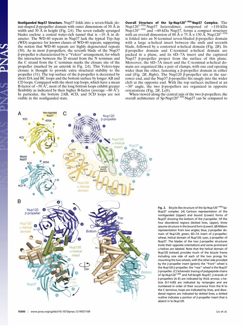
Nonliganded Nup37 Structure. Nup37 folds into a seven-blade do-nut-shaped β-propeller domain with outer dimensions of 50 Å inwidth and 30 Å in height (Fig. 2A). The seven radially arrangedblades enclose a central water-rich tunnel that is ∼10 Å in di-ameter. The WD-40 repeats in Nup37 lack the typical Trp-Asp(WD) sequence for known classes of WD-40 repeats, supportingthe notion that WD-40 repeats are highly degenerated repeats(30). As in most β-propellers, the seventh blade of the Nup37β-propeller is characterized by a “Velcro” arrangement, for whichthe interaction between the D strand from the N terminus andthe C strand from the C terminus marks the closure site of thepropeller (marked by an asterisk in Fig. 2A). This Velcro-typeclosure is thought to provide extra structural stability to thepropeller (31). The top surface of the β-propeller is decorated byshort DA and BC loops and the bottom surface by longer AB andCD loops. Compared with the short top loops, which have a meanB-factor of ∼50 Å2, most of the long bottom loops exhibit greaterflexibility as indicated by their higher B-factor (average ∼80 Å2).In particular, the bottom 2AB, 4CD, and 5CD loops are notvisible in the nonliganded state.
Overall Structure of the Sp-Nup1201–950·Nup37 Complex. TheNup1201–950·Nup37 heterodimer, composed of ∼110-kDaNup1201–950 and ∼40-kDa Nup37, forms a compact structurewith an overall dimension of 80 Å × 75 Å × 130 Å. Nup1201–950
is folded into an N-terminal seven-bladed β-propeller domainwith a large α-helical insert between the sixth and seventhblade, followed by a contorted α-helical domain (Fig. 2B). Itsβ-propeller domain and C-terminal α-helical domain arepacked in a plane, and its 6D–7A insert and the capturedNup37 β-propeller project from the surface of this plane.Moreover, the 6D–7A insert and the C-terminal α-helical do-main are organized like a pair of clamps, with one end openingwider than the other, fastening a β-propeller domain in eitherend (Fig. 2B, Right). The Nup120 β-propeller sits at the nar-rower end, and the Nup37 β-propeller fits snugly into the widercleft at the opposite end. With the top surfaces inclined at an∼30° angle, the two β-propellers are organized in oppositeorientations (Fig. 2B, Left).When viewed along the central axis of the two β-propellers, the
overall architecture of Sp-Nup1201–950·Nup37 can be compared to
Fig. 2. Bicycle-like structure of the Sp-Nup1201–950·Sp-Nup37 complex. (A) Cartoon representation of thenonliganded (Upper) and bound (Lower) forms ofNup37 showing the bottom of the β-propeller. Of thefour disordered regions (dotted lines, Upper), threeassume structure in thebound form (Lower). (B) Ribbonrepresentation from two angles; blue, β-propeller do-main of Nup120; green, 6D–7A insert of β-propeller;wheat, helical domain of Nup120; cyan, β-propeller ofNup37. The blades of the two β-propeller structures(note their opposite orientation) and some prominentα-helices are labeled. Note that the helical domain ofNup120 (wheat) provides much of the bicycle frameincluding one side of each of the two prongs formounting the twowheels,with theother side providedby the β-propeller insert (green); the “front” wheel isthe Nup120 β-propeller; the “rear”wheel is the Nup37β-propeller. (C) Schematic tracingof polypeptide chainsof Sp-Nup1201–950 and full-length Nup37; β-strands ofβ-propellers (A–E) are indicated by thick arrows; α-he-lices (h1–h30) are indicated by rectangles and arenumbered in order of their occurrence from the N tothe C terminus; loops are indicated by lines, and disor-dered regions are indicated by dotted lines; a dottedoutline indicates a portion of β-propeller insert that isabsent in Sc-Nup120.
16500 | www.pnas.org/cgi/doi/10.1073/pnas.1214557109 Liu et al.

a bicycle, in which Sp-Nup1201–950 supplies a front wheel (itsβ-propeller) and the main frame plus the prongs for the rearwheel (Fig. 2B, Left). The rear wheel is furnished by Nup37.Schematic tracings of the polypeptide chains of Nup1201–950 andof full-length Nup37 are shown in Fig. 2C.
Sp-Nup1201–950 Structure.Comparedwith the four-strand-per-bladearrangement seen in conventional β-propeller domains, the firstblade of the Nup120 β-propeller is characterized uniquely by an
extra β-strand (β1E) running parallel to the β1D strand at the outersurface. This feature also is present in the Sc-Nup120 propeller (22,28). Following this β1E strand, helix h2 (spanning residues 44–55,labeled in Fig. 2B) protrudes from the top surface, running roughlyparallel to the central axis of the β-propeller.Another distinct feature for Sp-Nup120 is a prominent insert
comprising helices h5–h9 (spanning residues 395–510) betweenthe sixth and seventh blade of its β-propeller domain (Fig. 2 B andC). Notably, the 1E–1A helix h2 runs antiparallel to h6 of the 6D–
Fig. 3. Detailedanalyses of the Sp-Nup1201–950·Nup37interactome. Interactions of specific residues by hydro-gen bonds (dotted lines), van der Waals interaction(thin lines), and electrostatic interactions (blue lines) areindicated. For reference, a boxed region of the cartoonstructure (a miniature equivalent of Fig. 2A, Right) isindicated on the left.
Fig. 4. Structural comparison of Sp-Nup1201–950 and Sc-Nup1201–729. The two sequences were aligned according to their secondary structures; residuenumbers are indicated. Identical residues are shaded in blue; an inserted stretch of about 50 residues in Sp-Nup120 is shaded in pink. Indicated secondarystructural elements for Sc-Nup120 are based on the crystal structure determined at 2.6-Å resolution [PDB ID:3F7F (22)]. Color codes are as in Fig. 1A. Secondarystructural elements are indicated: Arrows indicate β-strands numbered according to their location in the seven-bladed β-propeller; boxes indicate α-helicesnumbered in order from the N terminus to the C terminus; lines indicate loops, and dotted lines indicate disordered regions. The locations of residues of theSp-Nup120 interacting with Sp-Nup37 are indicated by green asterisks in the insert domain of the β-propeller and by wheat-colored circles in helical domain.
Liu et al. PNAS | October 9, 2012 | vol. 109 | no. 41 | 16501
BIOCH
EMISTR
Y

7A insert (Fig. 2B, Right) and is closely embraced by helices h6,h7, and h9. As a result, helix h2 and the five-helix insert fold intoa distinct helical bundle domain (Fig. 2B and Fig. S2).The C-terminal α-helical domain (spanning residues 544–950)
adopts an L-shaped structure composed of 21 α helices (h10–h30), with a bulging arm comprising helices h10–h22 (residues548–800) and a slender arm furnished by helices h23–h30 (resi-dues 810–950) (Fig. S3). The bulging arm is centered ona prominent hydrophobic helix hairpin, helices h17 and h19, withlengths of ∼50 Å and ∼34 Å, respectively. Helices h10–h16 coillike a snake around the base of the helix hairpin, distal to the Sp-Nup120 β-propeller domain, and the upper half of the helixhairpin packs closely against helices h20–h22. As a result, thishelix hairpin is almost fully buried and forms a prominent hy-drophobic core (Fig. 2B and Fig. S3).The slender arm of the L is formed by helices h23–h30 (Fig. 2 B
andC and Fig. S3). This region represents an extension of the helicalregion as compared with the previously reported Sc-Nup1201–729structure (Fig. 4). Helices h23–h30 are organized roughly into fourantiparallel pairs with h27 off pitch, stacking with h28 at a 45° angle.As a result, helices h23–h30 form a crescent shape with its convexsurface contacting Nup37 and the concave side surface largely oc-cupied by helix h27. Overall, these eight helices extend ∼40 Å inthe direction perpendicular to h17 (Fig. S3).
Structure of Sp-Nup120–Bound Nup37. Upon Sp-Nup120 binding,although the overall structure of Nup37 remains largely the same(the two structures could be aligned with an rmsd of 0.5 Å for371 Cα atoms), some disordered regions in nonliganded Nup37adopt an α-helical or loop structure (Fig. 2A and Fig. S4). Spe-cifically, on the bottom surface of Nup37, the 4CD and the 5CDloops are in close vicinity to the loop immediately ahead of helixh5 and of the extended h8–h9 loop region of the Sp-Nup120 6D–7A insert, respectively. On the top surface of Nup37, Ile251 of the4D-5A loop makes van der Waals contacts with Glu743 andLys744 in the loop following h19 of the Sp-Nup120 helical do-main (Figs. 3 and 4). It should be noted that the N-terminal fiveresidues of Nup37, instead of forming an extended β-strandpacked against the sixth blade of Nup37, become flexible uponSp-Nup120 binding, possibly because of a lack of crystal-packing contacts.
Sp-Nup120–Nup37 Interface. Sp-Nup120 interacts with Nup37through a concave surface contributed by the 6D–7A insert andthe α-helical domain of Sp-Nup120. The total buried surfacearea for the heterodimer is ∼4,900 Å2, with ∼2,100 Å2 of theburied surface area contributed by the interface between the 6D–7A insert and Nup37 (labeled “A” and “A′” in Fig. S5B) and theremaining ∼2,800 Å2 contributed by the interface between theα-helical domain and Nup37 (labeled “B” and “B′” in Fig. S5B).A detailed schematic view of the heterodimer interface is shownin Fig. 3. Binding of Nup37 to the enlarged 6D–7A insert of Sp-Nup120 is primarily through hydrophobic contacts and a fewhydrogen bonds, whereas the interface between Nup37 and thehelical domain of Nup120 involves, in addition, several electro-static interactions (Figs. 3 and 4 and Fig. S5D). Within this ex-tensive interface, some prominent hydrophobic centers are highlyconserved across species from S. pombe to human (Fig. S5C). Forexample, in the α-helical region, Phe136 of Nup37 stacks closelywith the side chain of Lys857 from Sp-Nup120. Other residuesfrom Sp-Nup120 (including Tyr849 and Cys861) and those fromNup37 (e.g., Thr165, Asn138, Asp162, and Asp163) furtherstrengthen the interaction. On the distal end of the curved in-terface of the helical bundle region, a prominent hydrophobiccore is formed centering on residues Phe283, Ile291, and Leu282(contributed by Nup37) and Tyr491, Tyr489, and Pro482 (con-tributed by Sp-Nup120). This extensive binding interface explainswhy the heterodimer resists high-salt (1 M NaCl, 1 M urea) orlow-salt challenge.
Structural Comparison of Sp-Nup120 and Sc-Nup120. The sevenblades of Sc-Nup120 and Sp-Nup120 propellers and most of theprominent helices in the α-helical regions in both structures can
be well aligned. However, two key regions of Sp-Nup120 standout as exceptions and ensnare Sp-Nup37: the enlarged 6D–7Ainsert of the Sp-Nup120 β-propeller (dotted outline in Fig. 2C)and the expanded C-terminal helical domain (Fig. 2B). Sp-Nup120’s 6D–7A insert contains ∼50 more amino acid residuesthan the 70-residue-long 6D–7A insert in Sc-Nup120. The en-largement of the helical insert of the Sp-Nup120 β-propellergives rise to two additional helices (h7 and h8) and an ∼21-Åelongation of helix h9 (Figs. 4 and 5). More importantly, helicesh8 and h9 and the loop connecting h8 and h9 make extensivecontacts with the fourth and fifth blades of the Nup37 propeller(Figs. 2B and 3).Comparison of the previously reported Sc-Nup1201–729 struc-
ture (22, 28) and the Sp-Nup1201–950 structure reveals sevenadditional α-helices at the C terminus of Sp-Nup120 (Fig. 4).This extended helical region of Sp-Nup120 harbors a series ofresidues distributed along helices h24–h26 and h29 that contactNup37 (Figs. 2–5). The structure of the corresponding region ofSc-Nup120 has not been determined. Based on the observationthat Sc-Nup120 alone is not sufficient to bind Nup37 (Fig. 1C),we suggest that Nup120 orthologs of S. pombe and Metazoaspecifically use features of these C-terminal α-helices as a partialbinding platform for Nup37. We also envisage that the remainingNup37 binding sites within the Sp-Nup120 β-propeller evolvedthrough the expansion of the 6D–7A insert (Fig. 5) by its in-creased contacts with Nup37. This expansion is conserved inNup120 orthologs that are competent to bind Nup37 (Fig. S6).
DiscussionWe report the atomic structures of a complex of two nucleoporins,Nup1201–950 and full-lengthNup37, aswell as of nonligandedNup37,all of S. pombe. These structures provide information not only aboutthe evolution of the coatomer module of the NPC but also aboutstructural changes that accompany the posttranslational assembly ofNup120·Nup37 as part of a hetero-octameric coatomer module.Localized adjacent to the cylindrical pore membrane with its
distinct embedded POMs, the coatomer module of the NPC un-derwent evolution toward higher complexity: A hetero-heptamerin S. cerevisiae evolved into a hetero-octamer in S. pombe and into a
Fig. 5. Superimposed structures of Sp-Nup1201–950·Nup37 and Sc-Nup1201–729.(A) Overlay of the Sp-Nup120 and Sc-Nup120 structures. (B) A 50°-rotated viewis shown. β-Propellers (pink for S. cerevisiae and blue for S. pombe) are shownin the Cα skeleton model except for cartoon representations for 1E–1A helix h2(blue) and helices in the 6D–7A insert (orange for S. cerevisiae and green forS. pombe). Prominent helices are labeled for Sp-Nup120 (lower case) and Sc-Nup120 (upper case). Helical domains are depicted in cartoonmodel (wheat forS. pombe and pink for S. cerevisiae). Blades of both β-propellers and somehelices of inserts and the helical domain are labeled. Nup37 is shown as surfacemodel in light gray. A 21-Å extension of h9 in Sp-Nup120 is indicated byan arrow.
16502 | www.pnas.org/cgi/doi/10.1073/pnas.1214557109 Liu et al.
hetero-nonamer in Metazoa. The heptamer-to-octamer or -non-amer transition involves the acquisition of Nup37 or of Nup37 andNup43, respectively. Although most analyses thus far have beencarried out with the S. cerevisiae hetero-heptamer, the constituentmembers of the coatomer module of other species generallyare identifiable by sequence homologies (32); most likely, allmodules also share a similar (electron microscopically defined)Y-shaped structure.Our biochemical studies together with structural analyses shed
light on the evolution of the Y-shaped complex. We first bio-chemically identified Sp-Nup120 as the binding partner forNup37 (Fig. 1). The structure of full-length Nup37 furtherrevealed that it folds into a canonical seven-bladed β-propellerthat showed several unstructured regions on its surface. Some ofthese regions become structured upon binding to Sp-Nup120.Immediately after cellular synthesis, these regions may be pro-tected temporarily by chaperones to allow an ordered assemblywith Sp-Nup120 as well as with other surrounding nucleoporinsor POMS. These unstructured regions increase surface entropyand pose problems for crystallization but likely endow thenucleoporin interactome with considerable plasticity to accom-modate transport substrates varying greatly in size.The overall architecture of the Sp-Nup1201–950·Nup37 com-
plex resembles the shape of a bicycle, to which Nup37 providesonly the rear wheel and Sp-Nup1201–950 contributes the mainframe and the front wheel (Fig. 2B, Left). However, the Sp-Nup120–derived front wheel, also a seven-bladed β-propeller, isdistinguished from Sc-Nup120 by an extension of about 50 res-idues in its 6D–7A insert. Strikingly, this 50-residue extensionprovides numerous contact sites (Figs. 3 and 4) to fasten the rearwheel and is conspicuously absent in S. cerevisiae but is present inS. pombe and Metazoa (33). Another noteworthy differencebetween the Sc- and Sp-Nup120 β-propellers is the presence inSp-Nup120 β-propellers of 1E–1A helix h2 (Figs. 2 and 4 andFig. S2), which projects from the β-propeller and stabilizes theadjacently located 50-residue extension. The equivalent region isdisordered in the crystal structure of Sc-Nup120 (22, 28).
Other principal contact sites for the Nup37 β-propeller arecontributed primarily by the C-terminal portion of the α-helicaldomain of Sp-Nup1201–950 (Figs. 3 and 4). Notably, the pre-viously determined crystal structure of the Sc-Nup1201-729 frag-ment lacked the equivalent region (refs. 22, 28 and Figs. 1 and 4).However, our finding that Sc-Nup120 does not bind to Sp-Nup37in vitro suggests that in Sc-Nup120 this region may have evolvedto exert other, yet to be determined, functions.Because β-propellers are classical interaction platforms for
other cofactors, the capture of Nup37 may facilitate a more ex-tensive integration of the coatomer module of the NPC into thenetwork of surrounding nucleoporins and POMs (Introduction).The addition of the Nup37-binding platform to the NPC ispredicted to give rise to additional functional properties of theNPC not present in S. cerevisiae. Determining what these prop-erties might be must await the identification and characterizationof other binding partners of Nup37. However, because S. pombe,like S. cerevisiae, undergoes closed mitosis, the acquisition ofNup37 in S. pombe (or its loss in S. cerevisiae) is presumed not torelate to the reversible NPC disassembly into subcomplexes thatoccurs in open mitosis, where the coatomer, other disassemblednucleoporins, and transport factors function in other capacities,such as the assemblies of kinetochores and spindles (34–36).
Materials and MethodsDetailed descriptions of protein expression, purification, crystallization, X-ray diffraction data collection, structure determination, and biochemicalanalyses are provided in SI Materials and Methods. In short, recombinantproteins were expressed in insect cells with an N-terminal cleavable His-tag.The proteins were purified by multiple rounds of chromatography approaches.The Sp-Nup37 structure was solved by SAD method. The Nup120.Nup37complex structure was solved by MR-SAD method.
ACKNOWLEDGMENTS. We thank Dr. David King (University of California, Ber-keley, CA) for protein analysis, Dr.Wuxian Shi (National Synchrotron Light Source,X29) for support duringdata collection, Dr. ValérieDoye (Université Paris Diderot,Paris) for providing Sp-Nup120 cDNA, and Dr. Shigehiro Kawashima (The Rocke-feller University, New York) for providing S. pombe culture.
1. Blobel G (1980) Intracellular protein topogenesis. Proc Natl Acad Sci USA 77:1496–1500.
2. Devos D, et al. (2004) Components of coated vesicles and nuclear pore complexesshare a common molecular architecture. PLoS Biol 2:e380.
3. Mans BJ, Anantharaman V, Aravind L, Koonin EV (2004) Comparative genomics, evolutionand origins of the nuclear envelope and nuclear pore complex. Cell Cycle 3:1612–1637.
4. Davis LI, Blobel G (1986) Identification and characterization of a nuclear pore complexprotein. Cell 45:699–709.
5. Cronshaw JM, Krutchinsky AN, Zhang W, Chait BT, Matunis MJ (2002) Proteomicanalysis of the mammalian nuclear pore complex. J Cell Biol 158:915–927.
6. Rout MP, et al. (2000) The yeast nuclear pore complex: Composition, architecture, andtransport mechanism. J Cell Biol 148:635–651.
7. Unwin PN, Milligan RA (1982) A large particle associated with the perimeter of thenuclear pore complex. J Cell Biol 93:63–75.
8. Gall JG (1967) Octagonal nuclear pores. J Cell Biol 32:391–399.9. Jarnik M, Aebi U (1991) Toward a more complete 3-D structure of the nuclear pore
complex. J Struct Biol 107:291–308.10. Solmaz SR, Chauhan R, Blobel G, Mel�cák I (2011) Molecular architecture of the
transport channel of the nuclear pore complex. Cell 147:590–602.11. Melcák I, Hoelz A, Blobel G (2007) Structure of Nup58/45 suggests flexible nuclear
pore diameter by intermolecular sliding. Science 315:1729–1732.12. Radu A, Moore MS, Blobel G (1995) The peptide repeat domain of nucleoporin Nup98
functions as a docking site in transport across the nuclear pore complex. Cell 81:215–222.
13. Chook YM, Blobel G (2001) Karyopherins and nuclear import. Curr Opin Struct Biol 11:703–715.
14. Cook A, Bono F, Jinek M, Conti E (2007) Structural biology of nucleocytoplasmictransport. Annu Rev Biochem 76:647–671.
15. Hoelz A, Debler EW, Blobel G (2011) The structure of the nuclear pore complex. AnnuRev Biochem 80:613–643.
16. Aitchison JD, Rout MP (2012) The yeast nuclear pore complex and transport throughit. Genetics 190:855–883.
17. Strambio-De-Castillia C, Niepel M, Rout MP (2010) The nuclear pore complex: Bridgingnuclear transport and gene regulation. Nat Rev Mol Cell Biol 11:490–501.
18. Kampmann M, Blobel G (2009) Three-dimensional structure and flexibility ofa membrane-coating module of the nuclear pore complex. Nat Struct Mol Biol 16:782–788.
19. Lutzmann M, Kunze R, Buerer A, Aebi U, Hurt E (2002) Modular self-assembly of a Y-shaped multiprotein complex from seven nucleoporins. EMBO J 21:387–397.
20. Siniossoglou S, et al. (2000) Structure and assembly of the Nup84p complex. J Cell Biol149:41–54.
21. Brohawn SG, Schwartz TU (2009) Molecular architecture of the Nup84-Nup145C-Sec13 edge element in the nuclear pore complex lattice. Nat Struct Mol Biol 16:1173–1177.
22. Seo HS, et al. (2009) Structural and functional analysis of Nup120 suggests ring for-mation of the Nup84 complex. Proc Natl Acad Sci USA 106:14281–14286.
23. Hsia KC, Stavropoulos P, Blobel G, Hoelz A (2007) Architecture of a coat for the nu-clear pore membrane. Cell 131:1313–1326.
24. Debler EW, et al. (2008) A fence-like coat for the nuclear pore membrane.Mol Cell 32:815–826.
25. Nagy V, et al. (2009) Structure of a trimeric nucleoporin complex reveals alternateoligomerization states. Proc Natl Acad Sci USA 106:17693–17698.
26. Fontoura BM, Blobel G, Matunis MJ (1999) A conserved biogenesis pathway for nu-cleoporins: Proteolytic processing of a 186-kilodalton precursor generates Nup98 andthe novel nucleoporin, Nup96. J Cell Biol 144:1097–1112.
27. Loïodice I, et al. (2004) The entire Nup107-160 complex, including three new mem-bers, is targeted as one entity to kinetochores in mitosis. Mol Biol Cell 15:3333–3344.
28. Leksa NC, Brohawn SG, Schwartz TU (2009) The structure of the scaffold nucleoporinNup120 reveals a new and unexpected domain architecture. Structure 17:1082–1091.
29. Adams PD, et al. (2010) PHENIX: A comprehensive Python-based system for macro-molecular structure solution. Acta Crystallogr D Biol Crystallogr 66:213–221.
30. van der Voorn L, Ploegh HL (1992) The WD-40 repeat. FEBS Lett 307:131–134.31. Chaudhuri I, Söding J, Lupas AN (2008) Evolution of the β-propeller fold. Proteins 71:
795–803.32. DeGrasse JA, et al. (2009) Evidence for a shared nuclear pore complex architecture
that is conserved from the last common eukaryotic ancestor. Mol Cell Proteomics 8:2119–2130.
33. Neumann N, Lundin D, Poole AM (2010) Comparative genomic evidence for a com-plete nuclear pore complex in the last eukaryotic common ancestor. PLoS ONE 5:e13241.
34. Zuccolo M, et al. (2007) The human Nup107-160 nuclear pore subcomplex contributesto proper kinetochore functions. EMBO J 26:1853–1864.
35. Orjalo AV, et al. (2006) The Nup107-160 nucleoporin complex is required for correctbipolar spindle assembly. Mol Biol Cell 17:3806–3818.
36. Mishra RK, Chakraborty P, Arnaoutov A, Fontoura BM, Dasso M (2010) The Nup107-160 complex and gamma-TuRC regulate microtubule polymerization at kinetochores.Nat Cell Biol 12:164–169.
Liu et al. PNAS | October 9, 2012 | vol. 109 | no. 41 | 16503
BIOCH
EMISTR
Y