structural insight into selectivity and resistance ... · structural insight into selectivity and...
TRANSCRIPT

Structural insight into selectivity and resistanceprofiles of ROS1 tyrosine kinase inhibitorsMonika A. Davarea,b,1, Nadeem A. Vellorec,d,1, Jacob P. Wagnera,b,1, Christopher A. Eidee,f,1, James R. Goodmane,Alexander Drilong, Michael W. Deiningerc,d, Thomas O’Harec,d,2,3, and Brian J. Drukere,f,2,3
aKnight Cancer Institute, Oregon Health & Science University, Portland, OR 97239; bDepartment of Pediatrics, Oregon Health & Science University, Portland,OR 97239; cHuntsman Cancer Institute, The University of Utah, Salt Lake City, UT 84112; dDivision of Hematology and Hematologic Malignancies, TheUniversity of Utah, Salt Lake City, UT 84112; eDivision of Hematology and Medical Oncology, Knight Cancer Institute, Oregon Health & Science University,Portland, OR 97239; fHoward Hughes Medical Institute, Portland, OR 97239; and gMemorial Sloan Kettering Cancer Center, New York, NY 10065
Contributed by Brian J. Druker, August 5, 2015 (sent for review April 10, 2015; reviewed by Nicholas Levinson and Ariel Lopez-Chavez)
Oncogenic ROS1 fusion proteins are molecular drivers in multiplemalignancies, including a subset of non-small cell lung cancer (NSCLC).The phylogenetic proximity of the ROS1 and anaplastic lymphomakinase (ALK) catalytic domains led to the clinical repurposing of theFood and Drug Administration (FDA)-approved ALK inhibitor crizoti-nib as a ROS1 inhibitor. Despite the antitumor activity of crizotinibobserved in both ROS1- and ALK-rearranged NSCLC patients, resis-tance due to acquisition of ROS1 or ALK kinase domain mutations hasbeen observed clinically, spurring the development of second-gener-ation inhibitors. Here, we profile the sensitivity and selectivity ofseven ROS1 and/or ALK inhibitors at various levels of clinical devel-opment. In contrast to crizotinib’s dual ROS1/ALK activity, cabozanti-nib (XL-184) and its structural analog foretinib (XL-880) demonstrate astriking selectivity for ROS1 over ALK. Molecular dynamics simulationstudies reveal structural features that distinguish the ROS1 and ALKkinase domains and contribute to differences in binding site andkinase selectivity of the inhibitors tested. Cell-based resistanceprofiling studies demonstrate that the ROS1-selective inhibitorsretain efficacy against the recently reported CD74-ROS1G2032R mu-tant whereas the dual ROS1/ALK inhibitors are ineffective. Takentogether, inhibitor profiling and stringent characterization of thestructure–function differences between the ROS1 and ALK kinasedomains will facilitate future rational drug design for ROS1- andALK-driven NSCLC and other malignancies.
ROS1 | ALK | kinase | inhibitor | structural modelling
Constitutively activated kinase fusion proteins that arise fromsomatic chromosomal rearrangements are frequent drivers of
malignant transformation in cancer and represent a targetable vul-nerability for clinical intervention. The clinical success of the tyrosinekinase inhibitor (TKI) imatinib in targeting the oncogenic BCR-ABL1 fusion protein in chronic myeloid leukemia (CML) motivatedefforts to identify and target oncogenic kinases in other cancers(1–3). One such setting is non-small cell lung cancer (NSCLC),where chromosomal rearrangements of the receptor tyrosine kinase(RTK) anaplastic lymphoma kinase (ALK) are found in 4–5% ofpatients (4, 5). The validation of rearranged ALK as an oncogenicdriver prompted the discovery and clinical implementation of cri-zotinib as the first clinical targeted inhibitor for use in ALK fusion-positive NSCLC (6, 7).Fusion proteins involving the highly related kinase ROS1, an
orphan RTK of the insulin receptor family, are present in ∼1%of NSCLC patients. ROS1 rearrangements span a variety of fu-sion partners across several other epithelial malignancies, in-cluding cholangiocarcinoma, gastric cancer, and ovarian cancer(4, 8). CD74-ROS1 is the most frequent ROS1 fusion detected inNSCLC. ROS1 fusion proteins are transforming drivers thatcontribute to tumorigenesis or tumor progression in multipleexperimental model systems (9–11).Approximately 75,000 and 15,000 new NSCLC patients per
year are anticipated to harbor tumors driven by rearranged ALKor ROS1, respectively. Although mutually exclusive in a given
tumor and considered to be distinct molecular subgroups (12),patients presenting with ROS1 or ALK fusion-driven lung cancershare clinical features, tend to be younger compared with otherNSCLC patients, and have little to no history of smoking. Thekinase domains of ROS1 and ALK display a high degree of se-quence homology (13), prompting investigation of crizotinib foractivity against ROS1 (12). Recent phase I data confirmed sig-nificant responses to crizotinib in ROS1-rearranged NSCLCpatients (14, 15).Despite the demonstrated clinical efficacy of TKI-based targeted
therapies in cancer, a universal challenge is emergence of drugresistance and ensuing disease progression. Resistance commonlyinvolves either acquisition of kinase domain mutations that com-promise inhibitor binding or activation of alternative pathways thatprovide compensatory cell survival signals. Concordant with pre-vious clinical experience from other TKIs, such as imatinib in CMLand erlotinib or gefitinib in lung cancer (16–18), resistance dueto acquisition of kinase domain mutations is frequently observed
Significance
Targeting oncogenic ROS1 fusion proteins with crizotinib hasshown promising clinical outcomes in non-small cell lung cancer(NSCLC) patients, but emergence of resistance to therapy hasbeen reported. By profiling the activity of clinically viable ROS1/anaplastic lymphoma kinase (ALK) inhibitors, we discovered thatthe Food and Drug Administration (FDA)-approved inhibitorcabozantinib potently inhibits native ROS1 and the crizotinib-resistant ROS1G2032R mutant, suggesting potential utility fortreatment of ROS1-rearranged lung cancer. Notably, cabozanti-nib is ineffective against the closely related ALK kinase. Molec-ular modeling shows specific structural differences between thekinase domains of ROS1 and ALK that explain selective bindingof cabozantinib to ROS1. These findings reveal limitations per-taining to the widely presumed inhibitory reciprocity of ROS1and ALK inhibitors and may facilitate rational design of newROS1-selective inhibitors.
Author contributions: M.A.D., N.A.V., C.A.E., T.O., and B.J.D. designed research; M.A.D.,N.A.V., J.P.W., C.A.E., and J.R.G. performed research; M.A.D., N.A.V., and J.P.W. contrib-uted new reagents/analytic tools; M.A.D., N.A.V., J.P.W., C.A.E., and T.O. analyzed data;and M.A.D., N.A.V., C.A.E., A.D., M.W.D., T.O., and B.J.D. wrote the paper.
Reviewers: N.L., University of Minnesota; and A.L.-C., University of Miami.
Conflict of interest statement: The Oregon Health & Science University has clinical trial contractswith Novartis and Bristol-Myers Squibb (BMS) to pay for patient costs, nurse and data managersalaries, and institutional overhead. B.J.D. does not derive salary, nor does his laboratory receivefunds, from these contracts. M.W.D. served on advisory boards and as a consultant for BMS,ARIAD, and Novartis.
Freely available online through the PNAS open access option.1M.A.D., N.A.V., J.P.W., and C.A.E. contributed equally to this work.2T.O. and B.J.D. contributed equally to this work.3To whom correspondence may be addressed. Email: [email protected] or [email protected].
This article contains supporting information online at www.pnas.org/lookup/suppl/doi:10.1073/pnas.1515281112/-/DCSupplemental.
www.pnas.org/cgi/doi/10.1073/pnas.1515281112 PNAS | Published online September 8, 2015 | E5381–E5390
BIOCH
EMISTR
YPN
ASPL
US

in crizotinib-treated NSCLC patients harboring ALK fusions (7,19–21). This experience has prompted the development of sev-eral second-generation ALK inhibitors capable of circumventingresistance. Furthermore, compared with ALK-rearranged lungcancers, ROS1-rearranged cancers are less frequent, and clinicalbenefit with crizotinib may be more durable. The median pro-gression-free survival for crizotinib-treated patients from phase Ievaluation of ROS1-rearranged NSCLC is 19.2 mo (15), com-pared with 7.7 mo for patients with ALK-rearranged disease(phase III data) (14). Given these factors, mechanisms of ac-quired resistance to crizotinib in the clinic may take longer toidentify, and only the CD74-ROS1G2032R mutation has beenreported to date (22).
We have previously reported that foretinib (XL-880) is apotent ROS1 inhibitor that retains efficacy against the crizo-tinib-resistant CD74-ROS1G2032R mutant in cell-based assays(23). In contrast to crizotinib’s dual ROS1/ALK efficacy, weobserved that foretinib is a poor ALK inhibitor. These findingsestablish that not all ROS1 inhibitors possess inhibitory reci-procity for ALK, and drug discovery efforts that use one kinaseas a proxy for the other may face limitations.Here, we report in vitro profiling of a panel of clinically relevant
ROS1 and ALK inhibitors. To complement cell-based resistanceprofiling of ROS1-selective inhibitors, structural comparison ofthe kinase domains of ROS1 and ALK and computational mod-eling of TKI binding to native and mutant kinase domains were
Table 1. Summary of ALK and ROS1 tyrosine kinase inhibitors in clinical development
E5382 | www.pnas.org/cgi/doi/10.1073/pnas.1515281112 Davare et al.

also performed. These results provide insights into therapeuticallyexploitable structural differences between ROS1 and ALK thatimpact inhibitor binding and design.
ResultsIn Vitro Profiling Reveals Differences in TKI Selectivity for ROS1 VersusALK.Given the clinical success with crizotinib in ALK fusion-drivenNSCLC and the fact that the ROS1 and ALK kinase domainsdisplay high sequence homology, there is an operating assumptionthat ALK inhibitors can be repurposed as ROS1 inhibitors (13).However, comprehensive sensitivity profiling of first- and second-generation ALK inhibitors against native and crizotinib-resistantROS1 has not been undertaken. We screened a panel of sevenTKIs at varying stages of clinical development (Table 1) againstBa/F3 cells transformed with CD74-ROS1 or EML4-ALK. Cabo-zantinib and foretinib both demonstrated a high degree of selec-tivity for ROS1 compared with ALK, potently inhibiting the growthof CD74-ROS1 cells (IC50 of 1.1 nM and 1.8 nM, respectively)while exhibiting minimal effect on EML4-ALK cells at the highestconcentration tested (2,500 nM) (Fig. 1A). Conversely, althoughBa/F3 EML4-ALK cells were confirmed to be sensitive to in-hibition by alectinib (IC50 of 12.3 nM), CD74-ROS1 cells werealectinib-insensitive (IC50 of 1,950 nM). The remaining TKIs [cri-zotinib, brigatinib (formerly AP26113), ceritinib, AZD3463)exhibited varying levels of inhibition for CD74-ROS1 and EML4-ALK cells, with brigatinib and AZD3463 displaying near equi-
potency for both (IC50 of 7.5 vs. 9.8 nM and 10.2 vs. 39.4 nM,respectively) (Fig. 1A). These results establish three categories withrespect to ROS1 and ALK inhibitor selectivity: ROS1-selective(cabozantinib, foretinib), dual ROS1/ALK (crizotinib, brigatinib,ceritinib, AZD3463), and ALK-selective inhibitors (alectinib) (Fig.1B and Table S1).Consistent with findings from cell proliferation assays, immu-
noblot analysis after short-term treatment of Ba/F3 CD74-ROS1cells with ROS1-selective and dual ROS1/ALK TKIs demon-strated concentration-dependent effects on ROS1 tyrosine phos-phorylation (Y2274) (Fig. 1C). Furthermore, although significantinhibition of ALK tyrosine phosphorylation (Y1278) occurred ina concentration-dependent manner in Ba/F3 EML4-ALK cellstreated with ALK-selective and dual ROS1/ALK TKIs, cabo-zantinib and foretinib did not reduce ALK phosphorylation atconcentrations up to 250 nM (Fig. 1C). Comparable selectivityfindings were observed in the human NSCLC cell lines HCC78and H3122, which harbor native SLC-ROS1 and EML4-ALK fu-sions, respectively. Downstream effector pathways (MAPK andAKT) activated by oncogenic ROS1 and ALK were also sup-pressed in a concentration-dependent manner (Fig. S1). Thesedata imply structural differences between the ROS1 and ALKkinase domains that dictate selectivity and efficacy of TKI binding.
A B
C
Cell v
iabilit
y(%
of un
treate
d)
CD74-ROS1G2032R
0.1 1 10 100 1000Concentration (nM)
020406080
100120 Crizotinib
Foretinib
Alectinib
CeritinibBrigatinib
AZD3463
Cabozantinib
CD74-ROS1 CD74-ROS1G2032R
Crizotinib
Foretinib
Cabozantinib
BrigatinibCeritin
ib
AZD3463Alectin
ib0.1
10100
100010000
IC50
(nM)
1
CD74
-ROS
1G203
2R
Vehic
le
nMCrizo
tinib
Foretini
b
Cabozan
tinib
Brigatini
b
Ceritinib
AZD3463
GAPDHpROS1
D
0 0.05
0.1 0.15
0 0.05
0.1 0.15
−12 −10 −8 −6 −4 −2
ROS1G2032R
Foretinib
ROS1G2032R
ROS1 ROS1ROS1Crizotinib
ROS1G2032R
CabozantinibE
Prob
abilit
y
−12 −10 −8 −6 −4 −2 −12 −10 −8 −6 −4 −2Docking score (kcal/mol)
ROS1-selective Dual ROS1/ALK ALK-
selective
101001010010100 10100 10100 10100
Fig. 2. ROS1-selective TKIs retain efficacy against the crizotinib-resistantROS1G2032R mutant. (A) Dose–response curves for proliferation of Ba/F3 CD74-ROS1G2032R cells after 72-h exposure to varying concentrations of cabozantinib,foretinib, crizotinib, brigatinib, ceritinib, AZD3634, and alectinib. Data arenormalized to vehicle-treated cells, and values shown are the mean ± SEM.(B) Scatter plot of cell proliferation IC50 values for each of the indicated TKIsagainst Ba/F3 cells expressing native CD74-ROS1 (blue) and CD74-ROS1G2032R
(green). Categories of selectivity profile are indicated above the plot. (C) Im-munoblot analysis of phospho-ROS1 from Ba/F3 CD74-ROS1G2032R cells aftertreatment with the indicated TKIs. (D) Overlay of crizotinib docking fromsimulated models with the actual ROS1:crizotinib complex. (E) Docking scorehistograms for native ROS1 and ROS1G2032R for crizotinib, foretinib, andcabozantinib. A threshold docking score of −6 is indicated by the verticaldashed orange line, where scores above or below this value correspond to pooror good binding conformations, respectively.
10000
CD74
-ROS
1A
C
B
CD74-ROS1
0.1 1 10 100 1000
CrizotinibForetinib
AlectinibCeritinibBrigatinib
Concentration (nM)
Cell v
iabilit
y(%
of un
treate
d)
AZD3463EML4-ALK
020406080
100120
0.1 1 10 100 1000Concentration (nM)
020406080
100120
0.1
10100
100010000
CD74-ROS1 EML4-ALK
IC50
(nM)
1
Crizotinib
Foretinib
Cabozantinib
BrigatinibCeritin
ib
AZD3463Alectin
ib
Cabozantinib
ROS1ALK F1174
Q1159
W1313
αC-Helix rotation (°)70 80 90100
300250200150100
50
Pock
et vo
lume (
Å3 )0-1-2-3
300250200150100
50
ROS1
ALK
ROS1-selective Dual ROS1/ALK ALK-
selective
pROS1
GAPDH
pALK
GAPDH
Vehic
le
Cabozantinib2.5 25 250 2.5 25 2502.5 25 250 2.5 25 2502.5 25 250 2.5 25 2502.5 25 250
Foretinib Crizotinib Brigatinib Ceritinib AZD3463 AlectinibnM
EML4
-ALK
D E F G
Fig. 1. Structural differences between the ROS1 and ALK kinase domainsunderlie the differential selectivity of TKIs. (A) Proliferation of Ba/F3 CD74-ROS1and EML4-ALK cells after 72-h exposure to cabozantinib, foretinib, crizotinib,brigatinib, ceritinib, AZD3634, and alectinib. Data are normalized to vehicle-treated control, and values shown are the mean ± SEM. (B) Scatter plot of cellproliferation IC50 values for each TKI against Ba/F3 cells expressing CD74-ROS1(blue) and EML4-ALK (orange). Categories of selectivity profile are indicatedabove the plot. (C) Immunoblot analysis of phospho-ROS1 from TKI-treated Ba/F3CD74-ROS1 cells (Upper) and phospho-ALK from TKI-treated Ba/F3 EML4-ALKcells (Lower). (D) Alignment of ROS1 (blue) and ALK (orange) using structuralhomology (based on Cα atoms). The A-loop is not shown. (E) Surface repre-sentation of ROS1 kinase, with P- and A-loops shown in ribbon representation(yellow). The protein surface is colored based on sequence identity betweenROS1 and ALK kinase, with red for identical sequence and blue for nonidenticalsequence. (F) Ribbon model depicting the rotation of αC-helix in ROS1 and ALK.(G) αC-helix rotation plotted against the volume of the specificity site for ROS1and ALK kinase calculated from molecular dynamic simulation.
Davare et al. PNAS | Published online September 8, 2015 | E5383
BIOCH
EMISTR
YPN
ASPL
US
Structural Differences in the Kinase Domains of ROS1 and ALK UnderlieTKI Selectivity. ROS1 and ALK share >64% overall sequence ho-mology in the kinase domain and ∼84% within the ATP bindingsite. Structural alignment (Fig. 1D and Fig. S2) demonstrated a lowoverall root mean square deviation (rmsd) of 2.3 Å between thetwo kinase domains. However, in comparison with the ATP bind-ing site, the specificity site (defined as the pocket enclosed betweenthe αC-helix and the catalytic DFG loop) (Fig. S3A) in ALK andROS1 showed multiple differences (Fig. 1E). To investigate thesesubtle differences, we performed molecular dynamics (MD) sim-ulations of the ROS1 and ALK kinase domains for 500 ns underexplicit solvent conditions using the catalytically active and inactiveconformations. Throughout this manuscript, “active” refers to theaspartic acid–phenylalanine–glycine (DFG)-in state and “inactive”refers to the DFG-out state of the kinase, irrespective of theconformation of the rest of the A-loop. Both ROS1 and ALK werestable during the course of the simulation, and no large-scaleconformational change was observed (Fig. S2). Root mean squarefluctuation (RMSF) of the protein was also measured from thesimulation (Fig. S3B). The αC-helix was stable in the active con-formation of both ROS1 and ALK whereas flexibility of the P-loopwas slightly higher compared with the corresponding inactiveconformation. Although MD simulation of the active state of ALKwas initialized using the A-loop in the autoinhibitory state, con-formations pertaining to the active A-loop were also explored in-termittently during the course of MD simulation. Based on thesimulations, the A-loop was overall significantly more flexible thanthe other structural elements of the ROS1 and ALK kinase do-mains. The active conformation of ALK showed reduced flexibilityin the A-loop compared with the inactive conformation due tostabilization through a network of aromatic/hydrophobic residues(Y1096, F1098, F1174, F1245, and F1271) at the specificity site(Fig. S3C). In particular, in the active state of ALK, A-loop residueY1278 engages in an aromatic stacking interaction with Y1096,which in turn is internally stabilized by four neighboring phenyl-alanine residues. Conversely, the inactive conformation of ROS1showed less flexibility in the A-loop compared with the activeconformation, largely due to increased proximity of the A-loop toboth the P-loop and the αC-helix compared with ALK (Fig. S3D).Conformational analysis of the specificity site using MD sim-
ulations further distinguished the inactive conformations ofROS1 and ALK with respect to pocket volume and αC-helixorientation (Fig. 1F). Specifically, ROS1 and ALK sampledmarkedly different average pocket volumes (186 Å3 and 135 Å3,respectively) (Fig. 1G), and the positioning of the bulky side-chain of Q2012 in ROS1 close to the C terminus of the αC-helixrestricts the αC-helix from collapsing into the specificity site. Incontrast, the equivalent position in ALK (C1182) lacks suchconstraints, thus reducing pocket volume. Also, in ROS1, theproximity of Q2012 to the αC-helix influences local changes andcontributes to a backbone hydrogen bond observed betweenM2001 and E1997 whereas ALK lacks such an interaction. Also,ROS1 and ALK differ in the identity of amino acid sidechainslining the specificity site pocket, providing an additional sourceof divergence in its size and shape. In particular, ROS1 M2001and L2010 correspond to ALK I1171 and V1180, respectively.These subtle differences in the specificity site could impact theorientation of the αC-helix, as evidenced by the ROS1 αC-helixsampling an average of 88° compared with 75° in ALK (Fig. 1G).These findings suggest that, despite the significant sequence
homology between ROS1 and ALK, differences in the rigidityand orientation of the αC-helix and A-loop contribute to speci-ficity site pocket volume, with the larger pocket in ROS1 capableof accommodating binding of larger scaffolds.
The CD74-ROSG2032R Mutation Confers Resistance to Dual ROS1/ALK TKIsbut Remains Sensitive to the ROS1-Selective Inhibitor Cabozantinib.Emergence of clinical resistance due to acquisition of a G2032R
mutation in the kinase domain of CD74-ROS1 was recentlydocumented in a crizotinib-refractory NSCLC patient, confirmingthat TKI vulnerability to kinase domain mutations extends totargeting of ROS1 (15, 22). We ranked TKIs in our panel for thecapability to block growth and survival of Ba/F3 CD74-ROS1G2032R
cells. We found that cabozantinib, a Food and Drug Administration(FDA)-approved inhibitor structurally related to foretinib (Table1), is a highly effective inhibitor of CD74-ROS1G2032R and exhibitsa fourfold higher potency compared with foretinib (IC50 of 15.3 vs.50.1 nM) (Fig. 2A). In contrast to the ROS1-selective TKIs, none ofthe dual ROS1/ALK inhibitors showed efficacy against the CD74-ROS1G2032R mutant, exhibiting ∼25- to 140-fold reduced sensitivitycompared with cells expressing native CD74-ROS1 (Fig. 2B andTable S1). These trends were confirmed by immunoblotting, wherecabozantinib was the most potent inhibitor of ROS1 autophos-phorylation in Ba/F3 CD74-ROS1G2032R cells (Fig. 2C).MD simulation has played a major role in uncovering intricate
details of biomolecular recognition at the atomic level (24, 25). Tobetter understand the effect of the G2032R mutation with respectto differential TKI binding, MD simulation of the ROS1G2032R
mutant was performed to relax the entire complex. The system wasstable over time, and no large-scale conformational changes wereobserved during the simulation. However, RMSF data revealedthat the substitution of arginine at position 2032 reduces theoverall flexibility of the protein (Fig. S4A). In particular, the mu-tant was highly stabilized by formation of a salt bridge between theguanidinium group of R2032 and the carboxyl group of β-strandresidue E1961. Additionally, although simulations using nativeROS1 revealed a hydrogen bond between residues R1948 andT1963, this interaction was lost in the G2032R mutant. Instead,R1948 of the mutant kinase reorients toward the ATP binding site,
Native
L1951A
M2029A
F2075A
K2111A
1
10
100ForetinibCabozantinib
Fold
incre
ase i
n IC 50
over
nativ
e CD7
4-RO
S1
A B
C D
−16 −10 −8 −6 −4 −2Docking score (kcal/mol)
−14 −12
0 0.05
0.1 0.15
Prob
abilit
y
0 0.05
0.1 0.15
ROS1:Cabozantinib ROS1:Foretinib
ROS1:Cabozantinib
Holo-simulationdockingensemble
Ba/F3 CD74-ROS1
Apo-simulationdockingensemble
L2028E2027
L1951K1980
M2001
F2004F2075
F2103 L2070D2102L2086
Fig. 3. Structural modeling reveals that the ROS1-selective TKIs cabozanti-nib and foretinib uniquely bind the inactive conformation of the ROS1 ki-nase domain. (A) Surface representation of the inactive conformation ofROS1 with the predicted binding of cabozantinib shown as a space-fillingmesh (green). The P- and A-loops are shown as ribbons in yellow. (B) Pre-dicted binding poses for cabozantinib and foretinib with inactive ROS1 (Leftand Right, respectively). For cabozantinib, a detailed view of specific contactresidues is shown (lower left). For foretinib, an alternative, reverse-orien-tation binding pose is also shown (Lower Right). The P-loop and A-loop areomitted to better illustrate TKI binding. (C) Docking score histograms forthe parental conformation docking ensemble and extended, induced-fitconformation docking of cabozantinib to ROS1. (D) Scatter plot showingfold-over-native cell proliferation IC50 values for cabozantinib (blue) andforetinib (red) against Ba/F3 CD74-ROS1 cells with the indicated structurallyimplicated differentiating residues mutated to alanine substitutions.
E5384 | www.pnas.org/cgi/doi/10.1073/pnas.1515281112 Davare et al.

forming an unusual ion-pair with R2032 that restricts P-loopflexibility. This interaction, although uncommon, has also beenobserved in other proteins as a stabilizing factor (26–28).To overcome challenges associated with incorporating receptor
flexibility into computational analysis of inhibitor binding modeand strength (29), we performed docking assessment on the entireconformational ensemble generated by the MD simulation (30).Consistent with the high degree of resistance conferred by theROS1G2032R mutant to crizotinib, modeling showed that R2032forms stable interactions that result in partial occupancy of thecrizotinib-binding site, creating a direct steric clash between theguanidinium group of R2032 and the pyrazole and piperidinemoieties of crizotinib. Additional stabilization and reorientation ofresidues such as L1951 also constrict the piperidine-binding region.Docking simulations performed on the native ROS1 MD ensem-ble accurately predicted the crizotinib binding pose (most favor-able docking score of −9.6 kcal/mol; systems with a score less than−6 kcal/mol are empirically categorized as good binders), as solvedpreviously by X-ray crystal structure (22) (Fig. 2D). Docking sim-ulations also distinguished the capacity for crizotinib binding be-tween native ROS1 and ROSG2032R, displaying poor dockingscores for the G2032R mutant (Fig. 2E). In contrast, docking ofcabozantinib and foretinib resulted in good binding scores for bothnative ROS1 and ROS1G2032R (Fig. 2E), again consistent with invitro results. Crizotinib resistance is mainly attributable to stericincompatibility between the partially solvated piperidine moiety ofcrizotinib and the arginine sidechain of residue 2032. By contrast,the quinoline group of cabozantinib is positioned at a substantialdistance from the arginine side chain of residue 2032, leavingsufficient space for this residue to engage in a salt bridge withE1961. This positioning was evident upon inspection of the dockedposes of cabozantinib, which suggest that the quinoline moiety ofcabozantinib is positioned at least 5 Å away from the R2032side chain. These findings extend our understanding of how the
G2032R mutant confers crizotinib resistance (22) and confirm theability of computational modeling to accurately predict the bindingselectivity of ROS1 TKIs.
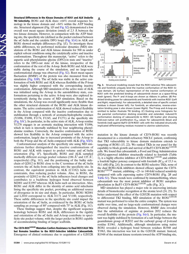
The ROS1-Selective TKIs Cabozantinib and Foretinib Bind the InactiveConformation of ROS1. Small-molecule, ATP-competitive TKIsgenerally engage with a target kinase in a type I or type II manner,corresponding to binding of the catalytically active or inactiveconformation, respectively. Analysis of the binding pose of cabo-zantinib in complex with ROS1 demonstrated a preference forthe inactive conformation, with the lowest docking score being−12 kcal/mol (Fig. 3A). Specifically, the quinoline moiety of cabo-zantinib was found to occupy the adenine-binding site, forming ahydrogen bond with the backbone atoms of E2027 and M2029,whereas the aryl linker makes aromatic stacking interactions withF2103 of the DFG motif (Fig. 3B). This observation is furtheraffirmed by the fact that a number of crystal structures of kinasesbound with quinolone- or quinazoline-based inhibitors showed thatthese fragments specifically occupy the adenine-binding site (31–33). Additional interactions include a hydrogen bond between thedicarboxamide group of cabozantinib (positioned close to gate-keeper residue L2026) and the catalytic K1980 residue and en-gagement of the fluorophenyl moiety (occupying the specificity site)with F2004 and F2075 in T-shaped and π–π aromatic stacking, re-spectively. Residues lining the specificity site (M2001, L2070, andI2100) provide additional hydrophobic interactions between cabo-zantinib and ROS1.To understand whether ROS1 displays an induced effect upon
cabozantinib binding, the ROS1:cabozantinib complex with thelowest docking score was subjected to MD simulation for 50 nsunder explicit solvent conditions, and the resulting trajectory wasanalyzed after stripping the solvent and the ligand molecule.Subsequent redocking of cabozantinib to the MD ensemble gen-erated from this holo-simulation revealed dramatically more
Freq
uenc
y amo
ng re
cove
red c
lones
with
muta
tions
(%)
CD74-ROS1 point mutantsrecovered in the presence of
cabozantinib(Starting from: native CD74-ROS1)
BA
DC
Freq
uenc
y amo
ng re
cove
red c
lones
with
muta
tions
(%)
CD74-ROS1 point mutantsrecovered in the presence of
foretinib(Starting from: native CD74-ROS1)
* No mutated clones recovered *0
20406080
100 80 nMn=0
F200
4E2
020
G203
2F2
075
V209
8D2
113
L222
3E2
232
Cabozantinib-resistant clones(Starting from: native CD74-ROS1)
10 20 40 80020406080
100
Concentration (nM)
Outgr
owth
(% of
well
s)
Total wellssurveyed (n): 96 192 384 384
40 80 160 320020406080
100
Concentration (nM)
Outgr
owth
(% of
well
s)
Foretinib-resistant clones(Starting from: CD74-ROS1G2032R)
Total wellssurveyed (n): 192 384 384 384
40 80 160320020406080
100
Concentration (nM)
Outgr
owth
(% of
well
s)
Cabozantinib-resistant clones(Starting from: CD74-ROS1G2032R)
Total wellssurveyed (n): 96 192 384 384
Freq
uenc
y amo
ng re
cove
red c
lones
with
comp
ound
muta
tions
(%)
CD74-ROS1 compound mutantsrecovered in the presence of
foretinib(Starting from: CD74-ROS1G2032R)
020406080
100 10 nMn=17
NF2
004
L201
0G2
032
F207
5V2
089
D211
3L2
223
E223
2
020406080
100 20 nMn=34 N
GCRV
F200
4L2
010
G203
2F2
075
V208
9D2
113
L222
3E2
232
020406080
100 40 nMn=15
XCICV
F200
4L2
010
G203
2F2
075
V208
9D2
113
L222
3E2
232
GV
020406080
100 80 nMn=9
F200
4L2
010
G203
2F2
075
V208
9D2
113
L222
3E2
232
VCI
V
I KG
020406080
100 40 nMn=2
F200
4E2
020
G203
2F2
075
V209
8D2
113
L222
3E2
232
GC
020406080
100 10 nMn=25
F200
4E2
020
G203
2F2
075
V209
8D2
113
L222
3E2
232
K IN
K
Freq
uenc
y amo
ng re
cove
red c
lones
with
comp
ound
muta
tions
(%)
020406080
100 40 nMn=6
G2032R
+0
20406080
100
* All clonesrecovered wereG2032R alone *
F200
4E2
020
F207
5V2
089
D211
3M2
134
CD74-ROS1 compound mutantsrecovered in the presence of
cabozantinib(Starting from: CD74-ROS1G2032R)
020406080
100 40 nMn=14
G2032R
+0
20406080
100
E197
4F2
004
I2009
E202
0F2
075
N211
2D2
113
R211
6W
2127
M212
8M2
134
P220
2L2
223
N222
4
TK KNG X K
020406080
100 80 nMn=42
ICLG2032R
+0
20406080
100
E197
4F2
004
I2009
E202
0F2
075
N211
2D2
113
R211
6W
2127
M212
8M2
134
P220
2L2
223
N222
4
KIC
NG XI
020406080
100 320 nMn=25
E197
4F2
004
I2009
E202
0F2
075
N211
2D2
113
R211
6W
2127
M212
8M2
134
P220
2L2
223
N222
4
VI
G2032R
G2032R
+0
20406080
100
CIV
G S
020406080
100 80 nMn=4
G2032R
+0
20406080
100
F200
4E2
020
F207
5V2
089
D211
3M2
134
NV
020406080
100 320 nMn=3
F200
4E2
020
F207
5V2
089
D211
3M2
134G2032R
+0
20406080
100
NGV
Foretinib-resistant clones(Starting from: native CD74-ROS1)
10 20 40 80020406080
100
Outgr
owth
(% of
well
s)
Total wellssurveyed (n): 96 96 96 96
Concentration (nM)
020406080
100 20 nMn=9
F200
4E2
020
G203
2F2
075
V209
8D2
113
L222
3E2
232
K V I
NGX
020406080
100 160 nMn=28
+0
20406080
100
E197
4F2
004
I2009
E202
0F2
075
N211
2D2
113
R211
6W
2127
M212
8M2
134
P220
2L2
223
N222
4
ICL
VCL
NGX IK T
G0
20406080
100 160 nMn=36
G2032R
+0
20406080
100
F200
4E2
020
F207
5V2
089
D211
3M2
134
V K C MNI
Fig. 4. In vitro mutagenesis screens suggest partially overlapping ROS1 point mutation and compound mutation resistance profiles for cabozantinib andforetinib. (A) Outgrowth summaries for cell-based resistance screens starting from Ba/F3 cells expressing native CD74-ROS1. (B) Outgrowth summaries for cell-based resistance screens starting from Ba/F3 CD74-ROS1G2032R cells. Breakdowns of frequency and spectra of mutant clones recovered in the presence ofincreasing concentrations of cabozantinib and foretinib are shown for assays starting from (C) native CD74-ROS1 and (D) CD74-ROS1G2032R cells. The numberof clones sequenced for each condition is indicated, and multiple substitutions at a single position are indicated as stacked bars.
Davare et al. PNAS | Published online September 8, 2015 | E5385
BIOCH
EMISTR
YPN
ASPL
US

favorable binding than the parent apo-simulation conformation fromwhich it was initialized, improving the docking score to −16 kcal/moland strongly suggesting induced conformational changes at thebinding site (Fig. 3C). MD simulation of the complex facilitatesrelaxation of the enzyme and ligand by removing any close con-tacts or strain observed in the initial docked pose. Subtle changesin the orientation and displacement of both the structural moietiesof the cabozantinib molecule and the residues within 4 Å ofcabozantinib significantly improved the binding score. For exam-ple, the dicarboxamide and cyclopropyl moieties of cabozantinibwere in close contact with residues D2102 and E1997 in the apo-simulated docking pose. Repositioning of these amino acids andneighboring residues resulted in optimized hydrogen bonding withthe dicarboxamide group as well as more favorable van der Waalsinteractions between the fluorophenyl group and specificity siteresidues L2070 and I2100. Also, slight readjustment in the positionof the quinoline moiety of cabozantinib relaxed a close contactwith residue L1951 observed in the starting point docked pose.Foretinib, a close structural analog of cabozantinib (Table 1),
was also docked to the ROS1 MD conformational ensemble.Analysis of the foretinib-docking pose showed a similar cabozantinib-like binding mode, with the additional morpholine moiety inforetinib interacting with residues K1976 and E2030 (Fig. 3B).This docking pose is further supported by the c-MET crystalstructure bound with foretinib, which showed a similar bindingconformation (34). However, closer examination of the foretinibdocked poses additionally revealed a reverse binding pose,wherein the morpholine group formed favorable interactions withthe αC-helix, whereas the fluorophenyl and quinoline moietiesinstead occupy the adenine-binding site and specificity site, re-spectively (Fig. 3B). Although intriguing, such inhibitor alternative-binding modes have also been visualized in other kinases (35, 36).For example, the crystal structure of SYK kinase revealed analternate binding pose for imatinib (PDB ID code 1XBB). Ex-amination of the top 200 conformations confirmed that ∼68% of
the docked poses maintained the cabozantinib-like pose whereas∼32% exhibited the reverse binding mode. Based on this puta-tive dual binding mode capacity for foretinib but not cabo-zantinib, we generated Ba/F3 CD74-ROS1 cell lines with alaninesubstitutions at select positions predicted from the modeling topreferentially disturb cabozantinib binding versus foretinibreverse pose binding. Accordingly, all four alanine mutants testeddemonstrated ∼2.5-fold to fivefold decreased sensitivity to cabo-zantinib compared with foretinib in vitro (Fig. 3D). This dis-covery suggests that foretinib may potentially engage the kinasein its reverse binding mode upon mutation although more rig-orous free energy calculations would be necessary to ascertain thisfinding. Overall, these findings suggest that ROS1-selective TKIsbind to the inactive conformation of ROS1 in a type II manner, incontrast to the type I binding mode exhibited by the dual ROS1/ALK inhibitor crizotinib.
ROS1-Selective TKIs Feature Largely Distinct Resistance ProfilesCompared with Dual ROS1/ALK Inhibitors. Acquired TKI resistancedue to point mutations is a frequent clinical challenge in manymalignancies, including NSCLC (37–39). Given our findings that,among the inhibitors tested, the ROS1-selective TKIs cabozantiniband foretinib uniquely retain potent activity against the CD74-ROS1G2032R mutant, we prospectively investigated potential mu-tations capable of conferring resistance to either or both TKIs.Accelerated cell-based resistance screens were performed startingfrom Ba/F3 cells expressing native CD74-ROS1 in the presenceof increasing concentrations of cabozantinib or foretinib. We ob-served a concentration-dependent reduction in the percentage ofwells that exhibited outgrowth with each TKI (Fig. 4A and TableS2). For both TKIs, sequencing of recovered clones for ROS1 ki-nase domain mutations revealed position 2113 in the A-loop as themost frequently mutated residue at all concentrations tested. Thespecific substitution at this position shifted from asparagine toglycine with increased TKI concentrations (Fig. 4C). With respect
F
D2113
P-loopC-helix
A-loopG2032
F2004
F2075
A B
C D
Crizotini
b
Cabozan
tinib
Foretini
b
Ceritinib
Brigatini
b
AZD3463
1
10
100
1000
NativeE1974KF2004CE2020KG2032R
CD74-ROS1 point mutants identified fromresistance screens for cabozantinib and foretinib
F2075VV2089MD2113GD2113NM2134I
F2075C
Native
G2032R/F2004CG2032R/E2020KG2032R/F2075CG2032R/F2075V
G2032R/D2113GG2032R/D2113NG2032R/M2134I
G2032R/V2089M
G2032R
G2032R-inclusive CD74-ROS1 compound mutants identifiedfrom resistance screens for cabozantinib and foretinib
1
10
100
1000
IC50
(nM)
Alectini
b
IC50
(nM)
E
0
0.05
0.1
Prob
abilit
y
0 5 10 15 0 5 10 15Distance (Å)
ROS1 ROS1D2113N
NativeE1974KF2004CE2020KG2032RF2075CF2075VV2089MD2113GD2113NM2134I
G2032R/F2004CG2032R/E2020KG2032R/F2075CG2032R/F2075VG2032R/V2089MG2032R/D2113GG2032R/D2113NG2032R/M2134I
≥100
1
75
25
Fold
incre
ase i
n IC 50
over
nativ
e CD7
4-RO
S1
CrizotinibCabozantinib Foretinib CeritinibBrigatinib AZD3463
50
CD74-ROS1 Alectinib
No efficacyagainst native
or mutantCD74-ROS1
ROS1-selective Dual ROS1/ALKALK-
selective
ROS1-selective Dual ROS1/ALK
ALK-selective
ROS1-selective Dual ROS1/ALK
ALK-selective
ROS1 ROS1D2113N
E1997R2107
E2120 D2113
E1997R2107
E2120N2113
Crizotini
b
Cabozan
tinib
Foretini
b
Ceritinib
Brigatini
b
AZD3463
Alectini
b
Fig. 5. Mutants recovered from resistance screensfor ROS1-selective TKIs, including those involvingposition D2113, confer varying levels of sensitivityto dual ROS1/ALK inhibitors. Scatter plots of cellproliferation IC50s for the indicated TKIs are shownfor Ba/F3 cells expressing (A) CD74-ROS1 point mu-tations and (B) G2032R-inclusive CD74-ROS1 com-pound mutations recovered in resistance screens forforetinib and cabozantinib. (C) Heat map depictingdifferential levels of sensitivity of the CD74-ROS1point mutations and G2032R-inclusive compoundmutations discovered from resistance screens. Theindicated color gradient represents fold increaseover IC50 for native CD74-ROS1 cells. (D) Ribbonstructure description of the inactive ROS1 kinasedomain, highlighting four positions implicated inTKI resistance and showing the Cα atom in van derWaals representation. The structural elements ofthe P-loop (magenta), A-loop (red), and αC-helix(green) are highlighted. (E) Predicted structuralconsequence of the ROS1D2113N mutation. Positions2113 and 2120, which form a unique salt-bridge inthe mutant kinase (Right), are shown as red balls.The two residues used to evaluate effects of themutation on the A-loop (R2107) and Cα-helix(E1997) are shown as sidechains in monitoring theshift of the activation loop to resemble a more typeI-like conformation in the mutant. (F) A-loop:Cα-helix–correlated conformational shift as measuredby distance profiles from simulation of the activeand inactive states of native ROS1 and ROS1D2113N.
E5386 | www.pnas.org/cgi/doi/10.1073/pnas.1515281112 Davare et al.

to cabozantinib, multiple substitutions of residues F2004 [cysteine(C)/valine (V)] and F2075 [cysteine (C)/isoleucine (I)/valine (V)]were recovered from concentrations up to 80 nM, consistent withfavorable interactions between the fluorophenyl group of the in-hibitor and these residues implicated from computational modelingof cabozantinib binding (Figs. 3B and 4C). Also in line with pre-dictions from modeling of variant foretinib binding poses, muta-tions at these two positions were recovered less frequently withforetinib than cabozantinib (Fig. 4C and Table S2). Notably, theF2075V mutation of ROS1 is analogous to the F359V mutation inthe kinase domain of ABL1, which is known to confer high-levelresistance to imatinib and nilotinib, both of which bind an inactiveconformation of the kinase (see Fig. S6) (40).A more recent issue that has gained attention in molecularly
targeted kinase inhibitor therapies is the risk for acquiring multi-drug-resistant compound mutations as an undesirable byproduct ofsequential TKI therapy (41–43). Given cabozantinib’s efficacyagainst the crizotinib-resistant CD74-ROS1G2032R mutant, a frac-tion of patients treated with second-line cabozantinib may harborG2032R or another mutation at baseline. To anticipate G2032R-inclusive ROS1 compound mutations that may confer resistance toROS1-selective TKIs, we performed resistance screens startingfrom Ba/F3 CD74-ROS1G2032R cells in the presence of cabozantinibor foretinib (Fig. 4B). Similar to screens starting from native CD74-ROS1, we observed a concentration-dependent reduction in thenumber of wells with outgrowth and the spectrum of mutated res-idues. Low frequency mutations identified in resistant clones re-covered from cabozantinib-treated wells included the following:E1974K, I2009L, E2020K, N2112K, R2116K, W2127*, M2128T,M2134I, L2223*, and N2224K (Fig. 4D and Table S3). Position2113 was the most frequently mutated site in tandem with G2032Rfor both TKIs, with clones recovered at concentrations as high as320 nM for foretinib and 640 nM for cabozantinib largely con-stricting to compound mutants pairing G2032R with F2004(I/V/C),F2075(C/I/V), or D2113G (Fig. 4D and Table S3).To evaluate the sensitivity of mutations identified in our re-
sistance screens for cabozantinib and foretinib to other ROS1TKIs, we rederived nine of the most frequently recovered singlemutants (E1974K, F2004C, E2020K, F2075C, F2075V, V2089M,D2113G, D2113N, and M2134I) and eight G2032R-inclusivecompound mutations (G2032R paired with F2004C, E2020K,F2075C, F2075V, V2089M, D2113G, D2113N, or M2134I) inBa/F3 CD74-ROS1 cells and tested their sensitivity against our panelof seven TKIs (Fig. 5 A and B and Table S4). All single mutantsshowed 2- to 30-fold decreased sensitivity to the ROS1-selective,type II inhibitors, cabozantinib and foretinib, but remained sensi-tive to the dual ROS1/ALK, putative type I binders crizotinib,brigatinib, ceritinib, and AZD3463. Consistent with the insensitivityof the CD74-ROS1G2032R mutant to all of the tested dual ROS1/ALK TKIs, we found that all G2032R-inclusive ROS1 compoundmutants also exhibit high-level resistance. By contrast, G2032R-inclusive CD74-ROS1 compound mutants displayed varyingdegrees of resistance to cabozantinib and foretinib. For example,among the most frequently recovered compound mutations forcabozantinib was G2032R/D2113N (Fig. 4D and Table S3), whichdemonstrated ∼15-fold increased IC50 (255.8 nM) for cabozanti-nib, compared with cells expressing G2032R or D2113N (Fig. 5 Aand B and Table S4). Overall, results from profiling of single mu-tants suggest that, whereas ROS1 kinase domain point mutationsinvolving residues F2004, F2075, and D2113 may confer resistanceto the ROS1-selective inhibitors cabozantinib and foretinib, theymay remain sensitive to dual ROS1/ALK TKIs. However, emer-gence of compound mutations poses a potential vulnerability forboth of these categories of ROS1 TKIs (Fig. 5 C and D).
The ROS1D2113N Mutant Induces an Altered A-Loop Conformation andCompromises Binding of Cabozantinib. Given the high frequencywith which the D2113N mutation was recovered from cell-based
resistance screens for CD74-ROS1, we performed MD simula-tions of this mutant to elucidate the local and global confor-mational changes induced in the ROS1 kinase domain. RMSFanalysis of the ROS1D2113N inactive conformation showed de-creased flexibility of the A-loop compared with native ROS1(Fig. S4B). This altered flexibility can be attributed to the localchanges observed in the A-loop upon mutation, indirectlyimpacting the binding site of ROS1-selective TKIs. The D2113residue in native ROS1 simultaneously interacts with the posi-tively charged R2116 and is repelled by another negativelycharged residue in the A-loop (E2120). The D2113N mutationnullifies this repulsion and instead creates a hydrogen bond be-tween these two residues (N2113 and E2120) (Fig. 5E), resultingin a significant shift and stabilization of the ROS1D2113N A-loopconformation and alteration of the αC-helix dynamics.To investigate the dynamics of the αC-helix relative to the
A-loop, the distance between residue E1997 (αC-helix) and R2107(A-loop) was monitored throughout the MD simulation. Notably,these residues are proximal and form a salt bridge in the inactivekinase, as observed in many other kinases adopting an SRC-likeinactive conformation (44–46), whereas they are further apart andlack such interaction in the active state (Fig. 5E). Although distanceprofiling of the active conformation from MD simulation showedthat both ROS1 and ROS1D2113N sampled similarly (14 and 15 Å,respectively) (Fig. 5F), conformational analysis of the inactivestate showed a bimodal distribution, with higher distances forROS1D2113N (averaged at 4.8 and 8.5 Å) compared with nativeROS1 (4 Å). These data suggest that the inactive state of nativeROS1 participates in salt bridge formation between E1997 andR2107 (as inferred from the reduced distance between them)and that the D2113N mutant displaces R2107 outside of the αC-helix and the interaction between E1997 and R2107 is lost. Dueto displacement of residue R2107 in ROS1D2113N, E1997 reor-ients its sidechain and partially occupies the specificity site,thereby creating a potential steric clash with type II binding in-hibitors. Although alterations in the specificity pocket due to theD2113N mutation present a possible resistance mechanism sce-nario for type II inhibitors, inherent limitations in the initiali-zation of the computational model or the finite accessibility ofthe simulation time scales cannot be completely ruled out.Foretinib and cabozantinib were docked to the inactive
ROS1D2113N MD ensemble and compared with native ROS1. Wefound that both inhibitors demonstrated significantly less favorabledocking scores for ROS1D2113N (Fig. S5) compared with nativeROS1. In contrast, crizotinib demonstrated equivalent, favorabledocking scores for both native ROS1 and ROS1D2113N (Fig. S5).Taken together, these results suggest that alterations of positionD2113 may represent a unique liability for ROS1-selective type IIinhibitors, possibly impacting the conformation of the specificitysite. Although speculative in nature, relevant experiments are war-ranted to further explore these differences.
DiscussionRational, molecularly guided clinical use of TKI therapies hassubstantially impacted patient outcomes in several cancer sub-types, including NSCLC (37, 47). The recent establishment ofrearranged ALK and ROS1 as distinct molecular diagnosticsubgroups of NSCLC, coupled with the clinical efficacy of cri-zotinib, has driven an explosion of new inhibitor development.Although several inhibitors have shown promising clinical activity(48–50), resistance to therapy has already surfaced. The highdegree of sequence homology between the catalytic domains ofALK and ROS1 suggests that ALK TKIs may be repurposed asROS1 inhibitors and vice versa, but our results establish limits tothis inhibitor design principle.Upon consideration of all available results for first- and second-
generation ALK and/or ROS1 TKIs, including the current study,we suggest three operational categories of selectivity: dual ROS1/
Davare et al. PNAS | Published online September 8, 2015 | E5387
BIOCH
EMISTR
YPN
ASPL
US

ALK, ROS1-selective, and ALK-selective. Among the dual ROS1/ALK TKIs, crizotinib and ceritinib both demonstrate crystallo-graphic evidence of a type I binding mode for ALK, and a similarbinding mode is observed for the ROS1:crizotinib complex (22, 51,52). Our computational studies reveal considerably greater struc-tural similarity between the catalytically active conformations ofALK and ROS1, and the chemical scaffolds of the newer dualROS1/ALK TKIs ceritinib, brigatinib, and AZD3463 are highlyrelated (Table 1), suggesting a common preferential type I bindingmode among dual ROS1/ALK inhibitors. Intriguingly, the ALK-selective inhibitor alectinib, which exhibits no ROS1 inhibitoryactivity, has also been reported to preferentially engage the activeconformation of ALK (53). Modeling of alectinib in active ALKsuggests that this lack of activity against ROS1 is due at least in partto two interactions not present in ROS1: (i) interaction of the ethylgroup of alectinib with a hydrophobic ALK hinge region residue(A1200) and (ii) unique access of alectinib’s nitrile group to thespecificity site (despite its type I binding mode) by interacting withE1167 of the ALK αC-helix. Notably, this analysis may also provideadditional context for understanding the mechanism behind thereported alectinib-resistant ALKV1180L mutation (54), which ispredicted to disrupt this nitrile-E1167 interaction.In contrast, the ROS1-selective inhibitors cabozantinib and
foretinib exhibit a type II binding mode and preferentially en-gage the inactive conformation of ROS1. This binding mode isprimarily due to structurally important differences that permitaccommodation of larger, type II inhibitor scaffolds. It is knownthat, in comparison with the specificity site of the inactive kinasedomain, the ATP binding site in the active conformation is highlyconserved among many kinases, forming the basis of selectivetype II inhibitor design (55). Notably, we observed that cabo-zantinib binding to ROS1 involves conformational selection,followed by an induced-fit mechanism to achieve optimized in-teraction. A recent study by Wilson et al. used an evolutionaryapproach to examine protein structure and identified a similarinduced-fit mechanism for imatinib that is critical to its selectivetype II binding of ABL1 compared with the highly related SRCkinase (56). Such a binding mechanism may also have implicationsfor additional biochemical properties, including kinetic on- and off-rates. Although our ensemble docking of the structurally relatedforetinib showed a similar initial binding pose to that of cabo-zantinib, an alternate reverse orientation binding mode was alsopresent. Conformational clustering revealed that the reversebinding mode exhibited by foretinib was present primarily in low-population clusters featuring a slightly more open specificity site.Further studies using free energy calculations and advanced sam-pling methods will be required for in depth characterization. To-gether, these findings suggest a potentially important theme inapplication of small-molecule TKI therapies, wherein structuraldifferences in the inactive conformations of related kinases mayexplain both the mechanistic subtleties and the contrasts seen inTKI binding affinity and inhibitory efficacy (56, 57).Consistent with differences in binding mode, distinct patterns
of kinase domain point mutation-based resistance are apparentfor the three categories of TKIs described here. In ALK-rearrangedNSCLC, crizotinib resistance due to acquisition of kinase domainmutations is routinely observed, with 11 substitutions spanning 9residues reported in clinical resistance (Table 1), in addition toseveral other residues implicated from in vitro resistance screeningassays (20, 21). Although clinical follow-up remains limited for thenewer inhibitors, a subset of crizotinib-resistant ALK mutations(those involving positions 1171, 1174, and 1202) have already beenimplicated in clinical resistance to the dual ROS1/ALK TKI cer-itinib and/or the ALK-selective TKI alectinib (Table 1) (21, 54, 58).This pattern is akin to the clinical experience in EGFR-driven lungcancer, where type I TKIs such as erlotinib evoke dramatic butinevitably transient responses, funneling to common point muta-tion-based failure (47). Although newer EGFR TKIs with different
binding properties (e.g., irreversible binding, such as afatinib) haveoffered some benefit, resistance and/or intolerance is still commonlyobserved. The only reported ROS1 mutation in clinical resistanceto date is CD74-ROS1G2032R (analogous to the resistant G1202Rmutation found in EML4-ALK) (Fig. S6), verified at autopsy in apatient with metastatic NSCLC that had been controlled withcrizotinib before relapse (22). Our finding that this mutation remainshighly sensitive to cabozantinib is consistent with a recent report(59). Given cabozantinib’s efficacy against both native and G2032R-mutant ROS1 fusions, an investigational new drug filing with theFDA for evaluation of cabozantinib in crizotinib-naive and -refractoryROS1 fusion-positive lung cancer has been initiated (clinical trialidentifier, NCT01639508), and a phase 2 trial is actively accruingpatients (clinical trial identifier, NCT01639508).Clinical experience with TKIs for ROS1- and ALK-driven
NSCLC, along with our in vitro profiling and structural modelingdata, suggests important themes regarding resistance. First, withrespect to dual ROS1/ALK inhibitors (type I mode binders),ROS1 mutations within the ATP binding site may represent asignificant resistance liability. All tested TKIs in this categorydemonstrated limited to no efficacy against cells expressing theCD74-ROS1G2032R mutant (Fig. 2 A and B). Conversely, ROS1-selective inhibitors (type II mode binders) remain highly effec-tive against ATP binding site mutations but are vulnerable tovariants that favor the active conformation of ROS1 or thosethat constrict the specificity site of inactive ROS1. As an exam-ple, mutations at the F2004 and F2075 sites of ROS1 reside inthe specificity pocket, which is occupied by the type II inhibitorscabozantinib and foretinib, but usually not by type I compounds.The nearly nonoverlapping resistance profiles of type I com-pared with type II inhibitors may open an important strategy forclinical management of resistance.Dual ROS1/ALK TKIs remain effective against nearly all
mutations in the specificity site that confer resistance to theROS1-selective inhibitors. However, in line with findings fromsequential therapy with ABL1 TKIs in CML (41–43), the po-tential emergence of G2032R-inclusive compound mutations inROS1 fusions could yield high-level resistance to multiple type Iand type II inhibitors (Fig. 5 B and C). At present, it is not knownwhether the ROS1G2032R mutation detected in a relapsed patientwill be the most prevalent resistance mutation for ROS1. Al-though the overall median duration of responses to crizotinib inROS1 fusion-positive patients seems longer than that observed inpatients harboring ALK fusions (15), resistance profiles are stillnascent and will take several years of clinical experience to fullyestablish. Nonetheless, the spectrum of ROS1 TKI-resistantmutations implicated clinically or from cell-based screening todate is noticeably restricted, compared with results from similarprofiling of ABL1 TKIs in CML (60, 61), suggesting a scenariomore like that seen with FLT3 TKIs in acute myeloid leukemia(62), where escape routes are confined to a relatively smaller set ofmutations. This finding has potential implications for continuedTKI development and the prospect of maximal control of ROS1mutation-mediated drug resistance. In addition to the activity ofROS1 inhibitors, clinical tolerability in the face of chronic dosingwill likewise serve as an important determinant of how and in whatorder to effectively use these treatments in the clinic. These clinicaldecisions may also ultimately dictate the durability of response andemergence of TKI-resistant mutations.Looking forward, consideration should be given to development
of new inhibitors for ROS1 and ALK. One possibility is to developand optimize type II inhibitors for ALK. Although a preclinicaltype II ALK scaffold has been described (63), to our knowledge,there are no reported clinical TKIs that engage the inactive con-formation of ALK. Whether such TKIs would demonstrate dif-ferential efficacy over their type I binding counterparts remains tobe seen although the possibility of a nonoverlapping resistanceprofile might offer additional options for patients with refractory
E5388 | www.pnas.org/cgi/doi/10.1073/pnas.1515281112 Davare et al.

ALK-rearranged disease. Another important consideration forTKI development is that, although targeting both ROS1 and ALKwith a single drug may seem appealing in terms of broader clinicalapplicability, attempting to target resistance mutations within theactive conformation TKI binding sites, such as G1202R in ALKand G2032R in ROS1, may prove difficult given overlappingresistance profiles of such type I inhibitors. Alternatively, struc-ture-based design of improved, potent ROS1-selective TKIs thataccommodate mutations, either by avoiding direct contact withsuch residues or by using additional productive contacts withinthe altered binding pocket, may prove a useful strategy.In summary, our in vitro profiling of a panel of clinical TKIs,
combined with computational structural modeling and resistancescreening, helps define therapeutically relevant differences betweenROS1 and ALK, providing a basis for further development of in-hibitors to circumvent resistance in NSCLC and other malignancies.
MethodsCell Viability Assays. All TKIs were prepared as 1-mM stocks in DMSO beforeeach experiment. Inhibitors were distributed at 2× concentration using aD300 Digital Dispenser (HP) [capable of accurately administering very smallvolumes (10 pL–150 nL)] into 384-well plates preloaded with 25 μL per wellof complete medium. Ba/F3 cells expressing CD74-ROS1 constructs wereseeded (800 cells per well; 25 μL) into drug plates using a Multidrop CombiReagent Dispenser (Thermo Scientific), and plates were incubated for 72 h at37 °C, 5% CO2. Viability was measured using a methanethiosulfonate-basedassay (CellTiter96 Aqueous One Solution; Promega) read on a Biotek Synergy2 plate reader. All experiments were performed at least two independenttimes in triplicate. Data were normalized using Microsoft Excel, and absoluteIC50 values were calculated with GraphPad Prism software using a nonlinearcurve fit equation modified using previously described parameters (64).
Immunoblot Analysis. Ba/F3 CD74-ROS1, CD74-ROS1G2032R, and EML4-ALK cells,as well as human NSCLC cell lines (HCC78 and H3122), were treated with theindicated concentrations of inhibitors for 2 h, pelleted, washed once in ice-coldPBS, and lysed in 200 μL of cell lysis buffer (Cell Signaling Technology) sup-plemented with 0.25% deoxycholate, 0.05% SDS, and protease and phos-phatase inhibitors. Equal amounts of protein were extracted with SDS samplebuffer for 15 min at 80 °C and run on 4–15% Tris-glycine precast gradient gels(Criterion; Bio-Rad). Proteins were transferred to Immobilon-FL membranes(Millipore) and probed with phospho-ROS1 [3078, 1:1,000; Cell SignalingTechnology (CST)], total ROS1 (3266, 1:1,000; CST), phospho-ERK1/2 (9101,1:1,000; CST), total ERK2 (sc-1647, 1:2,000; Santa Cruz), total ALK (3333, 1:1,000;CST), phospho-ALK (6941, 1:1,000; CST), phospho-Akt (4060, 1:1,000; CST), AKT(610860, 1:1,000; BD Transduction Laboratories), and GAPDH (AM4300,1:5,000; Ambion). Blots were imaged using either a LI-COR Odyssey imagingsystem or the Bio-Rad ChemiDoc imaging station according to the manufac-turer’s protocol for immunoblot detection with use of Infrared dye or horse-radish peroxidase-conjugated secondary antibodies, respectively.
Accelerated Cell-Based Resistance Screen. Ba/F3 cells expressing native CD74-ROS1 or CD74-ROS1G2032R were treated overnight with N-ethyl-N-nitrosourea(ENU) (50 μg/mL), pelleted, washed, resuspended in fresh media, and distrib-uted into 96-well plates (1 × 10⁵ cells per well) in 200 μL of complete mediumsupplemented with the indicated concentrations of either cabozantinib orforetinib. Wells were observed for media color change and cell growth underan inverted microscope daily for 4 wk, and wells exhibiting outgrowth weretransferred to 24-well plates containing 2 mL of complete medium supple-mented with the same concentration of cabozantinib or foretinib. At con-fluency, these wells were harvested, pelleted, and stored at −20 °C. GenomicDNA was extracted using a DNeasy Tissue Kit (Qiagen); for low inhibitor con-centrations wherein near 100% outgrowth of wells was observed, DNA wasextracted from a randomly selected subset of expanded clones. The CD74-ROS1kinase domain was amplified using primers CD74-ROS1 M13F1 (5′-GTA AAACGA CGG CCA GTG CTC TTC CAA CCC AAG AGG-3′) and ROS1-M13Kin1-Rev1(5′-CAG GAA ACA GCT ATG ACC GCC ATC TTC ACC TTC AAA GC-3′) and bi-directionally sequenced using M13F (5‘-GTA AAA CGA CGG CCA GTG-3′) andM13R primers (5‘-CAG GAA ACA GCT ATG ACC-3′). The resultant chromatographswere analyzed for mutations using Mutation Surveyor software (SoftGenetics).
Molecular Dynamics Simulations. Molecular dynamics (MD) simulation wasperformed using the Amber ff12SB force field (65) in NAMD simulation soft-ware (66). During the initial stages of model refinement, nonhydrogen atomswere restrained (100 kcal/mol/Å2), and the system was relaxed by energy min-imization using 1,000 steps of the steepest descent algorithm. During the nextstages of the heating, equilibration, and production run, all bonds involvinghydrogen atoms were restrained using the SHAKE algorithm (67). Periodicboundary conditions with particle mesh Ewald summation were used to handlethe long-range electrostatic interactions (real-space truncation at 9.0 Å and gridspacing of 1.0 Å) (68). All eight systems were heated from 100 to 300 K in 50 psusing a 1-fs time-step. After heating of all systems, 5 ns of equilibration wereperformed using the NPT ensemble with a 2-fs time-step. Temperature andpressure were controlled at 300 K and 1 atm using the Nosé–Hoover Langevinpiston algorithm (69) and Langevin dynamics (70), respectively. For each of theeight systems, three independent replicates were generated and simulated for500 ns using the NPT ensemble. Coordinates were saved every 10 ps, andconformational analysis was performed on the combined ensemble. A cumu-lative 12 μs simulation trajectory was generated for further analysis.
Conformational Analysis of ROS1 and ALK. The CPPTRAJ software of theAmberTools suite was used for postprocessing the MD-generated trajectories(71). For each system, a combined 1,500-ns (500 ns and three replicates) trajec-tory was analyzed for various molecular properties. Root mean square deviation(rmsd) and root mean square fluctuation (RMSF) of the protein were measuredusing a trajectory fitted on either the initial or average structure as reference.Structural fitting was performed using a least-square fitting method for theprotein backbone atoms. Rotation of the αC-helix was estimated using an angledefined by the Cα atoms of three residues located in the N and C terminus of theαC-helix and the rigid F-helix: Q1989, F2004, and W2145 for ROS1 and the cor-responding residues in ALK (Q1159, F1174, and W1313). The specificity sitepocket volume of ROS1 and ALK was measured using the POVME program (72),with both kinases’ trajectories aligned using the binding site as defined by thefollowing residues: L1951, A1978, K1980, E1997, M2001, L2028, G2032, L2086,and D2102 for ROS1 and equivalent residues in ALK based on structural align-ment. This qualitative estimate of the pocket volume is independent of thedefinition of the active site used here for alignment. Interatomic distances be-tween residues in the P-loop (F1956), αC-helix (E1997), and A-loop (R2107) ofROS1 and ALK (corresponding residues) were measured using the position of theCα atoms during the course of the simulation.
Molecular Docking Simulations. Ensemble docking was performed using theGlide program of Schrödinger’s package (Suite 2012, Maestro, version 9.3).Conformations (1,500) were extracted from each simulated system (one con-formation for every nanosecond), and a docking grid for the receptor wasgenerated using the binding site residues defined above (encompassing the ATPbinding site and specificity site). Ligands (crizotinib, foretinib, cabozantinib, andalectinib) were prepared using the Ligprep module of the Schrödinger’s package(version 3), docked using the GlideXP method (Glide version 5.8; Schrödinger,LLC), and analyzed for binding interactions (73). Parameterization of cabo-zantinib was performed using standard Amber protocol (74). Cabozantinib ge-ometry was optimized using Gaussian 09 (www.gaussian.com) at the HF/6–31G*level theory consistent with ff12SB force fields, and initial atomic charges werederived using restricted electrostatic potential (RESP) (75). Fifty nanoseconds ofimplicit solvent generalized-Born MD simulation was performed at 300 K, andthe resulting trajectory was clustered using the CPPTRAJ program (71). Repre-sentative structures from the top three clusters were selected, and the geometrywas optimized at the HF/6–31G* level. RESP charges were derived using themulticonformation charge fitting method. Antechamber was used to assignother parameters from the generalized Amber force field (74).
ACKNOWLEDGMENTS. We thank Matthew Zabriskie for technical assistanceand members of the B.J.D., M.W.D., and T.O. laboratories for valuablediscussion. M.W.D. receives research funding from Bristol-Myers Squibb,Celgene, Novartis, and Gilead. This work was supported by the HowardHughes Medical Institute (B.J.D.), and B.J.D. is supported by NIH Methodto Extend Research in Time Award R37CA065823. M.A.D. is supported by aHyundai Scholar Hope Grant. M.W.D. is supported by NIH Grants HL082978-01, 5 P01 CA049639-23, and R01 CA178397-01. T.O. is supported by NIHGrant R01 CA178397-01. N.A.V. acknowledges generous support from theCenter for High Performance Computing (The University of Utah) and com-puting allocations at the Extreme Science and Engineering Discovery Envi-ronment (XSEDE) supercomputers (Award TG-CHE120086). XSEDE is supportedby National Science Foundation Grant OCI-1053575.
1. Druker BJ (2009) Perspectives on the development of imatinib and the future ofcancer research. Nat Med 15(10):1149–1152.
2. Druker BJ, et al.; IRIS Investigators (2006) Five-year follow-up of patients receivingimatinib for chronic myeloid leukemia. N Engl J Med 355(23):2408–2417.
Davare et al. PNAS | Published online September 8, 2015 | E5389
BIOCH
EMISTR
YPN
ASPL
US

3. Nowell PC, Hungerford DA (1960) Chromosome studies on normal and leukemichuman leukocytes. J Natl Cancer Inst 25:85–109.
4. Shaw AT, Hsu PP, Awad MM, Engelman JA (2013) Tyrosine kinase gene rearrange-ments in epithelial malignancies. Nat Rev Cancer 13(11):772–787.
5. Grande E, Bolós MV, Arriola E (2011) Targeting oncogenic ALK: A promising strategyfor cancer treatment. Mol Cancer Ther 10(4):569–579.
6. AwadMM, Shaw AT (2014) ALK inhibitors in non-small cell lung cancer: Crizotinib andbeyond. Clin Adv Hematol Oncol 12(7):429–439.
7. Shaw AT, Engelman JA (2013) ALK in lung cancer: Past, present, and future. J ClinOncol 31(8):1105–1111.
8. Davies KD, Doebele RC (2013) Molecular pathways: ROS1 fusion proteins in cancer.Clin Cancer Res 19(15):4040–4045.
9. Arai Y, et al. (2013) Mousemodel for ROS1-rearranged lung cancer. PLoS One 8(2):e56010.10. Charest A, et al. (2006) ROS fusion tyrosine kinase activates a SH2 domain-containing
phosphatase-2/phosphatidylinositol 3-kinase/mammalian target of rapamycin signal-ing axis to form glioblastoma in mice. Cancer Res 66(15):7473–7481.
11. Saborowski A, et al. (2013) Mouse model of intrahepatic cholangiocarcinoma vali-dates FIG-ROS as a potent fusion oncogene and therapeutic target. Proc Natl Acad SciUSA 110(48):19513–19518.
12. Bergethon K, et al. (2012) ROS1 rearrangements define a unique molecular class oflung cancers. J Clin Oncol 30(8):863–870.
13. Chin LP, Soo RA, Soong R, Ou SH (2012) Targeting ROS1 with anaplastic lymphomakinase inhibitors: A promising therapeutic strategy for a newly defined molecularsubset of non-small-cell lung cancer. J Thorac Oncol 7(11):1625–1630.
14. Shaw AT, et al. (2013) Crizotinib versus chemotherapy in advanced ALK-positive lungcancer. N Engl J Med 368(25):2385–2394.
15. Shaw AT, et al. (2014) Crizotinib in ROS1-rearranged non-small-cell lung cancer.N Engl J Med 371(21):1963–1971.
16. O’Hare T, Deininger MW, Eide CA, Clackson T, Druker BJ (2011) Targeting the BCR-ABL signaling pathway in therapy-resistant Philadelphia chromosome-positive leu-kemia. Clin Cancer Res 17(2):212–221.
17. Yu HA, Riely GJ, Lovly CM (2014) Therapeutic strategies utilized in the setting of acquiredresistance to EGFR tyrosine kinase inhibitors. Clin Cancer Res 20(23):5898–5907.
18. Riely GJ, Politi KA, Miller VA, Pao W (2006) Update on epidermal growth factor re-ceptor mutations in non-small cell lung cancer. Clin Cancer Res 12(24):7232–7241.
19. Doebele RC, et al. (2012) Mechanisms of resistance to crizotinib in patients with ALKgene rearranged non-small cell lung cancer. Clin Cancer Res 18(5):1472–1482.
20. Heuckmann JM, et al. (2011) ALK mutations conferring differential resistance tostructurally diverse ALK inhibitors. Clin Cancer Res 17(23):7394–7401.
21. Lovly CM, Pao W (2012) Escaping ALK inhibition: Mechanisms of and strategies toovercome resistance. Sci Transl Med 4(120):120ps2.
22. Awad MM, et al. (2013) Acquired resistance to crizotinib from a mutation in CD74-ROS1. N Engl J Med 368(25):2395–2401.
23. Davare MA, et al. (2013) Foretinib is a potent inhibitor of oncogenic ROS1 fusionproteins. Proc Natl Acad Sci USA 110(48):19519–19524.
24. Baron R, McCammon JA (2013) Molecular recognition and ligand association. AnnuRev Phys Chem 64:151–175.
25. Dror RO, Dirks RM, Grossman JP, Xu H, Shaw DE (2012) Biomolecular simulation: Acomputational microscope for molecular biology. Annu Rev Biophys 41:429–452.
26. Magalhaes A, Maigret B, Hoflack J, Gomes JN, Scheraga HA (1994) Contribution ofunusual arginine-arginine short-range interactions to stabilization and recognition inproteins. J Protein Chem 13(2):195–215.
27. Neves MA, Yeager M, Abagyan R (2012) Unusual arginine formations in proteinfunction and assembly: Rings, strings, and stacks. J Phys Chem B 116(23):7006–7013.
28. Soetens J-CMC, Chipot C, Jansen G, Ángyán JG, Maigret B (1997) Effect of polariz-ability on the potential of mean force of two cations: The guanidinium−guanidiniumion pair in water. J Phys Chem 101(50):10910–10917.
29. Feixas F, Lindert S, Sinko W, McCammon JA (2014) Exploring the role of receptorflexibility in structure-based drug discovery. Biophys Chem 186:31–45.
30. Amaro RE, Baron R, McCammon JA (2008) An improved relaxed complex scheme for re-ceptor flexibility in computer-aided drug design. J Comput Aided Mol Des 22(9):693–705.
31. Shewchuk L, et al. (2000) Binding mode of the 4-anilinoquinazoline class of proteinkinase inhibitor: X-ray crystallographic studies of 4-anilinoquinazolines bound tocyclin-dependent kinase 2 and p38 kinase. J Med Chem 43(1):133–138.
32. Solomon VR, Lee H (2011) Quinoline as a privileged scaffold in cancer drug discovery.Curr Med Chem 18(10):1488–1508.
33. Zhang Q, et al. (2014) Identification of type II inhibitors targeting BRAF using privi-leged pharmacophores. Chem Biol Drug Des 83(1):27–36.
34. Qian F, et al. (2009) Inhibition of tumor cell growth, invasion, and metastasis by EXEL-2880 (XL880, GSK1363089), a novel inhibitor of HGF and VEGF receptor tyrosine ki-nases. Cancer Res 69(20):8009–8016.
35. Aronov AM, et al. (2007) Flipped out: Structure-guided design of selective pyr-azolylpyrrole ERK inhibitors. J Med Chem 50(6):1280–1287.
36. Atwell S, et al. (2004) A novel mode of Gleevec binding is revealed by the structure ofspleen tyrosine kinase. J Biol Chem 279(53):55827–55832.
37. Camidge DR, Pao W, Sequist LV (2014) Acquired resistance to TKIs in solid tumours:Learning from lung cancer. Nat Rev Clin Oncol 11(8):473–481.
38. Choi YL, et al.; ALK Lung Cancer Study Group (2010) EML4-ALK mutations in lungcancer that confer resistance to ALK inhibitors. N Engl J Med 363(18):1734–1739.
39. Chong CR, Jänne PA (2013) The quest to overcome resistance to EGFR-targetedtherapies in cancer. Nat Med 19(11):1389–1400.
40. O’Hare T, et al. (2005) In vitro activity of Bcr-Abl inhibitors AMN107 and BMS-354825against clinically relevant imatinib-resistant Abl kinase domain mutants. Cancer Res65(11):4500–4505.
41. Shah NP, et al. (2007) Sequential ABL kinase inhibitor therapy selects for compounddrug-resistant BCR-ABL mutations with altered oncogenic potency. J Clin Invest117(9):2562–2569.
42. Soverini S, et al. (2013) Unraveling the complexity of tyrosine kinase inhibitor-resistantpopulations by ultra-deep sequencing of the BCR-ABL kinase domain. Blood 122(9):1634–1648.
43. Zabriskie MS, et al. (2014) BCR-ABL1 compound mutations combining key kinasedomain positions confer clinical resistance to ponatinib in Ph chromosome-positiveleukemia. Cancer Cell 26(3):428–442.
44. Levinson NM, et al. (2006) A Src-like inactive conformation in the abl tyrosine kinasedomain. PLoS Biol 4(5):e144.
45. Shan Y, et al. (2009) A conserved protonation-dependent switch controls drugbinding in the Abl kinase. Proc Natl Acad Sci USA 106(1):139–144.
46. Xu W, Doshi A, Lei M, Eck MJ, Harrison SC (1999) Crystal structures of c-Src revealfeatures of its autoinhibitory mechanism. Mol Cell 3(5):629–638.
47. Pao W, Chmielecki J (2010) Rational, biologically based treatment of EGFR-mutantnon-small-cell lung cancer. Nat Rev Cancer 10(11):760–774.
48. Gadgeel SM, et al. (2014) Safety and activity of alectinib against systemic disease andbrain metastases in patients with crizotinib-resistant ALK-rearranged non-small-celllung cancer (AF-002JG): Results from the dose-finding portion of a phase 1/2 study.Lancet Oncol 15(10):1119–1128.
49. Gainor JF, et al. (2015) Alectinib salvages CNS relapses in ALK-positive lung cancer patientspreviously treated with crizotinib and ceritinib. J Thorac Oncol 10(2):232–236.
50. Shaw AT, et al. (2014) Ceritinib in ALK-rearranged non-small-cell lung cancer. N Engl JMed 370(13):1189–1197.
51. Cui JJ, et al. (2011) Structure based drug design of crizotinib (PF-02341066), a potentand selective dual inhibitor of mesenchymal-epithelial transition factor (c-MET) ki-nase and anaplastic lymphoma kinase (ALK). J Med Chem 54(18):6342–6363.
52. Friboulet L, et al. (2014) The ALK inhibitor ceritinib overcomes crizotinib resistance innon-small cell lung cancer. Cancer Discov 4(6):662–673.
53. Sakamoto H, et al. (2011) CH5424802, a selective ALK inhibitor capable of blockingthe resistant gatekeeper mutant. Cancer Cell 19(5):679–690.
54. Katayama R, et al. (2014) Two novel ALK mutations mediate acquired resistance tothe next-generation ALK inhibitor alectinib. Clin Cancer Res 20(22):5686–5696.
55. Davis MI, et al. (2011) Comprehensive analysis of kinase inhibitor selectivity. NatBiotechnol 29(11):1046–1051.
56. Wilson C, et al. (2015) Kinase dynamics: Using ancient protein kinases to unravel amodern cancer drug’s mechanism. Science 347(6224):882–886.
57. Zhao Z, et al. (2014) Exploration of type II binding mode: A privileged approach forkinase inhibitor focused drug discovery? ACS Chem Biol 9(6):1230–1241.
58. Lovly CM, et al. (2011) Insights into ALK-driven cancers revealed through develop-ment of novel ALK tyrosine kinase inhibitors. Cancer Res 71(14):4920–4931.
59. Katayama R, et al. (2015) Cabozantinib overcomes crizotinib resistance in ROS1fusion-positive cancer. Clin Cancer Res 21(1):166–174.
60. Bradeen HA, et al. (2006) Comparison of imatinib mesylate, dasatinib (BMS-354825),and nilotinib (AMN107) in an N-ethyl-N-nitrosourea (ENU)-based mutagenesis screen:High efficacy of drug combinations. Blood 108(7):2332–2338.
61. O’Hare T, et al. (2009) AP24534, a pan-BCR-ABL inhibitor for chronic myeloid leuke-mia, potently inhibits the T315I mutant and overcomes mutation-based resistance.Cancer Cell 16(5):401–412.
62. Smith CC, et al. (2012) Validation of ITD mutations in FLT3 as a therapeutic target inhuman acute myeloid leukaemia. Nature 485(7397):260–263.
63. Epstein LF, Chen H, Emkey R, Whittington DA (2012) The R1275Q neuroblastomamutant and certain ATP-competitive inhibitors stabilize alternative activation loopconformations of anaplastic lymphoma kinase. J Biol Chem 287(44):37447–37457.
64. Sebaugh JL (2011) Guidelines for accurate EC50/IC50 estimation. Pharm Stat 10(2):128–134.65. Case DA, et al. (2012) AMBER 12 (University of California, San Fransisco).66. Phillips JC, et al. (2005) Scalable molecular dynamics with NAMD. J Comput Chem
26(16):1781–1802.67. Miyamoto S, Kollman PA (1992) Settle: An analytical version of the SHAKE and
RATTLE algorithm for rigid water models. J Comput Chem 13(8):952–962.68. Darden T, York D, Pedersen L (1993) Particle mesh Ewald: An N·log(N) method for
Ewald sums in large systems. J Chem Phys 98(12):10089.69. Martyna GJ, Tobias DJ, Klein ML (1994) Constant pressure molecular dynamics algo-
rithms. J Chem Phys 101(5):4177.70. Pastor RW, Brooks BR, Szabo A (1988) An analysis of the accuracy of Langevin and
molecular dynamics algorithms. Mol Phys 65(6):1409–1419.71. Roe DR, Cheatham TE (2013) PTRAJ and CPPTRAJ: Software for processing and
analysis of molecular dynamics trajectory data. J Chem Theory Comput 9(7):3084–3095.
72. Durrant JD, Votapka L, Sørensen J, Amaro RE (2014) POVME 2.0: An enhanced tool fordetermining pocket shape and volume characteristics. J Chem Theory Comput 10(11):5047–5056.
73. Friesner RA, et al. (2006) Extra precision glide: Docking and scoring incorporating a modelof hydrophobic enclosure for protein-ligand complexes. J Med Chem 49(21):6177–6196.
74. Wang J, Wolf RM, Caldwell JW, Kollman PA, Case DA (2004) Development and testingof a general amber force field. J Comput Chem 25(9):1157–1174.
75. Bayly C, Cieplak P, Cornell W, Kollman P (1993) A well-behaved electrostatic potentialbased method using charge restraints for deriving atomic charges: The RESP model.J Phys Chem 97(40):10269–10280.
76. Jorgensen WLCJ, Madura JD, Impey RW, Klein ML (1983) Comparison of simple po-tential functions for simulating liquid water. J Chem Phys 79:926.
77. Joung IS, Cheatham TE, 3rd (2008) Determination of alkali and halide monovalent ionparameters for use in explicitly solvated biomolecular simulations. J Phys Chem B112(30):9020–9041.
E5390 | www.pnas.org/cgi/doi/10.1073/pnas.1515281112 Davare et al.