structural quality promotion of vanadium-containing biotite via gamma...
TRANSCRIPT
60
Structural Quality Promotion of Vanadium-Containing
Biotite via Gamma Irradiations
Khaled M. Elsabawy1,2,*
and Ahmed T.Tawfik1,3
1Materials Science Unit ,Chemistry Department ,Faculty of Science, Tanta University -
31725-Tanta-Egypt
2 Faculty of Science-Chemistry Department-Taif University-Taif–Alhawyah -888-Saudia
Arabia
3 Egyptian Environmental Affairs Agency-Egypt
ABSTRACT
Advanced synthesis of free fluoride V-biotite sample having the optimized formula
NaV2.5(Al,Si)4O10(OH)2 was synthesized by mixed solution-solid routes. The synthetic clay
was carefully characterized by different techniques including X-ray diffraction (XRD),
infrared absorption spectra (IR) and thermal analysis (TG/TDA).Furthermore micro-
structural features were monitored by both of scanning electron microscopy (SEM) and
AFM . The synthetic clay sample was exposed to two different gamma irradiation doses ,1st
dose was 2 MR and 2nd
dose ~ 4MR. Structural measurements after γ-irradiations gave
structure quality for synthetic V-biotite clay due to disappearance of some impurity phases
and unreacted oxides as result of absorbed gamma-ray dose increases from 2 to 4 MR .
Keywords: Biotite, Clay, XRD, TGA, SEM,AFM, Green synthesis.
SCIREA Journal of Materials Science
p://www.scirea.org/journal/Materialshtt
November 11, 2016
Volume 1, Issue1, October 2016
61
I. Introduction
Gamma, Laser irradiations doses were applied as structure promoters for different kinds of
matters such as glass, ceramics and mineral clays[1-3]. Rare earth doped phosphors have a
vital role as radiation detectors in many fields of fundamental and applied research, such as
clinical, personal, and environmental monitoring of ionizing radiation [4,5]. Various methods
of preparation have also been developed for easy synthesis of these materials to make them
available easily. While irradiation usually leads to the creation of structural defects, the
healing effect of irradiation is also known [6-7]. Ionizing radiation has been found to be
widely applicable in modifying the structure and properties of polymers and can be used to
tailor the performance of either bulk materials or surfaces. Improved luminescent
characteristics of some of aluminates have also found their place in optoelectronics. The
thermo-luminescence (TL) materials have been widely applied to defect studying and
dosimetry, such as the detection of ionizing radiation and dating in archaeology [8-13]. The
ability to detect and perform energy-dispersive spectroscopy of high-energy radiation such as
X-rays, γ-rays, and other uncharged and charged particles has improved dramatically in recent
years [14]. Alkaline earth aluminate ceramics are important host materials that have been
prepared and studied by several researchers for luminescence applications [15]. Alkaline earth
aluminate belongs to the spinel group of minerals (MAl2O4) with general chemical
composition, AB2O4, where A is a divalent atom [16]. The magnesium aluminate spinel
(MgAl2O4) has good thermal and mechanical properties, high hardness and low electrical loss,
and chemical properties. As such, it is currently utilized as a refractory for furnace walls and
firebricks and also has the potential for application as environment humidity sensors, laser
materials, and substrate in integrated electronics [17-21].
The major goal of these investigations is proving that irradiations doses can promote and
enhance structure of solid materials towards more regular structure eliminating defects could
be caused by impurity phases.
II. Experimental
II.1. Materials
All reagents used were of analytical grade (each purity >99%) Al2SiO5 from BHD Laboratory
reagent, Na2CO3 (GPR), and ammonium meta-vanadate, NH4VO3 (A.R. grade for laboratory
62
and research uses) were mixed in the chemical compositions corresponding to synthesizing
{NaV2.5 (Al,Si)4O10(OH)2}which have chemical formula of V-biotite.
II.2. Synthesis of V-Biotite Clay Sample
V-biotite was prepared carefully by using mixed solution/solid- state reaction technique using
nominal compositions of individual oxides in the main formula, such that aluminum silicate
Al2O3.SiO2, sodium carbonate (anhydrous) Na2CO3 were dissolved 50 ml of concentrated
solution of HNO3 then diluted to 100 ml by distill H2O solution (I). Ammonium meta-
vanadate NH4VO3 is also dissolved in the same amount of nitric acid with same dilution
factor , solution(2). The obtained solution (I+II) were mixed together and coprecipitated by
conc. ammonia/citrate solution, the hydroxylated precipitated product was filtered and dried
in oven at 110 °C.
The powders mixtures were mixed in an agate mortar for 1 h. The green compacted metals
powder was subjected to firing in a controlled atmosphere furnace to a temperature below its
melting point in sealed platinum container at 900-950°C for 9 h, followed by sintering step at
880 °C to allow packed metal powders to bond together and finally, the furnace was cooled
slowly down to room temperature and the material was kept in vacuum desiccators over silica
gel dryer.
II.3. Characterization of V-Biotite Clay Sample
II.3.1 X-ray diffraction investigation
XRD powder diffraction measurements were performed on an X'Pert SW. X-ray
diffractometer with filtered Cu Kα radiation (λ=1.54 A°), at 40 Kv and 30mA with a scanning
speed in the range of 2θ=5-70 (298 K).
II.3.2 Infrared spectra studies
The IR spectra of the clays were recorded between 4000 and 400 cm-1
using a KBr method
with a NICOLET 6700 FTIR thermoscientific spectrophotometer.
II.3.3. SEM and AFM Investigations
Scanning electron microscope measurement was carried out using small pieces of the
prepared sample by using a “Philips model XL 30 CP”. The sample was coated with gold.
Scanning electron microscope (SEM) was used to take micrographs of the clays. AFM
investigations made by di-innova (USA) instrument accompanied with particle size analyzer,
63
tapping non-contact mode was applied on small piece of the prepared clay after gamma
irradiations .
II.3.4. Thermal studies
Thermogravimetry (TGA) and differential thermal analysis (DTA) were carried out by using a
Shimadzu DTA-50H thermal analyzer. The sample was placed in platinum crucible (0.1 cm3)
the system were studied under nitrogen atmosphere with a heating rate 10 °C min-1
and
following rate at 20 ml min-1
, constant weight of sample 4.7 mg was used.
II.3.5. Electrical conductivity studies
The DC-electrical conductivity of synthetic V-biotite was measured using two terminals DC-
method. The pellets were inserted between spring loaded copper electrodes, A KEITHLEY
175 multimeter (ASA) was employed from 10-500 °C.
The temperature was measured by a calibrated chromel-alumel thermocouple placed firmly at
the sample. Measurements were conducted in such a way that at each temperature, sufficient
time was allowed to attain thermal equilibrium.
The number of conduction electron n is given by:
n = noe-Eg/KT
………………………………….…(2)
where nο is the concentration of atoms at the lattice site, E𝐠 is the band gap, and K is the
Boltezman constant (assuming E𝐠 ≈ KT), the conductivity relationship becomes
σ = (noe-Eg/KT
)eμ …………….….……………….….(3)
σ = Ae-Eg/KT
where A= nο eµ ………………..(4)
Log σ =Log A-(E𝐠 /2.303KT) ………….…….…..(5)
The plot of log σ against 1/T should give a straight line with a slop of –E/2.303K.
The measurements of σ as a function of temperature will permit calculation of the band gap
energy E𝐠 of materials behavior (conductor, semiconductor and insulator)
III. Results and Discussion
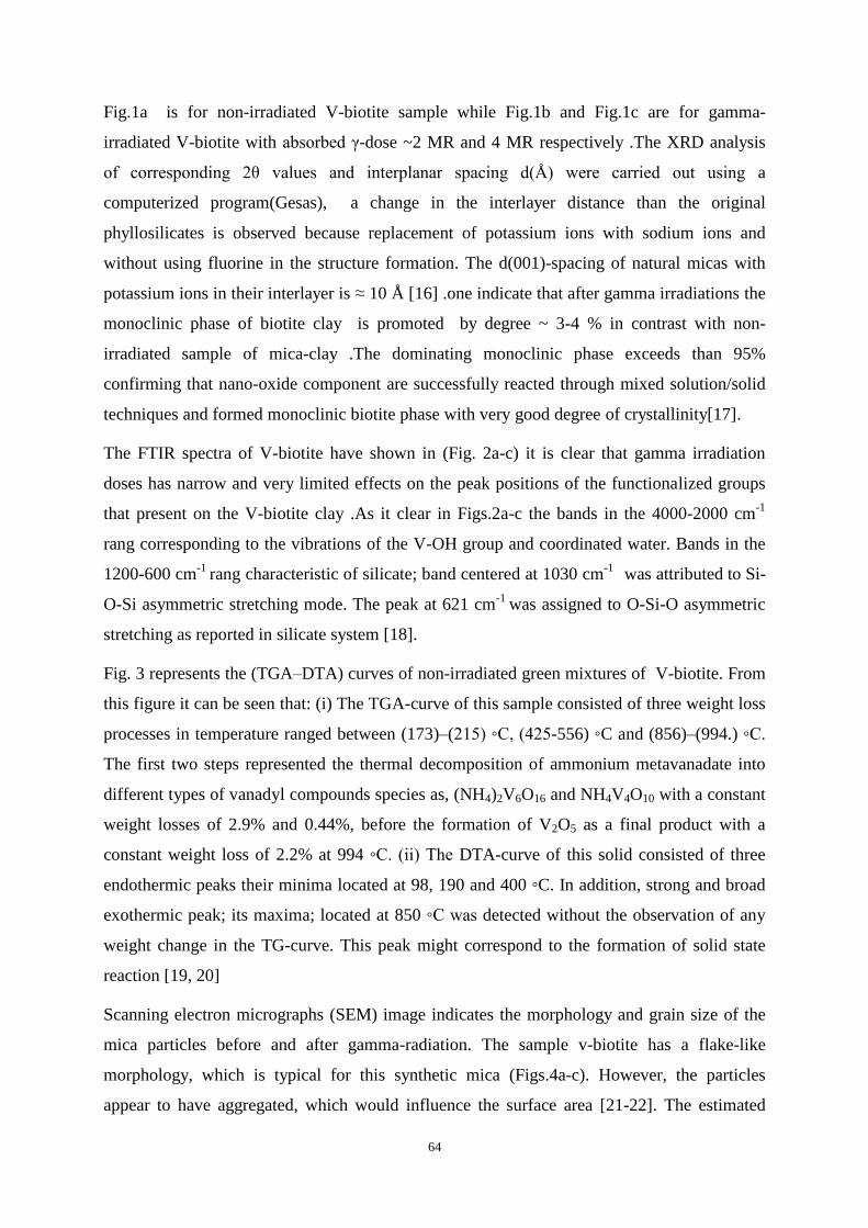
Fig.(1a-c): shows the XRD pattern recorded for synthetic free fluoride biotite which has the
formula NaV2.5(Al,Si)4O10 (OH)2
64
Fig.1a is for non-irradiated V-biotite sample while Fig.1b and Fig.1c are for gamma-
irradiated V-biotite with absorbed γ-dose ~2 MR and 4 MR respectively .The XRD analysis
of corresponding 2θ values and interplanar spacing d(Å) were carried out using a
computerized program(Gesas), a change in the interlayer distance than the original
phyllosilicates is observed because replacement of potassium ions with sodium ions and
without using fluorine in the structure formation. The d(001)-spacing of natural micas with
potassium ions in their interlayer is ≈ 10 Å [16] .one indicate that after gamma irradiations the
monoclinic phase of biotite clay is promoted by degree ~ 3-4 % in contrast with non-
irradiated sample of mica-clay .The dominating monoclinic phase exceeds than 95%
confirming that nano-oxide component are successfully reacted through mixed solution/solid
techniques and formed monoclinic biotite phase with very good degree of crystallinity[17].
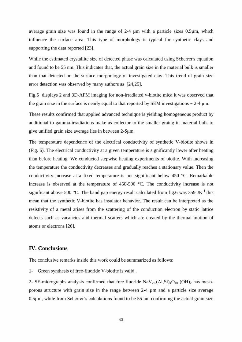
The FTIR spectra of V-biotite have shown in (Fig. 2a-c) it is clear that gamma irradiation
doses has narrow and very limited effects on the peak positions of the functionalized groups
that present on the V-biotite clay .As it clear in Figs.2a-c the bands in the 4000-2000 cm-1
rang corresponding to the vibrations of the V-OH group and coordinated water. Bands in the
1200-600 cm-1
rang characteristic of silicate; band centered at 1030 cm-1
was attributed to Si-
O-Si asymmetric stretching mode. The peak at 621 cm-1
was assigned to O-Si-O asymmetric
stretching as reported in silicate system [18].
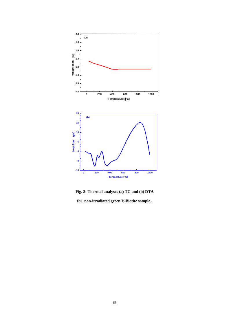
Fig. 3 represents the (TGA–DTA) curves of non-irradiated green mixtures of V-biotite. From
this figure it can be seen that: (i) The TGA-curve of this sample consisted of three weight loss
processes in temperature ranged between (173)–(215) ◦C, (425-556) ◦C and (856)–(994.) ◦C.
The first two steps represented the thermal decomposition of ammonium metavanadate into
different types of vanadyl compounds species as, (NH4)2V6O16 and NH4V4O10 with a constant
weight losses of 2.9% and 0.44%, before the formation of V2O5 as a final product with a
constant weight loss of 2.2% at 994 ◦C. (ii) The DTA-curve of this solid consisted of three
endothermic peaks their minima located at 98, 190 and 400 ◦C. In addition, strong and broad
exothermic peak; its maxima; located at 850 ◦C was detected without the observation of any
weight change in the TG-curve. This peak might correspond to the formation of solid state
reaction [19, 20]
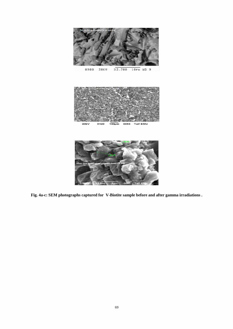
Scanning electron micrographs (SEM) image indicates the morphology and grain size of the
mica particles before and after gamma-radiation. The sample v-biotite has a flake-like
morphology, which is typical for this synthetic mica (Figs.4a-c). However, the particles
appear to have aggregated, which would influence the surface area [21-22]. The estimated
65
average grain size was found in the range of 2-4 µm with a particle sizes 0.5µm, which
influence the surface area. This type of morphology is typical for synthetic clays and
supporting the data reported [23].
While the estimated crystallite size of detected phase was calculated using Scherrer's equation
and found to be 55 nm. This indicates that, the actual grain size in the material bulk is smaller
than that detected on the surface morphology of investigated clay. This trend of grain size
error detection was observed by many authors as [24,25].
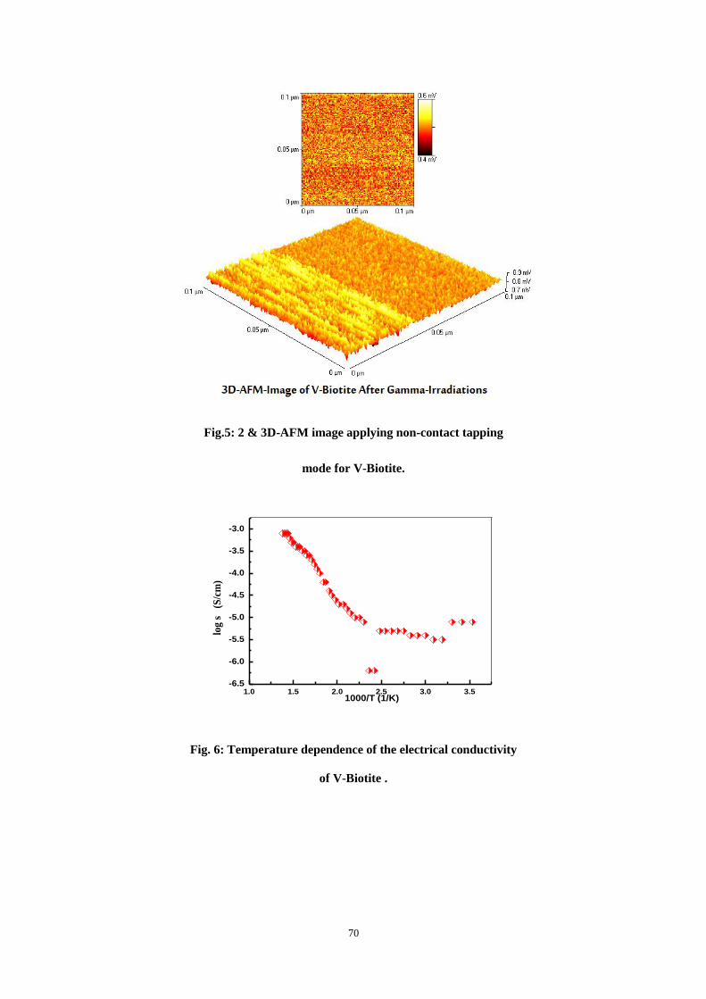
Fig.5 displays 2 and 3D-AFM imaging for non-irradiated v-biotite mica it was observed that
the grain size in the surface is nearly equal to that reported by SEM investigations ~ 2-4 μm.
These results confirmed that applied advanced technique is yielding homogeneous product by
additional to gamma-irradiations make as collector to the smaller graing in material bulk to
give unified grain size average lies in between 2-5μm.
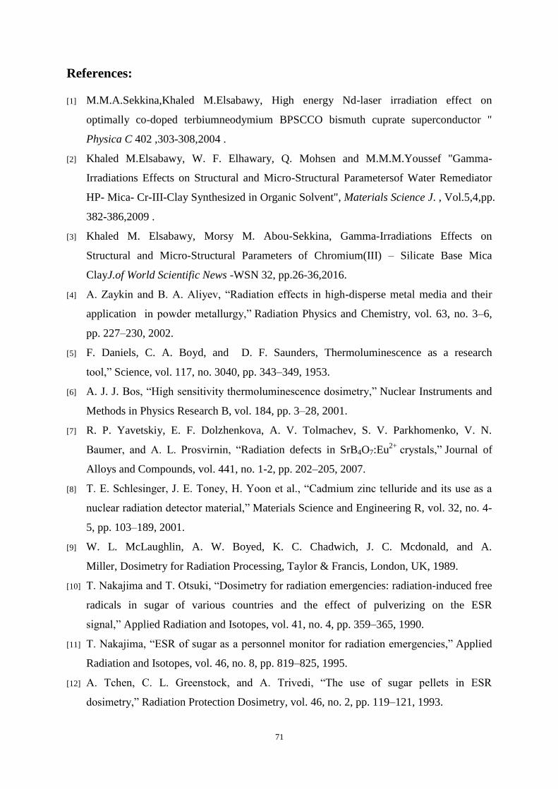
The temperature dependence of the electrical conductivity of synthetic V-biotite shows in
(Fig. 6). The electrical conductivity at a given temperature is significantly lower after heating
than before heating. We conducted stepwise heating experiments of biotite. With increasing
the temperature the conductivity decreases and gradually reaches a stationary value. Then the
conductivity increase at a fixed temperature is not significant below 450 °C. Remarkable
increase is observed at the temperature of 450-500 °C. The conductivity increase is not
significant above 500 °C. The band gap energy result calculated from fig.6 was 359 JK-1
this
mean that the synthetic V-biotite has insulator behavior. The result can be interpreted as the
resistivity of a metal arises from the scattering of the conduction electron by static lattice
defects such as vacancies and thermal scatters which are created by the thermal motion of
atoms or electrons [26].
IV. Conclusions
The conclusive remarks inside this work could be summarized as follows:
1- Green synthesis of free-fluoride V-biotite is valid .
2- SE-micrographs analysis confirmed that free fluoride NaV2.5(Al,Si)4O10 (OH)2 has meso-
porous structure with grain size in the range between 2-4 µm and a particle size average
0.5µm, while from Scherrer’s calculations found to be 55 nm confirming the actual grain size
66
in the material bulk is smaller than that detected on the surface morphology of investigated
clay.
3- Gamma-irradiations make as structure promoter as proved in our XRD and SEM
investigations.
4- AFM-investigations confirmed SEM which is reflecting structure homogeneity and success
of synthesizing technique.
Figures and Tables
10 20 30 40 50 60 70
Two Theta (Degree)
XRD-V-Biotite(a)
Gamma Dose~ 2 MR
(b)
Gamma Dose~ 4 MR
(c)
Fig. 1a-c: XRD patterns recorded for V-biotite before and after gamma irradiations.
67
4000 3500 3000 2500 2000 1500 1000 500
75
80
85
90
95
100
623
1030
2360
29203630
% T
ran
sm
itta
nc
e
Wavenumber (cm-1)
FTIR-Non-Irradiated-V-Biotite
Fig.2a
4000 3500 3000 2500 2000 1500 1000 500
75
80
85
90
95
100
105
488
750
955
1100
1390
1630
% T
rans
mitt
ance
Wavenumber (cm-1
)
FTIR for V-Biotite-After 1st
Gamma-irradiated dose ~ 2MR
Fig.2(b)
4000 3500 3000 2500 2000 1500 1000 50084
86
88
90
92
94
96
98
100
102
488
746
953
1100
14001690
2360
33203480
% T
ran
sm
itta
nc
e
Wavenumber (cm-1
)
FTIR of V-Biotite after 2nd gamma-irradiated dose ~ 4 MR
Fig.2c
Fig.2a-c: FTIR-spectra recorded for V-biotite sample before and
after two different doses of gamma ray .
68
0 200 400 600 800 10000.6
0.8
1.0
1.2
1.4
1.6
1.8
2.0
We
igh
t lo
ss
(
%)
Temperature C]
(a)
0 200 400 600 800 1000-10
-5
0
5
10
15
20
He
at
flo
w (µ
V)
Temperture [C]
(b)
Fig. 3: Thermal analyses (a) TG and (b) DTA
for non-irradiated green V-Biotite sample .
69
Fig. 4a-c: SEM photographs captured for V-Biotite sample before and after gamma irradiations .
70
Fig.5: 2 & 3D-AFM image applying non-contact tapping
mode for V-Biotite.
1.0 1.5 2.0 2.5 3.0 3.5-6.5
-6.0
-5.5
-5.0
-4.5
-4.0
-3.5
-3.0
1000/T (1/K)
log
s
(S
/cm
)
Fig. 6: Temperature dependence of the electrical conductivity
of V-Biotite .
71
References:
[1] M.M.A.Sekkina,Khaled M.Elsabawy, High energy Nd-laser irradiation effect on
optimally co-doped terbiumneodymium BPSCCO bismuth cuprate superconductor "
Physica C 402 ,303-308,2004 .
[2] Khaled M.Elsabawy, W. F. Elhawary, Q. Mohsen and M.M.M.Youssef "Gamma-
Irradiations Effects on Structural and Micro-Structural Parametersof Water Remediator
HP- Mica- Cr-III-Clay Synthesized in Organic Solvent", Materials Science J. , Vol.5,4,pp.
382-386,2009 .
[3] Khaled M. Elsabawy, Morsy M. Abou-Sekkina, Gamma-Irradiations Effects on
Structural and Micro-Structural Parameters of Chromium(III) – Silicate Base Mica
ClayJ.of World Scientific News -WSN 32, pp.26-36,2016.
[4] A. Zaykin and B. A. Aliyev, “Radiation effects in high-disperse metal media and their
application in powder metallurgy,” Radiation Physics and Chemistry, vol. 63, no. 3–6,
pp. 227–230, 2002.
[5] F. Daniels, C. A. Boyd, and D. F. Saunders, Thermoluminescence as a research
tool,” Science, vol. 117, no. 3040, pp. 343–349, 1953.
[6] A. J. J. Bos, “High sensitivity thermoluminescence dosimetry,” Nuclear Instruments and
Methods in Physics Research B, vol. 184, pp. 3–28, 2001.
[7] R. P. Yavetskiy, E. F. Dolzhenkova, A. V. Tolmachev, S. V. Parkhomenko, V. N.
Baumer, and A. L. Prosvirnin, “Radiation defects in SrB4O7:Eu2+
crystals,” Journal of
Alloys and Compounds, vol. 441, no. 1-2, pp. 202–205, 2007.
[8] T. E. Schlesinger, J. E. Toney, H. Yoon et al., “Cadmium zinc telluride and its use as a
nuclear radiation detector material,” Materials Science and Engineering R, vol. 32, no. 4-
5, pp. 103–189, 2001.
[9] W. L. McLaughlin, A. W. Boyed, K. C. Chadwich, J. C. Mcdonald, and A.
Miller, Dosimetry for Radiation Processing, Taylor & Francis, London, UK, 1989.
[10] T. Nakajima and T. Otsuki, “Dosimetry for radiation emergencies: radiation-induced free
radicals in sugar of various countries and the effect of pulverizing on the ESR
signal,” Applied Radiation and Isotopes, vol. 41, no. 4, pp. 359–365, 1990.
[11] T. Nakajima, “ESR of sugar as a personnel monitor for radiation emergencies,” Applied
Radiation and Isotopes, vol. 46, no. 8, pp. 819–825, 1995.
[12] A. Tchen, C. L. Greenstock, and A. Trivedi, “The use of sugar pellets in ESR
dosimetry,” Radiation Protection Dosimetry, vol. 46, no. 2, pp. 119–121, 1993.
72
[13] N. A. Atari and K. V. Ettinger, “Lyoluminescence of irradiated saccharides,” Radiation
Effects, vol. 20, no. 1-2, pp. 135–139, 1973.
[14] T. Nickel, E. Pitt, and A. Scharmann, “Lyoluminescence dosimetry with a sugar after
accidental gamma ray exposure,” Radiation Protection Dosimetry, vol. 35, no. 3, pp.
173–177, 1991.
[15] A. J. Walton, “Triboluminescence,” Advances in Physics, vol. 26, no. 6, pp. 887–948,
1977.
[16] G. Alzetta, I. Chudasek, and R. Scarmozzino, “Excitation of triboluminescence by
deformation of single crystals,” Physica Status Solidi A, vol. 1, no. 4, pp. 775–785,
1970.
[17] Y. Enomoto and H. Hashimoto, “Emission of charged particles from indentation fracture
of rocks,”Nature, vol. 346, no. 6285, pp. 641–643, 1990.
[18] T. Matsuzawa, Y. Aokiy, N. Takeuchi, and Y. Murayama, “A new long phosphorescent
phosphor with high brightness, SrAl2 O4: Eu2+
, Dy3+
,” Journal of the Electrochemical
Society, vol. 143, pp. 2670–2673, 1996.
[19] R. Dekkers and C. F. Woensdregt, “Crystal structural control on surface topology and
crystal morphology of normal spinel (MgAl2O4),” Journal of Crystal Growth, vol. 236, no.
1–3, pp. 441–454, 2002.
[20] C. Baudín, R. Martínez, and P. Pena, “High-temperature mechanical behavior of
stoichiometric magnesium spinel,” Journal of the American Ceramic Society, vol. 78, pp.
1857–1862,1995.
[21] M. Sindel, N. A. Travitzky, and N. Claussen, “Influence of magnesium-aluminum spinel
on the directed oxidation of molten aluminum alloys,” Journal of the American Ceramic
Society, vol. 73, pp. 2615–2618, 1990.
[22] Khaled M.Elsabawy,
Part-II 3D-AFM-Microstructural Featues and Suitability of
Sulfonyl-Urea Moeity as Center of Antidiabetic Drugs Families, , Inter. Journal of
Pharmaceutical Sci. (IJPMS) 1, Issue 1, pp. 1-6,2015.
[23] Khaled M. Elsabawy Micro-Structural Features Suitability of Gramicidin Drug as
Antibacterial Ointment Antibiotic Int.J. Pharm. Toxicology,5,1,pp.62-66,2015.
[24] Khaled M. Elsabawy, Impact of Double Irradiations on the Structural and Micro-
structural Features of some Selected Solid Phase -Milk Products „; Britsh Journal of
Research, 1,3 ,pp.124-133,2014.
73
[25] Khaled M. Elsabawy ,Spotlight on Nano-Structural Features of Solid-Phase
Moxifloxacin Antibiotic An AFM-Investigations ,Khaled M. Elsabawy and
Waheed F.El-Hawary,Int.J.of Phytopharmacology,5,4,pp.284-287,2014.