structural symmetry and protein function
TRANSCRIPT

P1: FDS
April 20, 2000 11:37 Annual Reviews AR098-05
?Annu. Rev. Biophys. Biomol. Struct. 2000. 29:105–53
Copyright c© 2000 by Annual Reviews. All rights reserved
STRUCTURAL SYMMETRY AND PROTEIN
FUNCTION
David S. Goodsell and Arthur J. OlsonDepartment of Molecular Biology, Scripps Research Institute, La Jolla, California 92037;e-mail: [email protected], [email protected]
Key Words oligomeric proteins, protein symmetry, protein structure/functionrelationships
■ Abstract The majority of soluble and membrane-bound proteins in modern cellsare symmetrical oligomeric complexes with two or more subunits. The evolution-ary selection of symmetrical oligomeric complexes is driven by functional, genetic,and physicochemical needs. Large proteins are selected for specific morphologicalfunctions, such as formation of rings, containers, and filaments, and for cooperativefunctions, such as allosteric regulation and multivalent binding. Large proteins are alsomore stable against denaturation and have a reduced surface area exposed to solventwhen compared with many individual, smaller proteins. Large proteins are constructedas oligomers for reasons of error control in synthesis, coding efficiency, and regulationof assembly. Symmetrical oligomers are favored because of stability and finite con-trol of assembly. Several functions limit symmetry, such as interaction with DNA ormembranes, and directional motion. Symmetry is broken or modified in many forms:quasisymmetry, in which identical subunits adopt similar but different conformations;pleomorphism, in which identical subunits form different complexes; pseudosymme-try, in which different molecules form approximately symmetrical complexes; andsymmetry mismatch, in which oligomers of different symmetries interact along theirrespective symmetry axes. Asymmetry is also observed at several levels. Nearly allcomplexes show local asymmetry at the level of side chain conformation. Severalcomplexes have reciprocating mechanisms in which the complex is asymmetric, but,over time, all subunits cycle through the same set of conformations. Global asymmetryis only rarely observed. Evolution of oligomeric complexes may favor the formationof dimers over complexes with higher cyclic symmetry, through a mechanism of pre-positioned pairs of interacting residues. However, examples have been found for allof the crystallographic point groups, demonstrating that functional need can drive theevolution of any symmetry.
CONTENTS
INTRODUCTION . . . . . . . . . . . . . . . . . . . . . . . . . . . . . . . . . . . . . . . . . . . . . . . . 106THE SYMMETRY OF OLIGOMERIC PROTEINS. . . . . . . . . . . . . . . . . . . . . . . . 107
Characteristics and Natural Occurrence of Symmetry Groups. . . . . . . . . . . . . . . 108
1056-8700/00/0610-0105$14.00 105
Ann
u. R
ev. B
ioph
ys. B
iom
ol. S
truc
t. 20
00.2
9:10
5-15
3. D
ownl
oade
d fr
om w
ww
.ann
ualr
evie
ws.
org
by W
IB63
22 -
Uni
vers
itaet
Bay
reut
h on
11/
16/1
0. F
or p
erso
nal u
se o
nly.

P1: FDS
April 20, 2000 11:37 Annual Reviews AR098-05
?106 GOODSELL ¥ OLSON
Structural Mechanisms of Oligomerization. . . . . . . . . . . . . . . . . . . . . . . . . . . . . 111WHY BUILD SYMMETRICAL PROTEINS?. . . . . . . . . . . . . . . . . . . . . . . . . . . . 112
Why Build Large Proteins?. . . . . . . . . . . . . . . . . . . . . . . . . . . . . . . . . . . . . . . . 114Why Build Oligomeric Proteins?. . . . . . . . . . . . . . . . . . . . . . . . . . . . . . . . . . . . 115Why Build Symmetrical Proteins?. . . . . . . . . . . . . . . . . . . . . . . . . . . . . . . . . . . 116Functional Niches for Small Monomeric Proteins. . . . . . . . . . . . . . . . . . . . . . . . 119
SYMMETRY AND COOPERATIVITY . . . . . . . . . . . . . . . . . . . . . . . . . . . . . . . . 120Allosteric Regulation. . . . . . . . . . . . . . . . . . . . . . . . . . . . . . . . . . . . . . . . . . . . 120Multivalent Binding . . . . . . . . . . . . . . . . . . . . . . . . . . . . . . . . . . . . . . . . . . . . . 122
MORPHOLOGICAL FUNCTIONS OF SYMMETRY. . . . . . . . . . . . . . . . . . . . . . 123Rulers, Rings, and Containers. . . . . . . . . . . . . . . . . . . . . . . . . . . . . . . . . . . . . . 123The Need to Be Large. . . . . . . . . . . . . . . . . . . . . . . . . . . . . . . . . . . . . . . . . . . . 127
FUNCTIONS THAT LIMIT SYMMETRY . . . . . . . . . . . . . . . . . . . . . . . . . . . . . . 128Directional Motion. . . . . . . . . . . . . . . . . . . . . . . . . . . . . . . . . . . . . . . . . . . . . . 128Interaction with DNA. . . . . . . . . . . . . . . . . . . . . . . . . . . . . . . . . . . . . . . . . . . . 129Membrane Interactions. . . . . . . . . . . . . . . . . . . . . . . . . . . . . . . . . . . . . . . . . . . 129
SYMMETRY BREAKING IN OLIGOMERIC PROTEINS . . . . . . . . . . . . . . . . . . 130Quasisymmetry. . . . . . . . . . . . . . . . . . . . . . . . . . . . . . . . . . . . . . . . . . . . . . . . 130Pleomorphic Symmetry. . . . . . . . . . . . . . . . . . . . . . . . . . . . . . . . . . . . . . . . . . 135Pseudosymmetry. . . . . . . . . . . . . . . . . . . . . . . . . . . . . . . . . . . . . . . . . . . . . . . 137Symmetry Mismatch. . . . . . . . . . . . . . . . . . . . . . . . . . . . . . . . . . . . . . . . . . . . 138
ASYMMETRY . . . . . . . . . . . . . . . . . . . . . . . . . . . . . . . . . . . . . . . . . . . . . . . . . . 141Local Asymmetry. . . . . . . . . . . . . . . . . . . . . . . . . . . . . . . . . . . . . . . . . . . . . . . 142Reciprocating Mechanisms. . . . . . . . . . . . . . . . . . . . . . . . . . . . . . . . . . . . . . . . 143Global Asymmetry. . . . . . . . . . . . . . . . . . . . . . . . . . . . . . . . . . . . . . . . . . . . . . 144
EVOLUTION OF OLIGOMERIC PROTEINS. . . . . . . . . . . . . . . . . . . . . . . . . . . . 146THE AESTHETICS OF SYMMETRY . . . . . . . . . . . . . . . . . . . . . . . . . . . . . . . . . 148
INTRODUCTION
Symmetry has played an important role in science from its very origins. TheGreeks, fascinated by the symmetry of vibrating strings, developed a quantitativeunderstanding of pitch and harmony, and Kepler formulated a simple mathematicaldescription of gravity that was based on the elliptical geometry of planetary orbits.Today, symmetry continues to permeate scientific thought. Physicists are lookingfor symmetries to unify an ever-growing menagerie of subatomic particles, anddevelopmental biologists are discovering how the simple symmetries of moleculardiffusion may combine to form complex body plans during embryogenesis. Somefields seem ripe for such symmetries, but when these fields are critically analyzed,the proposed symmetries never materialize. Kepler’s attempt to rationalize thepositions of the planets in the solar system based on Platonic solids is a historicalcase in point.
CA Coulson, a theoretical chemist and mathematician, described the utility andthe seduction of symmetry in his own field: “It is when symmetry interprets factsthat it serves its purpose; and then it delights us because it links our study of chem-istry with another world of the human spirit—the world of order, pattern, beauty,
Ann
u. R
ev. B
ioph
ys. B
iom
ol. S
truc
t. 20
00.2
9:10
5-15
3. D
ownl
oade
d fr
om w
ww
.ann
ualr
evie
ws.
org
by W
IB63
22 -
Uni
vers
itaet
Bay
reut
h on
11/
16/1
0. F
or p
erso
nal u
se o
nly.

P1: FDS
April 20, 2000 11:37 Annual Reviews AR098-05
?SYMMETRY AND FUNCTION 107
satisfaction” (as quoted in 34). In the many studies, both proven and spurious, inwhich researchers have attempted to find symmetry, the assumption has been madeimplicitly that such unifying symmetriesdo exist—that Nature herself is built bysymmetry from simpler components. Historically, many searches for symmetrywere motivated by belief in a divine creator with aesthetic sensibilities similar toour own. Most modern researchers, however, see symmetry as an emergent fea-ture of the general parsimony of our observed universe, resulting from the limitedmodes of interaction between a small number of building blocks as they assemble(or are assembled) into structures of greater complexity.
Symmetry has played a central role in biomolecular science since its earliesttriumphs. The structure of DNA reported by Watson and Crick in 1953, with itsdirect relationship of double-helical symmetry to genetic function, set the stageand perhaps overshadowed all that has followed. Indeed, Kendrew is said to havebeen disappointed in the “visceral” nature of myoglobin at low resolution, a disap-pointment that was more than compensated for by the symmetrical spiral tubes ofα-helices in the atomic-resolution structure. In this review, we explore the func-tional roles played by structural symmetry in macromolecules. For discussion ofother types of symmetry in molecular processes, such as the inherent symmetry ofreversible reactions, the reader might begin with the discussion by Garcia-Bellido(26).
THE SYMMETRY OF OLIGOMERIC PROTEINS
Symmetry is the rule rather than the exception for proteins. Most of the solubleand membrane-bound proteins found in living cells form symmetrical oligomericcomplexes with two or more identical subunits, and nearly all structural proteinsare symmetrical polymers of hundreds to millions of subunits. Svedberg has beencredited with the idea that proteins are composed of discrete subunits (90). In1967, Klotz presented a list of proteins presumed to form oligomers (48). This listwas expanded to∼300 entries (primarily soluble enzymes) in a 1975 review (49),underscoring the prevalence of oligomeric proteins in cells. In that compilation,over half of the oligomeric proteins are homodimers or homotetramers, presumedto form symmetrical complexes, and only∼15% were heterooligomers of differentchains. Klotz et al (49) also noted the relative scarcity of oligomers with oddnumbers of subunits. Goodsell attempted to quantify the prevalence of oligomericproteins in cells based on the concentration of soluble proteins inEscherichia coli,obtaining an average oligomerization state of about four (27), and a visual surveyof soluble proteins in the Protein Data Bank (PDB) underscored the prevalence ofsymmetrical, oligomeric species (28). Jones & Thornton tabulated the multimericstates of proteins in the July 1993 release of the PDB (43), finding a predominanceof monomers; of 970 total proteins, 66% were monomeric, 15% were dimeric,12% were tetrameric, and the remainder adopted other oligomeric states. Jones &Thornton noted, however, that the PDB over-represents small monomers, owingto the difficulties involved in protein crystallization.
Ann
u. R
ev. B
ioph
ys. B
iom
ol. S
truc
t. 20
00.2
9:10
5-15
3. D
ownl
oade
d fr
om w
ww
.ann
ualr
evie
ws.
org
by W
IB63
22 -
Uni
vers
itaet
Bay
reut
h on
11/
16/1
0. F
or p
erso
nal u
se o
nly.

P1: FDS
April 20, 2000 11:37 Annual Reviews AR098-05
?108 GOODSELL ¥ OLSON
TABLE 1 Natural occurrence of oligomeric proteins inEscherichia colia
Number of Number ofOligomeric state homooligomers heterooligomers Percent
Monomer 72 19.4
Dimer 115 27 38.2
Trimer 15 5 5.4
Tetramer 62 16 21.0
Pentamer 1 1 0.1
Hexamer 20 1 5.6
Heptamer 1 1 0.1
Octamer 3 6 2.4
Nonamer 0 0 0.0
Decamer 1 0 0.0
Undecamer 0 1 0.0
Dodecamer 4 2 1.6
Higher oligomers 8 2.2
Polymers 10 2.7
aThese data were compiled by using information at the SWISS-PROT annotated protein sequencedatabase (on the World Wide Web at www.expasy.ch/sport), with search tools developed byMichel Sanner. The list ofEscherichia coliK12 chromosomal entries (compiled by AmosBairoch including release 35.0 of the database and updates to May 1998) was searched forentries with explicit “subunit” annotations, yielding 617 entries. This corresponds to 16% of thetotal list, or 30% if “hypothetical” proteins are omitted. These individual protein chains werethen processed manually to create a list of 372 oligomeric species.
A survey of E. coli proteins in the SWISS-PROT annotated protein se-quence databank is included in Table 1. This survey includes soluble proteins,membrane-bound proteins, and structural proteins. Monomers are in the minority,composing only about one fifth of the protein species. Dimers and tetramers are farmore common. Homooligomers also predominate: 79% of oligomers with from2 to 12 subunits are homooligomers, whereas only 21% form heterooligomericcomplexes. As discussed in sections below, these homooligomeric complexes,when structures are known, associate by closed point group or helical symmetry.Asymmetric homooligomers are virtually unknown.
Characteristics and Natural Occurrence of Symmetry Groups
Early in the evolution of life, protein was selected as the basic material for buildingthe cellular machinery. With this selection came a choice of “handedness”—choosing one chirality of theα carbon over the other. The reason for the choice ofL-amino acids instead ofD-amino acids, and chiral amino acids instead of an achiralanalog, has been the subject of much scientific and philosophical discussion. Oneof the major consequences of the adoption of exclusivelyL-amino-acid proteins is
Ann
u. R
ev. B
ioph
ys. B
iom
ol. S
truc
t. 20
00.2
9:10
5-15
3. D
ownl
oade
d fr
om w
ww
.ann
ualr
evie
ws.
org
by W
IB63
22 -
Uni
vers
itaet
Bay
reut
h on
11/
16/1
0. F
or p
erso
nal u
se o
nly.

P1: FDS
April 20, 2000 11:37 Annual Reviews AR098-05
?SYMMETRY AND FUNCTION 109
that modern oligomeric proteins adopt only enantiomorphic symmetries; mirrorand inversion symmetries are disallowed.
Examples of most of the low-copy-number enantiomorphic point groups havebeen observed in naturally occurring proteins. All of the crystallographic pointgroups have been used, as shown by the examples in Figure 1. The choice of aparticular symmetry group can have a profound effect on the function and stabilityof the complex.
Cyclic Groups The cyclic groups contain a single axis of rotational symmetry,forming a ring of symmetrically arranged subunits. C1 symmetry (monomericproteins) and C2 symmetry (dimeric proteins) are common among proteins ofdiverse function. The higher cyclic groups are much more rare. Typically they areinvolved in functions that require directionality or sidedness, such as interactionwith membranes or rotational motion, or functions that require formation of ahollow tube or chamber.
Dihedral Groups The dihedral groups contain an axis of rotational symmetryand a perpendicular axis of two-fold symmetry. Dihedral symmetry is commonamong soluble cytoplasmic enzymes, particularly tetramers with D2 symmetry.Oligomers with dihedral symmetry have several different types of interface, in-cluding interfaces between oligomers related by the main rotational symmetry anddimeric interfaces related by the perpendicular two-fold axes. This provides a richinfrastructure from which to build allosteric control.
The choices for stability and interaction are potentially greater for dihedraloligomers than those available in cyclic oligomers with the same number of sub-units. Cyclic groups of four-fold or greater symmetry limit contacts betweensubunits. In most cases, there will be few cross contacts in a cyclic group, andonly neighboring subunits around the cyclic ring will be in contact. Imagine aC4 complex with subunits numbered sequentially around the ring. The nature ofthe symmetry makes difficult any contact between subunit 1 and subunit 3 andbetween subunits 2 and 4. In the dihedral group D2, however, these contacts areallowed (although observed less frequently than one might expect), and the com-plex is formed around three different dimeric interfaces, allowing many optionsfor regulation of this interaction and giving more opportunity for contact betweenall subunits, thereby increasing stability. This problem is even worse in the highersymmetries, where even larger ring structures are formed.
Cubic Groups Cubic symmetries contain three-fold symmetry combined withanother, nonperpendicular rotational axis, with three possibilities: tetrahedral,with three- and two-fold axes; octahedral, with three- and four-fold axes; andicosahedral, with three- and five-fold axes. Cubic symmetries, with their exactingstructural constraints, primarily play specialized roles in storage and transport.Crick & Watson (18, 19) first proposed that cubic symmetries, and icosahedralsymmetry in particular, are uniquely suited to creation of hollow shells, such asthe protein coats of simple spherical viruses.
Ann
u. R
ev. B
ioph
ys. B
iom
ol. S
truc
t. 20
00.2
9:10
5-15
3. D
ownl
oade
d fr
om w
ww
.ann
ualr
evie
ws.
org
by W
IB63
22 -
Uni
vers
itaet
Bay
reut
h on
11/
16/1
0. F
or p
erso
nal u
se o
nly.

P1: FDS
April 20, 2000 11:37 Annual Reviews AR098-05
?110 GOODSELL ¥ OLSON
Figure 1 Crystallographic point group symmetries. Examples of proteins with each of thecrystallographic point group symmetries have been found. Point group symbols are includedbelow each protein structure (e.g.C1andD2), and the number of identical subunits in each groupis included below and to the right of the structure (e.g.24 in octahedral groupO). One subunit isshaded in each example. Note that other noncrystallographic point groups are consistent with theenantiomorphic nature of proteins, including cyclic symmetriesC5andC7or higher and dihedralsymmetriesD5 andD7 or higher. Protein Data Bank accession codes for all structures used in thefigures (101) are accessible via the Internet (http://www.rcsb.org/pdb).
Ann
u. R
ev. B
ioph
ys. B
iom
ol. S
truc
t. 20
00.2
9:10
5-15
3. D
ownl
oade
d fr
om w
ww
.ann
ualr
evie
ws.
org
by W
IB63
22 -
Uni
vers
itaet
Bay
reut
h on
11/
16/1
0. F
or p
erso
nal u
se o
nly.

P1: FDS
April 20, 2000 11:37 Annual Reviews AR098-05
?SYMMETRY AND FUNCTION 111
Line, Plane, and Space GroupsThe addition of translational symmetry to rota-tional symmetries forms helical structures, symmetrical planes, and space-fillingcrystals. These symmetries are unbounded, in that they may be extended indefi-nitely until the organism runs out of room, runs out of subunits, or mechanicallystops the growth.
Line symmetries combine rotation with translation along the rotation axis, form-ing a helix. A perpendicular two-fold rotation axis may also be incorporated, toform a double helix or higher-order intertwined helices. Helices formed of proteinsubunits are widely used as structural elements. Pauling proposed that a subunitwith two complementary binding surfaces would form a hollow helical fibril (76).Structures from electron microscopy reveal this helical symmetry in, for exam-ple, microtubules, flagella, and tobacco mosaic virus. Helical interactions are alsoused to build tighter, narrower fibers, without the central hollow, by orienting thebinding surfaces such that only a small number of subunits compose each turn.Examples of this include actin fibrils and intermediate filaments.
Plane symmetries are formed when translation is applied in two spatial direc-tions and combined with rotational elements. Plane symmetries abound in dec-orative artwork; the elaborate tiling designs of MC Escher are prime examples.Plane arrays of proteins are found in biological membranes, such as connectionsat cellular gap junctions, which form a tight hexagonal array.
Space groups, although playing an indispensable role in protein structure deter-mination, are relatively rare in vivo. Collagen forms a natural three-dimensionallattice in connective tissue fibers, and a mutation in hemoglobin favors the for-mation of long, fibrous three-dimensional lattices that distort red blood cells insickle cell anemia. Small crystalline arrays may be found in hormone storagegranules and in peroxisomes. Perhaps the rigid uniformity of three-dimensionallattices precludes their widespread use as biological motifs; life is built on a moremalleable plan, allowing greater diversity of structure.
Structural Mechanisms of Oligomerization
The subunits in oligomeric proteins interact through highly specific contact sur-faces. Monod et al identify two types of contact surfaces in oligomeric complexes:isologous (or homologous) contacts and heterologous contacts (72). They defineisologous interfaces as those where identical surfaces on the two subunits interactand heterologous interfaces as those formed by different surfaces on the two sub-units. As they mention, “In a heterologous association, the domain of bonding hasno element of symmetry.” They note that isologous interfaces are limited to dimericassociations, where a two-fold axis crosses through the middle of the interface,and all other associations between two subunits are necessarily heterologous. Themajor consequence of the type of an interface, isologous or heterologous, is for theevolution of the interface, as described below in the section on evolution. The termsisologous and heterologous have fallen out of use in recent years. This is perhapsowing to the discovery of many oligomeric proteins with noncontiguous interfaces,such as theβ-subunit of the DNA polymerase III holoenzyme. Domain-swapped
Ann
u. R
ev. B
ioph
ys. B
iom
ol. S
truc
t. 20
00.2
9:10
5-15
3. D
ownl
oade
d fr
om w
ww
.ann
ualr
evie
ws.
org
by W
IB63
22 -
Uni
vers
itaet
Bay
reut
h on
11/
16/1
0. F
or p
erso
nal u
se o
nly.

P1: FDS
April 20, 2000 11:37 Annual Reviews AR098-05
?112 GOODSELL ¥ OLSON
dimers, in particular, strain the definition; these interfaces are split into two dis-crete units, and often the flexibility of the linkers connecting the two domains mayrelax the strict two-fold symmetry of the entire split interface.
The structural features of protein-protein interfaces have received extensivestudy. Crane presented two ideas based on insights from physics, before atomicstructures of proteins were known. “For a high degree of specificity, the contactor combining spots on the two particles must bemultipleandweak. Furthermore,those on one particle must have a geometrical arrangement which is complementaryto the arrangement of those on the other” (17). Chothia & Janin revealed, 25 yearslater, how these two principles were manifested in three protein structures: theinsulin dimer, the trypsin-PTI complex, and theαβ oxy-hemoglobin dimer (14).They noted that these three interfaces are complementary in shape and rely on theshielding of many hydrophobic groups for stabilization. Subsequent surveys, in-crementally larger as more structures were available, honed these principles (see 60and references therein).
Interfaces may be broken into two broad classes: interfaces between globularsubunits and interlocked interfaces. Note that the line dividing these two classesis often fuzzy. Most interfaces are formed between globular subunits that presum-ably fold as single subunits and then associate to form the oligomer. Superoxidedismutase (Figure 2a) is an example of this type of complex; the two subunits areessentially squashed spheres, pressed together to form the dimer. Interlocked in-terfaces, the second type, are composed of subunits that adopt much of their foldedstructure only after forming the complex. Dimeric cytokines such as interleukin10 (Figure 2b) are excellent examples; it is hard to imagine any structure in thesesubunits in the absence of the dimeric complex. The characteristics of these typesof interfaces are quite distinct (60). Interfaces between globular subunits may ormay not have hydrophobic cores, and most have a surprising amount of water scat-tered throughout the interface. Interlocked interfaces are indistinguishable fromthe interior of proteins: They often showβ strands intercalated into sheets of theirneighbors; together, the subunits form extensive hydrophobic cores; and buriedwater is relatively rare.
Interfaces that require extra stability may incorporate covalent attachments ormetal sites. Antibodies use disulfide linkages to glue their two flexible halvestogether. Metal ions are particularly common at the center of cyclic and dihedraloligomers with three-fold or greater symmetry. These ions are typically coordi-nated to symmetry-related sidechains from each subunit, sitting directly on therotational axis of symmetry. Examples include a zinc ion that stabilizes the insulinhexamer (2, 7) and divalent cations that stabilize viral capsids around three-foldaxes (74).
WHY BUILD SYMMETRICAL PROTEINS?
Given that oligomeric proteins are very common, there must be some selective ad-vantage driving the evolution of monomeric species into oligomers. This questionhas been discussed by many authors (see 49 and references therein) and was
Ann
u. R
ev. B
ioph
ys. B
iom
ol. S
truc
t. 20
00.2
9:10
5-15
3. D
ownl
oade
d fr
om w
ww
.ann
ualr
evie
ws.
org
by W
IB63
22 -
Uni
vers
itaet
Bay
reut
h on
11/
16/1
0. F
or p
erso
nal u
se o
nly.

P1: FDS
April 20, 2000 11:37 Annual Reviews AR098-05
?SYMMETRY AND FUNCTION 113
Figure 2 Interfaces of oligomeric proteins. Protein-protein interfaces are highly specific, beingformed of several dozen amino acids on the surface of each subunit. They are typically com-plementary in shape and chemical nature. Interfaces are most commonly formed between twoglobular subunits, as in superoxide dismutase (A). On the left is the dimeric protein, with onesubunit shaded. On the right is one subunit, rotated to show the interface region, with interfaceamino acids in white and solvent-exposed amino acids shaded. A minority of interfaces are formedby chains that interlock extensively, as in interleukin 10 (B). Again, the dimer is on the left andthe single subunit, colored to show the interface, is on the right.
perhaps most succinctly answered by Monod, who considered the driving forcesfor formation of symmetrical oligomeric complexes to be “...finiteness, stability,and self-assembly” (71). The only major topic missing from this list is the novelfunctional possibilities presented by oligomers. We divide the problem into a se-ries of questions: (a) Why build large proteins; (b) why build oligomeric proteins,and (c) why build symmetrical, oligomeric proteins?
The evolution of oligomeric proteins is bounded by two opposing forces.On one side, protein function typically drives evolution toward larger sizes. Onthe other hand, the mechanisms of protein synthesis tend to limit the length ofpolypeptide chains, favoring smaller proteins over larger ones. The typical size of30,000–50,000 Daltons (87) is a compromise between these opposing forces.
Ann
u. R
ev. B
ioph
ys. B
iom
ol. S
truc
t. 20
00.2
9:10
5-15
3. D
ownl
oade
d fr
om w
ww
.ann
ualr
evie
ws.
org
by W
IB63
22 -
Uni
vers
itaet
Bay
reut
h on
11/
16/1
0. F
or p
erso
nal u
se o
nly.

P1: FDS
April 20, 2000 11:37 Annual Reviews AR098-05
?114 GOODSELL ¥ OLSON
Why Build Large Proteins?
Large proteins have many advantages over smaller ones:
1. Morphological functionMany proteins have functions that require creationof very large, stable structures. These include long, thin structural elementsand large, hollow capsids and rings. Other proteins simply need to besterically large. These functions are described in detail in a later section.
2. Cooperative functionAllostery and multivalent associations are otherfunctions that create an evolutionary force selecting large proteins withseveral identical active sites rather than monomeric proteins with a singleactive site. These functions are described in detail in a later section.
3. Stability against denaturationLarge proteins, with their extensive internalinteractions, have more stable folded structures than very small proteins.Monod articulated this advantage: “...wherever order depends on veryweak interactions, it must be bought at the price of increasing number ofthese interactions,” where by “order,” Monod referred to a stable,stereospecific globular protein that folded spontaneously to the activeconformation (71). Protein stability involves a fine balance between theenthalpic stabilization by many weak nonbonded interactions and thecompeting effect of various entropic factors of conformational mobilityand solvation (65). In small proteins, the enthalpic compensations ofordered structure are not sufficient to offset the entropic cost ofconformational restriction. Proteins that are restrained to small sizes bytheir function (described below) thus use more extreme means to achievestability, such as covalent disulfide linkages or specific metal sites.
4. Reduction of surface areaIn general, it is preferable to reduce the proteinsurface area that is exposed to solvent, by creating a large protein withseveral identical active sites, versus several individual proteins. Thismay be accomplished in several ways: by creating a long polyprotein ofone chain with several functions, by creating an oligomer of severalnonidentical subunits, or by creating a homooligomer of identicalsubunits.
Reduction of surface area reduces the amount of solvent needed tohydrate proteins. A quick calculation can estimate the magnitude of thisproblem. Assume that the aqueous cytoplasm is composed entirely of20,000-Dalton subunits and that all oligomers are spherical in shape. Thusa monomer would be a sphere of radius 1.8 nm, a dimer would have aradius of 2.2 nm, and so on. Assume that the cytoplasm is 20% protein(24) and that the bound water of hydration is∼1.4 g/g of protein (15) or ahydration shell∼0.6 nm thick. If the aqueous cytoplasm is composedentirely of monomers, the hydrated proteins occupy 47% of the totalvolume, over twice the 20% volume occupied by the protein alone.Assuming this same 0.6-nm layer of hydration, the volume of the hydratedprotein drops to 40% for dimers, 35% for tetramers, and 30% for
Ann
u. R
ev. B
ioph
ys. B
iom
ol. S
truc
t. 20
00.2
9:10
5-15
3. D
ownl
oade
d fr
om w
ww
.ann
ualr
evie
ws.
org
by W
IB63
22 -
Uni
vers
itaet
Bay
reut
h on
11/
16/1
0. F
or p
erso
nal u
se o
nly.

P1: FDS
April 20, 2000 11:37 Annual Reviews AR098-05
?SYMMETRY AND FUNCTION 115
dodecamers. Thus, oligomerization can significantly reduce the amountof water bound to protein surfaces. However, this may not be a majorevolutionary driving force, because Clegg has shown that many cells canlose over half of their water without adverse effects (15).
The reduced surface area provided by an oligomeric protein providesprotection from degradation. Both insulin and proinsulin form hexamersin storage granules, stabilized by central zinc ions (7). In proinsulin, theouter surface is well covered by the connecting peptide, which is cleavedon maturation. The mature insulin, still in hexameric form, then forms acrystalline granule within the storage vesicle, further shielding themolecule from protease digestion. The crystallization also serves toenhance the conversion reaction, by removing mature insulin from thesoluble pool. When released into the blood, dilution, reduced levels ofzinc, and higher pH cause the hexamer to dissociate into the biologicallyactive monomers, which are rapidly degraded.
Reduced surface area also has been postulated to improve the diffusionof substrates to enzyme active sites. Substrates are thought to performa two-dimensional diffusive random walk along the surface uponencountering an enzyme, leading to more productive encounters withthe active site than simple three-dimensional diffusive encounters. Theprocess has been documented in simulations of superoxide dismutase, inwhich the surface around the active site forms a “funnel” that collectssubstrate (84). It has been postulated that the dimeric state of the proteinserves to hide the “unproductive” side of the enzyme.
Proteins with conserved function but a nonconserved oligomerizationstate might be examples of evolutionary selection based simply on theadvantages gained by reduction of surface area. For example,protocatechuate-3,4-dioxygenase contains a conserved (α-β-Fe3+)heterodimer core, but forms oligomeric complexes with 4, 5, or 12 (α-β)heterodimers in different species, all with no apparent cooperativeinteractions.
Why Build Oligomeric Proteins?
As noted above, many functions favor large proteins, requiring either one physi-cally large protein or favoring one large protein with several identical active sitesover many smaller proteins with individual active sites. These large proteins maybe constructed in one of three ways: as long, single chains; as heterooligomersof several smaller chains; or as homooligomers of identical chains. As shownin Table 1, Nature favors the latter choice, most often constructing large proteinsfrom many identical building blocks. Describing viral capsids, Crick & Watsonwrite “...the virus, when in the cell, finds it easier to control the production of alarge number of identical small protein molecules rather than that of one or twovery large molecules to act as its shell” (18). Several reasons that homooligomersare favored have been proposed:
Ann
u. R
ev. B
ioph
ys. B
iom
ol. S
truc
t. 20
00.2
9:10
5-15
3. D
ownl
oade
d fr
om w
ww
.ann
ualr
evie
ws.
org
by W
IB63
22 -
Uni
vers
itaet
Bay
reut
h on
11/
16/1
0. F
or p
erso
nal u
se o
nly.

P1: FDS
April 20, 2000 11:37 Annual Reviews AR098-05
?116 GOODSELL ¥ OLSON
1. Error control By building a large complex from many small subunits,translation errors may be reduced by discarding subunits with defects,providing an extra step for proofreading. These errors have beencharacterized and quantified in prokaryotes (reviewed in 57, 75). Missenseerrors, which change an amino acid at a given position, have been estimatedto occur at an average frequency of∼5 × 10⁻⁴ per codon. Approximatelyone in four proteins of 500 amino acids have a substituted amino acid, andproteins with 2000 amino acids nearly always have an error. Missenseerrors, however, are fairly harmless; the vast majority of single-sitemutations cause only a modest decrease in the protein’s functionality. In astudy of mutant bacteria with error-prone EF-Tu, a doubling of themissense error rate causes only a 10% decrease in bacterial growth rate.
Processivity errors, in which translation is terminated prematurely toyield a truncated protein, have a more significant impact. The prokaryoticprocessivity error frequency has been estimated at an average of3 × 10−4/codon, so about one in seven proteins with 500 amino acidswill be released before it is fully translated, and a protein with 3000amino acids will only rarely be translated in full.
2. Coding efficiencyHomooligomers provide a genetically compact way toencode the information needed to build a large protein: Association ofmany individual small subunits allows the creation of a large structure witha minimum of genetic space. Crick & Watson (19) proposed this idea andpredicted that spherical plant virus capsids are composed of many identicalsubunits on these grounds. They argued that the amount of RNA in thesesmall viruses—making the then-unproven assumption of a three-nucleotidecodon—is insufficient to encode a capsid composed of a large, singleprotein; therefore, they went on to predict (correctly) that these capsids arecomposed of subunits arranged with icosahedral symmetry. However, thelarge amount of noncoding DNA in eukaryotic genomes argues against thisbeing a major driving force in higher organisms (95).
3. Regulation of assemblyLarge assemblies built of many identical subunitshave attractive regulatory properties, because they are subject tosensitive phase transitions. For instance, actin is involved in many dynamicprocesses at the cell surface. A collection of actin-binding proteins controlthe nucleation, growth, termination, and disassembly of actin filaments,allowing fine spatial and temporal control (88). Similarly, microtubulesspontaneously switch between phases of growth and shrinkage, in a behaviortermed “dynamic instability” (69). This dynamic regulation of microtubulelength may have important physiological implications for mitosis (70).
Why Build Symmetrical Proteins?
The homooligomeric proteins found in modern cells are also highly symmetri-cal, with soluble oligomers forming closed complexes related by simple pointgroups, and extended polymers showing helical symmetry. Several features favor
Ann
u. R
ev. B
ioph
ys. B
iom
ol. S
truc
t. 20
00.2
9:10
5-15
3. D
ownl
oade
d fr
om w
ww
.ann
ualr
evie
ws.
org
by W
IB63
22 -
Uni
vers
itaet
Bay
reut
h on
11/
16/1
0. F
or p
erso
nal u
se o
nly.

P1: FDS
April 20, 2000 11:37 Annual Reviews AR098-05
?SYMMETRY AND FUNCTION 117
symmetrical complexes rather than asymmetric aggregates in the evolution ofoligomeric proteins:
1. Stability of associationBlundell & Srinivasan make an evocative commentin the proceedings of a recent symposium (8): “...generally, the lowestenergy state of an assembly is a symmetrical one.” This observation hasbeen demonstrated in systems of identical particles, in which the particlesinteract by nondirectional forces. For instance, in clusters of noble gasatoms, certain highly symmetrical assemblies are favored (40). However,given that the symmetry of oligomeric proteins is under the control ofevolution of function, this principle may not apply. For instance, if anonsymmetrical protein is essential for function, a complex will evolve inwhich the nonsymmetric association is the lowest energy state. Just thesame, many early analyses of quaternary structure tacitly assume thathomooligomers adopt closed point group symmetries because they areoptimally stable, with existence proofs as the justification (11, 49, 50, 72).
Cornish-Bowden & Koshland performed a thermodynamic analysisof oligomeric proteins to justify the prevalence of point group symmetries(16). They arranged four subunits into a square and defined two bindingsurfaces on each subunit, denoted P and Q. Two planar point groupsymmetries are possible, one related by twofold axes in the plane of thepage, with P-P interfaces and Q-Q interfaces, and one related by four-foldrotation, with all P-Q interfaces (Figure 3). Two asymmetric closedcomplexes are also possible by combining local two-fold axes with local90◦ rotations, containing a mixture of P-P, P-Q, and Q-Q interfaces. Bysurveying many different values for the binding strength of P-P, P-Q, andQ-Q interfaces, Cornish-Bowden & Koshland were able to show that thesymmetric arrangements are favored quite strongly, even given only modestdifferences in the binding energy between the three types of interfaceinteraction. In reality, the difference in interaction energies will be verylarge, so that oligomers with mixed symmetries requiring the formation oftwo types of interactions, like the P-P and P-Q pairings, are rarely observed.
The stability of closed, symmetrical oligomers is a consequenceof two factors: (a) the specificity of protein-protein interfaces favorssymmetrical complexes, and (b) the maximum numbers of intersubunitinteractions are formed in closed complexes. The many structuresof oligomeric proteins have revealed that protein-protein interaction sites,because they are composed of extended, complementary two-dimensionalsurfaces, are highly specific and directional. The directionality ofprotein-protein interfaces ensures that all homooligomers are symmetrical.A protein subunit is bound in one specific location and orientationrelative to its mate; no relative rotation, slipping around this surface like aclutch, is allowed. This can be compared to the difference between carbonatoms in cycloalkanes and those in benzene. In cycloalkanes, the C-Cbonds are relatively free to rotate, and most conformations of the molecule
Ann
u. R
ev. B
ioph
ys. B
iom
ol. S
truc
t. 20
00.2
9:10
5-15
3. D
ownl
oade
d fr
om w
ww
.ann
ualr
evie
ws.
org
by W
IB63
22 -
Uni
vers
itaet
Bay
reut
h on
11/
16/1
0. F
or p
erso
nal u
se o
nly.

P1: FDS
April 20, 2000 11:37 Annual Reviews AR098-05
?118 GOODSELL ¥ OLSON
P
P
Q
Q QP
PP
P
Q
Q
Q
PQ
P
P
P
P
Q
Q
Q
Q
QPP
P
P
P
Q
Q
Q
Q
(A)
(B)
Figure 3 Four planar arrangements for a tetramer. Subunits with two interfaces, P and Q,may be arranged in a plane of the page in four unique conformations. Shaded subunits areflipped around two-fold axes in the plane compared with unshaded subunits. As describedin the text, Cornish-Bowden & Koshland (16) used this model to validate the greater thermo-dynamic stability of the upper two symmetrical complexes over the lower two asymmetricalcomplexes.
are asymmetric. In benzene, however, delocalization over the C-C bondsdisallows rotation, enforcing symmetry. The specificity and directionality ofprotein-protein interfaces ensures that each subunit will interact identicallywith its neighbors, limiting the transformations between neighbors tocombinations of helical and cyclic (a special case with helical rise equalto zero) symmetries. (But, see the section on symmetry breaking below).
Given that homooligomers are symmetrical, with helical and cyclicsymmetries, closed point group symmetries will give the maximal stability
Ann
u. R
ev. B
ioph
ys. B
iom
ol. S
truc
t. 20
00.2
9:10
5-15
3. D
ownl
oade
d fr
om w
ww
.ann
ualr
evie
ws.
org
by W
IB63
22 -
Uni
vers
itaet
Bay
reut
h on
11/
16/1
0. F
or p
erso
nal u
se o
nly.

P1: FDS
April 20, 2000 11:37 Annual Reviews AR098-05
?SYMMETRY AND FUNCTION 119
over the entire oligomer. Caspar (11) proposed that “Specific bondingbetween the [identical] units necessarily leads to a symmetrical structure,since there will be only a limited number of ways to form the maximumnumber of most stable bonds,” where “bonds” refers to each unique protein-protein interaction surface. For instance, six subunits in an extended chainwill have only five stabilizing interactions, with two less-stable subunits at thedangling ends, whereas a ring of six will have six protein-protein interfaces.
2. Finite assemblyProteins must avoid unwanted aggregation. Point groupsymmetry provides a method to create oligomers of defined copy number.Helical symmetries and other symmetries with translational elements arenot bounded, however, and require special mechanisms to terminategrowth. Several disease states seem to be the result of pathologicalaggregation of mutant proteins, such as sickle-cell anemia, Alzheimer’sdisease, and prion-related diseases.
3. Folding efficiencyWolynes has speculated that symmetric proteinstructures provide fewer kinetic barriers to folding than do asymmetricstructures (95). Based on analogies with simple clusters of atoms,he argues that the energy landscape for folding of symmetric complexesmay be smoother than that of completely asymmetric structures.
Functional Niches for Small Monomeric Proteins
In most cases, evolution appears to drive proteins to larger size and thus to symmet-ric, oligomeric complexes. In some specialized classes of proteins, however, func-tional considerations have the opposite effect, favoring small, monomeric proteins:
1. Rapid diffusionCytochromec, ferredoxin, plastocyanin, and other solubleelectron transport proteins must be small and streamlined to diffuse rapidlyto their sites of action in the crowded environment inside cells.Extracellular hydrolases, hormones, and many toxins are small for thesame reason. These proteins are by and large monomeric, for the simplereason that it is difficult to create an oligomeric protein this small that canstill fold to form a functional protein and remain stable under harshenvironmental conditions.
2. Stability at low concentrationsOligomeric proteins are unstable at verylow concentrations, so secreted proteins are commonly monomeric. Ricinis an interesting exception. It is a heterodimer, with a B chain that binds tothe target cell surface and an A chain that inactivates eukaryotic ribosomes.The two subunits are connected by a disulfide bridge, but reduced ricin, ifapplied at concentrations at which the subunits associate, is even moretoxic than the disulfide-linked complex (61). Apparently, the disulfidebridge serves primarily to hold the subunits together at the lowconcentrations found as the toxin diffuses to its target.
Ann
u. R
ev. B
ioph
ys. B
iom
ol. S
truc
t. 20
00.2
9:10
5-15
3. D
ownl
oade
d fr
om w
ww
.ann
ualr
evie
ws.
org
by W
IB63
22 -
Uni
vers
itaet
Bay
reut
h on
11/
16/1
0. F
or p
erso
nal u
se o
nly.

P1: FDS
April 20, 2000 11:37 Annual Reviews AR098-05
?120 GOODSELL ¥ OLSON
SYMMETRY AND COOPERATIVITY
Oligomerization of proteins provides the opportunity for cooperative interactionbetween subunits. Allosteric regulation and multivalent binding are two advan-tages exploited by proteins.
Allosteric Regulation
Allosteric regulation encompasses two classes: (a) homotropic, in which bindingof a molecule to one subunit modulates the binding of the same type of molecule tothe other subunits, and (b) heterotropic, in which binding of an effector moleculechanges the conformation of the protein, modulating the binding of a secondtype of molecule. Hemoglobin is a familiar example of homotropic cooperativity,and enzymes such as phosphofructokinase and aspartate carbamoyltransferase areexamples of heterotropic regulation by allostery. The structural basis of allostericregulation has been the subject of several reviews (23, 63, 67, 79).
Symmetry arguments played a central role in the formulation of the conceptof allosteric regulation. The original model of Monod et al postulated two states,the relaxed (R)-state and the tense (T)-state (72). These two states are different inconformation and in their affinity for substrates, but the subunits within a given stateare related by perfect symmetry. In the model of Monod et al, the oligomer cyclesbetween two symmetrical states, all R to all T, and by assuming strict symmetry,an attractively simple mathematical model may be used to describe the behaviorof the system. Soon thereafter, Koshland et al proposed a sequential model inwhich one subunit at a time converts from the R to the T state, forming a series ofasymmetric intermediates between the fully symmetrical all-R and all-T states (55).The mathematics are necessarily more complex with sequential models. It remainsa surprise that the original two-state model of Monod et al worked so well andcontinues to be a reasonable first approximation in many cases.
Allosteric regulation requires a molecular geometry that allows the passing ofmessages from one subunit to the next or, better, from one subunit to all of therest [although allosteric regulation in monomers has been proposed, based on slowconformational changes and “memory” of the enzyme for its product-bound state(81)]. Perutz noted that there are few a priori constraints on the possible motions,even if one requires identical symmetry for the T and the R states (79). As long asall subunits shift similarly relative to the point group symmetry axes, the overallsymmetry will be preserved. Nonetheless, many of the cooperative enzymes thatshow allosteric motions use dihedral symmetry and show motions that are easilyrelated to the dihedral axes.
Many allosteric enzymes are composed of two rigid rings of subunits, whichthen associate back-to-back around a perpendicular two-fold axis to form thedihedral complex. The allosteric regulation occurs by rotating these two rings inrelation to one another about the cyclic axis and/or translating them along the axis.Fructose-1,6-bisphosphatase, a tetramer with D2 symmetry, shows a large motion
Ann
u. R
ev. B
ioph
ys. B
iom
ol. S
truc
t. 20
00.2
9:10
5-15
3. D
ownl
oade
d fr
om w
ww
.ann
ualr
evie
ws.
org
by W
IB63
22 -
Uni
vers
itaet
Bay
reut
h on
11/
16/1
0. F
or p
erso
nal u
se o
nly.

P1: FDS
April 20, 2000 11:37 Annual Reviews AR098-05
?SYMMETRY AND FUNCTION 121
Figure 4 Allosteric motions. Two types of allosteric motion are common in oligomeric proteins.The first is a rotational motion, exemplified by fructose-1,6-bisphosphatase (A). The upper dimerrotates by 15◦ in the T-to-R transition. The second is a pincher motion, exemplified by aspartatecarbamoyltransferase (B). Regulatory domains at the right and left of the complex flex open andseparate the catalytic subunits at the center.
of this sort (Figure 4a). The tetramer is formed of two stable dimers, and the R-to-Ttransition involves a rotation of 15◦ around the two-fold axis, piercing the stabledimers. Lifting the requirement for identical subunits, hemoglobin also fits thismodel, with the two stableα-β pairs rotating independent units.
A second approach uses a “pincher” motion similar to the changes seen inbacterial repressors. In the repressors, the effector binds at the interface betweentwo subunits in the dimer, causing the molecule to flex, repositioning the DNA-binding elements at the tips of the subunits. In allosteric proteins with dimericsymmetry, this type of motion is used for heterotropic regulation. The effectorbinds at or near the dimer interface, causing the two subunits to flex, and changesin conformation propagate to the active site, which may be quite distant fromthe effector site. Examples include glycogen phosphorylase (3) and chorismatemutase (89).
Ann
u. R
ev. B
ioph
ys. B
iom
ol. S
truc
t. 20
00.2
9:10
5-15
3. D
ownl
oade
d fr
om w
ww
.ann
ualr
evie
ws.
org
by W
IB63
22 -
Uni
vers
itaet
Bay
reut
h on
11/
16/1
0. F
or p
erso
nal u
se o
nly.

P1: FDS
April 20, 2000 11:37 Annual Reviews AR098-05
?122 GOODSELL ¥ OLSON
Allosteric enzymes with dihedral symmetry also show this type of pinchermovement. Dimeric units within the complex perform a similar flexing upon ef-fector binding. Often the active site is at the dimer interface that is remodeledin the transition. In aspartate carbamoyltransferase (29) with D3 symmetry, twotrimers stack on one another and are connected by three pincher-type interactionsat the points of a triangle. The regulatory motion separates and rotates the twotrimers along the threefold-symmetry axis (Figure 4b). BacterialL-lactate dehy-drogenase (41) with D2 symmetry may be thought of as two dimers. Each dimercontains a single effector binding site that lies on a dimer axis. Binding of fructose-1,6-bisphosphate to this site effects a pincher-type motion between two subunits,shifting the orientation of one dimer relative to the next by∼6◦ (note that thebinding of a single fructose-1,6,-bisphosphate molecule to a dimeric enzyme siteis an example of symmetry mismatch and pseudosymmetry, as described below).
Other enzymes with more complex functions use less easily characterized mo-tions. These enzymes still use dihedral symmetry, presumably because of theintimate nature of contacts possible, but they add intrasubunit-domain motions toincrease the vocabulary of motion that may be used. GroEL, with 14 subunits inD7 symmetry, is an excellent example. The subunits within one ring show positivecooperativity in the binding of ATP, whereas the two rings show negative coop-erativity, with ATP hydrolysis in one ring promoting ATP binding on the other.Large domain rotations have been observed both in ATP binding and in the bind-ing of the protein effector GroES (97). Pyruvate kinase similarly shows extensiverearrangement of domains within each subunit during the allosteric transition (68).
Dihedral symmetries appear to be far more conducive to allosteric regulationthan do the higher cyclic symmetries alone. The two solutions used by manyallosteric enzymes—rotation of two rings around the highest symmetry axis in adihedral group and pincherlike motions—are both consistent with dihedral sym-metry, but not with cyclic symmetry alone. The rarity of allosteric proteins withexclusively cyclic symmetry may result from the relative inefficiency of informa-tion transfer; the allosteric transition must propagate one subunit at a time aroundthe cyclic ring. Cyclic symmetries are used in allosteric proteins only when ne-cessitated by the function. The gap junction is an example; an irislike motionregulates the diameter of the pore.
Multivalent Binding
Cross-linking proteins rely on two or more functional sites arranged to maxi-mize their interaction with their targets. Structural cross-linkers have very specificshapes and symmetries that suit their function, and flexibility is often a key feature,allowing some latitude in the relative orientations of the objects linked together.Actin-binding proteins of several shapes are used to build different cellular struc-tures (66). Actin-bundling proteins, such asα-actinin, are short rods with bindingsites at each end. They link actin filaments into parallel bundles, for use in motil-ity and the shaping of cellular membranes. Gelation proteins, on the other hand,
Ann
u. R
ev. B
ioph
ys. B
iom
ol. S
truc
t. 20
00.2
9:10
5-15
3. D
ownl
oade
d fr
om w
ww
.ann
ualr
evie
ws.
org
by W
IB63
22 -
Uni
vers
itaet
Bay
reut
h on
11/
16/1
0. F
or p
erso
nal u
se o
nly.

P1: FDS
April 20, 2000 11:37 Annual Reviews AR098-05
?SYMMETRY AND FUNCTION 123
are typically large, flexible molecules with several actin-binding sites, which linkfilaments into polyhedral networks and give cytoplasm its gell-like nature.
Multivalent binding also increases the binding strength of a molecule to a singletarget, when the target displays multiple sites for binding and the protein containsseveral discrete binding sites. The overall binding strength is improved by reduc-tion of entropy. Once one site of the molecule has bound, the other sites are held inclose proximity to the target, making binding far more likely. Immune recognitiontakes advantage of this cooperativity (20, 30). Immunoglobulin, complement C1,and C-lectin mannose-binding proteins are designed to recognize a bacterial orviral surface and thus search for targets with several sites of binding within a givendistance.
This same type of “entropically favored” binding has been proposed as a pos-sible means of inducing curvature in membranes (39). A polyvalent moleculebinding to several sites on a membrane will induce a curvature concave towardsthe side of ligand binding. The energy of this interaction was estimated at about0.1 kcal/mol for a divalent molecule, suggesting that an array of these interactions,such as the array of matrix proteins that mediate viral budding in retroviruses,would be needed for a biologically significant effect.
The functional roles of these molecules place severe restrictions on their shapes.For molecules that bind multivalently to large targets, such as antibodies to cellsurfaces, the most efficient design has the binding sites oriented in a similar direc-tion. In this way, the binding sites are arranged to bind to adjacent sites, and the“tail” of the complex is available for recognition by subsequent steps in immunerecognition. The complement C1 protein is a case in point: It uses C6 symme-try to recognize bacterial surfaces. Given that the molecule will be built of sixsubunits, the C6 symmetry is far more effective than a D3 multipointed “jack.”Thus, molecules of the immune system are often built with rotational symmetry,but not higher symmetries. Cross-linkers, on the other hand, are most efficientwhen formed of two or more oppositely oriented binding sites, and thus they oftenshow dihedral symmetries. Thus, the S-lectins from plants are effective aggluti-nators, because their active sites point in opposite directions and bind to targets ondifferent cells.
MORPHOLOGICAL FUNCTIONS OF SYMMETRY
Symmetrical oligomers of identical subunits are used for a wide range of morpho-logical functions, in which the symmetrical shape of the complex is functionallyuseful.
Rulers, Rings, and Containers
Symmetry is often used to create objects of a given size, for use as rulers to measurenanoscale distances, rings to surround molecular targets, or containers to encloseobjects of a given size.
Ann
u. R
ev. B
ioph
ys. B
iom
ol. S
truc
t. 20
00.2
9:10
5-15
3. D
ownl
oade
d fr
om w
ww
.ann
ualr
evie
ws.
org
by W
IB63
22 -
Uni
vers
itaet
Bay
reut
h on
11/
16/1
0. F
or p
erso
nal u
se o
nly.

P1: FDS
April 20, 2000 11:37 Annual Reviews AR098-05
?124 GOODSELL ¥ OLSON
Figure 5 Sliding clamps of DNA polymerase. A ring of six average-sized subunits ordomains is perfect for surrounding a DNA strand, shown at the center. Two methods ofcreating this ring have been discovered: Theβ subunit of bacterial DNA polymerase III (left)is a dimer, with three similar domains in each subunit, and the eukaryotic processivity factorPCNA (right) is a trimer with two domains in each subunit. A single subunit is shaded ineach.
Repressors use cyclic two-fold symmetry to create allosteric “rulers” that accu-rately measure the repeat length of DNA. The two-fold symmetry makes allostericregulation particularly straightforward; like a pair of calipers, the binding of aninducer or effector at the “hinge” can change the distance between the two func-tional “tips.” These repressors carry with them the requirement for a palindromicDNA-binding site, matching the C2 symmetry of the repressor with the local C2symmetry of DNA. A more detailed discussion of the restrictions imposed by DNAsymmetry is included in a section below.
Three-fold and higher rotational axes of symmetry form pores or cavities thatare often put to functional use. Kelman et al note that several proteins that encircledouble-stranded DNA have six-fold rotational symmetry, with either six individualsubunits or six similar domains (47). They argue that six-fold symmetry is thebest compromise for the size of subunits (small enough to be economical andlarge enough to fold). The processivity factors of DNA polymerase (Figure 5)show approximate six-fold symmetry, with six average-sized domains arranged ina ring that clamps around the DNA strand. In theEscherichia coliβ-subunit ofpolymerase III holoenzyme, two subunits assemble to form a ring, each with threedomains (53). The eukaryotic processivity factor of DNA polymeraseδ, on theother hand, adopts an identical ring shape, but is composed of three subunits, eachwith two domains (56). Perhaps a hexameric ring of single-domain subunits hasyet to be discovered in another organism.
The size of the cavity may be estimated by using a simple approximation. First,the radius of the subunit (RSU) is calculated (86):
RSU= 3
√3νM
4πNA= 0.0665(nm)3
√M,
Ann
u. R
ev. B
ioph
ys. B
iom
ol. S
truc
t. 20
00.2
9:10
5-15
3. D
ownl
oade
d fr
om w
ww
.ann
ualr
evie
ws.
org
by W
IB63
22 -
Uni
vers
itaet
Bay
reut
h on
11/
16/1
0. F
or p
erso
nal u
se o
nly.

P1: FDS
April 20, 2000 11:37 Annual Reviews AR098-05
?SYMMETRY AND FUNCTION 125
whereν is the protein partial specific volume of 0.74 ml/g,M is the molecular massin Daltons, andNA is Avogadro’s number. They then assume that these spheresjust touch in the complex, yielding a cavity size (Rcavity):
Rcavity= RSU
(1
sin(π/n)− 1
),
wheren is the number of subunits in the ring. This probably overestimates the sizeof the cavity. We use a second approximation, which simplifies the extension ofthe approximation to cubic symmetries. We sum the diameters of then subunitsand use the sum as the circumference of the oligomeric ring on which the subunitcenters lie. The cavity size is then estimated as:
Rcavity= RSU
(n
π− 1
).
This calculation yields smaller cavity sizes, particularly for rings with three to sixsubunits, and it approximates the extensive contact between subunits. In fact, thecalculation for a trimer yields a nonphysical negative value, which is consistentwith many observed trimeric structures such as porin, which have protein atomsextending to the three-fold axis.
Oligomeric rings are used as pores through lipid bilayers. Examples include theconnexon, which forms a six-fold ring; the complement membrane attack complexand perforin, both of which form rings of variable size; and the nuclear pore, a largecomplex of proteins with eight-fold symmetry. Surprisingly, the trimeric bacterialporin does not use the oligomeric symmetry to form its pore—instead, each subunitforms a separate pore through the membrane, bounded by a largeβ-barrel. Ringsare also important for the creation of rotary motors. The large flagellar motorcomplexes ofE. coli andSalmonella typhimuriumare examples of motors used topower rotary motion, and ATP synthase is an example that is used oppositely as agenerator. As discussed below, rotary motors are limited to cyclic symmetry andlower symmetries. The trp RNA-binding attenuation protein, TRAP, may hold asurprise. It is a ring-shaped complex of 11 identical subunits, which, under controlof tryptophan concentration, negatively regulates the trp genes by binding to RNA.Mutagenesis studies have suggested that the RNA wraps around the perimeter ofthe ring, rather than threading through the hole (98).
Approximate ring structures may be built with other symmetries. For exam-ple, humanβ-trypsin is a tetramer with approximate D2 symmetry, stabilized bybinding to heparin, which forms a large pore along one of the two-fold axes (78).The active sites are oriented inwardly, opening onto the pore and limiting access.This has been proposed as the explanation for the resistance ofβ-trypsin to mostendogenous protease inhibitors. The processivity factors of DNA polymerase,mentioned above, are another example, with approximate C6 symmetry.
Monomeric proteins are used to store and transport individual small molecules—for example, the bacterial periplasmic binding proteins and metallothioneins,but, for trapping and storing larger molecules or larger numbers of molecules,
Ann
u. R
ev. B
ioph
ys. B
iom
ol. S
truc
t. 20
00.2
9:10
5-15
3. D
ownl
oade
d fr
om w
ww
.ann
ualr
evie
ws.
org
by W
IB63
22 -
Uni
vers
itaet
Bay
reut
h on
11/
16/1
0. F
or p
erso
nal u
se o
nly.

P1: FDS
April 20, 2000 11:37 Annual Reviews AR098-05
?126 GOODSELL ¥ OLSON
Figure 6 D7 symmetry to enclose a protein. Two proteins at opposite ends of the life ofprotein have similar symmetry.Top, the chaperone GroEL shows D7 symmetry, forming aprotein-sized cavity that assists in the folding of nascent proteins.Bottom, the proteasomealso shows D7 symmetry, forming a cavity that degrades obsolete proteins.
symmetrical oligomers are used. These containers have attractive properties; theymay be built at a defined size to act as a “sieve,” trapping only molecules of theproper size. They also may be built with defined chemical characteristics, creatinga custom environment within the enclosed space.
The simplest solution is the use of cyclic symmetry to form a “cup,” but, morecommonly, two of these cups assemble back-to-back, using dihedral symmetry.Two proteins with identical symmetry but opposite function incorporate this motif(Figure 6). The bacterial chaperonin GroEL shows D7 symmetry, forming twocups that guide the folding of immature proteins (97). The bacterial proteosomealso uses D7 symmetry, forming two cups that degrade obsolete proteins (64).Eukaryotic proteosomes use a similar overall morphology, but they are composedof a collection of similar proteins arranged in pseudo-D7 symmetry (32). Thechoice of this unusual symmetry group by both of these proteins is not as surprisingas it might seem; it is dictated by two structural constraints: the need for a cavityof 40–50A to house the substrate protein and the need to build the cavity fromtypically sized proteins. From calculations like those above, we might expect thatD6, D7, or D8 would be able to accommodate these functional constraints.
Ann
u. R
ev. B
ioph
ys. B
iom
ol. S
truc
t. 20
00.2
9:10
5-15
3. D
ownl
oade
d fr
om w
ww
.ann
ualr
evie
ws.
org
by W
IB63
22 -
Uni
vers
itaet
Bay
reut
h on
11/
16/1
0. F
or p
erso
nal u
se o
nly.

P1: FDS
April 20, 2000 11:37 Annual Reviews AR098-05
?SYMMETRY AND FUNCTION 127
Cubic symmetry is used to build containers of even larger size. Ferritin(Figure 1) uses octahedral symmetry to build a container for iron ions (91), andvirus capsids use icosahedral symmetry to create even larger containers. Whenmore space is needed, quasisymmetry may be used (see below). The lumazinesynthase-riboflavin synthase complex ofBacillus subtilisis a particularly unusualapplication of an icosahedral shell. The complex performs the two final reactionsin the synthesis of riboflavin. The complex is composed of an icosohedral capsidof 60β subunits, which carries out a condensation reaction that surrounds a trimerof α subunits, which then perform the final dismutation to form riboflavin (59).
The size of the cavity at the center of icosahedral capsids may be approximatedas above, by summing the areas of a great circle through the center of the subunitsand then calculating a sphere with similar overall surface area. The radius of theicosahedral cavity (Rico) is then:
Rico = RSU
(√60T
4− 1
),
whereT is the triangulation number (described below). Calculated values for Rico
showed an rms error of∼15% over a test set of 22 crystallographically determinedcapsids (data not shown). Similarly, cavities for tetrahedral and octahedral com-plexes may be estimated by using values of 12 and 24 instead of 60 and using anappropriate triangulation number.
The Need to Be Large
Occasionally, the outer diameter of the complex may be functionally important.Koshland suggests that early cells needed large proteins to reduce loss throughtheir leaky membranes (54). He postulates that early cells had not yet developedmethods for active transport, so that large, primitive pores were the major methodof transporting molecules into and out of the cell. Examples in modern organismsof the “need to be large” are difficult to find. The oxygen-carrying proteins ofinvertebrates, such as hemocyanin, form very large complexes. These proteinsshow allosteric control, but Perutz suggests that their high oligomerization state isto “prevent their passage through cell membranes,” or perhaps to prevent loss atcellular junctions (79).
Structural Elements Cells often use translational symmetries to build largestructural elements from average-sized protein units. The most common methodis the use of helical symmetry to construct filaments. Examples come in all sizes:thin filaments of actin, intermediate filaments of keratin and desmin, and thickfilaments of tubulin. Filaments with simple helical symmetry are directional, inthat one end may be distinguished from the other. This directionality is put to func-tional use in both actin and tubulin in their use as tracks for the molecular motorsmyosin and kinesin. Filaments that do not require directionality can incorporate
Ann
u. R
ev. B
ioph
ys. B
iom
ol. S
truc
t. 20
00.2
9:10
5-15
3. D
ownl
oade
d fr
om w
ww
.ann
ualr
evie
ws.
org
by W
IB63
22 -
Uni
vers
itaet
Bay
reut
h on
11/
16/1
0. F
or p
erso
nal u
se o
nly.

P1: FDS
April 20, 2000 11:37 Annual Reviews AR098-05
?128 GOODSELL ¥ OLSON
two-fold symmetry perpendicular to the helical axis. For instance, intermediatefilaments are formed of dimeric subunits that assemble in an approximate 42 helix,overlapping in a lap joint for maximal strength. Bacterial flagella are unusualcases; they form corkscrew-shaped superhelices, even though they are composedof a single type of flagellin subunit. As described below, a clever method ofsymmetry breaking is thought to be the mechanism.
One potential problem with helical symmetries is the lack of boundaries—how does the cell choose the proper length? For cytoskeletal elements, a complexseries of initiation and termination proteins controls the assembly and disassemblyof subunits. For tobacco mosaic virus, the solution is more direct. The RNApackaged in the virion acts as a “ruler,” building a virion of defined length.
A remarkable exception to the use of modular helices as structural elements isthe giant protein titin (58), the protein that limits the extension of muscle sarcom-eres. With>27,000 amino acids and a molecular mass of 3 million Daltons, titinproteins extend over a micrometer in length. Because each one is a single protein,it is important as a ruler for defining the size of muscle sarcomeres. It also thoughtto contain a stretchable element, which adds elasticity to muscle cells.
FUNCTIONS THAT LIMIT SYMMETRY
Many biochemical functions limit the level of symmetry that is possible, workingin opposition to the gains obtained from higher oligomerization and thus highersymmetry states. The result is an evolutionary tug of war, yielding the optimalstate for a given functional niche.
Directional Motion
Directional linear motion places functional limits on the symmetry adopted byprocessive protein machinery. Polymerases and ribosomes perform a directional,asymmetric reaction and thus are themselves without point group symmetry. Notethat individual subunits within these polymerases may have local symmetry, as forthe sliding clamps mentioned above. The functions of these subunits, however,are nondirectional. Bacterial DNA polymerase III is an unusual exception to thisobservation; it is a large complex of enzymes that has, overall, approximately two-fold symmetry (37). The two polymerase subunits, one acting on the leading strandand one on the lagging strand, associate in the active complex, along with a heli-case and several subunits that orchestrate the special needs of the lagging strand.This is an example of multivalent binding: The driving force for the dimerizationof two polymerases is the advantage of having two enzymes tethered in one place,for acting on two strands of DNA that are guaranteed to be spatially close to oneanother.
For filaments used as tracks for molecular motors, simple helical symmetry isthe highest that will allow unidirectional motion. Unidirectional motion along an
Ann
u. R
ev. B
ioph
ys. B
iom
ol. S
truc
t. 20
00.2
9:10
5-15
3. D
ownl
oade
d fr
om w
ww
.ann
ualr
evie
ws.
org
by W
IB63
22 -
Uni
vers
itaet
Bay
reut
h on
11/
16/1
0. F
or p
erso
nal u
se o
nly.

P1: FDS
April 20, 2000 11:37 Annual Reviews AR098-05
?SYMMETRY AND FUNCTION 129
intermediate filament or a double-stranded DNA helix is disallowed by symmetry.The motors themselves are also limited in symmetry; they cannot have symmetryelements that intersect with the helix axis. Similarly, rotary motors are limited tocyclic symmetry. Motors with dihedral symmetry would destroy the directionalityof motion, leading to a frustrated random walk.
Interaction with DNA
One might speculate on the aspects of B-DNA structure that are not involveddirectly in its function. The helical symmetry of DNA is not specified by itsfunction; it could adopt any form—left- or right-handed—and still transfer geneticinformation. The antiparallel orientation of the two strands, with local two-foldaxes perpendicular to the helix axis at each base pair, is also not specified byfunction. Two strands in parallel, with a two-fold symmetry parallel with thehelix axis, would also provide a mechanism for information transfer and wouldremove the need for discontinuous replication of the lagging strand (but mightreduce the opportunities for control imposed by the helically wound antiparalleldouble helix). The B-DNA structure is an example of a design locked in at an earlystage of evolution, perhaps not optimal, but unchangeable once incorporated.
The double-helical symmetry of DNA places limits on the symmetry of DNA-binding proteins. The sugar-phosphate backbones form a symmetrical doublehelix, but the local two-fold axes running through the center of each base pair,perpendicular to the helix axis, are broken at the atomic level by the noniden-tity of bases in each pair. Thus, DNA-binding protein may interact at severallevels. Proteins that bind nonspecifically to the backbone might be expected toshow dimeric symmetry. In fact, however, this is relatively rare. Most non-base-specific functions use other symmetries: Nucleases are primarily monomeric, andremodeling proteins, such as the nucleosome, are complex oligomers. The dimericsymmetry of DNA is used primarily in the binding of bacterial repressors and re-striction endonucleases, where the symmetry of the DNA backbone is mirrored ina self-complementary base sequence.
Alternatively, many monomeric proteins interact specifically with a given DNAsite, binding directionally to the local sequence and ignoring the overall symmetryof the DNA backbone. Many eukaryotic transcription factors fall into this class.The TATA-binding protein is an interesting hybrid of the two approaches (44). Atone level, it has two-fold pseudosymmetry, with two similar domains binding tosymmetric DNA backbones. At another level, however, it recognizes and bendsDNA in a sequence-specific and directional manner, initiating transcription in theproper direction from the TATA sequence.
Membrane Interactions
Biological membranes are nearly always asymmetric in function, separating“inside” from “outside.” The functions of membrane proteins are also asymmetric.
Ann
u. R
ev. B
ioph
ys. B
iom
ol. S
truc
t. 20
00.2
9:10
5-15
3. D
ownl
oade
d fr
om w
ww
.ann
ualr
evie
ws.
org
by W
IB63
22 -
Uni
vers
itaet
Bay
reut
h on
11/
16/1
0. F
or p
erso
nal u
se o
nly.

P1: FDS
April 20, 2000 11:37 Annual Reviews AR098-05
?130 GOODSELL ¥ OLSON
Receptors must distinguish between extracellular and intracellular sides, and direc-tional transport must be mediated by a protein that knows inside from outside. Thusthe proteins interacting with these surfaces and those embedded in the membraneare nearly always limited to cyclic symmetries, with the cyclic axis perpendicularto the plane of the membrane.
A few functions do not require this specificity, and one might expect to findexamples with higher symmetries. Channels that allow passive bidirectional trans-port might show dihedral symmetries. The gramicidin channel is one example,with two-fold symmetry such that the axis of symmetry is parallel to the surfaceinstead of perpendicular. Thus far, larger protein channels such as aquaporins,have shown cyclic symmetry.
SYMMETRY BREAKING IN OLIGOMERIC PROTEINS
Biological molecules often break from perfect symmetry to accomplish specificfunctional goals. There are cases in which identical subunits adopt similar butdifferent positions—this is termed quasisymmetry. Taken to extremes, identicalsubunits may be used to build several different structures, which is termed pleo-morphic symmetry. On the other hand, there are cases in which similar but differ-ent subunits perform identical roles—this is termed pseudosymmetry. And finally,in large complexes, components with different symmetry may be fitted together,forming a symmetry mismatch locally at the interface.
Quasisymmetry
Most viruses require capsids that are larger than can be created by 60 identical,moderately–sized proteins in perfect icosahedral symmetry. Some viruses answerthis need by creating capsids with multiple chains: For instance, poliovirus andrhinovirus capsids are composed of 60 copies of each of four different chains, allarranged in perfect icosahedral symmetry. Other viruses, however, have taken amore creative approach, using a single protein in several different quasiequivalentstructural roles. In these viruses, each of the 60 symmetrically identical positionsin the icosahedron is filled by a number of identical chains.
As quasisymmetry was originally conceived, these chemically identical chainsadopt positions that are approximately identical in the local environment, andsmall elastic deformations of the subunits allow similar contacts to be formed be-tween each. As stated by Klug: “... if each subunit in the final structure still formsthe same types of sets of bonds with its neighbors, then, although the units areno longer exactly equivalently related, they may be said to be quasi-equivalentlyrelated” (51). Caspar & Klug described a method for tiling icosahedra with tri-angular networks, creating steadily larger capsids composed of quasiequivalenttriangular subunits, thus introducing the concept of a “triangulation number” (12),as shown in Figure 7A and 7B. Using these triangular lattices, they showed that
Ann
u. R
ev. B
ioph
ys. B
iom
ol. S
truc
t. 20
00.2
9:10
5-15
3. D
ownl
oade
d fr
om w
ww
.ann
ualr
evie
ws.
org
by W
IB63
22 -
Uni
vers
itaet
Bay
reut
h on
11/
16/1
0. F
or p
erso
nal u
se o
nly.

P1: FDS
April 20, 2000 11:37 Annual Reviews AR098-05
?SYMMETRY AND FUNCTION 131
Figure 7 Triangulation number and quasisymmetry.A. With the basic triangular unitshown at top, composed of three subunits, a wide variety of quasisymmetrical icosahe-dral shells can be formed. The basicT = 1 icosahedron is the simplest. All subunits areidentical—one subunit is shown shaded at center. A larger shell may be formed by creatingan icosahedron with four triangular units tiled within each icosahedral face, as shown atthe bottom. Then, each of the triangles is not identical, and there are four unique subunits(shown shaded) in different local environments. Many other triangulation numbers arepossible, by tiling the triangular units differently within the icosahedral geometry.B. Twoexamples of viruses.Top, satellite tobacco necrosis virus, withT = 1 symmetry and asingle subunit in the asymmetric unit;bottom, tomato bushy stunt virus, withT = 3 sym-metry and three subunits in the asymmetric unit. Two faces of the underlying icosahedronare shown withdotted lines.
quasiequivalent lattices could be constructed for shells with 60T subunits, whereT = h2+ hk+ k2 andh andk are integers.
Some hint of the tolerances involved in assembly of large complexes fromsubunits that allow small local deformations is provided by the study of a bacterio-phage portal protein (92). This protein forms 12- or 13-fold cyclical oligomers. Itassembles sequentially, with subunits adding at an angle of 25.8◦ around the ring,just shy of the angle needed for a 14-fold ring. Apparently, small deviations inthis angle allow the ring to close 40% of the time into 12-fold rings (30◦/subunit)and 60% of the time into 13-fold rings (27.7◦/subunit).
The structures of viral capsids have revealed that this ideal for quasiequiva-lence is only rarely achieved. Although the triangular-network model has been
Ann
u. R
ev. B
ioph
ys. B
iom
ol. S
truc
t. 20
00.2
9:10
5-15
3. D
ownl
oade
d fr
om w
ww
.ann
ualr
evie
ws.
org
by W
IB63
22 -
Uni
vers
itaet
Bay
reut
h on
11/
16/1
0. F
or p
erso
nal u
se o
nly.

P1: FDS
April 20, 2000 11:37 Annual Reviews AR098-05
?132 GOODSELL ¥ OLSON
observed in nearly every icosahedral virus structure, the concept of minimalchanges and elastic deformations in structure between different quasiequivalentsubunits has fared less well (82). In reality, the viral protomers tend to show struc-tural “switches,” adopting two or more significantly different conformations thatmediate the different quasiequivalent contacts. There is considerable literaturedescribing the structural features used by viruses, as observed by electron micro-graph reconstruction and X-ray crystallography. Based on these works, there hasbeen a recent rebirth of interest in the field, as mutagenesis and computationalchemistry are applied. We do not attempt to review this burgeoning field, but wedo touch on a few interesting highlights. For more information, the reader mightstart with a recent review by Johnson & Speir (42).
Tomato bushy stunt virus (TBSV) is a classic example of a structural switch(Figure 8), allowing a single type of subunit to adapt to three different environ-ments in aT = 3 quasisymmetrical capsid (35). The subunit is composed of anN-terminal shell domain located at the interior of the capsid, connected by a flexiblelinker to a C-terminal domain that projects from the capsid surface. An N-terminalregion in the subunit undergoes an order-disorder transition based on the local sym-metry environment, being ordered in subunits that associate around icosahedraltwo-fold axes and disordered in subunits that associate through quasisymmetricaltwofold axes. The subunits make extensive contact with one another, and additionalstability is obtained by the ordered N-terminal arms, which associate between threesubunits at the quasi-six-fold axis.
Bluetongue virus (BTV) shows an interesting variation on quasisymmetry(Figure 9a). The virus is composed of several concentric protein shells (31).The outer shell adopts aT = 13 arrangement of surprising regularity, conformingclosely to the quasisymmetrical ideal. The inner shell is more unusual. It adoptsa T = 1 arrangement, but has two separate subunits in each equivalent position.These two subunits are bean shaped and are packed back-to-back in nonequivalent,asymmetric conformations. This arrangement is discussed in more detail below,under the heading of “Global Asymmetry.”
−−−−−−−−−−−−−−−−−−−−−−−−−−−−−−−−−−−−−−−−−−−−−−−−−−−−−−−−−→Figure 8 Structural switch in tomato bushy stunt virus. The three subunits in the asymmet-ric unit of tomato bushy stunt virus adopt slightly different conformations to accommodatethe geometric requirements of the quasisymmetrical positions. Two pairs of subunits areshown here, with the inside of the virion facing downward in each. Each subunit is com-posed of two domains, connected by a flexible linker. The C-terminal domain (at the topin each) forms a structure that protrudes from the spherical capsid, which is formed by theN-terminal domain (at the bottom in each). A, subunits arranged around the strict dimeraxis that runs through each icosahedral edge, with ordered N-terminal arms extending fromthe bottom of the complex; B, subunits arranged around a quasisymmetrical two-fold axisthat intersects the icosahedral face, with their disordered N-terminal arms. Notice the dif-ference in the orientation of the N-terminal domains, forming a flatter capsid surface in A(shown by the lines) and a more curved surface in B.
Ann
u. R
ev. B
ioph
ys. B
iom
ol. S
truc
t. 20
00.2
9:10
5-15
3. D
ownl
oade
d fr
om w
ww
.ann
ualr
evie
ws.
org
by W
IB63
22 -
Uni
vers
itaet
Bay
reut
h on
11/
16/1
0. F
or p
erso
nal u
se o
nly.

P1: FDS
April 20, 2000 11:37 Annual Reviews AR098-05
?SYMMETRY AND FUNCTION 133
Ann
u. R
ev. B
ioph
ys. B
iom
ol. S
truc
t. 20
00.2
9:10
5-15
3. D
ownl
oade
d fr
om w
ww
.ann
ualr
evie
ws.
org
by W
IB63
22 -
Uni
vers
itaet
Bay
reut
h on
11/
16/1
0. F
or p
erso
nal u
se o
nly.

P1: FDS
April 20, 2000 11:37 Annual Reviews AR098-05
?134 GOODSELL ¥ OLSON
Figure 9 Broken quasisymmetry in viruses. A, bluetongue virus, which can be thoughtof as aT = 1 virus with two subunits in the asymmetric unit (shaded). These two subunitsare packed back-to-back and adopt significantly different conformations. B, simian virus40 can be thought of as aT = 1 virus, with six subunits in the asymmetric unit, shownshaded. One of the six forms a pentamer with neighbors at the icosahedral five-fold axis,and five form a pentamer on the icosahedral face. The pentamers are connected by flexiblearms, seen surrounding the shaded pentamer.
Ann
u. R
ev. B
ioph
ys. B
iom
ol. S
truc
t. 20
00.2
9:10
5-15
3. D
ownl
oade
d fr
om w
ww
.ann
ualr
evie
ws.
org
by W
IB63
22 -
Uni
vers
itaet
Bay
reut
h on
11/
16/1
0. F
or p
erso
nal u
se o
nly.

P1: FDS
April 20, 2000 11:37 Annual Reviews AR098-05
?SYMMETRY AND FUNCTION 135
Simian virus 40 (SV40) is an extreme case, straining the concept of quasisym-metry to its limits. The subunits form stable pentamers with extensive intersubunitcontacts, but nearly all of the pentamer-pentamer contacts are mediated throughswapping of C-terminal arms (62). The pentamers form a shell with apparentT = 4quasisymmetry, but with a full pentamer at each vertex of the triangular lattice in-stead of an expected trimer. The flexibility of the C-terminal arms allows thisunusual break of symmetry to occur, as shown in Figure 9b. In some cases, armsare swapped between two pentamers, around a true icosahedral two-fold axis ora quasisymmetrical two-fold axis. Interactions between pentamers sitting on theicosahedral five-fold axes and their neighbors, however, form a three-way swapbetween three pentamers.
Bacterial flagella provide another interesting example of quasisymmetry.Flagella are long superhelical filaments used to propel bacteria through their en-vironment (93). Amazingly, these superhelical structures are often composed ofa single type of protein subunit, termed flagellin. This requires that identical sub-units adopt nonidentical environments, such that the subunits on the inner face ofthe superhelix are more crowded than those on the outer face. The current modelof flagellar structure requires flagellin to adopt two states (10, 45). When flagellinself-assembles into the filament, it forms distinctive longitudinal columns, seen byelectron micrograph reconstructions and X-ray diffraction (Figure 10). If an en-tire column switches conformation from a longer form to a shorter form, one sideof the filament is compressed, forcing the entire filament to adopt a superhelicalform. This model has been quite successful in predicting the various polymorphsobserved under different experimental conditions. As different numbers of longi-tudinal columns switch from one conformation to the other, different straight orcurly filaments are formed.
Pleomorphic Symmetry
Nature has also taken advantage of the idea of building several different structuresfrom a single type of modular subunit. The term “pleomorphic” has been borrowedfrom chemistry, referring to compounds that crystallize in several different habits.In the present context, the term refers to subunits that assemble into differentstructures.
A popular example of pleomorphism with covalent bonds is the spectrum ofdifferent buckminsterfullerines, in which a single carbon atom is the subunit. Byconstruction of lattices with six-fold and five-fold rings, a wide range of sym-metrical closed spheres and tubes, as well as diverse of asymmetric structures,may be constructed. The key that makes this diversity of structure possible is theability to form C-C linkages with a range of different C-C-C bond angles. If thecovalent bonding chemistry were so specific that bond angles were rigid at 120◦,only hexagonal nets would be possible. But since the allowable range is wider,easily allowing six-fold and five-fold rings to form, a larger range of geometriesis available to the final structures.
Ann
u. R
ev. B
ioph
ys. B
iom
ol. S
truc
t. 20
00.2
9:10
5-15
3. D
ownl
oade
d fr
om w
ww
.ann
ualr
evie
ws.
org
by W
IB63
22 -
Uni
vers
itaet
Bay
reut
h on
11/
16/1
0. F
or p
erso
nal u
se o
nly.

P1: FDS
April 20, 2000 11:37 Annual Reviews AR098-05
?136 GOODSELL ¥ OLSON
Figure 10 Model of flagellar superhelices.Left, flagella are formed from helical polymersof flagellin. Flagellin adopts two conformations, one of which has a shorter helical rise persubunit.Right, if an entire row of subunits, shown with the dotted line, shifts to the shorterconformation, it will distort the filament into a superhelix.
Several biological molecules show similar pleomorphism. Perhaps the mostfamiliar example is clathrin (Figure 11). The clathrin subunit is a trimeric triske-lion, with partially flexible arms. These triskelions then assemble, arm bindingto arm, to form closed geodesic structures that mediate the invagination of coatedpits. The structures show a wide range of geometry, from simple icosahedra toelongated ellipsoids, with five-fold and six-fold rings reminiscent of the smallestbuckminsterfullerines (38).
Many viruses also show pleomorphic forms, particularly when the environ-mental conditions are changed during assembly. Cowpea chlorotic mottle virus isnormally aT = 3 icosahedron, but changes in pH and ionic strength can causethe virion to reassemble into a variety of tubes, sheets, and multishelled particles(42). The HIV capsid may also be an example of pleomorphism. In a recent modelbased on electron microscopy of reconstructed cores, the cone-shaped HIV core
Ann
u. R
ev. B
ioph
ys. B
iom
ol. S
truc
t. 20
00.2
9:10
5-15
3. D
ownl
oade
d fr
om w
ww
.ann
ualr
evie
ws.
org
by W
IB63
22 -
Uni
vers
itaet
Bay
reut
h on
11/
16/1
0. F
or p
erso
nal u
se o
nly.

P1: FDS
April 20, 2000 11:37 Annual Reviews AR098-05
?SYMMETRY AND FUNCTION 137
Figure 11 Clathrin pleomorphism. Clathrin coats are formed of three-armed triskelions(top). These arrange into a variety of geodesic structures. Smaller structures are rich infive-fold rings (left), and larger structures add sixfold rings (right). The proposed locationof a triskelion is shown indotted linesin the right structure.
is proposed to be constructed of two different structures: a hexagonal network,rolled to form a cone, and two fullerine-type hemispherical caps at each end (25).
Pseudosymmetry
Pseudosymmetry refers to oligomers composed of two or more types of similarchains, such that the entire complex resembles an homooligomer. Hemoglobinis a familiar example, composed of twoα and two similarβ subunits. Theα2β2tetramer is strictly dimeric, but shows approximate D2 symmetry, as shown inFigure 12. Mammalian lactate dehydrogenase is another example, with oligomersformed with various mixtures of muscle and heart isoenzymes.
In the retroviral proteases, a symmetrical enzyme interacts with a pseudosym-metric substrate. These proteases are homodimers that form a long, two-fold–symmetric active site, with alternating hydrogen bond acceptors and hydrogenbond donors arrayed along its length. The substrate is a typical peptide withno internal symmetry. The peptide binds in extended form, interacting in a pseu-dosymmetrical manner with all of the hydrogen-bonding groups available from theprotein (94). BacterialL-lactate dehydrogenase is another example. The enzyme
Ann
u. R
ev. B
ioph
ys. B
iom
ol. S
truc
t. 20
00.2
9:10
5-15
3. D
ownl
oade
d fr
om w
ww
.ann
ualr
evie
ws.
org
by W
IB63
22 -
Uni
vers
itaet
Bay
reut
h on
11/
16/1
0. F
or p
erso
nal u
se o
nly.

P1: FDS
April 20, 2000 11:37 Annual Reviews AR098-05
?138 GOODSELL ¥ OLSON
Figure 12 Hemoglobin pseudosymmetry. Hemoglobin is a heterotetramer of twoα sub-units, shown inwhite, and twoβ subunits, shownshaded. The heme groups are shown indarker shading. A perfect two-fold axis relates the twoα and the twoβ subunits, shown asthe horizontal line. Since theα andβ subunits are similar in sequence and structure, theymay be related by pseudosymmetry, forming pseudo-two-fold axes vertically, shown withadotted line, and perpendicular to the plane of the paper through the center of the tetramer.
is a homotetramer with D2 symmetry, but only two molecules of the allostericactivator fructose-1,6-bisphosphate bind per oligomer. The binding site is formedby two subunits and is located exactly on one of the two-fold axes. The approx-imate dimeric symmetry of the substrate, with two phosphate groups extendingin opposite directions from a compact sugar, facilitates binding to the two-foldsymmetric site (41). Note that these interactions are also examples of symmetrymismatch, described below.
The human growth hormone receptor is an example of a pseudosymmetric as-sociation (22). The hormone itself is a small, monomeric protein. At the surfaceof a cell, it binds to two receptor molecules, forming a complex with approxi-mate dimeric symmetry. The two receptor molecules bind to different faces of thehormone molecule, using similar binding surfaces on the receptor. One mightexpect that a dimeric hormone would be easier to develop evolutionarily. Perhapsthe need for a small hormone, combined with the difficulty in creating a stabledimeric protein of small size, favors the evolution of a monomer.
Symmetry Mismatch
Large molecular complexes are often built of many different protein species. Insome cases, such as the ribosome, the complex is built of many different pro-teins (and nucleic acids) associating in an asymmetric manner. In other cases, theentire complex adopts a given symmetry, with identical numbers of each protein
Ann
u. R
ev. B
ioph
ys. B
iom
ol. S
truc
t. 20
00.2
9:10
5-15
3. D
ownl
oade
d fr
om w
ww
.ann
ualr
evie
ws.
org
by W
IB63
22 -
Uni
vers
itaet
Bay
reut
h on
11/
16/1
0. F
or p
erso
nal u
se o
nly.

P1: FDS
April 20, 2000 11:37 Annual Reviews AR098-05
?SYMMETRY AND FUNCTION 139
chain. The picornavirus capsids, formed of 60 copies of four different chains, arean example. Occasionally, however, the different components of these large com-plexes may adopt different symmetries, which then associate, forming a symmetrymismatch at the site of interaction.
Theα-keto acid dehydrogenase complexes, such as the pyruvate dehydrogenasecomplex linking glycolysis to the citric acid cycle, are good examples (80). Theyare composed of three different subunits, denoted E1, E2, and E3, which performsequential steps in the oxidative decarboxylation reaction. The pyruvate dehydro-genase complex fromE. coli is composed of a core of 24 E2 subunits arranged inoctahedral symmetry, forming a cube-shaped structure. A hole is formed at eachof the six faces of the cube, each with four-fold molecular symmetry. A dimer ofE3 is thought to bind within each of these holes, forming a mismatch of two-foldsymmetry within a four-fold symmetric environment.
Rotavirus is another example of a knob of lower symmetry fitting into a holeof higher symmetry (Figure 13). The viral capsid is composed of two concentricshells with typicalT = 13 icosahedral quasisymmetry. Many small holes areformed between subunits, and the holes are in register between the two shells.Sixty dimeric hemagglutinin VP4 molecules bind within one class of these holes,extending from the capsid surface. The outer end of these molecules shows typicaldimeric symmetry, but the inner end forms a globular structure with approximatehexagonal shape, fitting perfectly within the quasihexagonal holes (99).
Many examples of symmetry mismatch between monomeric proteins andoligomers with cyclic symmetry may be found. The pseudosymmetrical inter-actions of dimeric enzymes with monomeric substrates, described above, are ex-amples. In those, the approximate dimeric nature of the substrate softens themismatch. This is not so in ATP synthase, which has a three-fold symmetric com-plex composed of threeα and threeβ subunits, pierced by a monomericγ subunit.As described below, the interaction distorts the ring ofα andβ subunits, forc-ing eachα-β pair into a different conformation and mediating the unusual rotarymechanism of action (1). Cholera toxin (100) and the heat-labile enterotoxin ofE. coli (85) also each show a ring of subunits surrounding a monomeric subunit.
Symmetry mismatch has been evoked to explain several functional features ofprotein complexes. Hendrix proposed that a symmetry mismatch in bacteriophagecould be the mechanism used for DNA injection (36). The tails of these phagesshow six-fold rotational symmetry, but they attach to the icosahedral heads at oneunique vertex, through an axis of five-fold rotational symmetry. He proposed thatthis mismatch of symmetry might allow an ATP-driven motor to be formed at theinterface, which, by turning, would forcibly eject DNA from the head. He furtherproposed that similar mismatches might be important in the flagellar motor. Thisis certainly the case in ATP synthase, in which the monomeric rotor is turnedinside a three-fold symmetric ring (as described below), and the possibility ofrotation in the mechanism of intracellular proteolysis has been proposed based onthe seven-fold/six-fold symmetry mismatch of the components of theE. coli Clpchaperone-assisted protease (6).
Ann
u. R
ev. B
ioph
ys. B
iom
ol. S
truc
t. 20
00.2
9:10
5-15
3. D
ownl
oade
d fr
om w
ww
.ann
ualr
evie
ws.
org
by W
IB63
22 -
Uni
vers
itaet
Bay
reut
h on
11/
16/1
0. F
or p
erso
nal u
se o
nly.

P1: FDS
April 20, 2000 11:37 Annual Reviews AR098-05
?140 GOODSELL ¥ OLSON
Ann
u. R
ev. B
ioph
ys. B
iom
ol. S
truc
t. 20
00.2
9:10
5-15
3. D
ownl
oade
d fr
om w
ww
.ann
ualr
evie
ws.
org
by W
IB63
22 -
Uni
vers
itaet
Bay
reut
h on
11/
16/1
0. F
or p
erso
nal u
se o
nly.
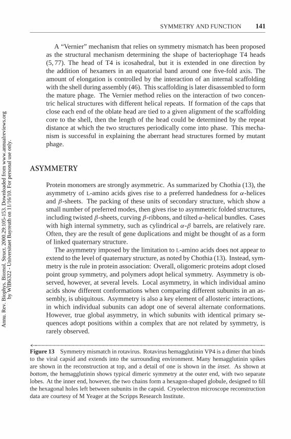
P1: FDS
April 20, 2000 11:37 Annual Reviews AR098-05
?SYMMETRY AND FUNCTION 141
A “Vernier” mechanism that relies on symmetry mismatch has been proposedas the structural mechanism determining the shape of bacteriophage T4 heads(5, 77). The head of T4 is icosahedral, but it is extended in one direction bythe addition of hexamers in an equatorial band around one five-fold axis. Theamount of elongation is controlled by the interaction of an internal scaffoldingwith the shell during assembly (46). This scaffolding is later disassembled to formthe mature phage. The Vernier method relies on the interaction of two concen-tric helical structures with different helical repeats. If formation of the caps thatclose each end of the oblate head are tied to a given alignment of the scaffoldingcore to the shell, then the length of the head could be determined by the repeatdistance at which the two structures periodically come into phase. This mecha-nism is successful in explaining the aberrant head structures formed by mutantphage.
ASYMMETRY
Protein monomers are strongly asymmetric. As summarized by Chothia (13), theasymmetry ofL-amino acids gives rise to a preferred handedness forα-helicesandβ-sheets. The packing of these units of secondary structure, which show asmall number of preferred modes, then gives rise to asymmetric folded structures,including twistedβ-sheets, curvingβ-ribbons, and tiltedα-helical bundles. Caseswith high internal symmetry, such as cylindricalα-β barrels, are relatively rare.Often, they are the result of gene duplications and might be thought of as a formof linked quaternary structure.
The asymmetry imposed by the limitation toL-amino acids does not appear toextend to the level of quaternary structure, as noted by Chothia (13). Instead, sym-metry is the rule in protein association: Overall, oligomeric proteins adopt closedpoint group symmetry, and polymers adopt helical symmetry. Asymmetry is ob-served, however, at several levels. Local asymmetry, in which individual aminoacids show different conformations when comparing different subunits in an as-sembly, is ubiquitous. Asymmetry is also a key element of allosteric interactions,in which individual subunits can adopt one of several alternate conformations.However, true global asymmetry, in which subunits with identical primary se-quences adopt positions within a complex that are not related by symmetry, israrely observed.
←−−−−−−−−−−−−−−−−−−−−−−−−−−−−−−−−−−−−−−−−−−−−−−−−−−−−−−−−−−−−−Figure 13 Symmetry mismatch in rotavirus. Rotavirus hemagglutinin VP4 is a dimer that bindsto the viral capsid and extends into the surrounding environment. Many hemagglutinin spikesare shown in the reconstruction at top, and a detail of one is shown in theinset. As shown atbottom, the hemagglutinin shows typical dimeric symmetry at the outer end, with two separatelobes. At the inner end, however, the two chains form a hexagon-shaped globule, designed to fillthe hexagonal holes left between subunits in the capsid. Cryoelectron microscope reconstructiondata are courtesy of M Yeager at the Scripps Research Institute.
Ann
u. R
ev. B
ioph
ys. B
iom
ol. S
truc
t. 20
00.2
9:10
5-15
3. D
ownl
oade
d fr
om w
ww
.ann
ualr
evie
ws.
org
by W
IB63
22 -
Uni
vers
itaet
Bay
reut
h on
11/
16/1
0. F
or p
erso
nal u
se o
nly.

P1: FDS
April 20, 2000 11:37 Annual Reviews AR098-05
?142 GOODSELL ¥ OLSON
Local Asymmetry
Nearly every crystal structure of an homooligomeric protein will show local dif-ferences in sidechain conformation and occasional small differences in backboneconformation. These may be observed as actual differences in atomic coordinates,if more than one subunit is found in the asymmetric unit, or as disordered residues,if the subunits are related by crystallographic symmetry. Often, these can be at-tributed to lack of constraints on the sidechain. Sidechains or even entire loopsthat are exposed to solvent will show largely different conformations in differentsubunits. Alternatively, crystal packing may order sidechains and flexible loopsin different conformations. These differences can provide valuable observationsof the mobility of proteins and the deformability of surface residues and loops.
Location near a symmetry axis can force breaking of local symmetry in a waythat is necessary for structural integrity. Often subunits will adopt different con-formations when near symmetry elements to optimize interactions across the axis.This was noted at the two-fold interface of insulin: “The two-fold-axis is not ex-actly obeyed, probably as a consequence of the very congested packing of residueswhich is observed here” (2). The flaps of HIV-1 protease provide another example,as shown in Figure 14. The peptide between ILE-50 and GLY-51 interacts withits symmetry mate across the two-fold axis; in one subunit, the amine hydrogenfaces across the axis; in the other, the peptide is rotated so that the carbonyl oxygenfaces across the axis, allowing formation of a hydrogen bond.
Local asymmetry seems to play a functional role in half-of-sites reactivity, ornegative cooperativity. In proteins that show half-of-sites reactivity, binding ofligand or effector to one subunit disfavors binding to the other subunit. Examplesinclude D-glyceraldehyde-3-phosphate dehydrogenase, alcohol dehydrogenase,
Figure 14 Local asymmetry in HIV-1 protease. Asymmetry of sidechains is often ob-served close to symmetry elements. The flaps of HIV-1 protease show asymmetry at ahydrogen bond (dotted line), that passes through the two-fold symmetry axis of dimericprotein, which runs top to bottom in the plane of the page, relating the flap in white to theshaded flap.
Ann
u. R
ev. B
ioph
ys. B
iom
ol. S
truc
t. 20
00.2
9:10
5-15
3. D
ownl
oade
d fr
om w
ww
.ann
ualr
evie
ws.
org
by W
IB63
22 -
Uni
vers
itaet
Bay
reut
h on
11/
16/1
0. F
or p
erso
nal u
se o
nly.

P1: FDS
April 20, 2000 11:37 Annual Reviews AR098-05
?SYMMETRY AND FUNCTION 143
arginine kinase, tyrosyl-tRNA synthetase, and many others (83). The crystal struc-tures of these enzymes show globally symmetric structures, with local asymmetryof loops, particularly around the active sites. The observation made by Moras et alseems to apply to the entire class: “The tetrameric molecule of glyceraldehyde-3-phosphate dehydrogenase has long been shown to have asymmetric propertieswhich almost certainly reflect themselves in small but definite differences of con-formation in the four polypeptide chains” (73).
Based on biochemical results that show half-of-sites reactivity, Degani &Degani propose that arginine kinase adopts a globally asymmetric structure (21).Crystal structures of this enzyme, however, show a symmetrical dimer. Crys-tal structure analyses have been reported for other proteins that show half-of-sites reactivity, including alcohol dehydrogenase, tyrosyl-tRNA synthetase, andD-glyceraldehyde-3-phosphate dehydrogenase. In all, the subunits are related bytwo-fold symmetry. The negative cooperativity appears to be effected throughsubtle motion sidechains, causing the two subunits to adopt different local struc-tures at the active site when complexed, but still retaining an overall symmetricalstructure when uncomplexed.
Reciprocating Mechanisms
Several examples of “reciprocating” mechanisms have been observed. In these,each subunit may adopt one or more states, but the state of one subunit is dependenton the states of the neighbors. For instance, envision a dimer in which each subunitmay adopt two conformations, A and B. The dimer interaction will be such that ifone subunit is in conformation A, the other must be in conformation B, and viceversa. Thus, at any given time, they do not adopt identical states, just like pistonson a crankshaft are all in different positions. However, if we take a time-averagedstructure, the three A-B pairs are identical. Also, in analogy with the pistons andcrankshaft, the reciprocating motion is processive; the conformation of one subunitwill be optimized for catalysis, and the conformation of the other subunit will beoptimized for binding to the next substrate.
ATP synthase is the best known example of this type of reciprocating engine.The reciprocating mechanism was proposed by Boyer (9), and the mechanism hasbeen revealed at the atomic level (1). ATP synthase is composed of aα3β3 ringof subunits, encircling a multisubunit membrane-bound axle. The reciprocatingmechanism cycles between three conformations: an O site with very low affinityfor ligands and no catalytic activity, an L site that binds ligands loosely and isinactive, and a T site that binds ligands tightly and performs the catalytic step. Inthe complex, one of the threeαβ heterodimers adopts each conformation at anygiven time, and physical turning of the axle converts one to the next.
The chaperonin GroEL acts, to a first approximation, through a two-state re-ciprocating mechanism (Figure 15). The asymmetric complex is composed of a14-subunit GroEL, arranged as two rings of seven subunits bound back-to-back
Ann
u. R
ev. B
ioph
ys. B
iom
ol. S
truc
t. 20
00.2
9:10
5-15
3. D
ownl
oade
d fr
om w
ww
.ann
ualr
evie
ws.
org
by W
IB63
22 -
Uni
vers
itaet
Bay
reut
h on
11/
16/1
0. F
or p
erso
nal u
se o
nly.

P1: FDS
April 20, 2000 11:37 Annual Reviews AR098-05
?144 GOODSELL ¥ OLSON
Figure 15 Reciprocating mechanism of GroEL:GroES. The asymmetric complex isformed of a 14-subunit GroEL and a conical GroES cap formed of seven subunits. Toa first approximation, the complex cycles between two conformations.Upper conforma-tion, the right half of GroEL is open, and the left half is capped by GroES, which inducesa significant conformational change in the GroEL subunits that it contacts.Lower confor-mation, the roles have reversed. The left side has lost GroES and is open, allowing thefolded protein to exit and new proteins to enter, and the right side is capped by GroES. Thecomplex is always asymmetric for the two sides, but, looking at a time average, both sidescycle through identical conformations.
with approximate D7 symmetry, and GroES, which forms a C7-symmetric cap onone ring of the GroEL complex (97). GroEL undergoes large deformations uponbinding of GroES, cycling between two conformations: acis conformation, towhich GroES is bound, which provides a closed space within which proteins fold,and thetrans conformation, which is open to the surrounding solvent, releasesand takes up polypeptides. One cycle allows about 15 seconds for the polypep-tide to fold, and requires 7 ATP molecules. Communication between the twohalves of the complex is mediated through a smooth tilt of subunits relative to theseven-fold axis, breaking the D7 symmetry of GroEL complex, but preserving theprotein-protein interactions that bind one ring to the other.
Global Asymmetry
Homooligomers with asymmetrically arranged subunits are remarkably rare. Onecan easily imagine an oligomer with nonintegral rotation symmetry or screw sym-metry that forms a complex of limited size, but without identical environments for
Ann
u. R
ev. B
ioph
ys. B
iom
ol. S
truc
t. 20
00.2
9:10
5-15
3. D
ownl
oade
d fr
om w
ww
.ann
ualr
evie
ws.
org
by W
IB63
22 -
Uni
vers
itaet
Bay
reut
h on
11/
16/1
0. F
or p
erso
nal u
se o
nly.

P1: FDS
April 20, 2000 11:37 Annual Reviews AR098-05
?SYMMETRY AND FUNCTION 145
Figure 16 Evolutionary argument against asymmetry.Right, in a symmetrical complex asshown, a portion of the protein surface is optimized through evolution to form an interface.Both subunits have a “knob” and a complementary “hole” that match across a two-fold-symmetry axis, shown with the small football symbol.Left, in the asymmetric complex,the surface forming the interface must be simultaneously optimized for interface contactand for interaction with solvent: the upper subunit in the complex has its “knob” formingan interface contact and its “hole” exposed to solvent, and in the lower subunit, the roles arereversed. Similar diagrams can be envisioned for other cyclic and screw-related asymmetriccomplexes.
each subunit. One can envision a strong evolutionary force against such asymmet-ric dimers. Compare the two dimers in Figure 16. In the two-fold related dimer,the interaction surface is identical in the two subunits: evolution would proceedby optimizing the face to be complementary with its mate. In the asymmetricdimer, on the other hand, the interface surface must be simultaneously optimizedto perform two roles. In one subunit, half of the “interface” will be in contact withthe neighbor, and half will be exposed to solvent; in the other subunit, these rolesare reversed. Thus, a large area must be evolutionarily optimized for interactionwith the neighborandwith solvent, and only half of this surface actually makescontact in the dimer.
Such a complex has been proposed for hexokinase, based on screw-relatedsubunits observed in crystal structures. However, the screw relationship is quitedifferent in two different crystal habits, arguing against this as the unique dimer-ization mode. A similar screw relationship has been proposed for arginine kinase,based on biochemical data showing half-of-sites reactivity.
The BTV core (Figure 9a) is the first X-ray crystallographic example of this typeof global asymmetry (31). In this structure, each position in aT = 1 icosahedronis filled with two subunits, bound back-to-back. The subunit is bean shaped andcomposed of three domains, and shows different conformations in the two differentasymmetric positions. It has yet to be determined whether the back-to-back dimeris a stable intermediate in the process of assembly.
HIV-1 reverse transcriptase shows a severe example of asymmetry, as shownin Figure 17. The active complex is a dimer of a 66,000-Dalton subunit and a51,000-Dalton subunit, the latter of which is a proteolyzed version of the first. Thecomplex has a single polymerase active site, a single RNase H proofreading site,
Ann
u. R
ev. B
ioph
ys. B
iom
ol. S
truc
t. 20
00.2
9:10
5-15
3. D
ownl
oade
d fr
om w
ww
.ann
ualr
evie
ws.
org
by W
IB63
22 -
Uni
vers
itaet
Bay
reut
h on
11/
16/1
0. F
or p
erso
nal u
se o
nly.

P1: FDS
April 20, 2000 11:37 Annual Reviews AR098-05
?146 GOODSELL ¥ OLSON
Figure 17 Asymmetry in human immunodeficiency virus-1 reverse transcriptase. Thereverse transcriptase of HIV-1 is composed of two subunits, a 66,000-Dalton subunit (white)and a 51,000-Dalton subunit (shaded), which is a proteolyzed version of the larger. Thetwo subunits adopt entirely different conformations in the complex.
and a single tRNA-binding site. Each subunit is composed of four similar sub-domains, which pack together in entirely different orientation in the two subunits(52). Owing to the strictures of viral economy, here one sequence plays two verydifferent structural roles.
EVOLUTION OF OLIGOMERIC PROTEINS
The remarkable solutions that cells have found to functional problems show thatthere are few limitations to what can be developed, given enough time. Just thesame, there have been attempts to justify the observed forms of symmetry withunderlying evolutionary arguments. Monod et al (72) presented one of the firstideas—that dimeric (isologous) interfaces are easier to create by mutation of exist-ing monomers than are interfaces in complexes of higher symmetry (heterologousinterfaces). They note that, on the surface of any monomer, pairs of comple-mentary residues are prepositioned for formation of dimeric contacts owing tosymmetry. For instance, a monomer with an arginine at one point on the surfaceand a glutamate some small distance away can immediately form two salt bridgeswith a second monomer, arginine on one to glutamate on the other, and glutamateon one to arginine on the other. The distance between the arginine and the gluta-mate on one subunit is necessarily identical with the distance between these tworesidues on the second subunit, providing the seed for a dimeric interface. This isnot the case with higher symmetries. In complexes with higher cyclic symmetry,the distance and orientation of a putative “seeding” pair are not rigidly definedaround the ring, so there exists the possibility of many nonproductive pairings.
Ann
u. R
ev. B
ioph
ys. B
iom
ol. S
truc
t. 20
00.2
9:10
5-15
3. D
ownl
oade
d fr
om w
ww
.ann
ualr
evie
ws.
org
by W
IB63
22 -
Uni
vers
itaet
Bay
reut
h on
11/
16/1
0. F
or p
erso
nal u
se o
nly.

P1: FDS
April 20, 2000 11:37 Annual Reviews AR098-05
?SYMMETRY AND FUNCTION 147
In polymeric complexes, the existence of a prepositioned pair is not possible, be-cause the two interacting surfaces are different, so evolution of a high-symmetrycomplex must begin with pairing of a single residue on each subunit. To give thesame strength of binding for this nascent oligomer, the high-symmetry interfacesmust have a fortuitous alignment of four residues on each subunit, two on one sideof the interface and two on the other.
Evolution of a D2 tetramer may be favored over that of a C4 tetramer by thissame argument. The D2 tetramer may be formed in two evolutionary steps, usingan existing complementary pair to seed a dimer interface between two monomers,and then, after this dimer has been optimized by evolution, using a second com-plementary pair on the dimer to seed a tetramer interface. The creation of a C4tetramer requires a concerted set of evolutionary steps, bringing the entire com-plex together at once. Hanson proposed that the requirement of evolution of anopen dimer as a first step towards creation of a ring would be an improbable eventand suggested that cyclic structures evolved sequentially by altering the relativeorientation of the binding faces: “In this way, a D2 isologous ring could evolve suc-cessively into a D3 and then a D4 ring, or ring contraction could take place” (33).Looking to the many structures of oligomeric proteins, we see that this type ofremodeling is probably not possible. It is difficult to postulate an evolutionarypathway that would change the angle between interfaces on a given subunit from180◦ to 120◦, either by repositioning an interface along the surface of a protein orby changing the angle by contracting the underlying protein fold.
Additional mutations favoring contact within interfaces are magnified in effectin oligomeric complexes. Monod et al write: “Because of the inherent coop-erativity of their structure, symmetrical oligomers should constitute particularlysensitive targets for molecular evolution, allowing much stronger selective pres-sures to operate in the random pursuit of functionally adequate structures” (72).A single mutation will form two new hydrogen bonds in a dimer, and single mu-tations will simultaneously relieve two close steric contacts. The effect is furthermagnified in higher-order oligomers.
These evolutionary forces are easily overcome by a specific functional need.Two observations give insight into the strength of these intrinsic structural forceson evolution relative to the strength of functional need. Arguing for a strongevolutionary preference for dimer interfaces, dimers and tetramers with D2 “dimer-of-dimers” symmetry are the most prevalent symmetries for soluble enzymes, evenfor those not showing significant allosteric behavior. Arguing against a strongforce, the processivity factors of DNA polymerase (Figure 5) use two differentsymmetries to fulfill the same need. In theE. coli β-subunit of polymerase IIIholoenzyme, two subunits assemble to form a ring with two interface patches. Theeukaryotic processivity factor of DNA polymeraseδ, on the other hand, adopts anidentical ring-shape, but it is composed of three subunits. An identical functionalneed is fulfilled by a dimer and by a trimer. Obviously, the evolutionary forcesseeding new interfaces are not strong enough to favor the dimer exclusively overthe trimer or vice-versa.
Ann
u. R
ev. B
ioph
ys. B
iom
ol. S
truc
t. 20
00.2
9:10
5-15
3. D
ownl
oade
d fr
om w
ww
.ann
ualr
evie
ws.
org
by W
IB63
22 -
Uni
vers
itaet
Bay
reut
h on
11/
16/1
0. F
or p
erso
nal u
se o
nly.

P1: FDS
April 20, 2000 11:37 Annual Reviews AR098-05
?148 GOODSELL ¥ OLSON
Xu et al discuss three possible modes for evolution of dimeric proteins (96),based on a survey of modern dimeric proteins. The first is the traditional mecha-nism, in which a stable monomeric species develops a dimerization site, perhapsthrough the use of complementary pairs of residues as described above. We mightexpect this mechanism to account for most of the oligomeric species observed to-day, such as superoxide dismutase in Figure 2a. It is not hard to image superoxidedismutase as a primitive species, stable as a monomer. The second pathway isthrough domain swapping, as first proposed by Bennett et al (4). The interleukinshown in Figure 2b may have evolved in this manner, with a swap across thenarrow linker at the center of the dumbbell-shaped molecule. The final mode forevolution of dimeric proteins is the most difficult of all—a one-step process, inwhich a dimeric species emerges fully formed. Xu et al propose several highly in-terlocked dimers, such as the gene V protein and the Trp aporepressor, as examplesof this mechanism. Because of their extensive interdigitation, they are probablynot stable as monomers, and the geometry of the backbone is not consistent withdomain swapping.
Unexpected symmetries may reveal a “frozen accident” of evolution (95)—asymmetry formed in the past and then unable to evolve further without loss offunction and a compromised organism. Hemoglobin provides a familiar example:One might expect that anα4 tetramer would be more genetically compact than theobservedα2β2 heterotetramer. Presumably, early in the evolution of vertebrates, agene duplication formed two copies of subunits for a tetrameric hemoglobin, andof the several competing forms—α4,β4, orα2β2—the heterotetramer was selected.
THE AESTHETICS OF SYMMETRY
Looking at much of decorative and fine art, we find that symmetry has a strongaesthetic appeal for many cultures. Symmetrical patterns, arrangements, and ob-jects adorn our rooms and define our architecture, and breaks from symmetry areincorporated deliberately and with artistic intent, because they will surprise andshock our expectations. When choosing vegetables or flowers, when looking atplants and animals, or when admiring the latest movie star, we favor the most sym-metric, seeking out the most “perfect” individuals. This predeliction for symmetrymay have its roots far back in our evolution, where symmetry was often a sign ofa healthy mate or fresh food.
Nature also selects for symmetry, for reasons of economy and control. Thesesymmetries occur at the molecular level, as described in this review, and extend tothe cellular and organismal level, for much the same reasons. Because symmetri-cal complexes are functionally more successful and economical than asymmetricobjects in many cases, our world is filled with five-pointed starfish, Y-shaped an-tibodies, spiral flowers, circular tree rings, geodesic diatoms, lenticular red bloodcells, and perfectly icosahedral viruses. These symmetries were perfected through
Ann
u. R
ev. B
ioph
ys. B
iom
ol. S
truc
t. 20
00.2
9:10
5-15
3. D
ownl
oade
d fr
om w
ww
.ann
ualr
evie
ws.
org
by W
IB63
22 -
Uni
vers
itaet
Bay
reut
h on
11/
16/1
0. F
or p
erso
nal u
se o
nly.

P1: FDS
April 20, 2000 11:37 Annual Reviews AR098-05
?SYMMETRY AND FUNCTION 149
functional pressures on evolution; their appeal to our senses is merely a happy sideeffect.
Of course, there remain symmetries that likely exist simply through serendipi-tous combinations of physical forces—not selected for functional need, but appear-ing fully formed once the universe took its current shape. The crystalline beauty ofminerals is one example, resulting from the minimization of inherent directionalinteractions of molecules. Similarly, theα-helix is another, with a structure definednot through evolutionary forces but through the intrinsic chemical geometry of thepolypeptide chain. The rainbow is perhaps the most glorious example—a gift ofNature existing simply through combination of the surface tension and refractiveindex of water.
ACKNOWLEDGMENTS
This work was funded by grant DEFG03 96ER2272 from the U.S. Department ofEnergy. This is publication 12361-MB from the Scripps Research Institute.
Visit the Annual Reviews home page at www.AnnualReviews.org
LITERATURE CITED
1. Abrahams JP, Leslie AGW, Lutter R, WalkerJE. 1994. Structure at 2.8 angstroms resolu-tion of F1-ATPase from bovine heart mito-chondria.Nature370:621–28
2. Adams MJ, Blundell TL, Dodson EJ,Dodson GG, Vijayan M, et al. 1969. Struc-ture of rhombohedral 2 zinc insulin crystals.Nature224:491–95
3. Barford D, Johnson LN. 1989. The allosterictransition of glycogen phosphorylase.Na-ture340:609–16
4. Bennett MJ, Schlunegger MP, Eisenberg D.1995. 3D domain swapping: a mechanismfor oligomer assembly.Protein Sci.4:2455–68
5. Berger B, Shor PW. 1998. On the struc-ture of the scaffolding core of bacteriophageT4 and its role in head length determina-tion. J. Struct. Biol.121:285–94
6. Beuron F, Maurizi MR, Belnap DM, KocsisE, Booy FP, et al. 1998. At sixes and sevens:characterization of the symmetry mismatchof the ClpAP chaperone-assisted protease.J. Struct. Biol.123:248–59
7. Blundell T, Dodson G, Hodgkin D, Mercola
D. 1972. Insulin: the structure in the crystaland its reflection in chemistry and biology.Adv. Protein Chem.26:279–402
8. Blundell TL, Srinivasan N. 1996. Symme-try, stability, and dynamics of multidomainand multicomponent protein systems.Proc.Natl. Acad. Sci. USA93:14243–48
9. Boyer PD. 1993. The binding site changemechanism for ATP synthase—some prob-abilities and possibilities.Biochim. Biophys.Acta1140:215–50
10. Calladine CR. 1978. Change of waveform inbacterial flagella: the role of mechanics atthe molecular level.J. Mol. Biol. 118:457–79
11. Caspar DLD. 1966. Design and assemblyof organized biological structure.Molecu-lar Architecture in Cell Physiology, Symp.Soc. Gen. Physiol.,pp. 191–207. New York:Prentice Hall
12. Caspar DLD, Klug A. 1962. Physical prin-ciples in the construction of regular viruses.Cold Spring Harbor Symp. Quant. Biol.27:1–24
13. Chothia C. 1991. Asymmetry in protein
Ann
u. R
ev. B
ioph
ys. B
iom
ol. S
truc
t. 20
00.2
9:10
5-15
3. D
ownl
oade
d fr
om w
ww
.ann
ualr
evie
ws.
org
by W
IB63
22 -
Uni
vers
itaet
Bay
reut
h on
11/
16/1
0. F
or p
erso
nal u
se o
nly.

P1: FDS
April 20, 2000 11:37 Annual Reviews AR098-05
?150 GOODSELL ¥ OLSON
structures.CIBA Found. Symp.162:36–5714. Chothia C, Janin J. 1975. Principles of
protein-protein recognition.Nature 256:705–8
15. Clegg JS. 1984. Properties and metabolismof the aqueous cytoplasm and its bound-aries.Am. J. Physiol.246:R133–51
16. Cornish-Bowden AJ, Koshland DE Jr.1971. The quaternary structure of proteinscomposed of identical subunits.J. Biol.Chem.246:3092–102
17. Crane HR. 1950. Principles and prob-lems of biological growth.Sci. MonthlyJune:376–89
18. Crick FHC, Watson JD. 1956. Structure ofsmall viruses.Nature177:473–75
19. Crick FHC, Watson JD. 1957. Virus struc-ture: general principles.CIBA Found.Symp. “The Nature of Viruses”,pp. 5–13.Boston: Little, Brown
20. Crothers DM, Metzger H. 1972. The in-fluence of polyvalency on the bindingproperties of antibodies.Immunochemistry9:341–57
21. Degani Y, Degani C. 1980. Enzymes withasymmetrically arranged subunits.TrendsBiochem. Sci.5:337–41
22. DeVos AM, Ultsch M, Kossiakoff AA.1992. Human growth hormone and ex-tracellular domain of its receptor: crystalstructure of the complex.Science255:306–12
23. Evans PR. 1991. Structural aspects of al-lostery.Curr. Opin. Struct. Biol.1:773–79
24. Fulton AB. 1982. How crowded is the cy-toplasm?Cell 30:345–47
25. Ganser BK, Li S, Klishko VY, Finch JT,Sundquist WI. 1999. Assembly and anal-ysis of conical models for the HIV-1 core.Science283:80–83
26. Garcia-Bellido A. 1996. Symmetriesthroughout organic evolution.Proc. Natl.Acad. Sci. USA93:14229–32
27. Goodsell DS. 1991. Inside a living cell.Trends Biochem. Sci.16:203–6
28. Goodsell DS, Olson AJ. 1993. Soluble pro-teins: size, shape and function.TrendsBiochem. Sci.18:65–68
29. Gouax JE, Lipscomb WN. 1990. Crystalstructures of phosphoacetamide ligated Tand phosphoacetamide and malonate lig-ated R states of aspartate carbamoyltrans-ferase at 2.8-A resolution and neutral pH.Biochemistry29:389–402
30. Greenbury CL, Moore DH, Nunn LAC.1965. The reaction with red cells of 7Srabbit antibody, its sub-units and their re-combinants.Immunology8:420–31
31. Grimes JM, Burroughs JN, Gouet P,Diprose JM, Malby R, et al. 1998. Theatomic structure of the bluetongue viruscore.Nature395:470–78
32. Groll M, Ditzel L, Lowe J, Stock D,Bochtler M, et al. 1997. Structure of the20S proteasome from yeast at 2.4A reso-lution. Nature386:463–71
33. Hanson KR. 1966. Symmetry of proteinoligomers formed by isologous associa-tion. J. Mol. Biol.22:405–9
34. Hargittai I, Hargittai M. 1995.Symmetrythrough the Eyes of a Chemist.New York:Plenum
35. Harrison SC, Olson AJ, Schutt CE, WinklerFK, Bricogne G. 1978. Tomato bushy stuntvirus at 2.9 Angstroms resolution.Nature276:368–73
36. Hendrix RW. 1978. Symmetry mismatchand DNA packaging in large bacte-riophages.Proc. Natl. Acad. Sci. USA75:4779–83
37. Herendeen DR, Kelly TJ. 1996. DNApolymerase III: running rings around thefork. Cell 84:5–8
38. Heuser J, Kirchhausen T. 1985. Deep-etchviews of clathrin assemblies.J. Ultrastruct.Res.92:1–27
39. Hewitt JA. 1977. On the influence of poly-valent ligands on membrane curvature.J.Theor. Biol.64:455–72
40. Hoare MR, Pal P. 1975. Physical clus-ter mechanics: statistical thermodynamicsand nucleation theory for monatomic sys-tems.Adv. Phys.24:645–78
41. Iwata S, Kamata K, Yoshida S, Minowa T,Ohta T. 1994. T and R states in the crystalsof bacterial L-lactate dehydrogenase reveal
Ann
u. R
ev. B
ioph
ys. B
iom
ol. S
truc
t. 20
00.2
9:10
5-15
3. D
ownl
oade
d fr
om w
ww
.ann
ualr
evie
ws.
org
by W
IB63
22 -
Uni
vers
itaet
Bay
reut
h on
11/
16/1
0. F
or p
erso
nal u
se o
nly.

P1: FDS
April 20, 2000 11:37 Annual Reviews AR098-05
?SYMMETRY AND FUNCTION 151
the mechanism for allosteric control.Na-ture Struct. Biol.1:176–85
42. Johnson JE, Speir JA. 1997. Quasi-equivalent viruses: a paradigm for proteinassemblies.J. Mol. Biol.269:665–75
43. Jones S, Thornton JM. 1996. Principlesof protein-protein interactions.Proc. Natl.Acad. Sci. USA93:13–20
44. Juo ZS, Chiu TK, Leiberman PM, BaikalovI, Berk AJ, Dickerson RE. 1996. How pro-teins recognize the TATA box.J. Mol. Biol.261:239–54
45. Kamiya R, Asakura S, Wakabayashi K,Namba K. 1979. Transition of bacterialflagella from helical to straight forms withdifferent subunit arrangements.J. Mol.Biol. 131:725–42
46. Kellenberger E. 1990. Form determinationof the heads of bacteriophages.Eur. J.Biochem.190:233–48
47. Kelman Z, Finkelstein J, O’Donnell M.1995. Why have six-fold symmetry?Curr.Biol. 5:1239–42
48. Klotz IM. 1967. Protein subunits: a table.Science155:697–98
49. Klotz IM, Darnall DW, Langerman NR,eds. 1975. Quaternary Structure of Pro-teins, pp. 293–411. New York: Academic
50. Klotz IM, Langerman NR, Darnall DW.1970. Quaternary structure of proteins.Annu. Rev. Biochem.39:25–62
51. Klug A. 1968. Point groups and thedesign of aggregates.Nobel Symp.“Sym-metry and Function of Biological Systemsat the Macromolecular Level” 11th, Stock-holm, pp. 425–36. Wiley & Sons, NewYork
52. Kohlstaedt LA, Wang J, Friedman JM, RicePA, Steitz TA. 1992. Crystal structure at3.5A resolution of HIV-1 reverse transcrip-tase complexed with an inhibitor.Science256:1783–89
53. Kong X-P, Onrust R, O-Donnell M,Kuriyan J. 1992. Three-dimensional struc-ture of the beta subunit ofE. coli DNApolymerase III holoenzyme: a slidingDNA clamp.Cell 69:425–37
54. Koshland DE. 1976. The evolution of func-tion in enzymes.Fed. Proc.35:2104–11
55. Koshland DE, Nemethy G, Filmer D. 1966.Comparison of experimental binding dataand theoretical models in proteins contain-ing subunits.Biochemistry5:365–85
56. Krishna TSR, Kong X-P, Gary S, Burg-ers PM, Kuriyan J. 1994. Crystal structureof the eukaryotic DNA processivity factorPCNA.Cell 79:1233–43
57. Kurland CG. 1992. Translational accuracyand the fitness of bacteria.Annu. Rev.Genet.26:29–50
58. Labeit S, Kolmerer B. 1995. Titins: giantproteins in charge of muscle ultrastructureand elasticity.Science270:293–96
59. Ladenstein R, Schneider M, Huber R, Bar-tunik H-D, Wilson K, et al. 1988. Heavyriboflavin synthase fromBacillus subtilis:crystal structure analysis of the icosahedralbeta-60 capsid at 3.3 A resolution.J. Mol.Biol. 203:1045–70
60. Larsen TA, Olson AJ, Goodsell DS. 1998.Morphology of protein-protein interfaces.Structure6:421–27
61. Lewis MS, Youle RJ. 1986. Ricin subunitassociation: thermodynamics and the roleof the disulfide bond in toxicity.J. Biol.Chem.261:11571–77
62. Liddington RC, Yan Y, Moulai J, Sahli R,Benjamin TL, Harrison SC. 1991. Struc-ture of simian virus 40 at 3.8-Angstromresolution.Nature354:278–84
63. Lipscomb WN. 1991. Structureand function of allosteric enzymes.CHEMTRACTS-Biochem. Mol. Biol.[Data Trace Chem. Publ.] 2:1–15
64. Lowe J, Stock D, Jap B, Zwickl P, Baumeis-ter W, Huber R. 1995. Crystal structure ofthe 20S proteosome from the archaeon T.acidophilum at 3.4A resolution.Science268:533–39
65. Lumry R, Rajender S. 1970. Enthalpy-entropy compensation phenomena in watersolutions of proteins and small molecules:a ubiquitous property of water.Biopoly-mers9:1125–227
Ann
u. R
ev. B
ioph
ys. B
iom
ol. S
truc
t. 20
00.2
9:10
5-15
3. D
ownl
oade
d fr
om w
ww
.ann
ualr
evie
ws.
org
by W
IB63
22 -
Uni
vers
itaet
Bay
reut
h on
11/
16/1
0. F
or p
erso
nal u
se o
nly.

P1: FDS
April 20, 2000 11:37 Annual Reviews AR098-05
?152 GOODSELL ¥ OLSON
66. Matsudaira P. 1991. Modular organiza-tion of actin crosslinking proteins.TrendsBiochem. Sci.16:87–92
67. Mattevi A, Rizzi M, Bolognesi M.1996. New structures of allosteric pro-teins revealing remarkable conformationalchanges.Curr. Opin. Struct. Biol.6:824–29
68. Mattevi A, Valentini G, Rizzi M, SperanzaML, Bolognesi M, Coda A. 1995. Crystalstructure ofEscherichia colipyruvate ki-nase type I: molecular basis of the allosterictransition.Structure3:729–41
69. Mitchison T, Kirschner M. 1984. Dynamicinstability of microtubule growth.Nature312:237–42
70. Mitchison TJ. 1988. Microtubule dynamicsand kinetochore function in mitosis.Annu.Rev. Cell Biol.4:527–49
71. Monod J. 1968. On symmetry and functionin biological systems.Nobel Symp. Symme-try Funct. Biol. Syst. Macromol. Lev., 11th,Stockholm,pp. 15–27. New York: Wiley
72. Monod J, Wyman J, Changeux J-P. 1965.On the nature of allosteric transitions:a plausible model.J. Mol. Biol. 12:88–118
73. Moras D, Olsen KW, Sabesan MN,Buehner M, Ford GC, Rossmann MG.1975. Studies of asymmetry in thethree-dimensional structure of lobsterD-glyceraldehyde-3-phosphate dehydro-genase.J. Biol. Chem.250:9137–62
74. Olson AJ, Bricogne G, Harrison SC. 1983.Structure of tomato bushy stunt virus IV.The virus particle at 2.9A resolution.J.Mol. Biol. 171:61–93
75. Parker J. 1989. Errors and alternatives inreading the universal genetic code.Micro-biol. Rev.53:273–98
76. Pauling L. 1953. Protein interactions: ag-gregation of globular proteins.Discov.Faraday Soc.13:170–76
77. Paulson JR, Laemmli UK. 1977. Morpho-genetic core of the bacteriophage T4 head.Structure of the core in polyheads.J. Mol.Biol. 111:459–85
78. Pereira PJB, Bergner A, Macedo-RibeiroS, Huber R, Matxchiner G, et al. 1998.Human beta-trypsin is a ring-like tetramerwith active sites facing a central pore.Na-ture392:306–11
79. Perutz MF. 1989. Mechanisms of coopera-tivity and allosteric regulation in proteins.Q. Rev. Biophys.22:139–236
80. Reed LJ, Hackert ML. 1990. Structure-function relationships in dihydrolipoamideacyltransferases.J. Biol. Chem.265:8971–74
81. Ricard J, Noat G. 1985. Kinetic co-operativity of monomeric mnemonical en-zymes.Eur. J. Biochem.152:557–64
82. Rossmann MG. 1984. Constraints on theassembly of spherical virus particles.Vi-rology134:1–11
83. Seydoux F, Malhotra OP, Bernhard SA.1974. Half-site reactivity.CRC Crit. Rev.Biochem.2:227–57
84. Sharp K, Fine R, Honig B. 1987. Com-puter simulations of the diffusion of a sub-strate to an active site of an enzyme.Sci-ence236:1460–64
85. Sixma TK, Pronk SE, Kalk KH, Wartna ES,Zanten BAM, et al. 1991. Crystal structureof a cholera toxin-related heat-labile en-terotoxin fromE. coli. Nature351:371–77
86. Srere PA. 1981. Protein crystals as amodel for mitochondrial proteins.TrendsBiochem. Sci.6:4–7
87. Srere PA. 1984. Why are enzymes so big?Trends Biochem. Sci.9:387–90
88. Stossel TP. 1989. From signal to pseudo-pod: How cells control cytoplasmic actinassembly.J. Biol. Chem.264:18261–64
89. Strater N, Hakansson K, Schnappauf G,Braus G, Lipscomb WN. 1996. Crystalstructure of the T state of allosteric yeastchorismate mutase and comparison withthe R state.Proc. Natl. Acad. Sci. USA93:3330–34
90. Svedberg T. 1929. Mass and size of proteinmolecules.Nature123:871
91. Theil EC. 1987. Ferritin: structure,gene regulation, and cellular function in
Ann
u. R
ev. B
ioph
ys. B
iom
ol. S
truc
t. 20
00.2
9:10
5-15
3. D
ownl
oade
d fr
om w
ww
.ann
ualr
evie
ws.
org
by W
IB63
22 -
Uni
vers
itaet
Bay
reut
h on
11/
16/1
0. F
or p
erso
nal u
se o
nly.

P1: FDS
April 20, 2000 11:37 Annual Reviews AR098-05
?SYMMETRY AND FUNCTION 153
animals, plants and microorganisms.Annu.Rev. Biochem.56:289–315
92. vanHeel M, Orlova EV, Dube P, Tavares P.1996. Intrinsic versus imposed curvaturein cyclical oligomers: the portal protein ofbacteriophage SPP1.EMBO J. 15:4785–88
93. Wilson DR. 1993. Bacterial flagellar fila-ments and their component flagellins.Can.J. Microbiol.39:451–72
94. Wlodawer A, Erickson JW. 1993. Struc-ture-based inhibitors of HIV-1 protease.Annu. Rev. Biochem.62:543–85
95. Wolynes PG. 1996. Symmetry and the en-ergy landscapes of biomolecules.Proc.Natl. Acad. Sci. USA93:14249–55
96. Xu D, Tsai C-J, Nussinov R. 1998. Mecha-nism and evolution of protein dimerization.Protein Sci.7:533–44
97. Xu Z, Horwich AL, Sigler PB. 1997. Thecrystal structure of the asymmetric GroEL-GroES-(ADP)7 chaperonin complex.Na-ture388:741–50
98. Yang M, Chen X-p, Militello K, Hoff-man R, Fernandez B, et al. 1997.Alanine-scanning mutagenesis ofBacillussubtilis trp RNA-binding attenuation pro-tein (TRAP) reveals residues involved intryptophan binding and RNA binding.J.Mol. Biol. 270:696–710
99. Yeager M, Berriman JA, Baker TS, Bel-lamy AR. 1994. Three-dimensional struc-
ture of the rotavirus haemagglutinin VP4by cryo-electron microscopy and differ-ence map analysis.EMBO J.13:1011–18
100. Zhang R-G, Scott DL, Westbrook ML,Nance S, Spangler BD, et al. 1995.The three-dimensional crystal structure ofcholera toxin.J. Mol. Biol.251:563–73
101. Protein Data Bank accession codes forstructures used in the figures. ThePDB may be reached at http://www.rcsb.org/pdb. Figure 1: pepsin, 5pep; alco-hol dehydrogenase, 2ohx; porin, 2por;neuraminidase, 1ivb; phosphofructoki-nase, 1pfk; aspartate carbamoyltrans-ferase, 1at1; glycolate oxidase, 1gox; glu-tamine synthetase, 2gls; protocatechuate-3,4-dioxygenase, 3pcg; ferritin, 1hrs;satellite tobacco necrosis virus, 2stv. Fig-ure 2 superoxide dismutase, 1xso; inter-leukin 10, 1ilk. Figure 4: fructose-1,6-bisphosphatase, 4fbp (T) and 5fbp (R); as-partate carbamoyltransferase, 4at1 (T) and1at1 (R). Figure 5:β subunit of RNA polIII, 2pol; PCNA, 1axc. Figure 6: GroEL,1der; proteasome, 1ryp. Figure 7: satellitetobacco necrosis virus, 2stv; tomato bushystunt virus, 2tbv. Figure 8: bluetonguevirus, 2btv; simian virus 40, 1sva. Figure11: hemoglobin, 4hhb. Figure 13: HIV-1protease, 7hvp. Figure 14: GroEL:GroES,1aon. Figure 16: reverse transcriptase,3hvt.
Ann
u. R
ev. B
ioph
ys. B
iom
ol. S
truc
t. 20
00.2
9:10
5-15
3. D
ownl
oade
d fr
om w
ww
.ann
ualr
evie
ws.
org
by W
IB63
22 -
Uni
vers
itaet
Bay
reut
h on
11/
16/1
0. F
or p
erso
nal u
se o
nly.

Annual Review of Biophysics and Biomolecular Structure Volume 29, 2000
CONTENTSMEASURING THE FORCES THAT CONTROL PROTEIN INTERACTIONS, Deborah Leckband 1STRUCTURE AND FUNCTION OF LIPID-DNA COMPLEXES FOR GENE DELIVERY, S. Chesnoy, L. Huang 27SIGNALING AND SUBCELLULAR TARGETING BY MEMBRANE-BINDING DOMAINS, James H. Hurley, Saurav Misra 49GCN5-RELATED N-ACETYLTRANSFERASES: A Structural Overview, Fred Dyda, David C. Klein, Alison Burgess Hickman 81STRUCTURAL SYMMETRY AND PROTEIN FUNCTION, David S. Goodsell, Arthur J. Olson 105
ELECTROKINETICALLY CONTROLLED MICROFLUIDIC ANALYSIS SYSTEMS, Luc Bousse, Claudia Cohen, Theo Nikiforov, Andrea Chow, Anne R. Kopf-Sill, Robert Dubrow, J. Wallace Parce
155DNA RECOGNITION BY Cys2His2 ZINC FINGER PROTEINS, Scot A. Wolfe, Lena Nekludova, Carl O. Pabo 183PROTEIN FOLDING INTERMEDIATES AND PATHWAYS STUDIED BY HYDROGEN EXCHANGE, S. Walter Englander 213QUANTITATIVE CHEMICAL ANALYSIS OF SINGLE CELLS, D. M. Cannon Jr, N. Winograd, A. G. Ewing 239THE STRUCTURAL BIOLOGY OF MOLECULAR RECOGNITION BY VANCOMYCIN, Patrick J. Loll, Paul H. Axelsen 265COMPARATIVE PROTEIN STRUCTURE MODELING OF GENES AND GENOMES, Marc A. Martí-Renom, Ashley C. Stuart, András Fiser, Roberto Sánchez, Francisco Melo, Andrej Sali 291FAST KINETICS AND MECHANISMS IN PROTEIN FOLDING, William A. Eaton, Victor Muñoz, Stephen J. Hagen, Gouri S. Jas, Lisa J. Lapidus, Eric R. Henry, James Hofrichter 327ATOMIC FORCE MICROSCOPY IN THE STUDY OF MACROMOLECULAR CRYSTAL GROWTH, A. McPherson, A. J. Malkin, Yu. G. Kuznetsov 361A DECADE OF CLC CHLORIDE CHANNELS: Structure, Mechanism, and Many Unsettled Questions, Merritt Maduke, Christopher Miller, Joseph A. Mindell 411DESIGNED SEQUENCE-SPECIFIC MINOR GROOVE LIGANDS, David E. Wemmer 439PULSED AND PARALLEL-POLARIZATION EPR CHARACTERIZATION OF THE PHOTOSYSTEM II OXYGEN-EVOLVING COMPLEX, R. David Britt, Jeffrey M. Peloquin, Kristy A. Campbell 463
Ann
u. R
ev. B
ioph
ys. B
iom
ol. S
truc
t. 20
00.2
9:10
5-15
3. D
ownl
oade
d fr
om w
ww
.ann
ualr
evie
ws.
org
by W
IB63
22 -
Uni
vers
itaet
Bay
reut
h on
11/
16/1
0. F
or p
erso
nal u
se o
nly.

ELECTROSTATIC MECHANISMS OF DNA DEFORMATION, Loren Dean Williams, L. James Maher III 497STRESS-INDUCED STRUCTURAL TRANSITIONS IN DNA AND PROTEINS, T. R. Strick, J.-F. Allemand, D. Bensimon, V. Croquette 523MOLECULAR MECHANISMS CONTROLLING ACTIN FILAMENT DYNAMICS IN NONMUSCLE CELLS, Thomas D. Pollard, Laurent Blanchoin, R. Dyche Mullins 545
UNNATURAL LIGANDS FOR ENGINEERED PROTEINS: New Tools for Chemical Genetics, Anthony Bishop, Oleksandr Buzko, Stephanie Heyeck-Dumas, Ilyoung Jung, Brian Kraybill, Yi Liu, Kavita Shah, Scott Ulrich, Laurie Witucki, Feng Yang, Chao Zhang, Kevan M. Shokat
577
Ann
u. R
ev. B
ioph
ys. B
iom
ol. S
truc
t. 20
00.2
9:10
5-15
3. D
ownl
oade
d fr
om w
ww
.ann
ualr
evie
ws.
org
by W
IB63
22 -
Uni
vers
itaet
Bay
reut
h on
11/
16/1
0. F
or p
erso
nal u
se o
nly.