structuralandfunctionalcharacterizationof ... · structuralandfunctionalcharacterizationof...
TRANSCRIPT

Structural and Functional Characterization ofTransmembrane Segment VII of the Na�/H� ExchangerIsoform 1*□S
Received for publication, June 27, 2006, and in revised form, July 20, 2006 Published, JBC Papers in Press, July 21, 2006, DOI 10.1074/jbc.M606152200
Jie Ding‡1,2, Jan K. Rainey‡§1,3, Caroline Xu‡, Brian D. Sykes‡§4, and Larry Fliegel‡5
From the ‡Department of Biochemistry and §Protein Engineering Network of Centres of Excellence, University of Alberta,Edmonton, Alberta T6G 2H7, Canada
The Na�/H� exchanger isoform 1 is an integral membraneprotein that regulates intracellular pH by exchanging one intra-cellular H� for one extracellular Na�. It is composed of anN-terminal membrane domain of 12 transmembrane segmentsand an intracellular C-terminal regulatory domain. We charac-terized the structural and functional aspects of the critical trans-membrane segment VII (TM VII, residues 251–273) by usingalanine scanning mutagenesis and high resolution NMR. Eachresidue of TM VII was mutated to alanine, the full-length pro-tein expressed, and its activity characterized. TMVII was sensi-tive to mutation. Mutations at 13 of 22 residues resulted inseverely reduced activity,whereas othermutants exhibited vary-ing degrees of decreases in activity. The impaired activitiessometimes resulted from low expression and/or low surface tar-geting. Three of the alanine scanningmutant proteins displayedincreased, and two displayed decreased resistance to theNa�/H� exchanger isoform 1 inhibitor EMD87580. The struc-ture of a peptide of TM VII was determined by using high reso-lution NMR in dodecylphosphocholine micelles. TM VII ispredominantly �-helical, with a break in the helix at the func-tionally critical residues Gly261–Glu262. The relative positionsand orientations of the N- and C-terminal helical segments areseen to vary about this extended segment in the ensemble ofNMRstructures.Our results show thatTMVII is a critical trans-membrane segment structured as an interrupted helix, with sev-
eral residues that are essential to both protein function and sen-sitivity to inhibition.
The mammalian Na�/H� exchanger isoform 1 (NHE1)6 is aubiquitous integral membrane protein mediating removal of asingle intracellular proton in exchange for one extracellularsodium ion (1). NHE1 has several cellular and physiologicalfunctions, including protecting cells from intracellular acidifi-cation (2, 3), promoting cell growth and differentiation (2), andregulating sodium fluxes and cell volume after osmotic shrink-age (4). The Na�/H� exchanger also plays a critical role in thedamage that occurs during ischemia and reperfusion and mayplay a key role inmediating heart hypertrophy. Inhibition of theexchanger protects themyocardium in these two forms of heartdisease (5, 6). Amiloride and its derivatives are inhibitors of theNHE1 isoformof theNa�/H� exchanger, and a new generationof Na�/H� exchanger inhibitors is being developed for clinicaltreatment of heart disease (7, 8). In addition to these manyphysiological roles, more recently, the Na�/H� exchanger hasbeen demonstrated to be involved in modulating cell motilityand invasiveness of neoplastic breast cancer cells (9) and hasbeen shown to be critical to cell motility in some cell types (10).NHE1 is composed of two domains as follows: anN-terminal
membrane domain of �500 amino acids and a C-terminal reg-ulatory domain of �315 amino acids (1, 6). The N-terminalmembrane domain is responsible for ionmovement and has 12transmembrane segments and 3 membrane-associated seg-ments (11) (Fig. 1A). How this domain binds and transportsNa� ions and protons is only recently starting to be elucidated.We have recently analyzed TM IV of the NHE1 isoform of theNa�/H� exchanger. We showed that prolines 167 and 168 arecritical to NHE1 function, targeting, and expression (12).Phe161 was shown to be a pore-lining residue critical to trans-port, and the structure of TM IV was shown to be atypical ofTM proteins, being composed of one region of �-turns, anextendedmiddle region, including Pro167–Pro168, plus a regionhelical in character (13).
* The costs of publication of this article were defrayed in part by the paymentof page charges. This article must therefore be hereby marked “advertise-ment” in accordance with 18 U.S.C. Section 1734 solely to indicate this fact.
The atomic coordinates and structure factors (code 2HTG) have been deposited inthe Protein Data Bank, Research Collaboratory for Structural Bioinformatics,Rutgers University, New Brunswick, NJ (http://www.rcsb.org/).
□S The on-line version of this article (available at http://www.jbc.org) containssupplemental text, Figs. S1 and S2, and supplemental Refs. 1–10.
1 Both authors contributed equally to this work.2 Supported by Alberta Heritage Foundation for Medical Research and the
Canadian Institutes of Health Research Strategic Training Institute in Mem-brane Proteins and Cardiovascular Disease.
3 Recipient of postdoctoral fellowships from the Natural Sciences and Engi-neering Research Council of Canada, the Alberta Heritage Foundation forMedical Research, and the Canadian Institutes of Health Research StrategicTraining Institute in Membrane Proteins and Cardiovascular Disease.
4 Recipient of support as a Canada Research Chair in Structural Biology.5 Supported by a grant from the Canadian Institutes of Health Research and a
Scientist Award from Alberta Heritage Foundation for Medical Research.To whom correspondence should be addressed: Dept. of Biochemistry,347 Medical Science Bldg., University of Alberta, Edmonton, Alberta T6G2H7, Canada. Tel.: 780-492-1848; Fax: 780-492-0886; E-mail: [email protected].
6 The abbreviations used are: NHE1, Na�/H� exchanger isoform 1; DIPSI,decoupling in the presence of scalar interactions; DPC, dodecylphospho-choline; DPC-d38, deuterated DPC; HA, hemagglutinin; HSQC, hetero-nuclear single quantum coherence spectroscopy; NOE, nuclear Over-hauser effect; NOESY, NOE spectroscopy; PBS, phosphate-buffered saline;TM, transmembrane (segment); TOCSY, total correlation spectroscopy;HPLC, high pressure liquid chromatography; t-Boc, tert-butyloxycarbonyl.
THE JOURNAL OF BIOLOGICAL CHEMISTRY VOL. 281, NO. 40, pp. 29817–29829, October 6, 2006© 2006 by The American Society for Biochemistry and Molecular Biology, Inc. Printed in the U.S.A.
OCTOBER 6, 2006 • VOLUME 281 • NUMBER 40 JOURNAL OF BIOLOGICAL CHEMISTRY 29817
by guest on June 18, 2018http://w
ww
.jbc.org/D
ownloaded from

TM VII is believed to extend from amino acids Ile251 toLeu273 (11) (Fig. 1B). We have shown that TMVII is critical forthe function of the NHE1 isoform of the Na�/H� exchanger(14). We demonstrated that Glu262 and Asp267 of TM VII arecritical for activity. Residues with a similar position and chargein TM VI were not important. The E262Q and D267N muta-tions destroyed activity of the NHE, whereas replacement withthe other negatively charged amino acid retained, butmodified,exchanger activity. In this study, we examine both structuraland functional aspects of TM VII of the NHE1 isoform of theNa�/H� exchanger. We use alanine scanning and insertionalmutagenesis (15, 16) to determine amino acid residues criticalfor Na�/H� exchanger activity, expression, targeting, andinhibitor resistance.We also useNMR to examine the structureof an isolated transmembrane segment. It must be acknowl-edged that an isolated membrane segment could have a differ-ent structure than that found in an intact membrane protein.However, isolated TM segments frommembrane proteins hav-ing multiple TM segments, including the cystic fibrosis trans-membrane conductance regulator (17, 18), bacteriorhodopsin
(19, 20), rhodopsin (21), and the fungal G-protein-coupledreceptor Ste2p (22), have been shown to be both functional andto have structures in good agreement with the segments in thecontext of the entire protein, where available. An importantcaveat is that solution conditionsmay need extensive screeningin order to achieve stabilization of the peptide in its physiolog-ical structure, for example, the variation in bacteriorhodopsinresults of Ref. 19 versusRef. 20 and our previous study of TM IVof NHE1 (13). Following from this body of work, the TM VIIpeptide was chemically synthesized and used to determine itsstructure in dodecylphosphocholine (DPC)micelles. The use ofDPCmicelles as amembranemimetic environment in the solu-tion state has been well established through structural studiesof both membrane-spanning and membrane-associated pro-teins or peptides (23, 24). Our study demonstrates that TMVIIis structured as an interrupted, likely kinked �-helix. We iden-tify residues critical in transport and those thatmodify inhibitorresistance. TMVII is distinctly different fromTM IV in both itsstructure and in functional aspects.
EXPERIMENTAL PROCEDURES
Materials—EMD87580 was a generous gift of Dr. N. Beier ofMerck. PWO DNA polymerase was from Roche Applied Sci-ence, and LipofectamineTM 2000 reagent was from Invitrogen.15N-Labeled, t-Boc-protected amino acids and deuterated sol-vents were purchased from Cambridge Isotope Laboratories(Andover, MA). Deuterated SDS and DPC were purchasedfrom C/D/N Isotopes (Pointe-Claire, Quebec, Canada) andused without further purification.Site-directed Mutagenesis—To examine and characterize
critical amino acids of TMVII of theNa�/H� exchanger,muta-tions were made to an expression plasmid containing a hemag-glutinin (HA)-tagged human NHE1 isoform of the Na�/H�
exchanger. The plasmid pYN4� contains the cDNA of theentire coding region of the Na�/H� exchanger (12). Two seriesof mutants were made (Table 1). One series of mutants wasmade in which all the residues of TM VII were mutated toalanine. A second series was for insertional mutagenesis,whereby alanine residues were inserted at critical locationsbetween residues of TM VII in the wild type pYN4�. Two ala-nine insertional mutants were made, one inserting an alaninebetween Gly261 and Glu262, and a second inserting an alaninebetween Leu264 and Leu265. A third insertional mutant had twomutations, a glutamate inserted betweenGly261 andGlu262 plusan N266D mutation. Site-directed mutagenesis using amplifi-cation with PWO DNA polymerase (Roche Applied Science)was followed by using the Stratagene (La Jolla, CA)QuikChangeTM site-directed mutagenesis kit. Mutations cre-ated a new restriction enzyme site for use in screening transfor-mants. DNA sequencing confirmed the mutations and fidelityof PCR.Cell Culture and Transfections—To examine Na�/H�
exchanger expression and activity, AP-1 cells were used thatlack an endogenousNa�/H� exchanger (14). TransfectionwithLipofectamineTM 2000 reagent (Invitrogen) was used to makestable cell lines of all mutants as described earlier (12). Trans-fected cells were selected using 800 �g/ml geneticin (G418),
FIGURE 1. Models of the Na�/H� exchanger. A, topological model of thetransmembrane domain of the NHE1 isoform of the Na�/H� exchanger asdescribed earlier (11). B, model of amino acids present in TM VII.
Structural and Functional Characterization of TM VII of NHE1
29818 JOURNAL OF BIOLOGICAL CHEMISTRY VOLUME 281 • NUMBER 40 • OCTOBER 6, 2006
by guest on June 18, 2018http://w
ww
.jbc.org/D
ownloaded from

and stable cell lines for experiments were regularly re-estab-lished from frozen stocks at passage numbers between 5 and 9.SDS-PAGEand Immunoblotting—ToconfirmNHE1 expres-
sion, cell lysates weremade fromAP-1 cells as described earlier(12). For Western blot analysis, equal amounts of up to 100 �gof each sample were resolved on 10% SDS-polyacrylamide gels.Nitrocellulose transfers were immunostained using anti-HAmonoclonal antibody (Roche Applied Science) and peroxidase-conjugated goat anti-mouse antibody (Bio/Can, Mississauga,Ontario, Canada). The enhanced chemiluminescenceWesternblotting and detection system (Amersham Biosciences) wasused to visualize immunoreactive proteins. Densitometricanalysis of x-ray films was using NIH Image 1.63 software(National Institutes of Health, Bethesda).Cell Surface Expression—Cell surface expression was meas-
uredwith sulfo-NHS-SS-biotin (Pierce) essentially as describedearlier (12). Briefly, the cell surfacewas labeledwith sulfo-NHS-SS-biotin, and immobilized streptavidin resin was used toremove plasma membrane Na�/H� exchanger. Equivalentamounts of the total and unbound proteins were analyzed bySDS-PAGE followed by Western blotting and densitometry asdescribed above. The relative amount of NHE1 on the cell sur-face was calculated by comparing both the 110- and the 95-kDaHA-immunoreactive species in Western blots of the total andunbound fractions.Na�/H� Exchange Activity—Na�/H� exchange activity was
measured using a PTI Deltascan spectrofluorometer. Theinitial rate of Na�-induced recovery of cytosolic pH (pHi)was measured after ammonium chloride-induced acute acidload using 2�,7�-bis(carboxyethyl)-5,6-carboxyfluorescein-AM(Molecular Probes Inc., Eugene, OR). Recovery was in the pres-
ence of 135mMNaCl andwasmeasured as described previously(13). There was no difference in the buffering capacities of sta-ble cell lines as indicated by the degree of acidification inducedby ammonium chloride. For some experiments cells weretreated with EMD87580 of varying concentrations. EMD87580was dissolved in water, and the inhibitory effect of EMD87580was documented using a two-pulse acidification assay. Cellswere treated with ammonium chloride two times and allowedto recover in NaCl-containing medium. One pulse was in theabsence of EMD87580 and one in the presence of inhibitor. Therate of recovery from acid load was compared � inhibitor.Where indicated, the activity of the Na�/H� exchangermutants was corrected for the level of protein expression andfor the targeting of the protein to the cell surface. Results areshown as mean � S.E., and statistical significance was deter-mined using the Mann-Whitney U test.Peptide Synthesis and Purification—TM VII peptides
(sequence, HINELLHILVFGESLLNDAVTVVLYKK; free Nterminus, amide-capped C terminus) with and without selec-tive 15N labels were synthesized and purified using previouslypublished t-Boc solid-phase techniques optimized for hydro-phobic membrane-spanning peptides (25), with the differencethat purification was carried out using a Zorbax 300 SB-C39.4-mm � 25-cm HPLC column (Agilent Technologies, PaloAlto, CA). Peptide identity was confirmed by matrix-assistedlaser desorption ionization mass spectrometry and by aminoacid analysis.NMR Spectroscopy and Structure Calculation—Samples for
structural study were obtained by dissolving �1 mM syntheticTMVII peptide in 90%H2O, 10%D2O solution containing�75mM DPC-d38. Chemical shifts were referenced to 2,2-dimeth-
TABLE 1Oligonucleotides used for site-directed mutagenesisMutated nucleotides are in lowercase letters and boldface type. Mutated amino acid residues are indicated using single letter notation, and new restriction sites areunderlined. Restriction sites deleted are indicated in parentheses. Amino acids flanking the insert are indicated where insertional mutagenesis occurred. Top, oligonucleo-tides used for alanine scanning mutagenesis. Bottom, synthetic oligonucleotides used for alanine insertional mutagenesis.
Mutation Oligonucleotide sequence (alanine scanning) Restriction site
I251A 5�-GAGGAAATTCACgcgAATGAGCTGCTG-3� BcgIN252A 5�-GGAAATTCACATCgcTGAGCTcCTGCACATCCTTG-3� SstIE253A 5�-TTCACATCAATGcGCTcCTcCACATCCTTGTTTTTG-3� BseRI1L254A 5�-CACATCAATGAagcttTGCACATCCTTG-3� HindIIIL255A 5�-CATCAATGAGCTagcGCACATCCTTG-3� NheIH256A 5�-CAATGAGCTGCTagcCATCCTTGTTTTTGG-3� NheII257A 5�-CAATGAGCTGCTGCAtgcCCTTGTTTTTGGG-3� SphIL258A 5�-GAGCTGCTGCAtATCgcTGTTTTTGGGGAG-3� (BsgI)V259A 5�-GCTGCACATCCTTgcaTTcGGGGAGTCCTTGC-3� BsmIF260A 5�-GCACATCCTTGTTgccGGcGAGTCCTTGCTC-3� NaeIG261A 5�-CATCCTTGTTTTTGcGGAaagCTTGCTCAATGACG-3� HindIIIE262A 5�-CCTTGTTTTTGGGGcaagCTTGCTCAATGACG-3� HindIIIS263A 5�-CTTGTTTTTGGGGAGgCCTTGCTCAATGAC-3� StuIL264A 5�-GTTTTTGGGGAGTCCgcttTaAATGACGCCGTCAC-3� DraIL265A 5�-GTTTTTGGGGAGTCCcTcgcgAATGACGCCGTCAC-3� NruIN266A 5�-TTGGGAGTCCTTGCTagcTGACGCCGTCACTG-3� NheID267A 5�-GTCCTTGCTCAATGctGCaGTCACTGTGGTCC-3� PstIV269A 5�-CTCAATGACGCCGcgACTGTGGTgCTGTATC-3� BcgIT270A 5�-CAATGACGCCGTCgCgGTcGTCCTGTATCAC-3� BsiEIV271A 5�-GACGCCGTCACTGcaGTCCTGTATCACC-3� PstIV272A 5�-GACGCCGTCACgGTcgcCCTGTATCACCTC-3� BsiEIL273A 5�-GTCACTGTGGTCgcGTATCACCTCTTTG-3� (AvaII)Mutation Oligonucleotide sequence Restriction site
G261-A-262E 5�-CACATCCTTGTTTTTGGcgccGAGTCCTTGCTCAATGACG-3� NarIL264-A-265L 5�-GGGAGTCCTTagcgCTCAATGACG-3� Eco47IIIG261-E-262E 5�-TGTTTTTGGGgagGAaagcttgcTCAATG-3� HindIIIN266D GTCCTTGCTCgAcgacgccGTCAC DrdI
Structural and Functional Characterization of TM VII of NHE1
OCTOBER 6, 2006 • VOLUME 281 • NUMBER 40 JOURNAL OF BIOLOGICAL CHEMISTRY 29819
by guest on June 18, 2018http://w
ww
.jbc.org/D
ownloaded from

yl-2-silapentane-5-sulfonic acid at 1.0 mM, with indirect refer-encing employed for 15N (26). Solution pH was adjusted to 4.8(deuterium isotope effects not taken into account), and allexperiments were carried out at 30 °C. One-dimensional 1H,natural abundance gradient-enhanced 1H-13C HSQC, TOCSY(60-ms mix; DIPSI spin lock), and NOESY (225–250-ms mix)experiments were acquired on the Canadian National HighField NMR Centre Varian INOVA 800-MHz spectrometer foreach sample. With the selectively 15N-labeled peptide, addi-tional three-dimensional 15N-edited NOESY-HSQC (250-msmix) and TOCSY-HSQC (57-ms mix, DIPSI spin lock) experi-ments were acquired at 500-MHz on a Varian Inova spectrom-eter. All experimentswere used as configuredwithin theVarianBioPack software package. Spectra were processed usingNMRPipe (27) and analyzed using Sparky 3 (T. D. Goddard andD. G. Kneller, University of California, San Francisco).Structure calculation was carried out in the python scripting
interface of XPLOR-NIH version 2.13 (28) using NOErestraints derived from the 225- and 250-msmixing time exper-iments at 800 MHz. Homonuclear NOESY peaks were manu-ally picked in Sparky, and volumes were calculated using Gaus-sian fits, with motion of the peak center generally allowed; insome cases (�1.5%) in theNOESY spectra, Sparky’s gaussian fitalgorithm did not find a convergent solution, and a summedsignal intensity was used instead over a manually specifiedregion. Initial NOE calibrationwas carried out empirically frompeak volumes to provide a value in the range of 1.8–6.0 Å; thiswas carried out separately for each spectrum in order to nor-malize for mixing time. These estimates were used to bin eachrestraint into one of strong (1.8–2.8 Å), medium (1.8–3.6 Å),weak (1.8–5.0 Å), or very weak (1.8–6.0 Å). Ambiguous assign-ments were handled using the XPLOR-NIH “or” statement,with the “sum” averaging option employed and the “number ofmonomers” parameter set to 1. The NOE term used the hard(square well) potential with a scaling factor of 20 for the hightemperature stage and a ramped scaling factor over the range1–30 for the cooling stage. The dihedral angle potential used aconstant scaling factor of 5 (rounds 1–8) or 25 (rounds 9–10)throughout the annealing protocol. Families of structures weregenerated using simulated annealing with a high temperaturestage at 3500 K of length 20 ps and a slow cooling stage goingfrom 3500 to 100 K in 25 K temperature steps for 2-ps stages.Time steps of 10 fs were used in each case.Structure calculations were carried out in two differentman-
ners. In one, a single extended polypeptide was generated andsubjected to simulated annealing. In the other, two extendedpolypeptides with identical TM VII primary sequence weregenerated. In this case, each intra-residue NOE was doubled toapply to each conformer. All other NOEs were made ambigu-ous, allowing satisfaction through the XPLOR-NIH summedaverage by any combination of intra-polypeptide and inter-polypeptide NOE restraints. To handle the multiple conforma-tions of the TM VII peptide being produced and to allow prac-tical generation and analysis of numerous ambiguousrestraints, an in-house tcl/tk script (freely available uponrequest) was used to iteratively refine the NOE restraints. Aswill be discussed below, use of two conformations allowed sat-isfaction of all NOE restraints, whereas a single conformation
required significant pruning of the family of NOE restraints.Analysis and NOE refinement are therefore only described forthe two-conformer calculation. Families of 50–200 structureswere generated, and NOE violations were analyzed over eachensemble of structures. A series of NOE restraint refinementswas carried out. Initially, violations �0.5 Å in �50% of struc-tures were lengthened by one category; over subsequentrounds, the violation threshold was subsequently decreasedincrementally to 0.05 Å and the number of violators to �10%.After 8 cycles of simulated annealing andNOE refinement, cal-culated XPLOR-NIH structure energies contained minimalcontributions from NOE violations; violations were notobserved in �10% of the ensemble for any NOEs, and magni-tudes of all observed violations were minimal. Two furthercycles of simulated annealing were carried out with increasedweighting on the dihedral angle restraints with further, veryminor, NOE restraint refinements carried out using the moststringent bounds given above between rounds 9 and 10. Fromthis ensemble of 60 two-conformer structures, the lowestenergy 33 ensemblemembers (selected based upon an arbitrarycutoff of 60 kcal/mol in XPLOR-NIH total energy values) con-taining 66 polypeptide conformers were retained for furtheranalysis. The final sets of restraints have been deposited in theProtein Data Bank (entry 2HTG) along with this ensemble ofstructures.
RESULTS
Alanine Scanning Mutagenesis—To determine which aminoacids of TMVIIwere critical to targeting, expression, and activ-ity, we mutated each amino acid of this transmembrane seg-ment to alanine. Fig. 2, A–C, illustrates aWestern blot demon-strating expression of the NHE alanine scanning mutants inAP-1 cells stably transfected with HA-tagged Na�/H�
exchanger. The expression levels of the Na�/H� exchangervaried slightly from one stable cell line to another. All of themutants expressed the protein although the levels varied from31 to 149% of the control. All mutants expressed the 110-kDaglycosylated form of the protein and a smaller 95-kDa unglyco-sylated form of the protein. We have earlier shown that ungly-cosylatedNa�/H� exchangermay be functional (29); therefore,the unglycosylated protein was included in analysis of the levelsof protein expression. The E262A mutant was found predomi-nantly as unglycosylated protein, similar to the E262Q muta-tion that we reporter earlier (14). All other mutants were foundas predominantly 110-kDa isoforms. Expression of L254A,E262A, V272A, and L273A was less than 60% of the controlvalue.We have shown earlier (13) that mutation of amino acids of
transmembrane segments can affect surface targeting of theNa�/H� exchanger. Therefore, we examined intracellular tar-geting of theNHE1 expressing cell lines within AP-1 cells. Cellswere treated with sulfo-NHS-SS-biotin and then lysed and sol-ubilized, and labeled proteins were bound to streptavidin-aga-rose beads. Equal amounts of total cell lysates and unboundlysates were separated by size using SDS-PAGE, and Westernblotting with anti-HA antibody identified the tagged NHE1protein. Fig. 3 ( first 24 panels) illustrates examples of the resultsand a summary of quantification of at least six experiments.
Structural and Functional Characterization of TM VII of NHE1
29820 JOURNAL OF BIOLOGICAL CHEMISTRY VOLUME 281 • NUMBER 40 • OCTOBER 6, 2006
by guest on June 18, 2018http://w
ww
.jbc.org/D
ownloaded from

Both the 110- and 95-kDa bands were included in the analysis.The Glu262 mutant was found principally in intracellular com-partments. Mostly this was as the 95-kDa unglycosylated formof the protein. Nonspecific binding of NHE1 protein to strepta-
vidin-agarose beads was �16%, so the values shown overesti-mate the level of surface protein.We determined how the mutations of the protein affected
the activity. The rate of recovery from an acute acid load wasdetermined as described earlier (30). Fig. 4A illustrates the ratesof recovery from stable cell lines transfected with either wildtype Na�/H� exchanger or mutants of TM VII. The rate ofrecovery is also shown when corrected for both the level ofexpression and surface targeting. The mutants fell into severalgeneral categories. Some had less than 50% of wild type activity,when measuring the activity uncorrected for expression levelsand targeting. This included Leu254, Leu255, His256, Ile257,Val259, Phe260, Gly261, Glu262, Asn266, Asp267, Thr270, Val271,and Val272.Within this group of mutants with reduced activity,there were subgroups of mutants. One subgroup had directeffects onNa�/H� exchanger activity. Even after correcting for
FIGURE 2. Western blot analysis of cell extracts from control and stablytransfected AP-1 cells with mutations in TM VII. Cell extracts were pre-pared from control and stably transfected cell lines. In all mutations the aminoacid indicated was changed to alanine. 100 �g of total protein was loaded ineach lane. Numbers below the lanes indicate the values obtained from densi-tometric scans of both the 110- and 95-kDa bands relative to wild type NHE.Results are mean � the S.E. of at least three measurements. A–C are extractsfrom stable cells lines of alanine scanning mutants of amino acids Ile251–Leu273. NHE refers to samples of cells extracts containing the wild typeNa�/H� exchanger. AP-1 refers to extracts from AP-1 cells not-transfectedwith Na�/H� exchanger. D contains control (NHE) cell extracts plus extractsfrom cells containing alanine insertional mutants. N266DIns261E containsthe mutant NHE with Asn266 changed to Asp and an glutamic acid insertedafter amino acid 261. Ins261A and Ins264A are extracts from cells with NHEcontaining alanine inserted after amino acids 261 and 264, respectively.
FIGURE 3. Subcellular localization of control NHE and TM VII mutants inAP-1 cells. Sulfo-NHS-SS-biotin-treated cells were lysed, and solubilized pro-teins were treated with streptavidin-agarose beads to bind labeled proteinsas described under “Experimental Procedures.” Equal samples of total celllysate (left side of panel) and unbound lysate (right side of panel) were run onSDS-PAGE. Western blotting with anti-HA antibody identified NHE1 protein.In all mutations the amino acid indicated was changed to alanine. Non-Sprefers to an experiment in which nonspecific binding to streptavidin-agarosebeads was carried out following the standard procedure but without labelingcells with biotin. The bottom row is from cells containing alanine insertionalmutations as described in Fig. 2. The percent of the total NHE1 protein thatwas found within the plasma membrane is indicated for each mutant. Fornonspecific binding the numbers indicate the amount of nonspecific bindingto streptavidin-agarose beads. The results are the mean � the S.E. of at leastsix determinations.
Structural and Functional Characterization of TM VII of NHE1
OCTOBER 6, 2006 • VOLUME 281 • NUMBER 40 JOURNAL OF BIOLOGICAL CHEMISTRY 29821
by guest on June 18, 2018http://w
ww
.jbc.org/D
ownloaded from

targeting and the level of expression, the activity was 50% ormore reduced comparedwith controls. This subgroup includedLeu255, Ile257, Val259, Phe260,Gly261, Asn266, Asp267, andThr270.MutantN266Awas not active in contrast to our previous report(14). We determined that the earlier results were because of aspontaneous reversion of themutation (not shown). A differentsubgroup had reduced expression or targeting to the cell sur-face, and when correcting for these factors, activity of themutants was increased to over 60% of controls. This subgroupincluded Leu254, His256, Glu262, Val271, and Val272. Thesemutants therefore had normal or relatively normal Na�/H�
exchanger activity butmutations that affected their targeting orexpression. For all of this subgroup except Glu262, most of the
abnormality in observed, uncorrected activity was because ofeffects on expression of the protein (Fig. 2). For the Glu262mutant, the reduced activity was because of effects on bothtargeting and expression levels. Glu262 was the only alaninescanning mutation that caused major effects on targeting.However, even after correcting for targeting and expression,the corrected activity of Glu262 was only 52% of the controllevels indicating there was still a significant defect in theNa�/H� exchanger activity. There was a different group ofmutants (Leu265, Val269, and Leu273) that had greater than 50%of control Na�/H� exchanger activity prior to correction foreffects on targeting. After correction for targeting and proteinexpression, they had activity that was greater than that ofcontrols.Amiloride analogs have been used as specific inhibitors of the
Na�/H� exchanger. A variety of studies have shown thatmuta-tions in specific amino acids of the transmembrane domainaffect the efficacy of these inhibitors, and such residues havebeen implicated in cation binding and transport by the protein(31). To determine the potential significance of the amino acidsof TM VII in amiloride efficacy, we examined the effect of themutations to alanine on the efficacy of the amiloride analogEMD87580. Initial experiments characterized the effect ofEMD87580 on the wild type Na�/H� exchanger expressed inAP-1 cells. An IC50 of 0.7 �M was obtained (not shown). Wethen did preliminary testing of the effect of this concentrationof EMB87580 on all Na�/H� exchanger mutants with enoughactivity (�30%) to be reproducibly measured. Several of themutants displayed either increased (Ile251, Val272, and Leu273)or decreased (Leu255 and Leu265) resistance to EMD87580,whereas others were not different from the wild type (notshown). Those displaying altered sensitivity to EMD87580wereanalyzed in further detail. The results are shown in Fig. 5. The
FIGURE 4. Na�/H� exchanger activity of cell lines expressing control andNHE1 mutants. NHE activity was measured as described under “Experimen-tal Procedures” in stable cell lines expressing NHE1. WT, wild type NHE1. A,alanine scanning mutants with the mutation indicated. Hatched bars indicatethe activity after correction for the level of expression and surface targeting.Solid bars are uncorrected. B, alanine insertional mutations as described in Fig.2. The activity of each mutant was measured and was then corrected relativeto controls for the level of expression of protein, for targeting to the cellsurface, and for both the level of expression (Ex) and targeting (Ta). The resultsare the mean � the S.E. of at least six determinations from two independentlymade cell lines.
FIGURE 5. Dose-response curves for inhibition by EMD87580 of the initialrates of recovery from an NH4Cl prepulse in stable cell lines expressingcontrol and NHE1 mutants. Results are presented as the percentage ofefflux from control cells without inhibitor. Increasing concentrations wereused. Results are the mean � the S.E. of at least six determinations. Cont,control.
Structural and Functional Characterization of TM VII of NHE1
29822 JOURNAL OF BIOLOGICAL CHEMISTRY VOLUME 281 • NUMBER 40 • OCTOBER 6, 2006
by guest on June 18, 2018http://w
ww
.jbc.org/D
ownloaded from

L265A and L255A mutants were more sensitive to inhibition(IC50 values, Leu265, 0.08 �M; Leu255, 0.28 �M) than the wildtype (0.7 �M) Na�/H� exchanger as was the Leu264 (IC50 0.64�M) mutant to a much smaller degree. In contrast the Val272and Ile251 mutants were more resistant to EMD87580 (IC50,Val272, 3.6 �M; Ile251, 2.2 �M), whereas the Leu273 mutant wasslightly more resistant (IC50, 1.8 �M) in comparison to the wildtype.Insertional Mutagenesis—Alanine insertional mutagenesis
within a transmembrane segment has been used to scan mem-brane domains of lactose permease (16), the Escherichia coliF1F0-ATP synthase (32), and other proteins (33–35). Twomutants were made with alanine insertions. Alanines wereinserted between amino acids 261 and 262 (Ins261A), and adifferent mutant had an alanine inserted between amino acids264 and 265 (Ins264A). A third related mutant had asparagine266 mutated to aspartic acid plus a glutamic acid insertedbetween amino acids 261 and 262 (N266DIns261E). The rea-soning behind the mutation was that if the positions of acidicresidues in TM VII were critical, an insertion of glutamic acidbetween Gly261 and Glu262 might conserve a required charge atthis position. In addition,mutation of Asn266 to an aspartic acidmight, in effect, result in replacement of Asp267 with an asparticacid that is shifted into the same position that was formerlyoccupied by Asp267. For all the insertional mutants, expressionof the protein was greatly decreased (Fig. 2D). Expression wasfrom 28 to 39% of the control levels. In addition, in all cases themajority of the protein expressed was as a deglycosylated95-kDa protein. In the doublemutant there was almost no 110-kDa protein, whereas in the insertions after amino acids 261and 264 there was relatively more 110-kDa protein. The pat-tern of expression of mostly 95-kDa protein was similar tothat obtained for substitution of Glu262 with alanine (Fig.2B). Surface expression of the insertional mutants was alsogreatly compromised. Ins261A and the double mutant wereboth � 80% intracellular in location. The Ins264A mutantwas �60% intracellular.The activity of insertional mutants was assayed as described
for alanine scanning mutations. The Ins264A mutant was vir-tually totally inactive, even after correction for targeting andexpression levels. The Ins261A mutant retained �25% activityafter corrections. Despite the apparent lack of expression of the110-kDa protein, the double mutant (N266DIns261E) retainedsignificantly more raw activity than either of the single inser-tional mutants. In addition, after correction for targeting andexpression levels, the mutant was as active as the wild type.Peptide Design and Conditions for NMR Spectroscopy—Pairs
of cationic residues at the N and C termini of extremely hydro-phobic peptides such as 24-mers of leucines have been shown toaid in peptide purification and handling (36). Therefore, wechose to add a pair of lysines to the C terminus of the sequencefollowing Tyr274. TheN terminus of the segment was chosen tobe the basic His250 residue, and we opted to keep a free N-ter-minal amine group; therefore, no additional cationic residueswere added at the N terminus. Although there is no biologicalrelevance to the numbering, the pair of lysine residues at the Cterminus are referred to herein as Lys275 and Lys276. A numberof TM VII peptides were prepared, either by fusion in the GB1
system (13, 37) or by chemical synthesis. In the former case, theyield of purified peptide upon cyanogen bromide cleavage wasextremely low. All high resolution structural studies weretherefore carried out using two different synthetic peptideswith identical sequences, one of which had no isotope labelsand one selectively 15N-labeled at residues Leu254, Leu258,Gly261, Leu264, Ala268, and Leu273.
A number of organic solvent conditions were initially tried asmembranemimetics for theTMVII peptides: amethanol/chlo-roform/water (4:4:1 v/v) mixture, dimethyl sulfoxide, acetoni-trile, chloroform, and mixtures of acetonitrile and hexafluoro-propanol. Note that the first two conditions were previouslyfound to provide stable solubilization of the TM IV segment ofNHE1 (13). SDS micelles (pH �5) also failed to solubilize TMVII. In many cases, promising one-dimensional 1H NMR spec-tra were obtained, but the peptide would come out of solutionafter �4–24 h. These were generally precipitates that could bereadily resuspended in solution, as opposed to irreversibleaggregates. The same phenomenon was noticed in fractionscontaining the TM VII peptide collected during HPLC purifi-cation in 0.5% trifluoroacetic acid/isopropyl alcohol/watermixtures. Conversely, DPC micelles containing TM VII stayedin solution for weeks at ambient temperature. Sample compo-nentswere�1mMpeptide,�75mMdeuteratedDPC, and 1mM2,2-dimethyl-2-silapentane-5-sulfonic acid (as a chemical shiftstandard) in 90%H2O, 10%D2O adjusted to pH�4.8 and stud-ied at 30 °C. Note that this temperature is lower than tempera-tures often employed for DPC micelle studies, allowing use ofthe cryogenically cooled triple-resonance probe on the 800-MHzCanadianNational High FieldNMRCentre spectrometerbut still providing extended stability of the samples and reten-tion of good spectral characteristics. This combination of fac-tors allowed determination of the structure of TM VII in DPCmicelles.Resonance Assignment and Structure Calculation—Sequen-
tial chemical shift assignments were carried out using TOCSYexperiments, including 15N-edited three-dimensional 1H-15NTOCSY-HSQC experiments for the isotope-labeled TM VIIpeptide, natural abundance 1H-13C HSQC experiments, andtwo-dimensional NOESY experiments (38, 39). Natural abun-dance 1H-15N HSQC was not feasible because of low signal-to-noise arising from the tumbling rate of the TM VII micelles.Poor coherence transfer from HN protons, characteristic ofJHN� in �-helices (40), made TOCSY and 15N-edited experi-ments less efficient and full unambiguous assignment difficultbecause of H� overlap. Despite these difficulties, we were ableto unambiguously assign all backbone HN and H� resonances(excluding the N-terminal NH3
�) and the vast majority of sidechain protons. (Unambiguous assignment of all Leu H� and H�
resonances was not possible; ambiguous NOE assignmentswere frequently employed in these cases.) Coupling from 15Nnuclei in the labeled sample in the either the indirect or bothdimensions served to aid in assignment of ambiguous 1H reso-nances. C� and C� (where applicable) chemical shifts were alsoassigned for all residues. Resonance assignments have beendeposited in the BioMagResBank.NOE build-up experiments carried out at 500 MHz over the
range of mixing times from 75 to 350 ms indicated an optimal
Structural and Functional Characterization of TM VII of NHE1
OCTOBER 6, 2006 • VOLUME 281 • NUMBER 40 JOURNAL OF BIOLOGICAL CHEMISTRY 29823
by guest on June 18, 2018http://w
ww
.jbc.org/D
ownloaded from
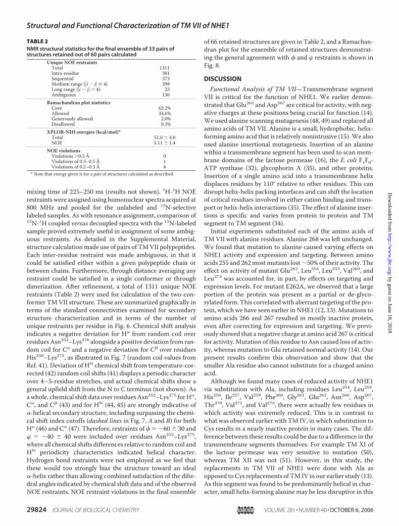
mixing time of 225–250 ms (results not shown). 1H-1H NOErestraints were assigned using homonuclear spectra acquired at800 MHz and pooled for the unlabeled and 15N-selectivelabeled samples. As with resonance assignment, comparison of15N-1H coupled versus decoupled spectra with the 15N-labeledsample proved extremely useful in assignment of some ambig-uous restraints. As detailed in the Supplemental Material,structure calculationmade use of pairs of TMVII polypeptides.Each inter-residue restraint was made ambiguous, in that itcould be satisfied either within a given polypeptide chain orbetween chains. Furthermore, through distance averaging anyrestraint could be satisfied in a single conformer or throughdimerization. After refinement, a total of 1311 unique NOErestraints (Table 2) were used for calculation of the two-con-former TMVII structure. These are summarized graphically interms of the standard connectivities examined for secondarystructure characterization and in terms of the number ofunique restraints per residue in Fig. 6. Chemical shift analysisindicates a negative deviation for H� from random coil overresiduesAsn252–Lys276 alongside a positive deviation from ran-dom coil for C� and a negative deviation for C� over residuesHis250–Lys275, as illustrated in Fig. 7 (random coil values fromRef. 41). Deviation of HN chemical shift from temperature-cor-rected (42) random coil shifts (41) displays a periodic characterover 4–5-residue stretches, and actual chemical shifts show ageneral upfield shift from the N to C terminus (not shown). Asawhole, chemical shift data over residuesAsn252–Lys275 forH�,C�, and C� (43) and for HN (44, 45) are strongly indicative of�-helical secondary structure, including surpassing the chemi-cal shift index cutoffs (dashed lines in Fig. 7, A and B) for bothH� (46) and C� (47). Therefore, restraints of � � �60� 30 and� � �40 � 40 were included over residues Asn252–Lys275,where all chemical shifts differences relative to randomcoil andHN periodicity characteristics indicated helical character.Hydrogen bond restraints were not employed as we feel thatthese would too strongly bias the structure toward an ideal�-helix rather than allowing combined satisfaction of the dihe-dral angles indicated by chemical shift data and of the observedNOE restraints. NOE restraint violations in the final ensemble
of 66 retained structures are given in Table 2, and a Ramachan-dran plot for the ensemble of retained structures demonstrat-ing the general agreement with � and � restraints is shown inFig. 8.
DISCUSSION
Functional Analysis of TM VII—Transmembrane segmentVII is critical for the function of NHE1. We earlier demon-strated that Glu262 and Asp267 are critical for activity, with neg-ative charges at these positions being crucial for function (14).We used alanine scanningmutagenesis (48, 49) and replaced allamino acids of TM VII. Alanine is a small, hydrophobic, helix-forming amino acid that is relatively nonintrusive (15).We alsoused alanine insertional mutagenesis. Insertion of an alaninewithin a transmembrane segment has been used to scan mem-brane domains of the lactose permease (16), the E. coli F1F0-ATP synthase (32), glycophorin A (35), and other proteins.Insertion of a single amino acid into a transmembrane helixdisplaces residues by 110° relative to other residues. This candisrupt helix-helix packing interfaces and can shift the locationof critical residues involved in either cation binding and trans-port or helix-helix interactions (35). The effect of alanine inser-tions is specific and varies from protein to protein and TMsegment to TM segment (34).Initial experiments substituted each of the amino acids of
TMVII with alanine residues. Alanine 268 was left unchanged.We found that mutation to alanine caused varying effects onNHE1 activity and expression and targeting. Between aminoacids 255 and 262mostmutants lost�50% of their activity. Theeffect on activity of mutant Glu262, Leu254, Leu255, Val269, andLeu273 was accounted for, in part, by effects on targeting andexpression levels. For mutant E262A, we observed that a largeportion of the protein was present as a partial or de-glyco-sylated form.This correlatedwith aberrant targeting of the pro-tein, which we have seen earlier in NHE1 (12, 13). Mutations toamino acids 266 and 267 resulted in mostly inactive protein,even after correcting for expression and targeting. We previ-ously showed that a negative charge at amino acid 267 is criticalfor activity.Mutation of this residue to Asn caused loss of activ-ity, whereas mutation to Glu retained normal activity (14). Ourpresent results confirm this observation and show that thesmaller Ala residue also cannot substitute for a charged aminoacid.Although we found many cases of reduced activity of NHE1
via substitution with Ala, including residues Leu254, Leu255,His256, Ile257, Val259, Phe260, Gly261, Glu262, Asn266, Asp267,Thr270, Val271, and Val272, there were actually few residues inwhich activity was extremely reduced. This is in contrast towhat was observed earlier with TM IV, in which substitution toCys results in a nearly inactive protein in many cases. The dif-ference between these results could be due to a difference in thetransmembrane segments themselves. For example TM XI ofthe lactose permease was very sensitive to mutation (50),whereas TM XII was not (51). However, in this study, thereplacements in TM VII of NHE1 were done with Ala asopposed toCys replacements of TM IV in our earlier study (13).As this segment was found to be predominantly helical in char-acter, small helix-forming alanine may be less disruptive in this
TABLE 2NMR structural statistics for the final ensemble of 33 pairs ofstructures retained out of 60 pairs calculated
Unique NOE restraintsTotal 1311Intra-residue 381Sequential 373Medium range (�i � j� � 4) 398Long range (�i � j� � 4) 23Ambiguous 136
Ramachandran plot statisticsCore 63.2%Allowed 34.6%Generously allowed 2.0%Disallowed 0.3%
XPLOR-NIH energies (kcal/mol)aTotal 51.0 � 4.8NOE 5.11 � 1.4
NOE violationsViolations �0.5 Å 0Violations of 0.3–0.5 Å 1Violations of 0.2–0.3 Å 4
a Note that energy given is for a pair of structures calculated as described.
Structural and Functional Characterization of TM VII of NHE1
29824 JOURNAL OF BIOLOGICAL CHEMISTRY VOLUME 281 • NUMBER 40 • OCTOBER 6, 2006
by guest on June 18, 2018http://w
ww
.jbc.org/D
ownloaded from

transmembrane segment than a Cys residue was in TM IV (13).Alanine may better preserve the overall character of TM VIIand be a better choice for examining the importance of sidechains and helical character, as compared with insertion of aCys residue because Cys has a much lower propensity than Alafor �-helix formation in a TM region (52, 53). The results sug-gest that many of the side chains of the amino acids do notappear to be especially critical for activity, although they some-times influenced expression levels and targeting.When consid-ering corrections for expression levels and targeting, the sub-group that had activity reduced 50% or more included onlyLeu255, Ile257, Val259, Phe260,Gly261, Asn266, Asp267, andThr270.The E262A mutation caused a very large decrease in activity,although much of this was because of aberrant targeting andexpression, and after these corrections activity was slightly over50% of control.Alanine insertional mutagenesis suggested that the overall
structure of TM VII was critical. Insertion of Ala after eitheramino acid 261 or 264 resulted in an almost completely inactiveprotein. Because of the importance of amino acid Asp267, we
reasoned that if we inserted a glu-tamic acid after amino acid 261, onthe initial assumption of an ideal�-helix, the helix might shift suchthat amino acid Asn266 was in theposition of Asp267. Mutation ofAsn266 toAspmight return a criticalacidic residue to this position. Inaddition, the glutamic acid insertionafter Gly261 might substitute forGlu262. In fact, we found that thisdouble mutant retained much moreactivity than simple insertion of ala-nine after 261. Upon correction fordefective targeting and expression,the activity was equivalent to that ofthe controls (Fig. 4B). This wassomewhat surprising because theproteinwas evenmore poorly glyco-sylated than the other insertionalmutants (Fig. 2D); however, we havepreviously shown that glycosylationis not essential to NHE1 function(29). Although having twoglutamate residues following oneanother in the mutant would not beconducive to �-helix formation (52,54), this region is extended ratherthan helical in the structure of TMVII that we have solved.An important characteristic of
theNHE1 protein is its sensitivity toinhibition by benzoyl guanidinetype of inhibitors. Sites in TM IV(55) and TM IX (56–58) alter sensi-tivity to inhibition. We reasonedthat because of the critical nature ofthis transmembrane segment, it
might also affect sensitivity to inhibition. Several of the residuesaltered the sensitivity. The maximum changes were a 10-foldincrease in sensitivity to inhibitionwithmutation of L265A anda 5-fold increase in resistance with the V272Amutation. Thesechanges are not as large as some reported earlier (58), but none-theless they altered the inhibitor efficacy significantly. Othermutations have reported significant effects on NHE inhibitorresistance but no effects on Na� affinity (59), suggesting thatthe inhibitor-binding site may be physically distinct but closelyrelated to the Na�-binding site (60). The contribution of manyregions of the NHE1 protein to inhibitor resistance, shown inthis and other studies, suggests that a number of differentregions of the protein likely come together to influence theprotein structure and thereby influence the NHE inhibitor-binding site. Likely, alterations in many amino acids and trans-membrane segments that affect the structure and function ofthe protein affect inhibitor binding. However, we did find thatthe effects were specific. Amino acids Leu265, Leu255, Ile251, andVal272 had significant effects on NHE1 inhibitor resistancewhereas others did not.
FIGURE 6. NOE restraint assignments for TM VII in DPC micelles. A, graphical summary of dXX NOE restraintsobserved in homonuclear NOESY experiments. (Figure was modified from CYANA (L.A. Systems, Inc.) output.)B, number of unique NOE restraints in final set of restraints on a per-residue basis (medium range restraints arebetween nuclei 2 and 4 residues apart in sequence; long range are 5 or more residues apart).
Structural and Functional Characterization of TM VII of NHE1
OCTOBER 6, 2006 • VOLUME 281 • NUMBER 40 JOURNAL OF BIOLOGICAL CHEMISTRY 29825
by guest on June 18, 2018http://w
ww
.jbc.org/D
ownloaded from

Structural Analysis of TMVII—Attempts to calculate a singleconformer in agreement with the assigned NOE data requiredremoval of a high proportion (34.4%) of the NOE restraint datato allow production of an ensemble of structures with low vio-lation statistics (data not shown). Two possible reasons for theinability to satisfy the NMR data were that the TM VII peptidewas sampling a number of conformations, as is frequentlyobserved with peptides or unstructured proteins, or that oli-gomerization of the TM VI peptide was taking place. In eithercase, NOE restraints would be observed that are not satisfied bya single conformation. Interestingly, we found that simultane-ous calculation of two conformers satisfied the NOE data setwithout significant dimer formation suggesting that samplingof multiple conformations rather than dimerization is respon-sible for reducing restraint violations. A similar result, butwith-out the ability to test for dimerization, could likely be achievedthrough ensemble-average structure calculation (61–64). Fur-ther details are supplied in the Supplemental Material.An ensemble of 66 TM VII structures (33 dual conformer
pairs) was obtained that satisfy the vast majority of observedNOE and chemical shift-derived � and � dihedral anglerestraints equally well (Table 2; Fig. 8). Despite strong chemicalshift evidence (Fig. 7; values deposited in the BioMagResBank),NOE connectivities (Table 2 and Fig. 6; restraints deposited inthe Protein Data Bank code 2HTG and RCSB codeRCSB038740) do not result in an uninterrupted helix over res-idues Asn252–Lys275. Superposition of all members of theensemble over the full length of the peptide is not possible.Rather, two distinct portions of the peptide show strong struc-tural convergence, as demonstrated by superposition of thepolypeptide backbone over the ensemble of structures. Thebreak point between these converged segments of TM VII is atGly261–Glu262. Minor variability in backbone structure is alsoobserved over the ensemble of structures at Leu254–Leu255,Leu265–Asn266, and Thr270–Val271, requiring separate super-positions for optimal root mean square deviation, as detailed inthe Supplemental Material. Residues highlighted in gray in Fig.8 with greater than usual dispersion of (�,�) angle are Ile251(corresponding to flexibility at the N terminus), Gly261 (corre-sponding to major break between helical segments), and Val271(corresponding to minor variability in positioning of the C ter-minus relative to N terminus of helical segment �Ser264–Leu273). Using the Kabsch-Sander secondary structure assign-ment method (65) as implemented in PROMOTIF (66), helicalstructure predominates over the converged segments N- andC-terminal to Gly261. In the N terminus, helical structure pre-dominates from Leu255–Phe260 and in the C terminus fromLeu264–Val272 (detailed analysis in the SupplementalMaterial).In a given two-conformer structure in agreement with theexperimental data, the relative positions and orientations of thetwo helical segments about Gly261–Glu262 tend to differ.The structure of TM VII in DPC micelles is therefore an
interrupted helix. A representative conformer from the ensem-ble of structures is shown in Fig. 9. The relative positioning andorientation of the N- and C-terminal helical stretches are quitevariable about Gly261 in the isolated TM segment. However, inthe protein environment this would likely be a well definedkink, with strong potential for an interruption in helicity over
the range Phe260–Ser263. It should be noted that in the prokary-oticNa�/H� exchangerNhaA (67), aswell as a number of othermembrane protein structures, kinked and interrupted helicesare thought to play crucial roles in function. The inherent flex-ibility of the TM VII segment about Gly261 also suggests thepotential for structural change in response to pH, as has beenhypothesized to be take place with the kinked and interruptedhelical transmembrane segments IV and XI of NhaA (67). Theexact role of TM VII in ion transport should become moreapparent as the pH sensormechanism ofNHE1 becomes betterunderstood.Correlating Structural and Functional Data—There are
interesting correlations between the structural and functional
FIGURE 7. Chemical shifts for TM VII in DPC micelles. Chemical shift differ-ences from random coil shifts (41) are shown for H� (A), C� (B), and C� (C).Dashed lines in A and B are chemical shift difference cutoffs considered signif-icant for the 1H (46) and 13C (47) chemical shift indices, respectively.
Structural and Functional Characterization of TM VII of NHE1
29826 JOURNAL OF BIOLOGICAL CHEMISTRY VOLUME 281 • NUMBER 40 • OCTOBER 6, 2006
by guest on June 18, 2018http://w
ww
.jbc.org/D
ownloaded from

data. One of the key areas of sensitivity to mutation was atamino acids Gly261 and Glu262 where we observed a strongbreak in helical structure. ForGlu262, we earlier showed that theconservative mutation to Asp allowed retention of most of thenormal properties of the protein, whereas switching touncharged Asn resulted in a dysfunctional protein (14). Thehelix-breaking nature of the GE pair of residues would likely beretained in both of these mutants (52, 54), giving further evi-dence that it is the acidic functionality ofGlu262 that is crucial toNHE1 activity. The insertion of an Ala between Gly261 andGlu262 also disrupts NHE1 activity, implying that both the posi-tion of Glu262 and the nonhelical nature of the Gly261–Glu262region are crucial. The ability of the Gly261–Ser263/Leu264region to undergo conformational change, reflected by variabil-ity about this region in the TMVII structure, may also be func-tionally crucial because the F260A,G261A, and E262Amutantswould all increase the helical propensity of this region, and allare highly disruptive to function.In converse to these residues, the activity of NHE1 is insen-
sitive to the S263A mutant. Ser263 shows more involvement inthe helix at the C terminus of TM VII than would be expectedbased upon helical propensities. The helical character of resi-due 263would be strengthened in the S263Amutant. Ser is alsoproposed to have a strong propensity for allowing close packingofTMhelices. If thiswere its role, a Ser3Alamutant is actuallyhighly conservative because Ala and Ser have very similar pack-ing characteristics (68). Replacement of the subsequent Leu264or Leu265 with Ala did not greatly inhibit activity, after correc-tion for expression and targeting was taken into account.Therefore, it seems that the local structure rather than thechemical character of side chains over Ser263–Leu265 is impor-tant for function, with the good possibility that substitution ofSer263 with a residue of lower helical packing capability wouldaffect overall NHE1 structure.
Substitution of Asn266, Asp267, or Thr270 with alanineinactivated NHE1. The negatively charged side chain ofAsp267 is critical to activity, because even the conservativeD267N mutant results in loss of activity (14). The �-helicalnature of the TM VII segment over the range includingAsn266, Asp267, and Thr270 means that all three side chainsfall on the same face of the segment (for example see theirpositions in Fig. 9). Negatively charged amino acids havebeen reported to be present in the funnel region of the pro-karyotic Na�/H� exchanger NhaA (67), and Asp267 couldplay a similar role in NHE1. Along these lines, Thr270 mayhave an analogous function to Thr132 of NhaA, which servesan important, although not essential, role in Na� binding(67). Therefore, it is possible that the side chains of thesethree residues are all involved in a cytoplasmic side ion fun-nel of NHE1 similar to that proposed for NhaA. Alterna-tively, the Asp267 side chain may form a critical salt bridgefor functional protein structure. As with Ser, Thr is proposedto allow close packing of TM segments in membranes (68),meaning that Thr270 may readily have a crucial packinginteraction with the same neighboring segment that wouldbe disrupted by replacement with the much smaller Ala sidechain. Insertion of an Ala between Leu264 and Leu265 woulddisrupt either role of these critical residues by shifting theregister of the entire helix off by one residue, and this inser-tional mutant indeed showed strongly perturbed expression,targeting, and activity.Our study gives a detailed structural and full functional
picture of TM VII. TM VII is a transmembrane segment thatis critical for NHE1 function. We demonstrate that 13 of 22residues were sensitive to mutation to alanine and that
FIGURE 8. Ramachandran plot for entire retained ensemble of TM VII pep-tide structures. Residues Ile251, Gly261, and Val271 are shown in gray as indi-cated; all other residues over the range Asn252–Lys276 are shown as open blackcircles.
FIGURE 9. Representative structure of TM VII peptide. Two different orien-tations of a representative ensemble member are shown side-by-side withcoloring covering the spectrum from blue (N terminus) to red (C terminus).The ensemble of structures shows a variety of orientations of the N- andC-terminal �-helical segments about Gly261; side chain positions are also typ-ically variable.
Structural and Functional Characterization of TM VII of NHE1
OCTOBER 6, 2006 • VOLUME 281 • NUMBER 40 JOURNAL OF BIOLOGICAL CHEMISTRY 29827
by guest on June 18, 2018http://w
ww
.jbc.org/D
ownloaded from

mutation of 5 residues altered sensitivity to inhibition byEMD87580. In contrast to TM IV of NHE1, TM VII is pre-dominantly �-helical. However, it has a pronounced break inhelicity in its central region, which includes the functionallyessential acidic Glu262 residue. The helical nature of the Cterminus of this segment positions three critical residues(Asn266, Asp267, and Thr270) on the same face of the segment.If these are determined to be involved in ion transport ratherthan structural stabilization of NHE1, this result provides astarting point for mechanistic elucidation of ion funnelingand transport. Future experiments may both further elabo-rate the role of TM VII in ion exchange and examine thestructure-function relationship for other TM segments ofthe NHE1 isoform of the Na�/H� exchanger.
Acknowledgments—We thank Jeffrey DeVries for maintenance of the500-MHz Varian Inova spectrometer; Jason Moses and Marc Genestof the Protein EngineeringNetwork of Centres of Excellence ChemistryLaboratory in Edmonton, Alberta, Canada, for synthesis, purifica-tion, and characterization of peptides; Charles Schwieters from theNational Institutes of Health (Bethesda) for helpful discussionsregarding XPLOR-NIH; and the CanadianNational High Field NMRCentre for their assistance and use of their facilities. Peptide synthesis,NMR spectroscopy, and structural calculations were funded by Pro-tein EngineeringNetwork of Centres of Excellence.Operation of Cana-dian National High Field NMR Centre is funded by Canadian Insti-tutes of Health Research, Natural Sciences and Engineering ResearchCouncil of Canada, and the University of Alberta.
REFERENCES1. Orlowski, J., and Grinstein, S. (1997) J. Biol. Chem. 272, 22373–223762. Grinstein, S., Rotin, D., and Mason, M. J. (1989) Biochim. Biophys. Acta
988, 73–973. Pouyssegur, J., Sardet, C., Franchi, A., L’Allemain, G., and Paris, S. (1984)
Proc. Natl. Acad. Sci. U. S. A. 81, 4833–48374. Shrode, L., Cabado, A., Goss, G., and Grinstein, S. (1996) in The Na�/H�
Exchanger (Fliegel, L., ed) pp. 101–122, R. G. Landes Co., Austin, TX5. Karmazyn, M., Liu, Q., Gan, X. T., Brix, B. J., and Fliegel, L. (2003)Hyper-
tension 42, 1171–11766. Fliegel, L. (2001) Basic Res. Cardiol. 96, 301–3057. Mentzer, R. M., Jr., Lasley, R. D., Jessel, A., and Karmazyn, M. (2003) Ann.
Thorac. Surg. 75, S700–S7088. Lang, H. J. (2003) inTheNa�/H� Exchanger, FromMolecular to Its Role in
Disease (Karmazyn, M., Avkiran, M., and Fliegel, L., eds) pp. 239–253,Kluwer Academic Publishers, Norwell, MA
9. Paradiso, A., Cardone, R. A., Bellizzi, A., Bagorda, A., Guerra, L., Tomma-sino, M., Casavola, V., and Reshkin, S. J. (2004) Breast Cancer Res. 6,R616–R628
10. Denker, S. P., and Barber, D. L. (2002) J. Cell Biol. 159, 1087–109611. Wakabayashi, S., Pang, T., Su, X., and Shigekawa, M. (2000) J. Biol. Chem.
275, 7942–794912. Slepkov, E. R., Chow, S., Lemieux, M. J., and Fliegel, L. (2004) Biochem. J.
379, 31–3813. Slepkov, E. R., Rainey, J. K., Li, X., Liu, Y., Cheng, F. J., Lindhout, D. A.,
Sykes, B. D., and Fliegel, L. (2005) J. Biol. Chem. 280, 17863–1787214. Murtazina, B., Booth, B. J., Bullis, B. L., Singh, D. N., and Fliegel, L. (2001)
Eur. J. Biochem. 268, 1–1315. Fleming, K. G., and Engelman, D. M. (2001) Proc. Natl. Acad. Sci. U. S. A.
98, 14340–1434416. Braun, P., Persson, B., Kaback, H. R., and von Heijne, G. (1997) J. Biol.
Chem. 272, 29566–2957117. Oblatt-Montal, M., Reddy, G. L., Iwamoto, T., Tomich, J. M., andMontal,
M. (1994) Proc. Natl. Acad. Sci. U. S. A. 91, 1495–1499
18. Wigley,W.C., Vijayakumar, S., Jones, J. D., Slaughter, C., andThomas, P. J.(1998) Biochemistry 37, 844–853
19. Hunt, J. F., Earnest, T. N., Bousche, O., Kalghatgi, K., Reilly, K., Horvath,C., Rothschild, K. J., and Engelman, D. M. (1997) Biochemistry 36,15156–15176
20. Katragadda, M., Alderfer, J. L., and Yeagle, P. L. (2001) Biophys. J. 81,1029–1036
21. Katragadda, M., Chopra, A., Bennett, M., Alderfer, J. L., Yeagle, P. L., andAlbert, A. D. (2001) J. Pept. Res. 58, 79–89
22. Naider, F., Khare, S., Arshava, B., Severino, B., Russo, J., and Becker, J. M.(2005) Biopolymers 80, 199–213
23. Damberg, P., Jarvet, J., and Graslund, A. (2001) Methods Enzymol. 339,271–285
24. Henry, G. D., and Sykes, B. D. (1994)Methods Enzymol. 239, 515–53525. Zhang, Y. P., Lewis, R. N., Hodges, R. S., and McElhaney, R. N. (1995)
Biophys. J. 68, 847–85726. Wishart, D. S., Bigam, C. G., Yao, J., Abildgaard, F., Dyson, H. J., Oldfield,
E., Markley, J. L., and Sykes, B. D. (1995) J. Biomol. NMR 6, 135–14027. Delaglio, F., Grzesiek, S., Vuister, G. W., Zhu, G., Pfeifer, J., and Bax, A.
(1995) J. Biomol. NMR 6, 277–29328. Schwieters, C. D., Kuszewski, J. J., Tjandra, N., and Clore, G. M. (2003) J.
Magn. Reson. 160, 65–7329. Haworth, R. S., Frohlich, O., and Fliegel, L. (1993) Biochem. J. 289,
637–64030. Li, X., Ding, J., Liu, Y., Brix, B. J., and Fliegel, L. (2004) Biochemistry 43,
16477–1648631. Karmazyn, M., Sawyer, M., and Fliegel, L. (2005) Curr. Drug Targets Car-
diovasc. Haematol. Disord. 5, 323–33532. Vik, S. B., Patterson, A. R., and Antonio, B. J. (1998) J. Biol. Chem. 273,
16229–1623433. Op De Beeck, A., Montserret, R., Duvet, S., Cocquerel, L., Cacan, R., Bar-
berot, B., Le Maire, M., Penin, F., and Dubuisson, J. (2000) J. Biol. Chem.275, 31428–31437
34. OpDe Beeck, A.,Molenkamp, R., Caron,M., Ben Younes, A., Bredenbeek,P., and Dubuisson, J. (2003) J. Virol. 77, 813–820
35. Mingarro, I., Whitley, P., Lemmon, M. A., and von Heijne, G. (1996) Pro-tein Sci. 5, 1339–1341
36. Davis, J. H., Clare, D.M., Hodges, R. S., and Bloom,M. (1983)Biochemistry22, 5298–5305
37. Lindhout, D. A., Thiessen, A., Schieve, D., and Sykes, B. D. (2003) ProteinSci. 12, 1786–1791
38. Cavanagh, J., Fairbrother, W., Palmer, A. G., III, and Skelton, N. J. (1996)Protein NMR Spectroscopy: Principles and Practice, Academic Press,New York
39. Wuthrich, K. (1986) NMR of Proteins and Nucleic Acids, Wiley Inter-science, New York
40. Cavanagh, J., Chazin, W. J., and Rance, M. (1990) J. Magn. Reson. 87,110–131
41. Wishart, D. S., Bigam, C. G., Holm, A., Hodges, R. S., and Sykes, B. D.(1995) J. Biomol. NMR 5, 67–81
42. Merutka, G., Dyson, H. J., and Wright, P. E. (1995) J. Biomol. NMR 5,14–24
43. Wishart, D. S., Sykes, B. D., and Richards, F. M. (1991) J. Mol. Biol. 222,311–333
44. Kuntz, I. D., Kosen, P. A., and Craig, E. C. (1991) J. Am. Chem. Soc. 113,1406–1408
45. Williamson, M. P. (1990) Biopolymers 29, 1423–143146. Wishart, D. S., Sykes, B. D., and Richards, F. M. (1992) Biochemistry 31,
1647–165147. Wishart, D. S., and Sykes, B. D. (1994) J. Biomol. NMR 4, 171–18048. Lu, Z. L., Saldanha, J. W., and Hulme, E. C. (2001) J. Biol. Chem. 276,
34098–3410449. Fraysse, A. S., Moller, A. L., Poulsen, L. R., Wollenweber, B., Buch-Peder-
sen, M. J., and Palmgren, M. G. (2005) J. Biol. Chem. 280, 21785–2179050. Dunten, R. L., Sahin-Toth, M., and Kaback, H. R. (1993) Biochemistry 32,
12644–1265051. He, M. M., Sun, J., and Kaback, H. R. (1996) Biochemistry 35,
12909–12914
Structural and Functional Characterization of TM VII of NHE1
29828 JOURNAL OF BIOLOGICAL CHEMISTRY VOLUME 281 • NUMBER 40 • OCTOBER 6, 2006
by guest on June 18, 2018http://w
ww
.jbc.org/D
ownloaded from

52. Jones, D. T., Taylor, W. R., and Thornton, J. M. (1994) Biochemistry 33,3038–3049
53. Liu, H., Cala, P. M., and Anderson, S. E. (1998) J. Mol. Cell. Cardiol. 30,685–697
54. Liu, L. P., and Deber, C. M. (1998) J. Biol. Chem. 273, 23645–2364855. Counillon, L., Franchi, A., and Pouyssegur, J. (1993) Proc. Natl. Acad. Sci.
U. S. A. 90, 4508–451256. Khadilkar, A., Iannuzzi, P., and Orlowski, J. (2001) J. Biol. Chem. 276,
43792–4380057. Orlowski, J., andKandasamy, R. A. (1996) J. Biol. Chem. 271, 19922–1992758. Noel, J., Germain, D., and Vadnais, J. (2003) Biochemistry 42,
15361–1536859. Wang, D., Balkovetz, D. F., andWarnock, D. G. (1995)Am. J. Physiol. 269,
C392–C402
60. Harris, C., and Fliegel, L. (1999) Int. J. Mol. Med. 3, 315–32161. Bonvin, A. M., and Brunger, A. T. (1995) J. Mol. Biol. 250, 80–9362. Bonvin, A. M. J. J., Boelens, R., and Kaptein, R. (1994) J. Biomol. NMR 4,
143–14963. Bruschweiler, R., Blackledge,M., and Ernst, R. R. (1991) J. Biomol. NMR 1,
3–1164. Wang, J. J., Hodges, R. S., and Sykes, B. D. (1995) J. Am. Chem. Soc. 117,
8627–863465. Kabsch, W., and Sander, C. (1983) Biopolymers 22, 2577–263766. Hutchinson, E. G., and Thornton, J. M. (1996) Protein Sci. 5, 212–22067. Hunte, C., Screpanti, E., Venturi, M., Rimon, A., Padan, E., andMichel, H.
(2005) Nature 435, 1197–120268. Eilers, M., Shekar, S. C., Shieh, T., Smith, S. O., and Fleming, P. J. (2000)
Proc. Natl. Acad. Sci. U. S. A. 97, 5796–5801
Structural and Functional Characterization of TM VII of NHE1
OCTOBER 6, 2006 • VOLUME 281 • NUMBER 40 JOURNAL OF BIOLOGICAL CHEMISTRY 29829
by guest on June 18, 2018http://w
ww
.jbc.org/D
ownloaded from

Jie Ding, Jan K. Rainey, Caroline Xu, Brian D. Sykes and Larry Fliegel Exchanger Isoform 1+/H+the Na
Structural and Functional Characterization of Transmembrane Segment VII of
doi: 10.1074/jbc.M606152200 originally published online July 21, 20062006, 281:29817-29829.J. Biol. Chem.
10.1074/jbc.M606152200Access the most updated version of this article at doi:
Alerts:
When a correction for this article is posted•
When this article is cited•
to choose from all of JBC's e-mail alertsClick here
Supplemental material:
http://www.jbc.org/content/suppl/2006/07/25/M606152200.DC1
http://www.jbc.org/content/281/40/29817.full.html#ref-list-1
This article cites 60 references, 18 of which can be accessed free at
by guest on June 18, 2018http://w
ww
.jbc.org/D
ownloaded from