structuralandinhibitionanalysisrevealsthemechanismof ... · (serum-free dmem), medium containing...
TRANSCRIPT
Structural and Inhibition Analysis Reveals the Mechanism ofSelectivity of a Series of Aggrecanase InhibitorsReceived for publication, June 2, 2009 Published, JBC Papers in Press, July 8, 2009, DOI 10.1074/jbc.M109.029116
Micky D. Tortorella1, Alfredo G. Tomasselli, Karl J. Mathis, Mark E. Schnute, Scott S. Woodard, Grace Munie,Jennifer M. Williams, Nicole Caspers, Arthur J. Wittwer, Anne-Marie Malfait, and Huey-Sheng Shieh2
From Pfizer Global Research and Development, St. Louis, Missouri 63017
Several inhibitors of a series of cis-1(S)2(R)-amino-2-indanol-based compounds were reported to be selective for the aggre-canases, ADAMTS-4 and -5 over other metalloproteases. Tounderstand the nature of this selectivity for aggrecanases, theinhibitors, along with the broad spectrum metalloproteaseinhibitormarimastat,were independently bound to the catalyticdomain of ADAMTS-5, and the corresponding crystal struc-tures were determined. By comparing the structures, it wasdetermined that the specificity of the relative inhibitors forADAMTS-5 was not driven by a specific interaction, such aszinc chelation, hydrogen bonding, or charge interactions, butrather by subtle and indirect factors, such as water bridging,ring rigidity, pocket size, and shape, as well as protein con-formation flexibility.
Osteoarthritis (OA)3 pathology includes degradation ofarticular cartilage, along with subchondral bone sclerosis andosteophyte formation, all contributing to impaired joint func-tion. Pain, restricted movement, and joint instability accom-pany these structural changes and often result in the need fortotal joint replacement. Current therapies alleviate the mild tomoderate pain and inflammation associated with OA, but donot protect the cartilage from further damage and have notdemonstrated an effect on disease progression (1). Therefore,therapeutics that prevent or slow the alteration of joint struc-ture and function will address a major unmet medical need.Loss of aggrecan, a macromolecular proteoglycan providing
cartilage with its properties of compressibility and resilience, isa major phenotype associated with OA and is believed to be acritical event in driving disease progression (2, 3). Both ex vivoand in vivo proof of concept studies support ADAMTS-4 andADAMTS-5, commonly referred to as aggrecanase-1 and -2,respectively, as the twomajor enzymes responsible for the pro-
teolytic breakdown of cartilage aggrecan (reviewed in Ref. 4).Blocking their activity may be an attractive strategy to stop orslow down the progression of the disease, as suggested by stud-ies in knock-out mice (5). Given the chronic nature of the dis-ease, long term treatment will be likely, demanding very safetherapeutic interventions only achievable with ADAMTS-4-and ADAMTS-5-specific inhibitors lacking off-target sideeffects. Designing selectivity has been very challenging and amajor source of difficulty is that at least 57 metalloproteases(MP) divided in three major families, 1) matrix metallopro-teases (MMP); 2) a disintegrin andmetalloproteinase (ADAM);and 3) a disintegrin and metalloproteinase with throm-bospondin motifs (ADAMTS), are present in humans.ADAMTS-4 and -5 belong to the ADAMTS family and sharecommon catalytic and structural features with the other MPmembers. These features include the highly conserved aminoacid sequence, HEXXHXXGXXH, harboring a catalytic zinccation, required for activation of the peptide bond towardhydrolysis. In addition, many MPs share significant structuraltopology in the active site, such as a flexible S1� loop. To com-plicate matters further, only a handful ofMP structures, mostlyin theMMP family, have been determined, and the functions ofmostMPs still remain unknown, a fact that has earned them anorphan status denomination. Lack of structural informationhas hindered the design for inhibitor specificity and withoutknown substrates formanyMPs, assays for screening inhibitorsare often not available, making determinations of selectivitydifficult (6, 7).Yao et al. (8, 9) reported the discovery of a series of (2R)-
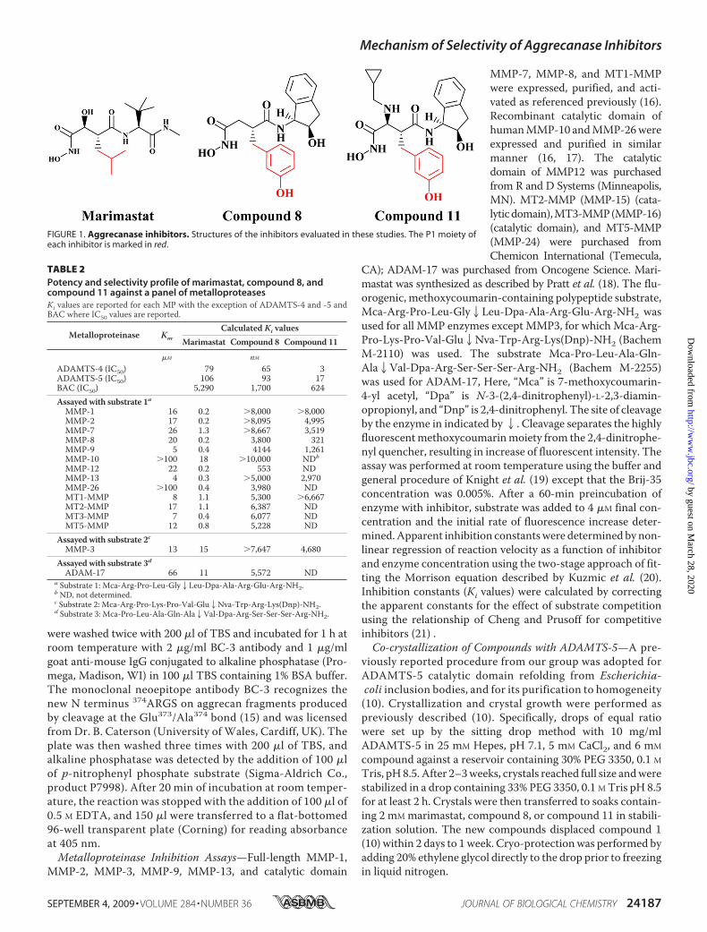
N4-hydroxy-2-(3-hydroxybenzyl)-N1-[(1S,2R)-2-hydroxy-,3-di-hydro-1H-inden-1-yl]butanediamide derivatives as potent andselective inhibitors of aggrecanase activity. Using a homologymodel of aggrecanase based on the active site of atrolysin Cand adamalysin II and docking compound 8, shown in Fig. 1,the authors concluded that the 3-hydroxyl group of inhibitor8 achieved selectivity through a specific hydrogen-bondinginteraction with Thr440 (numerical numbering is based onthe human sequence of ADAMTS-5) in the S1� pocket ofaggrecanase.Whereas both ADAMTS-4 and -5 have a threonine at this
position, MMP-1, -2, -3, -7, -8, -9, -10, -13, -14, -16, andADAM-17 have a valine, lending credence to the proposedhypothesis around selectivity. Recently, our group has estab-lished a protocol for crystallizing the catalytic domain ofADAMTS-5 and determined its three-dimensional structure(10). Thus, experimental validation or invalidation of thehypothesis that inhibitor 8 and related molecules form a spe-
The atomic coordinates and structure factors (codes 3HY7, 3HYG, and 3HY9)have been deposited in the Protein Data Bank, Research Collaboratory forStructural Bioinformatics, Rutgers University, New Brunswick, NJ (http://www.rcsb.org/).
1 To whom correspondence may be addressed: Pfizer Global Research andDevelopment, 700 Chesterfield Parkway, Chesterfield, MO 63017. Tel.: 636-293-9612; Fax: 636-247-6313; E-mail: [email protected].
2 To whom correspondence may be addressed: Pfizer Global Research andDevelopment, 700 Chesterfield Parkway, Chesterfield, MO 63017. Tel.: 636-247-6025; Fax: 636-247-7350; E-mail: [email protected].
3 The abbreviations used are: OA, osteoarthritis; ADAMTS, a disintegrin andmetalloproteinase with thrombospondin motifs; ADAM, a disintegrin andmetalloproteinase; MMP, matrix metalloproteinases; BNC, bovine nasalcartilage; IL-1�, interleukin-1�; PEG, polyethylene glycol; PDB, Protein DataBank; R.m.s., root mean square; BAC, bovine articular cartilage.
THE JOURNAL OF BIOLOGICAL CHEMISTRY VOL. 284, NO. 36, pp. 24185–24191, September 4, 2009© 2009 by The American Society for Biochemistry and Molecular Biology, Inc. Printed in the U.S.A.
SEPTEMBER 4, 2009 • VOLUME 284 • NUMBER 36 JOURNAL OF BIOLOGICAL CHEMISTRY 24185
by guest on March 28, 2020
http://ww
w.jbc.org/
Dow
nloaded from
cific hydrogen bondwith the hydroxyl group of threonine in theS1� pocket of aggrecanases could now be determined by pro-tease-inhibitor crystallographic analysis.In the current study, we wanted to confirm and extend the
selectivity profile of compound 8 and 11 against a wide array ofMPs. Moreover, we wanted to elucidate the key molecularinteractions responsible for the enhanced selectivity profile ofthis series of compounds. For this purpose we generated co-crystals and solved the structures of marimastat, compound 8,and compound 11 in complex with the catalytic domain ofrecombinant human ADAMTS-5.
EXPERIMENTAL PROCEDURES
Bovine Articular Cartilage (BAC) Assay—Cartilage was dis-sected from the metatarsophalangeal joints of young cowsobtained from the slaughterhouse. Cartilage was allowed toequilibrate for 3 days in Dulbecco’s modified Eagle’s medium(DMEM) supplemented with 10% fetal calf serum, penicillin(100 units/ml), and streptomycin (100 �g/ml), all purchasedfrom Invitrogen (Carlsbad, CA). Subsequently, cartilage wascut into 3 � 3-mm explants, weighing �10–20 mg each, andincubated in 96-well plates for 48 hwith either control medium(serum-free DMEM), medium containing interleukin-1�(IL-1� produced at Pfizer, as described, Ref. 11) (100 ng/ml) ormedium containing IL-1� (100 ng/ml) plus marimastat, com-pound 8, or compound 11 at 30 to 10,000 nM. At the end of theculture period, supernatants were collected and frozen at�20 °C until further assay. Glycosaminoglycan content of thesupernatants was determined using the dimethylmethyleneblue assay, as described by Farndale et al. (12). Shark chon-
droitin sulfate was used as a standard, and results wereexpressed as �g of GAG per mg of wet weight cartilage.Aggrecanase Inhibition Assay—Inhibition of ADAMTS-4 or
ADAMTS-5 cleavage at the Glu373/Ala374 site of aggrecan wasmeasured as follows. Full-length recombinant ADAMTS-4 and-5 were expressed in SF9 cells and purified as previouslydescribed (13, 14). Reaction mixtures of 100-�l final volume in96-well polypropylene plates contained purified bovine aggre-can, 0.2 nM ADAMTS-4 or ADAMTS-5, and inhibitor or 1.0%dimethyl sulfoxide vehicle control in 50 mM Tris buffer at pH7.5, containing 100 mM NaCl and 5 mM CaCl2. The concentra-tion of aggrecanwas 125nM for IC50 determinations.After a 6-hincubation at 37 °C, the reaction was stopped with the additionof 10 �l of 0.5 M EDTA, and 75 �l were transferred to a 96-wellpolyvinylidene difluoride (PVDF) membrane plate (Millipore,Billerica, MA) containing 75 �l of 20 mM carbonate-bicarbon-ate buffer at pH 9.6 (Sigma). Prior to the addition of the carbon-ate-bicarbonate buffer, the PVDF plates had been conditionedfor 5 min with 100 �l 70% ethanol in water and washed twotimes with 200 �l of water. Plate washing was facilitated byusing a Tecan Columbus Plus plate washer (Research TrianglePark, NC) modified with the addition of a vacuummanifold byFlush Tec (Cathedral City, CA). The samples were allowed tobind to the PVDF plates overnight at room temperature,washed twice with 200 �l of Tris-buffered saline (TBS) (Bio-Rad product 170-6435), and incubated for 3 h at 37 °C with 0.1units/ml chondroitinaseABC, 0.1 units/ml keratanase, and 0.01units/ml keratanase II (Associates of Cape Cod, East Falmouth,MA), in 100 �l of 50 mM Tris, 100 mM sodium acetate buffer atpH 6.5, containing 1% bovine serum albumin (BSA). The plates
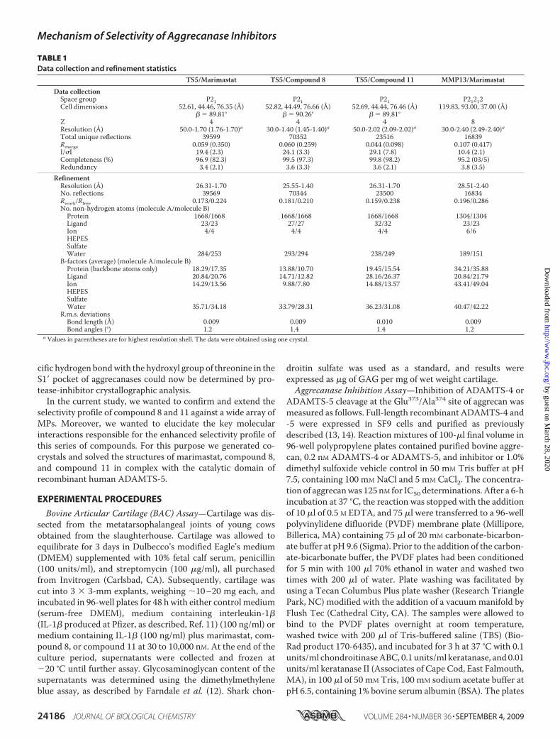
TABLE 1Data collection and refinement statistics
TS5/Marimastat TS5/Compound 8 TS5/Compound 11 MMP13/Marimastat
Data collectionSpace group P21 P21 P21 P21212Cell dimensions 52.61, 44.46, 76.35 (Å) 52.82, 44.49, 76.66 (Å) 52.69, 44.44, 76.46 (Å) 119.83, 93.00, 37.00 (Å)
� � 89.81° � � 90.26° � � 89.81°Z 4 4 4 8Resolution (Å) 50.0-1.70 (1.76-1.70)a 30.0-1.40 (1.45-1.40)a 50.0-2.02 (2.09-2.02)a 30.0-2.40 (2.49-2.40)aTotal unique reflections 39599 70352 23516 16839Rmerge 0.059 (0.350) 0.060 (0.259) 0.044 (0.098) 0.107 (0.417)I/�I 19.4 (2.3) 24.1 (3.3) 29.1 (7.8) 10.4 (2.1)Completeness (%) 96.9 (82.3) 99.5 (97.3) 99.8 (98.2) 95.2 (03/5)Redundancy 3.4 (2.1) 3.6 (3.3) 3.6 (2.1) 3.8 (3.5)
RefinementResolution (Å) 26.31-1.70 25.55-1.40 26.31-1.70 28.51-2.40No. reflections 39569 70344 23500 16834Rwork/Rfree 0.173/0.224 0.181/0.210 0.159/0.238 0.196/0.286No. non-hydrogen atoms (molecule A/molecule B)Protein 1668/1668 1668/1668 1668/1668 1304/1304Ligand 23/23 27/27 32/32 23/23Ion 4/4 4/4 4/4 6/6HEPESSulfateWater 284/253 293/294 238/249 189/151
B-factors (average) (molecule A/molecule B)Protein (backbone atoms only) 18.29/17.35 13.88/10.70 19.45/15.54 34.21/35.88Ligand 20.84/20.76 14.71/12.82 28.16/26.37 20.84/21.79Ion 14.29/13.56 9.88/7.80 14.88/13.57 43.41/49.04HEPESSulfateWater 35.71/34.18 33.79/28.31 36.23/31.08 40.47/42.22
R.m.s. deviationsBond length (Å) 0.009 0.009 0.010 0.009Bond angles (°) 1.2 1.4 1.4 1.2
a Values in parentheses are for highest resolution shell. The data were obtained using one crystal.
Mechanism of Selectivity of Aggrecanase Inhibitors
24186 JOURNAL OF BIOLOGICAL CHEMISTRY VOLUME 284 • NUMBER 36 • SEPTEMBER 4, 2009
by guest on March 28, 2020
http://ww
w.jbc.org/
Dow
nloaded from
were washed twice with 200 �l of TBS and incubated for 1 h atroom temperature with 2 �g/ml BC-3 antibody and 1 �g/mlgoat anti-mouse IgG conjugated to alkaline phosphatase (Pro-mega, Madison, WI) in 100 �l TBS containing 1% BSA buffer.The monoclonal neoepitope antibody BC-3 recognizes thenew N terminus 374ARGS on aggrecan fragments producedby cleavage at the Glu373/Ala374 bond (15) and was licensedfrom Dr. B. Caterson (University of Wales, Cardiff, UK). Theplate was then washed three times with 200 �l of TBS, andalkaline phosphatase was detected by the addition of 100 �lof p-nitrophenyl phosphate substrate (Sigma-Aldrich Co.,product P7998). After 20 min of incubation at room temper-ature, the reaction was stopped with the addition of 100 �l of0.5 M EDTA, and 150 �l were transferred to a flat-bottomed96-well transparent plate (Corning) for reading absorbanceat 405 nm.Metalloproteinase Inhibition Assays—Full-length MMP-1,
MMP-2, MMP-3, MMP-9, MMP-13, and catalytic domain
MMP-7, MMP-8, and MT1-MMPwere expressed, purified, and acti-vated as referenced previously (16).Recombinant catalytic domain ofhumanMMP-10 andMMP-26wereexpressed and purified in similarmanner (16, 17). The catalyticdomain of MMP12 was purchasedfrom R and D Systems (Minneapolis,MN). MT2-MMP (MMP-15) (cata-lyticdomain),MT3-MMP(MMP-16)(catalytic domain), and MT5-MMP(MMP-24) were purchased fromChemicon International (Temecula,
CA); ADAM-17 was purchased from Oncogene Science. Mari-mastat was synthesized as described by Pratt et al. (18). The flu-orogenic, methoxycoumarin-containing polypeptide substrate,Mca-Arg-Pro-Leu-Gly2Leu-Dpa-Ala-Arg-Glu-Arg-NH2 wasused for all MMP enzymes except MMP3, for which Mca-Arg-Pro-Lys-Pro-Val-Glu2Nva-Trp-Arg-Lys(Dnp)-NH2 (BachemM-2110) was used. The substrate Mca-Pro-Leu-Ala-Gln-Ala2Val-Dpa-Arg-Ser-Ser-Ser-Arg-NH2 (Bachem M-2255)was used for ADAM-17, Here, “Mca” is 7-methoxycoumarin-4-yl acetyl, “Dpa” is N-3-(2,4-dinitrophenyl)-L-2,3-diamin-opropionyl, and “Dnp” is 2,4-dinitrophenyl. The site of cleavageby the enzyme in indicated by2. Cleavage separates the highlyfluorescentmethoxycoumarinmoiety from the 2,4-dinitrophe-nyl quencher, resulting in increase of fluorescent intensity. Theassay was performed at room temperature using the buffer andgeneral procedure of Knight et al. (19) except that the Brij-35concentration was 0.005%. After a 60-min preincubation ofenzyme with inhibitor, substrate was added to 4 �M final con-centration and the initial rate of fluorescence increase deter-mined.Apparent inhibition constantswere determinedbynon-linear regression of reaction velocity as a function of inhibitorand enzyme concentration using the two-stage approach of fit-ting the Morrison equation described by Kuzmic et al. (20).Inhibition constants (Ki values) were calculated by correctingthe apparent constants for the effect of substrate competitionusing the relationship of Cheng and Prusoff for competitiveinhibitors (21) .Co-crystallization of Compounds with ADAMTS-5—A pre-
viously reported procedure from our group was adopted forADAMTS-5 catalytic domain refolding from Escherichia-coli inclusion bodies, and for its purification to homogeneity(10). Crystallization and crystal growth were performed aspreviously described (10). Specifically, drops of equal ratiowere set up by the sitting drop method with 10 mg/mlADAMTS-5 in 25 mM Hepes, pH 7.1, 5 mM CaCl2, and 6 mM
compound against a reservoir containing 30% PEG 3350, 0.1 M
Tris, pH8.5. After 2–3weeks, crystals reached full size andwerestabilized in a drop containing 33% PEG 3350, 0.1 M Tris pH 8.5for at least 2 h. Crystals were then transferred to soaks contain-ing 2 mM marimastat, compound 8, or compound 11 in stabili-zation solution. The new compounds displaced compound 1(10) within 2 days to 1week. Cryo-protectionwas performed byadding 20% ethylene glycol directly to the drop prior to freezingin liquid nitrogen.
FIGURE 1. Aggrecanase inhibitors. Structures of the inhibitors evaluated in these studies. The P1 moiety ofeach inhibitor is marked in red.
TABLE 2Potency and selectivity profile of marimastat, compound 8, andcompound 11 against a panel of metalloproteasesKi values are reported for each MP with the exception of ADAMTS-4 and -5 andBAC where IC50 values are reported.
Metalloproteinase Km
Calculated Ki valuesMarimastat Compound 8 Compound 11
�M nMADAMTS-4 (IC50) 79 65 3ADAMTS-5 (IC50) 106 93 17BAC (IC50) 5,290 1,700 624Assayed with substrate 1aMMP-1 16 0.2 �8,000 �8,000MMP-2 17 0.2 �8,095 4,995MMP-7 26 1.3 �8,667 3,519MMP-8 20 0.2 3,800 321MMP-9 5 0.4 4144 1,261MMP-10 �100 18 �10,000 NDb
MMP-12 22 0.2 553 NDMMP-13 4 0.3 �5,000 2,970MMP-26 �100 0.4 3,980 NDMT1-MMP 8 1.1 5,300 �6,667MT2-MMP 17 1.1 6,387 NDMT3-MMP 7 0.4 6,077 NDMT5-MMP 12 0.8 5,228 ND
Assayed with substrate 2cMMP-3 13 15 �7,647 4,680
Assayed with substrate 3dADAM-17 66 11 5,572 ND
a Substrate 1: Mca-Arg-Pro-Leu-Gly2Leu-Dpa-Ala-Arg-Glu-Arg-NH2.b ND, not determined.c Substrate 2: Mca-Arg-Pro-Lys-Pro-Val-Glu2Nva-Trp-Arg-Lys(Dnp)-NH2.d Substrate 3: Mca-Pro-Leu-Ala-Gln-Ala2Val-Dpa-Arg-Ser-Ser-Ser-Arg-NH2.
Mechanism of Selectivity of Aggrecanase Inhibitors
SEPTEMBER 4, 2009 • VOLUME 284 • NUMBER 36 JOURNAL OF BIOLOGICAL CHEMISTRY 24187
by guest on March 28, 2020
http://ww
w.jbc.org/
Dow
nloaded from
Structural Analysis—The crystalinformation, x-ray data, refinement,and final structure statistics of thesefive complex structures are listed inTable 1. Data were collected usingsynchrotron radiation in the IMCA-CAT beamlines 17ID and 17BM ofthe Advanced Photon Source atArgonne National Lab. The com-puter programs used for data proc-ess, refinement, ligand docking,and electron fitting are HKL2000,REFMAC, AFITT, and COOT,respectively.
RESULTS AND DISCUSSION
Potency and Selectivity Profile ofCompounds against a Panel ofMetalloproteases—Yao et al. (8, 9)showed 2 orders of magnitudeincreased selectivity of compounds8 and 11 for endogenous aggre-canase activity, using a partiallypurified preparation from IL-1�stimulated bovine cartilage, overMMP-1, -2, -8, and -9. In the cur-rent study, both compounds werescreened against recombinanthuman ADAMTS-4 and -5, theproteases responsible for theendogenous aggrecanase activityobserved in IL-1� stimulatedbovine cartilage, as well as anextensive range of other MP,including the soluble MMPs(MMP-1, -2, -3, -7, -8, -9, -10, -12,-13, -26), the membrane associ-ated MMPs (MT1-, MT2-, MT3-,and MT5-MMP) and ADAM-17.The potency and selectivity profileof compounds 8 and 11 were com-pared with marimastat, a knownnon-selective, broad spectrummet-alloprotease inhibitor (Fig. 1 andTable 2).Compound 8 inhibited both ADAMTS-4 and -5 with IC50 val-
ues of 65 and 93 nM, respectively, potencies comparable to mari-mastat. However, unlike marimastat, compound 8 showed a sig-nificant degree of selectivity over every other MP tested in thepanel, ranging from 10- to �100-fold, whereas marimastat inhib-ited every MP in the panel with similar affinity. Compound 11demonstrated increased potency over compound 8 against bothADAMTS-4 and -5, with IC50 values of 3 and 17 nM, and likecompound 8 showed approximately one to two orders of magni-tude selectivity against many other MPs tested. Collectively, thescreening data confirm that the series of molecules previouslydescribed by Yao (8, 9) are indeed selective for aggrecanases overotherMPs.
To confirm the potency of these compounds againstendogenous aggrecanase activity, each molecule was testedfor efficacy in blocking aggrecan breakdown/release in liveBAC explants following stimulation with IL-1�. Previously,we and others have shown that the majority of aggrecanaseactivity produced in bovine cartilage following IL-1� treat-ment is a combination of both ADAMTS-4 and -5, withADAMTS-4 accounting for �70% and ADAMTS-5 for�30% of the total activity (22). The most efficacious mole-cule in blocking aggrecan catabolism was compound 11, fol-lowed by compound 8 and then marimastat, with IC50 valuesof 0.6, 1.7, and 5.3 �M, respectively (Table 2). Not surpris-ingly, the rank order of efficacy in the BNC assay was con-
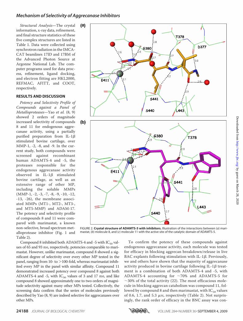
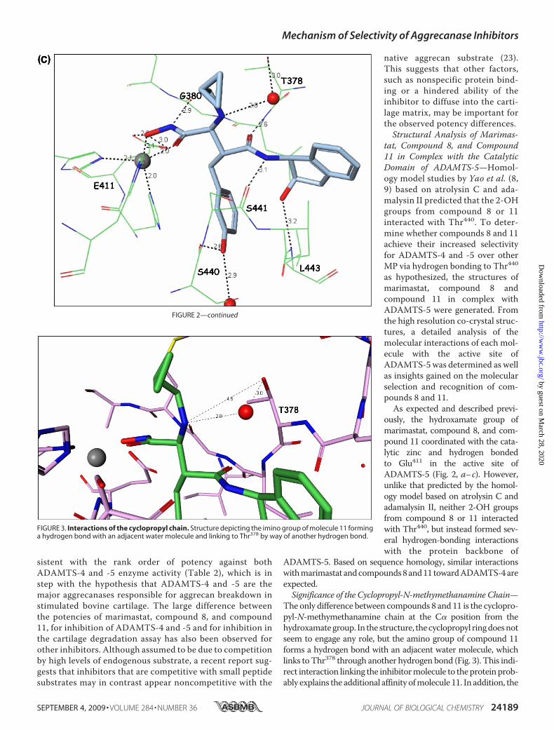
FIGURE 2. Crystal structure of ADAMTS-5 with inhibitors. Illustration of the interactions between (a) mari-mastat, (b) molecule 8, and (c) molecule 11 with the active site of the catalytic domain of ADAMTS-5.
Mechanism of Selectivity of Aggrecanase Inhibitors
24188 JOURNAL OF BIOLOGICAL CHEMISTRY VOLUME 284 • NUMBER 36 • SEPTEMBER 4, 2009
by guest on March 28, 2020
http://ww
w.jbc.org/
Dow
nloaded from
sistent with the rank order of potency against bothADAMTS-4 and -5 enzyme activity (Table 2), which is instep with the hypothesis that ADAMTS-4 and -5 are themajor aggrecanases responsible for aggrecan breakdown instimulated bovine cartilage. The large difference betweenthe potencies of marimastat, compound 8, and compound11, for inhibition of ADAMTS-4 and -5 and for inhibition inthe cartilage degradation assay has also been observed forother inhibitors. Although assumed to be due to competitionby high levels of endogenous substrate, a recent report sug-gests that inhibitors that are competitive with small peptidesubstrates may in contrast appear noncompetitive with the
native aggrecan substrate (23).This suggests that other factors,such as nonspecific protein bind-ing or a hindered ability of theinhibitor to diffuse into the carti-lage matrix, may be important forthe observed potency differences.Structural Analysis of Marimas-
tat, Compound 8, and Compound11 in Complex with the CatalyticDomain of ADAMTS-5—Homol-ogy model studies by Yao et al. (8,9) based on atrolysin C and ada-malysin II predicted that the 2-OHgroups from compound 8 or 11interacted with Thr440. To deter-mine whether compounds 8 and 11achieve their increased selectivityfor ADAMTS-4 and -5 over otherMP via hydrogen bonding to Thr440as hypothesized, the structures ofmarimastat, compound 8 andcompound 11 in complex withADAMTS-5 were generated. Fromthe high resolution co-crystal struc-tures, a detailed analysis of themolecular interactions of each mol-ecule with the active site ofADAMTS-5 was determined as wellas insights gained on the molecularselection and recognition of com-pounds 8 and 11.As expected and described previ-
ously, the hydroxamate group ofmarimastat, compound 8, and com-pound 11 coordinated with the cata-lytic zinc and hydrogen bondedto Glu411 in the active site ofADAMTS-5 (Fig. 2, a–c). However,unlike that predicted by the homol-ogy model based on atrolysin C andadamalysin II, neither 2-OH groupsfrom compound 8 or 11 interactedwith Thr440, but instead formed sev-eral hydrogen-bonding interactionswith the protein backbone of
ADAMTS-5. Based on sequence homology, similar interactionswithmarimastat andcompounds8and11towardADAMTS-4areexpected.Significance of the Cyclopropyl-N-methymethanamine Chain—
The only difference between compounds 8 and 11 is the cyclopro-pyl-N-methymethanamine chain at the C� position from thehydroxamategroup. In thestructure, thecyclopropyl ringdoesnotseem to engage any role, but the amino group of compound 11forms a hydrogen bond with an adjacent water molecule, whichlinks toThr378 through another hydrogen bond (Fig. 3). This indi-rect interaction linking the inhibitormolecule to theproteinprob-ably explains the additional affinity ofmolecule 11. In addition, the
FIGURE 2—continued
FIGURE 3. Interactions of the cyclopropyl chain. Structure depicting the imino group of molecule 11 forminga hydrogen bond with an adjacent water molecule and linking to Thr378 by way of another hydrogen bond.
Mechanism of Selectivity of Aggrecanase Inhibitors
SEPTEMBER 4, 2009 • VOLUME 284 • NUMBER 36 JOURNAL OF BIOLOGICAL CHEMISTRY 24189
by guest on March 28, 2020
http://ww
w.jbc.org/
Dow
nloaded from
Thr378 residue is quite unique to ADAMTSmembers and proba-bly confers some selectivity to the molecule. In most MMPs, thecorresponding residue is a nonpolar amino acid.Potency and Selectivity from the 2-Indanol Ring—From the
2-indanol and on, inhibitor molecules 8 and 11 form a nettedhydrogen bond network (Fig. 2, b and c). The hydroxyl group ofthe indanol ring forms a hydrogen bond with the backbone-NH- of Leu443. The amide -NH- in the inhibitor connects withthe backbone carbonyl group of Ser441, the amide carbonylgroup hydrogen bonds with the backbone -NH- of Leu379, andthe meta-hydroxyl of the phenyl ring makes additional hydro-gen bonds with Ser440 and water. This hydrogen-bonding net-work rigidifies the conformation of this part of the molecule,
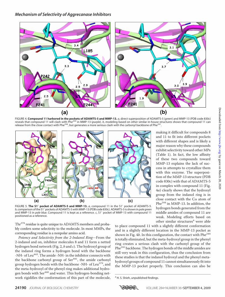
making it difficult for compounds 8and 11 to fit into different pocketswith different shapes and is likely amajor reason why these compoundsexhibit selectivity toward otherMPs(Table 1). In fact, the low affinityof these two compounds towardMMP-13 explains the lack of suc-cess in attempts to crystallize themwith this enzyme. The superposi-tion of the MMP-13 structure (PDBcode 830c) with that of ADAMTS-5in complex with compound 11 (Fig.4a) clearly shows that the hydroxylgroup from the indanol ring is inclose contact with the C� atom ofPhe244 in MMP-13. In addition, thehydrogen bonds generated from themiddle amides of compound 11 areweak. Modeling efforts based onother similar structures4 were able
to place compound 11 with a slightly different conformationand in a slightly different location in the MMP-13 pocket asshown in Fig. 4b. In this configuration, the contact with Phe244is totally eliminated, but themeta-hydroxyl group in the phenylring creates a serious clash with the carbonyl group of thePhe241 backbone. The hydrogen bonds of themiddle amides arestill very weak in this configuration, thus the conclusion fromthese studies is that the indanol hydroxyl and the phenyl meta-hydroxyl groups of compound11 cannot simultaneously fit intothe MMP-13 pocket properly. This conclusion can also be
4 H. S. Shieh, unpublished findings.
FIGURE 4. Compound 11 harbored in the pockets of ADAMTS-5 and MMP-13. a, direct superposition of ADAMTS-5 (green) and MMP-13 (PDB code 830c)reveals that compound 11 will clash with Phe244 in MMP-13 (purple). b, modeling based on other similar in-house structures shows that compound 11 canrelease from the close contact with Phe244, but generates a more serious clash with the carbonyl backbone of Phe241.
FIGURE 5. The S1� pocket of ADAMTS-5 and MMP-13. a, compound 11 in the S1� pocket of ADAMTS-5.b, comparison of the S1� pockets of ADAMTS-5 with MMP-13 (PDB code 830c). ADAMTS-5 is shown in pale greenand MMP-13 in pale blue. Compound 11 is kept as a reference. c, S1� pocket of MMP-13 with compound 11positioned as a reference.
Mechanism of Selectivity of Aggrecanase Inhibitors
24190 JOURNAL OF BIOLOGICAL CHEMISTRY VOLUME 284 • NUMBER 36 • SEPTEMBER 4, 2009
by guest on March 28, 2020
http://ww
w.jbc.org/
Dow
nloaded from
reached for MMP-1 (PDB code 966c) and likely extended toother MPs. Thus, structural studies conducted herein do notsupport selectivity arising from the binding of the meta-hy-droxyl group with the ADAMTS-specific Thr440 as proposedearlier (8, 9).Recognition of the P1� Moiety—The S1� pocket, also referred
to as the specificity pocket, is the recognition site for the differ-ent substrates for metalloproteinases. Marimastat, shown inFig. 1, has a very short P1�moiety, hence it can be recognized bymost of the metalloproteinases. The S1� pocket in ADAMTS-5is enclosed by a flexible loop including residues 426–436, thehelix C defined by residues 402 to 416, and part of helix A2.Several key residues, Leu438, Leu443, and Ile446 in the flexibleloop and Phe406, Thr407, His410, and Leu301 in the helices helpshape/form the pocket, as shown in Fig. 5a. The back side of thepocket in Fig. 5a is walled off essentially by the zinc-chelatingHis410 residue. The most significant feature of the ADAMTS-5pocket is its shallowness due to the presence of Leu438, Ile446,Leu301, and Phe406. Another feature is the restriction on the leftside of the pocket due to the S1� loop backbone. The S1� loop ofADAMTS-5 is smaller compared with the loop found inMMP-13 since it is two residues shorter, making penetrationthrough the loop difficult. The bigger S1� loop found inMMP-13 adopts a different conformation and its helix A2 tiltsaway from the pocket as shown in Fig. 5b, in such amanner as toopen up the channel. The channel pocket in MMP-13 actuallyextends through the loop and reaches out into the solventregion. However, the extension point does not start from themeta-position of the terminal phenyl ring, but rather from itspara-position as seen in Fig. 5c. The para-position exhibitsmore space, and the extension from this position has moreroom to maneuver to find the best fit with the protein in otherparts of the molecule. It will not be tethered to a limit or pro-hibited area as in themeta-position.Many para-extended com-pounds in this series of compounds do show strong affinitytoward MMP-13.4Conclusion—These studies pave the way for the design and
development for potent and selective inhibitors of ADAMTS-4and -5, which will be required for chronic OA therapy.
Acknowledgments—Use of the IMCA-CAT beamline 17-ID (or17-BM) at the Advanced Photon Source was supported by the com-panies of the IndustrialMacromolecularCrystallographyAssociationthrough a contract with theCenter for AdvancedRadiation Sources atthe University of Chicago.
REFERENCES1. Felson, D. T., and Kim, Y. J. (2007) Arthritis Rheum. 56, 1378–13832. Mankin, H. J., and Lippiello, L. (1970) J. Bone Joint Surg. 52, 424–4343. Pratta,M. A., Yao,W., Decicco, C., Tortorella,M. D., Liu, R. Q., Copeland,
R. A., Magolda, R., Newton, R. C., Trzaskos, J. M., and Arner, E. C. (2003)
J. Biol. Chem. 278, 45539–455454. Tortorella, M. D., and Malfait, A. M. (2008) Curr. Pharm Biotechnol. 9,
16–235. Glasson, S. S., Askew, R., Sheppard, B., Carito, B., Blanchet, T., Ma, H. L.,
Flannery, C. R., Peluso, D., Kanki, K., Yang, Z., Majumdar, M. K., andMorris, E. A. (2005) Nature 434, 644–648
6. Hooper, N. M. L., and Uwe (eds). (2005) The ADAM Family of Proteases,Vol 4, Birkhauser Verlag
7. Lagente, V., and Boichot, E. e. (2008)Matrix Metalloproteinases in TissueRemodelling and Inflammation, Vol. XI, Birkhauser
8. Yao, W., Chao, M., Wasserman, Z. R., Liu, R. Q., Covington, M. B., New-ton, R., Christ, D., Wexler, R. R., and Decicco, C. P. (2002) Bioorg Med.Chem. Lett 12, 101–104
9. Yao, W., Wasserman, Z. R., Chao, M., Reddy, G., Shi, E., Liu, R. Q., Cov-ington, M. B., Arner, E. C., Pratta, M. A., Tortorella, M., Magolda, R. L.,Newton, R., Qian, M., Ribadeneira, M. D., Christ, D., Wexler, R. R., andDeCicco, C. P. (2001) J. Med. Chem. 44, 3347–3350
10. Shieh, H. S., Mathis, K. J., Williams, J. M., Hills, R. L., Wiese, J. F., Benson,T. E., Kiefer, J. R.,Marino,M.H., Carroll, J. N., Leone, J.W.,Malfait, A.M.,Arner, E. C., Tortorella, M. D., and Tomasselli, A. (2008) J. Biol. Chem.283, 1501–1507
11. Yem, A. W., Curry, K. A., Tomich, C. S., and Deibel, M. R., Jr. (1988)Immunol. Invest. 17, 551–559
12. Farndale, R. W., Buttle, D. J., and Barrett, A. J. (1986) Biochim. BiophysActa 883, 173–177
13. Abbaszade, I., Liu, R. Q., Yang, F., Rosenfeld, S. A., Ross, O. H., Link, J. R.,Ellis, D. M., Tortorella, M. D., Pratta, M. A., Hollis, J. M., Wynn, R., Duke,J. L., George, H. J., Hillman, M. C., Jr., Murphy, K., Wiswall, B. H., Copel-and, R. A., Decicco, C. P., Bruckner, R., Nagase, H., Itoh, Y., Newton, R. C.,Magolda, R. L., Trzaskos, J. M., and Burn, T. C. (1999) J. Biol. Chem. 274,23443–23450
14. Tortorella,M.D., Burn, T.C., Pratta,M.A., Abbaszade, I., Hollis, J.M., Liu,R., Rosenfeld, S. A., Copeland, R. A., Decicco, C. P.,Wynn, R., Rockwell, A.,Yang, F., Duke, J. L., Solomon, K., George, H., Bruckner, R., Nagase, H.,Itoh, Y., Ellis, D. M., Ross, H., Wiswall, B. H., Murphy, K., Hillman, M. C.,Jr., Hollis, G. F., Newton, R. C., Magolda, R. L., Trzaskos, J. M., and Arner,E. C. (1999) Science 284, 1664–1666
15. Hughes, C. E., Caterson, B., Fosang, A. J., Roughley, P. J., and Mort, J. S.(1995) Biochem. J. 305, 799–804
16. Tortorella, M. D., Arner, E. C., Hills, R., Gormley, J., Fok, K., Pegg, L.,Munie, G., andMalfait, A. M. (2005) Arch. Biochem. Biophys. 444, 34–44
17. Rosenfeld, S. A., Ross, O. H., Corman, J. I., Pratta,M. A., Blessington, D. L.,Feeser, W. S., and Freimark, B. D. (1994) Gene 139, 281–286
18. Pratt, L. M., Beckett, R. P., Bellamy, C. L., Corkill, D. J., Cossins, J., Court-ney, P. F., Davies, S. J., Davidson, A. H., Drummond, A. H., Helfrich, K.,Lewis, C. N., Mangan, M., Martin, F. M., Miller, K., Nayee, P., Ricketts,M. L., Thomas, W., Todd, R. S., and Whittaker, M. (1998) Bioorg MedChem Lett 8, 1359–1364
19. Knight, C. G., Willenbrock, F., and Murphy, G. (1992) FEBS Lett. 296,263–266
20. Kuzmic, P., Elrod, K. C., Cregar, L. M., Sideris, S., Rai, R., and Janc, J. W.(2000) Anal. Biochem. 286, 45–50
21. Cheng, Y., and Prusoff,W. H. (1973) Biochem. Pharmacol. 22, 3099–310822. Tortorella, M. D., Malfait, A. M., Deccico, C., and Arner, E. (2001) Osteo-
arthritis and Cartilage 9, 539–55223. Wittwer, A. J., Hills, R. L., Keith, R. H., Munie, G. E., Arner, E. C., Anglin,
C. P., Malfait, A. M., and Tortorella, M. D. (2007) Biochemistry 46,6393–6401
Mechanism of Selectivity of Aggrecanase Inhibitors
SEPTEMBER 4, 2009 • VOLUME 284 • NUMBER 36 JOURNAL OF BIOLOGICAL CHEMISTRY 24191
by guest on March 28, 2020
http://ww
w.jbc.org/
Dow
nloaded from
Anne-Marie Malfait and Huey-Sheng ShiehWoodard, Grace Munie, Jennifer M. Williams, Nicole Caspers, Arthur J. Wittwer,
Micky D. Tortorella, Alfredo G. Tomasselli, Karl J. Mathis, Mark E. Schnute, Scott S.of Aggrecanase Inhibitors
Structural and Inhibition Analysis Reveals the Mechanism of Selectivity of a Series
doi: 10.1074/jbc.M109.029116 originally published online July 8, 20092009, 284:24185-24191.J. Biol. Chem.
10.1074/jbc.M109.029116Access the most updated version of this article at doi:
Alerts:
When a correction for this article is posted•
When this article is cited•
to choose from all of JBC's e-mail alertsClick here
http://www.jbc.org/content/284/36/24185.full.html#ref-list-1
This article cites 21 references, 5 of which can be accessed free at
by guest on March 28, 2020
http://ww
w.jbc.org/
Dow
nloaded from