structure and function of human erythrocyte … · anemia, which can be life threatening and...
TRANSCRIPT

1
STRUCTURE AND FUNCTION OF HUMAN ERYTHROCYTE
PYRUVATE KINASE: MOLECULAR BASIS OF NONSPHEROCYTIC
HEMOLYTIC ANEMIA
Giovanna Valentini‡,||*, Laurent R. Chiarelli||, Riccardo Fortin‡,
Manuela Dolzan‡, Alessandro Galizzi‡, Donald J. Abraham§,
Changqing Wang§, Paola Bianchi¶, Alberto Zanella¶
and Andrea Mattevi‡ *
‡ Dipartimento di Genetica e Microbiologia, Università di Pavia, via
Abbiategrasso 207, 27100 Pavia, Italy
|| Dipartimento di Biochimica, Università di Pavia, via Taramelli 3b,
27100 Pavia, Italy
§ Department of Medicinal Chemistry, Virginia Commonwealth
University, 800 East Leigh St. Suite 212 Richmond, VA 23219, USA
¶ Divisione di Ematologia, IRCCS Ospedale Maggiore di Milano, via
Francesco Sforza 35, 20122 Milano, Italy
*Corresponding authors: Andrea Mattevi or Giovanna Valentini,
Dipartimento di Genetica e Microbiologia, Università di Pavia, via
Abbiategrasso 207, 27100 Pavia, Italy; E-mail
[email protected], Fax +39-0382-528496
Running title: Human erythrocyte pyruvate kinase.
Copyright 2002 by The American Society for Biochemistry and Molecular Biology, Inc.
JBC Papers in Press. Published on April 17, 2002 as Manuscript M202107200 by guest on A
ugust 28, 2019http://w
ww
.jbc.org/D
ownloaded from

2
S u m m a r y
Deficiency of human erythrocyte R isozyme (RPK) is, together with
glucose 6-phosphate dehydrogenase deficiency, the most common
cause of the nonspherocytic hemolytic anemia. To provide a
molecular framework to the disease, we have solved the 2.7 Å
resolution crystal structure of human RPK in complex with fructose
1,6-bisphosphate, the allosteric activator, and phosphoglycolate, a
substrate analogue, and we have functionally and structurally
characterized eight mutants (G332S, G364D, T384M, D390N,
R479H, R486W, R504L, R532W) found in RPK-deficient patients. The
mutations target distinct regions of RPK structure, including domain
interfaces and catalytic and allosteric sites. The mutations affect to
different extent thermostability, catalytic efficiency and regulatory
properties. These studies are the first to correlate the clinical
symptoms with the molecular properties of the mutant enzymes.
Mutations greatly impairing thermostability and/or activity are
associated to severe anemia. Some mutant proteins exhibit
moderate changes in the kinetic parameters, which are sufficient to
cause mild-to-severe anemia, underlining the crucial role of RPK for
erythrocyte metabolism. Prediction of the effects of mutations is
difficult since there is no relation between the nature and location
of the replaced amino acid and the type of molecular perturbation.
by guest on August 28, 2019
http://ww
w.jbc.org/
Dow
nloaded from

3
Characterization of mutant proteins may serve as a valuable tool to
assist with diagnosis and genetic counseling.
by guest on August 28, 2019
http://ww
w.jbc.org/
Dow
nloaded from

4
I n t r o d u c t i o n
Pyruvate kinase (PK)1 catalyses the conversion of
phosphoenolpyruvate (PEP) to pyruvate with the synthesis of ATP.
The enzyme requires K+ and Mg++ (or Mn++) for activity (1,2,3). The
PK catalyzed reaction represents the last step of glycolysis with the
reaction product, pyruvate, being involved in a number of energetic
and biosynthetic pathways. PK is activated homotropically by PEP
and heterotropically by monophosphorylated or bisphosphorylated
sugars (2). In addition, Mg++, H+ and other cations modulate
enzymatic activity (4). The regulatory behavior of PK varies
depending on the enzyme source. Four PK isozymes have been
identified in mammals (5). The M1 (muscle) and M2 (fetal)
proteins are products of the alternative splicing of the same mRNA.
M2 PK is allosterically activated by fructose 1,6-bisphosphate (FBP)
and PEP while the M1 enzyme is exceptional in that it is the only
known PK that displays hyperbolic kinetics. The other two
mammalian PK isozymes, liver (L) and erythrocyte (R), are coded by
the same PKLR gene through the use of tissue specific alternate
promoters. Both R and L isozymes are activated by PEP and FBP (2).
The three-dimensional structures of several PKs from prokaryotic
and eukaryotic organisms have been elucidated (6,7,8,9,10). They
reveal a conserved architecture. PK is a 200 kDa tetramer with four
by guest on August 28, 2019
http://ww
w.jbc.org/
Dow
nloaded from

5
identical subunits, each consisting of four domains (Fig. 1); the
small N-terminal helical domain (absent in bacterial PKs), the A
domain with (β/α)8 barrel topology, the B domain which is inserted
between strand β3 and helix α3 of the A domain (β/α)8 barrel, and
the C domain with a α+β topology. This multidomain architecture is
instrumental to the regulation of PK activity. The enzyme activation
is thought to involve a combination of domain and subunit
rotations, coupled to alterations in the active site geometry. In this
mechanism, the residues located at the domain and subunit
interfaces are crucial in that they function in the communication
between activator-binding site and catalytic center (8,9,10).
Deficiency of human erythrocyte R isozyme (RPK) is, together with
glucose 6-phosphate dehydrogenase deficiency, the most common
cause of nonspherocytic hemolytic anemia (11). RPK deficiency
severely affects the erythrocyte metabolism, causing ATP depletion
which ultimately leads to hemolysis. Worldwide, more than 150
mutations in the gene coding RPK have been found in RPK-deficient
patients (12). The disease is transmitted as recessive trait and the
pathological symptoms occur only in homozygotes or compound
heterozygotes. The clinical manifestations vary from mild to severe
anemia, which can be life threatening and require continuous
transfusion therapy. Here, we describe the first crystal structure of
by guest on August 28, 2019
http://ww
w.jbc.org/
Dow
nloaded from
6
recombinant RPK2 and the biochemical characterization of eight
mutants found in patients subjected to clinical follow-up. These
studies allow a correlation between the clinical symptoms and the
molecular properties of the mutant enzymes.
Experimental procedures
Expression vectors
The vectors used to express RPK and its mutant and truncated forms
were derivatives of pTrcHisB (Invitrogen). RPK cDNA insert was
obtained from pGG1 (13) after introduction of Nco I-Nde I sites
around the ATG initiation codon. The mutagenic primer (14) used
to introduce the two sites into pGG1 was 5’-
CAAGGAGGCTGAAACCATGGCTAGCCAGGAGAACATATCATT. It altered
the second and the third codon of the insert. The underlined
sequences indicate the mutated bases. The selection primer used to
abolish the vector unique restriction site AflIII was 5’-
CGCAGGAAAGACCTTGGGAGCAAAAGGCC. The pTrcHisB with RPK
cDNA inserted in NcoI/EcoRI and designated pCW3 was mutagenised
to restore the codons previously changed. The mutagenic primer
was 5’-TAAGGAGGAATAAACCATGTCGATCCA GGAGAACATATCAT.
The selection was performed by digesting the parental pCW3 with
by guest on August 28, 2019
http://ww
w.jbc.org/
Dow
nloaded from

7
Nco I. The new plasmid, containing the correct insert, was named
pLC1. To obtain the desired RPK mutants, the pLC1 was subjected to
site-directed mutagenesis (14). The same selection primer (5’-
CCCCCCTGAATTCGAACCTT GGCTG) to abolish the unique HindIII site
was used in all cases. The specific mutagenic primers were:
5’-CTGGAGGTGAGCGACAGCATCATGGTGGCA for G332S;
5’-CTGCAACTTGGCGGACAAGCCTGTTGTCTG for G364D;
5’-CAAGCCCCGGC CAATGAGGGCAGAGACAAG for T384M;
5’-AGGGCAGAGACAAGCAATGTCGC CAATGCTG for D390N;
5’-CTGACCACAACTGGCCACTCAGCCCAGCTTCTG for R479H;
5’- AGCCCAGCTTCTGTCTTGGTACCGA CCTCGG for R486W;
5’-CTGCCCAGGCTGCC CTCCAGGTCCACTTAT for R504L;
5’-ATGATGTAAGATCGCTGGGTGCAATTTGGCA for R532W.
To obtain a truncated form of RPK, lacking the first 49 residues,
pCW3 was mutagenised by using the primer 5’-
TAAGGAGGAATAAACCATGGAGCTGGGCACTGCC TTCTTCC. This
sequence corresponds to that of the plasmid upstream of the
initiation triplet ATG and continues with that of the RPK insert
starting from the GAG codon of Glu at position 50. The selection
was performed by digesting pCW3 with NheI restriction enzyme. The
plasmid with the insert encoding the truncated RPK (50-574) was
designated pLC3. All inserts were sequenced.
by guest on August 28, 2019
http://ww
w.jbc.org/
Dow
nloaded from

8
Protein purification and enzymatic analysis
E.coli DH5α transformed with the specific expression vectors were
grown at 37°C in Luria-Bertani medium containing 100 µg /mL
ampicillin. When the culture optical density at 600 nm reached a
value of 0.5, the expression was induced by addition of isopropyl-β-
D-thiogalactopyranoside at a final concentration of 0.5 mM. The
induction time was 12 hours while the induction temperature was
30°C, with the exception of the mutants G332S, G364D, R504L, and
R532W, for which the induction temperature was 21°C. Wild-type
and mutant enzymes were purified by the procedure of Wang et al.
(13). Enzyme activities were measured at 37°C by the assay (13)
recommended by the International Committee for Standardization
in Hematology. Kinetic parameters were determined with the
Enzyme Kinetic ModuleTM 1.1 (SPSS Science Software Gmb). Thermal
stability was measured by incubating the enzyme (100-200 µg /mL)
at 53°C in a solution consisting of 20 mM potassium phosphate pH
6.5, 100 mM KCl, and 1 mM EDTA. Samples were removed at
intervals and immediately assayed.
by guest on August 28, 2019
http://ww
w.jbc.org/
Dow
nloaded from

9
Crystallography
Recombinant wild-type RPK was crystallized using the vapor
diffusion method at 22°C. Well solutions consisted of 50 mM
Mes/KOH pH 6.4, 10 mM MnSO4, and 10-14% w/v PEG8000.
Hanging drops were formed by mixing equal volumes of 12 mg/ml
protein in 50 mM KCl, 5 mM FBP, 5 mM phosphoglycolate, 20 mM
potassium phosphate pH 7.0 and well solutions. The crystals were
difficult to reproduce. The recombinant enzyme undergoes partial
proteolysis and about 50% of the purified protein chains lack the
first 47 amino acids (13). On this basis, we produced a mutant
truncated enzyme lacking the first 49 residues (see above).
Employment of the truncated protein greatly improved the
crystallization, which was carried out using the above-described
protocol. Crystals were obtained for the T384M, R479H and R486W
truncated mutants by the same protocol used for the truncated
wild-type RPK.
RPK crystals belong to space group P21 with one tetramer in the
asymmetric unit. Diffraction data were measured at 100 K on
beamline ID14-EH2 of the European Synchrotron Radiation Facility
(Grenoble, France) using a MarCCD detector and beamline B7WB of
DESY/EMBL (Hamburg, Germany) using a Mar Imaging Plate. Data
processing and reduction were carried out using MOSFLM (15) and
by guest on August 28, 2019
http://ww
w.jbc.org/
Dow
nloaded from

1 0
programs of the CCP4 suite (16). Data collection statistics are
reported in Table 1. The structure of the wild-type RPK was solved
by molecular replacement using the program Molrep (16). The
search model was the structure of rabbit muscle M1 PK in complex
with pyruvate (7; PDB entry 1PKN). Phases were improved by 4-fold
averaging (17) producing an electron density of excellent quality.
Model building was carried out with the program O (18). The model
was refined using Refmac (19). All measured data (no σ cut-off)
were employed and 2.5% of unique reflections were used to
monitor the progress of the refinement by Rfree validation. The
refined wild-type coordinates provided the starting model for the
refinement of the mutants. The set of reflections for calculation of
R free was identical to that of the wild-type structure refinement. A
summary of refinement statistics is presented in Table 1. Analysis
and inspection of the structures were carried out with O (18) and
programs of the CCP4 package (16). Figures were generated with
Molscript (20).
by guest on August 28, 2019
http://ww
w.jbc.org/
Dow
nloaded from
1 1
R e s u l t s
Position of Figure 1
The three-dimensional structure of RPK
The crystallographic studies were performed using a truncated RPK
in which the 49 N-terminal amino acids are absent. Use of the
truncated protein resulted in considerable improvement in the
reproducibility of the crystallization experiments. The truncated
protein exhibits kinetic properties virtually identical to those of
wild-type RPK. A more detailed analysis of this and other mutants
targeting the N-terminal residues will be published elsewhere.
The truncated recombinant RPK was crystallized in complex with
phosphoglycolate (a PEP analogue), FBP, Mn++ and K+. The presence
of the allosteric activators implies that the crystalline enzyme is in
the active R-state. The 2.7 Å resolution structure of RPK reveals the
typical four-domain subunit architecture found in all PKs of known
three-dimensional structure (Fig. 1a). The A (residues 85-159 and
263-431) and C domains (432-574) together with the small N-
terminal domain (57-84) form the main body of the subunit. The B
domain (160-262) is loosely packed to the rest of the molecule and
adopts slight different orientations (about 4°) in the four
by guest on August 28, 2019
http://ww
w.jbc.org/
Dow
nloaded from

1 2
crystallographically independent polypeptide chains. The four
subunits of the RPK tetramer are assembled to form a D2 symmetric
oligomer. The intersubunit interactions define two large contact
areas; the A/A’ interface involves the A domains of subunits related
by the vertical twofold axis, as defined in Figure 1b, while the C/C’
interface involves the C-domains of subunits interacting along the
horizontal axis.
The structure of RPK subunit closely resembles that of rabbit muscle
M1 PK, as expected from the 59% sequence identity between the two
proteins. The similarity is highest with M1 PK in complex with
pyruvate (7), with a root-mean-square difference of 1.2 Å for 512
Cα atom pairs. This M1 PK complex exhibits the same B domain
orientation found in RPK. In other M1 structures, the B domain is
either more open, as in the phospholactate complex (21), or more
closed, as in the complex with ATP (22,23).
Position of Figure 2
The allosteric site and the catalytic center
RPK was cocrystallized with FBP, phosphoglycolate and the K+ and
M n ++ ions. All ligands are clearly visible in the electron density map.
by guest on August 28, 2019
http://ww
w.jbc.org/
Dow
nloaded from

1 3
Phosphoglycolate, a potent PK inhibitor (9), is positioned in the PEP-
binding site, which is located at the top of the A domain (β/α)8
barrel, facing a cleft between the A and B domains (Fig. 1a). It is at
the heart of an intricate network of H-bonds, which involve protein
residues and the Mn++ and K+ cations (Fig. 2a). The phosphate group
is bound to the K+ atom and the side chain of Arg116 while the
carboxylate moiety is anchored through interactions with the Mn+ +
ion, the side chain of Thr371 and the main chain nitrogen atoms of
Gly338 and Asp339, which are located at the N-terminus of a short
helical segment belonging to loop 6 of the A domain (β/α)8 barrel.
This binding geometry is identical to that observed in the yeast PK-
phosphoglycolate complex (9) and closely resembles the binding of
pyruvate and phospholactate to rabbit M1 PK (for a discussion of
the implications of this binding mode for catalysis see References 7
and 21). These similarities are in keeping with the strict
conservation among the PK sequences of all residues surrounding
the substrate-binding site.
The FBP activator is hosted in the allosteric site in the C domain (Fig.
1a). The ligand is sandwiched between loops 475-479 and 557-566
(Fig. 2b) and extensively interacts with protein. The 6’-phosphate
group is engaged in a salt bridge with Arg532 while the 1’-
phosphate is H-bonded to the side chains of Thr475 and Ser480 and
by guest on August 28, 2019
http://ww
w.jbc.org/
Dow
nloaded from

1 4
the backbone nitrogen atoms of Thr476 and Thr477. Moreover, the
dipole of α -helix 480-486 points towards the 1’-phosphate, further
compensating the ligand negative charge. This geometry in FBP-
binding is identical to that found in yeast PK crystallized in the R-
state (9).
Rational for the mutagenesis studies
A survey of the missense mutations associated to the
nonspherocytic hemolytic anemia shows that most of them cluster
in specific regions of the protein three-dimensional structure: the
interface between the A and C domains, the A/A’ intersubunit
interface, the hydrophobic core of the A domain, and the FBP-
binding site (24). We generated eight RPK mutants (Fig. 1a),
targeting residues belonging to each of these regions of the protein.
Almost all selected mutations have been found in homozygote
patients. The kinetic, allosteric and thermostability parameters of
mutant proteins were evaluated (Table 2) and the crystal structures
of three mutants (T384M, R479H and R486W) were solved. The
mutations did not induce significant conformational changes in the
overall protein conformation and, therefore, we shall restrict the
description of the mutant structures mainly to the sites affected by
the mutations.
by guest on August 28, 2019
http://ww
w.jbc.org/
Dow
nloaded from
1 5
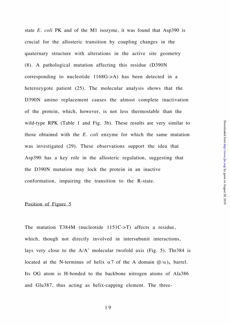
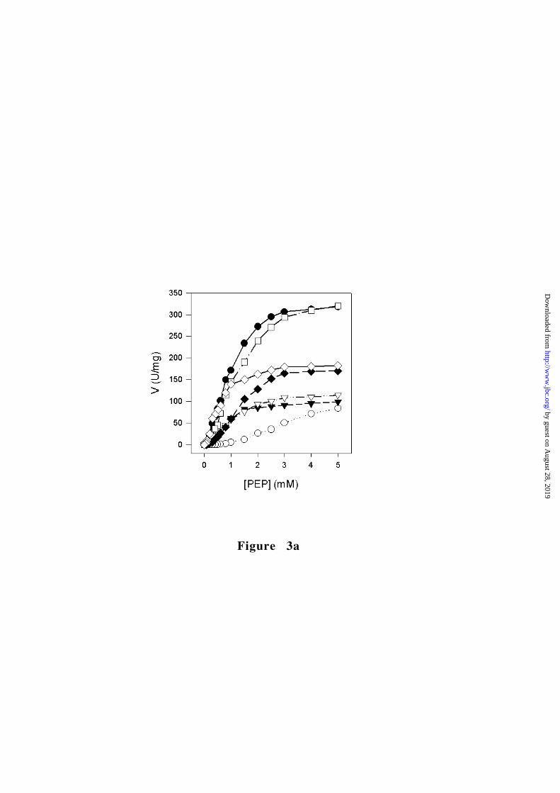
Position of Figure 3
G332S mutation in the A domain hydrophobic core
Many RPK mutations are localized in the hydrophobic core of the A
domain. An example is the G332S mutation (nucleotide 994G->A),
which affects a residue that is strictly conserved among PK
sequences. Gly332 is located on strand β6, being buried inside the
domain core (Fig. 1a). The G332S protein exhibits a 9-fold decrease
in the catalytic efficiency (5-fold for the FBP activated protein) and
is considerably less thermostable than the wild-type enzyme (Table
2 and Fig. 3). These substantially altered molecular properties
account for the clinical data. In homozygous form, the G332S
mutation leads to a severe anemia with the need of regular
transfusions (25,26).
Mutations at the A/C interface: G364D, R486W and R504L
The interface between the A and C domains is characterized by
many polar interactions that often involve charged side chains.
Many of these residues represent sites of pathological mutations,
which cause RPK-deficiency with variable levels of severity.
by guest on August 28, 2019
http://ww
w.jbc.org/
Dow
nloaded from

1 6
The mutation R504L (nucleotide 1511G->T) affects Arg504, a C-
domain residue which is partly solvent accessible and engaged in an
interdomain salt bridge with Asp281 (Fig. 1a). The R504L mutation
removes this interdomain interaction and introduces a hydrophobic
Leu side chain in a solvent exposed site close to a negatively charged
Asp. Such amino acid replacement is clearly unfavorable, providing
a rational for the extreme instability of the protein, which
prevented functional analysis (Table 2). This feature explains the
severe anemia found in RPK-deficient patients homozygous for this
mutation (27).
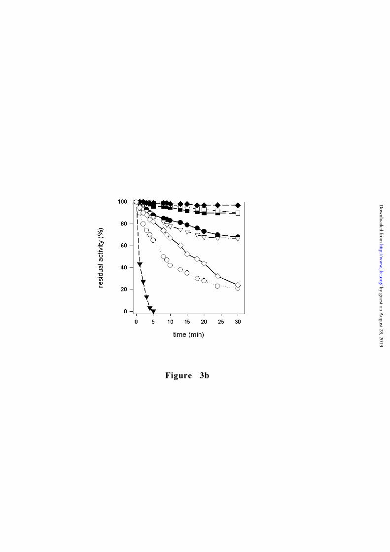
Position of Figure 4
The other two investigated mutants targeting the A/C interface
affect Gly364 and Arg486, which are part of a region of close
association between the A and C domains (Figs. 1a and 4a). Arg486
is H-bonded to the carbonyl oxygen of Leu362 at the C-terminus of
the A domain helix α6 while the neighboring Gly364 allows a sharp
turn of the polypeptide chain with a backbone conformation
(φ=85°,ψ=98°) that is unfavorable for a non-glycine residue. The
G364D (nucleotide 1091G->A) mutation has a drastic effect on the
enzyme stability, which is coupled to a 3-fold reduction of the
by guest on August 28, 2019
http://ww
w.jbc.org/
Dow
nloaded from
1 7
catalytic efficiency (Fig. 3b and Table 2). Given the tightly packed
environment and the backbone conformation of Gly364, it is
conceivable that introduction of a charged Asp side chain at this
site of the A/C interface can greatly perturb the domain assembly,
thus being deleterious for stability. Fully consistent with these
observations is the severe anemia found in patients homozygous for
G364D (28). Together with R504L, the G364D mutant highlights the
notion that the inter-domain interactions at the A/C interface are
critical for the stability of the protein.
R486W (nucleotide 1456C->T) is among the most frequent
mutations found in RPK-deficient patients (11). Characterization of
this mutant reveals that such a drastic amino acid replacement
results in small effects on the molecular properties. The mutant
three-dimensional structure shows that the Trp side chain is
accommodated without any structural perturbation. With respect to
the wild-type structure, no atomic movements larger than 0.25 Å
can be detected whereas the indole nitrogen atom is able to
establish a H-bond with the carbonyl oxygen atoms of Leu362 and
Asp361. Such structural conservation matches the limited changes
in biochemical parameters. The thermostability is even higher than
that of the wild-type protein (Fig. 3b) and the allosteric properties
are essentially unmodified (Table 2 and Fig. 3a). The only significant
by guest on August 28, 2019
http://ww
w.jbc.org/
Dow
nloaded from

1 8
perturbation is in the catalytic efficiency, which drops to 30% of the
value measured for the wild-type RPK (Table 2). These moderate
variations in the molecular parameters correlate with the clinical
symptoms since patients homozygous for the R486W mutation
generally exhibit a mild anemia (25).
The perturbed kinetics of the R486W protein is puzzling since
Arg486 is >20 Å away from the catalytic center (Fig. 1a), which is
left unperturbed by the mutation as shown by the mutant crystal
structure. The “long range” effect exercised by the R486W mutation
might reflect altered dynamic properties. It is known that the B
domain adopts different conformations depending on ligand binding
(21,22,23). The introduction of the Trp aromatic ring may restrict
the overall ability of the enzyme to undergo the conformational
changes occurring during catalysis, thereby perturbing the reaction
kinetics.
The A/A’ interface: T384M and D390N
Asp390 is a solvent inaccessible residue located in the A/A’
interface, at the heart of a H-bond network that involves Arg337
and Ser389’ (the prime symbol denotes a residue of a different
subunit). Based on the comparison between the structures of the T-
by guest on August 28, 2019
http://ww
w.jbc.org/
Dow
nloaded from
1 9
state E. coli PK and of the M1 isozyme, it was found that Asp390 is
crucial for the allosteric transition by coupling changes in the
quaternary structure with alterations in the active site geometry
(8). A pathological mutation affecting this residue (D390N
corresponding to nucleotide 1168G->A) has been detected in a
heterozygote patient (25). The molecular analysis shows that the
D390N amino replacement causes the almost complete inactivation
of the protein, which, however, is not less thermostable than the
wild-type RPK (Table 1 and Fig. 3b). These results are very similar to
those obtained with the E. coli enzyme for which the same mutation
was investigated (29). These observations support the idea that
Asp390 has a key role in the allosteric regulation, suggesting that
the D390N mutation may lock the protein in an inactive
conformation, impairing the transition to the R-state.
Position of Figure 5
The mutation T384M (nucleotide 1151C->T) affects a residue,
which, though not directly involved in intersubunit interactions,
lays very close to the A/A’ molecular twofold axis (Fig. 5). Thr384 is
located at the N-terminus of helix α7 of the A domain (β/α)8 barrel.
Its OG atom is H-bonded to the backbone nitrogen atoms of Ala386
and Glu387, thus acting as helix-capping element. The three-
by guest on August 28, 2019
http://ww
w.jbc.org/
Dow
nloaded from

2 0
dimensional structure of the T384M mutant reveals that the
mutation does not cause atomic shifts larger than 0.3 Å (Fig. 5). The
bulkier Met side chain is easily accommodated, the only change
being the removal of the helix-capping H-bonds. Also the kinetic
characterization shows limited variations, the main difference
between T384M and the wild-type protein being a 3-fold reduction
of the catalytic efficiency (5-fold for the FBP activated form; Table
2) mainly accounted by a reduction in K cat. Likewise, the mutation
does not alter the thermostability parameters (Fig. 3).
Thr384 is not part of the binding sites for PEP and ADP and the
crystal structure of the T384M protein shows that the active site
geometry is not affected by the mutation. Thus, the altered kinetics
displayed by the T384M mutant is difficult to rationalize. Modified
enzymatic parameters were observed also for the equivalent
mutation in the rabbit kidney isozyme (30). Thr384 is close to the
contact region between A and B domains (Fig. 1a) and, therefore,
may disturb the “closure” of the B domain occurring on ATP binding
(22). An alteration of the equilibrium between “open” and “closed”
B domain conformations may affect the enzymatic activity. It is
remarkable that homozygosity for the T384M mutation is
associated to anemia with mild-to-severe symptoms (31,32),
by guest on August 28, 2019
http://ww
w.jbc.org/
Dow
nloaded from

2 1
implying that even moderate changes in the enzyme catalytic power
can have pathological effects.
The allosteric site: R532W and R479H
The negative charges of FBP are compensated by the N-terminus of
helix 479-486 for the 1’-phosphate and Arg532 side chain for the
6’-phosphate (Fig. 2b). We have investigated two mutations that
target both these elements involved in FBP binding. The first of these
mutations is R532W (nucleotide 1594 C->T) that has been found in
compound heterozygotes, in which the other allele had a mutation
causing the truncation of the protein. The clinical symptoms in the
patients carrying the mutation were severe (33). Molecular analysis
of R532W protein indicates a complete loss in the responsiveness to
FBP, highlighting the essential of Arg532 role for activator binding
(Table 2). These perturbed allosteric properties are associated to a
decreased thermostability (Fig. 3b), possibly reflecting the
energetically unfavorable exposure on the protein surface of the
hydrophobic Trp residue.
The R479H mutation has been found in RPK-deficient patients
affected by severe anemia (34,35). The side chain of Arg479 is
located in the neighborhood of FBP although it does not directly
by guest on August 28, 2019
http://ww
w.jbc.org/
Dow
nloaded from

2 2
interact with the activator (Fig. 2b). The crystal structure of R479H
is identical to that of the wild-type protein, with the His side chain
being fully solvent-exposed. Similarly, the kinetic parameters (Table
2) appear to be essentially unaffected by the mutation. These
features are in contrast with the severe clinical symptoms (34,35).
An explanation for this riddle is given by the observation that the
mutation affects nucleotide 1436, which is located on a splicing site
at the 3’-end of exon 10. This fact together with our biochemical
analysis suggests that, rather than the amino acid replacement,
defects in mRNA splicing process are the actual cause of the RPK-
deficiency.
D i s c u s s i o n
Implications for the allosteric regulation
PK is a typical allosteric enzyme of the K-type. The allosteric signal
is transmitted across the long distance (>20 Å) separating the FBP-
binding site from the catalytic center. The exact mechanism of the
allosteric transition is not known in detail since no PK has been so
far crystallized in both T and R states. The comparison between the
structures of the T-state E. coli PK and of the rabbit M1 enzyme (8)
by guest on August 28, 2019
http://ww
w.jbc.org/
Dow
nloaded from

2 3
suggested that the allosteric transition involves modifications in the
relative orientations of the domains and subunits coupled to
conformational changes in the PEP-binding site. The X-ray analysis of
the T-state Leishmania mexicana PK (10) and the R-state yeast PK (9)
led to a further refinement of this model, allowing to discriminate
between the structural differences that are consequence of the
inherent divergence between eukaryotic and prokaryotic proteins
and the conformational changes that are genuinely due to the
allosteric transition. The mechanism of PK regulation has also been
the subject of many mutagenesis experiments (23 and 36 and
references therein). The general picture emerging from the mutant
analysis is that the intersubunit interactions at the A/A’ and C/C’
interfaces and the interdomain interactions at the A/B interface are
key to the allosteric responsiveness and to define the distribution of
the conformations between active and inactive states. Moreover, the
mutagenesis analyses combined with the crystallographic data
provide clear evidence for the idea that the T and R forms
correspond to ensembles of conformations characterized by
rotational flexibility of the B-domain (23).
Our study on human RPK is fully consistent with these features. The
crystal structure shows that also in RPK the B-domain is flexible,
adopting different orientations in the crystallographically
by guest on August 28, 2019
http://ww
w.jbc.org/
Dow
nloaded from

2 4
independent subunits. The key functional role of the A/A’ interface
is highlighted by D390N mutation, which targets a residue located in
the core of the A/A’ interface, producing an enzyme that retains a
stable tetrameric state but almost entirely lacks enzymatic activity.
Conversely, none of the mutations targeting residues at the A/C
interface alters the enzyme allosteric properties. Thus, in agreement
with the recent mutagenesis data on the yeast PK (36), this domain
interface appears to have little role in the transduction of the
allosteric signal, rather being important for the stability of the
domain assembly within the enzyme subunit.
Molecular basis of nonspherocytic hemolytic anemia
Characterization of mutant proteins shows that amino acid
substitutions can affect thermostability, catalytic efficiency and
response to the allosteric effector. Various regions of the RPK
structure, including domain interfaces and functional sites, are
affected by the pathological mutations (Fig. 1a). However, there
appears to be no relation between the nature and location of the
replaced amino acid and the type of molecular perturbation. For
instance, both R504L and R486W mutations affect Arg residues
involved in interdomain polar interactions at the A/C interface but
their effects are substantially different. The R504L protein is
by guest on August 28, 2019
http://ww
w.jbc.org/
Dow
nloaded from

2 5
extremely unstable while the R486W mutant is even slightly more
thermoresistant than the wild-type (Fig. 3b). These observations
emphasize the difficulty of predicting the consequences of
mutations simply from the location and the nature of the target
residues. They also warn against predictions of the effects of
mutations in human RPK based on the molecular analysis of other PK
isoenzymes.
The clinical manifestations of a genetic disease reflect the
interactions of a variety of physiological and environmental factors
and do not solely depend on molecular properties of the altered
molecule. Given this caution, it is evident that there is a general
correlation between the clinical manifestations and the biochemical
parameters of the mutant proteins. Mutants exhibiting strongly
perturbed kinetic and thermostability parameters (G332S, G364D,
R504L and R532W) are associated to severe RPK-deficiency.
Conversely, in the case of less abnormal molecular properties, the
disease has milder manifestations. It is remarkable that pathological
conditions are present in association to mutations such as T384M
or R486W, which are simply characterized by a moderate reduction
of the catalytic efficiency. The physiological concentrations of RPK
substrates and effectors are in the µM range (37). Therefore, in vivo
RPK operates in sub-saturating conditions, which may amplify the
by guest on August 28, 2019
http://ww
w.jbc.org/
Dow
nloaded from

2 6
effects of the different catalytic efficiencies between the wild-type
and mutant proteins (Fig. 3a).
In conclusion, our studies indicate that the functional parameters of
RPK are so finely tuned that even moderate molecular alterations
may significantly perturb cell metabolism. The correlation between
molecular and clinical parameters in PK-deficiency suggests that
biochemical characterization of mutant proteins may serve as a
valuable tool to understand and assist with diagnosis and genetic
counseling.
A c k n o w l e d g e m e n t s
We thank the staff of DESY/EMBL and ESRF for help during data
collection. This research was supported by grants from University
of Pavia (Progetto d’Ateneo “Nuove tecnologie molecolari e
cellulari”), Ministero della Ricerca Scientifica e Tecnologica
(Progetto Genomica Funzionale), and Allos Therapeutics Inc.,
Westminster, CO (USA).
by guest on August 28, 2019
http://ww
w.jbc.org/
Dow
nloaded from

2 7
R e f e r e n c e s
1. Kayne, F.J. (1973) The Enzymes, 3rd Ed., Academic Press Inc.,
New York and London 8 , 353-382
2. Fothergill-Gilmore, L.A., and Michels, P.A. (1992) Progr. Mol.
Biol. Biophys. 59 , 105-227
3. Gupta, R.K., Oesterling, R.M., and Mildvan, A.S. (1976)
Biochemistry 15 , 2881-2887
4. Mesecar, A.D., and Nowak, T. (1997) Biochemistry 36, 6792-
6 8 0 2
5. Hall, E.R., and Cottam, G.L. (1978) Int. J. Biochem. 9 , 785-793
6. Allen, S.C., and Muirhead, H. (1996) Acta Crystallogr. D52 , 499-
5 0 4
7. Larsen, T.M., Laughlin, L.T., Holden, H.M., Rayment, I., and Reed,
G.H. (1994) Biochemistry 33 , 6301-6309
8. Mattevi, A., Valentini, G., Rizzi, M., Speranza, M.L., Bolognesi,
M., and Coda A. (1995) Structure 3 , 729-741
by guest on August 28, 2019
http://ww
w.jbc.org/
Dow
nloaded from

2 8
9. Jurica, M.S., Mesecar, A., Heath, P.J., Shi, W., Nowak, T., and
Stoddard, B.L. (1998) Structure 6 , 195-210
10. Rigden, D.J., Phillips, S.E.V., Michels, P.A.M., and Fothergill-
Gilmore, L.A. (1999) J. Mol. Biol. 291 , 615-635
11. Zanella, A., and Bianchi, P. (2000) Baillieres Best Pract. Res. Clin.
Haematol . 13 , 57-81
12. Bianchi, P., and Zanella, A. (2000) Blood Cells Mol. Dis. 26 , 47-
5 3
13. Wang, C.Q., Chiarelli, L.R., Bianchi, P., Abraham, D.J., Galizzi, A.,
Mattevi, A., Zanella, A., and Valentini, G. (2001) Blood 98 , 3113-
3 1 2 0
14. Nickoloff, J.A., Deng, W.P., Miller, E.M., and Ray, F.A. (1996)
Methods Mol. Biol. 58 , 455-468
15. Leslie A.G. (1999) Acta Crystallogr. D55 , 1696-1702
by guest on August 28, 2019
http://ww
w.jbc.org/
Dow
nloaded from

2 9
16. Collaborative Computational Project Number 4 (1994) Acta
Crystallogr. D50 , 760-767
17. Cowtan, K.D., and Main, P. (1996) Acta Crystallogr. D52 , 43-48
18. Jones, T.A., Zou, J.Y., Cowan, S.W., and Kjeldgaard, M. (1991)
Acta Crystallogr. A47 , 110-119
19. Murshudov, G.N., Vagin, A.A., and Dodson, E.J. (1997) Acta
Crystallogr . D53 , 240-255
20. Kraulis, P.J. (1991) J.Appl.Crystallogr. 24 , 946-950
21. Larsen, T.M., Benning, M.M., Wesenberg, G.E., Rayment I., and
Reed, G.H. (1997) Arch. Biochem. Biophys. 345 , 199-206
22. Larsen, T.M., Benning, M.M., Rayment, I., and Reed, G.H. (1998)
Biochemistry 37 , 6247-6255
23. Wooll, J.O., Friesen, R.H., White, M.A., Watowich, S.J., Fox, R.O.,
Lee, J.C., and Czerwinski, E.W. (2001) J. Mol. Biol. 312 , 525-540
by guest on August 28, 2019
http://ww
w.jbc.org/
Dow
nloaded from

3 0
24. Mattevi, A., Bolognesi, M., and Valentini, G. (1996) FEBS Lett.
3 8 9 , 15-19
25. Zanella, A., Bianchi, P., Baronciani, L., Zappa, M., Bredi, E.,
Vercellati, C., Alfinito, F., Pelissero, G., and Sirchia, G. (1997) Blood
89 , 3847-3852
26. Lenzner, C., Nurnberg, P., Thiele, B.J., Reis, A., Brabec, V.,
Sakalova, A., and Jacobasch, G. (1994) Blood 83 , 2817-2822
27. Demina, A., Varughese, K.I., Barbot, J., Forman, L., and Beutler,
E. (1998) Blood 92 , 647-652
28. van Solinge, W.W., Kraaijenhagen, R.J., Rijksen, G., van Wijk, R.,
Stoffer, B.B., Gajhede, M., and Nielsen, F.C. (1997) Blood 90 , 4987-
4 9 9 5
29. Valentini, G., Chiarelli, L., Fortin, R., Speranza, M.L., Galizzi, A.,
and Mattevi, A. (2000) J. Biol. Chem. 275 , 18145-18152
30. Friesen, R.H., and Lee, J.C. (1998) J. Biol. Chem. 273 , 14772-
1 4 7 7 9
by guest on August 28, 2019
http://ww
w.jbc.org/
Dow
nloaded from

3 1
31. Kanno, H., Fujii, H., Hirono, A., and Miwa, S. (1991) Proc. Natl.
Acad. Sci. U.S.A. 88 , 8218-8221
32. Neubauer, B., Lakomek, M., Winkler, H., Parke, M., Hofferbert,
S., and Schröter, W. (1991) Blood 77 , 1871-1875
33. Lakomek, M., Huppke, P., Neubauer, B., Pekrun, A., Winkler, H.,
and Schroter, W. (1994) Ann. Hematol. 69 , 253-260
34. Kanno, H., Ballas, S.K., Miwa, S., Fujii, H., and Bowman, H.S.
(1994) Blood 83 , 2311-2316
35. Kanno, H., Wei, D.C., Chan, L.C., Mizoguchi, H., Ando, M.,
Nakahata, T., Narisawa, K., Fujii, H., and Miwa, S. (1994) Blood 84 ,
3505-3509
36. Fenton, A.W., and Blair, J.B. (2002) Arch. Biochem. Biophys.
3 9 7 , 28-39
37. Beutler, E. (1984) Red cell metabolism: A manual of biochemical
methods , Grune and Stratton, New York
by guest on August 28, 2019
http://ww
w.jbc.org/
Dow
nloaded from

3 2
38. Laskowski, R.A., MacArthur, M.W., Moss, D.S., and Thornton,
J.M. (1993) J.Appl.Crystallogr. 26 , 283-291
F o o t n o t e s
1 Abbreviations: PK, pyruvate kinase; RPK, human erythrocyte
pyruvate kinase; PEP, phosphoenolpyruvate; FBP, fructose 1,6-
bisphosphate .
2 PDB deposition: Atomic coordinates and structure factors have
been deposited in the Protein Data Bank (accession codes xxx and
xxx for native RPK, xxx and xxx for T384M, xxx and xxx for R479H,
and xxx and xxx for R486W).
by guest on August 28, 2019
http://ww
w.jbc.org/
Dow
nloaded from

3 3
Table 1. Data collection and refinement statistics
Native R479H T384M R486WSpace group P21 P21 P21 P21
Unit cell axes a,b,c, Å 7 4 . 41 7 2 . 18 5 . 5
7 4 . 01 7 1 . 88 5 . 1
7 6 . 31 7 3 . 08 5 . 8
7 3 . 71 7 1 . 28 5 . 0
β, ° 9 2 . 5 9 1 . 2 9 3 . 1 9 1 . 6Completenessa, % 9 5 . 9
( 8 6 . 5 )9 2 . 6
( 9 0 . 1 )9 3 . 7
( 9 1 . 3 )8 9 . 0
( 6 8 . 6 )
Measured reflections 1 1 8 7 5 2 9 8 9 0 1 1 1 6 3 0 5 7 6 3 9 0Unique reflections 5 5 0 9 1 5 0 7 0 1 5 2 7 2 1 4 2 5 1 2Resolution, Å 2 . 7 2 . 7 2 . 7 2 . 9Rsyma,b , % 7 . 6
( 2 4 . 1 )9 . 5
( 4 6 . 7 )7 . 9
( 5 4 . 6 )1 0 . 3
( 2 8 . 3 )
Protein atoms 1 5 4 3 1 1 5 1 8 1 1 5 4 3 5 1 5 2 1 8Solvent molecules 6 5 0 0 0Ligand atoms 1 2 4 1 2 4 1 2 4 1 2 4R-factorc, % 2 3 . 0 2 4 . 2 2 4 . 6 2 4 . 3Rfree
c, % 2 7 . 9 2 9 . 0 3 0 . 0 3 0 . 9rms bond lengths, Å 0 . 0 2 0 0 . 0 2 5 0 . 0 2 4 0 . 0 2 4rms bond angles, ° 2 . 0 2 . 4 2 . 8 2 . 9NCSd, domains A and C, Å subunit 1 – subunit 2 subunit 1 – subunit 3 subunit 1 – subunit 4
0 . 2 60 . 1 60 . 2 0
0 . 1 50 . 2 70 . 2 4
0 . 2 20 . 1 90 . 2 2
0 . 1 70 . 4 50 . 2 8
NCSd, domain B, Å subunit 1 – subunit 2 subunit 1 – subunit 3 subunit 1 – subunit 4
0 . 3 70 . 3 30 . 3 1
0 . 3 20 . 4 00 . 3 7
0 . 3 10 . 3 20 . 4 3
0 . 2 60 . 3 30 . 3 8
Ramachandran plote, %Most favoured regions 8 9 . 9 8 6 . 6 8 6 . 7 8 3 . 2Additionally allowed regions 9 . 5 1 2 . 4 1 2 . 6 1 5 . 6Disallowed regions 0 . 5 0 . 9 0 . 7 1 . 2
aValues in parentheses are for reflections in the highest resolution shell.
bR sym=∑ |Ii-<I>|/∑ Ii, where Ii is the intensity of ith observation and <I>
is the mean intensity of the reflection.
by guest on August 28, 2019
http://ww
w.jbc.org/
Dow
nloaded from

3 4
cR-factor=∑ |Fobs-Fcalc|/∑ |Fobs| where Fobs and Fcalc are the observed and
calculated structure factor amplitudes, respectively.
dRoot-mean-square deviation between Cα atoms of the non-
crystallographically symmetry related monomers present in the
asymmetric unit. Tight NCS restraints were applied throughout the
ref inement .
eAnalyzed with Procheck (38).
by guest on August 28, 2019
http://ww
w.jbc.org/
Dow
nloaded from

Table 2. Kinetic parameters of the wild-type and mutant RPKs
PEPa
FBP(-) 1mM FBP ADPb
_________________________________________ _______________________________________ ___________________________
Kcat S0.5 kcat/ S0.5 nH kcat S0.5 kcat/ S0.5 nH Kcat S0.5 kcat/ S0.5
Enzyme (s-1) (mM) (s-1/mM) (s-1) (mM) (s-1/mM) (s-1) (mM) (s-1/mM)
______________________________________________________________________________________________________________________________
Wild typec 355±12 1.10±0.04 323 1.60±0.16 355±11 0.18±0.020 1972 1.05±0.07 355±13 0.17±0.01 2080
G332S 137±6 3.79±0.2 36 2.31±0.12 152±5 0.38±0.04 389 1.08±0.08 111±7 0.49±0.03 226
G364D 104±7 0.93±0.03 112 1.54±0.03 118±6 0.75±0.02 153 1.39±0.03 115±6 0.16±0.04 718
T384M 149±10 1.24±0.09 120 1.50±0.03 172±7 0.36±0.07 383 1.00±0.16 135±9 0.15±0.02 900
D390N 0.48±0.04 1.40±0.05 0.34 1.65±0.01 0.55±0.05 0.34±0.009 1.6 1.15±0.02 0.45±12 0.25±0.01 1.8
R479H 390±8 1.10±0.03 454 2.09±0.02 386±8 0.08±0.003 4452 1.17±0.06 381±12 0.20±0.02 1905
R486W 195±4 1.69±0.06 116 2.07±0.11 203±10 0.40±0.07 492 1.32±0.03 218±13 0.24±0.02 908
R504L ND ND ND ND ND ND ND ND ND ND ND
R532W 183±5 0.63±0.03 290 1.41±0.11 187±8 0.66±0.06 275 1.48±0.14 189±12 0.20±0.04 945
by guest on August 28, 2019http://www.jbc.org/Downloaded from

3 6
Results are means (SE) for 3 determinations from 4 different
protein preparations
aKinetic parameters for PEP were obtained at fixed 1.5 mM ADP by
fitting data to the Hill plot.
bKinetic parameters for ADP were obtained at fixed 5 mM PEP by
fitting data to the Lineweaver-Burk plot
cData from (13).
by guest on August 28, 2019
http://ww
w.jbc.org/
Dow
nloaded from

3 7
Figure legends
Figure 1. Three-dimensional crystal structure of RPK. The N-
terminal domain is yellow, the A domain is red, the B domain is
cyan and the C domain is green. (a) The RPK subunit. The gray
spheres indicate the Cα atoms of the residues subjected to
mutagenesis. (b) The RPK tetramer. In this orientation, a molecular
twofold axis is perpendicular to the plane of the paper whereas the
other two molecular twofold axes are vertical and horizontal in the
pane of the paper (outlined by vertical and horizontal lines,
respectively).
Figure 2. The allosteric and catalytic sites of RPK. (a) Stereo view
of the active site with bound phosphoglycolate (outlined by gray
bonds), Mn++ and K+. With respect to Figure 1a, the model has been
rotated by approximately 30° around an axis horizontal in the plane
of the paper. (b ) Stereo view of the allosteric site with bound FBP
(gray bonds). The orientation is as in Figure 1a.
Figure 3. Characterization of RPK mutants. � wild-type, � G332S,
� G364D, ∇ T384M, � D390N, � R479H, � R486W, ✧ R532W. (a)
Steady-state kinetics of wild-type and mutant RPKs as a function of
PEP. (b) Thermal stability of wild-type and mutant RPKs. The residual
by guest on August 28, 2019
http://ww
w.jbc.org/
Dow
nloaded from

3 8
activity after incubation at 53°C is expressed as percentage of initial
activity.
Figure 4. The A/C interface in the region surrounding Arg486. The
orientation is as in Figure 1a. (a ) Stereo diagram of the wild-type
structure. (b ) Stereo diagram of the R486W mutant structure.
Figure 5. The A/A’ interface close to Thr384. The helices α7 of
twofold related subunits are shown. Residues of the opposite
subunits are denoted by the prime symbol. Superposed to Thr384 is
the Met side chain (gray bonds) of the crystal structure of the
T384M mutant. The orientation is as in Figure 1a.
by guest on August 28, 2019
http://ww
w.jbc.org/
Dow
nloaded from

3 9
R486W
R504L
R532W
D390N
T384M
Nter
Phosphoglycolate
Domain C
Domain A
Domain B
R479H
G332S
G364D
FBP
Domain
Figure 1a
by guest on August 28, 2019
http://ww
w.jbc.org/
Dow
nloaded from


4 1
T371 E315
R116
G338
Mn+
D339
K+
E315T371
R116
G338
Mn+
D339
K+
Figure 2a
by guest on August 28, 2019
http://ww
w.jbc.org/
Dow
nloaded from

4 2
R532
R479
Loop 475-479
Loop557-566
R479
R532Loop 475-479
557-566Loop
Figure 2b
by guest on August 28, 2019
http://ww
w.jbc.org/
Dow
nloaded from
4 5
L362
R486
G364
L362
R486
G364
Figure 4a
by guest on August 28, 2019
http://ww
w.jbc.org/
Dow
nloaded from

4 6
L362
W486
G364
L362
W486
G364
Figure 4b
by guest on August 28, 2019
http://ww
w.jbc.org/
Dow
nloaded from

4 7
T384 T384’T384’T384
Figure 5
by guest on August 28, 2019
http://ww
w.jbc.org/
Dow
nloaded from

Andrea MatteviGalizzi, Donald J. Abraham, Changqing Wang, Paola Bianchi, Alberto Zanella and
Giovanna Valentini, Laurent R. Chiarelli, Riccardo Fortin, Manuela Dolzan, Alessandrononspherocytic hemolytic anemia
Structure and function of human erythrocyte pyruvate kinase: Molecular basis of
published online April 17, 2002J. Biol. Chem.
10.1074/jbc.M202107200Access the most updated version of this article at doi:
Alerts:
When a correction for this article is posted•
When this article is cited•
to choose from all of JBC's e-mail alertsClick here
by guest on August 28, 2019
http://ww
w.jbc.org/
Dow
nloaded from