structure and function of the lung
TRANSCRIPT


STRUCTURE AND FUNCTION OF THE LUNG PRESENTER: Keagan Kirugo
DISCUSSANTS: The Circle

OBJECTIVES Airways and Airflow
Stability of the alveoli
Removal of inhaled particles
Blood-gas interface
Blood vessels and flow

A. AIRWAYS AND ALVEOLI

The airways comprises the conducting zone and the respiratory zones.
Conducting zone:
Trachea, bronchi, bronchioles and terminal bronchioles. These airways contain no alveoli and take no part and thus constitute the anatomic dead space.
Respiratory zone
Comprises the respiratory bronchioles which have alveoli budding from their walls, the alveolar ducts which are completely lined with alveoli. This zone is also called the acinus which is the functional unit of the lung.
The distance from the terminal bronchiole to the most distal alveolus is only a few mm but this zone makes up the most of the lung. Its volume is about 2.5-3 litres during rest.

During inspiration, the thoracic cavity is increased due to contraction of the diaphragm and action of the intercostal muscles to raise the ribs upwards and outwards.
Airflow in the conducting zone is by bulk flow. However diffusion takes over in the respiratory zone due to increased cross-sectional are brought about by increased branching. This makes the forward velocity slow down.
Elasticity of the lung enables it to return to its pre-inspiratory volume.
B. STABILITY OF THE ALVEOLI Relatively large forces develop on the alveoli that lead to its
collapse due to surface tension of the liquid lining the alveoli.
Instability is thus an expected consequence with the high number of alveoli (about 500 x 10^6).
Stability is however brought about by surfactant produced by the type II alveolar epithelial cells. Mechanism is by reducing surface tension.


C. REMOVAL OF INHALED PARTICLES

The lung presents a large surface of exposure due to its large surface area and its direct communication with the external environment.
PARTICLE SIZE FILTRATION SITE
Large Nose
Small Mucociliary apparatus
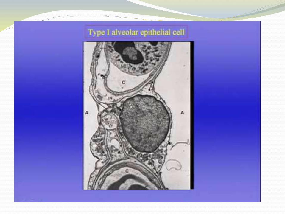
Very small Type I alveolar cells.

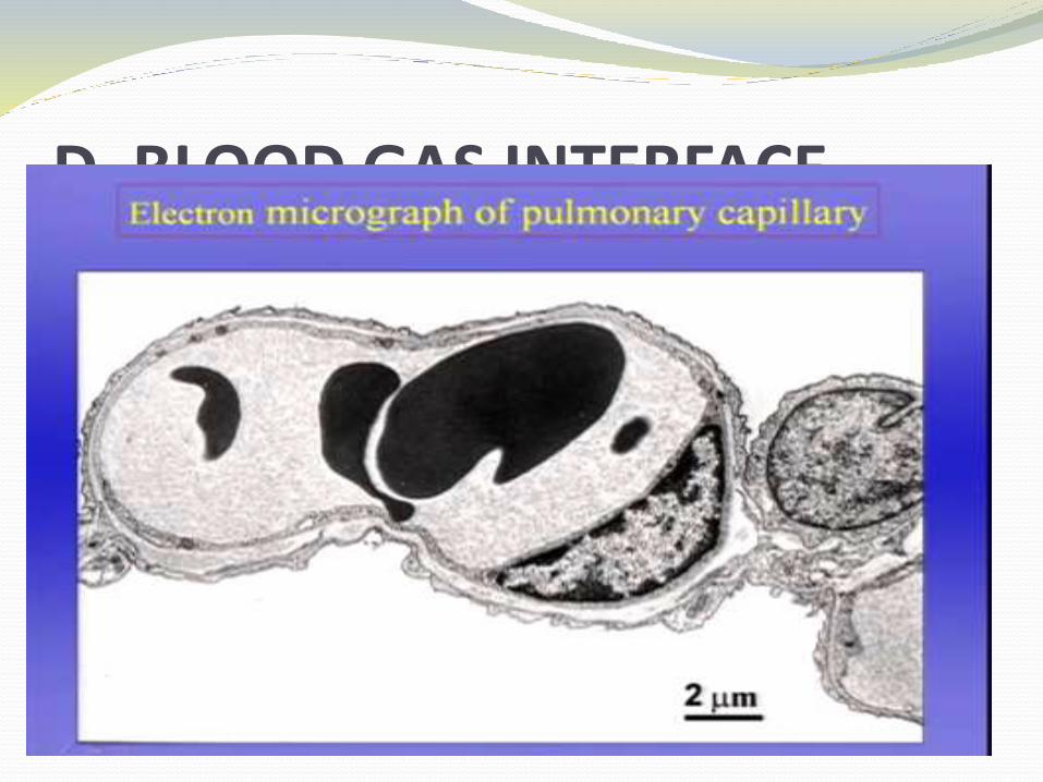
D. BLOOD GAS INTERFACE
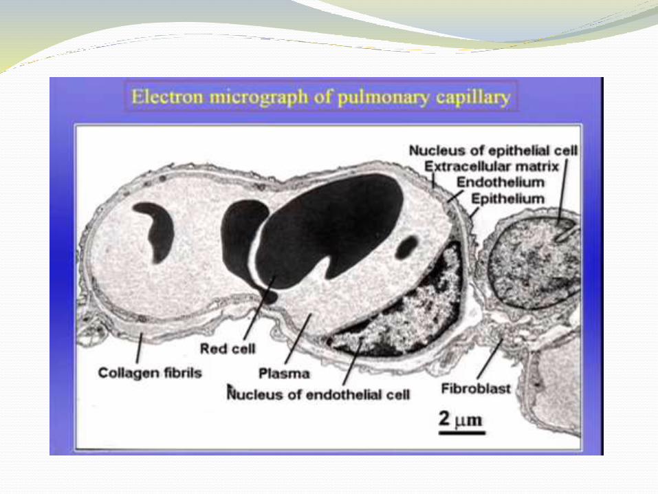


Surface area of the blood-gas barrier (BGB) is 50-100 sq metres. This is obtained by wrapping of the capillaries around the enormous number of alveoli. There are 500 x 10^6 alveoli each with almost 1/3 mm in diameter.
BGB is extremely thin <2 micron.

D. BLOOD VESSELS AND FLOW
Initially; the arteries, veins and bronchi run close together but towards the periphery of the lung, the veins move away to pass between the lobules whereas the arteries and bronchi travel together down to the centres of the lobules.

The capillaries form a dense network on the walls of the alveoli. The diameter is around 7-10 microns, just large enough to accommodate a red blood cell.
The lengths of the segments are so short that the dense network forms an almost continuous sheet of blood in the alveolar wall, a very efficient arrangement for gaseous exchange.

The thin BGB predisposes the capillaries to damage as in increased capillary pressure or increased lung volume during excessive inflation. This leads to leakage of plasma and red blood cells (rbcs) into the alveolar spaces.
Each rbc spends 0.75 seconds in the capillary network and during this time it traverses 2-3 alveoli. So efficient is the anatomy of gas exchange between the alveolar gas and capillary blood that this brief time is sufficient for virtually complete equilibration of O2 and CO2.

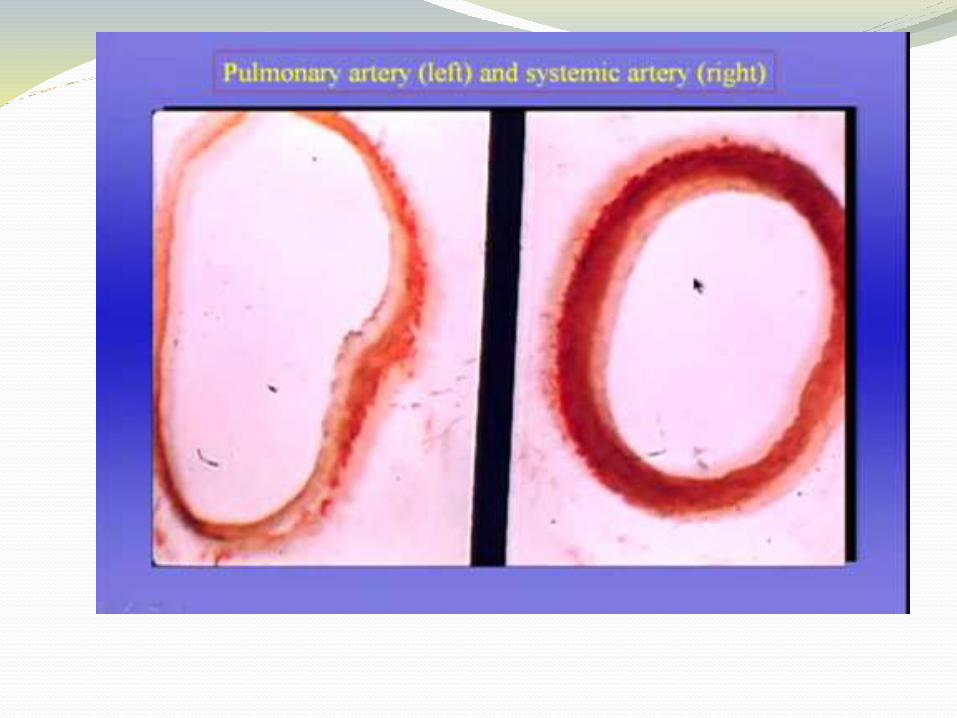
The pulmonary artery receives the whole output of the heart but the resistance is small. It thus has a mean pressure of almost 20 cm H2O (15mmHg) required for a flow of 6 litres/min.


Bronchial circulation is an additional blood system. The bronchial arteries are derived from the aorta and the intercostal arteries. They mainly supply the conducting zones as the respiratory zone is supplied by the pulmonary artery.
