structure and function predictions of the msa protein in
TRANSCRIPT

The University of Southern Mississippi The University of Southern Mississippi
The Aquila Digital Community The Aquila Digital Community
Faculty Publications
1-1-2007
Structure and Function Predictions of the Msa Protein in Structure and Function Predictions of the Msa Protein in
Staphylococcus aureus Staphylococcus aureus
Vijayaraj Nagarajan University of Southern Mississippi
Mohamed O. Elasri University of Southern Mississippi, [email protected]
Follow this and additional works at: https://aquila.usm.edu/fac_pubs
Part of the Bioinformatics Commons
Recommended Citation Recommended Citation Nagarajan, V., Elasri, M. O. (2007). Structure and Function Predictions of the Msa Protein in Staphylococcus aureus. BMC Bioinformatics, 8(S7), 1-9. Available at: https://aquila.usm.edu/fac_pubs/8478
This Article is brought to you for free and open access by The Aquila Digital Community. It has been accepted for inclusion in Faculty Publications by an authorized administrator of The Aquila Digital Community. For more information, please contact [email protected].

BioMed Central
Page 1 of 9(page number not for citation purposes)
BMC Bioinformatics
Open AccessProceedingsStructure and function predictions of the Msa protein in Staphylococcus aureusVijayaraj Nagarajan and Mohamed O Elasri*
Address: Department of Biological Sciences, The University of Southern Mississippi, Hattiesburg, MS, 39406, USA
Email: Vijayaraj Nagarajan - [email protected]; Mohamed O Elasri* - [email protected]
* Corresponding author
AbstractBackground: Staphylococcus aureus is a human pathogen that causes a wide variety of life-threatening infections using a large number of virulence factors. One of the major global regulatorsused by S. aureus is the staphylococcal accessory regulator (sarA). We have identified andcharacterized a new gene (modulator of sarA: msa) that modulates the expression of sarA. Geneticand functional analysis shows that msa has a global effect on gene expression in S. aureus. However,the mechanism of Msa function is still unknown. Function predictions of Msa are complicated bythe fact that it does not have a homologous partner in any other organism. This work aims atpredicting the structure and function of the Msa protein.
Results: Preliminary sequence analysis showed that Msa is a putative membrane protein. It wouldtherefore be very difficult to purify and crystallize Msa in order to acquire structure informationabout this protein. We have used several computational tools to predict the physico-chemicalproperties, secondary structural features, topology, 3D tertiary structure, binding sites, motifs/patterns/domains and cellular location. We have built a consensus that is derived from analysisusing different algorithms to predict several structural features. We confirm that Msa is a putativemembrane protein with three transmembrane regions. We also predict that Msa hasphosphorylation sites and binding sites suggesting functions in signal transduction.
Conclusion: Based on our predictions we hypothesise that Msa is a novel signal transducer thatmight be involved in the interaction of the S. aureus with its environment.
BackgroundIntroductionStaphylococcus aureus is an important human pathogenthat causes several diseases ranging from superficial skininfections to life-threatening diseases such as osteomyeli-tis and endocarditis. S. aureus is capable of infecting a
wide range of tissues in humans because of the largenumber of virulence factors and the complex regulatorynetworks that control them [1]. In addition, S. aureus isincreasingly resistant to multiple antibiotics thus becom-ing a growing threat to public health. There is an urgentneed to understand the complex regulatory networks used
from Fourth Annual MCBIOS Conference. Computational Frontiers in BiomedicineNew Orleans, LA, USA. 1–3 February 2007
Published: 1 November 2007
BMC Bioinformatics 2007, 8(Suppl 7):S5 doi:10.1186/1471-2105-8-S7-S5
<supplement> <title> <p>Proceedings of the Fourth Annual MCBIOS Conference. Computational Frontiers in Biomedicine</p> </title> <editor>Dawn Wilkins, Yuriy Gusev, Raja Loganantharaj, Susan Bridges, Stephen Winters-Hilt, Jonathan D Wren (Senior Editor)</editor> <note>Proceedings</note> </supplement>
This article is available from: http://www.biomedcentral.com/1471-2105/8/S7/S5
© 2007 Nagarajan and Elasri; licensee BioMed Central Ltd. This is an open access article distributed under the terms of the Creative Commons Attribution License (http://creativecommons.org/licenses/by/2.0), which permits unrestricted use, distribution, and reproduction in any medium, provided the original work is properly cited.

BMC Bioinformatics 2007, 8(Suppl 7):S5 http://www.biomedcentral.com/1471-2105/8/S7/S5
Page 2 of 9(page number not for citation purposes)
by S. aureus to cause disease. Regulatory networks areattractive therapeutic targets for future treatment of antibi-otic resistant infections.
Modulator of sarA (msa)One of the important global regulators of virulence in S.aureus is the Staphylococcal accessory regulator (sarA) [2].sarA regulates over 100 genes in S. aureus several of whichare associated with virulence [3]. sarA plays an importantrole in disease [4]. sarA itself is regulated by several locithat modulate its function. We recently identified a novelgene, msa, that modulate the function of sarA [5]. Weshowed that msa is essential for full expression of sarA andthat mutation of msa affected the expression of several vir-ulence factors in both sarA-dependent and sarA-independ-ent manners [5]. Microarray analyses of the msa mutantshow that Msa has a global effect on genes in S. aureus(unpublished data). These studies indicate that msa is animportant locus in S. aureus and that the characterizationof the Msa protein would be very useful in understandingstaphylococcal regulatory networks.
Computational toolsSeveral bioinformatics tools have been developed to pre-dict the structure and functional properties of bio mole-cules. These tools use a wide variety of algorithms topredict the properties of proteins at different levels [6,7].The accuracy of these bioinformatics tools has beenimproving; however, each tool has its own advantagesand disadvantages. A particular algorithm has its owncharacteristic specificity, sensitivity, robustness, computa-tional cost, etc. These characteristics can be tested againstbenchmarks of known datasets (e. g., Critical Assessmentof Techniques for Protein Structure Prediction – CASP). Inorder to make the most accurate predictions, severalmethods should be used to build a consensus.
The aim of this work is to predict the structure and func-tional properties of the Msa protein of S. aureus to thehighest possible accuracy. Our prediction results showthat the Msa is a putative integral membrane protein withthree probable transmembrane regions. We also predictthat the Msa contains phosphorylation sites in the loopregions (both inside and outside the membrane). The 3-Dstructure analysis of the Msa also predicts the presence ofputative binding sites. Thus, based on this computationalanalysis, and previous experimental data [5] we hypothe-sise that Msa might play a role in signal transduction. Thefact that Msa has no known homolog means that it wouldbe a novel signal transducer.
Results and discussionPrimary sequence analysisThe conceptually translated Msa protein is made of 133amino acids with a predicted molecular weight of 15.6571
kDa and an isoelectric point (pI) of 6.71. The GRAVYindex value 1.021 shows that Msa is probably an insolu-ble protein. The Codon adaptation index (CAI) value pre-dicts the Msa as a highly expressed protein. This isconsistent with experimental results described previouslyby our group [5].
Homology and similarityThe Msa is highly conserved among the different strains ofS. aureus (RF122, MRSA252, MSSA476, MW2, COL,Mu50, N315, and NCTC 8325). Even though there wereseveral variations in the nucleotide sequences, weobserved good conservation at the amino acid level. Mul-tiple sequence alignment and phylogenetic analysis ofboth nucleotide sequences (SAUSA300_1294,SACOL1436, SAOUHSC_01402, SAV1401, msa,SAS1342, MW1289, SAR1413, and SAB1257c) and pro-tein sequences (YP_493991, YP_186288, YP_499929,NP_371925, NP_374514, YP_043463, NP_646106) fromdifferent strains show that they are identical. The only twoexceptions were strains RF122 and MRSA252 whichshowed slight variations in the Msa sequences. In RF122,the protein sequence (YP_416734) was 97% similar to theMsa sequence from N315 while in MRSA252, the proteinsequence (YP_040815) was 98% similar to the Msa fromN315. The phylogeny of the Msa protein closely resem-bled that of the phylogeny of these organisms as deter-mined by Multi Locus Sequence Typing (MLST) [8]. Theposition and effect of mutations in the Msa proteinsequence of the strains MRSA252 and RF122 are discussedin the "3-D structure prediction and analysis" section.
Our similarity search results against several sequence andstructure databases, using different BLAST programs,showed that there were no significant closely relatedhomologs for the Msa protein, except for one in S. epider-midis. Even though there were no significant (based on E-value and score) homolog for Msa, BLAST also listed sev-eral membrane proteins with remote similarities (align-ment Score of 32–35 and E values scores from 0.91–10)only to the first few amino acids of the Msa protein (thatcorresponds to the predicted signal peptide region).
Localization predictionsAll the tools used to predict the cellular location of theprotein indicated that Msa is a putative membrane pro-tein. This prompted us to examine the sequence for pres-ence of signal peptide and potential cleavage sites in theMsa protein sequence. Seven out of eight signal peptideprediction tools indicated the presence of a potential sig-nal peptide in the Msa protein (Table 1). The majority ofthe programs also predicted an N-terminal cleavage sitebetween the amino acid 19 and 20.

BMC Bioinformatics 2007, 8(Suppl 7):S5 http://www.biomedcentral.com/1471-2105/8/S7/S5
Page 3 of 9(page number not for citation purposes)
Topology predictionsWe performed topology analysis on the Msa sequenceusing several prediction programs that yielded widely dis-crepant results (Table 2). Even though most programsfailed to recognize the signal peptide, a consensus topol-ogy emerged (Table 3). The predicted topology of the Msais IN-OUT with three putative transmembrane segments(from amino acid positions 27–47, 54–75, 108–125). TheN-terminal is predicted as present in the cytoplasmic sideof the membrane while the C-terminal is predicted as out-side the membrane. Our consensus topology also passedthe positive-inside rule and charge bias test [9], with acharge bias of +1 towards the inside of the membrane.
Secondary structure prediction results indicated the pres-ence of four distinct helical regions (Figure 1). One helicalregion corresponds to the cytoplasmic helix while theother three correspond to the integral membrane helices.These results are consistent with the predicted topology.
Domains/patterns/motifsWe searched for the presence of domains, patterns andmotifs in the Msa protein sequence, to gain insight into itsfunctions and structure. The SMART results showed thepresence of all the structural domains that we earlier iden-
tified using topology prediction programs and signal pep-tide prediction programs, viz. an N-terminal signalpeptide and three transmembrane regions. In addition,SMART also predicted the presence of a PreATP-graspdomain (d1gsa_1) from the SCOP database. Even thoughthis result had an E-value of 1.5, it was interesting becausethe predicted domain is a putative binding domain andfalls in the predicted cytoplasmic region of Msa (residues85–116). Our pattern search in the Msa protein sequence,using different programs against the PROSITE database,gave similar results (except for PPSearch, which did notpredict the Tyrosine kinase site at position 48), showingthe presence of three putative phosphorylation sites(Table 4). All of the predicted sites were found in theexposed regions of the Msa. Analysis of the location ofthese putative phosphorylation sites showed that two ofthe putative phosphorylation sites are outside the mem-brane while one of them is predicted in the cytoplasmicregion. We also observed that these putative phosphoryla-tion sites are highly conserved among different strains ofS. aureus. This suggests that Msa might be phosphorylatedby kinases in the cytoplasm as well as kinases on the out-side of the membrane (e.g. from the host cells). These pre-dictions further suggest that Msa might function as a
Table 1: Signal peptide and cleavage position prediction for the Msa protein
Signal Peptide Cleavage Position
SignalP Present 29PrediSi Present 29sigcleave Present 20PSORT Present 20Phobius Present 20SIG-Pred Present* 20iPSORT Present No predictionSOSUIsignal Absent No predictionConsensus Present 20
* Predicted as an eukaryotic signal peptide
Table 2: Topology predictions for the Msa protein
TopPred TMpred PHDhtm TMHMM SPLIT HMMTOP MEMSAT DAS TSEG
N-terminal
IN IN OUT IN IN IN IN - -
# of TMS 3 3 3 4 4 4 4 4 4TMS 1* 3–23 - 14–34 3–21 2–23 6–23 7–23 8–21 3–22TMS 2 27–47 29–47 - 25–47 27–47 28–47 30–47 27–44 24–47TMS 3 - 55–75 55–72 54–76 54–69 60–77 54–70 57–67** 54–75TMS 4 106–126 107–123 108–125 108–125 107–126 108–125 108–125 110–124 105–128
TMS, transmembrane segments* Analysis with several signal peptide prediction tools indicate that this TMS is a putative signal peptide** Probability not significant

BMC Bioinformatics 2007, 8(Suppl 7):S5 http://www.biomedcentral.com/1471-2105/8/S7/S5
Page 4 of 9(page number not for citation purposes)
signal transducer and provides important targets formutagenesis experiments to test this hypothesis.
Membrane bound receptors are important components ofsignal transduction in all living systems. The major classof receptors in eukaryotes contain seven transmembranesegments (7 TM). Prokaryotes use 7 TM class receptorsalso, however, a recent study showed that prokaryotescarry novel receptor classes that have transmembrane seg-ments ranging from one to eight [10,11]. The Msa proteinsequence did not have significant homology with any ofthe known receptors and experimental studies are under-way to evaluate its function as a signal transducer.
3-D structure prediction and analysisHomology based tertiary structure prediction for the Msaprotein failed, because of the lack of homologous struc-tures. We used fold recognition based structure predictionserver Phyre to model the tertiary structure of the Msa pro-tein. Visualization and analysis of the predicted structureusing Swiss-PDB Viewer (SPDBV) showed that the pre-dicted structure correlated with the other predicted struc-
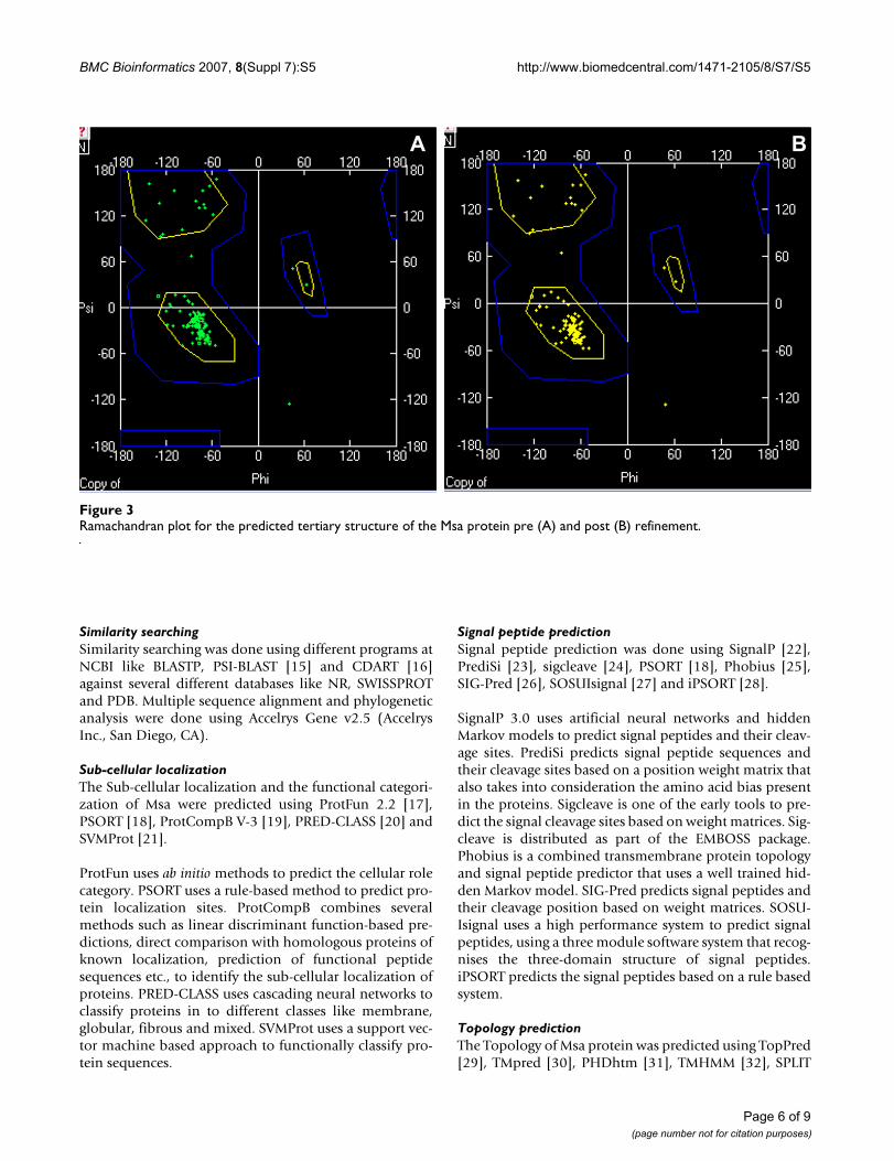
tural features of Msa in terms of the number and positionsof the transmembrane helices (Figure 2). We refined thepredicted structure by fixing side chains, fixing problem-atic loops, removal of amino acid clashes (bumps) andenergy minimization. The refinements did not yield anydrastic change in the initial predicted structure. This wasconfirmed by visually inspecting the structure and verify-ing the backbone structure using Ramachandran plot (Fig-ure 3) and computing the total energy difference betweenthe initial model and the refined model.
We analysed the predicted tertiary structure for clefts andbinding sites using ProFunc server and found putativebinding sites in the cytoplasmic region between the sec-ond and the third transmembrane helices (Figure 4A). Wealso used PINUP to predict putative interface residues inthe similar region (Figure 4B). Another binding site pre-diction server Q-SiteFinder also predicted similar bindingsite and binding site residues (Figure 4C).
ProFunc also predicted a "nest" near the putative phos-phorylation site (residues 47–50) which was predicted
Table 3: Consensus topology for the Msa protein including the N-terminal signal peptide prediction
N-terminal TMS 1 TMS 2 TMS 3
TopPred IN 27–47 - 106–126TMHMM IN 25–47 54–76 108–125TMpred IN 29–47 55–75 107–123SPLIT IN 27–47 54–69 107–126HMMTOP IN 28–47 60–77 108–125MEMSAT IN 30–47 54–70 108–125PHDhtm OUT 14–34 55–72 108–125TSEG No Prediction 24–47 54–75 105–128DAS No Prediction 27–44 54–67* 110–124Consensus IN 27–47 54–75 108–125
TMS, Transmembrane segment* Probability not significant
Consensus secondary structure predictions for the Msa proteinFigure 1Consensus secondary structure predictions for the Msa protein. Three transmembrane segments (TMS) and a cyto-plasmic helix are predicted.
TMS 1 TMS 2 TMS 3Cytoplasmic

BMC Bioinformatics 2007, 8(Suppl 7):S5 http://www.biomedcentral.com/1471-2105/8/S7/S5
Page 5 of 9(page number not for citation purposes)
outside the membrane [12]. The Msa has all the conservedresidues that make up the predicted "nest". The predicted"nest" in Msa shows features of an anion-binding site.Such "nests" are characteristic functional motifs, whichare found in ATP- or GTP binding proteins.
Multiple sequence alignment of the Msa protein sequencefrom 11 different strains of S. aureus revealed 12 muta-tions in strain RF122 and seven mutations in strainMRSA252 relative to consensus. Mutations at amino acidpositions 111, 131 and 133 were found in both MRSA252and RF122 strains. None of these mutations were found inthe predicted phosphorylation sites, predicted signal pep-tide sites or in the predicted anion-binding "nest". Butmany of the mutations were found both in the integralmembrane segments as well as in the other parts of theloop regions. Only one out of the 12 mutations had thereplacement (functionally different amino acid), while
others were substitutions (functionally similar aminoacids), in the strain RF122. In the strain MRSA152, twoout of seven mutations were replacements, while otherswere substitutions. MRSA strain had three mutations inthe predicted pre-ATP grasp domain, out of which onehad an amino acid replacement. RF122 strain had onlyone amino acid substitution in the pre-ATP grasp domain.This indicates that the predicted functional sites are con-strained from mutation.
ConclusionWe predict that Msa is a membrane protein with a cleava-ble N-terminal signal peptide sequence, followed by threeintegral transmembrane regions. The Msa is also predictedto have an IN-OUT topology with at least two putativephosphorylation sites, one outside the membrane andone in the cytoplasmic region. A putative binding site isalso predicted in the cytoplasmic region of Msa. Based onthese predictions we put forward a model for the Msa pro-tein (Figure 5). This model also prompted us to hypothe-sise that Msa might function as a novel signal transducerbetween the environment and the cytoplasm. This modelwill be used to design and execute experiments to confirmthe functions and topology of Msa and further our under-standing of its role in the pathogenesis of S. aureus.
MethodsFor a complete list of online tools used, see additional file1.
Primary sequence analysisWe used the protein sequence (Accession ID:NP_374514) obtained by conceptual translation of themsa open reading frame from the S. aureus N315 genome(NCBI database). The primary sequence analysis was per-formed using ProtParam, ProtScale [13] and SAPS [14].ProtScale was used to predict the Msa profile based on sev-eral amino acid scales. ProtParam computes propertieslike molecular weight, theoretical pI, instability index andgrand average of hydropathicity (GRAVY). SAPS predictssignificant features of protein sequences like charge-clus-ters, hydrophobic regions, compositional domains etc.
Predicted tertiary structure of the Msa protein showing the three transmembrane helicesFigure 2Predicted tertiary structure of the Msa protein show-ing the three transmembrane helices. Arrow indicates the predicted cleavage site for the putative signal peptide. N, N-terminus; C, C-terminus
N
C
Table 4: Prosite patterns predicted in the Msa protein
Protein Kinase C Casein Kinase II Tyrosine Kinase
PPSearch 99 49, 99 -PSITE 99 49, 99 48ScanProsite 99 49, 99 48Consensus 99 49, 99 48
Numbers denote residue position in the Msa protein sequence
BMC Bioinformatics 2007, 8(Suppl 7):S5 http://www.biomedcentral.com/1471-2105/8/S7/S5
Page 6 of 9(page number not for citation purposes)
Similarity searchingSimilarity searching was done using different programs atNCBI like BLASTP, PSI-BLAST [15] and CDART [16]against several different databases like NR, SWISSPROTand PDB. Multiple sequence alignment and phylogeneticanalysis were done using Accelrys Gene v2.5 (AccelrysInc., San Diego, CA).
Sub-cellular localizationThe Sub-cellular localization and the functional categori-zation of Msa were predicted using ProtFun 2.2 [17],PSORT [18], ProtCompB V-3 [19], PRED-CLASS [20] andSVMProt [21].
ProtFun uses ab initio methods to predict the cellular rolecategory. PSORT uses a rule-based method to predict pro-tein localization sites. ProtCompB combines severalmethods such as linear discriminant function-based pre-dictions, direct comparison with homologous proteins ofknown localization, prediction of functional peptidesequences etc., to identify the sub-cellular localization ofproteins. PRED-CLASS uses cascading neural networks toclassify proteins in to different classes like membrane,globular, fibrous and mixed. SVMProt uses a support vec-tor machine based approach to functionally classify pro-tein sequences.
Signal peptide predictionSignal peptide prediction was done using SignalP [22],PrediSi [23], sigcleave [24], PSORT [18], Phobius [25],SIG-Pred [26], SOSUIsignal [27] and iPSORT [28].
SignalP 3.0 uses artificial neural networks and hiddenMarkov models to predict signal peptides and their cleav-age sites. PrediSi predicts signal peptide sequences andtheir cleavage sites based on a position weight matrix thatalso takes into consideration the amino acid bias presentin the proteins. Sigcleave is one of the early tools to pre-dict the signal cleavage sites based on weight matrices. Sig-cleave is distributed as part of the EMBOSS package.Phobius is a combined transmembrane protein topologyand signal peptide predictor that uses a well trained hid-den Markov model. SIG-Pred predicts signal peptides andtheir cleavage position based on weight matrices. SOSU-Isignal uses a high performance system to predict signalpeptides, using a three module software system that recog-nises the three-domain structure of signal peptides.iPSORT predicts the signal peptides based on a rule basedsystem.
Topology predictionThe Topology of Msa protein was predicted using TopPred[29], TMpred [30], PHDhtm [31], TMHMM [32], SPLIT
Ramachandran plot for the predicted tertiary structure of the Msa protein pre (A) and post (B) refinementFigure 3Ramachandran plot for the predicted tertiary structure of the Msa protein pre (A) and post (B) refinement.
A B

BMC Bioinformatics 2007, 8(Suppl 7):S5 http://www.biomedcentral.com/1471-2105/8/S7/S5
Page 7 of 9(page number not for citation purposes)
[33], HMMTOP [34], MEMSAT [35], DAS [36] and TSEG[37]. We also computed the charge bias of the generatedmodels, based on the positive-inside rule [9].
TopPred II predicts the topology of a protein based on itshydrophobicity profile and positive-inside rule. TMpred
algorithm is based on the statistical analysis of TMbase, adatabase of naturally occurring transmembrane proteins,using a combination of several weight-matrices for scor-ing. PHDhtm uses a neural network based approach withthe evolutionary information to predict the locations ofthe transmembrane helices. TMHMM predicts transmem-
Binding site predictions for the Msa proteinFigure 4Binding site predictions for the Msa protein. (A) ProFunc predicted binding site (red); (B) PINUP predicted binding site (interface in green); (C) Q-SiteFinder predicted binding site and binding residues (pink)
A B C
Predicted model for the Msa protein showing structural and functional featuresFigure 5Predicted model for the Msa protein showing structural and functional features.
OUT
IN
P
Tyrosine kinase
phosphorylation siteCasein kinase
phosphorylation site P
Protein kinase
phosphorylation site
Binding site
(PreATP-grasp domain)
125
27
47 54
75 108
Signal peptide
cleavage site
NH2
COOH
20
48
49
P
99

BMC Bioinformatics 2007, 8(Suppl 7):S5 http://www.biomedcentral.com/1471-2105/8/S7/S5
Page 8 of 9(page number not for citation purposes)
brane regions based on the hidden Markov model. SPLIT4.0 predicts location of transmembrane helices by per-forming an automatic selection of optimal amino acidattribute and corresponding preference functions.HMMTOP 2.0 prediction is based on the hypothesis thatthe difference in the amino acid distributions in variousstructural parts determines the localization of the trans-membrane segments. MEMSAT applies a novel dynamicprogramming algorithm to recognize membrane topol-ogy models by expectation maximization. DAS uses densealignment surface method to predict transmembraneregions. TSEG uses a discriminant function to predict thetransmembrane segments.
Secondary structure predictionWe used the NPS (Network Protein Sequence Analysis)consensus secondary structure server [38]. This server runsthe input sequence against several different secondarystructure prediction tools and generates a consensus sec-ondary structure out of them.
Domains/patterns/motifs predictionSMART (Simple Modular Architecture Research Tool) [39]was used to identify the presence of any domains in theMsa protein. We used different pattern searching applica-tions (PPSearch [40], PSITE [41] and ScanProsite [42]),that use PROSITE [43] database, to predict functionallyrelevant patterns in Msa protein.
3-D Structure prediction and analysisInitial attempts to predict the tertiary structure of Msawere done using different approaches like homologymodelling, threading and ab initio. Automated homologymodelling servers Swiss-Model [44] and ModWeb [45]were used for homology modelling. Predictions describedin this study were done using fold recognition tools123D+ [46], GenThreader [47], a new version of 3-DPSSM (Phyre) [48].
The quality of the predicted structure was examined usingan online version of the WHATIF [49] program. Structurerefinement was done using both WHATIF and Swiss-PDBViewer [50]. Structure visualization was done using Swiss-PDB Viewer.
The 3-D structure of the Msa protein was analysed forclefts and binding surfaces using ProFunc [51], Q-Site-Finder [52], PINUP [53] and SuMo [54].
Meta serversWe also used meta servers like SCRATCH [55], ProSAL[56] and MetaPP [57] for predicting structure and func-tional properties of the Msa protein.
Competing interestsThe authors declare that they have no competing interests.
Authors' contributionsVN did the analysis and drafted the manuscript. MOEdirected the whole research and critically revised the man-uscript.
Additional material
AcknowledgementsThis study was supported by a grant (1R15AI062727-01A1) from the National Institute of Allergy and Infectious Diseases (NIAID) to MOE and by The Mississippi Functional Genomics Network (NIH/NCRR P20 RR016476). We extend our sincere thanks to the anonymous reviewers of this manuscript for their valuable suggestions.
This article has been published as part of BMC Bioinformatics Volume 8 Sup-plement 7, 2007: Proceedings of the Fourth Annual MCBIOS Conference. Computational Frontiers in Biomedicine. The full contents of the supple-ment are available online at http://www.biomedcentral.com/1471-2105/8?issue=S7.
References1. Novick RP: Autoinduction and signal transduction in the reg-
ulation of staphylococcal virulence. Mol Microbiol 2003,48(6):1429-1449.
2. Cheung AL, Projan SJ: Cloning and sequencing of sarA of Sta-phylococcus aureus, a gene required for the expression ofagr. J Bacteriol 1994, 176(13):4168-4172.
3. Dunman PM, Murphy E, Haney S, Palacios D, Tucker-Kellogg G, WuS, Brown EL, Zagursky RJ, Shlaes D, Projan SJ: Transcription profil-ing-based identification of Staphylococcus aureus genes reg-ulated by the agr and/or sarA loci. J Bacteriol 2001,183(24):7341-7353.
4. Blevins JS, Elasri MO, Allmendinger SD, Beenken KE, Skinner RA,Thomas JR, Smeltzer MS: Role of sarA in the pathogenesis ofStaphylococcus aureus musculoskeletal infection. InfectImmun 2003, 71(1):516-523.
5. Sambanthamoorthy K, Smeltzer MS, Elasri MO: Identification andcharacterization of msa (SA1233), a gene involved in expres-sion of SarA and several virulence factors in Staphylococcusaureus. Microbiology 2006, 152(Pt 9):2559-2572.
6. Rost B: Review: protein secondary structure prediction con-tinues to rise. J Struct Biol 2001, 134(2–3):204-218.
7. Pandey G, Kumar V, Steinbach M: Computational Approachesfor Protein Function Prediction: A Survey. Twin Cities: Depart-ment of Computer Science and Engineering, University of Minnesota; 2006.
8. Holden MT, Feil EJ, Lindsay JA, Peacock SJ, Day NP, Enright MC, Fos-ter TJ, Moore CE, Hurst L, Atkin R, et al.: Complete genomes oftwo clinical Staphylococcus aureus strains: evidence for therapid evolution of virulence and drug resistance. Proc NatlAcad Sci USA 2004, 101(26):9786-9791.
Additional file 1URL's of the tools and applications used in this study. URL's of the tools and applications used in this study. This file contains the list of URL's of all the online tools used to predict the structure and functional aspects of Msa protein.Click here for file[http://www.biomedcentral.com/content/supplementary/1471-2105-8-S7-S5-S1.txt]

BMC Bioinformatics 2007, 8(Suppl 7):S5 http://www.biomedcentral.com/1471-2105/8/S7/S5
Page 9 of 9(page number not for citation purposes)
9. von Heijne G: Membrane protein structure prediction. Hydro-phobicity analysis and the positive-inside rule. J Mol Biol 1992,225(2):487-494.
10. Galperin MY: A census of membrane-bound and intracellularsignal transduction proteins in bacteria: bacterial IQ, extro-verts and introverts. BMC Microbiol 2005, 5:35.
11. D'Souza M, Glass EM, Syed MH, Zhang Y, Rodriguez A, Maltsev N,Galperin MY: Sentra: a database of signal transduction pro-teins for comparative genome analysis. Nucleic Acids Res2007:D271-273.
12. Watson JD, Milner-White EJ: A novel main-chain anion-bindingsite in proteins: the nest. A particular combination of phi, psivalues in successive residues gives rise to anion-binding sitesthat occur commonly and are found often at functionallyimportant regions. J Mol Biol 2002, 315(2):171-182.
13. Gasteiger E, Hoogland C, Gattiker A, Duvaud S, Wilkins MR, AppelRD, Bairoch A: Protein Identification and Analysis Tools onthe ExPASy Server. In The Proteomics Protocols Handbook Editedby: Walker JM. Humana Press; 2005:571-607.
14. Brendel V, Bucher P, Nourbakhsh IR, Blaisdell BE, Karlin S: Methodsand algorithms for statistical analysis of protein sequences.Proc Natl Acad Sci USA 1992, 89(6):2002-2006.
15. Altschul SF, Madden TL, Schaffer AA, Zhang J, Zhang Z, Miller W, Lip-man DJ: Gapped BLAST and PSI-BLAST: a new generation ofprotein database search programs. Nucleic Acids Res 1997,25(17):3389-3402.
16. Geer LY, Domrachev M, Lipman DJ, Bryant SH: CDART: proteinhomology by domain architecture. Genome Res 2002,12(10):1619-1623.
17. Jensen LJ, Gupta R, Staerfeldt HH, Brunak S: Prediction of humanprotein function according to Gene Ontology categories. Bio-informatics 2003, 19(5):635-642.
18. Nakai K, Horton P: PSORT: a program for detecting sortingsignals in proteins and predicting their subcellular localiza-tion. Trends Biochem Sci 1999, 24(1):34-36.
19. Softberry [http://www.softberry.com]20. Pasquier C, Promponas VJ, Hamodrakas SJ: PRED-CLASS: cascad-
ing neural networks for generalized protein classificationand genome-wide applications. Proteins 2001, 44(3):361-369.
21. Cai CZ, Han LY, Ji ZL, Chen X, Chen YZ: SVM-Prot: Web-basedsupport vector machine software for functional classificationof a protein from its primary sequence. Nucleic Acids Res 2003,31(13):3692-3697.
22. Bendtsen JD, Nielsen H, von Heijne G, Brunak S: Improved predic-tion of signal peptides: SignalP 3.0. J Mol Biol 2004,340(4):783-795.
23. Hiller K, Grote A, Scheer M, Munch R, Jahn D: PrediSi: predictionof signal peptides and their cleavage positions. Nucleic AcidsRes 2004:W375-379.
24. von Heijne G: A new method for predicting signal sequencecleavage sites. Nucleic Acids Res 1986, 14(11):4683-4690.
25. Kall L, Krogh A, Sonnhammer EL: A combined transmembranetopology and signal peptide prediction method. J Mol Biol2004, 338(5):1027-1036.
26. SIG-Pred: Signal Peptide Prediction [http://www.bioinformatics.leeds.ac.uk/prot_analysis/Signal.html]
27. Gomi M, Sonoyama M, Mitaku S: High performance system forsignal peptide prediction: SOSUIsignal. Chem-Bio InformaticsJournal 2004, 4(4):142-147.
28. Bannai H, Tamada Y, Maruyama O, Nakai K, Miyano S: Extensivefeature detection of N-terminal protein sorting signals. Bio-informatics 2002, 18(2):298-305.
29. Claros MG, von Heijne G: TopPred II: an improved software formembrane protein structure predictions. Comput Appl Biosci1994, 10(6):685-686.
30. Hofmann K, Stoffel W: TMBASE – A database of membranespanning protein segments. Biol Chem Hoppe-Seyler 1993,374:166.
31. Rost B, Casadio R, Fariselli P, Sander C: Transmembrane helicespredicted at 95% accuracy. Protein Sci 1995, 4(3):521-533.
32. Krogh A, Larsson B, von Heijne G, Sonnhammer EL: Predictingtransmembrane protein topology with a hidden Markovmodel: application to complete genomes. J Mol Biol 2001,305(3):567-580.
33. Juretic D, Zoranic L, Zucic D: Basic charge clusters and predic-tions of membrane protein topology. J Chem Inf Comput Sci2002, 42(3):620-632.
34. Tusnady GE, Simon I: The HMMTOP transmembrane topologyprediction server. Bioinformatics 2001, 17(9):849-850.
35. Jones DT, Taylor WR, Thornton JM: A model recognitionapproach to the prediction of all-helical membrane proteinstructure and topology. Biochemistry 1994, 33(10):3038-3049.
36. Cserzo M, Wallin E, Simon I, von Heijne G, Elofsson A: Predictionof transmembrane alpha-helices in prokaryotic membraneproteins: the dense alignment surface method. Protein Eng1997, 10(6):673-676.
37. Kihara D, Shimizu T, Kanehisa M: Prediction of membrane pro-teins based on classification of transmembrane segments.Protein Eng 1998, 11(11):961-970.
38. Deleage G, Blanchet C, Geourjon C: Protein structure predic-tion. Implications for the biologist. Biochimie 1997,79(11):681-686.
39. Letunic I, Copley RR, Schmidt S, Ciccarelli FD, Doerks T, Schultz J,Ponting CP, Bork P: SMART 4.0: towards genomic data integra-tion. Nucleic Acids Res 2004:D142-144.
40. PPSearch [http://www.ebi.ac.uk/ppsearch/]41. Solovyev VV, Kolchanov NA: Search for functional sites using
consensus. In Computer analysis of Genetic macromolecules Edited by:Kolchanov NA, Lim HA. World Scientific; 1994:16-21.
42. de Castro E, Sigrist CJ, Gattiker A, Bulliard V, Langendijk-GenevauxPS, Gasteiger E, Bairoch A, Hulo N: ScanProsite: detection ofPROSITE signature matches and ProRule-associated func-tional and structural residues in proteins. Nucleic Acids Res2006:W362-365.
43. Hulo N, Bairoch A, Bulliard V, Cerutti L, De Castro E, Langendijk-Genevaux PS, Pagni M, Sigrist CJ: The PROSITE database. NucleicAcids Res 2006:D227-230.
44. Schwede T, Kopp J, Guex N, Peitsch MC: SWISS-MODEL: Anautomated protein homology-modeling server. Nucleic AcidsRes 2003, 31(13):3381-3385.
45. Sali A, Blundell TL: Comparative protein modelling by satisfac-tion of spatial restraints. J Mol Biol 1993, 234(3):779-815.
46. Alexandrov NN, Nussinov R, Zimmer RM: Fast protein fold rec-ognition via sequence to structure alignment and contactcapacity potentials. Pac Symp Biocomput 1996:53-72.
47. Bryson K, McGuffin LJ, Marsden RL, Ward JJ, Sodhi JS, Jones DT: Pro-tein structure prediction servers at University College Lon-don. Nucleic Acids Res 2005:W36-38.
48. Kelley LA, MacCallum RM, Sternberg MJ: Enhanced genome anno-tation using structural profiles in the program 3D-PSSM. JMol Biol 2000, 299(2):499-520.
49. Vriend G: WHAT IF: a molecular modeling and drug designprogram. J Mol Graph 1990, 8(1):52-56. 29.
50. Guex N, Peitsch MC: SWISS-MODEL and the Swiss-Pdb-Viewer: an environment for comparative protein modeling.Electrophoresis 1997, 18(15):2714-2723.
51. Laskowski RA, Watson JD, Thornton JM: ProFunc: a server forpredicting protein function from 3D structure. Nucleic AcidsRes 2005:W89-93.
52. Laurie AT, Jackson RM: Q-SiteFinder: an energy-based methodfor the prediction of protein-ligand binding sites. Bioinformatics2005, 21(9):1908-1916.
53. Liang S, Zhang C, Liu S, Zhou Y: Protein binding site predictionusing an empirical scoring function. Nucleic Acids Res 2006,34(13):3698-3707.
54. Jambon M, Imberty A, Deleage G, Geourjon C: A new bioinfor-matic approach to detect common 3D sites in protein struc-tures. Proteins 2003, 52(2):137-145.
55. SCRATCH [http://www.ics.uci.edu/%7Ebaldig/scratch/]56. ProSAL [http://xray.bmc.uu.se/sbnet/prosal.html]57. MetaPP [http://www.predictprotein.org/newwebsite/meta/
submit3.php]