structure and functionstructure and function of the...
TRANSCRIPT

Structure and Function of the KidneyStructure and Function of the Kidney
Ali J. Olyaei, PharmD, BCPSAssociate Professor of Medicine
Nephrology & HypertensionNephrology & HypertensionBoard Certified Clinical Pharmacotherapist
Division of Nephrology & Hypertension 1

The Normal GlomerulusThe Normal Glomerulus
• It consists of a tuft of anastomosing capillaries.
• Mesangium: gmesengial cells.
2

Glomerulonephritis
Most important group of generalisedparenchymal diseasesClassification is difficult - a mix of clinical and pathological descriptions
li i l fclinical featuresmorphology (eg various histology patterns)pathogenetic mechanisms (eg anti gbm disease)pathogenetic mechanisms (eg anti gbm disease)aetiology
Can be primary or secondary
3
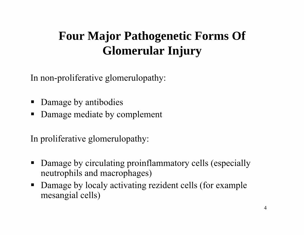
Four Major Pathogenetic Forms Of Glomerular Injury
In non-proliferative glomerulopathy:
Damage by antibodiesDamage by antibodiesDamage mediate by complement
In proliferative glomerulopathy:
Damage by circulating proinflammatory cells (especiallyDamage by circulating proinflammatory cells (especially neutrophils and macrophages)Damage by localy activating rezident cells (for example
i l ll )mesangial cells)4

Terminologies To Understand Glomerular Diseases
• Glomerulonephritis• DiffuseDiffuse• Focal
S l• Segmental• Membranous• Proliferative • Sclerosis
5
Sclerosis

Diffuse
• When all glomeruli of the kidney is involved in disease processinvolved in disease process.
6

Focal Wh l li f th kid i• When some glomeruli of the kidney is involved in disease process.
7

Segmental • When part of a glomerulous is involved in• When part of a glomerulous is involved in
disease process.
8
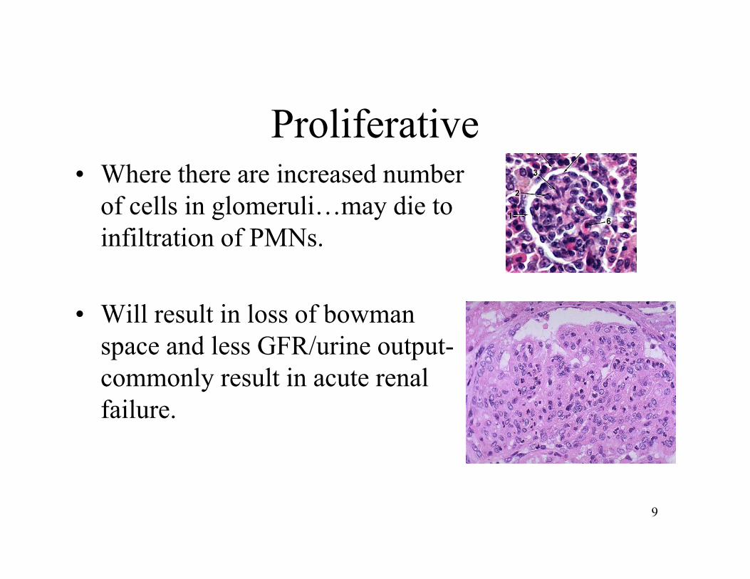
Proliferative • Where there are increased numberWhere there are increased number
of cells in glomeruli…may die to infiltration of PMNs.
• Will result in loss of bowman space and less GFR/urine output-commonly result in acute renal failure.
9

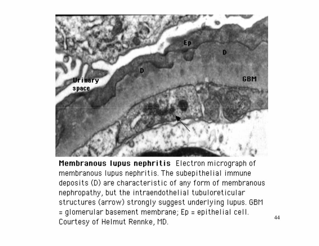
MEMBRANOUS GLOMERULONEPHRITIS (Thickened Basement Mem )(Thickened Basement Mem.)
10

Sclerosis (Trichrome stain)
I d ll bl l d i hi i11
• Increased collagen, blue colored in this stain.

Crescent
C t i f d b lif ti f ith li l ll d12
Crescent is formed by proliferation of epithelial cells and monocytes and fibrin.
DurationDuration
• Acute• eg: Acute Diffuse Proliferative• eg: Acute Diffuse Proliferative
glomerulonephritis.
• Chronic• Chronic • eg. Chronic Glomerulonephritis
13

Characteristics of common GlomerularCharacteristics of common Glomerular Diseases At Presentation
Heavyproteinuria
Proteinuria &haematuria
Predominanthaematuria
Minimal Change Lupus nephritis Acute post strep
Focal sclerosis
Membranous
Membrano-proliferative
Endocarditis
Crescentic(RPGN)
Haemolytic
Diabetes Mellitus
A l id i
Endocarditis
Henoch-Schonleinpurpura
Haemolyticuraemic syndrome
Amyloidosis purpura
14

Outcome of GlomerulonephritisOutcome of Glomerulonephritis
80
100
120
with
rena
l n
MPGN /FSGS / MGN /SLE
40
60
80
ntag
e st
ill w
func
tion /SLE
MCD /Mesang PGN / Post InfectiousRPGN
0
20
40
Per
cen RPGN
0 5 10 15
Years after apparent onset
15

Acute Nephritic Syndrome
Syndrome characterised in typical cases by:HaematuriaOliguriaEdemaH t iHypertensionReduced GFRProteinuriaFluid overload
16

Clinical Features of the Acute NephriticClinical Features of the Acute Nephritic Syndrome
Haematuria is usually macroscopic with pink or brown urine (like coca cola)Oliguria may be overlooked or absent in milder casesEdema is usually mild and is often just peri-orbital- weight gain may be detectedmay be detectedHypertension common and associated with raised urea and creatinineP t i i i i bl b t ll l th i th h tiProteinuria is variable but usually less than in the nephrotic syndrome
17
Aetiology of the Nephritic Syndrome
• Most common cause is acute post infectious glomerulonephritis
• Group A beta haemolytic streptococci of certain serotypes• Group A beta haemolytic streptococci of certain serotypes important in NZ
• IgA disease and henoch-schonlein purpura, crescentic glomerulonephritis and SLE can also present in this way
18

Management Issues In The Nephritic Syndrome
Appropriate investigations: skin and throat swabs,strepserology, complement, urea, creatinine electrolytes, urinalysis and CXRurinalysis and CXR
BP, urine output and daily weightfluid and diet managementtreat hypertension and fluid overloadtreat hypertension and fluid overloadtreat infection
19

The Nephrotic Syndrome
Is not a disease but a group of signs and symptoms seen in patients with heavy proteinuriapat e ts w t eavy p ote u apresents with oedemaproteinuria usually > 3.5g / 24hrs (>0.05g / kg / 24hrs in hild )children)
serum albumin < 30g/lother features: hyperlipidaemia, and hypercoaguable stateyp p , yp g
20

Acute GlomerulonephritisAcute Glomerulonephritis
A t P t t t lAcute Post-streptococcal Glomerulonephritis
Non-streptococcal causes
21

PoststreptococcalPoststreptococcalGlomerulonephritis
22

Case 1
• A 18 year-old man presents with hematuria. He also had cold and sore throat recently (two weeks ago). Physical exam is unremarkable. Labs: Cr 1.9 mg/dl, u/a with g ,large blood, - prot, 20-30. What is the most likely diagnosis?y g
23

P t t t l GNPoststreptococcal GN
U ll 10 d f h i i d 14• Usually occurs 10 days after pharyngitis and 14 days after skin infection(not synpharyngitic)(not synpharyngitic)
• Fallen incidence in US but common in some ruralFallen incidence in US, but common in some rural areas, poor hygiene places, and tropical countries
• Occurs more often in males and children
24
Clinical Presentation
• Most patients have milder disease• Classically presents with overt nephriticClassically, presents with overt nephritic
syndrome and oliguric ARF• Symptoms can include gross hematuria• Symptoms can include gross hematuria
(100% microscopic), HA, HTN (60-80%), hypervolemia and edema (80 90%)hypervolemia, and edema (80-90%)
25

Course• Irreversible Renal Failure rare – less than 1 % in
children, slightly higher in adults• Resolution usually quick plasma Cr usually returns• Resolution usually quick, plasma Cr usually returns
to previous levels by 3-4 weeks• Hematuria resolves usually within 3-6 months,
t i i f ll t l tproteinuria falls at a slower rate• Some patients experience htn, recurrent proteinuria,
and renal insufficiency 10-40 yrs aftery y• > 20% of adults may have some degree of persistent
proteinuria and or compromise of GFR 1 year out
26

Treatment
• Eliminate strep infxn with abx• Supportive therapySupportive therapy• Diuretics and antihypertensives to control
bp and extracellular fluid volumebp and extracellular fluid volume
27
Other Acute Glomerulonephritis
Non-streptococcal causes– Pneomococcal pneumonia
i i– Hepatitis B, C– Malaria
• Morphological features of these disease are similar to that of acute post streptococcal GN, only prognosis would be different.
28

Case 1
• A 25 year-old man presents with gross hematuria. He also has symptoms of URI y pthat started 2 days ago. Physical exam is unremarkable. Labs: Cr 1.5 mg/dl, large g , gblood, ++ prot, 20-30 dysmorphic RBCs/HPF. What is the most likely ydiagnosis?
29

IgA Nephropathy (Berger’s Disease)IgA Nephropathy (Berger s Disease)
• The most common cause of GN, esp in Asians, p• Exaggerated mucosal IgA response trapped in
glomeruli mesangial cell proliferation• Gross hematuria with URI• Microscopic hematuria & proteinuria common• Prognosis: generally good, bad if proteinuria > 2 g/d• Treatment:
ACE i hibi id l h h id fi h il– ACE inhibitors, steroids, cyclophosphamide, fish oil
30

Minimal Change Disease
• Syndrome: Nephrotic syndrome– Type of protenuria: selective (only albumin comes out).Type of protenuria: selective (only albumin comes out).– Due to loss of the normal charge barrier of GBM– Pathogenesis: Lymphokine production by T
cells• Most common cause of nephrotic syndrome in
children ( 2 6 years)children ( 2-6 years).• Light Microscopy :
– Normal glomeruliNormal glomeruli.– Lipid droplet in proximal tubular epithelium
31

RBCRBC
Effacement of foot processes due to loss of foot process(giving the appearance of fusion of the epithelial cell)
32

Membranous nephropathy (GN)
• Syndrome: Nephrotic syndrome• Most common nephrotic syndrome in ADULT.• Etiology:gy
– Idiopathic or genetic– Drug ( penicillamine)g ( p )– SLE, Diabetes mellitus– Adenocarcinoma of lung and colon.Adenocarcinoma of lung and colon.
33

Membranous GN
H&E stain: diffuse thickening of the capillary wall.
Sub epithelial deposit
34
Membranous GNMembranous GNClinical Features and Prognosis
• Some patient develop hypertension and hematuria.
• It has a variable and indolent course.• 40% patient progress to renal failure or end
stage renal disease after 2-20 years.• 10-30% with partial or complete remission of
proteinuria.• No or infrequent effect with steroid.
35

Wegener’s Granulomatosis
36

Upper Respiratory Tractpp p yNose
• Nasal crusting • FrequentFrequent
nosebleeds E i d• Erosion and perforation of the nasal septum. The bridge of the nose can collapse resulting in a “saddle–noseresulting in a saddle nose
deformity”.37

KidKidney• Glomerulonephritis w/ associated hematuria
and proteinuriap• Can lead to renal failure if not treated
aggressivelyaggressively • Renal masses (rare)
A i i di d bl d ll• Active urine sediment: red blood cell casts
38

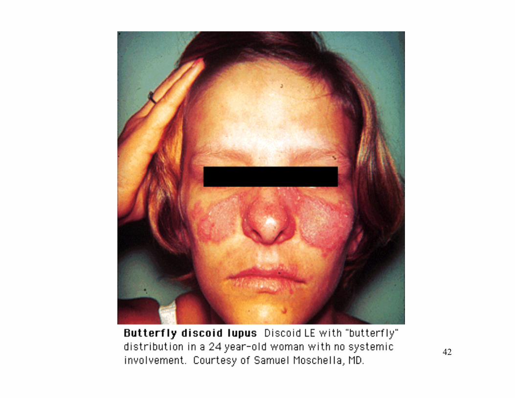
Systemic Lupus Erythematosus
39

Diagnostic criteria
American College of Rheumatology4/11 criteria (sens 85% specif 95%)4/11 criteria (sens 85%, specif 95%)“SOAP BRAIN MD”
S i i h l i• Serositis – heart, lung, peritoneum• Oral ulcers – painless esp palate• Arthritis – non-erosive• PhotosensitivityPhotosensitivity
40

Diagnostic criteria continued
• Blood disorders - ↓RBC (Coombs +), PLT, WCC, LymphocytesR l i l t t i i /± t• Renal involvement – proteinuria /± casts
• ANA – titer > 1:160• Immunologic phenomena LE cells anti dsDNA• Immunologic phenomena – LE cells, anti-dsDNA
Ab, anti-Sm Ab, antiphospholipid Ab, false WR +• Neurological disorders – seizures/ psychosisg p y• Malar rash – cheeks + nasal bridge• Discoid rash – rimmed with scaling, follicular
plugging41
42

43
44

45

Systemic Lupus Erythematosus
• Diagnosis:– clinical presentation - rash, arthralgia, fever, tiredness,
i tanaemia etc– hypocomplementaemia - (low C3 and C4)– antinuclear antibodies and anti DNA antibodiesa t uc ea a t bod es a d a t N a t bod es
• Treatment:– depends on histological severity (WHO class I - V)– nearly all get corticosteroids– WHO Class IV usually get corticosteroids and
cyclophosphamidecyclophosphamide
46

Focal segmental Glomerulosclerosis (FSGS)Glomerulosclerosis (FSGS).
47

Focal, segmental Glomerulosclerosis
Trichrome stain demonstrates blue, collagen Glomerulosclerosis , g
deposition.
48

Amyloidosis of Kidney
• Gross: waxy pale surface• LM:
– Pink hyaline like deposit • in mesangium
C d• Cogored– LM: brick red– Polarized light: apple green birefrengesPolarized light: apple green birefrenges
• Type of amyloid: yp y– Primary: Amyloid light chain ( Multiple myeloma)– Secondary (reactive): AA
49

Systemic Vasculitis
• Refers to classical polyarteritis nodosa, microscopic polyarteritis, Wegener’s granulomatosis, Henoch Schonlein purpura, Churg Strauss vasculitis and some cases ofpurpura, Churg Strauss vasculitis and some cases of rheumatoid arthritis
• Pauci immune rapidly progressive glomerulonephritis is common presentation sometimes with lung haemorrhagecommon presentation - sometimes with lung haemorrhage (not classical Goodpasture’s syndrome)
• Diagnosis has been made easier by anti neutrophil cytoplasmic antibody (ANCA) tests
50

51

Alport SyndromeAlport Syndrome • Hereditary form of GN accompanied by nerve deafness, y p y ,and various eye disorders
• Males tend to be affected more frequently and severely than females
• Most patients have an x-linked mode of inheritance, but autosomal dominant & recessive patterns also existbut autosomal dominant & recessive patterns also exist
• Morphology• focal segmental glomerulosclerosisfocal segmental glomerulosclerosis• GBM exhibits irregular areas of thickening or thinning with lamination and splitting of the lamina g p gdensa
52

Glomerular Diseases: Adjunctive Treatment to SlowGlomerular Diseases: Adjunctive Treatment to Slow Progression of Renal Disease
• BP control to < 130/80• ACE inhibitors (or ARBs) to max dosage or
combination if toleratedcombination if tolerated• Non DH Ca blocker to reduce proteinuria• Avoid dihydropyridine Ca channel blockers unless
combined with ACEIs (increase proteinuria)• Diuretics (↓BP, ↓edema)• Diet: Na restriction protein restriction (0 8 g/kg/d)Diet: Na restriction, protein restriction (0.8 g/kg/d)• Smoking cessation• ? Treatment of hyperlipidemia
53

Risk of Renal Disease Progression by SystolicRisk of Renal Disease Progression by Systolic BP: A Meta-Analysis of 11 RCTs
2 48
3.143
3.5 RR of 2xSCr or ESRD
*
*
2.48
1.832.08
2
2.5 *
11.23
0 5
1
1.5
0
0.5
<110 110-119 120-129 130-139 140-159 >160
Jafar TH et al. Ann Int Med 139:244, 2003 54

GN: Approach to Diagnosis
• History and physical (fluid-volume status, BP, signs of systemic disease)
• What is the syndrome?• Directed labs: hepatitis and lupus serologies p p g
(ANA), C3, C4• Renal biopsy in most patients with p y p
proteinuria > 1 gram/day or with nephritic features
55

Summaryy
• GN present with hematuria and/or proteinuria• Can be renal limited or systemic• History, physical exam, microscopic urinalysis
and directed serologies are essential in diagnosis• Renal biopsy needed in most cases, esp in
nephrotic syndrome and RPGNnephrotic syndrome and RPGN• Adjunctive treatment of proteinuria includes BP
lowering (<130/80), ACE inhibitors/ARBs orlowering ( 130/80), ACE inhibitors/ARBs or combination, and often diuretics for edema
56

Autosomal Dominant PolycysticAutosomal Dominant Polycystic Kidney Disease (ADPKD)
57

Autosomal Dominant Polycystic Kidney Disease (ADPKD)
58

Autosomal Dominant Polycystic KidneyAutosomal Dominant Polycystic Kidney Disease (ADPKD)
• Accounts for 7-10% of people on dialysis• 50% of sufferers have renal failure by age 60• Cysts may occur in liver and pancreas but do not usually
give problems• 5-10% have saccular cerebral aneurysms5 10% have saccular cerebral aneurysms• A family history of subarachnoid haemorrhage justifies
MRA or CT angio• Common complications include hypertension,
pyelonephritis, abdominal pain,haematuria and renal stones
59

Pathophysiology and GeneticsPathophysiology and Genetics
• Dominant inheritance often obvious from family tree but new mutations do occurM t ti f t l t 2 PKD 1 Ch 16 d• Mutations of at least 2 genes : PKD-1 on Chr 16 and PKD-2 on Chr 4
• Abnormal gene products are polycystin 1 and 2 which seem to be involved in cellular signaling
• Cyst cells immature and proliferate and secrete fluid. Ab t 1 2% f h i l d• About 1-2% of nephrons are involved
• Most cases diagnosed by ultrasound
60

Treatment of ADPKD
• No specific treatment• Diligent blood pressure control slows decline of
renal function• ACEi and ARB have no advantages over other
agents• Extended treatment of UTI necessary• Cyst aspiration does not improve kidney function
61

Acute and Chronic Urate NephropathyAcute and Chronic Urate Nephropathy• Acute nephropathy with overproduction of uric acid and
kidney obstruction with uric acid crystalskidney obstruction with uric acid crystals
• Can occur with treatment of malignant disease with cytotoxics, heat stroke and status epilepticus
• Treat with fluids and prophylaxis with allopurinolTreat with fluids and prophylaxis with allopurinol
• Role of uric acid in chronic renal failure disputed but does occur with some familial disorders
• Association between hyperuricaemia, hypertension vascularAssociation between hyperuricaemia, hypertension vascular disease, hyperlipidaemia and diabetes
62

Gout, Uric Acid and Renal Disease• Uric acid calculi, parenchymal deposits of uricUric acid calculi, parenchymal deposits of uric
acid and tubular obstruction with urate can cause renal damage
• An elevated plasma uric acid does not in itself seem to cause renal damage
• 1/4 of patients with gout get uric acid stones• 1/4 of patients with uric acid stones will have
gout
63

Acute Interstitial Nephritis
64

Acute Renal FailureAcute Renal FailurePrerenal Hypovolemia
Decreased cardiac outputRenal vasoconstriction
Intrinsic Acute Tubular NecrosisIntrinsic Acute Tubular NecrosisGlomerulonephritisVascular disorders
P t l Bladder NeckPostrenal Bladder NeckUreteralTubular
65

Most Common Ca sesMost Common Causes
•Drugs 71% (Antibiotics = 1/3)
•Infection 15%
•Idiopathic 8%•Idiopathic 8%
•TINU Syndrome 5%
•Sarcoidosis 1%
66

Drug Causes of AINAntibiotics Cephalosporins, Ciprofloxacin, Ethambutol,
Isoniazid, Macrolides, Penicillins, Rifampin, Sulfonamides, Tetracycline, Vancomycin
NSAIDs Almost all agents, including selective COX-2 inhibitorsinhibitors
Diuretics Furosemide, Thiazides, Triamterene
Miscellaneous Acyclovir, Allopurinol, Amlodipine, Azathioprine, Captopril Carbamazepine Clofibrate CocaineCaptopril, Carbamazepine, Clofibrate, Cocaine, Diltiazem, Famotidine, Indinavir, Mesalazine, Omeprazole, Phenteramine, Phenytoin, Pranlukast, Propylthioruacil, Quinine, RanitidinePropylthioruacil, Quinine, Ranitidine
67

Infectio s Ca ses of AINInfectious Causes of AIN
Bacterial Corynebacterium diphtheriae, legionella staphylococcilegionella, staphylococci, streptococci, yersinia
Viral CMV, EBV, HIV, HCV, HSV, , , , , ,hantaviruses, mumps, polyoma virus
Other Leptospira, mycobacterium, mycoplasma, rickettsia, syphilis toxoplasmosissyphilis, toxoplasmosis
68

Clinical PresentationClinical Presentation
AIN of any cause Drug-Induced AIN•Nausea
•Vomiting•Rash 15%•Fever 27%
•Malaise •Eosinophilia 23%
69

Laborator ManifestionsLaboratory Manifestions
•Acute rise in plasma creatinine concentration
•Eosinophilia and eosinophiluria
•Urine sediment: wbcs rbcs white cell castsUrine sediment: wbcs, rbcs, white cell casts
•Proteinuria (< 1 g/day)
•Signs of tubulointerstitial damage
70

TreatmentTreatment
•Discontinuation of offending agent
•CorticosteroidsP d i 1 /k f 40 60 1 2Prednisone 1 mg/kg to a max of 40-60 mg x 1-2
weeks
h l d i l 0 1 /d 3 dIV Methylprednisolone 0.5 – 1 g/day x 3 days
None (Ali’s recommendation)
71

PrognosisPrognosis
•Worse if methicillin-induced AIN
40% i h i l i f C•40% patients have persistent elevation of serum Cr
•Recovery less likely if prolonged period of renal failure, NSAID-induced AIN, interstitial granulomas or fibrosis on biopsy
72
Heme pigment-induced acute tubular necrosis
• Myoglobinuria: rhabdomyolysis.y g y y
• Hemoglobinuria: intravascular hemolysis• Hemoglobinuria: intravascular hemolysis.
73

Rhabdomyolysis
• The release of muscle cell contents as the result of traumatic or nontraumatic injury of skeletal muscle
• Physical findings may consist of • Tender, “doughy” muscles • Edema• weakness • Compartmental compression symptoms with signs and
symptoms of neurovascular compromise may developsymptoms of neurovascular compromise may develop, necessitating the need for emergent fasciotomy.
74
The majority of cases of rhabdomyolysis are nontraumaticnontraumatic
Alcohol abuse
Massive muscle compression from immobilization in drug induced coma
Drug inducedDrug-induced
Seizures
Occlusive peripheral vascular disease.
Combination therapy with itraconazole, simvastatin, and cyclosporineCombination therapy with itraconazole, simvastatin, and cyclosporine
Conversion from one fibric acid to another, or from one statin-fibrate combination to another
Detergent ingestion 75

Acute Kidney InjuryAcute Kidney Injury
76

Classical approach
• Three classic causes– Pre-renal failurePre renal failure
• Hypovolaemia, hypotension
– Intrinsic renal failure• Acute Tubular Necrosis, • Toxic injury
– Post-renal failure• Renal outflow tract obstruction
77
Acute Kidney Injury
• Spectrum of disorders from reduced function to established failure
• Multi-factorial causes• Associated with reduced Glomerular filtration and
Oliguria, which may initially be appropriate.• Involves sub lethal damage causing depolarization
and loss of normal function• Cell death through necrosis and apoptosis
78

RIFLE classificationGFR/Creatinine criteria Urine OutputGFR/Creatinine criteria Urine Output
criteriaR isk Increase in creatinine x1.5 UO < .5ml/kg/hr for
Or GFR decrease >25% 6hrs
I njury Increase in creatinine x 2Or GFR decrease >50%
UO < .5ml/kg/hr for 12hrs
F ailure Increase in creatinine x 3Or GFR decrease >75%
UO < .3ml/kg/hr for 24 hrs or Anuria for 12hrs
L oss Persistent ARF = complete loss of renal function > 4 weeks
E SRD End Stage Renal Disease > 3 monthsE SRD End Stage Renal Disease > 3 months79

Incidence In ICU
• 67% of Critical care patients have AKI• 12% class R• 27% class I• 28% class F28% class F
• However more than half of patients with• However more than half of patients with class R will progress to either class I or F
Hoste et al Crit Care 2006 80

Incidence in ICU
• Approximately 5% of patients in general intensive cares will require RRTq
• Equates to 200-300 per million population• Similar numbers to severe sepsis or ARDS• Similar numbers to severe sepsis or ARDS
• 10-20% of surviving patients require ongoing RRT beyond hospital discharge
81

Non -ICU ICUNon ICU
82

Sepsis and AKI
• Sepsis accounts for nearly 50% of all causes of AKI
• Combination of FactorsImmunological– Immunological
– ToxicInflammatory– Inflammatory
• Effect renal microvasculature and Tubular llcells
83

AKI and Mortality
• As already mentioned rising RIFLE class associated with increasing mortalityg y
• Despite advances in critical care medicine• Despite advances in critical care medicine and technology, patients who are treated with RRT still have a mortality of 50 60%with RRT still have a mortality of 50-60%
84
Prerenal Acute Renal FailurePrerenal Acute Renal Failure
85

Prerenal Acute Renal Failure
• GFR is reduced as a result of hemodynamic disturbances that decrease glomerular perfusiondecrease glomerular perfusion.
• The defining feature of prerenal ARF is the absence of cellularThe defining feature of prerenal ARF is the absence of cellular injury and the normalization of renal function with reversal of the altered hemodynamic factors.
86

Intravascular volume Altered intrarenaldepletion hemodynamics
Etiologies of Prerenal ARF
Decreased effective arterial blood volume
Abdominal compartment syndrome
87

Compensatory mechanisms Injury mechanisms
88

• Hx• P/EP/E• Urine sediment (usually normal, without cellular elements
or abnormal casts, unless chronic kidney disease is present)
• Una < 15 meq/L (>20 in ATN)• U/Pcreat > 20 (<15 in ATN)• U/Pcreat > 20 (<15 in ATN)• FeNa <1% (>1% in ATN)• UNa/K <1/4UNa/K <1/4 • BUN/creat >20
89

Prerenal Prolonged hypoperfusion ATN
90

Postrenal Acute Renal Failure
91

92

Etiologies of Postrenal ARFEtiologies of Postrenal ARFIntrinsic Extrinsic
Lower tract obstruction
Urethral stricture
Extrinsic obstruction
StoneP ill i
Retroperitoneal fibrosis
BPHProstate CA
TCC of bladderPapillary necrosis
Blood clotTCC
Aortic aneurysmRetroperitoneal or pelvic
malignancy
Bladder stonesBlood clot
Fungus ballNeurogenic bladder
Malpositioned catheter
93

Urine output?• The obstruction: • Complete Anuria
Normal
PolyuriaInComplete
Fluctuating
94

Complete obstruction
Recovery after relief of obstruction depends on:Recovery after relief of obstruction depends on:Severity Duration • Less than 1 wk duration, recovery complete.• Little or no recovery after 12 wk.
95

• Relief of obstruction may be accompanied by a post-obstructive diuresis;
• Excretion of salt and water retained during the obstruction.
• Persistent salt-wasting and impaired urinary concentrating ability .
96

Response to an Early Biomarker
Be Warned, Be Watchful
• Monitor intensively• Monitor fluid balance, urine outputMonitor fluid balance, urine output• Monitor blood pressure, cardiac function• Monitor electrolytes kidney function• Monitor electrolytes, kidney function
97

Response to an Early Biomarker
Do No Harm
• Avoid and treat hypotension• Avoid and treat hypovolemiaAvoid and treat hypovolemia• Avoid and treat oliguria• Avoid contrast agents• Avoid contrast agents• Avoid nephrotoxic medications
98

Response to an Early Biomarker
Early Intervention with CRRT
• Early fluid overload• Cytokine removal in sepsisCytokine removal in sepsis• Toxin removal after contrast administration
99

Myths
• Frusemide– Theoretically may reduce tubular injury
h i d / / l– Due to shutting down Na/K/Cl ATPase – Reduces oxygen demand– May help with fluid balanceMay help with fluid balance
• But– No clinical evidence– Accumulates in Oliguria– Nephrotoxic and Ototoxic
M t ll i t lit d d f RRT– May actually increase mortality and or need for RRT100

Myths
• Dopamine– Low dose Dopamine (2-3mcg/kg/min), knownLow dose Dopamine (2 3mcg/kg/min), known
as “renal dose”– No effect on mortality or need for Renal y
replacement therapy
101

Myths
• Vasopressors and AKI– Although Noradrenaline causesAlthough Noradrenaline causes
vasoconstriction with renal vasculature– No evidence of worsening AKIg– But should be used after adequate volume
resuscitation
102

Myths
• Mannitol– Currently no evidence of protective effectCurrently no evidence of protective effect– Causes an osmotic diuresis with may benefit
fluid balance
103