structure and genome release of twort-like …structure and genome release of twort-like myoviridae...
TRANSCRIPT
Structure and genome release of Twort-like Myoviridaephage with a double-layered baseplateJi�rí Nováceka, Marta �Siborováa, Martin Benešíkb, Roman Pant�ucekb, Ji�rí Doška�rb, and Pavel Plevkaa,1
aCentral European Institute of Technology, Masaryk University, 625 00 Brno, Czech Republic; and bDepartment of Experimental Biology, Faculty of Science,Masaryk University, 611 37 Brno, Czech Republic
Edited by Wolfgang Baumeister, Max Planck Institute of Biochemistry, Martinsried, Germany, and approved June 21, 2016 (received for review April 12, 2016)
Bacteriophages from the family Myoviridae use double-layeredcontractile tails to infect bacteria. Contraction of the tail sheathenables the tail tube to penetrate through the bacterial cell wall andserve as a channel for the transport of the phage genome into thecytoplasm. However, the mechanisms controlling the tail contractionand genome release of phages with “double-layered” baseplateswere unknown. We used cryo-electron microscopy to show thatthe binding of the Twort-like phage phi812 to the Staphylococcusaureus cell wall requires a 210° rotation of the heterohexameric re-ceptor-binding and tripod protein complexes within its baseplateabout an axis perpendicular to the sixfold axis of the tail. This rota-tion reorients the receptor-binding proteins to point away from thephage head, and also results in disruption of the interaction of thetripod proteins with the tail sheath, hence triggering its contraction.However, the tail sheath contraction of Myoviridae phages is notsufficient to induce genome ejection. We show that the end of thephi812 double-stranded DNA genome is bound to one protein sub-unit from a connector complex that also forms an interface betweenthe phage head and tail. The tail sheath contraction induces confor-mational changes of the neck and connector that result in disruptionof the DNA binding. The genome penetrates into the neck, but isstopped at a bottleneck before the tail tube. A subsequent structuralchange of the tail tube induced by its interaction with the S. aureuscell is required for the genome’s release.
bacteriophage | structure | contraction | Staphylococcus | genome release
The worldwide emergence of antibiotic-resistant pathogenicbacteria creates a need for new antibacterial treatments, in-
cluding phage-based therapy (1). Bacteriophage phi812 from thegenus Twort-like virus, subfamily Spounavirinae, can infect atleast 95% of Staphylococcus aureus strains, including thosestrains resistant to antibiotics (2). Thus, phi812 has the potentialto be used as antibacterial phage-therapy agent (3, 4).
Results and DiscussionCell Wall Recognition by Double-Layered Baseplate. The phi812 virionconsists of an isometric head 90 nm in diameter and a 240-nm-longcontractile tail ending with a baseplate (Fig. 1 andMovie S1). The tailand baseplate of the native phages are dynamic, as documented bythe frequent occurrence of phages with bent tails and baseplates ofdifferent diameters (SI Appendix, Fig. S1).Upon binding to S. aureus, phi812 baseplate proteins reorganize
into two layers parallel to the bacterial cell wall (Figs. 2C and 3 Gand J, SI Appendix, Fig. S2 and Table S1, and Movie S2). Thedouble-layered baseplate architecture, which is specific for thecontracted tail (Movie S3), is structurally distinct and has beenobserved in negative-stain micrographs of many Myoviridaephages (5–7). A characteristic feature of the double-layeredbaseplate is compact building blocks, which were previously de-scribed as thick legs of a “Victorian side table,” organized in twoconcentrical hexamers (8). In phi812, each of the legs is a complexof a trimer of receptor-binding proteins and a trimer of elongated“tripod” proteins (Fig. 2 and SI Appendix, Fig. S3). The two tri-mers have a common threefold axis and form a cone-shaped“receptor–tripod complex” (Fig. 2 and SI Appendix, Fig. S3). The
tripod proteins constitute the narrow tip of the cone and threeelongated protrusions that form peripheral regions of the re-ceptor–tripod complex base. Virulence associated proteins VrlCwere previously suggested to organize the structures of the double-layered baseplates (6, 8), and may therefore correspond to thetripod proteins. A complex of proteins gp122 and gp123 is acandidate for the phi812 VrlC (SI Appendix, Table S2). Sequenceidentities of virion proteins between phi812 and related staphy-lococcal phages K, A5W, ISP, and G1 are in the range of 99–100% (9). In addition, phi812 shares more than 30% sequenceidentity with the Staphylococcus phage Twort, Listeria phage A511,and Bacillus phage SPO1 (SI Appendix, Table S2). The receptor-binding protein complex of phi812 (gp125 and gp127) consists of acentral domain, three of which form a bowl-like structure in thecenter of the receptor–tripod complex base, a peripheral domainthat protrudes laterally from the central domain, and a receptor-binding domain that is visible at lower electron density levels thanthe rest of the receptor–tripod complex, indicating its flexibility (SIAppendix, Fig. S3).The tips of the receptor–tripod complexes from the outer hex-
amer bind to an elongated “boom” complex that extends in adirection perpendicular to the tail axis (Fig. 2C). The boomcomplex is attached to a “gooseneck” protein, six of which form aring around the tail tube (Figs. 2C and 3J). By extending theanalogy to a sailing ship, the tail tube forms the mast (Figs. 2C and3G and J). Each receptor–tripod complex from the inner hexameris attached to the gooseneck protein and the central part of theboom complex (Figs. 2C and 3J). Toward the phage head from thegooseneck proteins is a hexamer of “spacer” proteins that also
Significance
Resistance to antibiotics is widespread among pathogenic bacte-ria, including Staphylococcus aureus, which cause serious humandiseases. Bacteriophages from the Twort-like genus of the familyMyoviridae infect and kill pathogenic bacteria, and therefore areused to treat bacterial diseases. Detailed knowledge of the in-teractions of phages with bacterial cells is a prerequisite for theeffective and safe use of phages for medical purposes. However,the molecular details of the processes regulating infections bythese phages are not well understood. We used cryo-electronmicroscopy and tomography to describe the series of structuralchanges of a bacteriophage phi812 virion required to deliver itsgenome into the S. aureus cell.
Author contributions: J.N. and P.P. designed research; J.N., M.�S., and M.B. performedresearch; J.N. and P.P. analyzed data; and J.N., R.P., J.D., and P.P. wrote the paper.
The authors declare no conflict of interest.
This article is a PNAS Direct Submission.
Freely available online through the PNAS open access option.
Data deposition: CryoEM maps have been deposited in Electron Microscopy Data Bankand coordinates have been deposited in Protein Data Bank. For a list of deposition codes,see SI Appendix, Table S1.1To whom correspondence should be addressed. Email: [email protected].
This article contains supporting information online at www.pnas.org/lookup/suppl/doi:10.1073/pnas.1605883113/-/DCSupplemental.
www.pnas.org/cgi/doi/10.1073/pnas.1605883113 PNAS | August 16, 2016 | vol. 113 | no. 33 | 9351–9356
MICRO
BIOLO
GY
Dow
nloa
ded
by g
uest
on
May
26,
202
0

interact with tail sheath proteins (Figs. 2C and 3J). The boomcomplex provides an attachment site for the base of the tail fiber(Figs. 2C and 3J). The distal tip of the fiber is held in place byinteraction with the dimerization domain of one of the tripodproteins from the outer receptor–tripod complex hexamer (Figs.2C and 3J). The fiber has its long axis parallel to the axis of thetail, and the tip of the fiber is positioned in the same plane as thereceptor-binding domains of receptor-binding proteins (Figs. 2Cand 3J). Thus, the fibers contribute additional binding sites for theattachment of the phage to the bacterial cell wall.The biological relevance of the phi812 baseplate structure
determined from images of phi812 virions induced to contractin vitro was verified by the cryo-electron tomography of phagesattached to S. aureus cells (SI Appendix, Fig. S4). Focused ionbeam milling was used to prepare thin sections of cells for cryo-EM imaging (10).
Mechanism Controlling phi812 Tail Contraction. The native phi812baseplate has the overall shape of a compressed sphere with a di-ameter of 550 Å and height of 300 Å, and its subunit arrangement isquite different from the contracted baseplate (Fig. 2). In the nativebaseplate, the spacer proteins interact with both tail tube (gp104)and tail sheath (gp103) proteins (Fig. 2A, SI Appendix, Table S2,and Movie S4). Furthermore, the gooseneck proteins are in contactwith each other through a limited interface of 95 Å2 (at a 1σ con-tour level of the map), forming only a loosely connected ring aroundthe tail tube (Figs. 2A and 3C). Lateral protrusions of the gooseneck
proteins interact with the tip of the tail tube (Figs. 2A and 3C). Thegooseneck proteins also interact with receptor–tripod complexesfrom the inner hexamer and stabilize them tilted 20° away from thetail axis (Figs. 2A and 3C). The receptor-binding sites of receptor–tripod complexes from the outer hexamer point toward the phagehead (Figs. 2A and 3C) and require a 210° rotation to achieve theorientation they have in the contracted baseplate (Figs. 2C and 3J).Dimerization domains of tripod proteins from the outer hexamerreceptor–tripod complexes interact with tail sheath proteins, form-ing a disk proximal to the baseplate (Figs. 2A and 3C). Because thenative baseplate is attached to the tip of the tail tube, the interac-tions of tripod proteins with tail sheath proteins stabilize the tailsheath in its extended conformation. A 90-Å-long tubular density ofputative peptidase (11) is associated with each inner hexamer re-ceptor–tripod complex and gooseneck protein (Figs. 2A and 3C).These proteins are not present in the contracted baseplate, and dueto their enzymatic activity, they might facilitate penetration of thephage tail tube through the bacterial cell wall. In the native base-plate, the tail fibers are positioned between the receptor–tripodcomplexes from the outer hexamer and their receptor-binding tipspoint toward the phage head (Figs. 2A and 3C).Reference-free classification of cryo-micrographs identified an
alternative baseplate conformation that probably represents asnapshot of the gradual reorganization of the native baseplate intothe contracted one (Figs. 2B and 3D). Whereas the overallstructure of the intermediate is similar to the structure of thenative baseplate, the tips of tail fibers are exposed at the outerrim of the baseplate, and are therefore accessible for binding tothe bacterial cell wall (Figs. 2B and 3D and Movie S5).Based on a comparison of the native, intermediate, and con-
tracted baseplates, we propose the following sequence of stepsleading to the tail contraction. The initial attachment of phi812 tothe S. aureus cell wall is presumably formed by the tail fibers due totheir rotational freedom (Fig. 2 A and B). The tail contraction isconnected with a 20° rotation of the inner hexamer receptor–tripodFig. 1. Virions of bacteriophage phi812 in native conformation (A) and after
tail contraction (B). The heads are rainbow-colored according to the distancefrom the head center. The tails and baseplates are rainbow-colored accordingto the distance from the tail sixfold axis. The maps are contoured at 2σ. (C andD) Cryo-EM images of native and contracted phi812 virions. (E) Cryo-tomo-gram of phi812 virions attached to an S. aureus cell. (Scale bars, 1,000 Å.)
Fig. 2. Conformational changes of the phi812 baseplate induced by bindingto the host cell wall. Structures of phi812 baseplates in native (A), in-termediate (B), and contracted (C) conformations are shown. Bottom views(Top), side views (Middle), and selected details of the baseplates (Bottom)are shown. Individual components of the baseplates are color-codedaccording to the legend in Fig. 3. (A and C) Contacts of one baseplate wedgewith the tail tube. The maps are contoured at 0.75σ. (B, Inset) Contacts of thedimerization domain of tripod protein from the outer hexamer receptor–tripod complex with the head of tail sheath protein.
9352 | www.pnas.org/cgi/doi/10.1073/pnas.1605883113 Novácek et al.
Dow
nloa
ded
by g
uest
on
May
26,
202
0
complexes and disruption of their interaction with the lateraldomains of gooseneck proteins (Figs. 2 A and C and 3 C and G).The gooseneck proteins then form a tightly connected ring and nolonger interact with the complex located at the tip of the tail tube(Figs. 2C and 3G). Contacts between the spacer proteins and thetail tube proteins are also disrupted (Fig. 2C). Thus, all interac-tions that hold the baseplate at the tip of the tail tube are lost. Inparallel, the receptor–tripod complexes from the outer hexamerswing out 210° and bind to the cell wall. As a result of this rotation,the tripod proteins no longer stabilize the tail sheath proteins intheir native conformations (Figs. 2 A and C and 3 C and G).Subsequently, as the structural changes of tail sheath proteinspropagate along the tail, the whole tail sheath contracts, in afalling domino-like manner (12, 13), pulling the baseplate towardthe phage head.The proposed tail contraction-controlling mechanism depends
on the rotations of the outer hexamer receptor–tripod complexes.The two concentric hexamers of receptor–tripod complexes pointingtoward the cell wall give the baseplate its characteristic two-layeredstructure (8). Therefore, the mechanism regulating the phi812 tailcontraction is likely to be shared by other phages with double-layeredbaseplate architectures (5–7).
Comparison with Baseplates of Other Phages.Detailed characterizationof structure and mechanism of cell wall recognition is available forbaseplate of Myoviridae bacteriophage T4-infecting, Gram-negativeEscherichia coli (8, 14–18). The T4 baseplate has the shape of asemicircular dome in the native state and the shape of a single-layered, six-pointed star after the contraction (13, 17–19). T4 at-tachment to the bacterial cell wall is mediated by short- and long-tailfibers (13, 20, 21). Despite their overall different structures, the T4and phi812 baseplates display functional similarities. Conformationalchanges of long-tail fibers required for binding to the cell wall inducehinge movements of the gp9 trimer that provide an attachment sitefor the long-tail fibers on the T4 baseplate (22). The gp9 subunits areattached to gp10 proteins that form a structure resembling the boom
complex in phi812 (17). It was speculated previously that the con-formational change of gp9 triggers the tail sheath contraction (23). Itis therefore possible that in T4, the receptor-binding function of thereceptor-binding proteins has been completely replaced by long-tailfibers and tripod proteins transformed into gp9 performing struc-tural functions within the baseplate.The structure of the phi812 receptor-binding protein trimer
resembles the structure of the receptor-binding protein trimer ofthe bacteriophage p2 from the family Siphoviridae (24). Fur-thermore, the receptor-binding protein trimers of phages p2 and1358 point toward the bacteriophage head in the native state andrequire a 200° rotation to orient themselves toward the putativebacterial cell wall (24, 25). These structural and functional sim-ilarities reinforce the previously suggested common ancestry ofthe Siphoviridae and Myoviridae families (26).
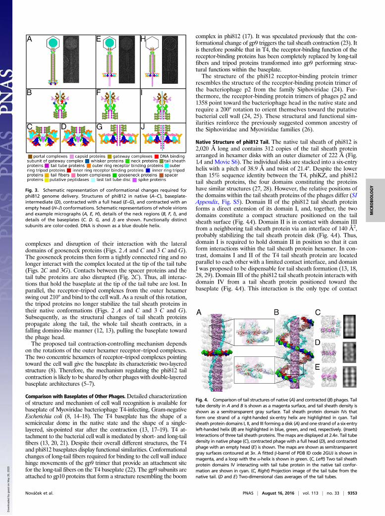
Native Structure of phi812 Tail. The native tail sheath of phi812 is2,020 Å long and contains 312 copies of the tail sheath proteinarranged in hexamer disks with an outer diameter of 222 Å (Fig.1A and Movie S6). The individual disks are stacked into a six-entryhelix with a pitch of 38.9 Å and twist of 21.4°. Despite the lowerthan 15% sequence identity between the T4, phiKZ, and phi812tail sheath proteins, the four domains constituting the proteinshave similar structures (27, 28). However, the relative positions ofthe domains within the tail sheath proteins of the phages differ (SIAppendix, Fig. S5). Domain II of the phi812 tail sheath proteinforms a direct extension of its domain I, and, together, the twodomains constitute a compact structure positioned on the tailsheath surface (Fig. 4A). Domain II is in contact with domain IIIfrom a neighboring tail sheath protein via an interface of 140 Å2,probably stabilizing the tail sheath protein disk (Fig. 4A). Thus,domain I is required to hold domain II in position so that it canform interactions within the tail sheath protein hexamer. In con-trast, domains I and II of the T4 tail sheath protein are locatedparallel to each other with a limited contact interface, and domainI was proposed to be dispensable for tail sheath formation (13, 18,28, 29). Domain III of the phi812 tail sheath protein interacts withdomain IV from a tail sheath protein positioned toward thebaseplate (Fig. 4A). This interaction is the only type of contact
Fig. 4. Comparison of tail structures of native (A) and contracted (B) phages. Tailtube density in A and B is shown as a magenta surface, and tail sheath density isshown as a semitransparent gray surface. Tail sheath protein domain IVs thatform one strand of a right-handed six-entry helix are highlighted in cyan. Tailsheath protein domains I, II, and III forming a disk (A) and one strand of a six-entryleft-handed helix (B) are highlighted in blue, green, and red, respectively. (Insets)Interactions of three tail sheath proteins. Themaps are displayed at 2.4σ. Tail tubedensity in native phage (C), contracted phage with a full head (D), and contractedphage with an empty head (E) is shown. The maps are shown as semitransparentgray surfaces contoured at 3σ. A fitted β-barrel of PDB ID code 2GUJ is shown inmagenta, and a loop with the α-helix is shown in green. (C, Left) Two tail sheathprotein domains IV interacting with tail tube protein in the native tail confor-mation are shown in cyan. (C, Right) Projection image of the tail tube from thenative tail. (D and E) Two-dimensional class averages of the tail tubes.
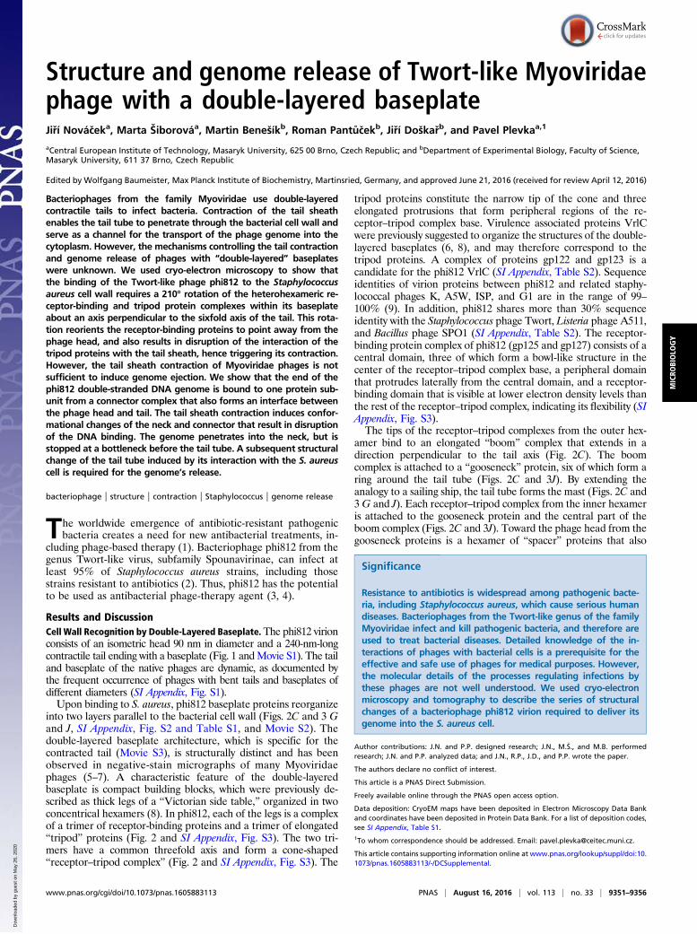
Fig. 3. Schematic representation of conformational changes required forphi812 genome delivery. Structures of phi812 in native (A–C), baseplate-intermediate (D), contracted with a full head (E–G), and contracted with anempty head (H–J) conformations. Schematic representations of whole virionsand example micrographs (A, E, H), details of the neck regions (B, F, I), anddetails of the baseplates (C. D. G, and J) are shown. Functionally distinctsubunits are color-coded. DNA is shown as a blue double helix.
Novácek et al. PNAS | August 16, 2016 | vol. 113 | no. 33 | 9353
MICRO
BIOLO
GY
Dow
nloa
ded
by g
uest
on
May
26,
202
0

between the consecutive tail sheath protein disks in the nativephi812 tail. Domain IV is positioned on the inside of the tailsheath and is in contact with the tail tube through its helix A (Fig.4 A and C and Movie S6). Tail tube proteins are organized into asix-entry helix with parameters identical to the parameters of thenative phi812 tail sheath (Fig. 4C). However, isolated tail sheathproteins of T4 and other Myoviridae phages spontaneously as-semble into so-called “polysheaths” that resemble contracted tailsheaths in their subunit organization (27, 28, 30, 31). Therefore,the structure of the native phi812 tail provides experimental proofof a previous speculation that assembly of the metastable Myo-viridae tail sheaths is determined by tail tube protein–tail sheathprotein interactions (32).The phi812 tail tube protein shares 16% sequence identity with
a putative tail tube protein from the Bacillus subtilismyophage-likeprophage with a known structure (PDB ID code 2GUJ). Theprophage tail tube protein consists of a six-stranded β-barrelcapped on one end by a loop containing two α-helices. Theβ-barrel and the helix-containing loop could be fitted separatelyinto the phi812 tail tube electron density map (Fig. 4C). The axisof the β-barrel lies in a plane perpendicular to the tail axis. Thesix tail tube proteins forming a disk interact with each othermainly through the “caps” on the top and bottom of the β-barrels(Fig. 4C). Strands 5 and 7 from the tail tube protein β-barrelinteract with helix A from domain IV of tail sheath protein.
Tail Sheath Contraction. The phi812 tail sheath contracts to 40%of its original length, resulting in insertion of the tail tube intothe S. aureus cell (Fig. 1). The tail sheath contraction is con-nected to a shift of the tail sheath proteins away from the tail axiscombined with an interdigitation of tail sheath proteins fromneighboring disks (Fig. 4B). The contracted tail sheath has pitchof 18.8 Å, twist of 30.7°, and outer diameter of 278 Å. Unlike thecase in the T4 phage (28), phi812’s tail sheath contraction isaccompanied by movements of its tail sheath protein domainsI–IV relative to each other (Fig. 4 A and B and SI Appendix, Fig.S6). The contacts between tail sheath proteins present in thenative tail are disrupted during the contraction, and new largerinteraction interfaces are formed. In the contracted tail, the tailsheath protein domain II is in contact with domain III of a tailsheath protein from an adjacent disk positioned toward thebaseplate (Fig. 4B and Movie S7).Helices A and B from the tail sheath protein domain IV form a
coiled coil with an axis parallel to the axis of the native tail (Fig.4A). However, the axis of the coiled coil is rotated 60° relative tothe tail axis in the contracted tail (Fig. 4B). Because the coiled-coilorientation determines the distance between the tail sheath pro-tein disks, the rotation brings the consecutive disks closer to eachother (Fig. 4B). Contacts of domain IV with the tail tube (Fig. 4C)are disrupted, and new contacts are formed between the domainIVs of tail sheath proteins from successive disks (Fig. 4B).The tail of the native phi812 can bend up to 90° (SI Appendix,
Fig. S1), enabling the phage to overcome mechanical stresses inthe extracellular environment. However, in the contracted tail,domains IV of the tail sheath proteins from the neighboring ringsare organized into a right-handed, six-entry helix, whereas theinteractions between domains II and III form a left-handed, six-entry helix (Fig. 4B and Movie S7). The two types of contactsconstitute a criss-crossing mesh holding the tail sheath straightand firm to enable efficient transport of the phage DNA into thebacteria (Fig. 1C).
Regulation of phi812 Genome Release. It has been shown previouslythat the genome ejection of Myoviridae phages is not directlyassociated with tail sheath contraction (13). Accordingly, weobserved phi812 virions with contracted tails and full heads (Fig.3 E and H). The tail tubes of contracted phi812 with full headsare similar to the tail tubes of native phages, but the tail tubes of
contracted phages with empty heads have a different structure,with a twist of 4.9° and pitch of 36.8 Å (Fig. 4 C–E). The axis ofthe β-barrel of the tail tube protein in the phage with the con-tracted tail and empty head is rotated 50° about an axis per-pendicular to the sixfold axis of the tail relative to its position inthe tail tube of the native phage (Fig. 4 C–E). The native phi812tail contains a spike complex attached to the end of the tail tube(Figs. 2A and 3C). The conformational change of the tail tubemay be induced by detachment of the spike or by interactions ofthe tail tube with the membrane or components of bacterialcytoplasm (33). The reorganization of the phi812 tail tube isreminiscent of the reorganization of the major tail protein of theSiphoviridae bacteriophage SPP1, which was proposed to trans-mit the signal required for the initiation of genome release (34).Bacteriophage tails are attached to heads through a connector
complex positioned at a special vertex in which a portal complexreplaces the pentamer of major capsid proteins (26). The phi812connector contains a dome-shaped “gateway” density interactingwith the portal complex (Figs. 3B and 5) with 12-fold symmetry(SI Appendix, Figs. S7 and S8 and Table S3), similar to othertailed phages (35–38). The gateway complex has sixfold sym-metry (Fig. 5 and SI Appendix, Figs. S7 and S8 and Table S3).The previously determined portal structure of T4 (35) could befitted into the map of the contracted phi812 particle with anempty head; however, the structural similarity is limited (SIAppendix, Fig. S9). In the native phage, both the portal andgateway complexes interact extensively with the fivefold sym-metrical capsid (Fig. 5A). The contacts deform the complexes tothe extent that they have pentagon-shaped cross-sections (Fig.5A and SI Appendix, Figs. S7 and S8 and Table S3).Along the axis of the portal complex in the native phi812 virion
is an elongated electron density that extends from the capsid to thegateway complex and has an average diameter of 20 Å (Fig. 5A).We interpret it to be the end region of the phage double-strandedDNA. The end of the genome asymmetrically binds to one of thegateway subunits (Figs. 3B and 5 A and D and Movie S8). It is thestrongest interaction between the DNA and protein to have beenidentified in the connector complex. Therefore, a disruption of thisinteraction probably regulates the DNA ejection as discussed below.The gateway subunits interact with a hexamer of “neck” pro-
teins positioned at the end of the phi812 tail (Figs. 3B and 5 andMovie S8). Because of the deviation of the gateway complex fromsixfold symmetry in the native phage, as described above, thegateway–neck interactions are asymmetrical (Fig. 5A and SI Ap-pendix, Fig. S7). It is notable that the strongest electron densityconnection between the two complexes involves the gatewaysubunit bound to the DNA (Figs. 3B and 5A). Whiskers are at-tached to the outer surface of the neck close to the head (Figs. 3Band 5A). In the direction toward the baseplate, the neck subunitsinteract with the domains IV of tail sheath proteins and with ahexamer of proteins on top of the tail tube (Figs. 3B and 5A).The phi812 neck links the tail sheath, tail tube, and connector,
and therefore has to transduce the conformational change trig-gering genome release from the tail to the gateway complex. Aftertail sheath contraction, the parts of the neck proteins that bind thetail sheath protein domain IV twist away from the tail axis tomaintain contacts with the expanded tail sheath (Fig. 5 A and B).Furthermore, whiskers are lost from the virion (Figs. 3F and 5Band Movie S8). The contacts of the portal and gateway subunitswith the capsid are loosened, and the two complexes relax to theirrespective 12-fold and sixfold symmetries (Fig. 5B and SI Appen-dix, Figs. S8 and S9 and Table S3). Most importantly, contacts ofthe DNA and the gateway subunit are disrupted, and the DNApenetrates into the neck (Figs. 3 B and F and 5B and Movie S8).However, the end of the DNA is stopped at a constriction betweenthe neck proteins and the tail tube (Figs. 3F and 5B).The neck and gateway complexes of contracted phages with empty
heads form an open channel with no narrow points (Figs. 3I and 5C).
9354 | www.pnas.org/cgi/doi/10.1073/pnas.1605883113 Novácek et al.
Dow
nloa
ded
by g
uest
on
May
26,
202
0

A comparison of the contracted phages with full and empty headsindicates that the conformational change of the tail tube inducesthe expansion of the central channel formed by the neck andgateway complexes (Figs. 3 F and I and 5 B and C and Movie S8).In summary, phi812 has a two-step regulation of genome release.First, the tail sheath contraction triggers the disruption of theDNA–gateway contacts and enables the penetration of DNA intothe neck. Second, the conformational change of the tail tube in-duces an expansion of the channel within the gateway and neck toenable genome ejection.
Note About phi812 Assembly. The unique conformation of theDNA-bound gateway subunit (Figs. 3B and 5A) is a specific in-dication that the head contains the packaged genome. Therefore,the specific gateway–neck binding may ensure attachment of thetail exclusively to the DNA-filled head.
Structure of phi812 Head. The icosahedral asymmetrical unit of thephi812 head contains 16 copies of the major capsid protein gp96and five and one-third copies of a cement protein (Fig. 1A). The3.8-Å resolution cryo-EM density map enabled the constructionof residues 28–460 out of 463 residues of the major capsid pro-tein (SI Appendix, Fig. S10 and Table S4). Residues 28–268 and400–460 constitute the characteristic HK97 fold comprising an
N-terminal arm, extended loop, peripheral domain, and axialdomain (39) (Fig. 6A). Residues 268–399 are inserted into theaxial domain and form the “I domain,” a protrusion on the outersurface of the capsid (40) (Fig. 6A). The I domain consists of twotwisted β-sheets which comprise strands β1, β3, β5, and β7, andβ2, β4, and β6, respectively (Fig. 6A). The long axis of the Idomain points from the quasi-sixfold axis of the major capsidprotein hexamer to a quasi-threefold axis of the capsid (Fig. 6F).The I domain of T4 is smaller than the I domain of phi812, and isnot in contact with minor capsid Soc proteins positioned aroundthe quasi-threefold axes of the capsid (41, 42). Furthermore, thecoat protein of Podoviridae bacteriophage P22 contains aninserted telokin-like domain that was speculated to stabilize theoverall fold of the P22 coat protein monomer (43). However, theinserted domain of P22 is built from several α-helices and doesnot resemble the I domain of phi812.The phi812 capsid is stabilized by interactions of the I domains
of major capsid proteins related by a quasi-threefold axis withone cement protein (gp161) (Fig. 6 E and F). This finding is incontrast to the HK97 major capsid proteins that are linked into a
Fig. 5. Structural changes of neck and connector complexes regulatingphi812 genome release. Cryo-EM reconstructions of neck and connectorregions from native phage (A), contracted phage with a full head (B), andcontracted phage with an empty head (C). Surface representations (Upper)and central sections (Lower) are shown for all three stages. Subunits arecolor-coded according to the legend in Fig. 3. (D) Top view of gatewaysubunit–DNA interaction. (E) Map of the contracted phage with an emptyhead and the difference density between the contracted phage with a fullhead and contracted phage with an empty head. The extra density is shownin gray and indicates the positions of bottlenecks in the neck of the con-tracted phage with a full head.
Fig. 6. Structures of the phi812 head, major capsid protein, and cementprotein. (A) Comparison of phi812 and HK97major capsid proteins. The phi812major capsid protein N-terminal arm is shown in dark blue, the extended loopis shown in cyan, the peripheral domain is shown in green, the axial domain isshown in yellow, and the I domain is rainbow colored from blue to red. Thesuperimposed structure of HK97 is displayed in gray. (B) Representative elec-tron density map contoured at 2.5σ with the phi812 major capsid proteinstructure in stick representation. (C) Cartoon representation of the cementprotein, rainbow-colored from the N terminus in blue to the C terminus in red.(D) Representative electron density map contoured at 4σ with the cementprotein structure in stick representation. (E) Positioning of cement proteinswithin the icosahedral asymmetrical unit imposes an icosahedral shape on thephi812 capsid. (E and F) Cement proteins are shown in red; major capsidprotein I domains that interact with the helix and β-strands 2, 3, and 4 of ce-ment protein are shown in dark blue; and I domains interacting with loopsbetween β-strands 4 and 5 and β-strands 6 and 7 of cement protein are shownin yellow. The major capsid protein I domains positioned around the icosa-hedral threefold symmetry axes are shown in green. Regions of major capsidproteins forming a pentamer around the icosahedral fivefold axis are shown inmagenta. Borders of an icosahedral asymmetrical unit are highlighted with adotted triangle. (F) Cement protein mediates contacts among major capsidproteins from three different major capsid protein hexamers.
Novácek et al. PNAS | August 16, 2016 | vol. 113 | no. 33 | 9355
MICRO
BIOLO
GY
Dow
nloa
ded
by g
uest
on
May
26,
202
0

chain mail-like structure by isopeptide bonds (39). Averaging ofthe five cement protein densities within the icosahedral asym-metrical unit resulted in a 4.2-Å resolution map that enabledconstruction of the 152-residue polyalanine structure (Fig. 6Cand SI Appendix, Fig. S10 and Table S4). The cement proteincontains a central α-helix flanked by a seven-stranded, antipar-allel β-sheet. The protein has three different interfaces forbinding the I domains of major capsid proteins (Fig. 6 C and F).The capsid of bacteriophage F1 that lacks cement protein pro-teins is more spherical than the capsid of phi812 (44). Therefore,we speculate that the icosahedral shape of phi812’s head is im-posed by cement proteins that push major capsid proteins todifferent distances from the quasi-threefold axes, and thus affectthe local curvature of the capsid (Figs. 1A and 5D). The cementprotein structure of phi812 is similar to the structure of thepreviously determined Soc protein from a T4-like phage (40) (SIAppendix, Fig. S12). However, it was reported that T4 Soc pro-teins form trimers around quasi-threefold axes and do not in-teract with the I domain of T4 major capsid proteins (40). TheT4 Soc protein increases the stability of the T4 capsid (40), and itis possible that phi812 cement protein has the same function,even though it has a different location within the capsid.
Methods SummaryBacteriophage phi812 was propagated on S. aureus strain CCM 8428 grown at37 °C in meat peptone broth. Bacteriophage solution at a concentration of1 mg/mL was vitrified on Quantifoil grids by plunging into liquid ethane. Im-ages were recorded with a Falcon II direct electron detection camera. Three-dimensional reconstructions were generated using EMAN2 (45).
ACKNOWLEDGMENTS. Access to computing and storage facilities owned byparties and projects contributing to the national grid infrastructure,MetaCentrum, provided under the program “Projects of Large Infrastructurefor Research, Development, and Innovations” (LM2010005), is greatly appre-ciated. This research was carried out under Project CEITEC 2020 (LQ1601)with financial support from the Ministry of Education, Youth, and Sportsof the Czech Republic under the National Sustainability Program II. This workwas supported by the IT4Innovations Centre of Excellence (CZ.1.05/1.1.00/02.0070) project, funded by the European Regional Development Fund andthe national budget of the Czech Republic via the Research and Develop-ment for Innovations Operational Program, as well as the Czech Ministry ofEducation, Youth, and Sports (MEYS CR) via the project Large Research, De-velopment, and Innovations Infrastructures (LM2011033). The research lead-ing to these results has received funding from the Grant Agency of the CzechRepublic Grant 15-21631Y and from EMBO Grant EMBO-IG 3041 (to P.P.). Weacknowledge the CF cryo-electron microscopy and tomography, supportedby the Czech Infrastructure for Integrative Structural Biology researchinfrastructure (LM2015043, funded by MEYS CR), for their support withobtaining scientific data presented in this paper.
1. Chambers HF, Deleo FR (2009) Waves of resistance: Staphylococcus aureus in theantibiotic era. Nat Rev Microbiol 7(9):629–641.
2. Pant�ucek R, et al. (1998) The polyvalent staphylococcal phage phi 812: Its host-rangemutants and related phages. Virology 246(2):241–252.
3. Alves DR, et al. (2014) Combined use of bacteriophage K and a novel bacteriophageto reduce Staphylococcus aureus biofilm formation. Appl Environ Microbiol 80(21):6694–6703.
4. Lu TK, Koeris MS (2011) The next generation of bacteriophage therapy. Curr OpinMicrobiol 14(5):524–531.
5. Chibani-Chennoufi S, Dillmann ML, Marvin-Guy L, Rami-Shojaei S, Brüssow H (2004)Lactobacillus plantarum bacteriophage LP65: A new member of the SPO1-like genusof the family Myoviridae. J Bacteriol 186(21):7069–7083.
6. Kutter EM, et al. (2011) Characterization of a ViI-like phage specific to Escherichia coliO157:H7. Virol J 8:430.
7. Arachchi GJ, et al. (2013) Characteristics of three listeriaphages isolated from NewZealand seafood environments. J Appl Microbiol 115(6):1427–1438.
8. Habann M, et al. (2014) Listeria phage A511, a model for the contractile tail ma-chineries of SPO1-related bacteriophages. Mol Microbiol 92(1):84–99.
9. O’Flaherty S, et al. (2004) Genome of staphylococcal phage K: A new lineage ofMyoviridae infecting gram-positive bacteria with a low G+C content. J Bacteriol186(9):2862–2871.
10. Villa E, Schaffer M, Plitzko JM, Baumeister W (2013) Opening windows into the cell:Focused-ion-beam milling for cryo-electron tomography. Curr Opin Struct Biol 23(5):771–777.
11. Eyer L, et al. (2007) Structural protein analysis of the polyvalent staphylococcal bac-teriophage 812. Proteomics 7(1):64–72.
12. Moody MF (1973) Sheath of bacteriophage T4. 3. Contraction mechanism deducedfrom partially contracted sheaths. J Mol Biol 80(4):613–635.
13. Leiman PG, Chipman PR, Kostyuchenko VA, Mesyanzhinov VV, Rossmann MG (2004)Three-dimensional rearrangement of proteins in the tail of bacteriophage T4 on in-fection of its host. Cell 118(4):419–429.
14. Kikuchi Y, King J (1975) Genetic control of bacteriophage T4 baseplate morpho-genesis. II. Mutants unable to form the central part of the baseplate. J Mol Biol 99(4):673–694.
15. Kikuchi Y, King J (1975) Genetic control of bacteriophage T4 baseplate morpho-genesis. I. Sequential assembly of the major precursor, in vivo and in vitro. J Mol Biol99(4):645–672.
16. Kikuchi Y, King J (1975) Genetic control of bacteriophage T4 baseplate morpho-genesis. III. Formation of the central plug and overall assembly pathway. J Mol Biol99(4):695–716.
17. Kostyuchenko VA, et al. (2003) Three-dimensional structure of bacteriophage T4baseplate. Nat Struct Biol 10(9):688–693.
18. Leiman PG, Kanamaru S, Mesyanzhinov VV, Arisaka F, Rossmann MG (2003) Structureand morphogenesis of bacteriophage T4. Cell Mol Life Sci 60(11):2356–2370.
19. Crowther RA, Lenk EV, Kikuchi Y, King J (1977) Molecular reorganization in thehexagon to star transition of the baseplate of bacteriophage T4. J Mol Biol 116(3):489–523.
20. Cerritelli ME, Wall JS, Simon MN, Conway JF, Steven AC (1996) Stoichiometry anddomainal organization of the long tail-fiber of bacteriophage T4: A hinged viraladhesin. J Mol Biol 260(5):767–780.
21. Simon LD, Anderson TF (1967) The infection of Escherichia coli by T2 and T4 bacte-riophages as seen in the electron microscope. II. Structure and function of the base-plate. Virology 32(2):298–305.
22. Kostyuchenko VA, et al. (1999) The structure of bacteriophage T4 gene product 9: Thetrigger for tail contraction. Structure 7(10):1213–1222.
23. Crowther RA (1980) Mutants of bacteriophage T4 that produce infective fibrelessparticles. J Mol Biol 137(2):159–174.
24. Sciara G, et al. (2010) Structure of lactococcal phage p2 baseplate and its mechanismof activation. Proc Natl Acad Sci USA 107(15):6852–6857.
25. Spinelli S, et al. (2014) Cryo-electron microscopy structure of lactococcal siphophage1358 virion. J Virol 88(16):8900–8910.
26. Veesler D, Cambillau C (2011) A common evolutionary origin for tailed-bacteriophagefunctional modules and bacterial machineries. Microbiol Mol Biol Rev 75(3):423–433.
27. Aksyuk AA, et al. (2011) Structural conservation of the myoviridae phage tail sheathprotein fold. Structure 19(12):1885–1894.
28. Aksyuk AA, et al. (2009) The tail sheath structure of bacteriophage T4: A molecularmachine for infecting bacteria. EMBO J 28(7):821–829.
29. Aksyuk AA, et al. (2012) Structural investigations of a Podoviridae streptococcusphage C1, implications for the mechanism of viral entry. Proc Natl Acad Sci USA109(35):14001–14006.
30. Moody MF (1967) Structure of the sheath of bacteriophage T4. I. Structure of thecontracted sheath and polysheath. J Mol Biol 25(2):167–200.
31. Arisaka F, Tschopp J, Van Driel R, Engel J (1979) Reassembly of the bacteriophage T4tail from the core-baseplate and the monomeric sheath protein P18: A co-operativeassociation process. J Mol Biol 132(3):369–386.
32. Aksyuk AA, Rossmann MG (2011) Bacteriophage assembly. Viruses 3(3):172–203.33. Leiman PG, Shneider MM (2012) Contractile tail machines of bacteriophages. Adv Exp
Med Biol 726:93–114.34. Plisson C, et al. (2007) Structure of bacteriophage SPP1 tail reveals trigger for DNA
ejection. EMBO J 26(15):3720–3728.35. Sun L, et al. (2015) Cryo-EM structure of the bacteriophage T4 portal protein assembly
at near-atomic resolution. Nat Commun 6:7548.36. Simpson AA, et al. (2000) Structure of the bacteriophage phi29 DNA packaging
motor. Nature 408(6813):745–750.37. Lebedev AA, et al. (2007) Structural framework for DNA translocation via the viral
portal protein. EMBO J 26(7):1984–1994.38. Olia AS, Prevelige PE, Jr, Johnson JE, Cingolani G (2011) Three-dimensional structure
of a viral genome-delivery portal vertex. Nat Struct Mol Biol 18(5):597–603.39. Wikoff WR, et al. (2000) Topologically linked protein rings in the bacteriophage HK97
capsid. Science 289(5487):2129–2133.40. Qin L, Fokine A, O’Donnell E, Rao VB, Rossmann MG (2010) Structure of the small
outer capsid protein, Soc: A clamp for stabilizing capsids of T4-like phages. J Mol Biol395(4):728–741.
41. Olson NH, Gingery M, Eiserling FA, Baker TS (2001) The structure of isometric capsidsof bacteriophage T4. Virology 279(2):385–391.
42. Fokine A, et al. (2005) Structural and functional similarities between the capsid pro-teins of bacteriophages T4 and HK97 point to a common ancestry. Proc Natl Acad SciUSA 102(20):7163–7168.
43. Parent KN, et al. (2010) P22 coat protein structures reveal a novel mechanism forcapsid maturation: Stability without auxiliary proteins or chemical crosslinks.Structure 18(3):390–401.
44. Hua J (2010) Regulation of capsid sizes of large tailed bacteriophages. PhD thesis(University of Pittsburgh, Pittsburgh, PA).
45. Tang G, et al. (2007) EMAN2: An extensible image processing suite for electronmicroscopy. J Struct Biol 157(1):38–46.
9356 | www.pnas.org/cgi/doi/10.1073/pnas.1605883113 Novácek et al.
Dow
nloa
ded
by g
uest
on
May
26,
202
0