structure and mechanism of the lincosamide antibiotic ... · structure article structure and...
TRANSCRIPT

Structure
Article
Structure and Mechanism of the LincosamideAntibiotic Adenylyltransferase LinBMariya Morar,1 Kirandeep Bhullar,1 Donald W. Hughes,1 Murray Junop,1 and Gerard D. Wright1,*1M.G. DeGroote Institute for Infectious Disease Research, Department of Biochemistry and Biomedical Sciences and the Department of
Chemistry, McMaster University, Hamilton, ON L8N 3Z5, Canada
*Correspondence: [email protected] 10.1016/j.str.2009.10.013
SUMMARY
Lincosamides make up an important class of anti-biotics used against a wide range of patho-gens, including methicillin-resistant Staphylococcusaureus. Predictably, lincosamide-resistant microor-ganisms have emerged with antibiotic modificationas one of their major resistance strategies. Inactivat-ing enzymes LinB/A catalyze adenylylation of thedrug; however, little is known about their mechanisticand structural properties. We determined two X-raystructures of LinB: ternary substrate– and binaryproduct–bound complexes. Structural and kineticcharacterization of LinB, mutagenesis, solvent iso-tope effect, and product inhibition studies areconsistent with a mechanism involving direct in-linenucleotidyl transfer. The characterization of LinBenabled its classification as a member of a nucleoti-dyltransferase superfamily, along with nucleotidepolymerases and aminoglycoside nucleotidyltrans-ferases, and this relationship offers further supportfor the LinB mechanism. The LinB structure providesan evolutionary link to ancient nucleotide polymer-ases and suggests that, like protein kinases and ace-tyltransferases, these are proto-resistance elementsfrom which drug resistance can evolve.
INTRODUCTION
The lincosamide class of antibacterials originates from a natural
product, lincomycin, and includes semisynthetic derivatives,
clindamycin and pirlimycin (Figure 1). This class was first charac-
terized in the 1960s and is now used for treatment of a broad
spectrum of infections (Rezanka et al., 2007; Hoeksema et al.,
1964). It is mostly active against gram-positive organisms, but
also finds use against selected gram-negative anaerobes and
protozoa. These antibiotics function by blocking microbial
protein synthesis via binding to the 23S rRNA of the 50S subunit
and mimicking the intermediate formed in the initial phase of the
elongation cycle (Schlunzen et al., 2001; Fitzhugh, 1998). The
most clinically relevant lincosamide, clindamycin, is frequently
used to treat infections caused by streptococci and staphylo-
cocci. It is particularly useful in treatment of connective tissue
infections because of its favorable skin and bone absorptivity
and action against strains producing necrotizing toxins (Spizek
Structure 17, 1649–16
et al., 2004). Clindamycin treatments have been limited in the
past because of rapid development of resistance and gastroin-
testinal side effects.
Recently, as the emergence of multidrug-resistant pathogens
has become a grave concern, lincosamide use has been revis-
ited. In particular, clindamycin has been found to be potent in
treating methicillin-resistant Staphylococcus aureus (MRSA),
a pathogen causing worldwide concern because of its increasing
rate of incidence and limited treatments (Bartlett, 2008; Johnson
and Decker, 2008). Needless to say, increased antibiotic use
leads to the development of resistance. Two of the most common
routes of resistance to lincosamides include antibiotic modifica-
tion, a route more prevalent in pathogenic gram-positive cocci
(Dutta and Devriese, 1982; Leclercq et al., 1985), and methylation
of the ribosomal 23S rRNA by Erm methyltransferases, observed
in many genera and resulting in coresistance to macrolide and
type B streptogramin antibiotics (Courvalin et al., 1985). Although
the latter mechanism is now relatively well understood (Tu et al.,
2005), the former has not been investigated in detail.
Organisms employing the antibiotic modification strategy
inactivate lincosamides via adenylylation catalyzed by enzymes
encoded by lin genes (Figure 2). Seven variations of the lin genes
have been reported, and these have been divided into two
groups on the basis of sequence analysis (Petinaki et al.,
2008). One group includes LinA and its variants (LnuC and
LnuD) and shows amino acid sequence homology with the
aminoglycoside antibiotic nucleotidyltransferase ANT(200)-Ia
(Petinaki et al., 2008). LinA is found in staphylococci and is
capable of adenylylation at either 30- or 40-OH of the methylthio-
lincosamide sugar of lincosamides (Brisson-Noel et al., 1988).
The second, smaller group, including LinB and LnuF, has
sequence similarity with the b-subunit of the DNA polymerase
(Pol b). LinB is found in enterococci, which are important patho-
gens in both human and veterinary medicine. These organisms
are frequently the cause of nosocomial abdominal, pelvic, and
postsurgical wound infections and are highly successful in
acquisition of antibiotic resistance elements, resulting in the
emergence of, among others, vancomycin-resistant enterococci
(Sood et al., 2008). Understanding antibiotic resistance determi-
nants from these organisms is therefore of great importance.
For LinB, clindamycin inactivation has been confirmed using
Kirby-Bauer disk assay, and the inactivation product has been
characterized by NMR (Bozdogan et al., 1999). The NMR anal-
ysis revealed that, unlike LinA, LinB modifies the 30-OH of meth-
ylthiolincosamide only. In this work, we determined two X-ray
crystal structures of Enterococcus faecium LinB. The ternary
complex with the substrate, clindamycin, and nonhydrolyzable
59, December 9, 2009 ª2009 Elsevier Ltd All rights reserved 1649

Structure
Characterization of LinB Structure and Function
ATP analog, AMPCPP, enabled the identification of the active
site and potential catalytic residues, which were subsequently
examined by site-directed mutagenesis. The enzyme activity
for the wild-type and mutated protein was quantified by determi-
nation of its steady-state kinetic parameters. The course of
the reaction was further probed by solvent isotope effect and
product inhibition experiments. On the basis of the structure-
function studies, LinB was classified as a member of the nucle-
otidyltransferase (NT) superfamily, which includes aminoglyco-
side NTs and nucleotide polymerases. Collectively, biochemical
and structural characterization and the comparison of LinB to
structural and functional homologs allowed a proposal for the
catalytic mechanism employed by this antibiotic resistance
enzyme. Furthermore, analysis of the relationship of LinB to the
NT superfamily members led to the classification of nucleotide
polymerases as protoresistance genes in context of the evolu-
tion of drug resistance.
RESULTS
Overall Structure of LinBThe ternary and binary complex structures of LinB were refined
to 2.0 A and 2.1 A resolution, respectively. The model of LinB
contains 267 residues per monomer, all of which are present in
the ternary complex. Residues 52–55, located in a small loop
region, are missing as a result of disorder in the binary complex
structure. The ligands for both complexes were modeled in
manually, according to the corresponding Fo-Fc difference elec-
tron density maps. The final quality of the models was verified
using PROCHECK (Laskowski et al., 1993), and the final R and
Rfree values are listed in Table 1.
A protomer of LinB consists of two domains: an N-terminal and
a C-terminal domain (Figure 3A). The N-terminal domain contains
a six-stranded mixed b sheet flanked by two a helices on each
side. The C-terminal domain is an a-helical bundle, which
consists of five a helices. An extensive loop region connects
the two domains.
Figure 1. Structures of the Common Linco-
samide Class Antibiotics
Figure was prepared using ChemDraw Ultra.
Figure 2. Reaction Catalyzed by LinB
Figure was prepared using ChemDraw Ultra. See
also Tables S2–S4.
The quaternary structure of LinB is
most likely a dimer. LinB forms a dimer
in the crystal by utilizing a noncrystallo-
graphic two-fold axis (Figure 3B). Analyt-
ical gel filtration chromatography data were consistent with
a dimeric form of LinB in solution. The formation of a dimer is
achieved by placement of the C-terminal bundle from one mono-
mer in the cleft formed by the two domains of the second mono-
mer. This swapped dimer arrangement results in formation of an
extensive hydrogen-bonding network: C-terminal helix, a6, from
one monomer interacts with the same helix of the second mono-
mer, as does the loop connecting helices a7 and a8. Helices a5
and a9 of the bundle lie perpendicular to the strands b2, b3, b4,
and b5 of the N-terminal domain of adjacent monomer. The
loops involved in connecting the domains within the monomer
are not part of the interface.
LinB Active SiteActive site of LinB is located in a large cleft situated at the dimer
interface (Figure 3B). In the ternary complex, clindamycin and
a nonhydrolyzable nucleotide analog, AMPCPP, are present,
as are two Mg2+ ions chelated by both substrates (Figure 3C).
The base and the ribose of the nucleotide are bound by the
loop connecting the N- and C-terminal domain of one monomer,
whereas the phosphate tail is sandwiched by the b sheet of the
N-terminal domain of one monomer and C-terminal helices of
the second monomer.
The base of AMPCPP forms no specific interactions with the
enzyme and is largely exposed to solvent with N6 and N7 forming
hydrogen bonds to water molecules. The 20-OH of the ribose
forms hydrogen bonds with water, whereas 30-OH accepts a
hydrogen bond from Lys32. Unlike the base and the sugar,
the phosphate tail is locked in through an intricate network
of interactions (Figure 3C). All three phosphates are chelated
by one Mg2+ ion (Mg1), and the a-phosphate is also chelated
by the second Mg2+ ion (Mg2). Two Ser residues from one mono-
mer and two Arg residues from the second monomer form
hydrogen bonds to b- and g-phosphates of the analog. Both
Mg2+ ions are hexacoordinated. In addition to the three phos-
phates, Mg1 is coordinated by Asp40, Glu42, and a water mole-
cule, whereas Mg2 is coordinated by Glu42, Glu89, Asp40,
1650 Structure 17, 1649–1659, December 9, 2009 ª2009 Elsevier Ltd All rights reserved
Structure
Characterization of LinB Structure and Function
30-OH of clindamycin, a-phosphate of AMPCPP, and a water
molecule. All residues involved in the binding of Mg2+ ions
come from the same monomer. Residues contributing to the
phosphate tail and Mg2+ ions binding are strictly conserved
among LinB enzymes.
Clindamycin binding is confined to one monomer only, sand-
wiched between the AMPCPP molecule and the N-terminal
b sheet. Unlike the base moiety of AMPCPP, the amino acyl
moiety of clindamycin forms extensive hydrophobic interactions:
partaking in these are Phe104, Tyr27, and Tyr44. This moiety
forms T-stack interaction with the purine of AMPCPP. The
peptide bond and the 40-OH of the clindamycin sugar moiety
form hydrogen bonds with Tyr44 and Tyr27, respectively. The
30-OH interacts with Mg2 and Glu89, whereas 20-OH interacts
with the a-phosphate of AMPCPP.
The structure of the binary complex with pyrophosphate (PPi)
reveals that the binding here is achieved via two Arg and two Ser
residues and a Mg2+ ion, exactly as seen for the b- and g-phos-
phates of the AMPCPP phosphate tail.
Kinetics Analysis and MutagenesisTurnover numbers and KM values for both substrates of LinB
were determined in this work (Table 2). The high micromolar
KM value for ATP and the low micromolar value for clindamycin
are comparable with those for kanamycin nucleotidyltrans-
ferases, which are related to LinB structurally and functionally
(Chen-Goodspeed et al., 1999). Enzyme inhibition was observed
at higher concentrations of clindamycin, which could not be
fitted to the standard substrate inhibition plot (data not shown).
These observations are indicative of substrate interactions with
LinB, which require further investigation.
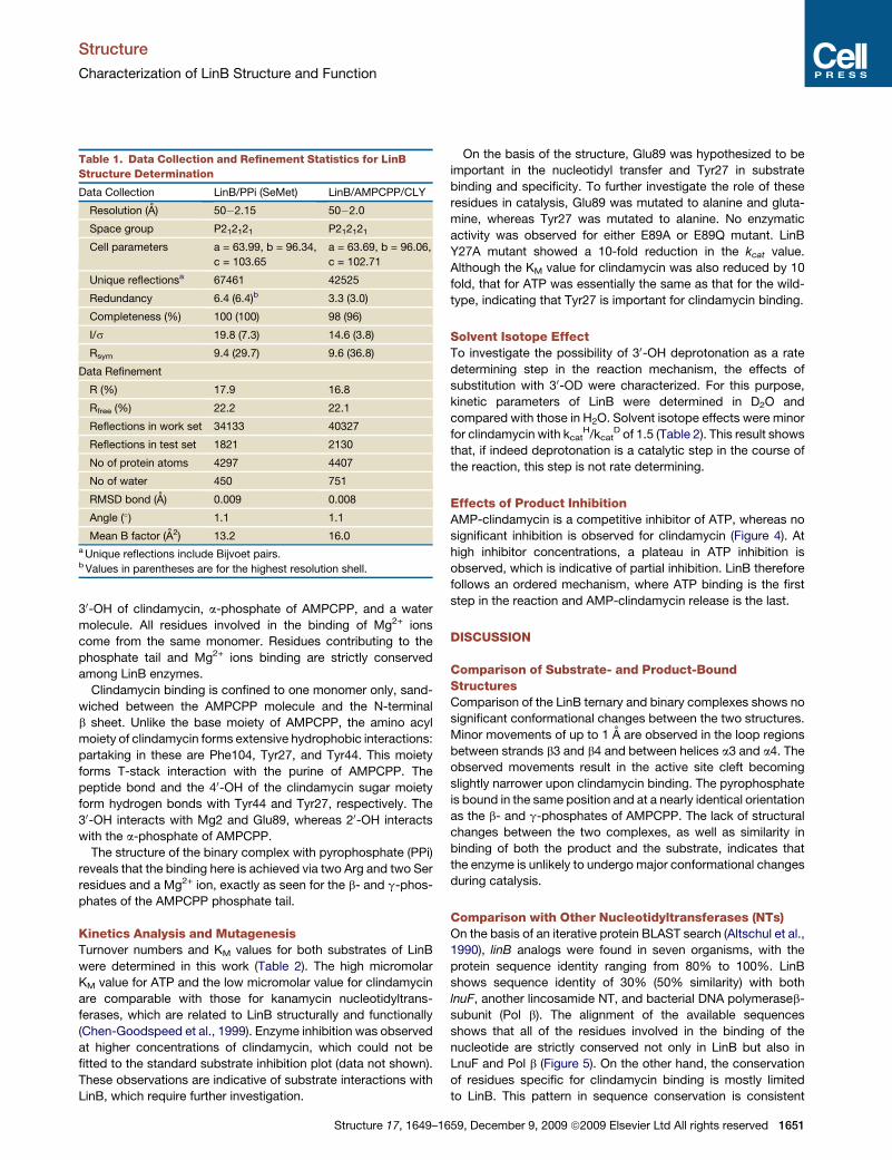
Table 1. Data Collection and Refinement Statistics for LinB
Structure Determination
Data Collection LinB/PPi (SeMet) LinB/AMPCPP/CLY
Resolution (A) 50�2.15 50�2.0
Space group P212121 P212121
Cell parameters a = 63.99, b = 96.34,
c = 103.65
a = 63.69, b = 96.06,
c = 102.71
Unique reflectionsa 67461 42525
Redundancy 6.4 (6.4)b 3.3 (3.0)
Completeness (%) 100 (100) 98 (96)
I/s 19.8 (7.3) 14.6 (3.8)
Rsym 9.4 (29.7) 9.6 (36.8)
Data Refinement
R (%) 17.9 16.8
Rfree (%) 22.2 22.1
Reflections in work set 34133 40327
Reflections in test set 1821 2130
No of protein atoms 4297 4407
No of water 450 751
RMSD bond (A) 0.009 0.008
Angle (�) 1.1 1.1
Mean B factor (A2) 13.2 16.0a Unique reflections include Bijvoet pairs.b Values in parentheses are for the highest resolution shell.
Structure 17, 1649–16
On the basis of the structure, Glu89 was hypothesized to be
important in the nucleotidyl transfer and Tyr27 in substrate
binding and specificity. To further investigate the role of these
residues in catalysis, Glu89 was mutated to alanine and gluta-
mine, whereas Tyr27 was mutated to alanine. No enzymatic
activity was observed for either E89A or E89Q mutant. LinB
Y27A mutant showed a 10-fold reduction in the kcat value.
Although the KM value for clindamycin was also reduced by 10
fold, that for ATP was essentially the same as that for the wild-
type, indicating that Tyr27 is important for clindamycin binding.
Solvent Isotope EffectTo investigate the possibility of 30-OH deprotonation as a rate
determining step in the reaction mechanism, the effects of
substitution with 30-OD were characterized. For this purpose,
kinetic parameters of LinB were determined in D2O and
compared with those in H2O. Solvent isotope effects were minor
for clindamycin with kcatH/kcat
D of 1.5 (Table 2). This result shows
that, if indeed deprotonation is a catalytic step in the course of
the reaction, this step is not rate determining.
Effects of Product InhibitionAMP-clindamycin is a competitive inhibitor of ATP, whereas no
significant inhibition is observed for clindamycin (Figure 4). At
high inhibitor concentrations, a plateau in ATP inhibition is
observed, which is indicative of partial inhibition. LinB therefore
follows an ordered mechanism, where ATP binding is the first
step in the reaction and AMP-clindamycin release is the last.
DISCUSSION
Comparison of Substrate- and Product-BoundStructuresComparison of the LinB ternary and binary complexes shows no
significant conformational changes between the two structures.
Minor movements of up to 1 A are observed in the loop regions
between strands b3 and b4 and between helices a3 and a4. The
observed movements result in the active site cleft becoming
slightly narrower upon clindamycin binding. The pyrophosphate
is bound in the same position and at a nearly identical orientation
as the b- and g-phosphates of AMPCPP. The lack of structural
changes between the two complexes, as well as similarity in
binding of both the product and the substrate, indicates that
the enzyme is unlikely to undergo major conformational changes
during catalysis.
Comparison with Other Nucleotidyltransferases (NTs)On the basis of an iterative protein BLAST search (Altschul et al.,
1990), linB analogs were found in seven organisms, with the
protein sequence identity ranging from 80% to 100%. LinB
shows sequence identity of 30% (50% similarity) with both
lnuF, another lincosamide NT, and bacterial DNA polymeraseb-
subunit (Pol b). The alignment of the available sequences
shows that all of the residues involved in the binding of the
nucleotide are strictly conserved not only in LinB but also in
LnuF and Pol b (Figure 5). On the other hand, the conservation
of residues specific for clindamycin binding is mostly limited
to LinB. This pattern in sequence conservation is consistent
59, December 9, 2009 ª2009 Elsevier Ltd All rights reserved 1651

Structure
Characterization of LinB Structure and Function
Figure 3. Overall Structure of LinB
(A) Ribbon diagram of the LinB monomer, color-coded by secondary structure. Helices are in blue, strands in green, and loops in yellow. Ligands, clindamycin,
and AMPCPP are shown in ball-and-stick representation.
(B) Ribbon diagram of the LinB dimer: one monomer is shown in magenta, and the second monomer is in pale green. See also Figure S1.
(C) Stereo view of the LinB active site. Clindamycin and AMPCPP, ligands in the ternary complex of LinB, and residues forming interactions with the ligands are
shown in ball-and-stick model. The ligands are color-coded, with C atoms in black, O atoms in red, N atoms in blue, S atoms in green, and P atoms in orange. The
residues are color-coded same as the ligands, except C atoms are in pale green. Mg2+ ions are shown as gray spheres, and water molecules as blue spheres.
Hydrogen-bonding interactions are shown as dashed lines. Residue labels denoted by asterisk come from the adjacent monomer. The figure was prepared using
Pymol and Photoshop.
with the ATP binding being the unifying feature for this group
of NTs.
Dali analysis (Holm et al., 2008) was performed in search of
structural homologs in the PDB (see Table S1 available online).
This search revealed that LinB shares similarity with antibiotic
NTs, of which S. aureus (Sa) KAN ANT(40) is the only one bio-
chemically characterized (Chen-Goodspeed et al., 1999; Peder-
Table 2. Summary of LinB Kinetic Parameters at pH 7.5
kcat (s�1) KM (mM) kcat/KM (M�1s�1)
Wild-type enzyme
Clindamycin (H2O) 0.31 ± 1 3 10�4 3.7 ± 0.44 9 3 104 ± 227
Clindamycin (D2O) 0.15 ± 7 3 10�4 9.4 ± 2.0
MgATP 0.6 ± 5 3 10�4 217 ± 23 2.8 3 103 ± 21
E89Q mutant <0.003
E89A mutant <0.003
Y27A mutant
Clindamycin 0.016 ± 2 3 10�4 73 ± 16 219 ± 13
MgATP 0.013 ± 2 3 10�4 441 ± 85 30 ± 2
Errors shown are standard errors to the fit of the data to Equation 1. See
also Figure S2.
1652 Structure 17, 1649–1659, December 9, 2009 ª2009 Elsevier Ltd
sen et al., 1995). DNA polyA polymerase (PAP) structures were
also detected (Martin et al., 2004; Meinke et al., 2008). Remark-
ably, all of these structures share less than 15% amino acid
sequence identity.
Of the four antibiotic NTs, two display similar quarternary orga-
nization, and all four share a similar fold and domain organization
as LinB (Figure 6A). Bacillus subtilis (Bs) NT and SaANT(40) are
most similar with LinB, whereas the Sulfolobus solfataricus (Ss)
NT lacks the C-terminal bundle and Exiguobacterium sibiricum
(Es) NT has an additional domain after the C-terminal bundle,
resulting in a greater dimer interface. On the basis of these
comparisons, the N-terminal domain appears to be the minimal
functional unit for the NT reaction with various structural acces-
sories available for fine tuning.
Analysis of primary and secondary structure shows that the
similarity extends to DNA polymerases PAP and Pol b, both of
which also have structural elements of the b sheet sandwiched
by a helices (Figure 6B). This similarity was initially observed
with the first structures of KAN ANT(40) (Sakon et al., 1993) and
DNA Pol b (Davies et al., 1994; Sawaya et al., 1994), resulting
in the identification of an ancient NT superfamily (Holm and
Sander, 1995). The superfamily also includes terminal deoxyNT
(TdT) (Delarue et al., 2002) and ATP:tRNA NT (CCAtr) (Okabe
All rights reserved
Structure
Characterization of LinB Structure and Function
et al., 2003), whose structures have been determined. This struc-
ture of LinB expands this superfamily by adding lincosamide NTs
to the growing collection.
Structural superposition reveals that not only is the fold
conserved but also the three catalytic acidic residues responsible
for the chelation of Mg2+ ions (Figure 6C). For LinB, these are
Figure 4. Product Inhibition Effect
(A) Double reciprocal plot of competitive inhibition of ATP by the purified
product AMP-clindamycin. Concentrations of AMPclindamycin are 0 mM
(C), 0.25 mM (,), 0.5 mM (-), 0.75 mM (6), 1 mM (:), and 1.5 mM (V). Inset
is a replot of KMapp versus KI.
(B) Lack of inhibition of clindamycin by AMP-clindamycin. Concentrations of
AMP-clindamycin are 0.25 mM (C), 0.375 mM (,), 0.5 mM (-), 0.75 mM
(6), and 0.9 mM (:).
Structure 17, 1649–1
strictly conserved Asp40, Glu42, and Glu89, forming interactions
with Mg1. The location of the Mg2+ ion and the geometry with
respect to the chelating residues is also maintained. Comparison
of nucleotide binding shows that the adenine base and the
ribose assume various conformations, whereas the geometry
and interactions of the phosphate tail are conserved. Conserva-
tion of the catalytic residues and that of the Mg2+ ion and
the phosphate tail positioning all indicate a similar mechanism
of action. As expected, residues responsible for the binding of
clindamycin are not conserved because of great variation in
the identity and the structure of second substrate within the
superfamily.
Mechanism of ActionIn this work, the biochemical studies show that LinB readily inac-
tivates clindamycin in vitro with the turnover of 0.3 s�1, and the
NMR analysis confirms the 30-OH of clindamycin as the site of
adenylylation. The map of the active site reveals the position
of the two substrates, allowing for further discussion of the
potential course of events. 30-OH of clindamycin is bound by
Glu89 and Mg1; the a-phosphate of AMPCPP also interacts
with Mg1, which, in turn, is chelated by Glu89 (Figure 3C). The
oxygen atom of the 30-OH of clindamycin is 3.4 A away from
the phosphorus atom of the AMPCPP a-phosphate. The angle
formed between the C30, O30, and Pa is 100�. The geometry of
binding and distances between the reacting groups point to
a direct in-line transfer of the adenylyl moiety onto clindamycin,
making an adenylylated enzyme or other similar intermediates
unlikely.
In-line AMP transfer mechanism is also supported by the
body of literature discussing functional and structural relatives
of LinB: ANTs and nucleotide polymerases. Ternary complex
of SaANT(40) with KAN and AMPCPP (Pedersen et al., 1995)
and kinetic isotope effect studies (Gerratana et al., 2001) argue
for direct in-line transfer for this enzyme. ANT(200)-Ia is one of
the earliest ANTs studied mechanistically and makes for an
insightful comparison. For this enzyme, inversion of phosphorus
configuration points to a simple direct nucleotidyl transfer (Van
Pelt et al., 1986). Both ANT(40) and ANT(200)-Ia obey ordered
mechanisms with the nucleotidylated product release being
the last and the rate-limiting step (Chen-Goodspeed et al.,
1999; Gates and Northrop, 1988). Our product inhibition
experiments show that LinB also follows an ordered Bi-Bi
mechanism.
Direct transfer could proceed via a dissociative mechanism
with the formation of a metaphosphate-like transition state and
pyrophosphate leaving prior to the attack onto clindamycin
(Figure 7). The alternative is the activation of the 30-OH for an
in-line transfer to a-phosphate of ATP. This route is associative
and would proceed via a pentacoordinated phosphate transition
state (Figure 7). Both transition states have been described in the
literature (Beard and Wilson, 2006; Thompson et al., 2002);
however, in the case of LinB, the latter mechanism is more likely
given the geometry of the active site.
Glu89 is proposed to be the catalytic base responsible for the
activation of the 30-OH. Mutagenesis of this residue confirmed
that it is essential for catalysis. Glu89 forms a hydrogen bond
with the reactive OH, and the abolishment of activity in a
conservative mutation to a Gln residue is further indicative of
659, December 9, 2009 ª2009 Elsevier Ltd All rights reserved 1653

Structure
Characterization of LinB Structure and Function
Figure 5. Alignment of LinB, LnuF, and DNA Pol b Sequences Retrieved from BLAST Search
Strictly conserved residues are highlighted gray, and conserved residues are in gray font. Secondary structural elements of LinB are shown at the top of the
sequence. Asterisks at the bottom of the sequence indicate residues involved in the nucleotide binding; number signs indicate residues involved in clindamycin
binding. The alignment was generated using ClustalW (Thompson et al., 1994) and rendered using ESPript (Gouet et al., 1999).
a function greater than binding and positioning of the substrate.
An associative mechanism has been described for ANT(40), the
structural homolog of LinB, through the application of kinetic
isotope effect experiments (Gerratana et al., 2001). Nucleotide
polymerases overall, and specific members of the NT super-
family to which LinB belongs, also operate via such a mecha-
nism, as illustrated by several crystal structures with trapped
intermediate analogs (Steitz, 1998; Doublie et al., 1998; Sawaya
et al., 1994; Batra et al., 2006). Although the mechanism with the
substrate activation via a catalytic base is favored, the solvent
isotope effect study shows that substrate deprotonation is not
rate determining. Our observation is consistent with those for
both ANT(40) and ANT(200)-Ia, where the product release is the
slow step of the reaction (Chen-Goodspeed et al., 1999; Gates
and Northrop, 1988).
1654 Structure 17, 1649–1659, December 9, 2009 ª2009 Elsevier Lt
Implications of the LinB Structure and Mechanismin the Evolution of Antibiotic Resistance GenesThe relationship of the antibiotic modifying genes, lincosamide
and aminoglycoside NTs, to nucleotide polymerases is now
evident through this work and others. This relationship is likely
the result of divergent evolution and is consistent with the
hypothesis that the NTs responsible for antibiotic resistance
arose from nucleotide polymerases. DNA and RNA polymerases
are ancient and vital molecules found in all kingdoms of life. On
the other hand, relatively few examples of lincosamide NTs are
known, suggesting that these are the result of adaptation of
certain organisms to their surrounding environment. The con-
cept of the antibiotic resistome (Wright, 2007) states that resis-
tance genes like linB that eventually make their way into patho-
genic organisms originate in environmental organisms where
d All rights reserved

Structure
Characterization of LinB Structure and Function
antibiotic producers, their neighbors, and related organisms
have been using small molecules such as antibiotics to interact
with each other for millennia. Bona fide resistance genes evolve
from precursor genes by natural selection. Borrowing the
vernacular of the oncogene field, we term these precursor genes
‘‘protoresistance’’ elements. Conservation of structure and
mechanism between antibiotic resistance NTs and nucleotide
polymerases described in this work indicates that, like protein
kinases (Hon et al., 1997) and protein acetyltransferases
(Wybenga-Groot et al., 1999), DNA polymerases are protoresist-
ance elements implicated in the evolution of drug resistance.
This information will facilitate future work to identify strategies,
such as small molecule inhibitor discovery and design (Daigle
et al., 1997), that can overcome antibiotic resistance.
EXPERIMENTAL PROCEDURES
Cloning, Expression, and Purification of LinB
The linB gene from E. faecium was synthesized with codon sequence
optimized for expression in Escherichia coli and subcloned into pET28a
expression vector (Novagen) by GenScript (USA). LinB containing plasmid
was transformed into E. coli B834(DE3) expression cell line using a standard
heat shock protocol. The resulting E. coli colonies were grown in 5 ml LB
starter cultures supplemented with 40 mg/mL kanamycin (KAN) overnight at
37�C. The starter cultures were subsequently used to inoculate 1 L of LB
culture with 40 mg/mL KAN. The 1 L culture was grown at 37�C until an
OD600nm of 0.8 was reached and then was chilled in ice-water bath for
15 min. The protein expression was induced in the chilled culture with 100 mM
IPTG, and the culture was further incubated overnight at 16�C. The cells were
harvested via centrifugation using Avanti J25 centrifuge (Beckman) and were
stored at �20�C until further use.
For protein purification, the cell pellet was resuspended in lysis buffer
(100 mM HEPES [pH 7.5], 300 mM NaCl, and 10 mM imidazole). The cells
were lysed via three passes through a French Pressure Cell (Aminco) at
13,000 psi, and the cell debris was removed via centrifugation at 30,000 g.
The cleared supernatant was loaded onto a 5 ml Ni-NTA column (QIAGEN)
and pre-equilibrated with lysis buffer at a flow rate of 1 mL/min. The column
was then washed with a 100 ml volume of the lysis buffer, followed by step
gradient wash with an elution buffer (100 mM HEPES [pH 7.5], 300 mM NaCl,
and 250 mM imidazole). The gradient consisted of a 10% increase in the
ratio of the elution buffer to the lysis buffer every 20 ml of wash. LinB eluted
in 30–50% volume of the elution buffer. The enzyme purity was verified
using SDS PAGE analysis, with the protein being approximately 90% pure.
The protein solution was dialyzed overnight at 4�C into a dialysis buffer
(10 mM Tris [pH 8.2] and 50 mM NaCl) and was concentrated to 50 mg/mL
via Amicon-Ultra centrifugal device from Millipore. The protein concentration
was determined using the Bradford Assay per the manufacturer’s instruc-
tions (BioRad). The enzyme was then frozen and stored at �20�C until
further use.
For structural studies, selenomethionine (SeMet) substituted protein
was prepared. E. coli B834(DE3) cells transformed with LinB-containing
pET28a plasmid were grown using the M9 SeMet high-yield growth media
kit from Orion Biosolutions. The growth protocol provided by the manufacturer
was followed. SeMet LinB was purified as described for the native enzyme,
the only modification being the addition of 1 mM b-mercaptoethanol to all
buffers.
Analytical Gel Filtration
The oligomeric state of LinB in solution was determined via analytical gel filtra-
tion using a Superdex 200 10/300 GL column (GE Healthcare). The column was
calibrated using a gel filtration LMW calibration kit (GE Healthcare) per the
manufacturer’s instructions. The elution buffer was 20 mM HEPES (pH 7.5)
and 150 mM NaCl for all proteins. LinB eluted at 14.9 mL, corresponding to
a molecular mass of 56 kDa. LinB elution profile and a table of elution volumes
and molecular masses for proteins used are shown in Figure S1.
Structure 17, 1649–16
Structure Determination
LinB was crystallized using a concentration of 13 mg/mL for native and
5 mg/mL for SeMet enzyme. The optimized crystallization conditions for
LinB included 20% PEG3350, 0.1 M Tris (pH 8.5), and 0.1 M magnesium
formate. Native LinB crystals were grown using hanging drop vapor diffusion
method at 23�C. For the ternary complex, LinB was cocrystallized with
1 mM AMPCPP and 0.5 mM clindamycin (CLI). SeMet LinB was crystallized
in the presence of 1 mM ATP. SeMet LinB crystal growth was induced by
streak-seeding with native LinB crystals. LinB crystallized in the orthorhombic
space group P212121, a = 63.99 A, b = 96.34 A, c = 103.65 A, 2 molecules per
asymmetric unit, and 52% solvent content. The crystals were frozen in liquid
nitrogen for data collection with the crystallization conditions supplemented
with 17% glycerol used as cryoprotectant.
Single wavelength anomalous dispersion data were collected for the
SeMet substituted crystals. The data were collected at the beamline X25C of
National Synchrotron Light Source (Brookhaven, NY) using 0.9798 nm wave-
length and 1� oscillation. HKL2000 program suite was used to index, integrate,
and scale the data (Otwinowski and Minor, 1997). Data collection statistics are
listed in Table 1. Ten Se sites of possible 16 were located and used for phasing
using AutoSolve program in the Phenix program suite (Adams et al., 2002). The
initial phases were used for electron density modification and automated model
building in AutoBuild program in Phenix, resulting in the Ca-trace for 300 resi-
dues. The remaining backbone and side chains were then built manually using
COOT software (Emsley and Cowtan, 2004). COOT was subsequently used in
manual model refinement. Refmac5 program of CCP4 suite was used for rigid
body and restrained refinement (Collaborative Computational Project, 1994;
Potterton et al., 2003). Refinement statistics are listed in Table 1.
The data for the LinB/CLI/AMPCPP ternary complex were collected using
RU300 rotating anode generator with the R-AXIS IV IP detector (MSC Ltd/
Rigaku); 7 min exposure, 1� oscillation, and 200 mm crystal to detector dis-
tance. The HKL2000 program suite was used to index, integrate, and scale
the data. The ternary complex structure was determined by molecular replace-
ment using the dimer of the SeMet LinB structure as the search model and Mol-
Rep from CCP4 program suite to perform rotation and translation function
calculations. COOT was used for manual refinement, and Refmac5 from
CCP4 was used for automated refinement of the structure. Noncrystallographic
symmetry restraints were used for the initial stages of the refinement. Data
collection and refinement statistics are listed in Table 1.
Kinetic Assays
The EnzCheck� pyrophosphate assay kit from Invitrogen, based on a previously
described method,was used to quantify the kinetic parameters for the wild-type
LinB (Upson et al., 1996). The kit was used as per Invitrogen protocol, and the
reaction progress was monitored for 10 min at 360 nm. The modifications to
the Invitrogen procedure follow. The 96-well flat bottom plates (Nalge NUNC
Int.) were used in the experimental set-up, with the total volume of the reaction
mixture being 250 mL. The reaction buffer was 50 mM HEPES (pH 7.5), 100 mM
KCl, and 2 mM MgCl2. The reaction was initiated using 10 ml of either clindamy-
cin or ATP. When monitoring clindamycin dependence, 3 mM ATP was used,
and final concentrations of clindamycin ranged between 3 and 300 mM. For
ATP characterization, 50 mM clindamycin was used, and the ATP concentration
ranged from 31 mM to 2 mM. For the calculations of the rate, a path length of 0.59
cm and an extinction coefficient of 11,000 M�1cm�1 were used. The initial rates
were described by using Michaelis-Menten kinetics, as shown in Equation 1,
with utility of Grafit 4 software (Erithacus Software, Staines, UK):
n =Vmax,½S�Km + ½S� : (1)
To assess the effects of site-directed mutagenesis (described below), Mal-
achite-Green spectrophotometric assay was used. A previously described
protocol for this assay was followed (De Leon et al., 2006). The activity of
LinB-Y27A mutant was quantified using EnzCheck� pyrophosphate assay.
Solvent Isotope Effect
Solvent isotope effects on the activity of LinB were assessed by quantifying the
enzyme’s kinetic parameters in 70% D2O. EnzCheck� pyrophosphate assay
was used with D2O used in place of H2O for the master mix dilutions.
59, December 9, 2009 ª2009 Elsevier Ltd All rights reserved 1655

Structure
Characterization of LinB Structure and Function
Figure 6. Comparison of LinB Structure with the Members of the NT Superfamily
(A) Structure of LinB is compared with aminoglycoside NT: SaANT(40) (1kny), and putative antibiotic NTs: SsNT (2rff) and EsNT (3c18) illustrating the conservation
of both the N and C-terminal domain. The N-terminal domains are in gray, C-terminal domains are in blue, and structural features missing from LinB are in
light-orange.
(B) A ribbon diagram of the LinB structure compared with PAP and Pol b, illustrating the conservation of the N terminal domain. The N-terminal domain is in gray,
a-helical bundle is in blue, and structural features missing from LinB are in light-orange.
1656 Structure 17, 1649–1659, December 9, 2009 ª2009 Elsevier Ltd All rights reserved

Structure
Characterization of LinB Structure and Function
Enzymatic Synthesis of Adenylylated Clindamycin
For the product inhibition studies, adenylylated clindamycin (AMP-clindamy-
cin) was synthesized and purified. Reaction mixture containing 100 ml of
50 mg/mL purified LinB, 20 mM HEPES (pH 7.5), 2 mM MgCl2, and 3 mM
ATP was initiated using 300 ml of 1.4 mg/mL clindamycin and incubated at
room temperature for 1 hr. To purify the reaction product, high-pressure liquid
chromatography was performed using DIONEX GP40 Gradient Pump.
Aliquots of reaction mixture (100 ml) were injected onto C-18 reverse phase
column (10 mm; 22 3 250 mm; GRACE VYDAC), and the pump flow rate was
set to 1.0 mL/min. AMP-clindamycin was eluted using 70% Solvent A
(0.03% trifluoroacetic acid in H2O) and 30% Solvent B (0.03% trifluoroacetic
acid in acetonitrile) in a linear gradient. Elution profile was monitored by
Figure 7. Possible Transition States for the LinB Reaction
Figure was prepared using ChemDraw Ultra.
Structure 17, 1649–1
a PDA-100 Photodiode Array Detector at 254 nm. Fractions were analyzed
using QTrap LC/MS/MS System (Applied Biosystems/MDS SCIEX), and those
of interest were pooled. Approximately 7 ml of purified product was collected
and lyophilized overnight, yielding 14 mg of purified AMP-clindamycin. Powder
sample was stored in�20�C and redissolved in appropriate buffers for further
use. The structure of the purified product was confirmed by 1H and 13C NMR
(Tables S2–S4).
Product Inhibition Studies
The pyrophosphate assay was employed for product inhibition studies. Five
different product concentrations were examined, and at each product concen-
tration, that of one substrate was kept constant and that of the second
substrate was varied. For the study of ATP inhibition, the concentrations of
AMP-clindamycin were 0.25, 0.5, 0.75, 1, and 1.5 mM; those of ATP were
62.5, 250, 500, and 1000 mM; and that of clindamycin was 50 mM. For the study
of clindamycin inhibition, the concentrations of AMP-clindamycin were 0.25,
0.375, 0.5, 0.75, and 0.9 mM; those of clindamycin were 6.25, 12.5, 18.75,
and 25 mM; and that of ATP was 2 mM.
Site-Directed Mutagenesis
QuikChange� Site-Directed Mutagenesis (Stratagene) was used to generate
mutants of LinB in the pET28a vector as per manufacturer instructions. Primers
used for mutagenesis are listed in Supplemental Experimental Procedures.
The mutagenesis results were confirmed via DNA sequencing (MOBIX,
McMaster University).
Determination of Minimal Inhibitory Concentration (MIC)
MIC of clindamycin for the E. coli strains containing plasmids with wild-type
and mutated LinB were determined by adapting the National Committee for
Clinical Laboratory Standards protocol. The modification was the use of
Luria-Bertani media for this experiment. Clindamycin has low activity against
E. coli, and as expected the MIC for all strains was 128 mg/mL. Then, the bacte-
rial growth was monitored every 30 min for 20 hr at 600 nm, showing impedi-
ment of growth during the exponential phase for strains containing LinB
mutants E89Q and Y27A, as well as empty pET28a vector (Figure S2).
ACCESSION NUMBERS
The coordinates and structure factors for the LinB/AMPCPP/CLI complex
structure have been deposited to the Protein Data Bank under accession
code 3JZ0, and that for LinB/PPi complex has been deposited under acces-
sion code 3JYY.
SUPPLEMENTAL DATA
Supplemental Data include two figures and five tables and can be found with
this article online at http://www.cell.com/structure/supplemental/S0969-
2126(09)00420-1.
ACKNOWLEDGMENTS
This work was supported by the Canadian Institutes of Health Research (grant
MT-13536 to G.D.W.), the Canada Research Chairs program (support to
G.D.W.), and Canadian Institutes of Health Research (grant MOP-89903 to
M.J.).
Received: August 7, 2009
Revised: October 13, 2009
Accepted: October 14, 2009
Published: December 8, 2009
(C) Superposition of the active sites of LinB, SaANT(40) (1kny), PAP (1q78), and Pol b (2fms). Nucleotides and the three acidic residues conserved within the
superfamily are in ball-and-stick representation, and Mg2+ ions are shown as spheres. The diagram is color-coded by atom type: C is gray for LinB, blue for
SaANT(40), green for PAP, and yellow for Pol b. Other atom types are colored the same as in Figure 1C. See also Table S1. The figure was prepared using Pymol
and Photoshop.
659, December 9, 2009 ª2009 Elsevier Ltd All rights reserved 1657
Structure
Characterization of LinB Structure and Function
REFERENCES
Adams, P.D., Grosse-Kunstleve, R.W., Hung, L.W., Ioerger, T.R., McCoy, A.J.,
Moriarty, N.W., Read, R.J., Sacchettini, J.C., Sauter, N.K., and Terwilliger, T.C.
(2002). PHENIX: building new software for automated crystallographic struc-
ture determination. Acta Crystallogr. D Biol. Crystallogr. 58, 1948–1954.
Altschul, S.F., Gish, W., Miller, W., Myers, E.W., and Lipman, D.J. (1990). Basic
local alignment search tool. J. Mol. Biol. 215, 403–410.
Bartlett, J.G. (2008). Methicillin-resistant Staphylococcus aureus infections.
Top. HIV Med. 16, 151–155.
Batra, V.K., Beard, W.A., Shock, D.D., Krahn, J.M., Pedersen, L.C., and
Wilson, S.H. (2006). Magnesium-induced assembly of a complete DNA
polymerase catalytic complex. Structure 14, 757–766.
Beard, W.A., and Wilson, S.H. (2006). Structure and mechanism of DNA
polymerase Beta. Chem. Rev. 106, 361–382.
Bozdogan, B., Berrezouga, L., Kuo, M.S., Yurek, D.A., Farley, K.A., Stockman,
B.J., and Leclercq, R. (1999). A new resistance gene, linB, conferring resis-
tance to lincosamides by nucleotidylation in Enterococcus faecium HM1025.
Antimicrob. Agents Chemother. 43, 925–929.
Brisson-Noel, A., Delrieu, P., Samain, D., and Courvalin, P. (1988). Inactivation
of lincosaminide antibiotics in Staphylococcus. Identification of lincosaminide
O-nucleotidyltransferases and comparison of the corresponding resistance
genes. J. Biol. Chem. 263, 15880–15887.
CCP4 (Collaborative Computational Project, Number 4). (1994). The CCP4
suite: programs for protein crystallography. Acta Crystallogr. D Biol. Crystal-
logr. 50, 760–763.
Chen-Goodspeed, M., Vanhooke, J.L., Holden, H.M., and Raushel, F.M.
(1999). Kinetic mechanism of kanamycin nucleotidyltransferase from Staphy-
lococcus aureus. Bioorg. Chem. 27, 395–408.
Courvalin, P., Ounissi, H., and Arthur, M. (1985). Multiplicity of macrolide-
lincosamide-streptogramin antibiotic resistance determinants. J. Antimicrob.
Chemother. 16 (Suppl A), 91–100.
Daigle, D.M., McKay, G.A., and Wright, G.D. (1997). Inhibition of aminoglyco-
side antibiotic resistance enzymes by protein kinase inhibitors. J. Biol. Chem.
272, 24755–24758.
Davies, J.F., 2nd, Almassy, R.J., Hostomska, Z., Ferre, R.A., and Hostomsky,
Z. (1994). 2.3 A crystal structure of the catalytic domain of DNA polymerase
beta. Cell 76, 1123–1133.
De Leon, G.P., Elowe, N.H., Koteva, K.P., Valvano, M.A., and Wright, G.D.
(2006). An in vitro screen of bacterial lipopolysaccharide biosynthetic enzymes
identifies an inhibitor of ADP-heptose biosynthesis. Chem. Biol. 13, 437–441.
Delarue, M., Boule, J.B., Lescar, J., Expert-Bezancon, N., Jourdan, N.,
Sukumar, N., Rougeon, F., and Papanicolaou, C. (2002). Crystal structures
of a template-independent DNA polymerase: murine terminal deoxynucleoti-
dyltransferase. EMBO J. 21, 427–439.
Doublie, S., Tabor, S., Long, A.M., Richardson, C.C., and Ellenberger, T.
(1998). Crystal structure of a bacteriophage T7 DNA replication complex at
2.2 A resolution. Nature 391, 251–258.
Dutta, G.N., and Devriese, L.A. (1982). Resistance to macrolide, lincosamide
and streptogramin antibiotics and degradation of lincosamide antibiotics in
streptococci from bovine mastitis. J. Antimicrob. Chemother. 10, 403–408.
Emsley, P., and Cowtan, K. (2004). Coot: model-building tools for molecular
graphics. Acta Crystallogr. D Biol. Crystallogr. 60, 2126–2132.
Fitzhugh, A.L. (1998). Antibiotic inhibitors of the peptidyl transferase center. 1.
Clindamycin as a composite analogue of the transfer RNA fragments L-Pro-
Met and the D-ribosyl ring of adenosine. Bioorg. Med. Chem. Lett. 8, 87–92.
Gates, C.A., and Northrop, D.B. (1988). Alternative substrate and inhibition
kinetics of aminoglycoside nucleotidyltransferase 200-I in support of a Theor-
ell-Chance kinetic mechanism. Biochemistry 27, 3826–3833.
Gerratana, B., Frey, P.A., and Cleland, W.W. (2001). Characterization of the
transition-state structure of the reaction of kanamycin nucleotidyltransferase
by heavy-atom kinetic isotope effects. Biochemistry 40, 2972–2977.
Gouet, P., Courcelle, E., Stuart, D.I., and Metoz, F. (1999). ESPript: analysis of
multiple sequence alignments in PostScript. Bioinformatics 15, 305–308.
1658 Structure 17, 1649–1659, December 9, 2009 ª2009 Elsevier Ltd
Hoeksema, H., Bannister, B., Birkenmeyer, R.D., Kagan, F., Magerlein, B.J.,
MacKellar, F.A., Schroeder, W., Slomp, G., and Herr, R.R. (1964). Chemical
studies of lincomycin. 1. The structure of lincomycin. J. Am. Chem. Soc. 86,
4223–4224.
Holm, L., and Sander, C. (1995). DNA polymerase beta belongs to an ancient
nucleotidyltransferase superfamily. Trends Biochem. Sci. 20, 345–347.
Holm, L., Kaariainen, S., Rosenstrom, P., and Schenkel, A. (2008). Searching
protein structure databases with DaliLite v.3. Bioinformatics 24, 2780–2781.
Hon, W.C., McKay, G.A., Thompson, P.R., Sweet, R.M., Yang, D.S., Wright,
G.D., and Berghuis, A.M. (1997). Structure of an enzyme required for aminogly-
coside antibiotic resistance reveals homology to eukaryotic protein kinases.
Cell 89, 887–895.
Johnson, M.D., and Decker, C.F. (2008). Antimicrobial agents in treatment of
MRSA infections. Dis. Mon. 54, 793–800.
Laskowski, R.A., MacArthur, M.W., Moss, D.S., and Thornton, J.M. (1993).
PROCHECK: a program to check the stereochemical quality of protein struc-
tures. J. Appl. Cryst. 26, 283–291.
Leclercq, R., Carlier, C., Duval, J., and Courvalin, P. (1985). Plasmid-mediated
resistance to lincomycin by inactivation in Staphylococcus haemolyticus.
Antimicrob. Agents Chemother. 28, 421–424.
Martin, G., Moglich, A., Keller, W., and Doublie, S. (2004). Biochemical and
structural insights into substrate binding and catalytic mechanism of mamma-
lian poly(A) polymerase. J. Mol. Biol. 341, 911–925.
Meinke, G., Ezeokonkwo, C., Balbo, P., Stafford, W., Moore, C., and Bohm, A.
(2008). Structure of yeast poly(A) polymerase in complex with a peptide from
Fip1, an intrinsically disordered protein. Biochemistry 47, 6859–6869.
Okabe, M., Tomita, K., Ishitani, R., Ishii, R., Takeuchi, N., Arisaka, F., Nureki,
O., and Yokoyama, S. (2003). Divergent evolutions of trinucleotide polymeriza-
tion revealed by an archaeal CCA-adding enzyme structure. EMBO J. 22,
5918–5927.
Otwinowski, Z., and Minor, W. (1997). Processing of X-ray diffraction data
collected in oscillation mode. Methods Enzymol. 276, 307–326.
Pedersen, L.C., Benning, M.M., and Holden, H.M. (1995). Structural investiga-
tion of the antibiotic and ATP-binding sites in kanamycin nucleotidyltransfer-
ase. Biochemistry 34, 13305–13311.
Petinaki, E., Guerin-Faublee, V., Pichereau, V., Villers, C., Achard, A.,
Malbruny, B., and Leclercq, R. (2008). Lincomycin resistance gene lnu(D) in
Streptococcus uberis. Antimicrob. Agents Chemother. 52, 626–630.
Potterton, E., Briggs, P., Turkenburg, M., and Dodson, E. (2003). A graphical
user interface to the CCP4 program suite. Acta Crystallogr. D Biol. Crystallogr.
59, 1131–1137.
Rezanka, T., Spizek, J., and Sigler, K. (2007). Medicinal use of lincosamides
and microbial resistance to them. Anti-Infect. Agents Med. Chem. 6, 133–144.
Sakon, J., Liao, H.H., Kanikula, A.M., Benning, M.M., Rayment, I., and Holden,
H.M. (1993). Molecular structure of kanamycin nucleotidyltransferase deter-
mined to 3.0-A resolution. Biochemistry 32, 11977–11984.
Sawaya, M.R., Pelletier, H., Kumar, A., Wilson, S.H., and Kraut, J. (1994).
Crystal structure of rat DNA polymerase beta: evidence for a common
polymerase mechanism. Science 264, 1930–1935.
Schlunzen, F., Zarivach, R., Harms, J., Bashan, A., Tocilj, A., Albrecht, R.,
Yonath, A., and Franceschi, F. (2001). Structural basis for the interaction of
antibiotics with the peptidyl transferase centre in eubacteria. Nature 413,
814–821.
Sood, S., Mahorta, M., Das, B.K., and Kapil, A. (2008). Enterococcal infections
and antimicrobial resistance. Indian J. Med. Res. 128, 111–121.
Spizek, J., Novotna, J., and Rezanka, T. (2004). Lincosamides: chemical struc-
ture, biosynthesis, mechanism of action, resistance, and applications. Adv.
Appl. Microbiol. 56, 121–154.
Steitz, T.A. (1998). A mechanism for all polymerases. Nature 391, 231–232.
Thompson, J.D., Higgins, D.G., and Gibson, T.J. (1994). CLUSTAL W:
improving the sensitivity of progressive multiple sequence alignment through
sequence weighting, position-specific gap penalties and weight matrix choice.
Nucleic Acids Res. 22, 4673–4680.
All rights reserved
Structure
Characterization of LinB Structure and Function
Thompson, P.R., Boehr, D.D., Berghuis, A.M., and Wright, G.D. (2002). Mech-
anism of aminoglycoside antibiotic kinase APH(30 )-IIIa: role of the nucleotide
positioning loop. Biochemistry 41, 7001–7007.
Tu, D., Blaha, G., Moore, P.B., and Steitz, T.A. (2005). Structures of MLSBK
antibiotics bound to mutated large ribosomal subunits provide a structural
explanation for resistance. Cell 121, 257–270.
Upson, R.H., Haugland, R.P., and Malekzadeh, M.N. (1996). A spectrophoto-
metric method to measure enzymatic activity in reactions that generate
inorganic pyrophosphate. Anal. Biochem. 243, 41–45.
Structure 17, 1649–1
Van Pelt, J.E., Iyengar, R., and Frey, P.A. (1986). Gentamicin nucleotidyltrans-
ferase. Stereochemical inversion at phosphorus in enzymatic 20-deoxyade-
nylyl transfer to tobramycin. J. Biol. Chem. 261, 15995–15999.
Wright, G.D. (2007). The antibiotic resistome: the nexus of chemical and
genetic diversity. Nat. Rev. Microbiol. 5, 175–186.
Wybenga-Groot, L.E., Draker, K., Wright, G.D., and Berghuis, A.M. (1999).
Crystal structure of an aminoglycoside 60-N-acetyltransferase: defining
the GCN5-related N-acetyltransferase superfamily fold. Structure 7,
497–507.
659, December 9, 2009 ª2009 Elsevier Ltd All rights reserved 1659