structure and mechanism of the mrp complex, an ancient
TRANSCRIPT

*For correspondence:
Competing interests: The
authors declare that no
competing interests exist.
Funding: See page 21
Received: 28 May 2020
Accepted: 30 July 2020
Published: 31 July 2020
Reviewing editor: Sriram
Subramaniam, University of
British Columbia, Canada
Copyright Steiner and
Sazanov. This article is distributed
under the terms of the Creative
Commons Attribution License,
which permits unrestricted use
and redistribution provided that
the original author and source are
credited.
Structure and mechanism of the Mrpcomplex, an ancient cation/protonantiporterJulia Steiner, Leonid Sazanov*
Institute of Science and Technology Austria, Klosterneuburg, Austria
Abstract Multiple resistance and pH adaptation (Mrp) antiporters are multi-subunit Na+ (or K+)/
H+ exchangers representing an ancestor of many essential redox-driven proton pumps, such as
respiratory complex I. The mechanism of coupling between ion or electron transfer and proton
translocation in this large protein family is unknown. Here, we present the structure of the Mrp
complex from Anoxybacillus flavithermus solved by cryo-EM at 3.0 A resolution. It is a dimer of
seven-subunit protomers with 50 trans-membrane helices each. Surface charge distribution within
each monomer is remarkably asymmetric, revealing probable proton and sodium translocation
pathways. On the basis of the structure we propose a mechanism where the coupling between
sodium and proton translocation is facilitated by a series of electrostatic interactions between a
cation and key charged residues. This mechanism is likely to be applicable to the entire family of
redox proton pumps, where electron transfer to substrates replaces cation movements.
IntroductionThe Na+/H+ antiporters are widely distributed secondary active transporters that use the proton
motive force to efflux intracellular sodium ions (Ito et al., 2017). Several protein families catalyse
this reaction and are mostly encoded by a single gene, such as the NHE family in eukaryotes and the
NhaA family in bacteria (Krulwich et al., 2011). Mrp antiporters are unique as they usually consist of
seven subunits (MrpABCDEFG) encoded in a single operon (Figure 1—figure supplement 1a).
Because of the operon’s distinctive properties, Mrp antiporters have been classified in a separate
category, cation:proton antiporter-3 (CPA3), in the transporter classification system (Saier et al.,
2016). They support intracellular pH homeostasis and Na+ efflux in alkali- and halophilic microorgan-
isms and are essential for their survival in challenging environments (Hamamoto et al., 1994;
Ito et al., 1999). At high pH, the pH component of the proton motive force is inverted from normal,
and so a substantial Dy component (electric potential) is crucial to drive proton translocation into
the cell (Krulwich et al., 2011). Antiporters with roles in alkaline pH homeostasis must catalyse elec-
trogenic antiport, in which the ratio of H+ entering the cell in exchange for Na+ moving out is
unequal, enabling proton entry to be driven by the Dy. For example, the stoichiometry for E. coli
NhaA is 2H+/1Na+ (Taglicht et al., 1993). The exact value for Mrp has not been fully established
experimentally due to challenges in purification of the intact complex (Morino et al., 2014) but is
likely to be also about 2, consistent with its function (Dzioba-Winogrodzki et al., 2009). This raises
the question as to why a so much more complicated protein assembly is needed to catalyse a similar
reaction. Since under the extreme environmental conditions Mrp is essential for cell survival and can-
not be replaced by single subunit antiporters (Cheng et al., 2016; Xu et al., 2018), one of the pro-
posals is that the Mrp complex can support cell growth at very high pH due to its large surface area,
where only a few available external protons can still be gathered for translocation into the cell
(Ito et al., 2017).
Steiner and Sazanov. eLife 2020;9:e59407. DOI: https://doi.org/10.7554/eLife.59407 1 of 24
RESEARCH ARTICLE

The largest two Mrp subunits, MrpA and MrpD, are homologous to each other, have 14 con-
served trans-membrane (TM) helices, and are thought to participate in proton translocation
(Mathiesen and Hagerhall, 2003). Their homologues (called antiporter-like subunits, which we will
abbreviate to APLS) are found in many proton-pumping protein complexes where they are present
in one to three (and recently discovered four [Chadwick et al., 2018]) copies per complex, depend-
ing on the energy availability and needs of the organism (Efremov and Sazanov, 2012). These
include bacterial (Baradaran et al., 2013; Friedrich et al., 1995) and mitochondrial respiratory
complex I (Fiedorczuk et al., 2016; Walker, 1992), NDH (NADH dehydrogenase-like) complex from
cyanobacteria (Laughlin et al., 2019; Pan et al., 2020; Schuller et al., 2019) and chloroplasts
(Sazanov et al., 1998), Fpo (F420:methanophenazine oxidoreductase) complex from archaea
(Baumer et al., 2000) as well as various membrane-bound hydrogenases (Efremov and Sazanov,
2012) including MBH (membrane-bound [NiFe]-hydrogenase) complex from archaea (Yu et al.,
2018). These modern enzymes represent some of the largest membrane protein complexes known
and are thought to have evolved from the unification of the membrane transporter Mrp-like module
with the soluble NiFe-hydrogenase module, sometimes followed by the addition of an electron input
module, such as the NAD-linked formate dehydrogenase in case of complex I (Efremov and Saza-
nov, 2012). The Mrp complex thus represents an ancient ancestor of diverse protein families and is
thought to have been among the few membrane proteins present in the last common ancestor of
prokaryotes (Sousa et al., 2016).
Structures of complex I (Agip et al., 2018; Baradaran et al., 2013; Fiedorczuk et al., 2016;
Zickermann et al., 2015), NDH (Schuller et al., 2019) and MBH (Yu et al., 2018) complexes have
been solved recently. These enzymes consist of two main domains – the Mrp-like membrane domain,
responsible for proton translocation (or sodium in case of MBH) and an attached hydrophilic redox
domain, responsible for electron transfer between substrates (e.g. NADH to quinone in case of
complex I). Therefore, the electron transfer and proton translocation processes are separated by
large distances (up to 200 A) and how they are coupled to each other remains a mystery. Upon solv-
ing the first structures of complex I, we proposed that redox reactions may drive proton transloca-
tion via long-range conformational changes (Baradaran et al., 2013; Efremov and Sazanov, 2011).
However, such changes have not been visualized till now despite significant efforts (Parey et al.,
2018). Electrostatic interactions between the key charged residues have also been proposed to play
an additional (Efremov and Sazanov, 2011) or main (Kaila, 2018; Verkhovskaya and Bloch, 2013)
role in the mechanism.
The structure of the universal common ancestor of these enzymes, the Mrp complex, has been
lacking so far. Clearly, it would be instrumental in resolving the coupling mechanism, which should
have common principles for this huge group of protein families. Furthermore, inactivation of the
Mrp complex strongly reduces pathogenicity of such problematic human pathogens as S. aureus
and P. aeruginosa (Krulwich et al., 2011), presenting Mrp as a valuable drug target. To address
these questions, we have determined the first, to our knowledge, atomic structure of the Mrp
complex.
Results
Structure determinationThe Mrp complex from Anoxybacillus flavithermus shows high sequence similarity to the well-charac-
terised Mrp complexes from Bacillus sp (Figure 4—figure supplements 3–4). The His-tagged Mrp
complex from A. flavithermus was recombinantly expressed in the antiporter-deficient E. coli strain
KNabc (Goldberg et al., 1987). The complex showed high Na+ (and to a lesser degree K+)/H+ anti-
port activity and purified to high homogeneity, predominantly as a dimer (Figure 1—figure supple-
ment 1b,f–j). It showed higher apparent stability than the Bacillus complex (Morino et al., 2014).
The protein tended to aggregate heavily in ice holes of cryo-EM grids. Therefore, we used grids
coated with a very thin layer of continuous carbon, which resulted in a uniform particle distribution
(Figure 1—figure supplement 2). However, the particles showed a strong preferred orientation,
with the hydrophilic protein surface attached to the carbon. To compensate for the associated loss
of information, data collection was performed with grids tilted (Tan et al., 2017) at 35˚, resulting in
excellent quality maps (Figure 1—figure supplements 2–4). The initial dataset was collected with
Steiner and Sazanov. eLife 2020;9:e59407. DOI: https://doi.org/10.7554/eLife.59407 2 of 24
Research article Structural Biology and Molecular Biophysics

protein purified in n-Dodecyl b-D-maltoside (DDM) detergent. Particles appeared as dimers of only
approximate C2 symmetry as the angle between monomers varied, resulting in several 3D classes
differing by that angle. After symmetry expansion in C2 point group in Relion, resulting pseudo-
monomer particles could be refined to 3.4 A resolution (Figure 1—figure supplement 2). This
allowed initial model building for most of the model, however, cryo-EM density at the edges of the
Figure 1. Overall structure of the Mrp complex. (a) Cryo-EM density of a monomer, coloured by subunit as indicated. (b) Cryo-EM density of a dimer,
with the model shown as a cartoon. (c) Model of the dimer, with each subunit coloured differently. Side view and view from the cytoplasm, where two
N-terminal helices of subunit MrpE, forming most of the dimer interface, are circled. (d) Schematic view of the Mrp monomer, MBH complex and
complex I. Homologs of Mrp subunits are coloured similarly as in c), with an additional MrpD-like subunit in complex I in orange.
The online version of this article includes the following figure supplement(s) for figure 1:
Figure supplement 1. Purification and characterization of Anoxybacillus flavithermus Mrp.
Figure supplement 2. Processing of the Mrp-DDM dataset.
Figure supplement 3. Processing of the Mrp-LMNG dataset.
Figure supplement 4. Examples of cryo-EM density of Mrp monomer in LMNG.
Steiner and Sazanov. eLife 2020;9:e59407. DOI: https://doi.org/10.7554/eLife.59407 3 of 24
Research article Structural Biology and Molecular Biophysics

monomer was fuzzy, with some TM helices (TMH) in the distal part of MrpA completely disordered.
Therefore we purified the complex in a milder detergent Lauryl Maltose Neopentyl Glycol (LMNG)
and collected a dataset again at 35˚ tilt. In this case dimers were overall ‘flatter’ than in DDM and
possibly closer to their native shape in the flat lipid bilayer (Figure 1—figure supplement 3). Com-
parisons of cryo-EM maps of various dimers did not reveal any specific differences in the overall
structure, apart from different apparent angles between the monomers. The best dimer class refined
to 3.7 A resolution (Figure 1b), while the best pseudo-monomer (after symmetry expansion) class
refined to 3.0 A with excellent density in all areas including previously disordered edges (Figure 1a).
This allowed us to build and refine a high quality atomic model of the Mrp dimer (97% complete
with only few terminal residues missing, Table 1, Supplementary file 1, Figure 1c).
Overall structureEach monomer consists of seven subunits with a total MW of 213 kDa and comprising 50 TM helices.
The largest two subunits, including the N-terminal part of MrpA (MrpAN, TMH1-16) and MrpD are
arranged next to each other in a very similar way as the APLS of complex I (Efremov and Sazanov,
2011), with the small subunits BCEFG and the C-terminal part of MrpA (MrpAC, TMH17-21) homolo-
gous to MBH subunits ABCDFG (Yu et al., 2018), attached ‘on the right’ of MrpD as shown in Fig-
ure 1. MBH subunit H is homologous to MrpD and MBH has its redox module attached ‘on the left’
of MbhH/MrpD, in contrast to complex I with the redox module on the right (Figure 1d). Thus, Mrp
has nearly all subunits, except MrpAN, in common with MBH. The fold of these Mrp subunits is
extremely well preserved in MBH, including all the key residues, which are conserved and essential
for activity (Figure 3a,c, Supplementary file 1). Similarly to MBH, subunits MrpC, AC, F and G each
fold into a three-helix sheet-like structure, forming four contiguous layers, flanked on one side by a
four-helix subunit MrpB (Figures 1c and 2d). MrpE caps the structure and is involved in
dimerization.
The similarity to complex I extends to the entire MrpA, MrpD and MrpC subunits, with the fold
and many key residues well conserved (Figure 3a,b, Supplementary file 1 and Figure 4—figure
supplement 2b). MrpA braces the complex with the long amphipathic helix HL (Figure 2d) extend-
ing from TMH15 to 16, similar to the arrangement in complex I. Among the three copies of APLS in
complex I, MrpA is more homologous to the complex I subunit Nqo12/NuoL/ND5 (T. thermophilus/
E. coli/mitochondrial nomenclature), while MrpD is closer to complex I Nqo14/NuoN/ND2 (Fig-
ure 4—figure supplement 2b). Similarly to APLS in complex I (Efremov and Sazanov, 2011), MrpA
and MrpD contain N-terminal (TMH4-8) and C-terminal (TMH9-13) proton translocation half-channels
that are related to each other by pseudosymmetry. Both half-channels contain lysine residues sitting
on symmetry-related discontinuous (interrupted by a ~6 residue loop) helices (LysTMH7 and
LysTMH12). These residues are likely the key to proton translocation because they are absolutely
conserved, essential for activity and sit in a strategic position in the centre of each half-channel cavity
(Sazanov, 2015). Key LysTMH7 forms a pair with a conserved TMH5 glutamate (GluTMH5), which is
thought to modulate the pKa of lysine (Efremov and Sazanov, 2011). A central key lysine sits on
another broken (by a p-bulge) helix TMH8 and connects the half-channels (Figure 4a,b). As in
complex I, these key residues are connected by additional polar residues and form a central hydro-
philic axis running through the middle of the membrane across the entire complex (Figure 4a,b).
The flexibility provided by the broken helices may have a role in the conformational coupling mecha-
nism and/or could help with pKa modulations of key residues.
As expected from sequence homology, the C-terminal MrpA TMH17-21 have the same fold as
the complex I subunit Nqo10/NuoJ/ND6, although the three essential for activity carboxylate resi-
dues from MrpA, E706, D792 and E796 (Supplementary file 1, Figure 4—figure supplement 3),
are not conserved in complex I (Figure 4—figure supplement 2b). Key E706 is replaced by Y59 in
complex I, where this essential tyrosine sits on a p-bulge in TMH3 of Nqo10. This p-bulge can be
wound up in mammalian complex I by a striking rotation of this helix, highly conserved in complex I
and probably critical for its mechanism (Agip et al., 2018; Letts et al., 2019). The p-bulge in the
corresponding helix is also present in the MBH complex (Yu et al., 2018). Interestingly, the bulge
appears to be absent in the Mrp, so it may not be a universal feature or it is possible that it may
appear in a different state of the complex, for example at high pH (current structure is solved at pH
6.0). Subunit MrpC fold is very similar to the complex I subunit Nqo11/NuoK/ND4L but instead of
Steiner and Sazanov. eLife 2020;9:e59407. DOI: https://doi.org/10.7554/eLife.59407 4 of 24
Research article Structural Biology and Molecular Biophysics

two histidines H37 and H40, conserved in Mrp, complex I has two essential glutamates (Figure 4—
figure supplement 2b).
A striking feature of the Mrp complex is the tightly intertwined interface between the two mono-
mers. To allow this, the two N-terminal TMH of MrpE (circled in Figure 1c) have swung out from
their respective positions in MBH (Figure 3a top), resulting in extensive interactions between subu-
nits MrpE and MrpB from both monomers. This shows that the Mrp complex clearly evolved to exist
Table 1. Cryo-EM data collection, refinement and validation statistics.
Data collection and processing
Magnification 105000
Voltage (kV) 300
Electron exposure (e-/A2) 85
Defocus range (mm) 0.6–2.3
Pixel size (A) 0.84
Symmetry imposed C1
Initial particle images (no.) 889272
Final particle images (no.) 285688
Map resolution (A) 2.98
FSC threshold 0.143
Map resolution range (A) 2.85–4.2
Refinement Monomer Dimer
Initial model used (PDB code) 6CFW, 4HEA 6CFW, 4HEA
Model resolution (A) 2.97 3.04
FSC threshold 0.5 0.5
Map sharpening B factor (A) �42, localresolution-filtered
�42
Model composition
Non-hydrogen atoms 15448 30250
Protein residues 1873 3730
Ligands 510 1020
Waters 242 0
B factors (A2)
Protein 88.4 74.4
Ligand 139.1 123.4
Waters 89.8 N/A
R.m.s. deviations
Bond lengths (A) 0.0080 0.0078
Bond angles (˚) 1.32 1.33
Validation
MolProbity score 1.47 1.31
Clashscore 3.58 2.17
Poor rotamers (%) 0.13 0.10
Ramachandran plot
Favoured (%) 95.44 95.45
Allowed (%) 4.56 4.55
Disallowed (%) 0 0
EMRinger score 3.73 3.61
Steiner and Sazanov. eLife 2020;9:e59407. DOI: https://doi.org/10.7554/eLife.59407 5 of 24
Research article Structural Biology and Molecular Biophysics

as a dimer in vivo, as also confirmed by the stability of the dimer contacts in all the 3D classes that
we observe and the dimer stability during purification in several species (Materials and methods and
Morino et al., 2014). Another notable feature of the structure is a dramatic tilt of about 35o of all
the TM helices in the small subunits, as compared to MrpAN and MrpD (Figure 2d). Such an unusual
fold is probably stabilised by the dimer architecture since the helices are tilted in opposite directions
at the interface where two protomers meet (Figure 1c).
Another striking feature is a very unusual surface distribution of hydrophobic lipid-exposed resi-
dues – starting at the edges of the dimer from the standard ~30 A wide hydrophobic belt as needed
to reside in the membrane, at the interface between the monomers the apparent belt thins to about
15–20 A. This is obvious from both the hydrophobicity of surface residues and from the calculated
surface charge distribution (Figure 2a,b). Consistently, the detergent belt, visible in low-resolution
maps (Figure 2—figure supplement 1h), gets very thin in this area. Overall the exposed hydropho-
bic belt is roughly linear (Figure 2a), consistent with the flatness of lipid bilayer. This suggests that
these dimers, with about 20 degrees apparent angle between the monomers, which refined to the
highest resolution (Figure 1—figure supplement 3), are probably close to the physiological state of
the complex.
The thinness of the dimer interface is mainly due to the two very short N-terminal TMHs of MrpE,
of only about 15 residues each, which form the bulk of the interface, resulting in only ~20 A total
Figure 2. Physicochemical properties of the Mrp complex. (a) Surface of the dimer is shown with residues coloured according to Eisenberg
hydrophobicity scale from white (hydrophobic) to red (hydrophilic). Apparent extent of the lipid membrane is outlined. (b) Surface charge distribution of
the dimer. Top is side view with cytosolic side up. Protein surface is shown coloured red for negative, white for neutral and blue for positive surface
charges, with the scale (-5 to +5 kbT/e) shown below. Likely areas of interactions with protons and sodium and are indicated. (c) Internal cavities in the
monomer, coloured according to charge. (d) Top, side view of the monomer with tunnels calculated in MOLE shown as red and orange surfaces.
Amphipathic helix HL from subunit MrpA is indicated. Bottom, view from the cytoplasm illustrating the high degree of tilt of all helices in the putative
Na+-translocating domain relative to helices in the H+-translocating domain (MrpAN/D). Amphipathic helix AH from subunit MrpE is indicated.
The online version of this article includes the following figure supplement(s) for figure 2:
Figure supplement 1. Local resolution, FSC curves, bound ions and membrane thinning.
Steiner and Sazanov. eLife 2020;9:e59407. DOI: https://doi.org/10.7554/eLife.59407 6 of 24
Research article Structural Biology and Molecular Biophysics

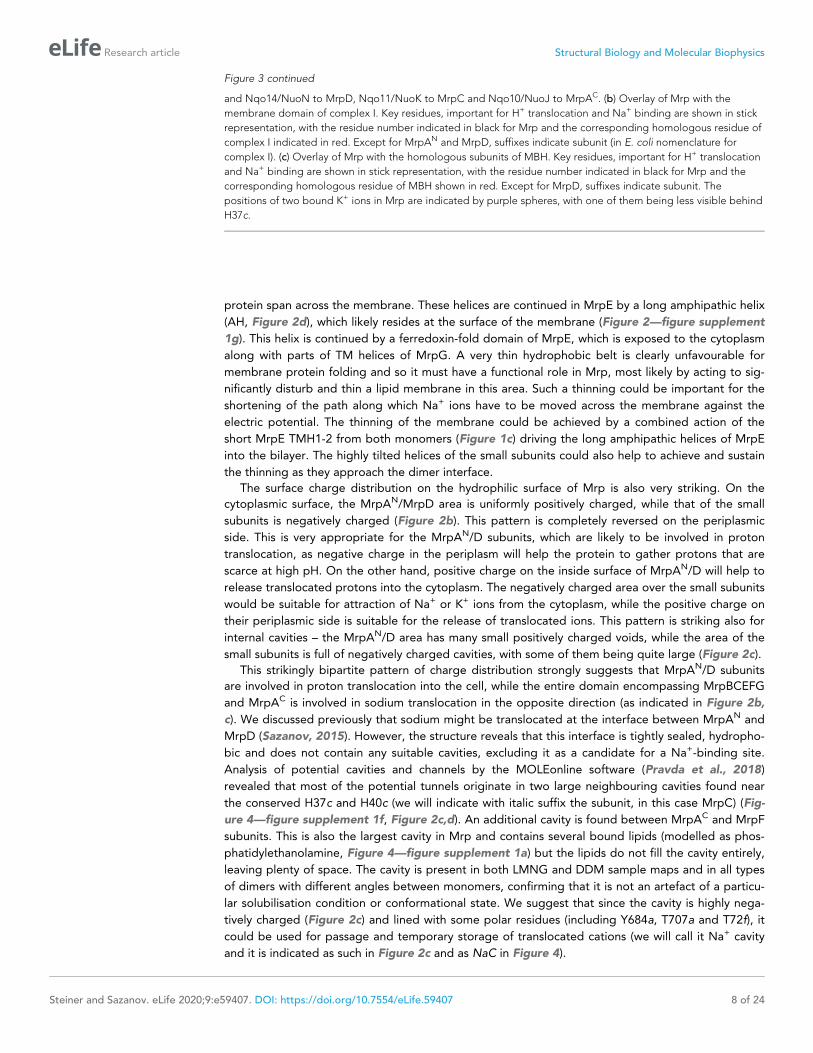
Figure 3. Overlay of Mrp with complex I and MBH complex. (a) The Mrp monomer is shown in the centre in side
view. Alignments to MBH are in the left column and to complex I in the right column. The top row shows view
from the cytosol, with the conserved domains underlined. The arrow indicates ‘swinging out’ of the MrpE TMH1-2
in comparison to MBH. The second row shows the side view, with Mrp subunits coloured as in the centre and
complex I/MBH in grey. In the bottom row only MBH and complex I are shown in the same orientation as above,
with subunits homologous to Mrp coloured as in Mrp and the rest grey. Quinone-binding subunit in complex I
(Nqo8/NuoH) is not present in Mrp and is highlighted in slate. Additional MrpD-like subunit in complex I (Nqo13/
NuoM) is in orange. MBH has its redox module attached to the Mrp-like domain on the opposite side compared
to complex I. MbhF is homologous to MrpB, MbhG to MrpC, MbhH to MrpD, MbhA to MrpE, MbhB to MrpF,
MbhC to MrpG and MbhD together with MbhE to MrpAC. Nqo12/NuoL is homologous to MrpAN, Nqo13/NuoM
Figure 3 continued on next page
Steiner and Sazanov. eLife 2020;9:e59407. DOI: https://doi.org/10.7554/eLife.59407 7 of 24
Research article Structural Biology and Molecular Biophysics
protein span across the membrane. These helices are continued in MrpE by a long amphipathic helix
(AH, Figure 2d), which likely resides at the surface of the membrane (Figure 2—figure supplement
1g). This helix is continued by a ferredoxin-fold domain of MrpE, which is exposed to the cytoplasm
along with parts of TM helices of MrpG. A very thin hydrophobic belt is clearly unfavourable for
membrane protein folding and so it must have a functional role in Mrp, most likely by acting to sig-
nificantly disturb and thin a lipid membrane in this area. Such a thinning could be important for the
shortening of the path along which Na+ ions have to be moved across the membrane against the
electric potential. The thinning of the membrane could be achieved by a combined action of the
short MrpE TMH1-2 from both monomers (Figure 1c) driving the long amphipathic helices of MrpE
into the bilayer. The highly tilted helices of the small subunits could also help to achieve and sustain
the thinning as they approach the dimer interface.
The surface charge distribution on the hydrophilic surface of Mrp is also very striking. On the
cytoplasmic surface, the MrpAN/MrpD area is uniformly positively charged, while that of the small
subunits is negatively charged (Figure 2b). This pattern is completely reversed on the periplasmic
side. This is very appropriate for the MrpAN/D subunits, which are likely to be involved in proton
translocation, as negative charge in the periplasm will help the protein to gather protons that are
scarce at high pH. On the other hand, positive charge on the inside surface of MrpAN/D will help to
release translocated protons into the cytoplasm. The negatively charged area over the small subunits
would be suitable for attraction of Na+ or K+ ions from the cytoplasm, while the positive charge on
their periplasmic side is suitable for the release of translocated ions. This pattern is striking also for
internal cavities – the MrpAN/D area has many small positively charged voids, while the area of the
small subunits is full of negatively charged cavities, with some of them being quite large (Figure 2c).
This strikingly bipartite pattern of charge distribution strongly suggests that MrpAN/D subunits
are involved in proton translocation into the cell, while the entire domain encompassing MrpBCEFG
and MrpAC is involved in sodium translocation in the opposite direction (as indicated in Figure 2b,
c). We discussed previously that sodium might be translocated at the interface between MrpAN and
MrpD (Sazanov, 2015). However, the structure reveals that this interface is tightly sealed, hydropho-
bic and does not contain any suitable cavities, excluding it as a candidate for a Na+-binding site.
Analysis of potential cavities and channels by the MOLEonline software (Pravda et al., 2018)
revealed that most of the potential tunnels originate in two large neighbouring cavities found near
the conserved H37c and H40c (we will indicate with italic suffix the subunit, in this case MrpC) (Fig-
ure 4—figure supplement 1f, Figure 2c,d). An additional cavity is found between MrpAC and MrpF
subunits. This is also the largest cavity in Mrp and contains several bound lipids (modelled as phos-
phatidylethanolamine, Figure 4—figure supplement 1a) but the lipids do not fill the cavity entirely,
leaving plenty of space. The cavity is present in both LMNG and DDM sample maps and in all types
of dimers with different angles between monomers, confirming that it is not an artefact of a particu-
lar solubilisation condition or conformational state. We suggest that since the cavity is highly nega-
tively charged (Figure 2c) and lined with some polar residues (including Y684a, T707a and T72f), it
could be used for passage and temporary storage of translocated cations (we will call it Na+ cavity
and it is indicated as such in Figure 2c and as NaC in Figure 4).
Figure 3 continued
and Nqo14/NuoN to MrpD, Nqo11/NuoK to MrpC and Nqo10/NuoJ to MrpAC. (b) Overlay of Mrp with the
membrane domain of complex I. Key residues, important for H+ translocation and Na+ binding are shown in stick
representation, with the residue number indicated in black for Mrp and the corresponding homologous residue of
complex I indicated in red. Except for MrpAN and MrpD, suffixes indicate subunit (in E. coli nomenclature for
complex I). (c) Overlay of Mrp with the homologous subunits of MBH. Key residues, important for H+ translocation
and Na+ binding are shown in stick representation, with the residue number indicated in black for Mrp and the
corresponding homologous residue of MBH shown in red. Except for MrpD, suffixes indicate subunit. The
positions of two bound K+ ions in Mrp are indicated by purple spheres, with one of them being less visible behind
H37c.
Steiner and Sazanov. eLife 2020;9:e59407. DOI: https://doi.org/10.7554/eLife.59407 8 of 24
Research article Structural Biology and Molecular Biophysics

Figure 4. Proton and cation translocation pathways. (a, b) View from the cytoplasm (a) and side view (b) with key residues proposed to be involved in
both pathways indicated. Waters predicted in Dowser software are shown as red spheres and the hydrogen bonds involving protonatable residues and
waters are shown as black dashes. Approximate pathways for H+ and Na+ translocation are indicated by arrows. Coupling points between MrpAN and
MrpD, and between MrpD and Na+-binding site are indicated by dashed ovals. (c) Details of Na+ pathway. The two experimentally identified bound K+
Figure 4 continued on next page
Steiner and Sazanov. eLife 2020;9:e59407. DOI: https://doi.org/10.7554/eLife.59407 9 of 24
Research article Structural Biology and Molecular Biophysics

One of the prominent tunnels identified by MOLE (red in Figure 2d) originates near H37c, passes
by the key E706a, through the Na+ cavity and then near the conserved D35f before exiting into the
cytoplasm. It is negatively charged at the origin, as would be appropriate for Na+, but has some nar-
row passages of about 1 A radius (Figure 4—figure supplement 1d), so the access must be regu-
lated. Another tunnel (orange in Figure 2d) originates in the cavity near H40c, which is also lined by
the key E137d (GluTMH5) and continues towards the periplasm at the interface between MrpD and
MrpC subunits (Figure 4—figure supplement 1e). Since H37c and H40c cavities are linked, these
two tunnels can form a channel for Na+ translocation across the membrane, with the passage
through the narrow restrictions regulated as part of the coupling mechanism.
Proton translocation pathwaysA striking pattern of charge distribution and conservation of the fold and key residues between pro-
ton-pumping complex I and Mrp (Figure 4—figure supplement 2b) leaves no doubt that proton
translocation channels are found in MrpAN and MrpD subunits, similarly to complex I and other
related redox proton pumps. High local resolution (~2.9 A) in the core of the structure allowed us to
model many bound water molecules (Figure 2—figure supplement 1a,f). They are found mostly on
the hydrophilic surfaces of the complex and along the entire central hydrophilic axis around key
charged residues, confirming the previously suggested hydration of the central axis
(Baradaran et al., 2013; Figure 4—figure supplement 1b). Since at 2.9 A resolution we are still lim-
ited in the identification of water molecules, for the analysis of the complete proton translocation
pathways we have modelled waters in Dowser (Zhang and Hermans, 1996; Figure 4b). Many exper-
imental and Dowser-predicted waters coincided, but some were identified only by Dowser. The anal-
ysis of connections between Grotthus-competent residues (K, H, E, D, T, S and Y [Khaniya et al.,
2020]) and waters (allowing for Grotthuss mechanism of proton transfer) revealed that the highly
hydrated cluster near MrpA LysTMH12 (K432) and LysTMH8 (K279), containing also conserved H273,
H369, and K377 (Figures 3b and 4b), is all interconnected and linked to the periplasm via the con-
served E433. The link to the cytoplasm is most likely along H273, which sits on TMH8. The potential
link is not continuous and must exist only temporarily during the catalytic cycle, allowing for protons
to be ejected into the cytoplasm. Analyses of MrpAN and MrpD and comparisons with known
complex I structures suggest that in APLS, links to the cytoplasm are achieved not along the centre
of the N-terminal half-channel as discussed originally (Efremov and Sazanov, 2011), but mostly
along TMH8, which has more polar residues in the area. TMH8 is suited to play a functional role due
to its p-bulge which is conserved in all APLS. The key LysTMH8 interacts with the backbone oxygen
of the p-bulge and is the only protonatable residue on TMH8 in MrpD and MrpD-like subunits of
complex I (Nqo13/NuoM/ND4 and Nqo14/NuoN/ND2), while in MrpA H273 is added to the p-bulge
and only this histidine is conserved in Nqo12/NuoL/ND5, presumably replacing LysTMH8.
The area between LysTMH12 (K392) and LysTMH8 (K250) in MrpD is also highly hydrated and
interlinked with participation of D333 and K337, which are replaced by one histidine in complex I
Nqo13 and Nqo14 subunits (Figure 3b). However, in contrast to MrpAN, LysTMH12d does not seem
to be linked to the periplasm due to the lack of polar residues and waters (either experimental or
Dowser-modelled) in this area. An analogous situation appears to exist in complex I, where only the
MrpA-like subunit Nqo12/NuoL/ND5 is clearly linked to the periplasm in nearly identical to MrpA
arrangement (Figure 3b), while Nqo13 and Nqo14 apparently lack such links, as similar analysis
shows. Therefore, it is possible that from the periplasm side all the protons enter the Mrp complex
Figure 4 continued
ions are shown as violet spheres. The key distance between one of the ions and GluTMH5d in the Na+/H+ coupling point is indicated. A large cavity
between MrpAC and MrpF is indicated as NaC.
The online version of this article includes the following figure supplement(s) for figure 4:
Figure supplement 1. Bound lipids, waters and ions; Na+ pathway.
Figure supplement 2. Conservation patterns.
Figure supplement 3. Sequence alignment of Mrp subunits.
Figure supplement 4. Sequence alignment of Mrp subunits.
Steiner and Sazanov. eLife 2020;9:e59407. DOI: https://doi.org/10.7554/eLife.59407 10 of 24
Research article Structural Biology and Molecular Biophysics

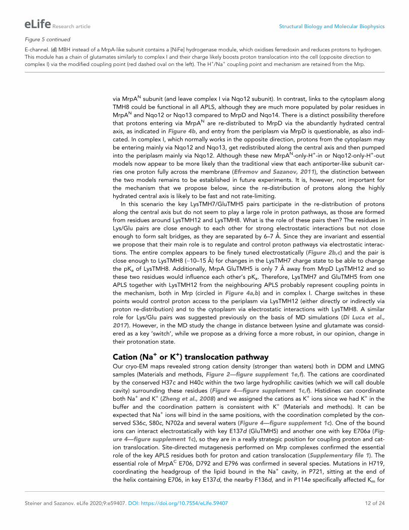
Figure 5. The antiport mechanism of the Mrp complex and its conservation in complex I and MBH. (a) A likely state of the Mrp complex in the current
structure, as a part of the catalytic cycle. Key glutamates from the central axis are shown as red circles and key lysines as blue circles, with charged state
depicted by filled circle and neutral state by empty circle. Key helices where these residues sit are numbered and shown as broken where applicable.
Coupling points are indicated by red dashed ovals. Electrostatic interactions are indicated as thick dashed lines. Possible lateral re-distribution of
protons between MrpA and MrpD is indicated as alternative to the direct entry of protons to MrpD from the periplasm. Na+ ion (blue) is bound in the
coupling point and the large Na+ cavity mid-pathway is indicated as an empty oval. A sequence of events leading up to this state is described in the
text. (b) An alternative state of the Mrp complex as another part of the catalytic cycle. Na+ ion is expelled into the periplasm and coupling points
become neutral. (c) Complex I contains one additional MrpD-like subunit (orange, Nqo13/NuoM), with the change of LysTMH12 to glutamate. The
coupling points between the APLS are conserved and the coupling point with Na+ is replaced by coupling to the charge of glutamates in the
Figure 5 continued on next page
Steiner and Sazanov. eLife 2020;9:e59407. DOI: https://doi.org/10.7554/eLife.59407 11 of 24
Research article Structural Biology and Molecular Biophysics
via MrpAN subunit (and leave complex I via Nqo12 subunit). In contrast, links to the cytoplasm along
TMH8 could be functional in all APLS, although they are much more populated by polar residues in
MrpAN and Nqo12 or Nqo13 compared to MrpD and Nqo14. There is a distinct possibility therefore
that protons entering via MrpAN are re-distributed to MrpD via the abundantly hydrated central
axis, as indicated in Figure 4b, and entry from the periplasm via MrpD is questionable, as also indi-
cated. In complex I, which normally works in the opposite direction, protons from the cytoplasm may
be entering mainly via Nqo12 and Nqo13, get redistributed along the central axis and then pumped
into the periplasm mainly via Nqo12. Although these new MrpAN-only-H+-in or Nqo12-only-H+-out
models now appear to be more likely than the traditional view that each antiporter-like subunit car-
ries one proton fully across the membrane (Efremov and Sazanov, 2011), the distinction between
the two models remains to be established in future experiments. It is, however, not important for
the mechanism that we propose below, since the re-distribution of protons along the highly
hydrated central axis is likely to be fast and not rate-limiting.
In this scenario the key LysTMH7/GluTMH5 pairs participate in the re-distribution of protons
along the central axis but do not seem to play a large role in proton pathways, as those are formed
from residues around LysTMH12 and LysTMH8. What is the role of these pairs then? The residues in
Lys/Glu pairs are close enough to each other for strong electrostatic interactions but not close
enough to form salt bridges, as they are separated by 6–7 A. Since they are invariant and essential
we propose that their main role is to regulate and control proton pathways via electrostatic interac-
tions. The entire complex appears to be finely tuned electrostatically (Figure 2b,c) and the pair is
close enough to LysTMH8 (~10–15 A) for changes in the LysTMH7 charge state to be able to change
the pKa of LysTMH8. Additionally, MrpA GluTMH5 is only 7 A away from MrpD LysTMH12 and so
these two residues would influence each other’s pKa. Therefore, LysTMH7 and GluTMH5 from one
APLS together with LysTMH12 from the neighbouring APLS probably represent coupling points in
the mechanism, both in Mrp (circled in Figure 4a,b) and in complex I. Charge switches in these
points would control proton access to the periplasm via LysTMH12 (either directly or indirectly via
proton re-distribution) and to the cytoplasm via electrostatic interactions with LysTMH8. A similar
role for Lys/Glu pairs was suggested previously on the basis of MD simulations (Di Luca et al.,
2017). However, in the MD study the change in distance between lysine and glutamate was consid-
ered as a key ‘switch’, while we propose as a driving force a more robust, in our opinion, change in
their protonation state.
Cation (Na+ or K+) translocation pathwayOur cryo-EM maps revealed strong cation density (stronger than waters) both in DDM and LMNG
samples (Materials and methods, Figure 2—figure supplement 1e,f). The cations are coordinated
by the conserved H37c and H40c within the two large hydrophilic cavities (which we will call double
cavity) surrounding these residues (Figure 4—figure supplement 1c,f). Histidines can coordinate
both Na+ and K+ (Zheng et al., 2008) and we assigned the cations as K+ ions since we had K+ in the
buffer and the coordination pattern is consistent with K+ (Materials and methods). It can be
expected that Na+ ions will bind in the same positions, with the coordination completed by the con-
served S36c, S80c, N702a and several waters (Figure 4—figure supplement 1c). One of the bound
ions can interact electrostatically with key E137d (GluTMH5) and another one with key E706a (Fig-
ure 4—figure supplement 1c), so they are in a really strategic position for coupling proton and cat-
ion translocation. Site-directed mutagenesis performed on Mrp complexes confirmed the essential
role of the key APLS residues both for proton and cation translocation (Supplementary file 1). The
essential role of MrpAC E706, D792 and E796 was confirmed in several species. Mutations in H719,
coordinating the headgroup of the lipid bound in the Na+ cavity, in P721, sitting at the end of
the helix containing E706, in key E137d, the nearby F136d, and in P114e specifically affected Km for
Figure 5 continued
E-channel. (d) MBH instead of a MrpA-like subunit contains a [NiFe] hydrogenase module, which oxidises ferredoxin and reduces protons to hydrogen.
This module has a chain of glutamates similarly to complex I and their charge likely boosts proton translocation into the cell (opposite direction to
complex I) via the modified coupling point (red dashed oval on the left). The H+/Na+ coupling point and mechanism are retained from the Mrp.
Steiner and Sazanov. eLife 2020;9:e59407. DOI: https://doi.org/10.7554/eLife.59407 12 of 24
Research article Structural Biology and Molecular Biophysics

Na+, indicating a role for these residues in the Na+ pathway. P114e sits on a loop in the ferredoxin-
like domain of MrpE, which also contains H131e near H32g. These conserved histidines may bind
Na+ in the part of MrpE/G near the AH helix. This area at the tip of the monomer, where the mem-
brane is likely to be thinned, is as highly conserved as the core of APLS, attesting to its functional
importance (Figure 4—figure supplement 2a). It is also the most negatively charged area on the
cytoplasmic surface (Figure 2b) and is likely to represent the entry point for cations. Conserved D29f
is one of the residues responsible for the negative charge here and its mutation to alanine
completely abolished the activity (Morino et al., 2010).
On the basis of these considerations and according to the analysis of cavities and tunnels by
MOLE as described above, we propose that the Na+ pathway (which applies also to K+) starts near
H42g/H131e, passes by the highly conserved D35f, traverses the large Na+ cavity between MrpF
and MrpAC, passes by the key E706a and enters the double cavity where at least two cations, as we
observe, can be coordinated between E706a, H37c, H40c and E137d (Figure 4c, Figure 4—figure
supplement 1c,f). The double cavity most likely represents a point of coupling between proton and
cation translocation (circled in Figure 4c), as here Na+ ion(s) sit in a position analogous to the
LysTMH12 in APLS coupling points. This way, the proton translocation through MrpD (already cou-
pled to MrpAN via K248a/E167a/K392d coupling point) can directly be coupled to Na+ translocation.
The Na+ path continues towards the periplasm at the interface between TMH5d, TMH3c and
TMH21a, ending with the key E796a and D792a at the exit into the periplasm (Figure 4c). T75c is
facing E796a and is essential for growth in high salt (Supplementary file 1).
The role of conserved D35f was not clear from previous mutations in Bacillus subtilis Mrp, as sub-
stitutions were highly detrimental when the complex was expressed in E. coli (perhaps the assembly
of the complex was affected) but not so in B. subtilis itself (Kajiyama et al., 2009). However, in B.
subtilis only conservative mutations to polar residues E and N were explored and so the effects
could be expected to be mild. To clarify this, we generated mutants of A. flavithermus Mrp complex
by mutating D35f to L, a hydrophobic residue of roughly similar size to D, and expressed the com-
plex in E. coli similarly to WT Mrp. The mutation completely deactivated the complex as shown by
the absence of growth at high NaCl concentrations (Figure 1—figure supplement 1j). The dimer
seemed to be de-stabilised as well, although the monomer was fully assembled (Figure 1—figure
supplement 1d,e). To try to clarify how important the dimer is for the function, we generated MrpE
L41W and MrpG S72W mutants. They were chosen because there are no salt bridges linking the
monomers but these residues form close contacts between the two monomers and we expected
that an introduction of bulky tryptophanes may disrupt the dimer. This, however, did not happen
and the activity was not affected (Figure 1—figure supplement 1d,e,j). Therefore, the dimer can
withstand some perturbations but it may not be absolutely essential for the mechanism per se, as a
very similar fold exists as a monomer in MBH. The role of the Mrp dimer may have more to do with
the stabilisation of the unfavourable membrane-thinning fold as discussed above. Nevertheless, our
data suggests that invariably conserved D35f is important for activity, consistent with its position on
the Na+ path (Figure 4c). Furthermore, this aspartate is found right opposite the invariable and
essential for activity (Morino et al., 2010) P86g, which breaks TMH3 of MrpG in half and bends it at
a point where this helix contacts the MrpE ferredoxin-fold domain with its essential P114. Such archi-
tecture suggests that conformational interactions are important in this area, perhaps as a part of gat-
ing for Na+ entry and exit.
Summing up, surface and cavity charge distribution, analysis of channels and cavities, patterns of
sequence conservation and mutagenesis results, cryo-EM density for bound cations and basic mech-
anistic considerations all overwhelmingly support the Na+ path as depicted in Figure 4b,c. The key
residues are arranged as a kind of ladder descending from the cytoplasm to the periplasm along the
repeating pattern of the three-helix fold of the small subunits. In addition to the already discussed
key residues, many further, mostly conserved, polar residues line the path all along the way, as
expected for a cation pathway (Figure 4—figure supplement 1f). The path differs from the proposal
for the MBH complex, where the suggested entry point roughly coincided with our view (around
D35f), while the exit was proposed to be directly ‘below’, near D59f (D59b in MBH) (Yu et al.,
2018). However, the area around D59f (not universally conserved residue) is exposed to the peri-
plasm and is separated from D35f by several layers of highly hydrophobic residues, therefore this
proposal for the Na+ path is extremely unlikely. Instead, we propose that the Na+ pathway in MBH
is the same as in Mrp, as all the key residues and fold are very well conserved (Figure 3c). The
Steiner and Sazanov. eLife 2020;9:e59407. DOI: https://doi.org/10.7554/eLife.59407 13 of 24
Research article Structural Biology and Molecular Biophysics

double cavity in the Na+/H+ coupling point is of similar size and also negatively charged in MBH.
The only difference is that H40c from Mrp is replaced by D37g in MBH, a residue which is also capa-
ble of coordinating Na+. D37g, H41g, E69d, N34g and about six nearby serines are ideally arranged
in MBH to coordinate two cations similarly to Mrp (Figure 3c). In fact, in the deposited cryo-EM den-
sity of the MBH complex (EMD-7468) there is a clear density for a potential Na+ ion coordinated by
E69d (E706a homologue). The cavity between MbhB (MrpF) and MbhD (MrpAC) is also present but
is not as large as the Na+ cavity in Mrp, perhaps because of a lesser demand for temporary Na+ stor-
age in MBH. Residues that we propose to be involved in the coupling and exit points of the Na+
path (from E706a to D792a, Figure 3c) were instead suggested to be a part of an additional H+
translocation path in MBH, working in the opposite direction to MbhH/MrpD (Yu et al., 2018). This
is again very unlikely because the site in the coupling point is extremely well suited to be a Na+ bind-
ing site due to all the overwhelming evidence listed above. The main argument in support for an
additional H+ translocation path in MBH came from the similarity to complex I, where this pathway
(E-channel) is not well established and should be working in the same direction as MrpD/Nqo14 and
not in the opposite. This is hardly strong evidence, along with the absence of any credible exit point
into the cytoplasm, since the proposed direct path for H+ from the double cavity towards the cyto-
plasm (Yu et al., 2018) is blocked by several layers of hydrophobic residues both in MBH and Mrp.
In summary, we suggest that the Na+ path depicted in Figure 4 is common for Mrp and MBH and
that the additional H+ path proposed for MBH does not exist.
DiscussionImportantly, a striking electrostatic imbalance of H+ and Na+ modules of Mrp (Figure 2b,c), the
arrangement of key residues in the H+ and Na+ pathways, the separation of H+ cross-pathways from
the LysTMH7/GluTMH5 coupling points and a remarkable similarity of the MrpAN/MrpD and MrpD/
Na+ coupling points (Figure 4a,b) all collectively suggest that electrostatic interactions are the main
driving force in the antiport mechanism.
Overall however, the electrostatic forces are likely to be amplified by coordinated conformational
changes, because most of the key residues (including all four LysTMH8 and LysTMH12) sit on breaks
of TM helices and interact with exposed backbone oxygen atoms from such breaks, so even small
movements of these helices will affect each other and pKa’s of these residues. Therefore, the Mrp
complex is likely to exist in two different conformational states, one of which is conductive to Na+
binding from the cytoplasm and another is associated with Na+ release into the periplasm. However,
the difference between the states can be really minor, because changes in pKa’s can be associated
with only small changes in the local environment. In order to ascertain the charge state of the key
residues in our current structure, we analysed PROPKA (Bas et al., 2008) predictions and the
appearance of cryo-EM density, especially of E/D residues, since due to radiation damage carboxy-
lates lose side-chain density in cryo-EM when they are in a charged state (but not if in a salt bridge)
(Baker and Rubinstein, 2010; Grant and Grigorieff, 2015). These analyses suggest that both in
MrpAN and MrpD LysTMH7/GluTMH5 pairs have both residues charged, LysTMH12 is charged and
LysTMH8 is probably neutral. We also know that two cations (K+ in our structure but usually Na+ in
vivo) are bound in the double cavity in the H+/Na+ coupling point, with one of them likely to be des-
tined for translocation into the periplasm, meaning that there is one additional positive charge due
to the bound cation (indicated as a filled blue circle and Na+ in Figure 5).
These considerations allow us to arrive at a model for the Mrp state in the current structure as
depicted in Figure 5a and a mechanism as follows. The distribution of charges in Figure 5a would
result from Na+ charge effectively lowering the pKa of MrpD GluTMH5 (7 A away, Figure 4—figure
supplement 1c), so that it donates its proton to MrpD LysTMH7 (6 A away). At the same time both
LysTMH12 would have been protonated from the periplasm. Positive charge on MrpD LysTMH12
lowers the pKa of MrpA GluTMH5 so that it donates its proton to MrpA LysTMH7, mirroring events
in MrpD. Assuming LysTMH8 was protonated before, now the positive charge both on LysTMH7
and LysTMH12 will force it to lose the proton to the cytoplasm both in MrpAN and MrpD, giving us
the state in Figure 5a. Next, if Na+ is translocated towards the periplasm, MrpD LysTMH7/GluTMH5
pair will become neutral, as Na+ charge has been removed and the proton from LysTMH7 can go
back to GluTMH5. The absence of positive charge on LysTMH7 and the corresponding change in
charge balance will now allow for MrpD LysTMH8 to be protonated by LysTMH12. In its turn, the
Steiner and Sazanov. eLife 2020;9:e59407. DOI: https://doi.org/10.7554/eLife.59407 14 of 24
Research article Structural Biology and Molecular Biophysics

absence of charge on MrpD LysTMH12 will allow for the MrpA LysTMH7/GluTMH5 pair to become
neutral, and then for MrpA LysTMH8 to become protonated by MrpA LysTMH12 in a similar
sequence of events, with the system arriving to the state depicted in Figure 5b. The system is now
reset, and the arrival of another Na+ ion from the cytoplasm to the coupling site will set events in
motion leading to state in Figure 1a, accompanied by the translocation of two protons into the cyto-
plasm. Here we discussed how Na+ ion would drive proton translocation, but of course the reverse
cycle is also applicable, and so proton translocation into the cell, driven by D, would result in Na+
being pumped out.
We believe that this mechanism explains all the properties of Mrp-catalysed antiport on the basis
of prominent structural features. The ability to couple the translocation of two H+ inside the cell in
exchange for one Na+ moving outside is achieved by the inter-MrpAN-MrpD coupling point. A simi-
lar coupling principle, but with a cation binding site instead of LysTMH12 and an opposing direction-
ality, is employed at the MrpD-MrpG interface. Coordinating/amplifying conformational interactions
are possible because a break in TMH7 directly contacts a break in TMH8, while the loop from
the TMH12 break directly contacts GluTMH5 from neighbouring APLS. Helix HL connects TMH7s on
the cytoplasmic side (Figure 2d) and additionally, striking b-hairpin (bH) elements connect APLS on
the periplasmic side both in Mrp and in complex I. Links to bH both of TMH5 (D157a and D128d)
and TMH8 (D104a and D75d) are critical for activity (Supplementary file 1). Finally, a striking tilt of
TM helices in the Na+-translocating part of the complex as compared to the H+-translocating
domain (Figure 2d) might have a role in coupling if helices change the tilt during the catalytic cycle.
This two-APLS unit can be elegantly extended to three (Sazanov, 2015) or four (Chadwick et al.,
2018) APLS in complex I-like enzymes or shortened to one in MBH (or FHL-1 or Ech enzymes
[Efremov and Sazanov, 2012]), with the same mechanism applicable. The main difference to Mrp
would be that in complex I (or membrane-bound hydrogenases [Efremov and Sazanov, 2012])
instead of a positive charge of Na+, the coupling is achieved with a net negative charge being pro-
duced as two protons are abstracted during electron transfer and reduction of quinone to quinol or
reduction of protons to hydrogen gas. The linking of three APLS units in complex I allows for the
translocation of three protons in a similar to Mrp mechanism, while the fourth proton is likely translo-
cated via the E-channel, as depicted in Figure 5c. In complex I the additional APLS subunit Nqo13/
NuoM is unique as it has a conserved GluTMH12 instead of a lysine, which may allow this subunit to
operate in an anti-phase with the other two APLS. Such a feature may be important to prevent
excessive build-up of electrostatic imbalance in the membrane domain of complex I during the cata-
lytic cycle. The E-channel structurally coincides with the exit path for Na+ (Figure 3b) while the entry
part is not homologous and is replaced in complex I with many glutamates (hence the E-channel)
leading on towards the quinone-binding site as a putative proton ‘relay’.
In MBH the redox [NiFe]-hydrogenase module is attached to a single APLS unit from the opposite
site as compared to complex I (Figure 3a). As noted above, the additional H+ pathway proposed for
MBH (Yu et al., 2018) is very unlikely from structural considerations, and in any case it would lead to
a futile cycle of protons coming in via MbhH and exiting via this additional pathway. It was discussed
previously that the hydrogenase module of MBH evolves H2 and generates a proton gradient,
whereas the Mrp module transforms it into a Na+ gradient (McTernan et al., 2014). We suggest
that the Na+ pathway in MBH is the same as we propose for Mrp, while the energy of the redox
reaction (which is quite small for MBH with DE = 60 mV [Yu et al., 2018]) is used to boost the trans-
location of a proton into the cell via MbhH, a standard APLS with all the key residues conserved
(Figure 3c). The reversal of the redox module compared to complex I would then make complete
sense because the redox energy would be used in MBH to drive proton translocation in the opposite
direction as compared to complex I or FHL-type hydrogenases (Efremov and Sazanov, 2012), and
so from the electrostatics point of view the charge would need to be delivered to APLS from the
opposite side. Thus, instead of two APLS units driving Na+ in Mrp, in MBH one APLS unit supported
by the redox reaction would drive Na+ in a similar to Mrp mechanism (Figure 5d).
In conclusion, the Mrp structure revealed basic operating principles of this ancient antiport sys-
tem, which forms the basis of a huge variety of modern redox proton and sodium pumps. The mech-
anism that we propose, based on electrostatics and supported by conformational interactions, is
likely to be applicable to all members of this group.
Steiner and Sazanov. eLife 2020;9:e59407. DOI: https://doi.org/10.7554/eLife.59407 15 of 24
Research article Structural Biology and Molecular Biophysics

Materials and methods
Key resources table
Reagent type(species) or resource Designation Source or reference Identifiers Additional information
Gene(Thermus thermophilus)
HB8,ATCC 27634,DSM 579
Uniprot Nqo12: Q56227Nqo13: Q56228Nqo14: Q56229
Multiple sequencealignments
Gene(Pyrococcus furiosus)
COM1,DSM 3638
pdbFASTA sequence
6CFW Multiple sequencealignments
Gene(Bacillus pseudofirmus)
Strain OF4 Uniprot MrpA: Q9RGZ5MrpB: Q9RGZ4MrpC: Q9RGZ3MrpD: Q9RGZ2MrpE: Q9RGZ1MrpF: Q9RGZ0MrpG: Q9RGY9
Multiple sequencealignments
Gene(Bacillus subtilis)
168 Uniprot MrpA: Q9K2S2MrpB: O05259MrpC: O05260MrpD: O05229MrpE: Q7WY60MrpF: O05228MrpG: O05227
Multiple sequencealignments
Gene(Staphylococcus aureus)
1280 Uniprot MrpA: Q9ZNG6MrpB: P60678MrpC: P60682MrpD: P60686MrpE: P60690MrpF: P60694MrpG: P60698
Multiple sequencealignments
Gene(Thermosynechococcuselongatus)
BP-1 Uniprot NdhF1: Q8DKX9NdhD1: Q8DKY0
Multiiple sequencealignments
Sequence-based reagent
MrpF D35L This paper PCR primers Forward: CGCTTATTTTTTACTATATATGTTGAAAAAAAATGAAACReverse: GCAAGCGTAATGCCCATCGCTAAGAGCGCGATAATACGATCCGForward: CGGATCGTATTATCGCGCTCTTAGCGATGGGCATTACGCTTGCReverse: ATATAGTAAAAAATAAGCG
Sequence-based reagent
MrpG S72W This paper PCR primers Forward: CGCTTATTTTTTACTATATATGTTGAAAAAAAATGAAACReverse: CACGATGCCAAGCAATAGACGCCAGTTGAAATGGTTATTTTCAATGForward: CATTGAAAATAACCATTTCAACTGGCGTCTATTGCTTGGCATCGTGReverse: ATATAGTAAAAAATAAGCG
Sequence-based reagent
MrpE L41W This paper PCR primers Forward: CGCTTATTTTTTACTATATATGTTGAAAAAAAATGAAACReverse: GCGCGAATGGAAAAAGCGACGCCATATAAAAAGAATAAACAGCCCGATCATGTACForward: GTACATGATCGGGCTGTTTATTCTTTTTATATGGCGTCGCTTTTTCCATTCGCGCReverse: ATATAGTAAAAAATAAGCG
Strain, strainbackground(Escherichia coli)
KNabc doi:10.1073/pnas.84.9.2615 Expression andassay strain
Continued on next page
Steiner and Sazanov. eLife 2020;9:e59407. DOI: https://doi.org/10.7554/eLife.59407 16 of 24
Research article Structural Biology and Molecular Biophysics

Continued
Reagent type(species) or resource Designation Source or reference Identifiers Additional information
Transfected construct(Anoxybacillusflavithermus)
DSM 21510/WK1 Other UniprotMrpA: B7GL84MrpB: B7GL83MrpC: B7GL82MrpD: B7GL98MrpE: B7GL97MrpF: B7GL96MrpG: B7GIG3
Prof. Masahiro Ito(Graduate School of LifeSciences, ToyoUniversity, Japan)
Software, algorithm SerialEM doi:10.1016/j.jsb.2005.07.007 Data Acquisition Softwarefor a variety of data fromelectron microscopes
Software, algorithm Relion doi:10.1016/j.jsb.2012.09.006 Cryo-EM processingsoftware
Software, algorithm CTFFIND4 doi:10.1016/j.jsb.2015.08.008 Defocus estimationsoftware
Software, algorithm Gctf doi:10.1016/j.jsb.2015.11.003 Per-particle CTFestimation software
Software, algorithm USCF Chimera doi:10.1002/jcc.20084 Visualisation software ofmolecular structures andcryo-EM maps
Software, algorithm Coot doi:10.1107/S0907444904019158 Software formodel building
Software, algorithm PHENIX doi:10.1107/S0907444909052925 Structurerefinement software
Software, algorithm MotionCor2 doi:10.1038/nmeth.4193 Whole frame imagemotion correctionsoftware
Expression and purification of MrpPlasmid DNA, termed AF_Mrp, encoding Mrp from thermophilic Anoxybacillus flavithermus WK1
with a C-terminal His-tag on MrpG was kindly provided by Prof. Masahiro Ito (Graduate School of
Life Sciences, Toyo University, Ouragun, Gunma 374–0193, Japan). AF_Mrp was expressed in 50
litres of LBK media (1% tryptone, 0.5% yeast extract, 83 mM KCl, pH 7.5) supplemented with 100
mg/mL ampicillin, 25 mg/mL kanamycin and antifoam using a fermenter. The bacterial culture was
grown for 16–17 hr at 37˚C, while keeping at constant pH of 7.3. The agitation was adjusted to keep
the dissolved oxygen concentration between 2% - 10%. The next day, the cells were harvested by
centrifugation (5000 x g for 30 min at 4˚C) and the cell pellets were stored at �80˚C.
Preparing membranes from E. coli KNabcFrozen cell pellets were thawed in ice-cold water and resuspended in 20 mM HEPES-KOH pH 7.0, 5
mM MgCl2 and 10% glycerol. The cells were homogenized twice at 30,000 psi, using a high-pressure
cell disruption Constant System pressure cell TS 1.1. DNases I (0.3 mg/ml) and proteinase inhibitor
cocktail (5 tablets of EDTA-free Complete Ultra inhibitor [Roche]) were added to the lysate. The
lysate was clarified by centrifugation at 26,000 x g for 30 min at 4˚C. Membranes were obtained by
ultracentrifugation of the supernatant at 180,000 x g for 2 hr at 4˚C. Pelleted membrane fractions
were resuspended in 20 mM HEPES-KOH pH 7.0, 5 mM MgCl2, 20% glycerol and stored at �80˚C.
Membrane solubilisationMembranes (10 mg/ml) were thawed in ice-cold water and solubilized by incubation for 1 hr at 4˚C
in 20 mM HEPES-KOH pH 7.0, 5 mM MgCl2, 10% glycerol to which 0.3 M KCl and 1% (w/v) Lauryl
Maltose Neopentyl Glycol (LMNG) or n-Dodecyl b-D-maltoside (DDM) had been added. After ultra-
centrifugation at 180,000 x g for 15 min, the supernatant was diluted two-fold with 20 mM HEPES-
KOH pH 7.0, 5 mM MgCl2, 10% glycerol, 0.3 M KCl and imidazole was added up to a concentration
of 20 mM.
Steiner and Sazanov. eLife 2020;9:e59407. DOI: https://doi.org/10.7554/eLife.59407 17 of 24
Research article Structural Biology and Molecular Biophysics

PurificationSolubilised membranes were loaded onto a 5 ml TALON (GE Healthcare) column, previously equili-
brated with five column volumes (CV) of 20 mM HEPES-KOH pH 7.0, 5 mM MgCl2, 10% glycerol, 0.3
M KCl, 20 mM imidazole and 0.05% LMNG or 0.05% DDM. The column was washed with 15 CV of
equilibration buffer and the protein was eluted with 10 CV 20 mM HEPES-KOH pH 7.0, 5 mM
MgCl2, 10% glycerol, 0.3 M KCl, 200 mM imidazole and 0.05% LMNG or 0.05% DDM. Fractions con-
taining Mrp were pooled and applied onto a Superose 6 10/300 GL (GE Healthcare) previously equil-
ibrated with 2 CV of 20 mM Bis-Tris pH 6.0, 5 mM MgCl2, 0.15 M KCl and 0.05% LMNG or 0.05%
DDM, and eluted isocratically. The dimer eluted at around 12.5 ml and was diluted to 0.16 mg ml�1
for grid preparation. Purification of the Mrp complex was performed more than five times, each time
from a different batch of cells, and all attempts of replication were successful.
Electron microscopyCopper grids (Quantifoil mesh 300, R 0.6/1) were covered with a 1.2 nm thin layer of continuous car-
bon. Grids were glow discharged in air at 30 mA for 5 s. 3 mL of protein sample were applied to the
grids, blotted for 7 s at 4˚C and 100% humidity and quickly plunged into liquid ethane using a FEI
Vitrobot IV. Grids were stored in liquid nitrogen. Images were collected at 35˚ tilt using a 300 kV
Titan Krios electron microscope equipped with a Gatan K3 camera and an energy filter set to a slit
width of 20 eV at the Institute of Science and Technology Austria. Micrographs were collected with
the FEI EPU package for the LMNG dataset or SerialEM for the DDM dataset, at a nominal magnifi-
cation of 105,000 x, resulting in a calibrated physical pixel size of 0.84 A per pixel. Defocus values
varied from 0.6 mm to 2.3 mm. A total dose of 85 e-/A2 was fractionated into 85 frames for the
LMNG-dataset. A total dose of 90 e-/A2 was fractionated into 88 frames for the DDM-dataset.
Image processing of the LMNG-dataset760 and 3126 movies were collected in normal- and super-resolution respectively. Processing was
done in Relion 3.0.7 (Scheres, 2012). Movie frames were motion-corrected, dose-weighted and
super-resolution images were binned two-fold using MotionCor2 (Zheng et al., 2017). Contrast
transfer function (CTF) parameters were determined for aligned micrographs using
CTFFIND4 (Rohou and Grigorieff, 2015). After a manual inspection of the Thon rings, bad micro-
graphs showing ice rings were excluded from further analysis, yielding 755 good micrographs for
normal resolution and 3096 for super resolution movies. Auto-picking with 3D references, which
were the 3D auto-refined dimer map from the DDM dataset in C2 symmetry low-pass-filtered to 30
A, resulted in 198565 particles for normal resolution and 693504 particles for super resolution micro-
graphs. The coordinates of the particles were then used for a per-particle CTF estimation using
Gctf (Zhang, 2016). Star files from normal and super-resolution micrographs were merged after Gctf
estimation. At this stage and at all stages during the entire processing of both LMNG and DDM
datasets, when particles were re-extracted after classification or refinement, the duplicates were
removed using 100 A minimum inter-particle distance. 2D classification was attempted but it did not
improve the results. 3D classification of all picked particles, extracted in a 256 pixel box (down-sam-
pled to 1.68 A pixel) was carried out in C1 symmetry with a 3D auto-refined dimer map from the
DDM dataset as initial reference, filtered to 30 A. This resulted in four good classes with 606671 par-
ticles. The best class was selected yielding 264961 particles. Particles were re-extracted in a 512
pixel box (0.84 A pixel). An initial 3D auto-refinement in C2, with local angular searches and a 3D
auto-refined dimer map from the DDM dataset filtered to 30 A as a reference resulted in a map with
an overall resolution of 5.5 A.
MonomerThe resolution of the dimer map was limited due to variable angle between the monomers, resulting
in the loss of true C2 symmetry for the entire particle pool. The particles were therefore symmetry-
expanded according to the C2 point group, meaning that the particle number was enlarged twice
because each dimer particle produced two monomer particles. Particles were re-extracted with re-
centring on a monomer in a 512 pixel box (0.84 A pixel). Removal of duplicates during re-extraction
resulted in the loss of a few particles, which were probably coming from two neighboring dimers,
and so the final number of particles is slightly less than double. 3D classification of these monomers,
Steiner and Sazanov. eLife 2020;9:e59407. DOI: https://doi.org/10.7554/eLife.59407 18 of 24
Research article Structural Biology and Molecular Biophysics

with local angular searches and a monomer map (excised in Chimera from the auto-refined dimer
map and filtered to 8 A) as a reference resulted in two good classes, with combined 285688 par-
ticles. These particles were re-extracted in a 512 pixel box (0.84 A pixel) for masked 3D auto-refine-
ment in C1, with local searches and the same monomer map as during 3D classification as a
reference, which resulted in a map with an overall resolution of 3.16 A. After post processing and
polishing, 3D auto-refinement was repeated with local angular searches and a monomer map as a
reference, resulting in a map with an overall resolution of 3.05 A. The final resolution after post proc-
essing was 2.98 A.
DimerThe processing of the dimer was done in the same way as the processing of the monomer up to the
first 3D classification. After 3D classification the best class was selected, extracted in a 256 pixel box
(down-sampled to 1.68 A pixel) and another round of 3D classification was done in C1 symmetry
with global searches and a dimer map (a 3D auto-refined dimer map from the DDM dataset filtered
to 30 A) as initial reference. 3D classification resulted in one best class with 59328 particles. Masked
3D auto-refinement of this class, re-extracted in a 512 pixel box (0.84 A pixel) was conducted in C2
symmetry with a map of this class filtered to 8 A as a reference, resulting in a map with an overall
resolution of 3.7 A. The final resolution after post processing was 3.74 A. Other dimer classes could
be refined to resolutions of about 4 A and differed only by the angle between the monomers.
Image processing of the DDM-dataset1544 movies were collected in super-resolution mode. Processing was done in Relion 3.0.2. Movie
frames were motion corrected, dose weighted and binned two-fold using MotionCor2. CTF parame-
ters were determined for each micrograph from non-dose-weighted, aligned images using
CTFFIND4. The data was manually examined and micrographs showing poor power spectra, large
portions of carbon or extensive ice-contaminations were excluded, yielding 1255 good micrographs.
Auto-picking with 3D references filtered to 30 A resulted in 226371 particles. The 3D references
came from a 3D auto-refined map that was generated in a previous low-resolution test data set. The
coordinates of the particles were then used for a per-particle estimation using Gctf. Particles were
extracted in a 256 pixel box (down-sampled to 1.68 A pixel). 2D classification resulted in four good
classes with 150138 particles. 3D classification was carried out in C1 symmetry using as initial refer-
ence a 30 A low-pass-filtered map that was generated in C2 symmetry using initial model generation
tool in Relion.
MonomerAfter 3D classification two good classes were selected, resulting in 140351 particles. The particles
were then symmetry-expanded according to the C2 point group, re-extracted with re-centring on a
monomer in a 512 pixel box (0.84 A pixel) and duplicates were removed, resulting in 272878 par-
ticles. 3D classification without a mask in C1 symmetry, a monomer map (excised in Chimera from
the best 3D class dimer map and filtered to 8 A) as a reference and local searches resulted in one
good class with 83340 particles. Another masked 3D auto-refinement with particles re-extracted in a
512 pixel box (0.84 A pixel), starting with local searches in C1 symmetry, followed by post-process-
ing resulted in a map with a resolution of 3.7 A. The map revealed that helices were left-handed
(50:50 chance of that since the initial dimer model was generated de novo) and so this map and all
further reference maps for monomers and dimers had their hand inverted for further processing. Par-
ticle-polishing and 3D auto-refinement improved the resolution to 3.41 A.
DimerAfter first 3D classification, two good classes with 140351 particles were selected. Several rounds of
3D classification were performed with the reference model in the correct hand (initial model with
inverted hand and filtered to 30 A), which resulted in one good class with 89240 particles. Further
3D classification with global searches in C1 symmetry resulted in three classes with 10125, 21450
and 25395 particles per class. Masked 3D auto-refinement with particles extracted in a 512 pixel box
(0.84 A pixel), in C2 symmetry and local searches was performed with each class. After post-process-
ing, this resulted in maps with resolutions of 7.5 A, 8.0 A and 4.3 A.
Steiner and Sazanov. eLife 2020;9:e59407. DOI: https://doi.org/10.7554/eLife.59407 19 of 24
Research article Structural Biology and Molecular Biophysics

Atomic model buildingThe initial model was generated using the cryo-EM structure of the MBH complex (Yu et al., 2018)
and the crystal structure of complex I from Thermus thermophilus (Baradaran et al., 2013). Homol-
ogy models were created for all subunits of Mrp with Phyre2 server (Kelley et al., 2015) using chain
T from Thermus thermophilus complex I as a template for the N-terminal part of MrpA and MbhD
together with MbhE for the C-terminal part of MrpA. MbhF, MbhG, MbhH, MbhA, MbhB, MbhC
from MBH and were used as templates for MrpB, MrpC, MrpD, MrpE, MrpF and MrpG, respectively.
The homology models were fit into our cryo-EM map using USCF Chimera (Pettersen et al., 2004).
Morphing was used to adjust the model to fit the cryo-EM map using PHENIX
software (Adams et al., 2010). The model was then manually corrected using Coot (Emsley and
Cowtan, 2004) and refined against the cryo-EM map in real space using PHENIX with our protocol
for cryo-EM structure refinement which allows electron radiation-damaged carboxyl side-chains to
acquire high B-factors, so they don’t distort the backbone (Letts et al., 2019). Densities for several
lipids could be detected. Based on the appearance of their density and the prevalence of phosphati-
dylethanolamine among E. coli lipids, phosphatidylethanolamine was modelled into these densities.
The initial model was built into the DDM-dataset monomer density and then extended and com-
pleted in the LMNG-dataset monomer density. For accurate modelling of water molecules, particu-
larly to avoid false positives, we filtered the LMNG monomer map by local resolution in Relion and
resampled it at 0.5 A per pixel (akin to the water modelling procedure in phenix.douse). After this
procedure, water molecules displayed strong signals (~2 rmsd), had nearly spherical densities, were
not clashing with other atoms and participated in hydrogen bonds, which are all strongly indicative
of real water molecules. This allowed automatic placement of water molecules in COOT, which were
then all checked and corrected manually, to leave only waters with clear density and fulfilling geome-
try criteria. K+ ions were placed on the basis of density and a coordination pattern by nitrogen atoms
from histidines and oxygen atoms from waters or serines and glutamine. The average coordination
distance was about 2.8–3.0 A, consistent with known values for K+ (Harding, 2002). Mg was present
in the crystallisation buffer but was excluded as a candidate for bound ions since it should have
stronger density and is normally coordinated by at least several negatively charged residues, with
average coordination distances of about 2.2 A (Zheng et al., 2008). One of K ions (#1, to the left in
Figure 2—figure supplement 1e,f) is found in the identical positions in LMNG and DDM maps,
while another one (#2, to the right in Figure 2—figure supplement 1e,f) is shifted by about 3 A.
However, ion #2 is still located within the same cavity and is coordinated by the same residues in
both cases (H40c, H37c and N702a). The difference is probably due to partly disordered/disturbed
DDM structure. Therefore we conclude that DDM density supports the notion that cavity is able to
coordinate two K+ ions, but for a detailed discussion we use K+ ions from the higher resolution and
more complete LMNG model.
For the final refinement of the dimer, two monomer densities at 3.0 A resolution were combined
in Chimera after their fit into the 3.7 A resolution dimer density. Two monomer models were fit into
this composite map and refined in one final round.
Introduction of site-directed mutationsGibson assembly (Gibson et al., 2009) was used for the construction of plasmids with point muta-
tions in AF_Mrp (MrpE L41W, MrpG S72W and MrpF D35L). The point mutations in MrpE and MrpG
were chosen to disrupt the Mrp dimer. The point mutation in MrpF was chosen due to the putative
involvement of this residue in Na+ translocation. Each mutated plasmid was generated by combining
two big DNA fragments of similar size, which were produced by two independent PCRs using
AF_Mrp as template by means of Gibson assembly. The Gibson assembly method requires that the
DNA fragments have ~20 base pairs overlaps with the adjacent fragment. The overlaps were added
to the ends of the fragments by means of long (~40 base pairs) primers, which also contained the
point mutation. The sequence of the mutated plasmids was confirmed by sequencing.
Preparation of everted membrane vesiclesEverted membrane vesicles (EMV) were prepared as previously described (Ambudkar et al., 1984),
with some changes. E. coli KNabc cells were transformed with the respective plasmid and grown in
LBK medium containing the respective antibiotics for 16 hr at 37˚C. The cells were harvested at 4000
Steiner and Sazanov. eLife 2020;9:e59407. DOI: https://doi.org/10.7554/eLife.59407 20 of 24
Research article Structural Biology and Molecular Biophysics

x g and the pellet was washed two times with 10 mM Bis-Tris-Propane-Sulfate pH 7.5, 5 mM MgCl2,
140 mM choline chloride and 10% glycerol and resuspended in the same buffer. Constant System
pressure cell was used to prepare EMV by passing the resuspended cells through the cell disruptor
for a single time at 10,000 psi. The cell suspension was centrifuged at 36,000 x g for 15 min followed
by a centrifugation at 180,000 x g for 1.5 hr. The same procedure was done for the control, which
were non-transformed KNabc cells. The EMV were suspended at 20 mg/ml and stored at �80˚C.
Antiport assayNa+/H+ antiport activity assay was performed as previously described (Morino et al., 2008;
Swartz et al., 2007). 66 mg of EMV were suspended in 2 ml 10 mM Bis-Tris-Propane-Sulfate, 140
mM choline chloride, 5 mM MgCl2, 1 mM acridine orange at pH 7.5, pH 8.5 or pH 9.5. Measurements
were conducted with excitation and emission at 420 and 500 nm, respectively, using a Spectramax
M2e Plate and Cuvette reader. Tris-succinate at pH 7.5, pH 8.5 or pH 9.5 was added to a final con-
centration of 2.5 mM to initiate respiration. After fluorescence quenching of acridine orange, a
steady-state was reached and NaCl or KCl was added to a final concentration of 2.5 mM. The Na+
dependent fluorescence dequenching is indicative for the Na+/H+ antiport activity. NH4Cl was
added to a concentration of 10 mM to bring the fluorescence back to baseline. Assays were con-
ducted in triplicates with three independent membrane preparations.
Growth experimentsGrowth experiments were conducted as previously described (Kosono et al., 2006). E. coli KNabc
cells were transformed with the respective plasmid and incubated for 16 hr at 37˚C in LBK medium
containing 0–1200 mM NaCl. KNabc cells lack the three main sodium proton antiporters and can
withstand a salt concentration of up to 200–300 mM. A functional Mrp antiporter complements the
inability of KNabc cells to grow at higher salt concentrations.
AcknowledgementsThis research was supported by the Scientific Service Units (SSU) of IST Austria through resources
provided by the Electron Microscopy Facility (EMF), the Life Science Facility (LSF) and the IST high-
performance computing cluster. We thank Dr Victor-Valentin Hodirnau and Daniel Johann Gutl from
IST Austria for assistance with collecting cryo-EM data. We thank Prof. Masahiro Ito (Graduate
School of Life Sciences, Toyo University, Japan) for a kind provision of plasmid DNA encoding Mrp
from A. flavithermus WK1. JS is a recipient of a DOC Fellowship of the Austrian Academy of Scien-
ces at the Institute of Science and Technology, Austria.
Additional information
Funding
Funder Grant reference number Author
Austrian Academy of Sciences DOC fellowship Julia Steiner
The funders had no role in study design, data collection and interpretation, or the
decision to submit the work for publication.
Author contributions
Julia Steiner, Formal analysis, Funding acquisition, Investigation, Visualization, Methodology, Writing
- review and editing; Leonid Sazanov, Conceptualization, Resources, Supervision, Funding acquisi-
tion, Validation, Investigation, Visualization, Methodology, Writing - original draft, Project adminis-
tration, Writing - review and editing
Author ORCIDs
Leonid Sazanov https://orcid.org/0000-0002-0977-7989
Steiner and Sazanov. eLife 2020;9:e59407. DOI: https://doi.org/10.7554/eLife.59407 21 of 24
Research article Structural Biology and Molecular Biophysics

Decision letter and Author response
Decision letter https://doi.org/10.7554/eLife.59407.sa1
Author response https://doi.org/10.7554/eLife.59407.sa2
Additional files
Supplementary files. Supplementary file 1. Supplementary tables 1– 3.
. Transparent reporting form
Data availability
Structure of the Mrp dimer is deposited in PDB with PDB ID 6Z16, with corresponding cryo-EM den-
sity maps in EMDB (EMD-11027).
The following datasets were generated:
Author(s) Year Dataset title Dataset URLDatabase andIdentifier
Steiner J, SazanovLA
2020 Structure of the Mrp antiportercomplex
https://www.rcsb.org/structure/6Z16
RCSB Protein DataBank, ID6Z16
Steiner J, SazanovLA
2020 Structure of the Mrp antiportercomplex
https://www.ebi.ac.uk/pdbe/entry/emdb/EMD-11027
Electron MicroscopyData Bank, EMD-11027
ReferencesAdams PD, Afonine PV, Bunkoczi G, Chen VB, Davis IW, Echols N, Headd JJ, Hung LW, Kapral GJ, Grosse-Kunstleve RW, McCoy AJ, Moriarty NW, Oeffner R, Read RJ, Richardson DC, Richardson JS, Terwilliger TC,Zwart PH. 2010. PHENIX: a comprehensive Python-based system for macromolecular structure solution. ActaCrystallographica Section D Biological Crystallography 66:213–221. DOI: https://doi.org/10.1107/S0907444909052925, PMID: 20124702
Agip AA, Blaza JN, Bridges HR, Viscomi C, Rawson S, Muench SP, Hirst J. 2018. Cryo-EM structures of complex Ifrom mouse heart mitochondria in two biochemically defined states. Nature Structural & Molecular Biology 25:548–556. DOI: https://doi.org/10.1038/s41594-018-0073-1, PMID: 29915388
Ambudkar SV, Zlotnick GW, Rosen BP. 1984. Calcium efflux from Escherichia coli evidence for two systems. TheJournal of Biological Chemistry 259:6142–6146. PMID: 6373751
Baker LA, Rubinstein JL. 2010. Radiation damage in electron cryomicroscopy. Methods in Enzymology 481:371–388. DOI: https://doi.org/10.1016/S0076-6879(10)81015-8, PMID: 20887865
Baradaran R, Berrisford JM, Minhas GS, Sazanov LA. 2013. Crystal structure of the entire respiratory complex I.Nature 494:443–448. DOI: https://doi.org/10.1038/nature11871, PMID: 23417064
Bas DC, Rogers DM, Jensen JH. 2008. Very fast prediction and rationalization of pKa values for protein-ligandcomplexes. Proteins: Structure, Function, and Bioinformatics 73:765–783. DOI: https://doi.org/10.1002/prot.22102, PMID: 18498103
Baumer S, Ide T, Jacobi C, Johann A, Gottschalk G, Deppenmeier U. 2000. The F420H2 dehydrogenase fromMethanosarcina mazei is a Redox-driven proton pump closely related to NADH dehydrogenases. The Journalof Biological Chemistry 275:17968–17973. DOI: https://doi.org/10.1074/jbc.M000650200, PMID: 10751389
Chadwick GL, Hemp J, Fischer WW, Orphan VJ. 2018. Convergent evolution of unusual complex I homologswith increased proton pumping capacity: energetic and ecological implications. The ISME Journal 12:2668–2680. DOI: https://doi.org/10.1038/s41396-018-0210-1, PMID: 29991762
Cheng B, Meng Y, Cui Y, Li C, Tao F, Yin H, Yang C, Xu P. 2016. Alkaline response of a halotolerant alkaliphilicHalomonas strain and functional diversity of its na+(K+)/H+ antiporters. The Journal of Biological Chemistry291:26056–26065. DOI: https://doi.org/10.1074/jbc.M116.751016, PMID: 27777302
Di Luca A, Gamiz-Hernandez AP, Kaila VRI. 2017. Symmetry-related proton transfer pathways in respiratorycomplex I. PNAS 114:E6314–E6321. DOI: https://doi.org/10.1073/pnas.1706278114, PMID: 28716925
Dzioba-Winogrodzki J, Winogrodzki O, Krulwich TA, Boin MA, Hase CC, Dibrov P. 2009. The Vibrio choleraemrp system: cation/proton antiport properties and enhancement of bile salt resistance in a heterologous host.Journal of Molecular Microbiology and Biotechnology 16:176–186. DOI: https://doi.org/10.1159/000119547,PMID: 18311075
Efremov RG, Sazanov LA. 2011. Structure of the membrane domain of respiratory complex I. Nature 476:414–420. DOI: https://doi.org/10.1038/nature10330, PMID: 21822288
Steiner and Sazanov. eLife 2020;9:e59407. DOI: https://doi.org/10.7554/eLife.59407 22 of 24
Research article Structural Biology and Molecular Biophysics

Efremov RG, Sazanov LA. 2012. The coupling mechanism of respiratory complex I — A structural andevolutionary perspective. Biochimica Et Biophysica Acta (BBA) - Bioenergetics 1817:1785–1795. DOI: https://doi.org/10.1016/j.bbabio.2012.02.015
Emsley P, Cowtan K. 2004. Coot: model-building tools for molecular graphics. Acta Crystallographica. Section D,Biological Crystallography 60:2126–2132. DOI: https://doi.org/10.1107/S0907444904019158, PMID: 15572765
Fiedorczuk K, Letts JA, Degliesposti G, Kaszuba K, Skehel M, Sazanov LA. 2016. Atomic structure of the entiremammalian mitochondrial complex I. Nature 538:406–410. DOI: https://doi.org/10.1038/nature19794,PMID: 27595392
Friedrich T, Steinmuller K, Weiss H. 1995. The proton-pumping respiratory complex I of Bacteria andmitochondria and its homologue in chloroplasts. FEBS Letters 367:107–111. DOI: https://doi.org/10.1016/0014-5793(95)00548-N, PMID: 7796904
Gibson DG, Young L, Chuang RY, Venter JC, Hutchison CA, Smith HO. 2009. Enzymatic assembly of DNAmolecules up to several hundred kilobases. Nature Methods 6:343–345. DOI: https://doi.org/10.1038/nmeth.1318, PMID: 19363495
Goldberg EB, Arbel T, Chen J, Karpel R, Mackie GA, Schuldiner S, Padan E. 1987. Characterization of a na+/H+antiporter gene of Escherichia coli. PNAS 84:2615–2619. DOI: https://doi.org/10.1073/pnas.84.9.2615,PMID: 3033655
Grant T, Grigorieff N. 2015. Measuring the optimal exposure for single particle cryo-EM using a 2.6 areconstruction of Rotavirus VP6. eLife 4:e06980. DOI: https://doi.org/10.7554/eLife.06980, PMID: 26023829
Hamamoto T, Hashimoto M, Hino M, Kitada M, Seto Y, Kudo T, Horikoshi K. 1994. Characterization of a generesponsible for the na+/H+ antiporter system of alkalophilic Bacillus species strain C-125. MolecularMicrobiology 14:939–946. DOI: https://doi.org/10.1111/j.1365-2958.1994.tb01329.x, PMID: 7715455
Harding MM. 2002. Metal-ligand geometry relevant to proteins and in proteins: sodium and potassium. ActaCrystallographica Section D Biological Crystallography 58:872–874. DOI: https://doi.org/10.1107/S0907444902003712, PMID: 11976508
Ito M, Guffanti AA, Oudega B, Krulwich TA. 1999. Mrp, a multigene, multifunctional locus in Bacillus subtilis withroles in resistance to cholate and to na+ and in pH homeostasis. Journal of Bacteriology 181:2394–2402.DOI: https://doi.org/10.1128/JB.181.8.2394-2402.1999, PMID: 10198001
Ito M, Morino M, Krulwich TA. 2017. Mrp antiporters have important roles in diverse Bacteria and archaea.Frontiers in Microbiology 8:2325. DOI: https://doi.org/10.3389/fmicb.2017.02325, PMID: 29218041
Kaila VRI. 2018. Long-range proton-coupled electron transfer in biological energy conversion: towardsmechanistic understanding of respiratory complex I. Journal of the Royal Society Interface 15:20170916.DOI: https://doi.org/10.1098/rsif.2017.0916
Kajiyama Y, Otagiri M, Sekiguchi J, Kudo T, Kosono S. 2009. The MrpA, MrpB and MrpD subunits of the mrpantiporter complex in Bacillus subtilis contain membrane-embedded and essential acidic residues. Microbiology155:2137–2147. DOI: https://doi.org/10.1099/mic.0.025205-0, PMID: 19389778
Kelley LA, Mezulis S, Yates CM, Wass MN, Sternberg MJ. 2015. The Phyre2 web portal for protein modeling,prediction and analysis. Nature Protocols 10:845–858. DOI: https://doi.org/10.1038/nprot.2015.053, PMID: 25950237
Khaniya U, Gupta C, Cai X, Mao J, Kaur D, Zhang Y, Singharoy A, Gunner MR. 2020. Hydrogen bond networkanalysis reveals the pathway for the proton transfer in the E-channel of T. thermophilus complex I. BiochimicaEt Biophysica Acta (BBA) - Bioenergetics 1861:148240. DOI: https://doi.org/10.1016/j.bbabio.2020.148240
Kosono S, Kajiyama Y, Kawasaki S, Yoshinaka T, Haga K, Kudo T. 2006. Functional involvement of membrane-embedded and conserved acidic residues in the ShaA subunit of the multigene-encoded na+/H+ antiporter inBacillus subtilis. Biochimica Et Biophysica Acta (BBA) - Biomembranes 1758:627–635. DOI: https://doi.org/10.1016/j.bbamem.2006.04.012
Krulwich TA, Sachs G, Padan E. 2011. Molecular aspects of bacterial pH sensing and homeostasis. NatureReviews Microbiology 9:330–343. DOI: https://doi.org/10.1038/nrmicro2549, PMID: 21464825
Laughlin TG, Bayne AN, Trempe JF, Savage DF, Davies KM. 2019. Structure of the complex I-like molecule NDHof oxygenic photosynthesis. Nature 566:411–414. DOI: https://doi.org/10.1038/s41586-019-0921-0,PMID: 30742075
Letts JA, Fiedorczuk K, Degliesposti G, Skehel M, Sazanov LA. 2019. Structures of respiratory supercomplex I+III2 Reveal Functional and Conformational Crosstalk. Molecular Cell 75:1131–1146. DOI: https://doi.org/10.1016/j.molcel.2019.07.022, PMID: 31492636
Mathiesen C, Hagerhall C. 2003. The ’antiporter module’ of respiratory chain complex I includes the MrpC/NuoKsubunit – a revision of the modular evolution scheme. FEBS Letters 549:7–13. DOI: https://doi.org/10.1016/S0014-5793(03)00767-1, PMID: 12914915
McTernan PM, Chandrayan SK, Wu CH, Vaccaro BJ, Lancaster WA, Yang Q, Fu D, Hura GL, Tainer JA, AdamsMW. 2014. Intact functional fourteen-subunit respiratory membrane-bound [NiFe]-hydrogenase complex of thehyperthermophilic archaeon Pyrococcus furiosus. Journal of Biological Chemistry 289:19364–19372.DOI: https://doi.org/10.1074/jbc.M114.567255, PMID: 24860091
Morino M, Natsui S, Swartz TH, Krulwich TA, Ito M. 2008. Single gene deletions of mrpA to mrpG and mrpEpoint mutations affect activity of the mrp na+/H+ antiporter of alkaliphilic Bacillus and formation of hetero-oligomeric mrp complexes. Journal of Bacteriology 190:4162–4172. DOI: https://doi.org/10.1128/JB.00294-08,PMID: 18408029
Morino M, Natsui S, Ono T, Swartz TH, Krulwich TA, Ito M. 2010. Single site mutations in the hetero-oligomericmrp antiporter from alkaliphilic Bacillus pseudofirmus OF4 that affect na+/H+ antiport activity, sodium
Steiner and Sazanov. eLife 2020;9:e59407. DOI: https://doi.org/10.7554/eLife.59407 23 of 24
Research article Structural Biology and Molecular Biophysics

exclusion, individual mrp protein levels, or mrp complex formation. Journal of Biological Chemistry 285:30942–30950. DOI: https://doi.org/10.1074/jbc.M110.118661, PMID: 20624916
Morino M, Suzuki T, Ito M, Krulwich TA. 2014. Purification and functional reconstitution of a seven-subunit mrp-type na+/h+ antiporter. Journal of Bacteriology 196:28–35. DOI: https://doi.org/10.1128/JB.01029-13,PMID: 24142251
Pan X, Cao D, Xie F, Xu F, Su X, Mi H, Zhang X, Li M. 2020. Structural basis for electron transport mechanism ofcomplex I-like photosynthetic NAD(P)H dehydrogenase. Nature Communications 11:610. DOI: https://doi.org/10.1038/s41467-020-14456-0, PMID: 32001694
Parey K, Brandt U, Xie H, Mills DJ, Siegmund K, Vonck J, Kuhlbrandt W, Zickermann V. 2018. Cryo-EM structureof respiratory complex I at work. eLife 7:e39213. DOI: https://doi.org/10.7554/eLife.39213, PMID: 30277212
Pettersen EF, Goddard TD, Huang CC, Couch GS, Greenblatt DM, Meng EC, Ferrin TE. 2004. UCSF Chimera?Avisualization system for exploratory research and analysis. Journal of Computational Chemistry 25:1605–1612.DOI: https://doi.org/10.1002/jcc.20084
Pravda L, Sehnal D, Tousek D, Navratilova V, Bazgier V, Berka K, Svobodova Varekova R, Koca J, Otyepka M.2018. MOLEonline: a web-based tool for analyzing channels, tunnels and pores (2018 update). Nucleic AcidsResearch 46:W368–W373. DOI: https://doi.org/10.1093/nar/gky309, PMID: 29718451
Rohou A, Grigorieff N. 2015. CTFFIND4: fast and accurate defocus estimation from electron micrographs.Journal of Structural Biology 192:216–221. DOI: https://doi.org/10.1016/j.jsb.2015.08.008, PMID: 26278980
Saier MH, Reddy VS, Tsu BV, Ahmed MS, Li C, Moreno-Hagelsieb G. 2016. The transporter classificationdatabase (TCDB): recent advances. Nucleic Acids Research 44:D372–D379. DOI: https://doi.org/10.1093/nar/gkv1103, PMID: 26546518
Sazanov LA, Burrows PA, Nixon PJ. 1998. The plastid ndh genes code for an NADH-specific dehydrogenase:isolation of a complex I analogue from pea thylakoid membranes. PNAS 95:1319–1324. DOI: https://doi.org/10.1073/pnas.95.3.1319, PMID: 9448329
Sazanov LA. 2015. A giant molecular proton pump: structure and mechanism of respiratory complex I. NatureReviews Molecular Cell Biology 16:375–388. DOI: https://doi.org/10.1038/nrm3997, PMID: 25991374
Scheres SH. 2012. RELION: implementation of a bayesian approach to cryo-EM structure determination. Journalof Structural Biology 180:519–530. DOI: https://doi.org/10.1016/j.jsb.2012.09.006, PMID: 23000701
Schuller JM, Birrell JA, Tanaka H, Konuma T, Wulfhorst H, Cox N, Schuller SK, Thiemann J, Lubitz W, Setif P,Ikegami T, Engel BD, Kurisu G, Nowaczyk MM. 2019. Structural adaptations of photosynthetic complex I enableferredoxin-dependent electron transfer. Science 363:257–260. DOI: https://doi.org/10.1126/science.aau3613,PMID: 30573545
Sousa FL, Nelson-Sathi S, Martin WF. 2016. One step beyond a ribosome: the ancient anaerobic core. BiochimicaEt Biophysica Acta (BBA) - Bioenergetics 1857:1027–1038. DOI: https://doi.org/10.1016/j.bbabio.2016.04.284
Swartz TH, Ito M, Ohira T, Natsui S, Hicks DB, Krulwich TA. 2007. Catalytic properties of Staphylococcus aureusand Bacillus members of the secondary cation/proton antiporter-3 (Mrp) family are revealed by an optimizedassay in an Escherichia coli host. Journal of Bacteriology 189:3081–3090. DOI: https://doi.org/10.1128/JB.00021-07, PMID: 17293423
Taglicht D, Padan E, Schuldiner S. 1993. Proton-sodium stoichiometry of NhaA, an electrogenic antiporter fromEscherichia coli. The Journal of Biological Chemistry 268:5382–5387. PMID: 8383669
Tan YZ, Baldwin PR, Davis JH, Williamson JR, Potter CS, Carragher B, Lyumkis D. 2017. Addressing preferredspecimen orientation in single-particle cryo-EM through tilting. Nature Methods 14:793–796. DOI: https://doi.org/10.1038/nmeth.4347, PMID: 28671674
Verkhovskaya M, Bloch DA. 2013. Energy-converting respiratory complex I: on the way to the molecularmechanism of the proton pump. The International Journal of Biochemistry & Cell Biology 45:491–511.DOI: https://doi.org/10.1016/j.biocel.2012.08.024, PMID: 22982742
Walker JE. 1992. The NADH:ubiquinone oxidoreductase (complex I) of respiratory chains. Quarterly Reviews ofBiophysics 25:253–324. DOI: https://doi.org/10.1017/S003358350000425X, PMID: 1470679
Xu N, Zheng Y, Wang X, Krulwich TA, Ma Y, Liu J. 2018. The lysine 299 residue endows the multisubunit Mrp1antiporter with dominant roles in na+ Resistance and pH Homeostasis in Corynebacterium glutamicum. Appliedand Environmental Microbiology 84:e00110-18. DOI: https://doi.org/10.1128/AEM.00110-18, PMID: 29523552
Yu H, Wu CH, Schut GJ, Haja DK, Zhao G, Peters JW, Adams MWW, Li H. 2018. Structure of an ancientrespiratory system. Cell 173:1636–1649. DOI: https://doi.org/10.1016/j.cell.2018.03.071, PMID: 29754813
Zhang K. 2016. Gctf: real-time CTF determination and correction. Journal of Structural Biology 193:1–12.DOI: https://doi.org/10.1016/j.jsb.2015.11.003, PMID: 26592709
Zhang L, Hermans J. 1996. Hydrophilicity of cavities in proteins. Proteins: Structure, Function, and Genetics 24:433–438. DOI: https://doi.org/10.1002/(SICI)1097-0134(199604)24:4<433::AID-PROT3>3.0.CO;2-F, PMID: 9162944
Zheng H, Chruszcz M, Lasota P, Lebioda L, Minor W. 2008. Data mining of metal ion environments present inprotein structures. Journal of Inorganic Biochemistry 102:1765–1776. DOI: https://doi.org/10.1016/j.jinorgbio.2008.05.006, PMID: 18614239
Zheng SQ, Palovcak E, Armache JP, Verba KA, Cheng Y, Agard DA. 2017. MotionCor2: anisotropic correction ofbeam-induced motion for improved cryo-electron microscopy. Nature Methods 14:331–332. DOI: https://doi.org/10.1038/nmeth.4193, PMID: 28250466
Zickermann V, Wirth C, Nasiri H, Siegmund K, Schwalbe H, Hunte C, Brandt U. 2015. Structural biologymechanistic insight from the crystal structure of mitochondrial complex I. Science 347:44–49. DOI: https://doi.org/10.1126/science.1259859, PMID: 25554780
Steiner and Sazanov. eLife 2020;9:e59407. DOI: https://doi.org/10.7554/eLife.59407 24 of 24
Research article Structural Biology and Molecular Biophysics