structure-based discovery of nanog variant with enhanced ... · enhanced properties to promote...
TRANSCRIPT

Structure-based discovery of NANOG variant withenhanced properties to promote self-renewal andreprogramming of pluripotent stem cellsYohei Hayashia,1, Laura Cabonib,1, Debanu Dasc,d, Fumiaki Yumotob,e, Thomas Claytonc,f, Marc C. Dellerc,f,Phuong Nguyenb, Carol L. Farrc,f, Hsiu-Ju Chiuc,d, Mitchell D. Millerc,d, Marc-André Elsligerc,f, Ashley M. Deaconc,d,Adam Godzikc,g,h, Scott A. Lesleyc,f,i, Kiichiro Tomodaa, Bruce R. Conklina,j, Ian A. Wilsonc,f, Shinya Yamanakaa,k,2,and Robert J. Fletterickb,2
aGladstone Institutes of Cardiovascular Disease, San Francisco, CA 94158; bDepartment of Biochemistry and Biophysics, and jDepartments of Medicine,Anatomy, Medical Genetics, and Cellular and Molecular Pharmacology, University of California, San Francisco, CA 94158; cJoint Center for StructuralGenomics, fDepartment of Integrative Structural and Computational Biology, The Scripps Research Institute, La Jolla, CA 92037; dStanford SynchrotronRadiation Lightsource, Stanford Linear Accelerator Center National Accelerator Laboratory, Menlo Park, CA 94025; eStructural Biology Research Center, KEKHigh Energy Accelerator Research Organization, Tsukuba, Ibaraki 305-0801, Japan; gCenter for Research in Biological Systems, University of California,San Diego, La Jolla, CA 92037; hProgram on Bioinformatics and Systems Biology, Sanford-Burnham Medical Research Institute, La Jolla, CA 92037; iProteinSciences Department, Genomics Institute of the Novartis Research Foundation, San Diego, CA 92121; and kDepartment of Reprogramming Science, Centerfor iPS Cell Research and Application, Kyoto University, Kyoto 606-8507, Japan
Contributed by Robert J. Fletterick, February 11, 2015 (sent for review November 18, 2014; reviewed by Hiromitsu Nakauchi)
NANOG (from Irish mythology Tír na nÓg) transcription factorplays a central role in maintaining pluripotency, cooperating withOCT4 (also known as POU5F1 or OCT3/4), SOX2, and other pluri-potency factors. Although the physiological roles of the NANOGprotein have been extensively explored, biochemical and biophys-ical properties in relation to its structural analysis are poorly un-derstood. Here we determined the crystal structure of the humanNANOG homeodomain (hNANOG HD) bound to an OCT4 promoterDNA, which revealed amino acid residues involved in DNA recogni-tion that are likely to be functionally important.We generated a seriesof hNANOG HD alanine substitution mutants based on the protein–DNA interaction and evolutionary conservation and determined theirbiological activities. Some mutant proteins were less stable, resultingin loss or decreased affinity for DNA binding. Overexpression of theorthologous mouse NANOG (mNANOG) mutants failed to maintainself-renewal of mouse embryonic stem cells without leukemia inhib-itory factor. These results suggest that these residues are critical forNANOG transcriptional activity. Interestingly, one mutant, hNANOGL122A, conversely enhanced protein stability and DNA-binding affin-ity. The mNANOG L122A, when overexpressed in mouse embryonicstem cells, maintained their expression of self-renewal markers evenwhen retinoic acid was added to forcibly drive differentiation. Whenoverexpressed in epiblast stem cells or human induced pluripotentstem cells, the L122A mutants enhanced reprogramming intoground-state pluripotency. These findings demonstrate that structuraland biophysical information on key transcriptional factors providesinsights into themanipulation of stem cell behaviors and a frameworkfor rational protein engineering.
NANOG | crystal structure | pluripotent stem cells | DNA-binding |reprogramming
NANOG (from Irish mythology Tír na nÓg, Land of EternalYouth) is a key transcription factor regulating pluripotency
in mammalian early embryos and pluripotent stem cells. Coop-erating with other master regulators of pluripotency, NANOGplays a central role in pluripotency (1–3) and forms autoregulatoryloops to maintain ES cell (ESC) identity (4–7). NANOG wasinitially identified from its ability to confer mouse (m)ESC self-renewal without dependence on leukemia inhibitory factor (LIF)when overexpressed in mESCs (8, 9). Disruption of the NANOGgene in mESCs compromises their pluripotency (8); however,mESCs can maintain their self-renewal without NANOG (10).NANOG expression marks fully reprogrammed induced pluripo-tent stem cells (iPSCs) during reprogramming mammalian somaticcells into a pluripotent state (11); however, NANOG is dispensable
for generating iPSCs from somatic cells both exogenously (12)and endogenously (13, 14). In contrast, NANOG robustly pro-motes reprogramming of epiblast stem cells (EpiSCs) (15, 16)or human iPSCs (HiPSCs), which have primed state pluripotencywith distinct gene expression patterns and cell signaling depen-dence, into ground-state pluripotency (17–20).NANOG is composed of 305 amino acids, including a central
homeodomain (HD). HDs are ∼60 amino acid DNA-bindingdomains that are found in the highly conserved HOX genes (alsoknown as homeotic genes), and in other transcription factors thatare less evolutionarily conserved (21). The HD structure iscomposed of an unstructured N-terminal arm, a bundle of three
Significance
Maintenance and reprogramming of pluripotency are amongthe most important issues in stem cell biology and regenerativemedicine. Pluripotency is governed by several key transcriptionfactors regulating transcription of other factors. Among these,regulation of OCT4 transcription by NANOG (from Irish myth-ology Tír na nÓg) is a critical interaction. We present here thecrystal structure of human NANOG homeodomain in complexwith the OCT4 promoter DNA and, through a series of ration-ally designed mutations, we identify key functional residues inthe protein–DNA interaction, protein stability, and mainte-nance of mouse ESC self-renewal. Furthermore, we describe amutation, NANOG L122A, which enhances DNA binding affinity,protein stability, mouse ESC self-renewal, and reprogramming intoground state from primed state pluripotency.
Author contributions: Y.H., L.C., I.A.W., S.Y., and R.J.F. designed research; Y.H., L.C., D.D.,and F.Y. performed research; P.N. contributed new reagents/analytic tools; Y.H., L.C., D.D.,F.Y., T.C., M.C.D., C.L.F., H.-J.C., M.D.M., M.-A.E., A.M.D., A.G., S.A.L., K.T., and B.R.C.analyzed data; and Y.H., L.C., and D.D. wrote the paper.
Reviewers included: H.N., The Institute of Medical Science, The University of Tokyo.
Conflict of interest statement: S.Y. is a scientific advisor of iPS Academia Japan withoutsalary.
Freely available online through the PNAS open access option.
Data deposition: The atomic coordinates and structure factors have been deposited in theProtein Data Bank, www.pdb.org (PDB ID code 4RBO). Expression profiling by highthroughput sequencing has been deposited in the Gene Expression Omnibus (GEO) da-tabase, www.ncbi.nlm.nih.gov/geo (accession no. GSE66127).1Y.H. and L.C. contributed equally to this work.2To whom correspondence may be addressed. Email: [email protected] [email protected].
This article contains supporting information online at www.pnas.org/lookup/suppl/doi:10.1073/pnas.1502855112/-/DCSupplemental.
4666–4671 | PNAS | April 14, 2015 | vol. 112 | no. 15 www.pnas.org/cgi/doi/10.1073/pnas.1502855112
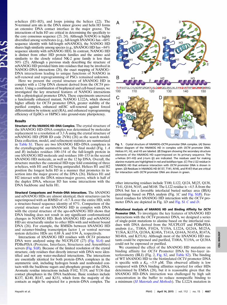
α-helices (H1–H3), and loops joining the helices (22). TheN-terminal arm sits in the DNA minor groove and helix H3 formsan extensive DNA contact interface in the major groove. Theinteractions of helix H3 are critical in determining the specificity tothe core consensus sequence (23, 24). Although NANOG is highlydiversified among vertebrates (e.g., full-length hNANOG has ∼60%sequence identity with full-length mNANOG), the NANOG HDshares high similarity among species (e.g., hNANOGHD has ∼84%sequence identity with mNANOG HD). In contrast, NANOG HDis distinct from other HD protein families and the amino acidsimilarity to the closely related NK-2 gene family is less than50% (25). Although a previous study describing the structure ofmNANOGHD provided hints into residues that may be involved inNANOG–DNA interactions (26), the exact mapping of NANOG–DNA interactions leading to unique functions of NANOG inself-renewal and reprogramming of PSCs remained unknown.Here we present the crystal structure of hNANOG HD in
complex with a 12-bp DNA element derived from the OCT4 pro-moter. Using a combination of biophysical and cell-based assays, weinvestigated the key structural features of NANOG interactionswith a physiological promoter DNA. These experiments uncovereda functionally enhanced mutant, NANOG L122A, which showedhigher affinity for OCT4 promoter DNA, greater stability of thepurified complex, enhanced mESC self-renewal against forceddifferentiation by retinoic acid (RA), and enhanced reprogrammingefficiency of EpiSCs or HiPSCs into ground-state pluripotency.
ResultsStructure of the hNANOG HD–DNA Complex. The crystal structure ofthe hNANOG HD–DNA complex was determined by molecularreplacement to a resolution of 3.3 Å using the crystal structure ofmNANOG HD (PDB ID code 2VI6) (26) as the search model.Data collection, model, and refinement statistics are summarizedin Table S1. There are two hNANOG HD–DNA complexes inthe crystallographic asymmetric unit. The final model (Fig. 1 Aand B) includes residues 100–154 of the full-length protein inone hNANOG HD molecule and residues 100–153 in the secondhNANOG HD molecule, as well as the 12 bp DNA. Overall, thestructure matches the canonical HD-type fold consisting of threeα-helices, with H1 and H2 being antiparallel. Helices H1 and H2position the longest helix H3 in a manner that promotes its in-sertion into the major groove of the DNA (26). Helices H1 andH2 interact with the DNA minor/major groove, which is half ofthe duplex DNA, whereas H3 has some interactions with theDNA backbone and helix H1.
Structural Comparisons and Protein–DNA Interactions. The hNANOGand mNANOG HDs are almost identical; their structures can besuperimposed with an RMSD of ∼0.7 Å over the entire HD, witha structure-based sequence identity of 87%. Comparison of thecrystal structure of our hNANOG HD in complex with DNAwith the crystal structure of the apo-mNANOG HD shows thatDNA binding does not result in any significant conformationalchanges in NANOG HD. Both hNANOG HD and mNANOGHD are structurally similar to other HDs with and without boundDNA (26). For example, the RMSDs between hNANOG HDand octamer-binding transcription factor 1 or ventral nervoussystem defective HDs are 0.88 Å and 0.94 Å, respectively.Interactions of hNANOG HD residues with OCT4 promoter
DNA were analyzed using the NUCPLOT (27) (Fig. S1A) andPDBePISA (Proteins, Interfaces, Structures and Assemblies)servers (Fig. S1B). Because of the limited resolution of the crystalstructure, only residues that directly interact with DNA were iden-tified and not any water-mediated interactions. The interactionsare essentially identical for both protein–DNA complexes in theasymmetric unit, including hydrogen bonds and nonbonded con-tacts with the backbone sugars and phosphate groups of the DNA.Aromatic residue interactions include F102, Y119, and Y136 thatcontact phosphates in the DNA backbone. Basic residues includeK137, K140, R147, and K151, which form the majority of thecontacts as might be expected for a protein–DNA complex. The
other interacting residues include T100, L122, Q124, M125, Q138,T141, Q144, N145, and M148. The L122 residue is ∼4.5 Å from theDNA but has a favorable interfacial buried surface area (BSA)percentage based on PISA analysis (Fig. 1C and Fig. S1B). Fea-tured residues for hNANOG HD interaction with the OCT4 pro-moter DNA are depicted in Fig. 1D and Fig. S1 C and D.
Mutational Analysis of hNANOG HD and Binding Affinity for OCT4Promoter DNA. To investigate the key features of hNANOG HDinteractions with the OCT4 promoter DNA, we designed a seriesof single-point mutations to alanine based on the interfacial resi-dues with DNA derived from the NUCPLOT and PDBePISAanalysis (i.e., T100A, F102A, Y119A, L122A, Q124A, M125A,Y136A, K137A, Q138A, K140A, T141A, Q144A, N145A, R147A,M148A, and K151A). Although most of the hNANOG HD mu-tants could be expressed and purified, T100A, Y119A, or Q138Acould not be expressed or purified.We examined the effect of the hNANOG HD mutations on
binding affinity for OCT4 Promoter DNA by bio-layer in-terferometry (BLI) (Fig. 2, Fig. S2, and Table S2). The bindingof WT hNANOG HD to the biotinylated OCT4 promoter DNAis specific with a KD ∼5.9 μM. This observed affinity is lowcompared with DNA binding affinities reported for other HDsdetermined by EMSA (28), but it is reasonable given that thehNANOG HD–DNA interaction was challenged by high saltconcentration in the buffer to reduce nonspecific binding toa minimum (SI Materials and Methods). The L122A mutation in
Fig. 1. Crystal structure of hNANOG–OCT4 promoter DNA complex. (A) Stereoribbon diagram of the hNANOG HD in complex with OCT4 promoter DNA.Helices H1, H2, and H3 are labeled. (B) Diagram showing the secondary structureelements of the hNANOG HD superimposed on its primary sequence. Theα-helices (H1–H3) and β-turn (β) are indicated. The residues used for makingalaninemutants are highlighted in red and boldface type. (C) The L122 residue inhNANOG HD that enhance interaction with OCT4 promoter DNA is shown ingreen. (D) Residues in hNANOG HD (K137, T141, N145, and R147) that are criticalfor interaction with OCT4 promoter DNA are shown in green.
Hayashi et al. PNAS | April 14, 2015 | vol. 112 | no. 15 | 4667
CELL
BIOLO
GY
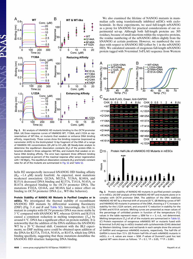
helix H2 unexpectedly increased hNANOG HD binding affinity(KD ∼1.4 μM) nearly fourfold. As expected, most mutationsweakened association: Q124A, M125A, Y136A, K140A, andK151A decreased DNA binding and K137A, T141A, N145A, orR147A abrogated binding to the OCT4 promoter DNA. Themutations F102A, Q144A, and M148A had a minor effect onbinding to OCT4 promoter DNA (i.e., WT-like behavior).
Protein Stability of NANOG HD Mutants in Purified Complex or inmESCs. We investigated the thermal stability of recombinanthNANOG HD mutants by differential scanning fluorimetry(DSF) (Fig. 3 A and B and Table S2). Interestingly, the L122Amutant in complex withOCT4 promoter DNA was more stable by3 °C compared with hNANOG WT, whereas Q144A and K151Acaused a consistent reduction in melting temperature (Tm) byaround 6 °C. DNA has a significant effect on protein stability. It isnoteworthy that the addition of the DNA stabilizes the hNANOGWT by a 20 °C thermal shift. Consistent with the BLI experi-ments, no DSF melting curve could be obtained upon addition ofthe DNA for K137A, T141A, N145A, or R147A, which lose DNAbinding specificity, suggesting that these mutations destabilize thehNANOG HD structure hampering DNA binding.
We also examined the lifetime of NANOG mutants in mam-malian cells using translationally inhibited mESCs with cyclo-heximide. In these experiments, we used full-length mNANOGas a proxy for hNANOG for practical considerations of our ex-perimental set-up. Although both full-length proteins are 305residues, because of small insertions within the respective proteins,the residue numbering of the mNANOG differs slightly from thehNANOG at certain positions. However, we numbered the resi-dues with respect to hNANOG HD (offset by 1 in the mNANOGHD). We calculated amounts of exogenous full-length mNANOGprotein tagged with N-terminal 3×FLAG sequence from Western
Fig. 2. BLI analysis of hNANOG HD mutants binding to the OCT4 promoterDNA. (A) Dose–response curves of hNANOG WT, Y136A, and L122A as rep-resentatives of WT-like, or mutants that weaken or enhance DNA bindingaffinity, respectively. These curves show the binding response (expressed asnanometer shift) to the biotinylated 12-bp fragment of the DNA of a rangeof hNANOG HD concentrations (20 μM to 0.5 μM). (B) Steady-state analysis todetermine the equilibrium dissociation constants (KD) of the protein–DNA in-teraction divided in three categories: WT-like, and mutants that weaken or en-hance DNA binding affinity. The error bars represent three different bindingcycles expressed as percent of the maximal response after sensor regenerationwith 1 MMgCl2. The equilibrium dissociation constants (KD) and kinetic constantrates for all of the mutants are summarized in Fig. S2 and Table S2.
Fig. 3. Protein stability of NANOG HD mutants in purified protein complexor in mESCs. (A) DSF analysis of the hNANOG HDWT and mutants alone or incomplex with OCT4 promoter DNA. The addition of the DNA stabilizeshNANOG HDWT by a thermal shift of around 20 °C. (B) Melting curves of WTand hNANOG HDmutants in presence of the DNA, showing a 3 °C increase instability for the L122A variant, and around 6 °C reduction in stability for theQ144A and K151A variants. Melting curves are presented as mean values ofthe percentage of unfolded protein in function of the temperature (°C);values in the table represent mean ± SEM for n = 3; n.d., not determined.Melting temperature (Tm) of all of the mutants are summarized in Table S2.(C) Protein expression of exogenous mNANOG WT or mutants fused withN-terminal 3×FLAG tag in mESCs treated with cycloheximide (CHX) detectedby Western blotting. Green and red bands in each sample show the amountof GAPDH and exogenous mNANOG mutants, respectively. The half-life ofGAPDH is more than 72 h. (D) Protein half-life of each mNANOG mutants inmESCs. n = 4, values are mean + SEM. P values of Dunnett’s post hoc testagainst WT were shown as follows: +P < 0.1, *P < 0.05, ***P < 0.001.
4668 | www.pnas.org/cgi/doi/10.1073/pnas.1502855112 Hayashi et al.
blotting analysis (Fig. 3 C and D and Table S2). The exogenousWT mNANOG protein half-life in mESCs was 3.1 ± 0.5 h, which isconsistent with previous studies of human and mouse NANOG (ei-ther exogenous or endogenous) in ESCs (29–31). The half-life ofsome mNANOGmutants (e.g., F102A, Y119A, K137A, and R147A)was significantly shorter than WT. In contrast, the half-life of L122A,which showed stronger DNA binding, was 11.5 ± 0.7 h. These resultssuggested that the protein lifetime of mNANOG mutants in mESCswere similar to the thermal stability of purified hNANOG mutants,which was regulated by their DNA-binding strength.
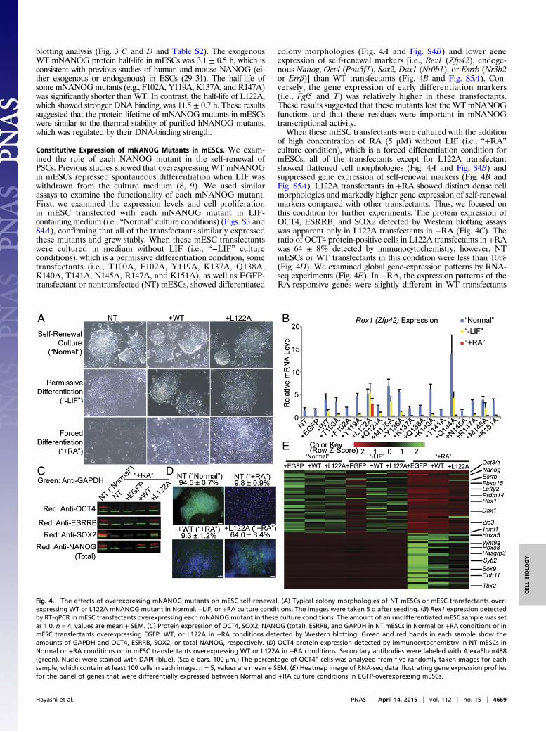
Constitutive Expression of mNANOG Mutants in mESCs. We exam-ined the role of each NANOG mutant in the self-renewal ofPSCs. Previous studies showed that overexpressing WT mNANOGin mESCs repressed spontaneous differentiation when LIF waswithdrawn from the culture medium (8, 9). We used similarassays to examine the functionality of each mNANOG mutant.First, we examined the expression levels and cell proliferationin mESC transfected with each mNANOG mutant in LIF-containing medium (i.e., “Normal” culture conditions) (Figs. S3 andS4A), confirming that all of the transfectants similarly expressedthese mutants and grew stably. When these mESC transfectantswere cultured in medium without LIF (i.e., “−LIF” cultureconditions), which is a permissive differentiation condition, sometransfectants (i.e., T100A, F102A, Y119A, K137A, Q138A,K140A, T141A, N145A, R147A, and K151A), as well as EGFP-transfectant or nontransfected (NT) mESCs, showed differentiated
colony morphologies (Fig. 4A and Fig. S4B) and lower geneexpression of self-renewal markers [i.e., Rex1 (Zfp42), endoge-nous Nanog, Oct4 (Pou5f1), Sox2, Dax1 (Nr0b1), or Esrrb (Nr3b2or Errβ)] than WT transfectants (Fig. 4B and Fig. S5A). Con-versely, the gene expression of early differentiation markers(i.e., Fgf5 and T) was relatively higher in these transfectants.These results suggested that these mutants lost the WTmNANOGfunctions and that these residues were important in mNANOGtranscriptional activity.When these mESC transfectants were cultured with the addition
of high concentration of RA (5 μM) without LIF (i.e., “+RA”culture condition), which is a forced differentiation condition formESCs, all of the transfectants except for L122A transfectantshowed flattened cell morphologies (Fig. 4A and Fig. S4B) andsuppressed gene expression of self-renewal markers (Fig. 4B andFig. S5A). L122A transfectants in +RA showed distinct dense cellmorphologies and markedly higher gene expression of self-renewalmarkers compared with other transfectants. Thus, we focused onthis condition for further experiments. The protein expression ofOCT4, ESRRB, and SOX2 detected by Western blotting assayswas apparent only in L122A transfectants in +RA (Fig. 4C). Theratio of OCT4 protein-positive cells in L122A transfectants in +RAwas 64 ± 8% detected by immunocytochemistry; however, NTmESCs or WT transfectants in this condition were less than 10%(Fig. 4D). We examined global gene-expression patterns by RNA-seq experiments (Fig. 4E). In +RA, the expression patterns of theRA-responsive genes were slightly different in WT transfectants
Fig. 4. The effects of overexpressing mNANOG mutants on mESC self-renewal. (A) Typical colony morphologies of NT mESCs or mESC transfectants over-expressing WT or L122A mNANOG mutant in Normal, −LIF, or +RA culture conditions. The images were taken 5 d after seeding. (B) Rex1 expression detectedby RT-qPCR in mESC transfectants overexpressing each mNANOGmutant in these culture conditions. The amount of an undifferentiated mESC sample was setas 1.0. n = 4, values are mean + SEM. (C) Protein expression of OCT4, SOX2, NANOG (total), ESRRB, and GAPDH in NT mESCs in Normal or +RA conditions or inmESC transfectants overexpressing EGFP, WT, or L122A in +RA conditions detected by Western blotting. Green and red bands in each sample show theamounts of GAPDH and OCT4, ESRRB, SOX2, or total NANOG, respectively. (D) OCT4 protein expression detected by immunocytochemistry in NT mESCs inNormal or +RA conditions or in mESC transfectants overexpressing WT or L122A in +RA conditions. Secondary antibodies were labeled with AlexaFluor488(green). Nuclei were stained with DAPI (blue). (Scale bars, 100 μm.) The percentage of OCT4+ cells was analyzed from five randomly taken images for eachsample, which contain at least 100 cells in each image. n = 5, values are mean + SEM. (E) Heatmap image of RNA-seq data illustrating gene expression profilesfor the panel of genes that were differentially expressed between Normal and +RA culture conditions in EGFP-overexpressing mESCs.
Hayashi et al. PNAS | April 14, 2015 | vol. 112 | no. 15 | 4669
CELL
BIOLO
GY
and more different in L122A transfectants compared with EGFPtransfectants. These results suggested that L122A transfectantsshowed partial resistant activity against RA-induced differentia-tion, which was not found in WT mNANOG transfectants.
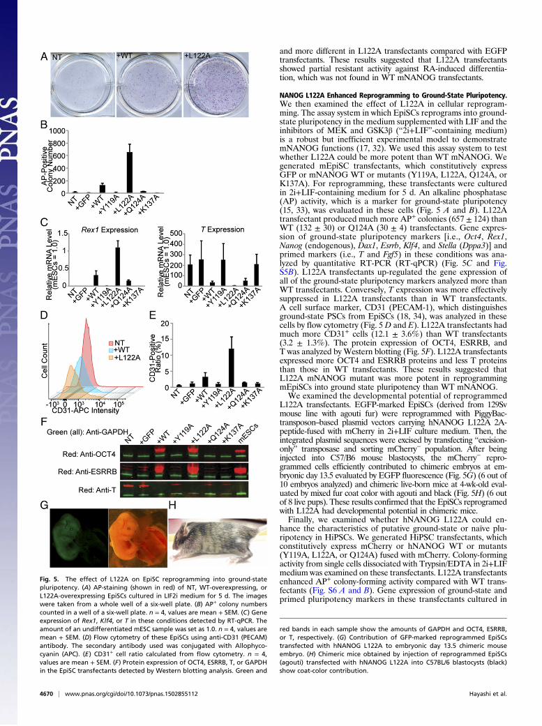
NANOG L122A Enhanced Reprogramming to Ground-State Pluripotency.We then examined the effect of L122A in cellular reprogram-ming. The assay system in which EpiSCs reprograms into ground-state pluripotency in the medium supplemented with LIF and theinhibitors of MEK and GSK3β (“2i+LIF”-containing medium)is a robust but inefficient experimental model to demonstratemNANOG functions (17, 32). We used this assay system to testwhether L122A could be more potent than WT mNANOG. Wegenerated mEpiSC transfectants, which constitutively expressGFP or mNANOG WT or mutants (Y119A, L122A, Q124A, orK137A). For reprogramming, these transfectants were culturedin 2i+LIF-containing medium for 5 d. An alkaline phosphatase(AP) activity, which is a marker for ground-state pluripotency(15, 33), was evaluated in these cells (Fig. 5 A and B). L122Atransfectant produced much more AP+ colonies (657 ± 124) thanWT (132 ± 30) or Q124A (30 ± 4) transfectants. Gene expres-sion of ground-state pluripotency markers [i.e., Oct4, Rex1,Nanog (endogenous), Dax1, Esrrb, Klf4, and Stella (Dppa3)] andprimed markers (i.e., T and Fgf5) in these conditions was ana-lyzed by quantitative RT-PCR (RT-qPCR) (Fig. 5C and Fig.S5B). L122A transfectants up-regulated the gene expression ofall of the ground-state pluripotency markers analyzed more thanWT transfectants. Conversely, T expression was more effectivelysuppressed in L122A transfectants than in WT transfectants.A cell surface marker, CD31 (PECAM-1), which distinguishesground-state PSCs from EpiSCs (18, 34), was analyzed in thesecells by flow cytometry (Fig. 5 D and E). L122A transfectants hadmuch more CD31+ cells (12.1 ± 3.6%) than WT transfectants(3.2 ± 1.3%). The protein expression of OCT4, ESRRB, andT was analyzed by Western blotting (Fig. 5F). L122A transfectantsexpressed more OCT4 and ESRRB proteins and less T proteinsthan those in WT transfectants. These results suggested thatL122A mNANOG mutant was more potent in reprogrammingmEpiSCs into ground state pluripotency than WT mNANOG.We examined the developmental potential of reprogrammed
L122A transfectants. EGFP-marked EpiSCs (derived from 129Svmouse line with agouti fur) were reprogrammed with PiggyBac-transposon–based plasmid vectors carrying hNANOG L122A 2A-peptide-fused with mCherry in 2i+LIF culture medium. Then, theintegrated plasmid sequences were excised by transfecting “excision-only” transposase and sorting mCherry− population. After beinginjected into C57/B6 mouse blastocysts, the mCherry− repro-grammed cells efficiently contributed to chimeric embryos at em-bryonic day 13.5 evaluated by EGFP fluorescence (Fig. 5G) (6 out of10 embryos analyzed) and chimeric live-born mice at 4-wk-old eval-uated by mixed fur coat color with agouti and black (Fig. 5H) (6 outof 8 live pups). These results confirmed that the EpiSCs reprogramedwith L122A had developmental potential in chimeric mice.Finally, we examined whether hNANOG L122A could en-
hance the characteristics of putative ground-state or naïve plu-ripotency in HiPSCs. We generated HiPSC transfectants, whichconstitutively express mCherry or hNANOG WT or mutants(Y119A, L122A, or Q124A) fused with mCherry. Colony-formingactivity from single cells dissociated with Trypsin/EDTA in 2i+LIFmedium was examined on these transfectants. L122A transfectantsenhanced AP+ colony-forming activity compared with WT trans-fectants (Fig. S6 A and B). Gene expression of ground-state andprimed pluripotency markers in these transfectants cultured in
Fig. 5. The effect of L122A on EpiSC reprogramming into ground-statepluripotency. (A) AP-staining (shown in red) of NT, WT-overexpressing, orL122A-overexpressing EpiSCs cultured in LIF2i medium for 5 d. The imageswere taken from a whole well of a six-well plate. (B) AP+ colony numberscounted in a well of a six-well plate. n = 4, values are mean + SEM. (C) Geneexpression of Rex1, Klf4, or T in these conditions detected by RT-qPCR. Theamount of an undifferentiated mESC sample was set as 1.0. n = 4, values aremean + SEM. (D) Flow cytometry of these EpiSCs using anti-CD31 (PECAM)antibody. The secondary antibody used was conjugated with Allophyco-cyanin (APC). (E) CD31+ cell ratio calculated from flow cytometry. n = 4,values are mean + SEM. (F) Protein expression of OCT4, ESRRB, T, or GAPDHin the EpiSC transfectants detected by Western blotting analysis. Green and
red bands in each sample show the amounts of GAPDH and OCT4, ESRRB,or T, respectively. (G) Contribution of GFP-marked reprogrammed EpiSCstransfected with hNANOG L122A to embryonic day 13.5 chimeric mouseembryo. (H) Chimeric mice obtained by injection of reprogrammed EpiSCs(agouti) transfected with hNANOG L122A into C57BL/6 blastocysts (black)show coat-color contribution.
4670 | www.pnas.org/cgi/doi/10.1073/pnas.1502855112 Hayashi et al.

2i+LIF culture medium for 5 d was analyzed by RT-qPCR (Fig.S6C). L122A transfectants significantly up-regulated the geneexpression of KLF2, endogenous NANOG, OCT4, PRDM14, andTFCP2L1 more thanWT transfectants. These results suggested thatL122A enhanced the characteristics of putative ground state ornaïve pluripotency in HiPSCs.
DiscussionIn this study, we describe the crystal structure of the hNANOGHD in complex with a 12-bp fragment of the OCT4 promoterDNA. Through alanine scanning, we evaluate the effect of keyamino acids in the NANOGHD–DNA interface on DNA-bindingaffinity, protein stability in the purified complex, protein lifetimein mESCs, and mESC self-renewal. From these results (summa-rized in Table S2), we classify the role of each of these residues asoutlined in SI Discussion.The L122A mutant showed higher binding affinity to OCT4
promoter DNA, higher thermal stability in the complex, greaterprotein lifetime in mESCs, enhanced resistance against RA-induced differentiation in mESCs, and enhanced reprogram-ming ability to ground-state pluripotency from EpiSCs andHiPSCs. These results suggest that enhanced DNA-bindingactivity of L122A lessens the off rate of NANOG from DNAto favor the transcriptionally active state in mammalian stem cells.Conversely, WT NANOG’s relatively weak binding activity andmodest protein stability may contribute to prompt and properdifferentiation of epiblast or germ cells in developing embryos (10,35) and to heterogeneous and fluctuating expression in PSC cul-ture, which has been mainly explained by feedback loops in tran-scription networks (36, 37).In conclusion, we demonstrate that an engineered key tran-
scriptional factor based on structural and biophysical informationimproved their performance in stem cell self-renewal and repro-gramming. Several key transcriptional factors have been identifiedfor regulating stem cell behavior and for reprogramming somaticcells into specific cell lineages. Using our approach for these factorsmay be beneficial to stem cell biology and regenerative medicine.
Materials and MethodsThe hNANOG HD construct (residues 94–162) was overexpressed in Escher-ichia coli (BL21Star, DE3; Invitrogen) and purified as a complex with a 12-bpfragment of the NANOG binding site in the OCT4 promoter (5′-GGCC-CATTCAAG-3′/3′-CCGGGTAAGTTC-5′). The cloning, expression, purification,and crystallization of hNANOG HD–DNA complex were carried out usingstandard protocols in the R.J.F. laboratory and at the Joint Center forStructural Genomics (JCSG; www.jcsg.org). mESCs, RF8 line (a gift fromR. V. Farese Jr., Harvard University, Boston) were cultured in FCS-based culturemedium supplemented with LIF without feeder cells. EPiSCs, EpiSC-5 line (a giftfrom Paul Teser, Case Western Reserve University, Cleveland) were cultured inN2B27 medium (StemCells) supplemented with basic FGF (10 ng/mL; Millipore)and Activin A (10 ng/mL; R&D Systems). All of the protocols of mouseexperiments were approved by the Institutional Animal Care and Use Com-mittee at University of California, San Francisco. Details of the materials,methods, and associated references are in SI Materials and Methods. See TableS3 for the DNA oligos and primers used for each mutation.
ACKNOWLEDGMENTS. We thank all members of the Joint Center forStructural Genomics (JCSG) for their general contributions to the structuralwork; K. Essex, Y. Miyake, and D. Singer for administrative support; S. Sami andC. Kime for technical assistance; and the Gladstone Flow Cytometry, Stem Cell,Genomics, Transgenic, and Bioinformatics Cores for technical support. Thiswork was funded by the National Institute of Health (NIH), National Institutesof General Medical Sciences (NIGMS), Protein Structure Initiative Grants U54GM094586 (to the JCSG) and U01 GM094614 (to R.J.F.). Portions of thisresearch were carried out at the Stanford Synchrotron Radiation Lightsource(SSRL), a Directorate of the Stanford Linear Accelerator Center NationalAccelerator Laboratory and an Office of Science User Facility operated forthe US Department of Energy Office of Science by Stanford University. TheSSRL Structural Molecular Biology Program is supported by the US Departmentof Energy’s Office of Biological and Environmental Research, and by the NIH,National Center for Research Resources, Biomedical Technology Program(Grant P41RR001209), and NIGMS; Postdoctoral Fellowship for ResearchAbroad of Japan Society for the Promotion of Science (JSPS) and Universityof California, San Francisco’s Program for Breakthrough Biomedical Research(to Y.H.); and the National Heart, Lung, and Blood Institute/NIH (GrantUO1HL098179), the L. K. Whittier Foundation, and the Roddenberry Founda-tion (to S.Y.). The Gladstone Institutes received support from a National Centerfor Research Resources Grant RR18928.
1. Loh YH, et al. (2006) The Oct4 and Nanog transcription network regulates pluri-potency in mouse embryonic stem cells. Nat Genet 38(4):431–440.
2. Boyer LA, et al. (2005) Core transcriptional regulatory circuitry in human embryonicstem cells. Cell 122(6):947–956.
3. Jiang J, et al. (2008) A core Klf circuitry regulates self-renewal of embryonic stem cells.Nat Cell Biol 10(3):353–360.
4. Rodda DJ, et al. (2005) Transcriptional regulation of nanog by OCT4 and SOX2. J BiolChem 280(26):24731–24737.
5. Pan G, Li J, Zhou Y, Zheng H, Pei D (2006) A negative feedback loop of transcriptionfactors that controls stem cell pluripotency and self-renewal. FASEB J 20(10):1730–1732.
6. Zhou Q, Chipperfield H, Melton DA, Wong WH (2007) A gene regulatory network inmouse embryonic stem cells. Proc Natl Acad Sci USA 104(42):16438–16443.
7. Kim J, Chu J, Shen X, Wang J, Orkin SH (2008) An extended transcriptional networkfor pluripotency of embryonic stem cells. Cell 132(6):1049–1061.
8. Mitsui K, et al. (2003) The homeoprotein Nanog is required for maintenance of plu-ripotency in mouse epiblast and ES cells. Cell 113(5):631–642.
9. Chambers I, et al. (2003) Functional expression cloning of Nanog, a pluripotencysustaining factor in embryonic stem cells. Cell 113(5):643–655.
10. Chambers I, et al. (2007) Nanog safeguards pluripotency and mediates germline de-velopment. Nature 450(7173):1230–1234.
11. Okita K, Ichisaka T, Yamanaka S (2007) Generation of germline-competent inducedpluripotent stem cells. Nature 448(7151):313–317.
12. Takahashi K, Yamanaka S (2006) Induction of pluripotent stem cells from mouseembryonic and adult fibroblast cultures by defined factors. Cell 126(4):663–676.
13. Schwarz BA, Bar-Nur O, Silva JC, Hochedlinger K (2014) Nanog is dispensable for thegeneration of induced pluripotent stem cells. Curr Biol 24(3):347–350.
14. Carter AC, Davis-Dusenbery BN, Koszka K, Ichida JK, Eggan K (2014) Nanog-independentreprogramming to iPSCs with canonical factors. Stem Cell Rev 2(2):119–126.
15. Brons IG, et al. (2007) Derivation of pluripotent epiblast stem cells from mammalianembryos. Nature 448(7150):191–195.
16. Tesar PJ, et al. (2007) New cell lines from mouse epiblast share defining features withhuman embryonic stem cells. Nature 448(7150):196–199.
17. Silva J, et al. (2009) Nanog is the gateway to the pluripotent ground state. Cell 138(4):722–737.18. Rugg-Gunn PJ, et al. (2012) Cell-surface proteomics identifies lineage-specific markers
of embryo-derived stem cells. Dev Cell 22(4):887–901.19. Gillich A, et al. (2012) Epiblast stem cell-based system reveals reprogramming synergy
of germline factors. Cell Stem Cell 10(4):425–439.
20. Takashima Y, et al. (2014) Resetting transcription factor control circuitry towardground-state pluripotency in human. Cell 158(6):1254–1269.
21. Gehring WJ, Affolter M, Bürglin T (1994) Homeodomain proteins. Annu Rev Biochem63:487–526.
22. Gehring WJ, et al. (1994) Homeodomain-DNA recognition. Cell 78(2):211–223.23. Berger MF, et al. (2008) Variation in homeodomain DNA binding revealed by high-
resolution analysis of sequence preferences. Cell 133(7):1266–1276.24. Noyes MB, et al. (2008) Analysis of homeodomain specificities allows the family-wide
prediction of preferred recognition sites. Cell 133(7):1277–1289.25. Harvey RP (1996) NK-2 homeobox genes and heart development. Dev Biol 178(2):203–216.26. Jauch R, Ng CK, Saikatendu KS, Stevens RC, Kolatkar PR (2008) Crystal structure and
DNA binding of the homeodomain of the stem cell transcription factor Nanog. J MolBiol 376(3):758–770.
27. Luscombe NM, Laskowski RA, Thornton JM (1997) NUCPLOT: A program to generateschematic diagrams of protein-nucleic acid interactions. Nucleic Acids Res 25(24):4940–4945.
28. Weiler S, et al. (1998) Site-directed mutations in the vnd/NK-2 homeodomain. Basis of var-iations in structure and sequence-specific DNA binding. J Biol Chem 273(18):10994–11000.
29. Moretto-Zita M, et al. (2010) Phosphorylation stabilizes Nanog by promoting its in-teraction with Pin1. Proc Natl Acad Sci USA 107(30):13312–13317.
30. Ramakrishna S, et al. (2011) PEST motif sequence regulating human NANOG forproteasomal degradation. Stem Cells Dev 20(9):1511–1519.
31. Abranches E, Bekman E, Henrique D (2013) Generation and characterization of anovel mouse embryonic stem cell line with a dynamic reporter of Nanog expression.PLoS ONE 8(3):e59928.
32. Silva J, et al. (2008) Promotion of reprogramming to ground state pluripotency bysignal inhibition. PLoS Biol 6(10):e253.
33. van Oosten AL, Costa Y, Smith A, Silva JC (2012) JAK/STAT3 signalling is sufficient anddominant over antagonistic cues for the establishment of naive pluripotency. NatCommun 3:817.
34. Robson P, Stein P, Zhou B, Schultz RM, Baldwin HS (2001) Inner cell mass-specific expression ofa cell adhesion molecule (PECAM-1/CD31) in the mouse blastocyst. Dev Biol 234(2):317–329.
35. Yamaguchi S, Kimura H, Tada M, Nakatsuji N, Tada T (2005) Nanog expression inmouse germ cell development. Gene Expr Patterns 5(5):639–646.
36. Kalmar T, et al. (2009) Regulated fluctuations in nanog expression mediate cell fatedecisions in embryonic stem cells. PLoS Biol 7(7):e1000149.
37. MacArthur BD, et al. (2012) Nanog-dependent feedback loops regulate murine em-bryonic stem cell heterogeneity. Nat Cell Biol 14(11):1139–1147.
Hayashi et al. PNAS | April 14, 2015 | vol. 112 | no. 15 | 4671
CELL
BIOLO
GY