structure of dicot rootcms.gcg11.ac.in/attachments/article/61/structure of roots and stems... ·...
TRANSCRIPT

Structure Of Dicot Root


Root Cap - shaped like a thimble this structure covers the tip of the root and provides protection as the root drives into the soil. These cells are produced by the root apical meristem. The outer cells of the root cap are continuously being worn away and new cells are added to the inner portion. The cells may be covered with a lubricating slime. Root apical meristem - the cells here divide rapidly to form new cells. These new cells later mature into more specialized root tissues. Region or Zone of Elongation root tissues. Region or Zone of Elongation - in this region the cells previously produced by the meristem undergo rapid elongation. Elongation (along with production of new cells) results in root growth which pushes the root further into the soil. Within the region of elongation just behind the meristem you will find the following undifferentiated tissues:

Protoderm - undifferentiated tissue on the exterior of the root which will mature to become the epidermis Procambium - undifferentiated tissue in the central part of the root which will mature to become the the vascular cylinder Ground meristem - undifferentiated tissue lying between the protoderm and procambium that will mature to become the cortex
Region of differentiation - here the root becomes thicker and Region of differentiation - here the root becomes thicker and secondary or lateral roots are developed. In this region the protoderm, procambium and ground meristem cells undergo differentiation into specialized cells. Root-hairs begin to form in the region of differentiation. Root hairs are fine outgrowths of epidermis. They increase the area of absorption of the root.


A transverse section passing through the root of Sunflower reveals the following details.
EpiblemaEpiblema is the outermost covering of the root formed by single layer of compactly arranged, barrel-shaped, parenchyma cells. The cells are characteristically thin-walled since they are involved in absorption of water. A cuticle and stomata are absent. Some of the epiblema cells are produced into long unicellular projections called root hairs. Hence, the into long unicellular projections called root hairs. Hence, the epiblema is also known as piliferous layer.
CortexCortex is a major component of the ground tissue of root. It is represented by several layers of loosely arranged parenchyma cells. Intercellular spaces are prominent. The cortex is mainly meant for storage of water. The cells also allow a free movement of water into the xylem vessels.

EndodermisIt is the innermost layer of cortex formed by compactly arranged barrel-shaped cells. Some of the cells in the endodermis are thin-walled and are known as passage cells. The passage cells allow water to pass into the xylem vessels. The remaining cells in the endodermis are characterised by the presence of thickening on their xylem vessels. The remaining cells in the endodermis are characterised by the presence of thickening on their radial walls. These thickenings are known as casparianthickenings. They are formed by the deposition of a waxy substance called suberin. The casperian thickenings play an important role in creating and maintaining a physical force called root pressure.

SteleStele consists of pericycle, conjunctive tissue and vascular bundles.PericyclePericycle is a region that lies immediately below the endodermis. It is represented by a single layer of parenchyma cells.Conjunctive TissueConjunctive tissue is represented by a group of radiallyarranged parenchyma cells found in between the vascular Conjunctive tissue is represented by a group of radiallyarranged parenchyma cells found in between the vascular bundles. The cells are specialised for storage of water.Vascular BundlesVascular bundles are described as radial and tetrarch. There are four bundles each of xylem and phloem occurring alternately. Xylem is described as exarch.PithPith is absent in the older root.

Diagnostic Features of a Dicot Root* Presence of thin walled cells in the epiblema. * Absence of cuticle, and stomata. * Presence of unicellular root hairs.* Absence of hypodermis. * Presence of passage cells and casparian thickenings in the endodermis. * Presence of uniseriate pericycle made up of parenchyma. * Presence of uniseriate pericycle made up of parenchyma. * Presence of conjuctive tissue. * Absence of pith. * Presence of radial vascular bundles exhibiting tetrachcondition with exarch xy

Structure Of Dicot Stem


Dicotyleconus Stem (Sunflower Stem)
Internally the dicot stem is differentiated into following regions.
1. EpidermisIt is single layered outer most protective covering made up of tangentially flattened thin walled parenchymatous cells. They lack tangentially flattened thin walled parenchymatous cells. They lack intercellular spaces and chloroplast. They are covered with cuticle and bear multicellular hairs. At places are present minute pores called. stomata, each having two kidney shaped guard cells to facilitate gaseous exchange and transpiration.

2. CortexIt is a multicellular tissue present between epidermis and vascular tissues. It is differentiated into three regions.
(a) HypodermisOutmost collenchymatous part of cortex. They have angular thickenings and so lack intercellular spaces. They provide mechanical strength and support to the stem. Some cells might posses chloroplasts and perform photosynthesis.posses chloroplasts and perform photosynthesis.
(b) General cortexIt is a parenchymatous tissue lie below the collenchymatoushypodermis. It forms the bulk of cortex. The cells are thin walled and posses conspicuous intercellular spaces. The cortex contains a number of mucilage ducts running all along the stem. The cells of cortex store food and also provide mechanical strength in fully turgid conditions. The mucilage ducts conserve water.

(c) EndodermisIt is the innermost layer of cortex made up of barrel shaped cells compactly arranged without any intercellular spaces. The cells are filled with starch and are also called starch sheath. The radial and transverse walls posses bands of lignin called casparian strips. Due to this endodermis forms a water-tight layer preventing loss of water from vascular tissue to cortex.
3. PericycleIt is a few layered thick boundary of vascular system below endodermis. It is made up of sclerenchymatous cells lying above each vascular bundle as a bundle cap alternating with parenchyma. The sclerenchyma provides strength to the stem and parenchyma stores food.

4. Vascular systemIt lies below the pericycle around the pith in the form of a ring of vascular bundles or fasciles. They are wedge shaped having pholem on outside and xylem towards inside with cambium between the two. The cambium is called fascicular cambium.
The vascular bundles are conjoint, collateral and open. The vascular bundles are conjoint, collateral and open. The pholem has all the four types and conducts food. Xylem is also complete and is endarch. It conducts water and dissolved minerals from roots to various parts of plant and also provide mechanical strength. Cambium consists of thin walled cells and is responsible for increase in girth by producing secondary xylem and secondary pholem.

5. Medullary/Pith raysThe regions between the vascular bundles are made up of parenchymatous cells and are called medullary rays or pith rays. They connect pith cortex and pericycle and are involved in radial conduction of water, minerals and food.
6. Pith/Medulla6. Pith/MedullaIt is the central part of stem consisting of parenchymatous cells having conspicuous intercellular spaces. The cells store food.

Structure Of Monocot Stem


T.S. of a Monocot Stem (Maize)


Epidermis
Epidermis is the outermost covering of the stem represented by a single layer of compactly arranged, barrel-shaped parenchyma cells. Intercellular spaces are absent. Trichomes are absent. A cuticle is present. The epidermis contains numerous minute openings called stomata.Hypodermis
Hypodermis is a region that lies immediately below the epidermis. It is represented by a few layers of compactly arranged It is represented by a few layers of compactly arranged sclerenchyma
Ground Tissue
Ground tissue is a major component of the stem. It is undifferentiated. The ground tissue is represented by several layers of loosely arranged parenchyma cells enclosing prominent intercellular spaces. The ground tissue is meant for storage of food.

Vascular BundlesThey are found irregularly scattered in the ground tissue. Towards the periphery, the bundles are smaller in size while towards the centre, they are larger in size. The smaller bundles are younger, while the larger ones are older. Hence, the arrangement is described as centrifugal. Each vascular bundle has a covering called bundle sheath formed by a single layer of sclerenchyma cells. The vascular bundle encloses both xylem and phloem. Xylem is found towards the inner surface and phloem phloem. Xylem is found towards the inner surface and phloem towards the outer surface. Cambium is absent. Hence the vascular bundles are described as conjoint, collateral and closed. In the xylem, there are two metaxylem and two protoxylemvessels arranged in 'the shape of 'Y'. The lower protoxylemvessel is non functional and remains as a water filled cavity called lisigenous cavity or protoxylem cavity. Xylem is described as endarch. In the phloem, only sieve tubes, companion cells and phloem fibres are present. Phloem parenchyma is absent.

Diagnostic Features of a Monocot Stem
* Absence of trichomes. * Presence of stomata. * Presence of a hypodermis made up of sclerenchyma. * Presence of undifferentiated ground tissue. * Presence of numerous vascular bundles irregularly scattered with cerifugal arrangement. * Vascular bundles are conjoint, collateral & closed with * Vascular bundles are conjoint, collateral & closed with endarch xylem. * Presence of only two protoxylem & two metaxylemvessels in each bundle. * Presence of a lysigenous cavity. * Absence of phloem parenchyma. * Presence of a bundle sheath made up of sclerenchyma.

Structure Of Monocot Root

T.S. of a Monocot Root
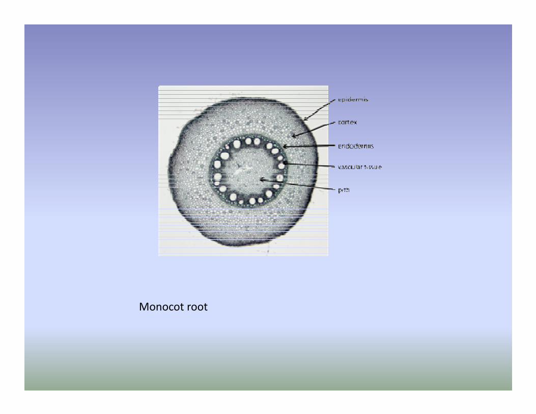
Monocot root

EpiblemaEpiblema is the outermost covering of the root formed by a single layer of compactly arranged, barrel-shaped parenchyma cells. The cells are characteristically thin-walled since they are involved in absorption of water. A cuticle and stomata are absent. Some of the epiblema cells are produced into long unicellular projections called root hairs. Hence, epiblema is also known as piliferouslayer.
CortexCortex is a major component of the ground tissue of root. It is Cortex is a major component of the ground tissue of root. It is represented by several layers of loosely arranged parenchyma cells. Intercellular spaces are prominent. The cortex is mainly meant for storage of water. The cells also allow a free movement of water into the xylem vessels.

Endodermis :
It is the innermost layer of cortex formed by compactly arranged barrel-shaped cells. Some of the cells in the endodermis are thin-walled and are known as passage cells. The passage cells allow water to pass into the xylem vessels. The remaining cells in the endodermis are characterised by the presence of thickening on their radial walls. These The remaining cells in the endodermis are characterised by the presence of thickening on their radial walls. These thickenings are known as casparian thickenings. They are formed by the deposition of a waxy substance called suberin. The casparian thickenings play an important role in creating and maintaining a physical force called root pressure.

SteleStele is the central cylinder of the root consisting of pericycle, conjunctive tissue, pith and vascular bundles.
PericyclePericycle is the outermost covering of the stele represented by a single layer of parenchyma cells.Pericycle is the outermost covering of the stele represented by a single layer of parenchyma cells.
Conjunctive tissueIt is represented by loosely arranged parenchyma cells found in between the vascular bundles. The cells are specialized for storage of water.

PithPith is the innermost region of the root representing the central axis. It is composed of few loosely arranged parenchyma cells.
Vascular bundlesVascular bundles are radial in arrangement. There are Vascular bundles are radial in arrangement. There are eight bundles each of xylem and phloem. Hence, the condition is described as polyarch. Xylem is described as exarch.

Diagnostic Features of a Monocot Root
* Presence of thin walled cells in the epiblema. * Absence of cuticle and stomata. * Presence of unicellular root hairs. * Presence of passage cells and casparian thickenings in the endodermis. the endodermis. * Presence of parenchyma cells in the pericycle. * Presence of conjuctive tissue. * Presence of a distinct pith. * Presence of radial vascular bundles with polyarchcondition and an exarch xylem.

Thank You