structure of shigella ipgb2 in complex with human rhoa
TRANSCRIPT
Structure of Shigella IpgB2 in Complex with Human RhoAIMPLICATIONS FOR THE MECHANISM OF BACTERIAL GUANINE NUCLEOTIDE EXCHANGEFACTOR MIMICRY*□S
Received for publication, January 26, 2010, and in revised form, March 11, 2010 Published, JBC Papers in Press, April 2, 2010, DOI 10.1074/jbc.M110.107953
Bjorn U. Klink‡, Stephan Barden‡, Thomas V. Heidler‡, Christina Borchers§, Markus Ladwein¶,Theresia E. B. Stradal§�, Klemens Rottner¶, and Dirk W. Heinz‡1
From the ‡Division of Structural Biology, ¶Cytoskeleton Dynamics Group, and §Signaling and Motility Group, Helmholtz Zentrumfur Infektionsforschung, D-38124 Braunschweig and the �Institute of General Zoology and Genetics, University of Munster,D-48149 Munster, Germany
A common theme in bacterial pathogenesis is the manipula-tion of eukaryotic cells by targeting the cytoskeleton. This is inmost cases achieved either by modifying actin, or indirectly viaactivation of key regulators controlling actin dynamics such asRho-GTPases. A novel group of bacterial virulence factorstermed the WXXXE family has emerged as guanine nucleotideexchange factors (GEFs) for these GTPases. The precise mecha-nism of nucleotide exchange, however, has remained unclear.Herewe report the structure of theWXXXE-protein IpgB2 fromShigella flexneri and its complex with human RhoA. We unam-biguously identify IpgB2 as a bacterial RhoA-GEF and dissectthe molecular mechanism of GDP release, an essential prereq-uisite for GTP binding. Our observations uncover that IpgB2induces conformational changes on RhoA mimicking DbI- butnot DOCK family GEFs. We also show that dissociation of theGDP�Mg2� complex is preceded by the displacement of themetal ion to the �-phosphate of the nucleotide, diminishing itsaffinity to the GTPase. These data refine our understanding ofthe mode of action not only of WXXXE GEFs but also of mam-malian GEFs of the DH/PH family.
Remodeling of the actin cytoskeleton is essential for eukary-otic life, taking place in processes as diverse as division,motility,or cell-cell communication. The actin cytoskeleton is com-posed of actin monomers and helical filaments constantlyassembling and disassembling at their ends, which is tightlyregulated by multiple signaling pathways employing hundredsof actin-binding proteins. Most if not all signaling pathwaysconverge on small GTPases of the Rho family, which are longrecognized as key signaling switches inducing different actinfilament structures (1–3). As effector interaction occurs in theGTP-bound state, their intrinsic GTPase activity is counterbal-
anced by guanine nucleotide exchange factors (GEFs)2 drivingGTP re-loading by increasing the intrinsic nucleotide dissocia-tion rate (4). Activation of the name-giving family memberRhoA induces cell contraction. This drives the formation ofstress fiber bundles and focal adhesions in fibroblasts or endo-thelial cells, exerting force onto the substratum or neighboringcells (5). In neurons for instance, RhoA activation inducesgrowth cone retraction (6). Other Rho family members triggerthe formation of cell protrusions antagonistic to RhoA, withRac1 and Cdc42 inducing lamellipodia and filopodia, respec-tively (7, 8).Given their importance in cell physiology, Rho-GTPases
have also emerged as key targets of pathogens modifying cellsignaling or actin remodeling for their own needs (9). Forinstance, multiple bacterial toxins inhibiting RhoA, such as C3exoenzyme of Clostridium botulinum, have served as invalu-able tools to study RhoA function in vivo (10, 11). As an alter-native to inhibition, Rho-GTPase signaling pathways drivingdifferent types of actin reorganization are also frequentlybypassed or activated by pathogens, at least transiently, allow-ing them to induce their tight adherence to or entry intoeukaryotic cells (12–14). Pathogens often modulate host pro-tein functions by virulence factors acting as perfect mimics ofmammalian proteins. For example, the Salmonella toxin SopEcan activate Cdc42 in a similar fashion as Dbl-like mammalianGEFs (15). Interestingly, SopE does not display any significantstructural or sequence similarity to the mimicked mammalianprotein.An intriguing novel family of bacterial effectors shares a
WXXXE sequencemotif, themembers of which were originallyclassified as GTPase mimics, despite the lack of any sequencehomology or guanine nucleotide binding (16, 17). Prototypicmembers of the family include IpgB1 and IpgB2 from Shigellaflexneri andMap frompathogenicEscherichia coli, which are alltranslocated into the host by type III secretion. Interestingly,these proteins were reported to induce filopodia (for Map) orlamellipodia and stress fiber formation, indicative of Rac1 andRhoA activity in the case of IpgB1 and IpgB2, respectively (16,18). In another study, IpgB1 was described to drive Rac1 and
* This work was funded in part by the Deutsche Forschungsgemeinschaft(Grants SFB621 to K. R. and SPP1150 to T. E. B. S.).
The atomic coordinates and structure factors (codes 3LYQ, 3LW8, 3LWN, and3LXR) have been deposited in the Protein Data Bank, Research Collaboratoryfor Structural Bioinformatics, Rutgers University, New Brunswick, NJ(http://www.rcsb.org/).
□S The on-line version of this article (available at http://www.jbc.org) containssupplemental Figs. 1– 4.
1 Supported by the Fonds der Chemischen Industrie. To whom correspon-dence should be addressed: Division of Structural Biology, HelmholtzZentrum fur Infektionsforschung, Inhoffenstrasse 7, D-38124 Braun-schweig, Germany. Tel.: 49-531-6181-7000; Fax: 49-531-6181-7099; E-mail:[email protected].
2 The abbreviations used are: GEF, guanine nucleotide exchange factor; MBP,maltose-binding protein; PFA, formaldehyde; RBD, Rho-binding domain;P-loop, phosphate-binding loop; Sif, Salmonella-induced filament; DH/PH,Dbl homologous/pleckstrin homologous fragment of PDZ-RhoGEF;r.m.s.d., root mean square deviation; ROCK, RhoA-effector Rho kinase.
THE JOURNAL OF BIOLOGICAL CHEMISTRY VOL. 285, NO. 22, pp. 17197–17208, May 28, 2010© 2010 by The American Society for Biochemistry and Molecular Biology, Inc. Printed in the U.S.A.
MAY 28, 2010 • VOLUME 285 • NUMBER 22 JOURNAL OF BIOLOGICAL CHEMISTRY 17197
by guest on February 14, 2018http://w
ww
.jbc.org/D
ownloaded from
Cdc42 activation (19). In addition, more recently identifiedfamily members such as EspM and EspT have been interpretedto activate rather than mimic RhoA and Rac1/Cdc42, respec-tively (20, 21), which was recently verified in the case of EspM2(22). The finding that the WXXXE protein SifA displays a foldsimilar to the bacterial GEF SopE (15, 23) provided the firststructural evidence for WXXXE proteins acting as GEFs.Finally, Map has recently been defined as Cdc42-GEF (24),refuting the mimicry hypothesis and questioning the relevanceof IpgB2 binding to downstream effectors of RhoA (16).Here we provide crystal structures of IpgB2 both in its free
form and in complex with human RhoA. Our data reveal themechanism and structurally decipher the sequence of eventsinducing GDP release catalyzed by this novel family of bacterialGEFs, constituting yet another remarkable example of conver-gent evolution, a common theme in bacterial pathogenesis (12,25, 26).
EXPERIMENTAL PROCEDURES
Cloning, Expression, and Purification—IpgB2 (residues1–188, NCBI gene ID 56404000 was cloned into the expressionvector pET-M41 (kindly provided by Gunter Stier, EMBL, Hei-delberg). This pET-derived vector allows expressing the pro-tein of interest as a fusion to the C terminus of His-taggedmaltose binding protein (MBP) with a linker sensitive totobacco etch virus protease. RhoA (residues 2–181, NCBI geneID 10835049) and ROCK I were cloned into a modified pET-28c vectorwith anHis8 tag and a linker sensitive to tobacco etchvirus protease fused to the N terminus. The original ROCK Iconstruct was purchased from GENEART and was optimizedfor protein production in E. coli. It encoded for amino acid res-idues 947–1015 according toNCBI gene ID4885583. IpgB2 andRhoAwere produced in E. coliTuner (DE3) cells in LBmediumcontaining additional 2 g/liter glucose after induction with 1mM isopropyl 1-thio-�-D-galactopyranoside. The expressiontemperature was 15 °C for IpgB2 and 27 °C for RhoA. ROCK Iwas produced in E. coli Tuner (DE3) cells at 22 °C in 2�YTmedium after induction with 0.8 mM isopropyl 1-thio-�-D-ga-lactopyranoside. Selenomethionine-substituted IpgB2was pro-duced inminimalmedium (1 g/literNH4Cl, 3 g/liter KH2PO4, 6g/liter Na2HPO4�7H2O) mixed 10:1 with supplement medium(21.82 g/liter D-glucose monohydrate, 0.45 g MgSO4�7H2O,10.45 mg/liter Fe2(SO4)3�H2O, 10 mg/liter thiamine) and 50mg/liter L-selenomethionine, with the pH adjusted to pH 7.4.Harvested cells containing IpgB2 were resuspended in buffer(50 mM HEPES, pH 7.5, 250 mM NaCl, 8.7% (v/v) glycerol, 7.5mM imidazole, 250mM trisodium citrate, and 10mM �-mercap-toethanol). In the case of RhoA, the buffer composition was 30mM Tris/HCl, pH 7.5, 50 mMNaCl, 5 mMMgCl2, 5% (v/v) glyc-erol, 10 mM �-mercaptoethanol, and 0.05 mM GDP, and in thecase of ROCK I the buffer contained 50 mM Tris/HCl, pH 8.5,10% (v/v) glycerol, 20% (w/v) sucrose, 0.2 mM Na2S2O4, 2 mM
MgCl2, and2 mM dithiothreitol. To all resuspension buffers, aprotease inhibitor mixture (Roche Applied Science, completeEDTA free) and a small amount of DNase I (�1mg) was added.The suspended cells were mechanically disrupted using a TSseries bench top cell homogenizer (Constant Systems Ltd.), andthe soluble cell extract after centrifugation (60min, 37,000 � g,
277 K) was purified by affinity chromatography using nickel-nitrilotriacetic acid-agarose. During affinity chromatography,the buffer of ROCK I was changed to a buffer containing 50mM
Tris/HCl, pH7.5, 150mMNaCl, 0.1% (v/v)TritonX-100, 10mM
MgCl2, and 0.2 mM dithiothreitol. All elution buffers addition-ally contained 250 mM imidazole. IpgB2 and RhoA were mixedwith tobacco etch virus protease in a molar ratio of 100:1 anddialyzed against buffer for 12 h at 277 K. ROCK I was firstpurified by anion chromatography on a Mono Q 5/5 column(Amersham Biosciences) and then treated equally. During dial-ysis, the imidazole concentrationwas reduced, and the proteinswere digested to cleave off the fusion tags. The tags wereremoved by (negative) affinity chromatography using nickel-nitrilotriacetic acid-agarose, and IpgB2 and RhoA were furtherpurified by gel-filtration chromatography on a Superdex 75 col-umn (Amersham Biosciences). During gel-filtration chroma-tography, the buffer was exchanged against 200 mM trisodiumcitrate/citric acid, pH 6.0, and 10 mM dithiothreitol in the caseof IpgB2 and against 30 mM Tris/HCl, pH 7.5, 50 mM NaCl, 5mM MgCl2, 5% (v/v) glycerol, 5 mM dithiothreitol in the case ofRhoA.Nucleotide Exchange and GTPase and GEF Assays—GTPase
and GEF assays were performed according to methodsdescribed by Eberth and Ahmadian (27). RhoA loaded with dif-ferent nucleotides was prepared by a modified method similaras described by Hutchinson and Eccleston (28), utilizing theenhanced nucleotide dissociation rate of RhoA in buffers lack-ing Mg2� (29).Recombinant Protein Pulldown Experiments—His-MBP-
fused IpgB2 was incubated with 5 �l of nickel-nitrilotriaceticacid-agarose (Qiagen) per milligram of fusion protein for 12 hat 4 °C in buffer containing 50 mM HEPES, pH 7.5, 250 mM
NaCl, 8.7% (v/v) glycerol, 250 mM trisodium citrate, 10 mM
�-mercaptoethanol. The resin was washed in a gravity flow col-umn with 5 column volumes of buffer to remove unbound pro-tein. Aliquots of 35 �l of packed resin mixed 1:1 (v/v) withglycerol were used in a typical binding assay with a 1-ml totalreaction volumeor stored at�70 °Cuntil usage. The resin com-plexed with His-MBP-IpgB2 was incubated with recombinanttagless test protein (RhoA, ROCK I (Rho-binding domain(RBD))) for 10 min. The agarose was pelleted at 1000 � g, thesupernatant was removed, and the agarose was washed threetimes to remove unbound test protein. Proteins were elutedwith buffer additionally containing 250 mM imidazole, sepa-rated by SDS-PAGE, and visualized by Coomassie staining. Toexclude unspecific binding of the test protein to the resin, allexperiments were repeated with resin without complexedHis-MBP-IpgB2.Preparation of Soluble Bacterial WXXXE Effector Proteins—
As untruncated wild-type IpgB2 was not soluble in standardbuffers after cleavage of the MBP fusion tag, buffer conditionswere optimized to obtain soluble, untagged IpgB2. 96 condi-tions froma standard crystallization screen (Classics Suite,Qia-gen) were tested for optimization. The MBP-tagged IpgB2 wasconcentrated to �10 mg/ml, and 1.2 �l of protein solution wasmixed with 1.2 �l of H2O, 0.3 �l of tobacco etch virus Protease(�2.5 mg/ml), and 1.2 �l of reservoir solution from the crystal-lization screen in batch experiments. After incubation at 4 °C
RhoA Activation by the Bacterial GEF IpgB2
17198 JOURNAL OF BIOLOGICAL CHEMISTRY VOLUME 285 • NUMBER 22 • MAY 28, 2010
by guest on February 14, 2018http://w
ww
.jbc.org/D
ownloaded from
for 3 days, the solution wasmixed with 10 �l of H2O, and insol-uble particles were pelleted by centrifugation. 10 �l of superna-tant of each experiment was separated by SDS-PAGE and visu-alized by Coomassie staining. In almost all tested conditions,complete cleavage of the MBP tag was observed, and approxi-mately one-third of all conditions contained varying quantitiesof soluble IpgB2. Buffer components that repetitively werepresent in conditions leading to soluble IpgB2were individuallytested in further cleavage experiments. Citrate proved to be themost successful buffer component at concentrations of 100–200mM, pH 5–7. Fresh IpgB2 was produced as described aboveusing buffer systems based on this finding, which yielded �100mg of soluble MBP-fused IpgB2 per liter bacterial culture and�10 mg of untagged IpgB2 per liter after purification to appar-ent homogeneity. Purified, untagged IpgB2 can also be handledin bufferswithout citrate (e.g. 10mMTris/HCl, pH7.0) at 4 °C aslong as no mechanical or oxidizing stress is applied to the pro-tein. The proper folding state of purified IpgB2 was tested by15N-, 13C-NMR spectroscopy on appropriately labeled IpgB2(data not shown), indicating that the protein accommodates acompact, folded state in solution.The buffer systems found for IpgB2 also allowed purification
of the homologous WXXXE protein Map in full length, andallowed production of MBP-tagged IpgB1 (data not shown).IpgB1, however, slowly precipitates upon cleavage of the fusiontag and might require further buffer optimization.Cell Assays—cDNA of the constitutively active variant Q14V
of humanRhoAwas a kind gift fromLauraMachesky (Glasgow,UK), cDNAof IpgB2was amplified by PCR from genomicDNAof S. flexneri (serotype 5), and the mutants of IpgB2 were cus-tom-synthesized (Genescript, Piscataway, NJ). All cDNAs weresubcloned into pEGFP-C1 (Clontech, Mountain View, CA).The fidelity of all constructs was verified by sequencing. Fortransfections and fluorescence microscopy, SV40 large T-anti-gen-immortalized mouse embryonic fibroblasts were grown inDulbecco’s modified Eagle’s medium, 4.5 g/liter glucose(Invitrogen) supplemented with 10% fetal bovine serum(Sigma) and 2 mM glutamine. Transfections were carried outwith FuGENE 6 (RocheApplied Science) according to theman-ufacturer’s protocols. Cells were plated on glass coverslips andfixed 16–24 h post transfection. Cells were fixed with 4% form-aldehyde in phosphate-buffered saline for 20 min, permeabi-lizedwith amixture of 0.1%TritonX-100 and formaldehyde for45 s, and stained for the actin cytoskeleton using Alexa594-labeled phalloidin (Molecular Probes, Invitrogen). Epifluores-cence images were acquired using an inverted microscope(Axiovert 100TV, Carl Zeiss, Jena, Germany) equipped with a100�/1.3 numerical aperture PlanNeofluar objective and aback-illuminated charge-coupled device camera (CoolSNAPK4, Photometrics, Tucson, AZ) driven byMetaMorph software(Molecular Devices, Downingtown, PA), and further processedusing Adobe Photoshop CS and Illustrator software (AdobeSystems, Mountain View, CA).Crystallization—Native and selenomethionine-substituted
IpgB2 and its complex with RhoA were crystallized using thehanging drop, vapor-diffusion technique at 4 °C. Initial sphe-roidal precipitate of free IpgB2 could be optimized by adding 10mM FeCl3 to the protein in gel-filtration chromatography
buffer, which was sufficient to obtain crystal growth withoutany additional precipitate. The crystal quality could beimproved with low concentrations of polyethylene glycol 3350and isopropanol. The final composition was 1 �l of 89 mg/mlIpgB2 in gel-filtration chromatography buffer mixed with 1 �lof reservoir containing 100mM trisodiumcitrate/citric acid, pH5.5, 10 mM NaCl, 4–7% isopropanol, 3–5% polyethylene glycol3350, and 10mM FeCl3. The crystal morphology was optimizedby streak-seeding from initial crystallization conditions con-taining micro crystals. Crystals of IpgB2 in complex with RhoAwere obtained from a (stoichiometric)mixture of 25�l of RhoA(19.3 mg/ml) with 5.7 �l of IpgB2 (89.6 mg/ml). 1 �l of thisprotein solution was mixed with 1 �l of reservoir solution con-taining 100 mM Hepes, pH 7.5, and 20% (w/v) polyethyleneglycol 3350 for crystallization of complex A. To obtain complexB, excessMg2� was removed from the mixture prior to crystal-lization by five cycles of 20-fold dilution with buffer containing30 mM Tris/HCl, pH 7.5, 50 mMNaCl, 5% (v/v) glycerol, 20 mM
EDTA, 0.05 mM GDP, and 5 mM dithiothreitol following con-centration to the original volume using aVivaspin concentratorunit (Sartorius, 5-kDa cutoff). The final concentration of theIpgB2�RhoA complex was 20.9 mg/ml. Crystals of complex Aand B were cryoprotected in reservoir buffer with an additional20% (v/v) glycerol and frozen in liquid nitrogen. ComplexCwasobtained by treatment of complex B crystals with a buffer con-taining 100 mM LiSO4, 100 mMHEPES, pH 7.5, 30 mM Tris, pH7.0, 22.5% (w/w) polyethylene glycol 3350, 100 mM NaCl, 12%(v/v) glycerol, and 40 mM EDTA. Complex C crystals weredirectly frozen from this solution after 2–5 min of incubationtime (30).Free IpgB2 crystallized as cuboids with dimensions of up to
0.2 � 0.2 � 0.2 mm3 belonging to space group P4212. IpgB2 incomplex with RhoA crystallized as rhomboids with dimensionsof �0.2 � 0.2 � 0.08 mm3. Complex A crystals belonged tospace group P21. Despite similar crystal morphology, complexB crystals belonged to space group P212121. Complex C crystalshad a gel-like consistency and melted within �10 min afterpreparation. When frozen prior to complete disintegration,they showed an improved diffraction behavior compared withcomplex B. Changes in the crystal lattice led to the new spacegroup P21212.Data Collection, Structure Determination, and Refinement—
X-ray diffraction data were collected at the beamlines ID23-1and ID23-2 of the European SynchrotronRadiation Facility andby using a Rigaku MicroMax 007HF (home-source) x-ray gen-erator equipped with a Saturn 944� charge-coupled devicedetector. Data were processed and scaled using the XDS pro-gram package (31, 32). SHELXC, -D, and -E (33, 34) wereused for structure solution and phasing of free selenomethi-onine-substituted IpgB2, and an initial model was built withAutoBuild of PHENIX (35). The structures of IpgB2 in com-plex with RhoA were solved by molecular replacement withMOLREP (36) using the partially refined structure of free IpgB2and the structure of RhoA (37) as searchmodels. TheMOLREPsolution was automatically corrected using ARP/wARP (38),manually completed in COOT (39), and refined with REF-MAC5 (40). The figureswere prepared using PyMOL (41). Datastatistics are summarized in Table 1. Similarities between dif-
RhoA Activation by the Bacterial GEF IpgB2
MAY 28, 2010 • VOLUME 285 • NUMBER 22 JOURNAL OF BIOLOGICAL CHEMISTRY 17199
by guest on February 14, 2018http://w
ww
.jbc.org/D
ownloaded from
ferent structures were evaluated based on the r.m.s.d. of com-parable residues using the DaliLite server (42).Protein Data Bank Accession Numbers—The atomic coordi-
nates and structure factors (code 3LYQ for free IpgB2, 3LW8for IpgB2�RhoA complex A, 3LWN for complex B, and 3LXRfor complex C) have been deposited in the RCSB Protein DataBank.
RESULTS
IpgB2 Is a GEF for Human RhoA—Consistent with the pro-posed function of IpgB2 acting as a GEF for RhoA, a weak but
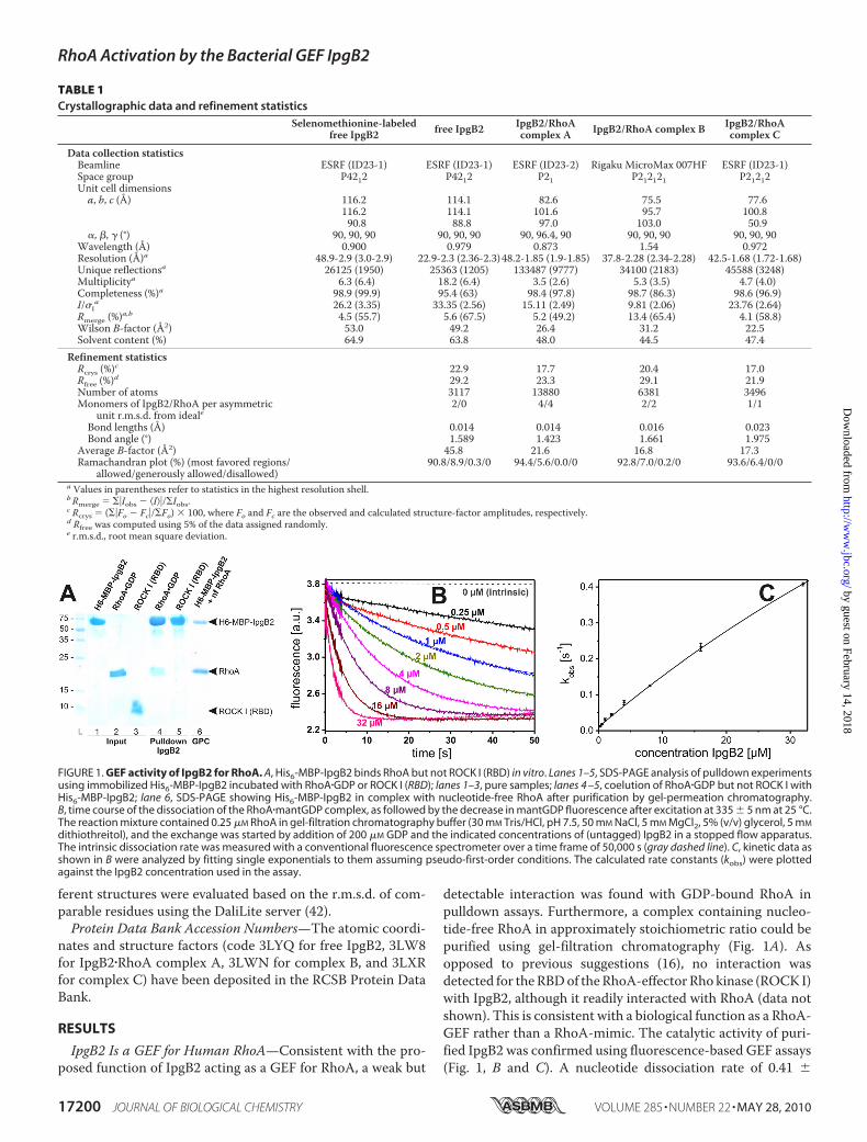
detectable interaction was found with GDP-bound RhoA inpulldown assays. Furthermore, a complex containing nucleo-tide-free RhoA in approximately stoichiometric ratio could bepurified using gel-filtration chromatography (Fig. 1A). Asopposed to previous suggestions (16), no interaction wasdetected for theRBDof theRhoA-effector Rho kinase (ROCK I)with IpgB2, although it readily interacted with RhoA (data notshown). This is consistent with a biological function as a RhoA-GEF rather than a RhoA-mimic. The catalytic activity of puri-fied IpgB2 was confirmed using fluorescence-based GEF assays(Fig. 1, B and C). A nucleotide dissociation rate of 0.41 �
FIGURE 1. GEF activity of IpgB2 for RhoA. A, His6-MBP-IpgB2 binds RhoA but not ROCK I (RBD) in vitro. Lanes 1–5, SDS-PAGE analysis of pulldown experimentsusing immobilized His6-MBP-IpgB2 incubated with RhoA�GDP or ROCK I (RBD); lanes 1–3, pure samples; lanes 4 –5, coelution of RhoA�GDP but not ROCK I withHis6-MBP-IpgB2; lane 6, SDS-PAGE showing His6-MBP-IpgB2 in complex with nucleotide-free RhoA after purification by gel-permeation chromatography.B, time course of the dissociation of the RhoA�mantGDP complex, as followed by the decrease in mantGDP fluorescence after excitation at 335 � 5 nm at 25 °C.The reaction mixture contained 0.25 �M RhoA in gel-filtration chromatography buffer (30 mM Tris/HCl, pH 7.5, 50 mM NaCl, 5 mM MgCl2, 5% (v/v) glycerol, 5 mM
dithiothreitol), and the exchange was started by addition of 200 �M GDP and the indicated concentrations of (untagged) IpgB2 in a stopped flow apparatus.The intrinsic dissociation rate was measured with a conventional fluorescence spectrometer over a time frame of 50,000 s (gray dashed line). C, kinetic data asshown in B were analyzed by fitting single exponentials to them assuming pseudo-first-order conditions. The calculated rate constants (kobs) were plottedagainst the IpgB2 concentration used in the assay.
TABLE 1Crystallographic data and refinement statistics
Selenomethionine-labeledfree IpgB2 free IpgB2 IpgB2/RhoA
complex A IpgB2/RhoA complex B IpgB2/RhoAcomplex C
Data collection statisticsBeamline ESRF (ID23-1) ESRF (ID23-1) ESRF (ID23-2) Rigaku MicroMax 007HF ESRF (ID23-1)Space group P4212 P4212 P21 P212121 P21212Unit cell dimensionsa, b, c (Å) 116.2 114.1 82.6 75.5 77.6
116.2 114.1 101.6 95.7 100.890.8 88.8 97.0 103.0 50.9
�, �, � (°) 90, 90, 90 90, 90, 90 90, 96.4, 90 90, 90, 90 90, 90, 90Wavelength (Å) 0.900 0.979 0.873 1.54 0.972Resolution (Å)a 48.9-2.9 (3.0-2.9) 22.9-2.3 (2.36-2.3)48.2-1.85 (1.9-1.85) 37.8-2.28 (2.34-2.28) 42.5-1.68 (1.72-1.68)Unique reflectionsa 26125 (1950) 25363 (1205) 133487 (9777) 34100 (2183) 45588 (3248)Multiplicitya 6.3 (6.4) 18.2 (6.4) 3.5 (2.6) 5.3 (3.5) 4.7 (4.0)Completeness (%)a 98.9 (99.9) 95.4 (63) 98.4 (97.8) 98.7 (86.3) 98.6 (96.9)I/�I
a 26.2 (3.35) 33.35 (2.56) 15.11 (2.49) 9.81 (2.06) 23.76 (2.64)Rmerge (%)a,b 4.5 (55.7) 5.6 (67.5) 5.2 (49.2) 13.4 (65.4) 4.1 (58.8)Wilson B-factor (Å2) 53.0 49.2 26.4 31.2 22.5Solvent content (%) 64.9 63.8 48.0 44.5 47.4
Refinement statisticsRcrys (%)c 22.9 17.7 20.4 17.0Rfree (%)d 29.2 23.3 29.1 21.9Number of atoms 3117 13880 6381 3496Monomers of IpgB2/RhoA per asymmetric
unit r.m.s.d. from ideale2/0 4/4 2/2 1/1
Bond lengths (Å) 0.014 0.014 0.016 0.023Bond angle (°) 1.589 1.423 1.661 1.975
Average B-factor (Å2) 45.8 21.6 16.8 17.3Ramachandran plot (%) (most favored regions/
allowed/generously allowed/disallowed)90.8/8.9/0.3/0 94.4/5.6/0.0/0 92.8/7.0/0.2/0 93.6/6.4/0/0
a Values in parentheses refer to statistics in the highest resolution shell.bRmerge � �Iobs � I��/Iobs.c Rcrys � (�Fo � Fc�/Fo) � 100, where Fo and Fc are the observed and calculated structure-factor amplitudes, respectively.d Rfree was computed using 5% of the data assigned randomly.e r.m.s.d., root mean square deviation.
RhoA Activation by the Bacterial GEF IpgB2
17200 JOURNAL OF BIOLOGICAL CHEMISTRY VOLUME 285 • NUMBER 22 • MAY 28, 2010
by guest on February 14, 2018http://w
ww
.jbc.org/D
ownloaded from
0.005 s�1 was measured at the highest molar excess of IpgB2over RhoA (128-fold), corresponding to a �104-fold accelera-tion of nucleotide exchange by IpgB2 comparedwith the intrin-sic rate of 3.8� 0.2� 10�5 s�1. This activity is well in the rangeobserved for the bacterial GEF SopE (15, 43) and more than anorder of magnitude higher when compared with mammalianGEFs like p115RhoGEF and p190RhoGEF (44).Structure of IpgB2—IpgB2 consists of two distinct domains:
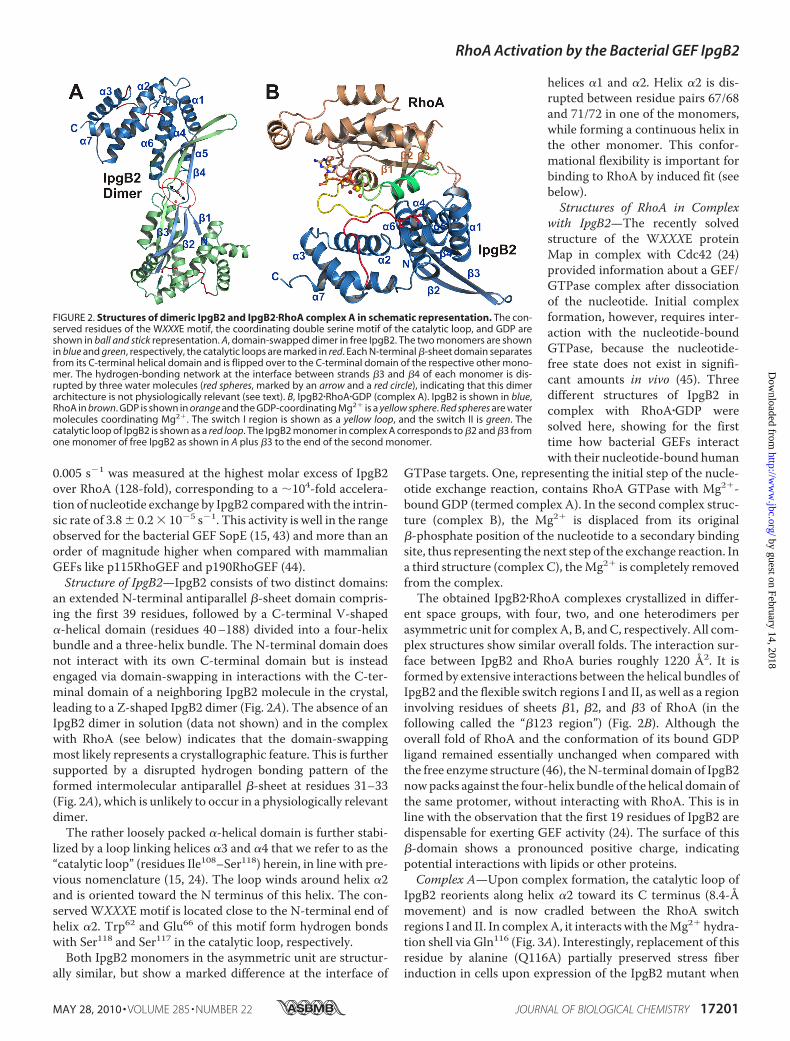
an extended N-terminal antiparallel �-sheet domain compris-ing the first 39 residues, followed by a C-terminal V-shaped�-helical domain (residues 40–188) divided into a four-helixbundle and a three-helix bundle. The N-terminal domain doesnot interact with its own C-terminal domain but is insteadengaged via domain-swapping in interactions with the C-ter-minal domain of a neighboring IpgB2 molecule in the crystal,leading to a Z-shaped IpgB2 dimer (Fig. 2A). The absence of anIpgB2 dimer in solution (data not shown) and in the complexwith RhoA (see below) indicates that the domain-swappingmost likely represents a crystallographic feature. This is furthersupported by a disrupted hydrogen bonding pattern of theformed intermolecular antiparallel �-sheet at residues 31–33(Fig. 2A), which is unlikely to occur in a physiologically relevantdimer.The rather loosely packed �-helical domain is further stabi-
lized by a loop linking helices �3 and �4 that we refer to as the“catalytic loop” (residues Ile108–Ser118) herein, in line with pre-vious nomenclature (15, 24). The loop winds around helix �2and is oriented toward the N terminus of this helix. The con-served WXXXE motif is located close to the N-terminal end ofhelix �2. Trp62 and Glu66 of this motif form hydrogen bondswith Ser118 and Ser117 in the catalytic loop, respectively.
Both IpgB2 monomers in the asymmetric unit are structur-ally similar, but show a marked difference at the interface of
helices �1 and �2. Helix �2 is dis-rupted between residue pairs 67/68and 71/72 in one of the monomers,while forming a continuous helix inthe other monomer. This confor-mational flexibility is important forbinding to RhoA by induced fit (seebelow).Structures of RhoA in Complex
with IpgB2—The recently solvedstructure of the WXXXE proteinMap in complex with Cdc42 (24)provided information about a GEF/GTPase complex after dissociationof the nucleotide. Initial complexformation, however, requires inter-action with the nucleotide-boundGTPase, because the nucleotide-free state does not exist in signifi-cant amounts in vivo (45). Threedifferent structures of IpgB2 incomplex with RhoA�GDP weresolved here, showing for the firsttime how bacterial GEFs interactwith their nucleotide-bound human
GTPase targets. One, representing the initial step of the nucle-otide exchange reaction, contains RhoA GTPase with Mg2�-bound GDP (termed complex A). In the second complex struc-ture (complex B), the Mg2� is displaced from its original�-phosphate position of the nucleotide to a secondary bindingsite, thus representing the next step of the exchange reaction. Ina third structure (complex C), theMg2� is completely removedfrom the complex.The obtained IpgB2�RhoA complexes crystallized in differ-
ent space groups, with four, two, and one heterodimers perasymmetric unit for complexA, B, andC, respectively. All com-plex structures show similar overall folds. The interaction sur-face between IpgB2 and RhoA buries roughly 1220 Å2. It isformed by extensive interactions between the helical bundles ofIpgB2 and the flexible switch regions I and II, as well as a regioninvolving residues of sheets �1, �2, and �3 of RhoA (in thefollowing called the “�123 region”) (Fig. 2B). Although theoverall fold of RhoA and the conformation of its bound GDPligand remained essentially unchanged when compared withthe free enzyme structure (46), theN-terminal domain of IpgB2nowpacks against the four-helix bundle of the helical domain ofthe same protomer, without interacting with RhoA. This is inline with the observation that the first 19 residues of IpgB2 aredispensable for exerting GEF activity (24). The surface of this�-domain shows a pronounced positive charge, indicatingpotential interactions with lipids or other proteins.Complex A—Upon complex formation, the catalytic loop of
IpgB2 reorients along helix �2 toward its C terminus (8.4-Åmovement) and is now cradled between the RhoA switchregions I and II. In complexA, it interacts with theMg2� hydra-tion shell via Gln116 (Fig. 3A). Interestingly, replacement of thisresidue by alanine (Q116A) partially preserved stress fiberinduction in cells upon expression of the IpgB2 mutant when
FIGURE 2. Structures of dimeric IpgB2 and IpgB2�RhoA complex A in schematic representation. The con-served residues of the WXXXE motif, the coordinating double serine motif of the catalytic loop, and GDP areshown in ball and stick representation. A, domain-swapped dimer in free IpgB2. The two monomers are shownin blue and green, respectively, the catalytic loops are marked in red. Each N-terminal �-sheet domain separatesfrom its C-terminal helical domain and is flipped over to the C-terminal domain of the respective other mono-mer. The hydrogen-bonding network at the interface between strands �3 and �4 of each monomer is dis-rupted by three water molecules (red spheres, marked by an arrow and a red circle), indicating that this dimerarchitecture is not physiologically relevant (see text). B, IpgB2�RhoA�GDP (complex A). IpgB2 is shown in blue,RhoA in brown. GDP is shown in orange and the GDP-coordinating Mg2� is a yellow sphere. Red spheres are watermolecules coordinating Mg2�. The switch I region is shown as a yellow loop, and the switch II is green. Thecatalytic loop of IpgB2 is shown as a red loop. The IpgB2 monomer in complex A corresponds to �2 and �3 fromone monomer of free IpgB2 as shown in A plus �3 to the end of the second monomer.
RhoA Activation by the Bacterial GEF IpgB2
MAY 28, 2010 • VOLUME 285 • NUMBER 22 JOURNAL OF BIOLOGICAL CHEMISTRY 17201
by guest on February 14, 2018http://w
ww
.jbc.org/D
ownloaded from
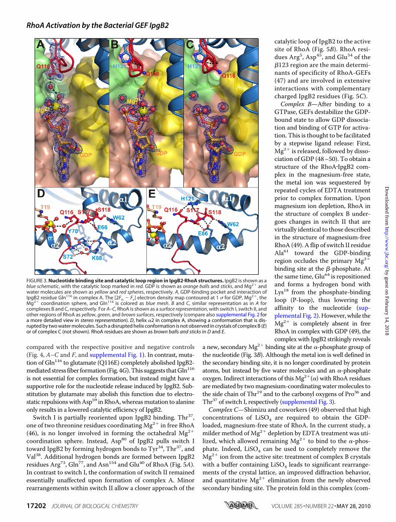
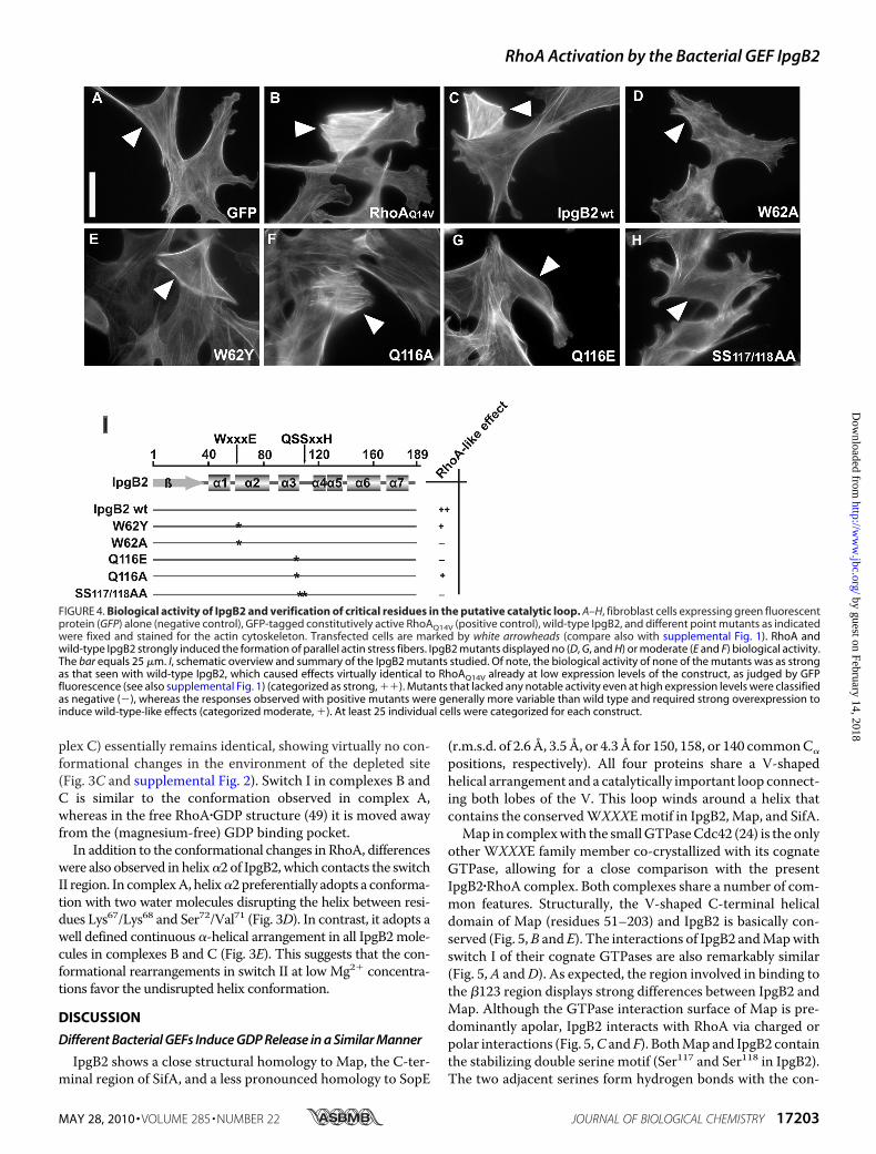
compared with the respective positive and negative controls(Fig. 4, A–C and F, and supplemental Fig. 1). In contrast, muta-tion of Gln116 to glutamate (Q116E) completely abolished IpgB2-mediatedstress fiber formation (Fig. 4G).This suggests thatGln116is not essential for complex formation, but instead might have asupportive role for the nucleotide release induced by IpgB2. Sub-stitution by glutamate may abolish this function due to electro-static repulsionswithAsp59 inRhoA,whereasmutation to alanineonly results in a lowered catalytic efficiency of IpgB2.Switch I is partially reoriented upon IpgB2 binding. Thr37,
one of two threonine residues coordinatingMg2� in free RhoA(46), is no longer involved in forming the octahedral Mg2�
coordination sphere. Instead, Asp80 of IpgB2 pulls switch Itoward IpgB2 by forming hydrogen bonds to Tyr34, Thr37, andVal38. Additional hydrogen bonds are formed between IpgB2residues Arg73, Gln77, and Asn154 and Glu40 of RhoA (Fig. 5A).In contrast to switch I, the conformation of switch II remainedessentially unaffected upon formation of complex A. Minorrearrangements within switch II allow a closer approach of the
catalytic loop of IpgB2 to the activesite of RhoA (Fig. 5B). RhoA resi-dues Arg5, Asp45, and Glu54 of the�123 region are the main determi-nants of specificity of RhoA-GEFs(47) and are involved in extensiveinteractions with complementarycharged IpgB2 residues (Fig. 5C).Complex B—After binding to a
GTPase, GEFs destabilize the GDP-bound state to allow GDP dissocia-tion and binding of GTP for activa-tion. This is thought to be facilitatedby a stepwise ligand release: First,Mg2� is released, followed by disso-ciation of GDP (48–50). To obtain astructure of the RhoA�IpgB2 com-plex in the magnesium-free state,the metal ion was sequestered byrepeated cycles of EDTA treatmentprior to complex formation. Uponmagnesium ion depletion, RhoA inthe structure of complex B under-goes changes in switch II that arevirtually identical to those describedin the structure of magnesium-freeRhoA (49). A flip of switch II residueAla61 toward the GDP-bindingregion occludes the primary Mg2�
binding site at the �-phosphate. Atthe same time, Glu64 is repositionedand forms a hydrogen bond withLys18 from the phosphate-bindingloop (P-loop), thus lowering theaffinity to the nucleotide (sup-plemental Fig. 2).However,while theMg2� is completely absent in freeRhoA in complex with GDP (49), thecomplexwith IpgB2 strikingly reveals
a new, secondary Mg2� binding site at the �-phosphate group ofthe nucleotide (Fig. 3B). Although the metal ion is well defined inthe secondary binding site, it is no longer coordinated by proteinatoms, but instead by five water molecules and an �-phosphateoxygen. Indirect interactions of thisMg2�(�) with RhoA residuesaremediated by twomagnesium-coordinatingwatermolecules tothe side chain of Thr19 and to the carbonyl oxygens of Pro36 andThr37 of switch I, respectively (supplemental Fig. 3).Complex C—Shimizu and coworkers (49) observed that high
concentrations of LiSO4 are required to obtain the GDP-loaded, magnesium-free state of RhoA. In the current study, amilder method ofMg2� depletion by EDTA treatment was uti-lized, which allowed remaining Mg2� to bind to the �-phos-phate. Indeed, LiSO4 can be used to completely remove theMg2� ion from the active site: treatment of complex B crystalswith a buffer containing LiSO4 leads to significant rearrange-ments of the crystal lattice, an improved diffraction behavior,and quantitative Mg2� elimination from the newly observedsecondary binding site. The protein fold in this complex (com-
FIGURE 3. Nucleotide binding site and catalytic loop region in IpgB2�RhoA structures. IpgB2 is shown as ablue schematic, with the catalytic loop marked in red. GDP is shown as orange balls and sticks, and Mg2� andwater molecules are shown as yellow and red spheres, respectively. A, GDP-binding pocket and interaction ofIpgB2 residue Gln116 in complex A. The [2Fo � Fc] electron density map contoured at 1 � for GDP, Mg2�, theMg2� coordination sphere, and Gln116 is colored as blue mesh. B and C, similar representation as in A forcomplexes B and C, respectively. For A–C, RhoA is shown as a surface representation, with switch I, switch II, andother regions of RhoA as yellow, green, and brown surfaces, respectively (compare also supplemental Fig. 2 fora more detailed view in stereo representation). D, helix �2 in complex A, showing a conformation that is dis-rupted by two water molecules. Such a disrupted helix conformation is not observed in crystals of complex B (E)or of complex C (not shown). RhoA residues are shown as brown balls and sticks in D and E.
RhoA Activation by the Bacterial GEF IpgB2
17202 JOURNAL OF BIOLOGICAL CHEMISTRY VOLUME 285 • NUMBER 22 • MAY 28, 2010
by guest on February 14, 2018http://w
ww
.jbc.org/D
ownloaded from
plex C) essentially remains identical, showing virtually no con-formational changes in the environment of the depleted site(Fig. 3C and supplemental Fig. 2). Switch I in complexes B andC is similar to the conformation observed in complex A,whereas in the free RhoA�GDP structure (49) it is moved awayfrom the (magnesium-free) GDP binding pocket.In addition to the conformational changes in RhoA, differences
were also observed in helix�2 of IpgB2, which contacts the switchII region. In complexA, helix�2preferentially adopts a conforma-tion with two water molecules disrupting the helix between resi-dues Lys67/Lys68 and Ser72/Val71 (Fig. 3D). In contrast, it adopts awell defined continuous �-helical arrangement in all IpgB2mole-cules in complexes B and C (Fig. 3E). This suggests that the con-formational rearrangements in switch II at low Mg2� concentra-tions favor the undisrupted helix conformation.
DISCUSSION
Different Bacterial GEFs Induce GDP Release in a Similar Manner
IpgB2 shows a close structural homology to Map, the C-ter-minal region of SifA, and a less pronounced homology to SopE
(r.m.s.d. of 2.6Å, 3.5Å, or 4.3Å for 150, 158, or 140 commonC�
positions, respectively). All four proteins share a V-shapedhelical arrangement and a catalytically important loop connect-ing both lobes of the V. This loop winds around a helix thatcontains the conservedWXXXEmotif in IpgB2,Map, and SifA.Map in complexwith the smallGTPaseCdc42 (24) is the only
other WXXXE family member co-crystallized with its cognateGTPase, allowing for a close comparison with the presentIpgB2�RhoA complex. Both complexes share a number of com-mon features. Structurally, the V-shaped C-terminal helicaldomain of Map (residues 51–203) and IpgB2 is basically con-served (Fig. 5,B and E). The interactions of IpgB2 andMapwithswitch I of their cognate GTPases are also remarkably similar(Fig. 5,A andD). As expected, the region involved in binding tothe �123 region displays strong differences between IpgB2 andMap. Although the GTPase interaction surface of Map is pre-dominantly apolar, IpgB2 interacts with RhoA via charged orpolar interactions (Fig. 5,C and F). BothMap and IpgB2 containthe stabilizing double serine motif (Ser117 and Ser118 in IpgB2).The two adjacent serines form hydrogen bonds with the con-
FIGURE 4. Biological activity of IpgB2 and verification of critical residues in the putative catalytic loop. A–H, fibroblast cells expressing green fluorescentprotein (GFP) alone (negative control), GFP-tagged constitutively active RhoAQ14V (positive control), wild-type IpgB2, and different point mutants as indicatedwere fixed and stained for the actin cytoskeleton. Transfected cells are marked by white arrowheads (compare also with supplemental Fig. 1). RhoA andwild-type IpgB2 strongly induced the formation of parallel actin stress fibers. IpgB2 mutants displayed no (D, G, and H) or moderate (E and F) biological activity.The bar equals 25 �m. I, schematic overview and summary of the IpgB2 mutants studied. Of note, the biological activity of none of the mutants was as strongas that seen with wild-type IpgB2, which caused effects virtually identical to RhoAQ14V already at low expression levels of the construct, as judged by GFPfluorescence (see also supplemental Fig. 1) (categorized as strong, ��). Mutants that lacked any notable activity even at high expression levels were classifiedas negative (�), whereas the responses observed with positive mutants were generally more variable than wild type and required strong overexpression toinduce wild-type-like effects (categorized moderate, �). At least 25 individual cells were categorized for each construct.
RhoA Activation by the Bacterial GEF IpgB2
MAY 28, 2010 • VOLUME 285 • NUMBER 22 JOURNAL OF BIOLOGICAL CHEMISTRY 17203
by guest on February 14, 2018http://w
ww
.jbc.org/D
ownloaded from
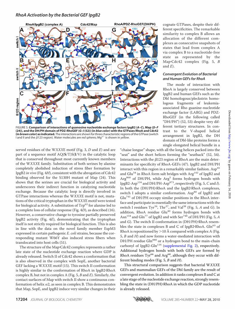
served residues of the WXXXE motif (Fig. 3, D and E) and arepart of a sequence motif AQ(S/T)S(I/V) in the catalytic loopthat is conserved throughout most currently known membersof the WXXXE family. Substitution of both serines by alaninecompletely abolished induction of stress fiber formation byIpgB2 in vivo (Fig. 4H), consistent with the abrogation of Cdc42binding observed for the S130H mutant of Map (24). Thisshows that the serines are crucial for biological activity andunderscores their indirect function in catalyzing nucleotideexchange. Because the catalytic loop is directly involved inGTPase interactions whereas the WXXXE motif is not, muta-tions of the critical tryptophan in theWXXXEmotif were testedfor biological activity. A substitution of Trp62 for alanine led toa complete loss of cellular response (Fig. 4D), as described (16).However, a conservative change to tyrosine partially preservedIpgB2 activity (Fig. 4E), demonstrating that the tryptophanitself is not strictly required for biological function. This is alsoin line with the data on the novel family member EspM3expressed in certain pathogenic E. coli strains, because the cor-responding mutant W66Y also induced stress fibers whentranslocated into host cells (51).The structure of theMap�Cdc42 complex represents a rather
late state of the nucleotide exchange reaction where GDP isalready released. Switch II of Cdc42 shows a conformation thatis also observed in the complex with SopE, another bacterialGEF lacking aWXXXE motif (15). This switch II conformationis highly similar to the conformation of RhoA in IpgB2�RhoAcomplex B, but not in complex A (Fig. 5, B and E). Similarly, thecontact surfaces of Map with switch II show a continuous con-formation of helix �2, as seen in complex B. This demonstratesthat Map, SopE, and IpgB2 induce very similar changes in their
cognate GTPases, despite their dif-ferent specificities. The remarkablesimilarity to complex B allows anallocation of the different com-plexes as consecutive snapshots ofstates that lead from complex Avia complex B to a nucleotide-freestate as represented by theMap�Cdc42 complex (Fig. 5, Band E).
Convergent Evolution of Bacterialand Human GEFs for RhoA
The mode of interaction withRhoA is largely conserved betweenIpgB2 and human GEFs such as theDbl homologous/pleckstrin homo-logous fragments of leukemia-associated Rho guanine-nucleotideexchange factor (LARG) and PDZ-RhoGEF (in the following called“DH/PH”) (52, 53) despite very dif-ferent tertiary structures. In con-trast to the V-shaped helicalarrangement in IpgB2, the DHdomain of Dbl-like proteins forms asingle elongated helical bundle in a
“chaise longue” shape, with all the long helices packed into the“seat” and the short helices forming the “seatback” (52, 54).Interactions with the �123 region of RhoA are the main deter-minants for specificity of RhoA-GEFs (47). IpgB2 and DH/PHinteract with this region in a remarkably similar fashion. Asp45
and Glu54 in RhoA form salt bridges with Arg142 of IpgB2 andArg868 of DH/PH, while Arg5 forms hydrogen bonds withIpgB2-Asp134 and DH/PH-Asp873, respectively (Fig. 5,C and I).In both the (DH/PH)�RhoA and the IpgB2�RhoA complexes,switch I adopts a similar conformation. Asp80 of IpgB2 andGlu741 of DH/PH occupy similar positions in the RhoA inter-face andparticipate in essentially the same interactionswith theswitch I residues Tyr34, Thr37, and Val38 (Fig. 5, A and G). Inaddition, RhoA residue Glu40 forms hydrogen bonds withAsn154 and Gln77 of IpgB2 and with Ser748 of DH/PH (Fig. 5, Aand G). The switch II conformation in (DH/PH)�RhoA resem-bles the state in complexes B and C of IpgB2�RhoA. Gln63 ofRhoA is repositioned by�10 Å compared with complex A (Fig.5, B and H) and now forms a water-mediated interaction withDH/PH residue Gln880 or a hydrogen bond to the main-chaincarbonyl of IpgB2-Gln116 (supplemental Fig. 2), respectively.Additional hydrogen bonds with both GEFs are formed byRhoA residues Tyr66 and Arg68, although they occur with dif-ferent binding modes (Fig. 5, B and H).This structural comparison suggests that bacterial WXXXE
GEFs and mammalian GEFs of the DbI family are the result ofconvergent evolution. In addition it ranks complexes B andC ata late stage of the nucleotide exchange reaction, strongly resem-bling the state in (DH/PH)�RhoA in which the GDP nucleotideis already released.
FIGURE 5. Comparison of interactions of guanosine nucleotide exchange factors IpgB2 (A–C), Map (D–F(24)), and the DH/PH domain of PDZ-RhoGEF (G–I (52)) (in blue color) with the GTPases RhoA and Cdc42(in brown color) as indicated. The interactions are shown for three characteristic regions of the GTPase (switchI and II and the �123 region). Water molecules are red spheres; Mg2� is shown in yellow.
RhoA Activation by the Bacterial GEF IpgB2
17204 JOURNAL OF BIOLOGICAL CHEMISTRY VOLUME 285 • NUMBER 22 • MAY 28, 2010
by guest on February 14, 2018http://w
ww
.jbc.org/D
ownloaded from
Role of the WXXXE Motif in Bacterial GEFs
The crystal structures of SifA and a Map�Cdc42 complexalready revealed that WXXXE proteins are GEFs instead ofGTPasemimics (23, 24, 30). Information onmechanistic detailsand GTPase selectivity of WXXXE proteins, however, is stilllimited. The structures of free andRhoA-complexed IpgB2 pre-sented in this work together with the structures of SifA andMap�Cdc42 now provide a framework for themode of action ofbacterialWXXXE proteins. Although theWXXXEmotif is onlyindirectly required for catalytic activity, residues of the catalyticloop form direct interactions with the cognate GTPase. Forthis, one important function of theWXXXEmotif is to stabilizethe catalytic loop by forming interactions with the double ser-ine motif (Ser117/Ser118). This allows productive interactionswith the cognate GTPase, e.g. of the catalytically important cat-alytic loop residue Gln116 with the Mg2� hydration shell. Thedouble serine motif is strictly required for activity, and interac-tions involving the WXXXE motif are strikingly conserved inIpgB2 and Map. SifA most likely utilizes a similar stabilization,despite harboring a more extended catalytic loop that is stabi-lized by several proline residues, leading to a different overallloop conformation (supplemental Fig. 4A). His255 of the cata-lytic loop in SifA is oriented toward Trp197 and Glu201 of theWXXXE motif, suggesting that it substitutes the hydrogenbonding potential of the two serine residues present in IpgB2,Map, and numerous otherWXXXE proteins (supplemental Fig.4B). However, the question arises whether the significant struc-tural differences in the catalytic loop of SifA compared withIpgB2 suggest that RhoA might not be the main target of SifA,as recently proposed by Ohlsen et al. (23). An overlay withIpgB2�RhoA reveals highly unfavorable interactions betweenSifA and RhoA, particularly within the specificity determiningregions (supplemental Fig. 4C). Interestingly, phenotypesinduced by SifA in host cells as the formation of Salmonella-induced filaments (Sifs) do not typically coincide with RhoAactivation but could derive, rather, from interference with Rab-GTPase signaling.We conclude that the WXXXE motif in all family members
forms an essential part of a conserved structural motif, which iscomplemented by the catalytic loop motif AQ(S/T)S(I/V) orrelated residues in the distinct sequence of SifA. Interactionsbetween bothmotifs promote a functionally competent confor-mation of the catalytic loop that is required for productiveinteractions with the cognate GTPase. The role of the differentcatalytic loop in SifA still requires further analysis and mightreveal other GTPases as targets of SifA whichmight not belongto the Rho family.
GEF Mechanism of Bacterial WXXXE Proteins
Various families of mammalian Rho-GTPase specific GEFsexist that activate their cognate GTPases by different mecha-nisms. The largest of those is the Dbl family of GEFs containinga DH/PH domain. In contrast to other Rho-specific GEFs likeproteins from the DOCK family (55), DbI proteins do notdirectly insert residues into the active site of their cognateGTPase (4). Bacterial WXXXE GEFs utilize a similar mecha-nism for GTPase activation, despite having a different overall
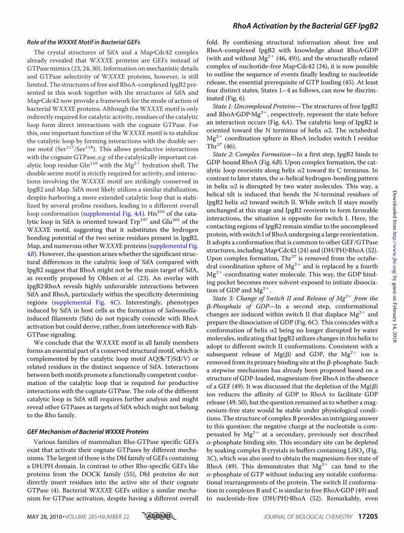
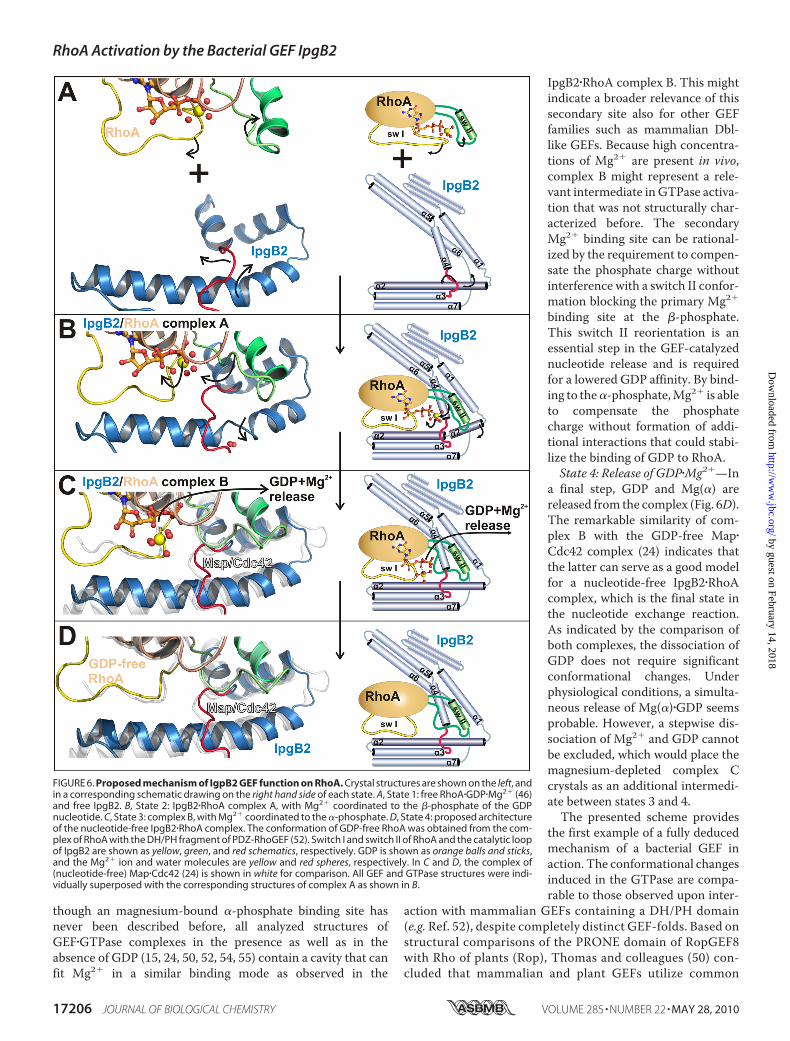
fold. By combining structural information about free andRhoA-complexed IpgB2 with knowledge about RhoA�GDP(with and without Mg2� (46, 49)), and the structurally relatedcomplex of nucleotide-free Map�Cdc42 (24), it is now possibleto outline the sequence of events finally leading to nucleotiderelease, the essential prerequisite of GTP loading (45). At leastfour distinct states, States 1–4 as follows, can now be discrim-inated (Fig. 6).State 1: Uncomplexed Proteins—The structures of free IpgB2
and RhoA�GDP�Mg2�, respectively, represent the state beforean interaction occurs (Fig. 6A). The catalytic loop of IpgB2 isoriented toward the N terminus of helix �2. The octahedralMg2� coordination sphere in RhoA includes switch I residueThr37 (46).State 2: Complex Formation—In a first step, IpgB2 binds to
GDP-bound RhoA (Fig. 6B). Upon complex formation, the cat-alytic loop reorients along helix �2 toward its C terminus. Incontrast to later states, the�-helical hydrogen-bonding patternin helix �2 is disrupted by two water molecules. This way, ahelical tilt is induced that bends the N-terminal residues ofIpgB2 helix �2 toward switch II. While switch II stays mostlyunchanged at this stage and IpgB2 reorients to form favorableinteractions, the situation is opposite for switch I. Here, thecontacting regions of IpgB2 remain similar to the uncomplexedprotein, with switch I of RhoAundergoing a large reorientation.It adopts a conformation that is common to other GEF/GTPasestructures, includingMap�Cdc42 (24) and (DH/PH)�RhoA (52).Upon complex formation, Thr37 is removed from the octahe-dral coordination sphere of Mg2� and is replaced by a fourthMg2�-coordinating water molecule. This way, the GDP bind-ing pocket becomes more solvent-exposed to initiate dissocia-tion of GDP and Mg2�.State 3: Change of Switch II and Release of Mg2� from the
�-Phosphate of GDP—In a second step, conformationalchanges are induced within switch II that displace Mg2� andprepare the dissociation of GDP (Fig. 6C). This coincides with aconformation of helix �2 being no longer disrupted by watermolecules, indicating that IpgB2 utilizes changes in this helix toadopt to different switch II conformations. Consistent with asubsequent release of Mg(�) and GDP, the Mg2� ion isremoved from its primary binding site at the�-phosphate. Sucha stepwise mechanism has already been proposed based on astructure ofGDP-loaded,magnesium-free RhoA in the absenceof a GEF (49). It was discussed that the depletion of the Mg(�)ion reduces the affinity of GDP to RhoA to facilitate GDPrelease (49, 50), but the question remained as towhether amag-nesium-free state would be stable under physiological condi-tions. The structure of complexBprovides an intriguing answerto this question: the negative charge at the nucleotide is com-pensated by Mg2� at a secondary, previously not described�-phosphate binding site. This secondary site can be depletedby soaking complex B crystals in buffers containing LiSO4 (Fig.3C), which was also used to obtain the magnesium-free state ofRhoA (49). This demonstrates that Mg2� can bind to the�-phosphate of GTP without inducing any notable conforma-tional rearrangements of the protein. The switch II conforma-tion in complexes B and C is similar to free RhoA�GDP (49) andto nucleotide-free (DH/PH)�RhoA (52). Remarkably, even
RhoA Activation by the Bacterial GEF IpgB2
MAY 28, 2010 • VOLUME 285 • NUMBER 22 JOURNAL OF BIOLOGICAL CHEMISTRY 17205
by guest on February 14, 2018http://w
ww
.jbc.org/D
ownloaded from
though an magnesium-bound �-phosphate binding site hasnever been described before, all analyzed structures ofGEF�GTPase complexes in the presence as well as in theabsence of GDP (15, 24, 50, 52, 54, 55) contain a cavity that canfit Mg2� in a similar binding mode as observed in the
IpgB2�RhoA complex B. This mightindicate a broader relevance of thissecondary site also for other GEFfamilies such as mammalian Dbl-like GEFs. Because high concentra-tions of Mg2� are present in vivo,complex B might represent a rele-vant intermediate in GTPase activa-tion that was not structurally char-acterized before. The secondaryMg2� binding site can be rational-ized by the requirement to compen-sate the phosphate charge withoutinterference with a switch II confor-mation blocking the primary Mg2�
binding site at the �-phosphate.This switch II reorientation is anessential step in the GEF-catalyzednucleotide release and is requiredfor a lowered GDP affinity. By bind-ing to the�-phosphate,Mg2� is ableto compensate the phosphatecharge without formation of addi-tional interactions that could stabi-lize the binding of GDP to RhoA.State 4: Release of GDP�Mg2�—In
a final step, GDP and Mg(�) arereleased from the complex (Fig. 6D).The remarkable similarity of com-plex B with the GDP-free Map�Cdc42 complex (24) indicates thatthe latter can serve as a good modelfor a nucleotide-free IpgB2�RhoAcomplex, which is the final state inthe nucleotide exchange reaction.As indicated by the comparison ofboth complexes, the dissociation ofGDP does not require significantconformational changes. Underphysiological conditions, a simulta-neous release of Mg(�)�GDP seemsprobable. However, a stepwise dis-sociation of Mg2� and GDP cannotbe excluded, which would place themagnesium-depleted complex Ccrystals as an additional intermedi-ate between states 3 and 4.The presented scheme provides
the first example of a fully deducedmechanism of a bacterial GEF inaction. The conformational changesinduced in the GTPase are compa-rable to those observed upon inter-
action with mammalian GEFs containing a DH/PH domain(e.g. Ref. 52), despite completely distinct GEF-folds. Based onstructural comparisons of the PRONE domain of RopGEF8with Rho of plants (Rop), Thomas and colleagues (50) con-cluded that mammalian and plant GEFs utilize common
FIGURE 6. Proposed mechanism of IgpB2 GEF function on RhoA. Crystal structures are shown on the left, andin a corresponding schematic drawing on the right hand side of each state. A, State 1: free RhoA�GDP�Mg2� (46)and free IpgB2. B, State 2: IpgB2�RhoA complex A, with Mg2� coordinated to the �-phosphate of the GDPnucleotide. C, State 3: complex B, with Mg2� coordinated to the �-phosphate. D, State 4: proposed architectureof the nucleotide-free IpgB2�RhoA complex. The conformation of GDP-free RhoA was obtained from the com-plex of RhoA with the DH/PH fragment of PDZ-RhoGEF (52). Switch I and switch II of RhoA and the catalytic loopof IpgB2 are shown as yellow, green, and red schematics, respectively. GDP is shown as orange balls and sticks,and the Mg2� ion and water molecules are yellow and red spheres, respectively. In C and D, the complex of(nucleotide-free) Map�Cdc42 (24) is shown in white for comparison. All GEF and GTPase structures were indi-vidually superposed with the corresponding structures of complex A as shown in B.
RhoA Activation by the Bacterial GEF IpgB2
17206 JOURNAL OF BIOLOGICAL CHEMISTRY VOLUME 285 • NUMBER 22 • MAY 28, 2010
by guest on February 14, 2018http://w
ww
.jbc.org/D
ownloaded from
intermediate states in small G protein/GEF reactions. Ourwork shows that this notion can now be extended to bacterialWXXXE GEFs.
Conclusions
We report the first crystal structure of a freeWXXXE fam-ily member. The structure of IpgB2 from S. flexneri com-bined with its complexes with RhoA allows a structuraldescription of the sequence of events accompanying GDPrelease induced by this bacterial GEF, which is an essentialprerequisite for GTP loading. A function as a GTPase mim-icry protein, as proposed previously for IpgB2, can now beclearly ruled out. In addition, our data show the formation ofan intermediate state with a magnesium ion relocalized fromthe �-phosphate to the �-phosphate of GDP. This way, thephosphate charge gets compensated without interferencewith a low GDP-affinity state, induced by switch II blockingtheMg2�-binding site at the �-phosphate. It is plausible thatthe molecular mechanism of GDP release involves a simul-taneous dissociation of GDP�Mg2�(�) from the GEF�GTPasecomplex. In such a scenario, stable association of a complexcontaining GDP but lacking Mg2� is not required to explainthe GEF-induced nucleotide release. The depicted mecha-nism might also be considered as a model for mammalianRhoA-GEFs, which have not been crystallized as GEF�RhoA�GDP complexes so far.
Acknowledgments—We thank Thorsten Luhrs for performing NMRexperiments on 15N- and 13C-labelled IpgB2,ManfredNimtz formassspectrometry, Christiane Ritter for technical support during thestopped flow experiments, and Joachim Reichelt and Kevin Walklingfor technical assistance. We also thank Gunhild Layer (TechnicalUniversity Braunschweig (TU)) for sharing equipment and JohannesWalther (TU) for technical support during high-performance liquidchromatography experiments. We express our gratitude to the staff ofbeamlines ID14-4, ID23-1, ID23-2 and ID29 of the European Syn-chrotron Radiation Facility, Grenoble, France, of BL14.1 and BL14.2,Berliner Elektronen-Speicherring Gesellschaft fur Synchrotronstrahl-ung (BESY), Berlin, Germany, and of Deutsches Elektronen Synchro-tron (DESY), Hamburg, Germany, for access to beam time and assist-ance during data collection.
REFERENCES1. Ladwein, M., and Rottner, K. (2008) FEBS Lett. 582, 2066–20742. Hall, A. (1998) Science 279, 509–5143. Burridge, K., and Wennerberg, K. (2004) Cell 116, 167–1794. Rossman, K. L., Der, C. J., and Sondek, J. (2005)Nat. Rev.Mol. Cell. Biol. 6,
167–1805. Chrzanowska-Wodnicka, M., and Burridge, K. (1996) J. Cell Biol. 133,
1403–14156. Gallo, G., and Letourneau, P. C. (2004) J. Neurobiol. 58, 92–1027. Nobes, C. D., and Hall, A. (1995) Biochem. Soc. Trans. 23, 456–4598. Ridley, A. J., Paterson, H. F., Johnston, C. L., Diekmann, D., and Hall, A.
(1992) Cell 70, 401–4109. Aktories, K., and Barbieri, J. T. (2005) Nat. Rev. Microbiol. 3, 397–41010. Vogelsgesang,M., Pautsch, A., and Aktories, K. (2007)Naunyn Schmiede-
bergs Arch. Pharmacol. 374, 347–36011. Ridley, A. J., and Hall, A. (1992) Cell 70, 389–39912. Galan, J. E. (2009) Cell Host. Microbe 5, 571–57913. Rottner, K., Stradal, T. E., and Wehland, J. (2005) Dev. Cell 9, 3–17
14. Boquet, P., and Lemichez, E. (2003) Trends Cell Biol. 13, 238–24615. Buchwald, G., Friebel, A., Galan, J. E., Hardt, W. D., Wittinghofer, A., and
Scheffzek, K. (2002) EMBO J. 21, 3286–329516. Alto, N. M., Shao, F., Lazar, C. S., Brost, R. L., Chua, G., Mattoo, S., Mc-
Mahon, S. A., Ghosh, P., Hughes, T. R., Boone, C., and Dixon, J. E. (2006)Cell 124, 133–145
17. Hayward, R. D., and Koronakis, V. (2006) Cell 124, 15–1718. Handa, Y., Suzuki,M., Ohya, K., Iwai, H., Ishijima, N., Koleske, A. J., Fukui,
Y., and Sasakawa, C. (2007) Nat. Cell Biol. 9, 121–12819. Ohya, K., Handa, Y., Ogawa, M., Suzuki, M., and Sasakawa, C. (2005)
J. Biol. Chem. 280, 24022–2403420. Arbeloa, A., Blanco, M., Moreira, F. C., Bulgin, R., Lupez, C., Dahbi, G.,
Blanco, J. E.,Mora, A., Alonso,M. P.,Mamani, R. C., Gomes, T. A., Blanco,J., and Frankel, G. (2009) J. Med. Microbiol. 58, 988–995
21. Bulgin, R. R., Arbeloa, A., Chung, J. C., and Frankel, G. (2009) Cell Micro-biol. 11, 217–229
22. Arbeloa, A., Garnett, J., Lillington, J., Bulgin, R. R., Berger, C., Lea, S. M.,Matthews, S., and Frankel, G. (2009) Cell Microbiol., in press
23. Ohlson,M. B.,Huang, Z., Alto,N.M., Blanc,M. P., Dixon, J. E., Chai, J., andMiller, S. I. (2008) Cell Host. Microbe 4, 434–446
24. Huang, Z., Sutton, S. E.,Wallenfang, A. J., Orchard, R. C.,Wu, X., Feng, Y.,Chai, J., and Alto, N. M. (2009) Nat. Struct. Mol. Biol. 16, 853–860
25. Stebbins, C. E., and Galan, J. E. (2001) Nature 412, 701–70526. Shames, S. R., Auweter, S. D., and Finlay, B. B. (2009) Int. J. Biochem. Cell
Biol. 41, 380–38927. Eberth, A., and Ahmadian, M. R. (2009) Curr. Protoc. Cell Biol., Chapter
14, 14.9.1–14.9.2528. Hutchinson, J. P., and Eccleston, J. F. (2000) Biochemistry 39,
11348–1135929. Zhang, B., Zhang, Y., Wang, Z., and Zheng, Y. (2000) J. Biol. Chem. 275,
25299–2530730. Diacovich, L., Dumont, A., Lafitte, D., Soprano, E., Guilhon, A. A., Bignon,
C., Gorvel, J. P., Bourne, Y., and Meresse, S. (2009) J. Biol. Chem. 284,33151–33160
31. Kabsch, W. (1993) J. Appl. Crystallogr. 26, 795–80032. Kabsch, W. (1988) J. Appl. Crystallogr. 21, 916–92433. Pape, T., and Schneider, T. R. (2004) J. Appl. Crystallogr. 37, 843–84434. Sheldrick, G. M. (2008) Acta Crystallogr. Sect. A 64, 112–12235. Adams, P. D., Gopal, K., Grosse-Kunstleve, R. W., Hung, L. W., Ioerger,
T. R., McCoy, A. J., Moriarty, N. W., Pai, R. K., Read, R. J., Romo, T. D.,Sacchettini, J. C., Sauter, N. K., Storoni, L. C., and Terwilliger, T. C. (2004)J. Synchrotron. Radiat. 11, 53–55
36. Collaborative Computational Project Number 4 (1994) Acta Crystallogr.D Biol. Crystallogr. 50, 760–763
37. Dvorsky, R., Blumenstein, L., Vetter, I. R., and Ahmadian, M. R. (2004)J. Biol. Chem. 279, 7098–7104
38. Lamzin, V. S., and Wilson, K. S. (1993) Acta Crystallogr. D Biol. Crystal-logr. 49, 129–147
39. Emsley, P., and Cowtan, K. (2004)Acta Crystallogr. D Biol. Crystallogr. 60,2126–2132
40. Murshudov, G. N., Vagin, A. A., and Dodson, E. J. (1997)Acta Crystallogr.D Biol. Crystallogr. 53, 240–255
41. DeLano, W. L. (2002) The PyMOL Molecular Graphics System, DeLanoScientific LLC, San Carlos, CA
42. Holm, L., and Park, J. (2000) Bioinformatics 16, 566–56743. Rudolph, M. G., Weise, C., Mirold, S., Hillenbrand, B., Bader, B., Witting-
hofer, A., and Hardt, W. D. (1999) J. Biol. Chem. 274, 30501–3050944. Hausler, L. C. (2004) Aktivierung von GTPasen der Rho-Familie. Ph.D.
Thesis, Bochum, Ruhr-Universitat Bochum45. Cherfils, J., and Chardin, P. (1999) Trends Biochem. Sci. 24, 306–31146. Wei, Y., Zhang, Y., Derewenda, U., Liu, X., Minor, W., Nakamoto, R. K.,
Somlyo, A. V., Somlyo, A. P., andDerewenda, Z. S. (1997)Nat. Struct. Biol.4, 699–703
47. Snyder, J. T., Worthylake, D. K., Rossman, K. L., Betts, L., Pruitt, W. M.,Siderovski, D. P., Der, C. J., and Sondek, J. (2002) Nat. Struct. Biol. 9,468–475
48. Freymann, D. M., Keenan, R. J., Stroud, R. M., and Walter, P. (1999) Nat.Struct. Biol. 6, 793–801
RhoA Activation by the Bacterial GEF IpgB2
MAY 28, 2010 • VOLUME 285 • NUMBER 22 JOURNAL OF BIOLOGICAL CHEMISTRY 17207
by guest on February 14, 2018http://w
ww
.jbc.org/D
ownloaded from
49. Shimizu, T., Ihara, K., Maesaki, R., Kuroda, S., Kaibuchi, K., and Hako-shima, T. (2000) J. Biol. Chem. 275, 18311–18317
50. Thomas, C., Fricke, I., Scrima, A., Berken, A., andWittinghofer, A. (2007)Mol. Cell 25, 141–149
51. Arbeloa, A., Bulgin, R. R., MacKenzie, G., Shaw, R. K., Pallen, M. J.,Crepin, V. F., Berger, C. N., and Frankel, G. (2008) Cell Microbiol. 10,1429–1441
52. Derewenda, U., Oleksy, A., Stevenson, A. S., Korczynska, J., Dauter, Z.,
Somlyo, A. P., Otlewski, J., Somlyo, A. V., and Derewenda, Z. S. (2004)Structure 12, 1955–1965
53. Kristelly, R., Gao, G., and Tesmer, J. J. (2004) J. Biol. Chem. 279,47352–47362
54. Worthylake, D. K., Rossman, K. L., and Sondek, J. (2000) Nature 408,682–688
55. Yang, J., Zhang, Z., Roe, S. M., Marshall, C. J., and Barford, D. (2009)Science 325, 1398–1402
RhoA Activation by the Bacterial GEF IpgB2
17208 JOURNAL OF BIOLOGICAL CHEMISTRY VOLUME 285 • NUMBER 22 • MAY 28, 2010
by guest on February 14, 2018http://w
ww
.jbc.org/D
ownloaded from
Ladwein, Theresia E. B. Stradal, Klemens Rottner and Dirk W. HeinzBjörn U. Klink, Stephan Barden, Thomas V. Heidler, Christina Borchers, Markus
EXCHANGE FACTOR MIMICRYFOR THE MECHANISM OF BACTERIAL GUANINE NUCLEOTIDE
IpgB2 in Complex with Human RhoA: IMPLICATIONSShigellaStructure of
doi: 10.1074/jbc.M110.107953 originally published online April 2, 20102010, 285:17197-17208.J. Biol. Chem.
10.1074/jbc.M110.107953Access the most updated version of this article at doi:
Alerts:
When a correction for this article is posted•
When this article is cited•
to choose from all of JBC's e-mail alertsClick here
Supplemental material:
http://www.jbc.org/content/suppl/2010/04/02/M110.107953.DC1
http://www.jbc.org/content/285/22/17197.full.html#ref-list-1
This article cites 51 references, 12 of which can be accessed free at
by guest on February 14, 2018http://w
ww
.jbc.org/D
ownloaded from