structured reporting quality storyboard rsna 2016 final · 2017. 1. 19. · quality storyboard-...
TRANSCRIPT
1/13/2017
1
Implementing a Structured Reporting Initiative
Using a Collaborative and Iterative Approach and Phased Template
Rollout
Quality Storyboard- Education Exhibit RSNA 2016
Shlomit Goldberg-Stein, MDWilliam Walter, MDE. Stephen Amis Jr., MDMeir H. Scheinfeld, MD, PhD
Purpose
• To describe the implementation of a structured reporting initiative at a large multi-site, academic medical center radiology department
• To provide a blueprint of how to successfully achieve structured reporting using a collaborative multistep approach
1/13/2017
2
What is a Structured Radiology Report?
Report with a consistent, standard format
Expected data elements (such as patient name, medical record number, time study performed etc.) are present
Normal exam reports for a given exam are always the same
My normal and your normal exam reports do not sound different referring physicians don’t have to “interpret” your report
May or may not include standard lexicon (standardized language)
Why should Structured Reporting be used?
There is a growing body of evidence that structured reporting improves the quality and value of radiologists’ major work product, the radiology report:
Radiologists can compare reports more easilyThe report structure functions as a checklist, ensuring that images are interpreted in a consistent mannerReferring clinicians can find “what they are looking for” more easily
Radiology leaders have called for optimization of reporting and the delivery of actionable information in radiology reports as a critical link in the imaging value chain
1/13/2017
3
Structured reporting can break down sophisticated tasks into manageable and automated pieces
A uniform lexicon helps diminish complexity
Structured reporting makes it easier to concentrate on the images, and errors in both perception and interpretation would decrease
Ordering physicians would also find the reports to be more readable
Ellenbogen PH. J Am Coll Radiol. 2013 Sep;10(9):641.
• Compared content, clarity, and clinical usefulness of conventional (i.e., free-form) and structured radiology reports of body CT’s
• Reports were evaluated by referring physicians, attending radiologists and radiology fellows
• Mean content and clarity satisfaction ratings were significantly better ( p <.0001) for structured reports.
• Conclusion: Referring clinicians and radiologists found that structured reports had better content and greater clarity than conventional reports
Radiology. 2011 Jul;260(1):174-81.
1/13/2017
4
Free text vs. Structured Reporting• “Some radiologists believe their true value lies in
crafting free-text reporting according to personal preferences. This idiosyncratic approach undermines actionable reporting because referring physicians are left having to navigate reports to find key actionable information, which inevitably varies from one radiologist to another”
• Structured reporting allows for a consistent and predictable report format, with easy navigation through the report to extract the necessary information
• May be disease-specific so that critical information will always be included in the report
• Promotes adherence to radiology practice guidelines
Boland G, Enzmann D, Duszak R, Actionable Reporting, JACR. 2014 Sep;11(9):844-5.
SettingUrban tertiary care medical center which performs 650,000 radiology exams/year
Includes 3 hospitals and 5 outpatient imaging facilities
Department includes 88 faculty, 36 residents, 13 fellows
Powerscribe 360 (Nuance, Burlington, MA) is used as the voice recognition system
A single PACS and RIS system (Centricity, GE healthcare, Chicago, IL) are used at all sites
Prior to the Structured Reporting initiative, all attending and resident radiologists used a mix of free text and personally developed or cloned reporting templates, based on personal preference
1/13/2017
5
The challenges
Technical
Organizational
The human component
Balance the need for some uniformity, with the need to respect each radiologists expertise and opinion
Achieve systemic consistency while preserving physician autonomy
How can we motivate a large group of radiologists to agree on
Structured Reporting?
Consensus-building efforts are criticalImplement structured reporting such that radiologists will prefer to use structured reports rather than creating, maintaining, and using their own macros or report templates
1/13/2017
6
Outline of our processPlanning
Educate staff and trainees (8 weeks)
Create multiple teams (2 weeks)
Survey users (6 weeks)
Template creation and multi-step refinement (5 steps, 8-44 weeks)
Template Implementation
Evaluation of our implementation and radiologists’ compliance
Planning Three ground rules were decided upon in consultation with the department chairman
1. The initiative would be limited to cross-sectional exams (CT, MRI, US), with the goal of having ≥ 90% of dictated studies having structured templates
2. Structured reporting would be implemented using a consensus building process (rather than a top down) approach
3. Reports would be structured but no restrictions would be made on the actual language used
Two co-chairs were selected to lead the project
No financial incentives or penalties would be linked to this initiative
1/13/2017
7
Educate (8 weeks)Committee co-chairs formally presented concepts of structured reporting to staff radiologists and trainees, including relevant publications that examined both the benefits and challenges of structured reporting
Defined goals of the structured reporting initiative were publicized
Attempts made to directly address specific concerns from radiologists who were skeptical of the new initiative
Create multiple teams (2 weeks)The co-chairs formed subcommittees of subspecialist radiologists and additional stakeholder radiologists (off-site from main campus) to allow for focused input into template creation for both choosing content and appropriate language
Subcommittees also included resident representatives for trainee input
These radiologists were relied upon to integrate the concerns and needs of their referring clinician base into the process
Continued on next slide
1/13/2017
8
Create multiple teams (2 weeks)6 subcommittees were formed:
Abdominal, Cardiothoracic, Musculoskeletal, Pediatric, Ultrasound, Neuroradiology
Some subcommittees worked independently, others relied more heavily on the co-chairs
For each template created, a formal template trial process allowed for feedback from all readers (both attendings and trainees)
There were no administrators or administrative assistants involved in the process.
Survey users (6 weeks)A department-wide survey was distributed electronically to all staff and trainees to collect data on user navigation preferences
Survey questions included Preferred method of moving between fields in a template
Use of “pick lists”
Use of field names
Use of blank fields
This was done to help inform global template structure and formatting decisions
1/13/2017
9
Template creation and multi-step refinement
Step 1: Template drafting (4-30 weeks) Step 2: Limited trial and voting (1-6 weeks) Step 3: Site-wide trial (1-3 weeks)Step 4: Template finalization (1-2 weeks)Step 5: Final technical checks (1-3 weeks)
Step 1: Template drafting (4-30 weeks)
The creation of every structured reporting template was intentionally a multi-step process
Template drafts were created, edited, trialed, and re-trialed until the greatest possible consensus was achieved
After initial template drafting by sub-specialty physician champion(s), the committee co-chairs edited the draft to meet defined criteria, including a standard header with radiology information system (RIS) data merge fields for patient and exam information, contrast material information, and clear and grammatically correct language
Pick-list options for common abnormalities and variants were crafted to allow for quick and typo-free report creation
1/13/2017
10
Step 2: Limited trial and voting (1-6 weeks)
A limited-user, two-week testing period for high-frequency readers of that exam type was first performed to test usability of the template and obtain focused feeedback
We reasoned that the best way to determine whether a template was ready for use was to attempt to use it in clinical practice
Draft templates were configured such that a normal workflow could be followed, with auto-population of the template with the launch of an exam for all trial radiologists
A simple email-based mechanism for feedback was established, with all comments and suggestions sent to the committee co-chairs and/or subcommittee members
These were compiled and distributed formally in a forum allowing for discussion or voting on details (when necessary)
Step 3: Site-wide trial (1-3 weeks)
The revised template draft then entered a site-wide two-week trial, open to all radiologists responsible for reading that exam type
This included rare readers (i.e. those reading that exam type only when on-call, a few days a month).
All trainees were also invited to participate in this trial
Again, all written template feedback was compiled by the committee co-chairs and formally distributed for discussion and voting (when necessary)
1/13/2017
11
Step 4: Template finalization (1-2 weeks)
The compiled feedback and voting results were shared again with stakeholders, together with a final template draft, revised by the committee co-chairs
The co-chairs optimized and standardized the voice command fields, managed the naming of template pick-list options, and established standard language macros as required
Step 5: Final technical checks (1-3 weeks)
Collaboration with radiology department coders was necessary to ensure matching of templates to proper exam codes
This was essential for the correct exam template to auto-populate at the launch of each exam
In some cases, the addition of new exam codes was required
1/13/2017
12
Template implementation We required careful collaboration with the information technology department to ensure an uninterrupted exam interpretation workflow
All finalized exam templates were initiated site-wide at a defined “go-live” time, which was publicized in advance
User level accounts were then updated by the committee co-chairs, in order to avoid any conflicting personal templates from populating instead of the approved structured template
Template implementation
Education of the technical staff was also required to ensure accurate coding of studies on exam completion, such that the correct template would properly auto-populate with exam launch
For example, different exam codes and corresponding distinct templates were created for “CT Chest with Contrast” and “CT Chest for PE”
1/13/2017
13
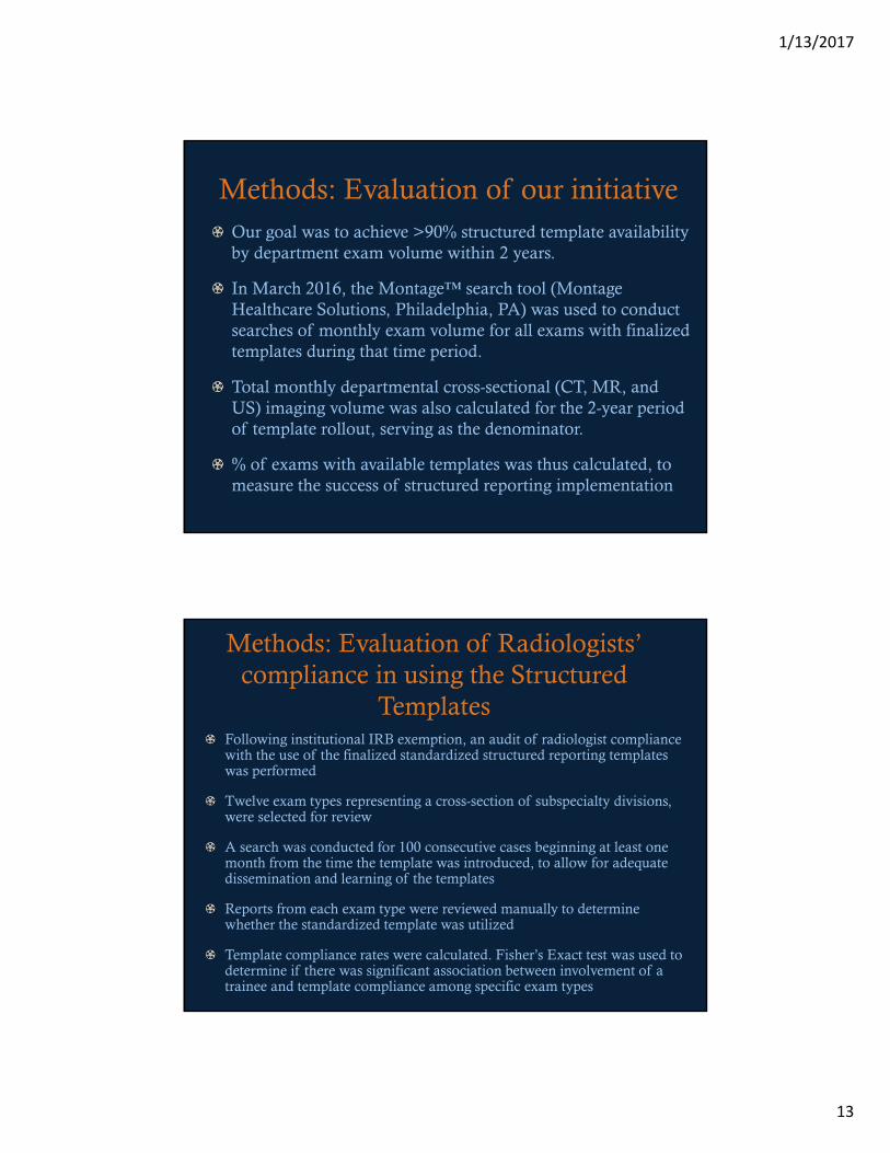
Methods: Evaluation of our initiative Our goal was to achieve >90% structured template availability by department exam volume within 2 years.
In March 2016, the Montage™ search tool (Montage Healthcare Solutions, Philadelphia, PA) was used to conduct searches of monthly exam volume for all exams with finalized templates during that time period.
Total monthly departmental cross-sectional (CT, MR, and US) imaging volume was also calculated for the 2-year period of template rollout, serving as the denominator.
% of exams with available templates was thus calculated, to measure the success of structured reporting implementation
Methods: Evaluation of Radiologists’ compliance in using the Structured
Templates Following institutional IRB exemption, an audit of radiologist compliance with the use of the finalized standardized structured reporting templates was performed
Twelve exam types representing a cross-section of subspecialty divisions, were selected for review
A search was conducted for 100 consecutive cases beginning at least one month from the time the template was introduced, to allow for adequate dissemination and learning of the templates
Reports from each exam type were reviewed manually to determine whether the standardized template was utilized
Template compliance rates were calculated. Fisher’s Exact test was used to determine if there was significant association between involvement of a trainee and template compliance among specific exam types
1/13/2017
14
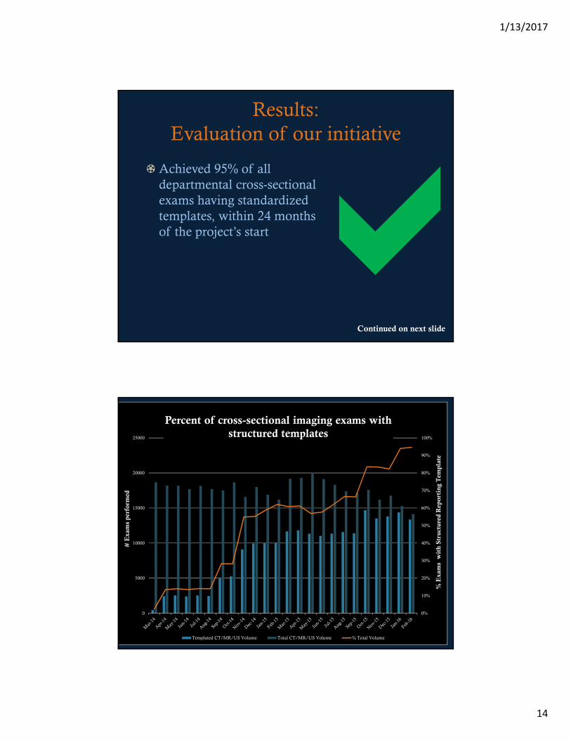
Results:Evaluation of our initiative
Achieved 95% of all departmental cross-sectional exams having standardized templates, within 24 months of the project’s start
Continued on next slide
0%
10%
20%
30%
40%
50%
60%
70%
80%
90%
100%
0
5000
10000
15000
20000
25000%
Exa
ms
wit
h S
tru
ctu
red
Rep
ort
ing
Tem
pla
te
# E
xam
s p
erfo
rmed
Percent of cross-sectional imaging exams with structured templates
Templated CT/MR/US Volume Total CT/MR/US Volume % Total Volume
1/13/2017
15
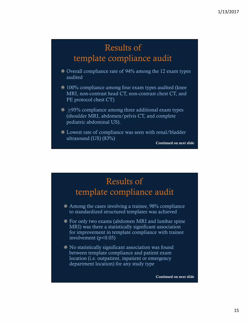
Results oftemplate compliance audit
Overall compliance rate of 94% among the 12 exam types audited
100% compliance among four exam types audited (knee MRI, non-contrast head CT, non-contrast chest CT, and PE protocol chest CT)
>95% compliance among three additional exam types (shoulder MRI, abdomen/pelvis CT, and complete pediatric abdominal US).
Lowest rate of compliance was seen with renal/bladder ultrasound (US) (83%)
Continued on next slide
Results of template compliance audit
Among the cases involving a trainee, 98% compliance to standardized structured templates was achieved
For only two exams (abdomen MRI and lumbar spine MRI) was there a statistically significant association for improvement in template compliance with trainee involvement (p<0.05)
No statistically significant association was found between template compliance and patient exam location (i.e. outpatient, inpatient or emergency department location) for any study type
Continued on next slide
1/13/2017
16
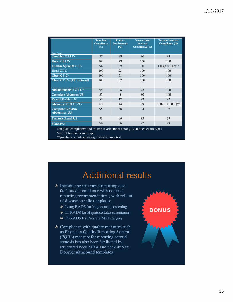
Exam Type*
Template Compliance
(%)
Trainee Involvement
(%)
Non-trainee Involved
Compliance (%)
Trainee-Involved Compliance (%)
Shoulder MRI C- 97 49 96 98
Knee MRI C- 100 49 100 100
Lumbar Spine MRI C- 94 39 90 100 (p < 0.05)**
Head CT C- 100 23 100 100
Chest CT C- 100 31 100 100
Chest CT C+ (PE Protocol) 100 52 100 100
Abdominopelvic CT C+ 96 48 92 100
Complete Abdomen US 85 6 80 100
Renal/Bladder US 83 12 82 92
Abdomen MRI C+/C- 88 44 79 100 (p < 0.001)**
Complete Pediatric Abdominal US
95 38 94 97
Pediatric Renal US 91 46 93 89
Mean (%) 94 36 92 98
Template compliance and trainee involvement among 12 audited exam types*n=100 for each exam type.**p-values calculated using Fisher’s Exact test.
Additional resultsIntroducing structured reporting also facilitated compliance with national reporting recommendations, with rollout of disease-specific templates:
Lung-RADS for lung cancer screening
Li-RADS for Hepatocellular carcinoma
PI-RADS for Prostate MRI staging
Compliance with quality measures such as Physician Quality Reporting System (PQRS) measure for reporting carotid stenosis has also been facilitated by structured neck MRA and neck duplex Doppler ultrasound templates
BONUS
1/13/2017
17
ConclusionsImplementing Structured Reporting at our large multi-site Radiology Department required consensus building, education, and technical optimization for success
A collaborative and iterative approach achieved an overall compliance rate of 94%
Our experiences will serve as a model to other institutions attempting to improve the quality of their reports by implementing structured reporting
Further studies are required to determine whether our reports are indeed more actionable, and whether in turn patient care has improved
ReferencesWeiss DL, Langlotz CP. Structured reporting: patient care enhancement or productivity nightmare? Radiology (2008); 249: 739–747
Schwartz LH, Panicek DM, Berk AR, Li Y, Hricak H. Improving Communication of Diagnostic Radiology Findings through Structured Reporting. Radiology 2011; 260:174-181
Hawkins CM, Hall S, Zhang B, Towbin AJ. Creation and Implementation of Department-Wide Structured Reports: An Analysis of the Impact on Error Rate in Radiology Reports J Digit Imaging (2014) 27:581–587
Walter WR, Goldberg-Stein S, Levsky JM, Cohen HW, Scheinfeld MH. A Default Normal Chest CT Structured Reporting Field for Coronary Calcifications Does Not Cause Excessive False-Negative Reporting. JACR 2015;(12) 783-787
Brook OR, Brook A. Vollmer CM, Kent TS, Norberto S, Pedrosa I. Structured Reporting of Multiphasic CT for Pancreatic Cancer: Potential Effect on Staging and Surgical Planning. Radiology 2015; 464-472
Marcovici PA, Taylor GA. Journal club: Structured radiology reports are more complete and more effective than unstructured reports. AJR 2014; 203: 1265-71
Continued on next slide
1/13/2017
18
ReferencesNaik SS, Hanbidge A, Wilson SR. Radiology reports: examining radiologist and clinician preferences regarding style and content. AJR 2001; 176:591-598
Hawkins CM, Hall S, Salisbury S, Towbin AJ. Prepopulated radiology report templates: a prospective analysis of error rate and turnaround time. J Digit Imaging 2012; 4: 313–319
Boland GW, Duszak R. Structured Reporting and Communication. JACR 2015; (12) 1286-1288
Boland GW, Enzmann DR, Duszak R. Actionable Reporting. JACR 2014; (11): 844-84
Larson DB, Towbin AJ, Pryor RM, Donnelly LF. Improving consistency in radiology reporting through the use of department-wide standardized structured reporting. Radiology 2013; 267: 240–250
Guimaraes CV, DeFlorio RM, Averill LW, Walters KE, Beasley RA, Donnelly LF. Implementation of Standardized Reports Within a Pediatric Health Care System With Geographically Dispersed Sites. JACR 2015;12:1293-1295
Tran L, Wadhwa A, Mann E. Implementation of Structured Radiology Reports. JACR 2016; 13 (3): 296-299