structures of bacillomycin d and bacillomycin l
TRANSCRIPT

1600 THE JOURNAL OF ANTIBIOTICS DEC. 1984
STRUCTURES OF BACILLOMYCIN D AND BACILLOMYCIN L
PEPTIDOLIPID ANTIBIOTICS FROM
BACILLUS SUBTILIS
FRANCOISE PEYPOUX, MARIE-THERESE POMMIER, BHUPESH C. DASt, FRANCOISE BESSON,
LUCIEN DELCAMBEtt and GEORGES MICHEL
Laboratoire de Biochimie Microbienne, Universite de Lyon I,
43, Boulevard du 11 Novembre 1918, 69622 Villeurbanne Cedex, France tlnstitut de Chimie des Substances Naturelles ,
Gif sur Yvette, France ttC .P.E.M., Boulevard de la Constitution,
32, Liege, Belgique
(Received for publication July 7, 1984)
The complete structures of bacillomycin D and bacillomycin L were revised by FAB
mass spectrometry and by Edman degradation of the derivatives resulting from the N-bro-
mosuccinimide reaction. The homologous components of both series of antibiotics were
separated by HPLC and the ,3-amino acids were identified by capillary gas chromatography.
Bacillomycin D and bacillomycin L are peptidolipid antibiotics isolated from strains of Bacillus
subtilis"tt. Their structural determination by chemical methods indicated that they consist of a hepta-
peptide chain linked to a liposoluble 3-amino acid3,4). Among the amino acids of the peptidic moie-
ties, aspartyl, glutamyl, asparaginyl and glutarninyl residues were found and the following structures had
been proposed:
C14 or C15 13-amino acid L-Asx D-Tyr D-Asx
L-Thr D-Ser L-Glx X4
Bacillomycin D: L-AsxL-Asp, D-Asx=D-Asn, X4=L-Pro, L-GIx=L-Glu
Bacillomycin L: L-Asx=L-Asp, D-AsxD-Asp, X4=L-Ser, L-Glx=L-Gln
The presence of amide groups on the dicarboxylic amino acids was suggested from the formation of
a,w-diamino acids by the reaction of RESSLER and KASHELIKAR5) and the free carboxyl groups were esti-
mated by titration with the hydroxymate method applied to methyl esters6).
Recently, these antibiotics were studied by fast atom bombardment (FAB) mass spectrometry and
the molecular weights were found one mass unit less than the expected values. This difference could be
due to the presence of an Asn or a Gln residue instead of an Asp or a Glu residue and such a discre-
pancy is not surprising in view of the imprecision of quantitative methods used in the previous work. On the other hand, reinvestigation of homologous 8-amino acids from iturin A using HPLC and
NMR spectrometry by IS0GAI et al. showed that they consist of a mixture of n-C13, n-C14, anteiso-C15,
iso-C15, n-C15 and n-C18 (3-amino acids with n-C14 and iso-C13 as major components7). More recently,
WINKELMANN et al. isolated from a strain of B. subtilis a peptidolipid complex of the iturin group con-
taining six 8-amino acids with a high proportion of iso-C168).
1601VOL. XXXVII NO. 12 THE JOURNAL OF ANTIBIOTICS
These new results prompted us to reinvestigate the structure of both the peptidic and the lipophilic
moieties of bacillomycin D and of bacillomycin L.
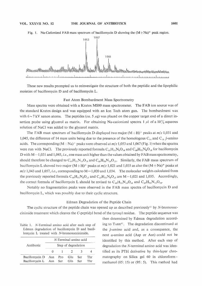
Fig. 1. Na-Cationized FAB mass spectrum of bacillomycin D showing the (M+Na)+ peak region.
10531067
1081
Fast Atom Bombardment Mass Spectrometry
Mass spectra were obtained with a Kratos MS80 mass spectrometer. The FAB ion source was of
the standard Kratos design and was equipped with an Ion Tech atom gun. The bombardment was
with 6-7 kV xenon atoms. The peptides (ca. 5 ,ug) was placed on the copper target end of a direct in-
sertion probe using glycerol as matrix. For obtaining Na-cationized spectra 1 icl of a 10% aqueous
solution of NaCl was added to the glycerol matrix.
The FAB mass spectrum of bacillomycin D displayed two major (M +H)+ peaks at m/z 1,031 and
1,045, the difference of 14 mass units being due to the presence of the homologous C14 and C15 8-amino
acids. The corresponding (M-Na)+ peaks were observed at m/z 1,053 and 1,067 (Fig. 1) when the spectra
were run with NaCl. The previously reported formula C48H73N9O16 and C49H75N9O16 for bacillomycin
D with M = 1,031 and 1,045, i.e., one mass unit higher than the values obtained by FAB mass spectrometry,
should therefore be changed to C48H74N10O15 and C48H76N10O15. Similarly, the FAB mass spectrum of
bacillomycin L showed two major (M +H)+ peaks at m/z 1,021 and 1,035 as also the (M+Na)+ peaks at
m/z 1,043 and 1,057, i.e., corresponding to M=1,020 and 1,034. The molecular weights calculated from
the previously reported formula C48H71N9O17 and C47H73N9O17 are M=1,021 and 1,035. Accordingly,
the correct formula of bacillomycin L should be revised to C46H72N10O16 and C47H74N10O16.
Notably no fragmentation peaks were observed in the FAB mass spectra of bacillomycin D and
bacillomycin L, which was possibly due to their cyclic structure.
Edman Degradation of the Peptide Chain
The cyclic structure of the peptide chain was opened up as described previously3) by N-bromosuc-
cinimide treatment which cleaves the C-peptidyl bond of the tyrosyl residue. The peptide sequence was
then determined by Edman degradation accord-
ing to TARR9). The degradation discontinued at
the a-amino acid and, as a consequence, the
next a-amino acid (Asp or Asn) could not be
identified by this method. After each step of
degradation the N-terminal amino acid was iden-
tified as its PTH derivative by thin-layer chro-
matography on Silica gel 60 in chloroform -
methanol (85: 15) or (95: 5). This method had
Table 1. N-Terminal amino acid after each step of
Edman degradation of bacillomycin D and bacil-
lomycin L treated with N-bromosuccinimide.
Antibiotic
Bacillomycin D
Bacillomycin L
N-Terminal amino acid
Step of degradation
0
Asn
Asn
1
Pro
Ser
2
Glu
Gln
3
Ser
Ser
4
Thr
Thr
1602 THE JOURNAL OF ANTIBIOTICS DEC. 1984
Fig. 2. Gas chromatogram of N-trifluoroacetyl n-amino acyl methyl esters on a 50 m SP 2100 fused-silica capillary column.
Temp : 180°C for 15 minutes then programmation from 180°C to 240°C at VC/minute. A: iturin A, B: bacillomycin D, C: bacillomycin L.
A
B
C
Time (minutes)
Table 2. Percentage of j,-amino acids in antibiotics.
Peak
1
2
3
4
5
Nature of carbon chain
n-C14
iso-C15
anteiso-C15
iso-C16
n-C16
Iturin A
34
30.7
12.1
8.5
9.8
Bacillomycin D
47.6
22.7
12.5
3.3
8.8
Bacillomycin L
38.9
25.2
15.4
10.1
6.1
1603VOL. XXXVII NO. 12 THE JOURNAL OF ANTIBIOTICS
been used previously with bacillomycin D3) and the results are presented with those of bacillomycin L
in Table 1.
Thus, in bacillomycin D, the free carboxyl group belongs to the L-glutamyl residue thereby indicat-
ing that the L-Asx residue linked to the COOH group of the R-amino acid must be an L-asparaginyl re-
sidue. In bacillomycin L both L-Asx and L-Glx residues of the sequenced peptide chain are amidated
and the free carboxyl group must be that of L-Asp residue linked to the COOH group of the p-amino
acid.
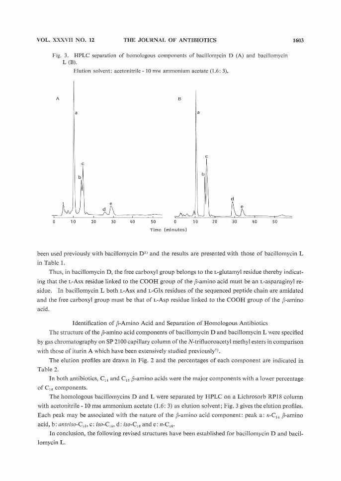
Fig. 3. HPLC separation of homologous components of bacillomycin D (A) and bacillomycin L (B).
Elution solvent: acetonitrile - 10 mm ammonium acetate (1.6: 3).
A B
Time (minutes)
Identification of p-Amino Acid and Separation of Homologous Antibiotics
The structure of the P-amino acid components of bacillomycin D and bacillomycin L were specified
by gas chromatography on SP 2100 capillary column of the N-trifluoroacetyl methyl esters in comparison
with those of iturin A which have been extensively studied previously7).
The elution profiles are drawn in Fig. 2 and the percentages of each component are indicated in
Table 2.
In both antibiotics, C14 and C15 R-amino acids were the major components with a lower percentage
of C16 components.
The homologous bacillomycins D and L were separated by HPLC on a Lichrosorb RP18 column
with acetonitrile - 10 mm ammonium acetate (1.6: 3) as elution solvent; Fig. 3 gives the elution profiles.
Each peak may be associated with the nature of the A-amino acid component: peak a: n-C14 R-amino
acid, b: anteiso-C15, c: iso-C15, d: iso-C16 and e: n-C16.
In conclusion, the following revised structures have been established for bacillomycin D and bacil-
lomycin L.
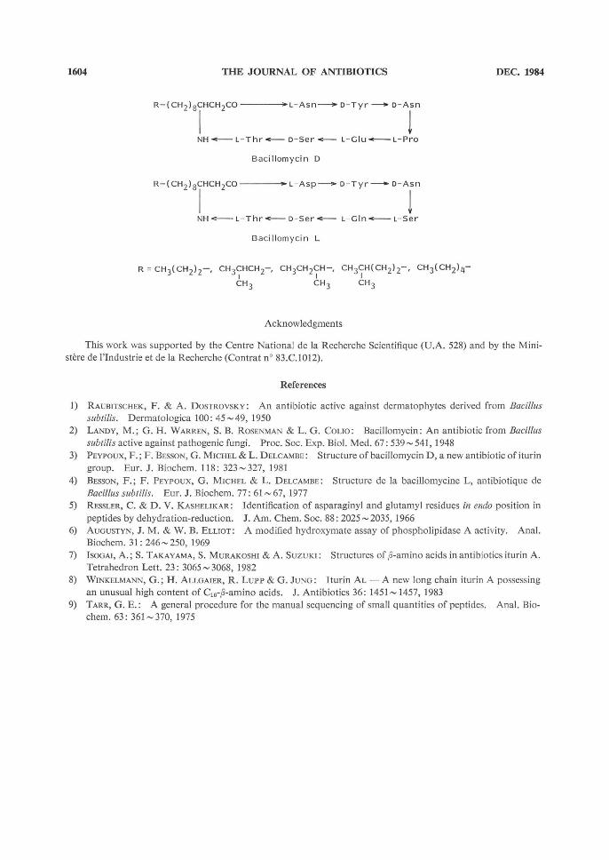
1604 THE JOURNAL OF ANTIBIOTICS DEC. 1984
R-(CH2)3CHCH2CO L-Asn D-Tyr D-Asn
NH L-Thr D-Ser L-Glu L-Pra
Bacillomycin D
R-(CH2)8CHCH2CO L-Asp D-Tyr D-Asn
NH L-Thr D-Ser L-Gln L-Ser
Bacillomycin L
R=CH3(CH2)2-, CH3CHCH2-, CH3CH2CH-, CH3CH(CH2)2-, CH3(CH2)4-
CH3 CH3 CH3
Acknowledgments
This work was supported by the Centre National de la Recherche Scientifique (U.A. 528) and by the Mini-sters de l'Industrie et de la Recherche (Contrat n° 83.C.1012).
References
1) RAUBITSCHEK, F. & A. DOSTROVSKY: An antibiotic active against dermatophytes derived from Bacillus subtilis. Dermatologica 100: 45-.49, 1950
2) LANDY, M.; G. H. WARREN, S. B. ROSENMAN & L. G. COLIO: Bacillomycin: An antibiotic from Bacillus subtilis active against pathogenic fungi. Proc. Soc. Exp. Biol. Med. 67: 539 541, 1948
3) PEYPOUx, F.; F. BESSON, G. MICHEL & L. DELCAMBE: Structure of bacillomycin D, anew antibiotic of iturin
group. Eur. J. Biochem. 118: 323327, 1981 4) BESSON, F.; F. PEYPOUX, G. MICHEL & L. DELCAMBE: Structure de la bacillomycine L, antibiotique de
Bacillus subtilis. Eur. J. Biochem. 77: 61 - 67, 1977 5) RESSLER, C. & D. V. KASHELIKAR: Identification of asparaginyl and glutamyl residues in endo position in
peptides by dehydration-reduction. J. Am. Chem. Soc. 88: 2025 - 2035, 1966 6) AUGUSTYN, J. M. & W. B. ELLIOT: A modified hydroxymate assay of phospholipidase A activity. Anal. Biochem. 31: 246 - 250, 1969 7) ISOGAI, A.; S. TAKAYAMA, S. MURAKOSHI & A. SuzuKI : Structures of 13-amino acids in antibiotics iturin A. Tetrahedron Lett. 23: 3065-3068, 1982 8) WINKELMANN, G.; H. ALLGAIER, R. LuPP & G. JUNG: Iturin AL - A new long chain iturin A possessing an unusual high content of C16-3-amino acids. J. Antibiotics 36: 1451~ 1457, 1983
9) TARR, G. E.: A general procedure for the manual sequencing of small quantities of peptides. Anal. Bio- chem. 63: 361 - 370, 1975