study of tissues characteristics · 1 9/23/2004 s. davenport © 1 study of tissues four primary...
TRANSCRIPT

1
9/23/2004 S. Davenport © 1
Study of Tissues
Four primary tissuesEpithelia, Connective, Muscle,
Nervous
9/23/2004 S. Davenport © 2
EPITHELIAL TISSUE
CharacteristicsStudy according to structureStudy according to locationStudy according to formation of glands
9/23/2004 S. Davenport © 3
Characteristics of Epithelia
• Identification and functions of epithelial tissues involves at least seven characteristics:– Cellularity– Polarity– Attachment– Avascular– Regeneration
9/23/2004 S. Davenport © 4
CELLULARITY
Epithelia are composed mostly of cells. They have very little extracellular material between the cells.
Stratified squamous epitheliumAreolar connective tissue
9/23/2004 S. Davenport © 5
POLARITY• Unequal distribution of organelles resulting in
structural specialization• Example – Simple columnar epithelium
– Surface modified for absorption (microvilli)– Cytoplasm processes absorbed materials and transcytosis– Base of cell modified for exocytosis
Simple columnar epithelium from intestine9/23/2004 S. Davenport © 6
ATTACHMENTThe membranes are always attached to an underlying connective tissue layer at a thin region called the basement membrane. The basement membrane is noncellular and consists of materials produced by the epithelial cells and the adjacent connective tissue.
Pseudostratified ciliated columnar epithelium
Basement membrane
2
9/23/2004 S. Davenport © 7
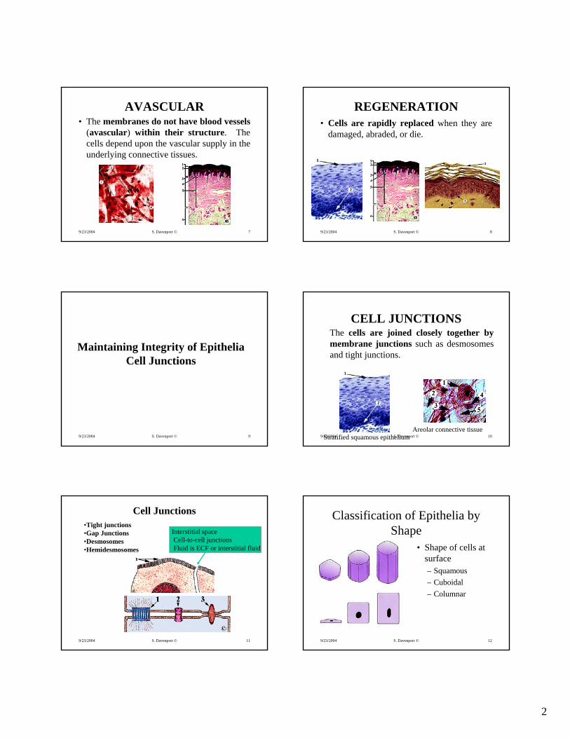
AVASCULAR• The membranes do not have blood vessels
(avascular) within their structure. The cells depend upon the vascular supply in the underlying connective tissues.
9/23/2004 S. Davenport © 8
REGENERATION• Cells are rapidly replaced when they are
damaged, abraded, or die.
9/23/2004 S. Davenport © 9
Maintaining Integrity of EpitheliaCell Junctions
9/23/2004 S. Davenport © 10
CELL JUNCTIONSThe cells are joined closely together by membrane junctions such as desmosomesand tight junctions.
Stratified squamous epitheliumAreolar connective tissue
9/23/2004 S. Davenport © 11
Cell Junctions•Tight junctions•Gap Junctions•Desmosomes•Hemidesmosomes
Interstitial spaceCell-to-cell junctionsFluid is ECF or interstitial fluid
9/23/2004 S. Davenport © 12
Classification of Epithelia by Shape
• Shape of cells at surface– Squamous– Cuboidal– Columnar
3
9/23/2004 S. Davenport © 13
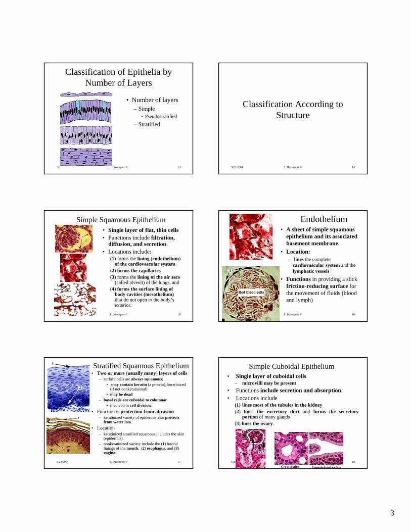
Classification of Epithelia by Number of Layers
• Number of layers– Simple
• Pseudostratified
– Stratified
9/23/2004 S. Davenport © 14
Classification According to Structure
9/23/2004 S. Davenport © 15
Simple Squamous Epithelium• Single layer of flat, thin cells• Functions include filtration,
diffusion, and secretion. • Locations include:
(1) forms the lining (endothelium)of the cardiovascular system
(2) forms the capillaries,(3) forms the lining of the air sacs
(called alveoli) of the lungs, and (4) forms the surface lining of
body cavities (mesothelium) that do not open to the body’s exterior.
9/23/2004 S. Davenport © 16
Endothelium• A sheet of simple squamous
epithelium and its associated basement membrane.
• Location:– lines the complete
cardiovascular system and the lymphatic vessels
• Functions in providing a slick friction-reducing surface for the movement of fluids (blood and lymph)
9/23/2004 S. Davenport © 17
Stratified Squamous Epithelium• Two or more (usually many) layers of cells
– surface cells are always squamous: • may contain keratin (a protein), keratinized
(if not nonkeratinized)• may be dead
– basal cells are cuboidal to columnar• involved in cell division.
• Function is protection from abrasion– keratinized variety of epidermis also protects
from water loss. • Location
– keratinized stratified squamous includes the skin (epidermis).
– nonkeratinized variety include the (1) buccallinings of the mouth, (2) esophagus, and (3)vagina.
9/23/2004 S. Davenport © 18
Simple Cuboidal Epithelium• Single layer of cuboidal cells
– microvilli may be present• Functions include secretion and absorption. • Locations include
(1) lines most of the tubules in the kidney(2) lines the excretory duct and forms the secretory
portion of many glands(3) lines the ovary.

4
9/23/2004 S. Davenport © 19
Transitional Epithelium• Several to many layers of cells
– depending upon the mechanical stress placed upon it
• Functions in allowing the organ it lines (urinary bladder) to easily change shape.
• Locations include: lines (1) the urinary bladder(2) renal pelvis, the central urine-
containing cavity of the kidney (3) the ureters (tubes that connect the
kidneys to the bladder). 9/23/2004 S. Davenport © 20
Simple Columnar Epithelium(Nonciliated)
• Single layer of columnar cells– Microvilli and goblet cells may be present
• Functions include secretion and absorption• Locations include lining of
(1) the digestive tract from the stomach to the anus(2) the excretory ducts of some glands(3) the interior of the gallbladder.
Lining of the small intestine
9/23/2004 S. Davenport © 21
Pseudostratified Columnar Epithelium• Single layer of columnar cells of different heights.
– All cells are in contact with the basement membrane.
• Functions include secretion and the movement ofsubstances (mucus) over the surface by cilia.
• Locations include1) lines most of upper respiratory tract.
9/23/2004 S. Davenport © 22
Classification According to Formation of Glands
9/23/2004 S. Davenport © 23
Glandular Epithelia
• A gland is one or more cells which produce and secrete a product that is called a secretion. – The method of secretion is used to classify
glands into the following two types: (1) exocrine (2) endocrine.
9/23/2004 S. Davenport © 24
Endocrine Glands• Endocrine glands secrete their products
into the blood; they are ductless. Their secretions are called hormones. Examples include the thyroid, anterior pituitary, and adrenal glands.

5
9/23/2004 S. Davenport © 25
Exocrine Glands• Exocrine glands secrete their products
into a duct that opens to the surface of the covering or lining membrane. Examples include the sweat, oil (sebaceous), salivary, and goblet glands (cells).
9/23/2004 S. Davenport © 26
Modes of Secretion• Merocrine glands
– Secrete by exocytosis as product is produced– Pancreas, sweat glands, salivary glands
• Holocrine glands– Accumulate product until they rupture (mitotic)– Sebaceous (oil) glands
• Apocrine glands– Accumulate product at surface and pinched off– Unclear as to human location. Possibly some
secretion in mammary glands.
9/23/2004 S. Davenport © 27
Connective TissuesFeaturesCharacteristicsClassification
Connective Tissues ProperCartilageBoneBlood
9/23/2004 S. Davenport © 28
General Features
• Widely distributed tissues of the body. • Locations include bones, tendons, ligaments,
cartilage, blood, and the abundant loose connective tissues (such as adipose) located in and around other tissues.
• Functions include providing (1) a framework, (2) support, (3) binding, (4) protection, (5) insulation, and (6) transportation (specifically for blood).
9/23/2004 S. Davenport © 29
Characteristics
• Identification and functions of connective tissues involves at least three characteristics:– Cellularity– Matrix– Vascularity
• Three fundamental elements of connective tissues– Cells– Ground substance– Fibers
• (Ground substance and fibers are components of matrix)
9/23/2004 S. Davenport © 30
Cellularity– Cells are dispersed in a substance called the
extracellular matrix– In addition to the structural cells of the connective tissue,
many other types of cells may be present.– Connective tissues are not organized into distinctive
cellular membranes (as seen in the organization of cells of epithelia). However, membranes or “sheets” of connective tissue are common in the body.
Stratified squamous epitheliumAreolar connective tissue

6
9/23/2004 S. Davenport © 31
Cellularity - Types of Cells in Connective Tissues
• Structural cells– May be named (by prefix) according to type of tissue
• Chondro - cartilage• Osteo - bone• Fibro – structural cells of connective tissue proper• Hemo – blood
– Activity of cell may be indicated by suffix• Cyte – mature (or maintaining) cell of the tissue• Blast – cell actively dividing and producing tissue substance• Clast – cells that are active in “breakdown”
• Associated cells include– White blood cells (neutrophils, eosinophils, basophils),
mast cells, plasma cells, macrophages, fat cells9/23/2004 S. Davenport © 32
Matrix of Connective Tissue• Nonliving extracellular material
– is abundant in connective tissues– characteristics are responsible for the nature of the
specific connective tissue.
• Consists of• Ground substance• Fibers
AreolarHyaline cartilage
Bone
9/23/2004 S. Davenport © 33
Ground Substance of Connective Tissue
• Occupies the area around the cells and fibers– Interstitial fluid– Complex mixture of proteins (structural and
organizational).
Dense regular (tendon)Areolar, wm
9/23/2004 S. Davenport © 34
Vascularity of Connective Tissue• Varying degrees of vascularity.
– high vascularity, such as found in areolar, to no vascularity, such as found in cartilage tissues.
Hyaline cartilage
Bone
Areolar (wm)
9/23/2004 S. Davenport © 35
CLASSIFICATION• Based upon three characteristics of matrix:
– (1) the types of fibers, – (2) the type of ground substance– (3) the structural arrangement.
• According to these characteristics classified into three types: – (1) Connective tissue proper, – (2) Fluid connective tissues– (4) Supporting connective tissues
9/23/2004 S. Davenport © 36
Connective Tissue Proper• Connective tissue “proper” include the “typical”
connective tissues which are all connective tissues except blood, bone, and cartilage
• The matrix is characterized by – (1) being flexible, – (2) having a viscous ground substance, and – (3) by having abundant fibers.
• The structural cells are either fibroblasts or fibrocytes. • The two subclasses of connective tissue proper are
– (1) Loose connective tissue• Areolar, adipose, reticular
– (2) Dense connective tissue• Dense regular, dense irregular, elastic

7
9/23/2004 S. Davenport © 37
Fibers• Fibers are distinctive protein threads
embedded in the ground substance. • Three common fibers
– (1) collagen, – (2) elastic, – (3) reticular.
9/23/2004 S. Davenport © 38
Collagen Fibers• Most abundant of the three fibers
– Their long collagen protein structure makes them appear as fine clear threads in fresh preparations. Thus, they are often called “white fibers.”
• Functions include– (1) providing structural framework– (2) providing strength.
Dense regular (tendon)Areolar, wm
9/23/2004 S. Davenport © 39
Elastic Fibers• Long fibers made of the protein elastin
– appear yellow in fresh preparations. Thus, they are often called “yellow fibers.”
• Functions include – allowing the tissue to stretch and recoil.
Elastic cartilageElastic fibers (artery)
9/23/2004 S. Davenport © 40
Reticular Fibers• Similar to collagenous fibers
– are thinner and more branched• Functions include
(1) providing a structural framework(2) providing strength.
Reticular fibers, lymph node
9/23/2004 S. Davenport © 41
LOOSE CONNECTIVE TISSUES
• Areolar• Adipose• Reticular
9/23/2004 S. Davenport © 42
Areolar• Structural cells are fibrocytes.
– Other cell types include mast cells(produce histamine) and macrophages (leukocytes which function in phagocytosis).
• Matrix contains– Abundant collagenous and elastic
fibers loosely dispersed in the ground substance.
• Functions include– (1) attaching, (2) packing (3)
supporting tissues and organs. • Locations:
– widely distributed throughout the body. It attaches the skin to underlying tissues, surrounds and supports many organs such as blood vessels, and glands.
8
9/23/2004 S. Davenport © 43
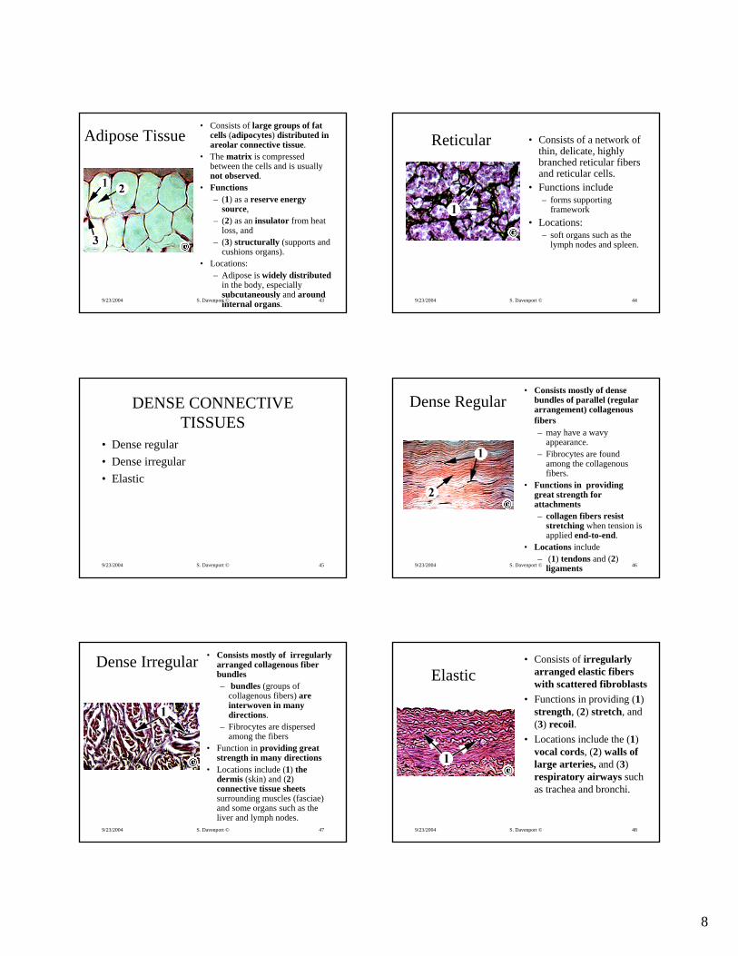
Adipose Tissue• Consists of large groups of fat
cells (adipocytes) distributed in areolar connective tissue.
• The matrix is compressed between the cells and is usually not observed.
• Functions – (1) as a reserve energy
source, – (2) as an insulator from heat
loss, and – (3) structurally (supports and
cushions organs).• Locations:
– Adipose is widely distributedin the body, especially subcutaneously and around internal organs. 9/23/2004 S. Davenport © 44
Reticular • Consists of a network of thin, delicate, highly branched reticular fibers and reticular cells.
• Functions include– forms supporting
framework • Locations:
– soft organs such as the lymph nodes and spleen.
9/23/2004 S. Davenport © 45
DENSE CONNECTIVE TISSUES
• Dense regular• Dense irregular• Elastic
9/23/2004 S. Davenport © 46
Dense Regular• Consists mostly of dense
bundles of parallel (regular arrangement) collagenousfibers– may have a wavy
appearance. – Fibrocytes are found
among the collagenousfibers.
• Functions in providing great strength for attachments– collagen fibers resist
stretching when tension is applied end-to-end.
• Locations include– (1) tendons and (2)
ligaments
9/23/2004 S. Davenport © 47
Dense Irregular • Consists mostly of irregularly arranged collagenous fiber bundles– bundles (groups of
collagenous fibers) are interwoven in many directions.
– Fibrocytes are dispersed among the fibers
• Function in providing great strength in many directions
• Locations include (1) the dermis (skin) and (2) connective tissue sheetssurrounding muscles (fasciae) and some organs such as the liver and lymph nodes.
9/23/2004 S. Davenport © 48
Elastic• Consists of irregularly
arranged elastic fibers with scattered fibroblasts
• Functions in providing (1) strength, (2) stretch, and (3) recoil.
• Locations include the (1)vocal cords, (2) walls of large arteries, and (3)respiratory airways such as trachea and bronchi.

9
9/23/2004 S. Davenport © 49
FLUID CONNECTIVE TISSUEBlood and Lymph
9/23/2004 S. Davenport © 50
Blood
• Consists of formed elements and plasma– Formed elements are the cells
(erythrocytes, or RBCs, and leukocytes,or WBCs) and platelets, the non-cellular elements (cell fragments)
• Matrix is fluid component called plasma.• Functions include transportation of
– (1) oxygen,– (2) carbon dioxide, – (3) nutrients, – (4) wastes, and – (5) hormones;– (6) and promotes immune and
inflammatory responses(leukocytes).
• Location– cardiovascular system (heart and the
blood vessels)
9/23/2004 S. Davenport © 51
SUPPORTING CONNECTIVE TISSUES
9/23/2004 S. Davenport © 52
CARTILAGE• Characteristics
– a matrix which is semisolid and slightly flexible – consists mostly of collagen fibers embedded in a protein
ground substance– avascular– chondrocytes are the structural cells of mature cartilage– perichondrium, a membrane of dense irregular connective
tissue, forms the surface of most cartilage. • Functions as a (1) supportive and (2) structural connective
tissue• Three types of cartilage are presented for study:
– Hyaline cartilage– Fibrocartilage– Elastic cartilage
9/23/2004 S. Davenport © 53
GROWTH OF CARTILAGE
Interstitial growthGrowth from within the cartilage tissue– Dividing (mitotic) cells and additional matrix
Appositional growthGrowth occurs at the surface of cartilage (perichondrium). Layers of chondrocytesand matrix are added to the tissue.
9/23/2004 S. Davenport © 54
Hyaline Cartilage
• Consists of abundant collagenous fibers that are embedded in ground substance. – This gives the matrix a firm structure
with an amorphous appearance (you cannot identify the detail of the fibers or ground substance).
• Functions in providing (1) support, (2) a structural framework, and (3) cushion.
• Locations– locations include (1) where the ribs
connect to the sternum (called costal cartilage), (2) the ends of long bones(called articular cartilage), (3) the tip of the nose, and (4) the framework of larger respiratory airways.

10
9/23/2004 S. Davenport © 55
Fibrocartilage• Consists of dense, compact,
collagenous fiber bundles with little ground substance. – The fiber bundles are wavy and
nearly parallel with chondrocyteslocated along their surface.
• Functions include (1) providing strength and (2) resisting compression.
• Locations include (1) the fibro-cartilage discs (intervertebral discs) that separate the vertebrae, (2) part of the knee joint and (3) the symphysispubis (connects the two pubic bones).
9/23/2004 S. Davenport © 56
Elastic Cartilage
• Consists of abundant collagenousand elastic fibers embedded in ground substance. – The ground substance blends
with the collagenous fibers making them invisible.
– The elastic fibers usually stain dark blue.
– Chondrocytes are distributed among the fibers.
• Functions include providing (1) support and (2) flexibility.
• Locations include the (1) external ear and the (2) epiglottis (cartilage structure that closes the opening to the airway when swallowing food).
9/23/2004 S. Davenport © 57
BONE• Contains collagenous fibers and calcium salts
– calcium salts make bone tissue hard,– collagenous fibers give it strength
• Functions include– (1) providing protection– (2) serving as attachment sites for muscles and connective
tissues– (3) providing reserves for minerals– (4) blood cell production (marrow)– (5) providing a site for fat deposit (yellow marrow).
• Two structural types of bone tissue, – (1) compact– (2) spongy 9/23/2004 S. Davenport © 58
Bone Tissue• Compact bone
– organized in units called Haversiansystems (osteons).
• Haversian system contains a – Haversian (central) canal which
contains blood vessels.– Matrix consists of mineral salts
(about 2/3 mostly tricalciumphosphate, or hydroxyapatite) and 1/3 collagen fibers
– Lamellae (concentric rings of matrix) surround each Haversian canal
– Osteocytes in circular-rows separate the lamellae
– Canaliculi (small canals ) interconnect the osteocytes.
• Spongy bone– organized into plates called trabeculae.
9/23/2004 S. Davenport © 59
MEMBRANESEpithelia may be classified according to
formation of membranes. Membranes contain both epithelial and underlying connective tissue.
Epithelial membranesSerous membranesCutaneous membraneSynovial membranes
9/23/2004 S. Davenport © 60
Mucous membranes (mucosae)• Membranes composed of an epithelial tissue and its
associated connective tissue– Epithelium varies according to location– Connective tissue layer called the lamina propria.
• Locations: line body cavities that open to the exterior– such as the reproductive, digestive, and respiratory tracts.
• Functions include secretion and absorption.
Simple columnar from small intestine Stratified squamous of mouth

11
9/23/2004 S. Davenport © 61
Serous Membranes• Simple squamous epithelium (mesothelium)
attached to loose connective tissue• Locations: line body cavities that do not open
to the exterior– the pleura, pericardial, and peritoneal membranes
• Function in the maintenance of serous fluids.
Simple squamous, surface view 9/23/2004 S. Davenport © 62
Cutaneous Membrane• The skin
– an epithelium called the epidermis (keratinized stratified squamous epithelium)
– connective tissue layer called the dermis (mostly dense irregular connective tissue).
• Location; covers the body. • Functions include protection from abrasion,
waterproofing, and isolation from the environment.
9/23/2004 S. Davenport © 63
Synovial Membranes
• Synovial membranes line joint cavities (is not located on the articular surfaces of bones) – Synovial membranes produce synovial fluid– Synovial fluid lubricates synovial (fluid) joints.
9/23/2004 S. Davenport © 64
Connective Tissue Framework of the Body
• Provide for strength and stability• Maintain positions of internal organs• Provides a route for distribution of blood vessels,
lymphatic vessels, and nerves.– Superficial fascia or subcutaneous layer
• Areolar tissue and adipose tissue– Deep fascia
• Dense irregular connective tissue• Surrounds organs, attaches abdominal pelvic organs, serves to
bind organs such as muscles into bone.
9/23/2004 S. Davenport © 65
Muscle Tissue
SkeletalCardiacSmooth
9/23/2004 S. Davenport © 66
Muscle Tissue1
• Three types of muscle tissues – (1) skeletal, – (2) cardiac,– (3) smooth.(A less frequently used scheme classifies muscle into two types; 1) Striated and 2)
Smooth. Skeletal and cardiac would then be placed under “striated.”)
• Cells are called fibers because of their long structure– Cells contract due the interaction of the contractile proteins
actin and myosin– Generate tension by shortening (contracting)

12
9/23/2004 S. Davenport © 67
Muscle Tissue2• Controlled by
– Nervous system– Autoregulation
• Myogenic – contraction in response to local stimulus such as change in blood pressure (local hyperemia). Especially seen in smooth and cardiac muscle.
• Pacemakers – contraction in response to electrical changes within the muscle itself . Especially, seen in smooth and cardiac muscle.
– Hormones• may influence rate and/or force of contraction. Especially seen in
cardiac muscle.
• Functions include– body movements such as locomotion – movement internal materials (blood, urine, food stuffs, etc.)– generation of heat (body temperature regulation)
9/23/2004 S. Davenport © 68
Skeletal Muscle1
• The fibers (cells) are characterized by being – (1) long– (2) parallel– (3) cylindrical,– (4) multinucleate
• Location: – Fibers are organized to form the
skeletal muscles– Skeletal muscles are attached to
bone by dense connective tissue (tendons).
• Function in the production of voluntary body movements
9/23/2004 S. Davenport © 69
Skeletal Muscle2
• Myofibrils– Long rod-like elements of the fiber which consist
mostly of thin and thick protein filaments• Striations
– alternating dark and light cross bands resulting mostly from the arrangement of the thin and thick filaments
9/23/2004 S. Davenport © 70
Skeletal Muscle3
• A and I Bands– A band is the dark cross-band (or striation)– I band is the light cross-band (or striation)
• Z line (disc)– Protein plate located in center of each I band
• Sarcomere– The functional unit of contraction– Is identified as the region between two successive Z lines
9/23/2004 S. Davenport © 71
Cardiac Muscle
• Cardiac muscle fibers are characterized by being– (1) cylindrical, – (2) uninucleate mostly (some
cells are binucleate), – (3) striated, – (4) branching.Intercalated disks – sites where
cells are connected end-to-end• Location
– Muscle of the heart• Function
– in producing the heart’s involuntary contractions..
9/23/2004 S. Davenport © 72
Smooth Muscle
• Fibers are characterized by being – (1) long, – (2) spindle shaped, – (3) uninucleate, – (4) nonstriated (smooth).
• Location– in the walls of many of the hollow
organs such as the (1) esophagus, (2) stomach, (3) intestines, (4) urinary bladder, and (5) blood vessels
• Functions– produces involuntary contractions
which move and/or regulate passage of materials in the organs.

13
9/23/2004 S. Davenport © 73
Nerve Tissue“Typical” motor neuron and neuroglia
StructureFunction
9/23/2004 S. Davenport © 74
Nerve Tissue
• Nervous tissue forms the nervous system– consists mostly of the (1) brain, (2) spinal
cord, and (3) nerves. • Two fundamental types of cells form the
basis of nervous tissue: • (1) neurons (nerve cells) • (2) neuroglia
9/23/2004 S. Davenport © 75
Neurons • Most neurons have – (1) cell body (soma)– (2) cell processes
• An axon and many dendrites
• Functions include– Generation, inhibition and
transmission of electrical events
• Neuroglial cells– nonconductive and mostly function
in (1) supporting and (2) insulating the nervous tissue. There are also several different types of neuroglial cells.
9/23/2004 S. Davenport © 76
Inflammation and Regeneration
9/23/2004 S. Davenport © 77
Inflammation
• Beginning of healing of damaged tissue• Coordinates activities of several tissue• Characterized by
– Increased blood flow (redness and warmth)– Increased vessel permeability (swelling)– PainInfection would involve presence of pathogens,
such as bacteria.
9/23/2004 S. Davenport © 78
Regeneration
• Regeneration involves the replacement of damaged tissue.– Fibroblasts invade area and produce collagen
fibers (becomes a scar or fibrous tissue)– Tissue cells are replace depending upon the
type of tissue. Some tissues are amitotic and are replaced by fibrous tissue, fibrosis occurs.