study on rehardening of demineralized dentin with …...1 study on rehardening of demineralized...
TRANSCRIPT
1
Study on Rehardening of Demineralized Dentin with Pulp-capping Agents
Using a New Hardness Determination System
MATSUDA Tomoyuki1), ONDA Kohei2), TANIMOTO Hiroaki2),
YOSHIKAWA Kazushi2) and YAMAMOTO Kazuyo2)
1) Graduate School of Dentistry, Department of Operative Dentistry,
Osaka Dental University.
2) Department of Operative Dentistry, Osaka Dental University.
Corresponding author: Dr. Matsuda, Department of Operative Dentistry,
8-1 Kuzuha-hanazonocho, Hirakata, Osaka 573-1121, Japan
TEL: +81-72-864-3077, FAX: +81-72-864-3177
E-mail: [email protected]
2
Abstract
Purpose
We measured the hardness of demineralized dentin over time using the
Cariotester to determine the effectiveness of several types of pulp-capping agent
for IPC.
Methods
Extracted human molars were used to prepare dentin samples with a diameter
10 mm and thickness of 2 mm. Sound dentin samples were immersed in lactic acid
solution and were regarded as demineralized when the value obtained using the
Cariotester was approximately 20 KNH. HY-Bond Temporary Cement Soft (Shofu),
Neodyne-α (Neo Dental Chemical Products), Dycal (Dentsply), and Calcipex Plain
II (Nippon Shika Yakuhin) with a 60% calcium hydroxide mixture were used as
the pulp-capping agents in the present study. Each pulp-capping agent was
applied to the surface of demineralized dentin and covered with base cement.
After the base cement solidified, pulp-capped dentin samples and controls were
divided into 2 groups: those placed in a container with 100% humidity and those
immersed in remineralization solution, and stored at 37℃ in a thermostatic
chamber for 1 and 3 months. The hardness of the capping agent-applied region
was then measured. Data were analyzed using a one-way analysis of variance and
Tukey’s test (α=0.05). Pulp-capping agent-applied surfaces were also observed
under SEM.
3
Results
The hardness of demineralized dentin increased with the application of Dycal,
Calcipex plain II, and a 60% calcium hydroxide mixture, and mineralized
substance-like aggregates were deposited on the surface of and between collagen
fibrils. It was suggested that the remineralization of demineralized dentin
depended on the calcium hydroxide concentration contained in a pulp-capping
agent.
Conclusion
The hardness of demineralized dentin increased and remineralization features
were noted following the application of a pulp-capping agent contained over 27%
calcium hydroxide.
Key words: Knoop hardness, pulp-capping agent, remineralization

4
Introduction
The concept of minimal intervention (MI): the conservation of sound dentin to
minimize invasion, has recently become widely pervasive in caries treatment.
Terashima1) and Sato2,3) described the division of carious dentin into the outer
layer, in which the tooth substance is demineralized by cariogenic bacteria,
leading to the collapse of collagen fibrils, and the inner layer, in which the tooth
substance is partially demineralized by the indirect influence of cariogenic
bacteria. Caries detector solutions have been developed to distinguish between
these two layers and are widely used for caries removal4,5). The outer layer stained
with caries detectors should be removed, whereas active conservation of the
non-stained inner layer has been reported to induce physiological
remineralization after restoration6-8). However, pulpectomy is selected when
caries progresses to deep dentin close to the pulp because the removal of all
infected dentin exposes the pulp. Indirect pulp capping (IPC) is recommended in
these cases in an attempt to conserve dental pulp and avoid pulpectomy9). In IPC,
infected dentin near the pulp is intentionally left and a tannin-fluoride
mixture-combined carboxylate cement or calcium hydroxide preparation is
applied to promote the sterilization and remineralization of residually infected
dentin and formation of tertiary dentin (reparative dentin). The remineralization
of demineralized dentin via the application of a tannin-fluoride mixture-combined
carboxylate cement and calcium hydroxide preparation has already been
confirmed using various approaches, such as hardness and X-ray radiographical,
bacteriological, and histopathological methods10-17). Although hardness is a
clinically important index, only a few studies have used it as an objective index.
5
A hardness measurement device for carious dentin, the Cariotester, has
recently been designed by Shimizu18,19) et al. and released by SAMEIME co.
Carious dentin hardness can be directly measured in the oral cavity using the
Cariotester, which has facilitated the introduction of an objective index, hardness,
into caries treatment.
In the present study, the hardness of demineralized dentin was measured over
time using the Cariotester to investigate the effectiveness of several types of
pulp-capping agents for IPC. In addition, Pulp-capping agents were applied to
demineralized dentin prepared using a lactic acid solution and the Knoop
hardness of the pulp-capping agent-applied surfaces was measured after 1 and 3
months. Pulp-capping agent-applied surfaces were also observed under SEM and
the effectiveness of the agents tested was determined.
Materials and Methods
1. Experimental samples
The subject teeth were human molars that were extracted at the Department of
Oral Surgery, Osaka Dental University Hospital, and stored in physiological
saline at -40C. They were defrosted under running water immediately before
being used in the experiments. The occlusal surface of each tooth was
macroscopically observed, and teeth with caries, white turbidity, coloration, and
crack were excluded. This study was approved by the Medical Ethics Committee
of Osaka Dental University (approval number: 110742).
6
2. Experimental methods
1) Hardness measurement
To investigate time-course changes in the hardness induced by application of a
pulp-capping agent, using Cariotester SUK-971 (Cariotester, SANEIME co.), the
hardness of sound dentin, demineralized dentin, and dentin 1 and 3 months after
pulp-capping was measured following the manufacturer’s instructions. The
measurement range was set at the center within a 3-mm diameter on the enamel
side in the sound dentin samples, and a site within a 3-mm diameter from the
center in the demineralized and pulp-capped dentin samples. The hardness was
measured at 5 sites in each sample, and the mean of the 5 values was adopted.
The number of samples was 3 for each condition.
2) Preparation of samples
The roots of the human molars were cut at 3 mm apical from the anatomical
cervical line, the dental pulp was removed, and the coronal enamel and root
dentin were cut vertically to the tooth axis using a model trimmer. The enamel
side of the exposed dentin and the lateral side of the pulp cavity were polished
using waterproof polishing paper #1000 to prepare a dentin sample with a
diameter 10 mm and thickness 2 mm. The hardness was measured on the enamel
side of the dentin sample using the Cariotester, and the tooth was regarded as a
sound dentin sample when the value obtained was approximately 60 KNH.

7
3) Preparation of demineralized dentin
The major organic acid produced by cariogenic bacteria, lactic acid (Kishida),
was used to decalcify sound dentin samples. The enamel side of sound dentin
samples was immersed in 50 ml of 20 mM lactic acid solution for 10 hours while
being aspirated from the lateral side of the pulp cavity at 0.01 MPa using an
aspirator (MDA-006, Ulvac). The sound sample was then sufficiently washed with
distilled water, and the hardness was measured on the enamel side using the
Cariotester. Samples were regarded as demineralized when the value obtained
was approximately 20 KNH.
4) Preparation of pulp-capped dentin samples
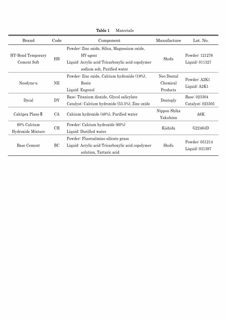
The pulp-capping agents and cement tested are shown in Table 1. As
commercially available products, HY-Bond Temporary Cement Soft (Shofu),
Neodyne-α (Neo Dental Chemical Products), Dycal (Dentsply), and Calcipex plain
II (Nippon Shika Yakuhin) were tested following the manufacturers’ instructions.
To investigate the influence of calcium hydroxide concentration on the
remineralization of demineralized dentin, a 60% calcium hydroxide mixture was
prepared by mixing 0.6 g of calcium hydroxide (Kishida) with 1 ml of distilled
water, and tested as a pulp-capping agent. To prepare pulp-capped dentin samples,
each pulp-capping agent was applied to the surface of demineralized dentin and
by coverage with base cement (Shofu). Demineralized dentin samples covered
with BC only and no pulp-capping agent were prepared as controls. After BC
solidified, the pulp-capped dentin samples and controls were divided into 2
groups: those placed in a container with 100% humidity (DW group) and those
8
immersed in remineralization solution (1.5 mmol/l CaCl2, 0.9 mmol/l KH2PO4, 130
mmol/l KCl, 20 mmol/l HEPES) adjusted to pH 7.0 with KOH (RS group), and
stored at 37C in a thermostatic chamber for 1 and 3 months. After storage, BC
and pulp-capping agents were removed from the pulp-capped dentin samples and
controls, without touching the capping agent-applied region, and the hardness of
the capping agent-applied region was measured.
3. Observation under SEM
To observe the surface of the pulp-capped dentin samples after hardness
measurement, the samples were fixed and dehydrated with an alcohol series
following the standard method, and then freeze-dried using a t-BuOH freeze dryer,
VFD21S (VD). The samples were then subjected to gold vapor deposition using an
ion sputtering device, E-1030 (Hitachi), and observed under a field-emission
scanning electron microscope, S-4000 (Hitachi). The surfaces of the sound and
demineralized dentin samples were similarly processed and observed.
4. Statistical analysis
Data were analyzed using a one-way analysis of variance and Tukey’s test
(α=0.05).
Results
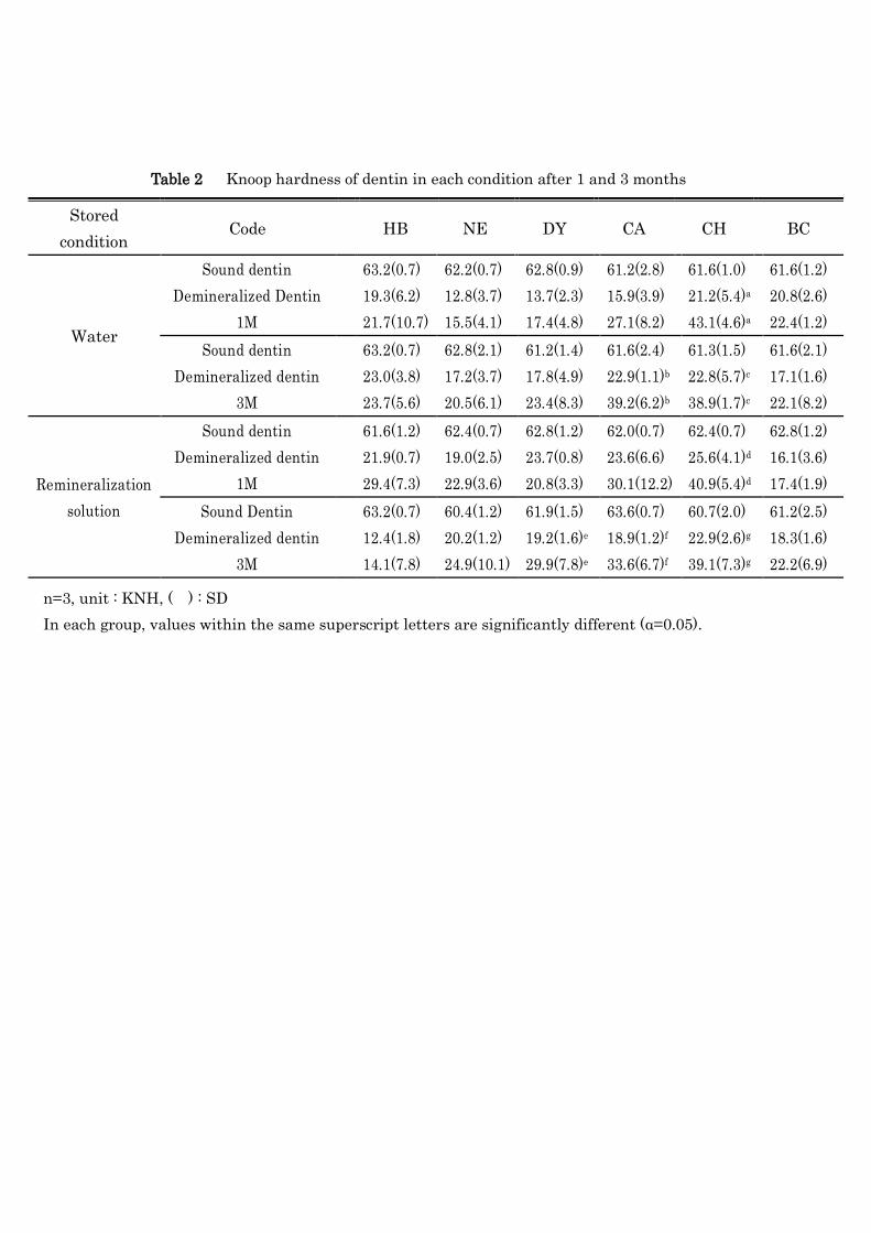
1. Hardness measurement
The hardness of samples, as measured using the Cariotester, is shown in Table 2.
1) Hardness of sound and demineralized dentin samples

9
The mean hardness levels of sound and demineralized dentin samples were
62.1±0.9 and 19.4±3.6 KNH, respectively. Hardness was significantly lower in all
demineralized dentin samples than in sound dentin samples.
2) Hardness of pulp-capped dentin samples
(1) HB-applied dentin
Hardness levels after 1 and 3 months were 21.7±10.7 and 23.7±5.6 KNH in the
DW group, and 29.4±7.3 and 14.1±7.8 KNH in the RS group, respectively. No
significant differences were observed in hardness levels between demineralized
dentin samples and DW or the RS groups at 1 or 3 months.
(2) NE-applied dentin
Hardness levels after 1 and 3 months were 15.5±4.1 and 20.5±6.1 KNH in the
DW group, and 22.9±3.6 and 24.9±10.1 KNH in the RS group, respectively. No
significant differences were observed in hardness levels between demineralized
dentin samples and DW or the RS groups at 1 or 3 months.
(3) DY-applied dentin
Hardness levels after 1 and 3 months were 17.4±4.8 and 23.4±8.3 KNH in the
DW group, and 20.8±3.3 and 29.9±7.8 KNH in the RS group, respectively. No
significant differences were observed in hardness levels between demineralized
dentin samples and the DW group at 1 or 3 months. No significant difference was
observed in hardness levels between the RS group and demineralized dentin
samples at 1 month, but was significantly higher in the RS group at 3 months.
(4) CA-applied dentin
10
Hardness levels after 1 and 3 months were 27.1±8.2 and 39.2±6.2 KNH in the
DW group, and 30.1±12.2 and 33.6±6.7 KNH in the RS group, respectively.
Hardness was not significantly different from that of the demineralized dentin
samples in the DW or the RS group at 1 month, but it was significantly increased
at 3 months.
(5) CH-applied dentin
Hardness levels after 1 and 3 months were 43.1±4.6 and 38.9±1.7 KNH in the
DW group, and 40.9±5.4 and 39.1±7.3 KNH in the RS group, respectively.
Hardness levels were significantly increased in both the DW and RS groups at 1
and 3 months than in demineralized dentin samples.
(6) Control
Hardness levels after 1 and 3 months were 22.4±1.2 and 22.1±8.2 KNH in the
DW group, and 17.4±1.9 and 22.2±6.9 KNH in the RS group, respectively. No
significant difference was observed in hardness levels between demineralized
dentin samples and the DW or RS groups at 1 or 3 months.
2. Observation under SEM
1) Sound dentin samples
SEM observation of the sound dentin samples are shown in Figure 1 (left). The
sample surface was densely covered with mineralized substances, such as
hydroxyapatite, and no collagen fibril was exposed.

11
2) Demineralized dentin samples
SEM observation of the demineralized dentin samples are shown in Figure 1
(light). Collagen fibrils were exposed due to demineralization by lactic acid.
Striation was observed on most collagen fibrils, but was absent on the others. The
slight retention of mineralized substances was observed on the collagen fibril
surface; however, no mineralized substance was present between collagen fibrils.
3) Pulp-capped dentin samples
(1) HB-applied samples
SEM observations of the dentin are shown in Figure 2. In the DW group,
collagen fibrils were exposed after 1 month, similar to those in demineralized
dentin samples, and no mineralized substance-like aggregates adhered to the
collagen fibril surface. The slight deposition of mineralized substance-like
aggregates was observed on the collagen fibril surface at 3 months. In the RS
group, collagen fibrils were exposed at 1 month, similar to those in the DW group;
however, mineralized substance-like aggregates were deposited on the collagen
fibril surface. The deposition of aggregates on the collagen fibril surface had
progressed at 3 months, and the space between collagen fibrils was more likely to
be sealed by these aggregates.
(2) NE-applied samples
SEM observations of the dentin are shown in Figure 3. In the DW group,
although collagen fibrils were exposed at 1 and 3 months, almost no deposition of
mineralized substance-like aggregates was noted. In the RS group, similar to
those in the DW group, collagen fibrils were exposed and almost no deposition of
12
mineralized substance-like aggregates was noted at 1 month. Some collagen
fibrils were exposed at 3 months and no mineralized substance-like aggregate was
deposited in some regions; however, the sample surface was more likely to be
covered by mineralized substance-like aggregates.
(3) DY-applied samples
SEM observations of the dentin are shown in Figure 4. In the DW group,
collagen fibrils were exposed at 1 month, and mineralized substance-like
aggregates were deposited on the surfaces of some collagen fibrils. The striation of
collagen fibrils was also noted. The deposition of aggregates on the collagen fibril
surface was more extensive at 3 months than at 1 month, and the space between
collagen fibrils was more likely to be sealed by the aggregates. In the RS group,
the deposition of mineralized substance-like aggregates was observed on the
collagen fibril surface at 1 month, and the space between collagen fibrils was more
likely to be sealed by the aggregates. This deposition had progressed further at 3
months, the space between collagen fibrils was sealed by the aggregates, and the
sample surface was densely covered.
(4) CA-applied samples
SEM observations of dentin are shown in Figure 5. In the DW group,
mineralized substance-like aggregates were deposited on the collagen fibril
surface and between collagen fibrils at 1 month. These depositions had further
progressed at 3 months, and the sample surface was densely covered by the
aggregates. In the RS group, similar to those in the DW group, mineralized
substance-like aggregates were deposited on the collagen fibril surface and
between collagen fibrils at 1 month, and had densely covered the sample surface

13
by 3 months.
(5) CH-applied samples
SEM observations of the dentin are shown in Figure 6. In the DW group,
mineralized substance-like aggregates were deposited on the collagen fibril
surface and between collagen fibrils at 1 month, and had densely covered the
sample surface by 3 months. In the RS group, similar to those in the DW group,
mineralized substance-like aggregates were deposited on the collagen fibril
surface and between collagen fibrils at 1 month, and had densely covered the
sample surface by 3 months.
(6) Control (BC-applied samples)
SEM observations of the dentin are shown in Figure 7. In the DW group, similar
to those in demineralized dentin samples, collagen fibrils were exposed at 1 and 3
months, and no mineralized substance-like aggregate had formed on the collagen
fibril surface. In the RS group, the deposition of mineralized substance-like
aggregates on the collagen fibril surface was slightly higher than that in the DW
group at 1 month; however, most collagen fibrils were exposed, which was similar
to that observed in the DW group. The deposition of mineralized substance-like
aggregates was slightly more extensive at 3 months than at 1 month, but was still
very limited.

14
Discussion
Although the remineralization of demineralized dentin after IPC is an
important issue in caries treatment, it has not yet been fully elucidated. Although
caries in extracted teeth are used as a material to investigate various problems, it
is difficult to collect a large number of carious teeth with a similar grade of
demineralized dentin. The artificial preparation of demineralized dentin similar
to spontaneous caries may be very useful to solve the problems described above.
Although many studies have attempted to prepare artificial carious dentin20-28),
caries was limited to the superficial layer in many of these reports, and the
preparation of artificial carious dentin with a thickness similar to that of
spontaneous caries has not yet been established. We used lactic acid as a solution
to decalcify dentin because it is an organic acid produced at high levels by
cariogenic bacteria29,30). To prepare demineralized dentin with a thickness similar
to that of caries, teeth were immersed in lactic acid solution aspirated from the
lateral side of the pulp cavity using an aspirator, which reduced the Knoop
hardness of the lateral side of the pulp cavity. We also investigated time-course
changes in the Knoop hardness of the pulp-capping agent-applied surface. Similar
investigations of the hardness of the lateral side of the pulp cavity may facilitate
the preparation of demineralized dentin samples with a constant thickness
similar to that of carious dentin, which would increase the significance of studies
on the depth of remineralization of carious dentin. Pulp-capped dentin samples
were stored in remineralization solution under conditions of 100% humidity.
Dental pulp fluid has been shown to act as remineralization solution in vital
teeth31). Even though the composition of the remineralization solution used was
15
not completely consistent with that of dental pulp fluid, we used it to investigate
the influence of Ca, contained in dental pulp fluid, on remineralization.
HY-Bond Temporary Cement Soft is a tannin-fluoride mixture (HY
agent)-combined carboxylate cement that is mainly used for temporary
cementation in dental practice; however, Nagamine13). reported that HY
agent-combined carboxylate cement promoted the remineralization of residual
carious dentin. The guidelines for caries treatment established by the Japanese
Society of Conservative Dentistry also recommend pulp-capping using HY
agent-combined carboxylate cement in IPC9). Thus, we tested this product as a
pulp-capping agent. No increase was observed in the hardness of demineralized
dentin in either the DW or RS group; however, the deposition of mineralized
substance-like aggregates was observed over time in both groups on SEM. HY
agents have been shown to strengthen organic substances and increase the acid
resistance of collagen fibrils32). These effects may have strengthened collagen
fibrils and promoted the deposition of mineralized substance-like aggregates.
Although hardness was not improved during the application period in this study,
the deposition of aggregates on collagen fibrils may be further promoted by
prolonging the application period, thereby increasing hardness.
Neodyne-α is zinc oxide eugenol cement. The liquid component, eugenol, has
superior analgesic sedative effects on dental pulp, and is used as a temporary
sealing or lining material in dental practice for caries that reach the deep dentin
and cause pulpitis. Since its powder component contains approximately 19%
calcium hydroxide and the overall content may be approximately 15%33), this
product was tested as a pulp-capping agent. No increase was observed in the

16
hardness of demineralized dentin in either the DW or RS group, and no deposition
of mineralized substance-like aggregates on the collagen fibril surface was noted
in the DW group on SEM. However, the sample surface was mostly covered by
mineralized substance-like aggregates in the RS group, although some collagen
fibrils were exposed, which indicated that the surface was remineralized. In
addition, the striation of collagen fibrils disappeared after 1 month but
reappeared after 3 months, which may have been due to the inhibition of
hydrolysis of collagen fibrils by the protein astringent action of zinc oxide34). The
presence or absence of the striation of collagen fibrils is considered to be related to
the remineralization of dentin, with decreased remineralization due to damage to
collagen fibrils being reported previously35). The application of Neodyne to carious
dentin may contribute to the long-term physiological remineralization of carious
dentin by preventing the hydrolysis of collagen fibrils.
Dycal is a calcium hydroxide pulp-capping agent. Since the base, containing
56.7% titanium dioxide, and catalyst paste, containing 53.5% calcium hydroxide
and 9.7% zinc oxide, are mixed36), the overall calcium hydroxide content may be
approximately 27%. An increase in hardness was not observed in the DW group,
but was at 3 months in the RS group, and mineralized substance-like aggregates
were densely deposited on the collagen fibril surface on SEM, which indicated
that the application of Dycal and subsequent 3-month immersion in
remineralization solution remineralized demineralized dentin. The deposition of
mineralized substance-like aggregates on the collagen fibril surface was also
noted in the distilled water group on SEM and was promoted over time,
suggesting that hardness increases with the prolongation of the application

17
period.
Clacipex Plain II is a calcium hydroxide root canal filler mainly used in dental
practice to fill the root canal. We tested it as a pulp-capping agent in the present
study because it contains approximately 48% calcium hydroxide. An increase in
hardness was observed after 3 months in both the DW and RS groups, and the
deposition of mineralized substance-like aggregates on and between collagen
fibrils was observed on SEM. These results suggest that demineralized dentin
wass remineralized 3 months after the application of a pulp-capping agent
containing approximately 24% calcium hydroxide.
A 60% calcium hydroxide mixture was prepared by mixing 0.6 g of calcium
hydroxide and 1 ml of distilled water as an experimental pulp-capping agent to
investigate the influence of calcium hydroxide concentrations on the
remineralization of demineralized dentin. An increase was observed in hardness
after 1 month in both the DW and RS groups, and mineralized substance-like
aggregate deposition was observed on and between collagen fibrils on SEM. These
results indicated that demineralized dentin was remineralized 1 month after the
application of a pulp-capping agent containing 60% calcium hydroxide. Therefore,
the application of a pulp-capping agent containing approximately 60% or higher
calcium hydroxide may increase hardness over the deep layer of thick
demineralized dentin.
The base cement was a glass ionomer cement, which is used as a lining material
in dental practice. The sustained release of fluoride ions is a characteristic of glass
ionomer cement, and has been shown to promote remineralization37-41). We tested
the base cement as a pulp-capping agent-covering material in the present study.
18
Samples covered only with the base cement without a pulp-capping agent were
also prepared as a control because the influence of fluoride ions slowly released
from the covering base cement on demineralized dentin remineralization was
considered. No increase was observed in hardness in either the DW or RS group.
The deposition of mineralized substance-like aggregates on and between collagen
fibrils was confirmed in the remineralization solution group after 3 months on
SEM, which suggested that prolonging the application period may promote
aggregate deposition and increase the hardness of demineralized dentin.
In caries treatment, demineralized dentin is mechanically removed using a
spoon excavator or round bar, and the removal range is decided based on finger
sensation. Histopathological studies have classified caries into the first and
second layers of carious dentin, and this has facilitated the development of caries
detectors, which are now widely used as an index for demineralized dentin
removal. However, finger sensation and differentiation using a caries detector
depend on the operator’s subjective judgment, such that demineralized dentin
may be left in place. Thus, using an objective index, hardness, is necessary.
Dentin hardness has been measured using micro-hardness meters, such as
Mohs, Vickers, and Knoop hardness meters42-46). However, these studies were
performed using extracted teeth; dentin hardness has not yet been measured in
vital teeth in the oral cavity. Shimizu18,19) et al. recently developed a hardness
measurement device for carious dentin, the Cariotester, which is now
commercially available. The direct measurement of the Knoop hardness of carious
dentin in the oral cavity using the Cariotester has facilitated the introduction of
an objective index, hardness, into caries treatment.

19
Many cases in which dental caries has progressed to deep dentin close to the
pulp have been reported in dental practice. Pulpectomy was previously indicated
for these cases when caries caused pulp exposure. However, a recent study of
dental pulp clarified its high healing capacity and the reversibility of dental pulp
inflammation47,48); therefore, the importance of dental pulp protection has been
recognized and IPC is now recommended. Infected dentin near the pulp is
intentionally left in IPC, even in caries progressing to deep dentin close to the
pulp. This is then treated with a tannin-fluoride mixture-combined carboxylate
cement or calcium hydroxide preparation; through which residually infected
dentin is sterilized and remineralized and tertiary dentin (reparative dentin)
formation is promoted, with the aim of dental pulp conservation. Although this
is limited to cases with no pulp symptoms, favorable outcomes were obtained in
most cases when IPC achieved restoration without pulp exposure. The
effectiveness of IPC has been investigated using not only hardness, but also
various other methods, such as X-ray radiography and bacteriological and
histopathological examinations. However, these were performed using samples
prepared from extracted teeth; studies using carious dentin in the oral cavity have
not yet been conducted. If the presence or absence of the remineralization of
residual demineralized dentin and dentin restoration can be confirmed by
measuring the hardness of demineralized dentin in the oral cavity over time using
the Cariotester, pulp exposure may be prevented at re-entry.

20
Conclusions
The hardness of various dentin samples was measured using a novel Knoop
hardness measurement system, Cariotester, and the sample surfaces were
observed using SEM. The following conclusions were obtained:
1. When only lining with base cement was applied without pulp-capping, the
hardness of demineralized dentin did not increase, and no remineralization
occurred.
2. When the calcium hydroxide concentration was low, the hardness of
demineralized dentin did not increase, but mineralized substance-like aggregate
formation was observed.
3. When a pulp-capping agent containing calcium hydroxide at or above a specific
concentration was applied to demineralized dentin, the hardness of the
demineralized dentin increased and features of remineralization were observed.
It was suggested that the remineralization of demineralized dentin depended on
the calcium hydroxide concentration contained in a pulp-capping agent.
We would like to thank the members of the Department of Operative Dentistry,
Osaka Dental University, for their advice and help. This study was supported in
part by Osaka Dental University Research Funds (12-03).

21
Reference
1) Terashima S. Differentiation of two layers of carious dentin by staining. J
Stomatol Soc Jpn 1970; 37: 279-286. (in Japanese)
2) Sato Y. Removal of infected dentin using fuchsin staining as a guide: 1.
Experiment with extracted carious teeth. J Stomatol Soc Jpn 1973; 40: 420-427.
(in Japanese)
3) Sato Y. Removal of infected dentin using fuchsin staining as a guide: 2.
Experiment with vital carious teeth. J Stomatol Soc Jpn 1974; 41: 202-211. (in
Japanese)
4) Sano H. Relationship between caries detector staining and structual
characteristics of carious dentin. J Stomatol Soc Jpn 1987; 54: 241-270. (in
Japanese)
5) Sano H, Nakajima M and Tagami J. Diagnosis and removal of dental caries
using caries detector.Yasuda N and Tagami J.Practice in prosthodontics
separate volume. Search for new cariology and operative dentistry.Ishiyaku
Shuppan: Tokyo; 1997.89-92. (in Japanese)
6) Katou S. Recalcification of artificially demineralized dentin in living dog teeth.
J Stomatol Soc Jpn 1968; 35: 613-625. (in Japanese)
7) Miyauchi H. Physiological recalcification of carious dentin. J Stomatol Soc Jpn
1976; 43: 384-393. (in Japanese)
8) Yamamoto Y. An in vitro study on the remineralization of inner carious dentin:
The remineralization effect of glass ionomer cement. Tsurumi Univ Dent J
1984; 10: 57-74. (in Japanese)
9) The Japanese society of conservative dentistry. Clinical guidelines for treating

22
caries in adults following a minimal intervention policy-Evidence and
consensus based report. 1st ed. Nagasue Shoten: kyoto; 2009. 52-69. (in
Japanese)
10) Leung RL, Loesche WJ, Charbeneu GT. Effect of dycal on bacteria in carious
lesions. J Am Dent Assoc 1980;100:193-197.
11) Ogawa F and Machida Y. Clinical observation of pulp capping on deep carious
lesions. J Tokyo Dent Soc 1984; 84: 1963-1970. (in Japanese)
12) Goto J. Clinical and histological study of indirect pulp capping on deep carious
lesions. Jpn J Ped Dent 1985; 23: 926-938. (in Japanese)
13) Nagamine M. Studies on treatment of deep carious lesions utilizing
polycarboxylate cement combined with tannin-fluoride preparation. J
Okayama Dent Soc 1993; 12: 1-25. (in Japanese)
14) Leksell E, Ridell K, Cvek M, Mejare I. Pulp exposure after stepwise versus
direct complete excavation of deep carious lesions in young posterior
permanent teeth. Endod Dent Traumatol 1996;12: 192-196.
15) Bjorndal L, Thylstrup A. A clinical and microbiological study of deep carious
lesions during stepwise excavation using long treatment interval. Caries Res
1997; 31:411-417.
16) Bjorndal L, Thylstrup A. A practice-based study on stepwise excavation of deep
carious lesions in permanent teeth: a 1-year follow-up study. Community Dent
Oral Epidemiol 1998; 26: 122-128.
17) Bjorndal L, Larsen T. Changes in the cultivable flora in deep carious lesions
following a stepwise excavation procedure. Caries Res 2000; 34:502-508.
18) Shimizu A, Honda K, Natsumi Y and Hasegawa M. Development and

23
evaluation of a practical device for detecting hardness of carious dentin. Jpn J
Conserv Dent 2006; 49: 45-51. (in Japanese)
19) Shimizu A, Kinoshita N, Honda K, Abe T and Hasegawa M. A new trial for
determining the hardness of carious dentin. Jpn J Conserv Dent 2008; 51:
565-570. (in Japanese)
20) Satake S, Hasegawa M, Shimizu A and Yoshioka W. Production of a caries-like
lesion and remineralization of the lesion. Part 1. Caries-like lesion produced by
high viscosity etching gel and rehardening of the lesion. Jpn J Conserv Dent
1990; 33: 1540-1549. (in Japanese)
21) Wefel JS, Heilman JR, Jordan TH. Comparisons of in vitro root caries models.
Caries Res 1995; 29: 204-209.
22) Iijima Y and Takagi O. The reaction of enamel and root dentine samples after
demineralization and remineralization. J Dent Hlth 1996; 46: 290-296.
23) Konishi M. Creation of artificial carious dentin using lactic acid and
collagenase. Jpn J Conserv Dent 1999; 42: 144-157. (in Japanese)
24) Shimizu A, Maeda T, Natsumi Y, Honda K and Hasegawa M. Attempts at
producing artificial caries-like lesion in dentin. Part 1 An investigation on
decalcification method and period. Jpn J Conserv Dent 1999; 42: 1116-1122. (in
Japanese)
25) Urayama A, Yoshiyama M, Kimochi T, Matsuoka N, Ozaki k and Matsuo T.
Resin infiltration into artificial carious dentin. Jpn J Conserv Dent 1999; 42:
529-535. (in Japanese)
26) Itota T, Iwai Y, Okamoto M, Tashiro Y, Nakabo S, Nishitani Y, Nagamine M,
ToriiYand Yoshiyama M. Remineralization of demineralized dentin by a
24
fluoride-releasing adhesive system. Jpn J Conserv Dent 2001; 44: 175-181. (in
Japanese)
27) Shimizu A, Maeda T, Natsumi Y, Hasegawa M and Honda K. Attempts at
producing artificial caries-like lesion in dentin. Part 2 Decalcification using
buffer solution and/or acid gel. Jpn J Conserv Dent 2001; 44: 583-589. (in
Japanese)
28) Yamazaki T, Miake Y, Ishikawa T, Hiruma N and Yanagisawa N. The effect of
mineralized seaweed on remineralization of artificially demineralized dentin.
J Hard Tissue Biol 2011; 20: 11-16. (in Japanese)
29) Hanada N. Consider etiology of dental caries as disease group: A proposal.
Dental Outlook 1997; 89: 309-313. (in Japanese)
30) Nikifruk G. Abe K, Kameyama Y, Kitano S, Sakaki T and Takeuchi H.
Understanding dental caries: Etiology and mechanisms basic and aspects. 1st
ed. Gakken Shoin: Tokyo; 1987. 149. (in Japanese)
31) Shellis RP. Effects of a supersaturated pulpal fluid on the formation of
caries-like lesions on the roots of human teeth. Caries Res 1994, 28: 14-20.
32) Yamaga M, Hori N, Koide T and Daito M. The effect of tannin-fluoride
preparation (HY agent) on typeⅠcollagen. Jpn J Ped Dent 1996; 34: 208-213.
(in Japanese)
33) Makiishi T, Aida S, Matsui K, Yamauchi K, Hirai Y, Takahashi K and Ishikawa
T. A clinical study on the zinc oxide eugenol cement “Neodyne α”. J Tokyo Dent
Soc 1980; 80: 1045-1050. (in Japanese)
34) Amano Y.The outline of the fundamental method in endodontic care. Toda T,
Nakamura H, Suda H and Katsuumi I. Endodontics. 3rd ed. Ishiyaku

25
Shuppan: Tokyo; 2010, 65. (in Japanese)
35) Saito T, Toyooka H, Matsuda K, Yamauchi M and Crenshaw MA. In vitro
mineral induction by insoluble dentin matrix: A role of phosphate group and
carboxyl group on mineral induction.Jpn J Conserv Dent 1997; 40: 1461-1468.
(in Japanese)
36) Tronstad L. Reaction of the exposed pulp to Dycal treatment. Oral Surg Oral
Med Pathol. 1974; 38: 945-953.
37) Minami K, Nagai Y, Inaba D, Someya Y, Matsuda K and Yonemitsu M. Effect
of fluoride releasing restorative materials on remineralization of root dentin in
vitro. J Dent Hlth. 2001; 51: 293-297. (in Japanese)
38) Tominaga T, Mukai Y and Teranaka T. Time-course study of fluoride-releasing
restorative materials on dentin remineralization and acid resistance in vitro.
Jpn J Conserv Dent 2007; 50: 808-817. (in Japanese)
39) Ten Cate JM, van Duinen RN. Hypermineralization of dentinal lesions
adjacent to glass-ionomer cement restorations. J Dent Res 1995; 74:
1266-1271.
40) Hatibovic-Kofman S, Suljac JP, Koch G. Remineralization of natural carious
lesions with a glass ionomer cement. Swed Dent J 1997; 21: 11-17.
41) Bynum AM, Donly KJ. Enamel de/remineralization on teeth adjacent to
fluoride releasing materials without dentifrice exposure. ASDC J Dent Child
1999; 66: 89-92.
42) Miake K, Higashi S, Fukuyama S, Ebihara N, TakoA, Senchi H and Nabeya S.
Microhardness studies on human teeth. Part 2. Microhardness of enamel and
dentin of permanent molars. J Tokyo Dent Soc 1967; 67: 961-986. (in Japanese)
26
43) Nishimura F, Okazaki K, Kono Y, Komori K and Nomoto S. Compressive
behavior and microvickers hardnes of human enamel and dentin. J J Dent
Mater 1986; 5: 449-454. (in Japanese)
44) Saito H. Study on the method of hardness test and the distribution of
micro-vickers hardness of human teeth. J J Dent Mater 1991; 10: 241-265. (in
Japanese)
45) Willems G, Lambrechts P, Braem M, Celis JP, Vanherle G. A classification of
dental composites according to their morphological and mechanical
characteristics. Dent Mater 1992; 8: 310-319.
46) Akimoto N, Ohmori K, Yokoyama G, Momoi Y and Kohno A. Study on
microhardness of human dentin using nano-indentation tester. Jpn J Conserv
Dent 1999; 42: 96-101. (in Japanese)
47) Kidd EAM. Caries removal and the pulpo-dentinal complex. Dent Update
2000; 27: 476-482.
48) Ranly DM, Garcia-Godoy F. Current and potential pulp therapies for primary
and young permanent teeth. J Dent. 2000; 28: 153-161.

27
新規 Knoop硬さ測定システムによる覆髄剤の有効性の検討
松田有之 1),恩田康平 2),谷本啓彰 2),吉川一志 2),山本一世 2)
1)大阪歯科大学大学院歯学研究科 歯科保存学専攻
2)大阪歯科大学歯科保存学講座
責任著書者連絡先:松田有之
〒573-1121 大阪府枚方市楠葉花園町 8-1
大阪歯科大学大学院歯学研究科歯科保存学専攻
TEL: 072-864-3077, FAX: 072-864-3177
E-mail: [email protected]
覆髄剤が脱灰象牙質の硬さに与える影響について
28
抄録
目的
Minimal Intervention(MI)の概念に基づき,齲蝕が深部象牙質にまで進行し歯髄に
近接する場合,歯髄に近接する深部齲蝕象牙質を保存し,露髄を回避する目的で暫間
的間接覆髄法(IPC)が行われる.本研究では,新規 Knoop硬さ測定システムである
カリオテスターSUK-971(三栄エムイー)を用いて象牙質試料の硬さを測定し,覆髄
剤が軟化象牙質へ与える影響を検討した.
材料および方法
ヒト抜去大臼歯から直径 10mm,厚さ 2mmの象牙質試料を作製し,象牙質試料の
歯髄腔側からアスピレーターで吸引しながら,エナメル質側を 20mM乳酸溶液に浸
漬して,エナメル質側の硬さが 20KNH程度となる脱灰象牙質試料を作製した.脱灰
象牙質試料に,ハイボンドテンポラリーセメントソフト(松風),ネオダイン-α(ネ
オ製薬),ダイカル(デンツプライ三金),カルシペックス プレーンⅡ(日本歯科薬
品),60%水酸化カルシウム混和物(キシダ)を貼付し,ベースセメント(松風)で
被覆したものを覆髄試料,覆髄剤を貼付せずベースセメントのみで被覆したものをコ
ントロールとして作製し,湿度 100%容器中または石灰化溶液中で 1か月間および 3
か月間保管後,脱灰象牙質の硬さを測定した.試料数は各条件につき 3試料とし,得
られた値は一元配置分散分析および Tukeyの検定にて統計解析を行った(α=0.05).
また,硬さ測定後,覆髄剤貼付部の SEM画像の観察を行った.
29
結果および考察
ダイカル,カルシペックスプレーンⅡ,60%水酸化カルシウム混和物を貼付した脱
灰象牙質試料では硬さが向上し,SEM画像でも石灰化物の緻密な沈着が認められた.
コントロール,ハイボンドテンポラリーセメントソフト,ネオダイン-αを貼付した
脱灰象牙質試料では,硬さは向上せず,SEM画像でも石灰化物の緻密な沈着は認め
られなかった.このことから,水酸化カルシウムを 27%以上含有する覆髄剤を脱灰象
牙質に応用することで,コラーゲン線維表面に石灰化物の沈着が起こり,脱灰象牙質
が硬化したと考えられる.また,カリオテスターを用いることで, IPC後の象牙質
の硬さの継時的変化をチェアーサイドで客観的に評価することが可能になると考え
られる.
結論
覆髄剤貼付による脱灰象牙質の再石灰化は,覆髄材に含有される水酸化カルシウム
の含有濃度に影響されることが示唆された.
キーワード:Knoop硬さ,覆髄剤,再石灰化
30
Figure legend
Table 1 Materials
The materials tested as pulp-capping agents in this study.
Table 2 Knoop hardness of dentin in each condition after 1 and 3 months
The unit is Knoop hardness of dentin in each condition. ( ) means SD of
Knoop hardness. The number of samples was 3 for each condition. In each
group, values within the same superscript letters are significantly different
(α=0.05).
Fig. 1 SEM images of sound and demineralized dentin
The sample surface of sound dentin was densely covered with mineralized
substances, such as hydroxyapatite, and no collagen fibrils were exposed. The
collagen fibrils of demineralized dentin samples were exposed due to
demineralization by lactic acid. Striation was observed on most collagen fibrils,
but was absent on the others. The slight retention of mineralized substances was
observed on the collagen fibril surface, but no mineralized substance was present
between collagen fibrils.
Fig. 2 SEM images of dentin applied HB
In the DW group, collagen fibrils were exposed after 1 month, similar to those in
demineralized dentin samples, and no mineralized substance-like aggregates
adhered to the collagen fibril surface. The slight deposition of mineralized
substance-like aggregates was observed on the collagen fibril surface at 3 months.

31
In the RS group, collagen fibrils were exposed at 1 month, similar to those in
the DW group; however, mineralized substance-like aggregates were deposited on
the collagen fibril surface. The deposition of aggregates on the collagen fibril
surface had progressed at 3 months, and the space between collagen fibrils was
more likely to be sealed by the aggregates.
Fig. 3 SEM images of the dentin applied NE
In the DW group, collagen fibrils were exposed at 1 and 3 months, and almost
no deposition of mineralized substance-like aggregates occurred.
In RS group, similar to those in DW group, collagen fibrils were exposed and
almost no deposition of mineralized substance-like aggregates was noted at 1
month. Some collagen fibrils were exposed at 3 months and no mineralized
substance-like aggregates were deposited in some regions; however, the sample
surface was more likely to be covered by mineralized substance-like aggregates.
Fig. 4 SEM images of the dentin applied DY
In the DW group, collagen fibrils were exposed at 1 month, and mineralized
substance-like aggregates were deposited on the surfaces of some collagen fibrils.
The striation of collagen fibrils was also noted. The deposition of aggregates on
the collagen fibril surface was more extensive at 3 months than at 1 month, and
the space between collagen fibrils was more likely to be sealed by the aggregates.
In the RS group, the deposition of mineralized substance-like aggregates was
observed on the collagen fibril surface at 1 month, and the space between collagen
fibrils was more likely to be sealed by the aggregates. Deposition progressed
32
further by 3 months, the space between collagen fibrils was sealed by the
aggregates, and the sample surface was densely covered.
Fig. 5 SEM images of the dentin applied CA
In the DW group, mineralized substance-like aggregates were deposited on the
collagen fibril surface and between collagen fibrils at 1 month. Deposition had
further progressed at 3 months, and the sample surface was densely covered by
the aggregates.
In the RS group, similar to those in the DW group, mineralized substance-like
aggregates were deposited on the collagen fibril surface and between collagen
fibrils at 1 month, and deposition had progressed and aggregates densely covered
the sample surface at 3 months.
Fig. 6 SEM images of the dentin applied CH
In the DW group, mineralized substance-like aggregates were deposited on the
collagen fibril surface and between collagen fibrils at 1 month, and deposition had
progressed and aggregates densely covered the sample surface at 3 months.
In the RS group, similar to those in the DW group, mineralized substance-like
aggregates were deposited on the collagen fibril surface and between collagen
fibrils at 1 month, and deposition had progressed and aggregates densely covered
the sample surface at 3 months.
Fig. 7 SEM images of the dentin applied BC
In the DW group, similar to those in demineralized dentin samples, collagen
33
fibrils were exposed at 1 and 3 months, and mineralized substance-like
aggregates did not form on the collagen fibril surface.
In the RS group, the deposition of mineralized substance-like aggregates on the
collagen fibril surface at 1 month was slightly higher than that in the DW group;
however, most collagen fibrils were exposed, similar to those in the DW group. The
deposition of mineralized substance-like aggregates was slightly more at 3
months than at 1 month, but was still very limited.
Brand Code Component Manufacture Lot. No.
HY-Bond Temporary
Cement Soft HB
Powder: Zinc oxide, Silica, Magnesium oxide,
HY-agent
Liquid: Acrylic acid-Tricarboxylic acid copolymer
sodium solt, Purified water
Shofu Powder: 121276
Liquid: 011327
Neodyne-α NE
Powder: Zinc oxide, Calcium hydroxide (19%),
Rosin
Liquid: Eugenol
Neo Dental
Chemical
Products
Powder: A2K1
Liquid: A2K1
Dycal DY Base: Titanium dioxide, Glycol salicylate
Catalyst: Calcium hydroxide (53.5%), Zinc oxide Dentsply
Base: 023304
Catalyst: 023305
Calcipex PlaneⅡ CA Calcium hydroxide (48%), Purified water Nippon Shika
Yakuhinn A6K
60% Calcium
Hydroxide Mixture CH
Powder: Calcium hydroxide (60%)
Liquid: Distilled water Kishida G22464D
Base Cement BC
Powder: Fluoroalmino silicate grass
Liquid: Acrylic acid-Tricarboxylic acid copolymer
solution, Tartaric acid
Shofu Powder: 051214
Liquid: 031397
Table 1 Materials
Stored
condition Code
HB NE DY CA CH BC
Water
Sound dentin 63.2(0.7) 62.2(0.7) 62.8(0.9) 61.2(2.8) 61.6(1.0) 61.6(1.2)
Demineralized Dentin 19.3(6.2) 12.8(3.7) 13.7(2.3) 15.9(3.9) 21.2(5.4)a 20.8(2.6)
1M 21.7(10.7) 15.5(4.1) 17.4(4.8) 27.1(8.2) 43.1(4.6)a 22.4(1.2)
Sound dentin 63.2(0.7) 62.8(2.1) 61.2(1.4) 61.6(2.4) 61.3(1.5) 61.6(2.1)
Demineralized dentin 23.0(3.8) 17.2(3.7) 17.8(4.9) 22.9(1.1)b 22.8(5.7)c 17.1(1.6)
3M 23.7(5.6) 20.5(6.1) 23.4(8.3) 39.2(6.2)b 38.9(1.7)c 22.1(8.2)
Remineralization
solution
Sound dentin 61.6(1.2) 62.4(0.7) 62.8(1.2) 62.0(0.7) 62.4(0.7) 62.8(1.2)
Demineralized dentin 21.9(0.7) 19.0(2.5) 23.7(0.8) 23.6(6.6) 25.6(4.1)d 16.1(3.6)
1M 29.4(7.3) 22.9(3.6) 20.8(3.3) 30.1(12.2) 40.9(5.4)d 17.4(1.9)
Sound Dentin 63.2(0.7) 60.4(1.2) 61.9(1.5) 63.6(0.7) 60.7(2.0) 61.2(2.5)
Demineralized dentin 12.4(1.8) 20.2(1.2) 19.2(1.6)e 18.9(1.2)f 22.9(2.6)g 18.3(1.6)
3M 14.1(7.8) 24.9(10.1) 29.9(7.8)e 33.6(6.7)f 39.1(7.3)g 22.2(6.9)
Table 2 Knoop hardness of dentin in each condition after 1 and 3 months
n=3, unit : KNH, ( ) : SD
In each group, values within the same superscript letters are significantly different (α=0.05).
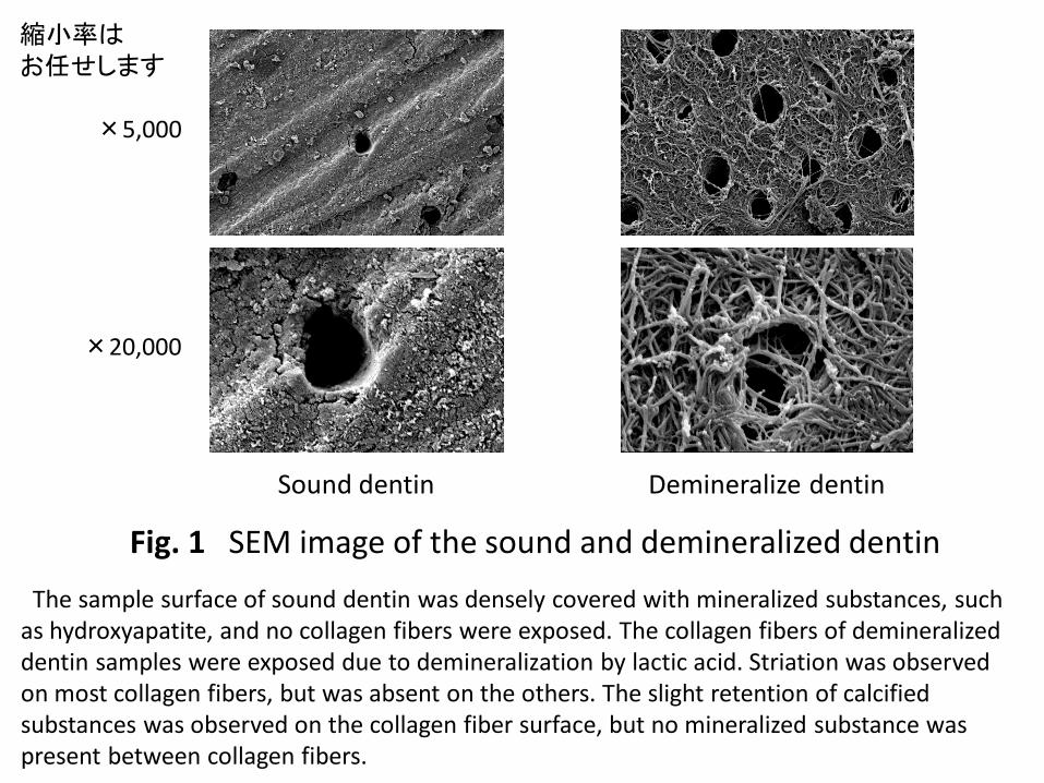
Fig. 1 SEM image of the sound and demineralized dentin
Sound dentin Demineralize dentin
The sample surface of sound dentin was densely covered with mineralized substances, such as hydroxyapatite, and no collagen fibers were exposed. The collagen fibers of demineralized dentin samples were exposed due to demineralization by lactic acid. Striation was observed on most collagen fibers, but was absent on the others. The slight retention of calcified substances was observed on the collagen fiber surface, but no mineralized substance was present between collagen fibers.
縮小率は お任せします
×5,000
×20,000

1M 3M
1M 3M
1M 3M
1M 3M
Fig. 2 SEM image of the dentin applied HB
×5,000
×20,000
DW RS
×5,000
×20,000
In the DW group, collagen fibers were exposed after 1 month, similar to those in demineralized dentin samples, and no mineralized substance-like aggregates adhered to the collagen fiber surface. The slight deposition of mineralized substance-like aggregates was observed on the collagen fiber surface at 3 months. In the RS group, collagen fibers were exposed at 1 month, similar to those in the DW group; however, mineralized substance-like aggregates were deposited on the collagen fiber surface. The deposition of aggregates on the collagen fiber surface had progressed at 3 months, and the space between collagen fibers was more likely to be sealed by the aggregates.
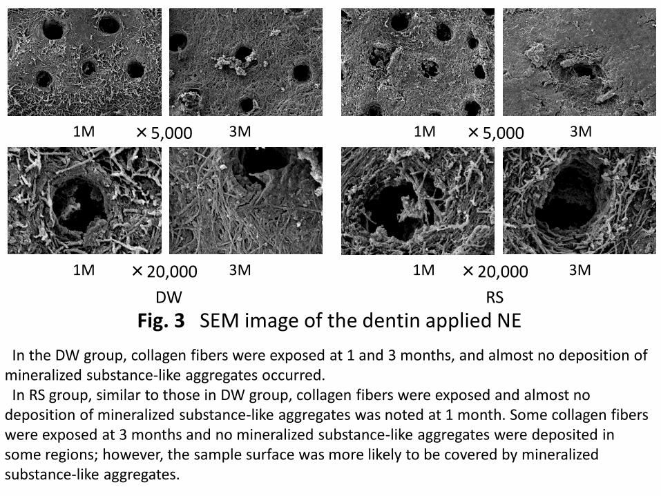
Fig. 3 SEM image of the dentin applied NE
1M 3M 1M 3M
1M 3M 1M 3M
×5,000
×20,000
DW RS
×5,000
×20,000
In the DW group, collagen fibers were exposed at 1 and 3 months, and almost no deposition of mineralized substance-like aggregates occurred. In RS group, similar to those in DW group, collagen fibers were exposed and almost no deposition of mineralized substance-like aggregates was noted at 1 month. Some collagen fibers were exposed at 3 months and no mineralized substance-like aggregates were deposited in some regions; however, the sample surface was more likely to be covered by mineralized substance-like aggregates.

Fig. 4 SEM image of the dentin applied DY
1M 3M
1M 3M 1M 3M
×5,000
×20,000
DW RS
×20,000
1M 3M ×5,000
In the DW group, collagen fibers were exposed at 1 month, and mineralized substance-like aggregates were deposited on the surfaces of some collagen fibers. The striation of collagen fibers was also noted. The deposition of aggregates on the collagen fiber surface was more extensive at 3 months than at 1 month, and the space between collagen fibers was more likely to be sealed by the aggregates. In the RS group, the deposition of mineralized substance-like aggregates was observed on the collagen fiber surface at 1 month, and the space between collagen fibers was more likely to be sealed by the aggregates. Deposition progressed further by 3 months, the space between collagen fibers was sealed by the aggregates, and the sample surface was densely covered.

Fig. 5 SEM image of the dentin applied CA
1M 3M
1M 3M
×5,000
×20,000
DW
1M 3M ×5,000
1M 3M
RS
×20,000
In the DW group, mineralized substance-like aggregates were deposited on the collagen fiber surface and between collagen fibers at 1 month. Deposition had further progressed at 3 months, and the sample surface was densely covered by the aggregates. In the RS group, similar to those in the DW group, mineralized substance-like aggregates were deposited on the collagen fiber surface and between collagen fibers at 1 month, and deposition had progressed and aggregates densely covered the sample surface at 3 months.
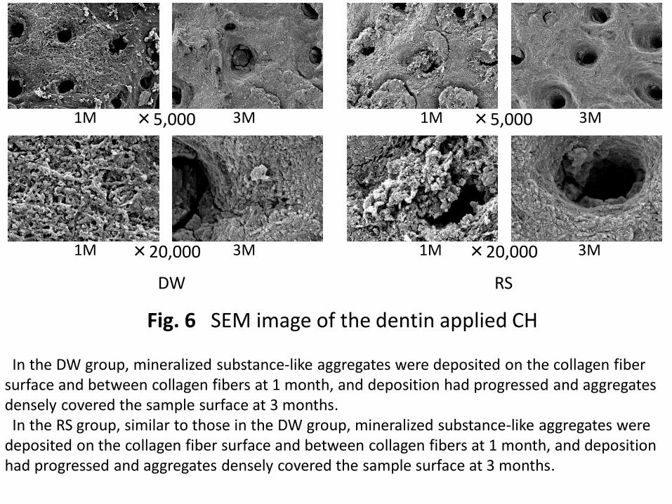
Fig. 6 SEM image of the dentin applied CH
1M 3M
1M 3M
×5,000
×20,000
DW
1M 3M
RS
×20,000
1M 3M ×5,000
In the DW group, mineralized substance-like aggregates were deposited on the collagen fiber surface and between collagen fibers at 1 month, and deposition had progressed and aggregates densely covered the sample surface at 3 months. In the RS group, similar to those in the DW group, mineralized substance-like aggregates were deposited on the collagen fiber surface and between collagen fibers at 1 month, and deposition had progressed and aggregates densely covered the sample surface at 3 months.

Fig. 7 SEM image of the dentin applied BC
1M 3M
1M 3M
×5,000
×20,000
DW
1M 3M
RS
×20,000
1M 3M ×5,000
In the DW group, similar to those in demineralized dentin samples, collagen fibers were exposed at 1 and 3 months, and mineralized substance-like aggregates did not form on the collagen fiber surface. In the RS group, the deposition of mineralized substance-like aggregates on the collagen fiber surface at 1 month was slightly higher than that in the distilled water group; however, most collagen fibers were exposed, similar to those in the distilled water group. The deposition of mineralized substance-like aggregates was slightly more at 3 months than at 1 month, but was still very limited.