studying the chlorophyll fluorescence in cyanobacteria ... · cyanobacteria with membrane computing...
TRANSCRIPT
Studying the Chlorophyll Fluorescence inCyanobacteria with Membrane ComputingTechniques
Ioan Ardelean1, Daniel Dıaz-Pernil2, Miguel A. Gutierrez-Naranjo3,Francisco Pena-Cantillana3, Iris Sarchizian4
1Institute of Biology Bucharest, Romanian Academy, 060031 Bucharest, [email protected]
2Research Group on Computational Topology and Applied MathematicsDepartment of Applied Mathematics - University of Sevilla, 41012, [email protected]
3Research Group on Natural ComputingDepartment of Computer Science and Artificial IntelligenceUniversity of Sevilla, 41012, [email protected], [email protected]
4Scoala Gimnaziala nr.38 Dimitrie Cantemir, Constanta, [email protected]
Summary. In this paper, we report a pioneer study of the decrease in chlorophyll fluo-rescence produced by the reduction of MTT (a dimethyl thiazolyl diphenyl tetrazoliumsalt) monitored using an epifluorescence microscope coupled to automate image analysisin the framework of P systems. Such analysis has been performed by a family of tissueP systems working on the images as data input.
1 Introduction
Membrane Computing has many features that makes it suitable for the study andthe implementation of algorithms of digital images. One of them is that, usually,these algorithms can be parallelized and locally solved. Regardless how large thepicture is, many algorithmic processes can be performed in parallel in differentlocal areas. Another interesting feature is that the local information needed fora pixel transformation can also be easily encoded in the data structures used inMembrane Computing.
Recently, a new research line has been open by applying well-known MembraneComputing techniques for solving problems from digital images. For example, seg-mentation is a well-known problem in the process of digital images which tries to
10 I. Ardelean et al.
assign a label to every pixel in an image in such way that pixels with the samelabel share certain visual characteristics. Segmentation has shown its utility, forexample, in bordering tumors and other pathologies or computer-guided surgery.In [11, 12, 13, 15, 31] we can find several approaches to this problem with Mem-brane Computing techniques. Other problems as thresholding [10] or smoothing[25] has also been considered in the framework of membrane computing. Specialattention deserves [18], where the symmetric dynamic programming stereo (SDPS)algorithm [19] for stereo matching was implemented by using simple P moduleswith duplex channels or [33], where the authors combine Membrane Computingand quantum-inspired algorithms for image processing.
In [2], a first approach of the application of Membrane Computing techniques tothe study of images from Microbiology was presented. Automated image analysisis increasingly used in Microbiology to quantify important parameters for researchand application. The most studied so far are the cell numbers, cell volumes, fre-quencies of dividing cells, in situ classification of bacteria, enumeration of activelyrespiring bacteria, characterization of bacterial growth on solid medium, viabilityand physiological activity in biofilms (e.g. [9, 17, 29, 30]).
In [2], the focus was the study of the application of Membrane Computingtechniques to the problem of counting cells. The whole process is a combinationof different techniques of processing images (binarization, segmentation, noise re-duction . . . ) which can be performed by different families of P systems. The finalalgorithm is a sequence of partial processes which can be performed by MembraneComputing techniques, and the application of such processes can be seen as aglobal machine which takes as input a digital image showing a biological entity(usually, a photograph taken with a microscopy in a wet lab) and the output is thenumber of cells in the picture.
In this paper, we focus on the problem of considering the intensity of color incyanobacteria, a phylum of bacteria that obtain their energy through photosyn-thesis, by using algorithms based on Membrane Computing techniques. We reporta new study of the decrease in chlorophyll fluorescence produced by the reductionof MTT (a dimethyl thiazolyl diphenyl tetrazolium salt) monitored using an epi-fluorescence microscope coupled to automate image analysis in the framework ofP systems The different stages of the analysis have been performed by a family oftissue P systems working on the images as data input.
Such families of P systems used in the stages of the process have inspired par-allel software programs which have been developed by using a device architecturecalled CUDATM, (Compute Unified Device Architecture). CUDATM is a generalpurpose parallel computing architecture that allows the parallel NVIDIA1 Graph-ics Processors Units (GPUs) to solve many complex computational problems in amore efficient way than on a CPU. GPUs constitute nowadays a solid alternativefor high performance computing, and the advent of CUDA allows programmers afriendly model to accelerate a broad range of applications. This novel architecturehas been previously used to implement parallel software that simulates the behav-1 http://www.nvidia.com.
Studying the Chlorophyll Fluorescence in Cyanobacteria 11
ior of P systems [5, 6, 7, 8, 24, 25], and, in a similar way to other implementations,the obtained results in the problem of detecting cyanobacteria are quite promising.
The paper is organized as follows: Next, we recall the computational modelused to design the different families of P systems that performs the stages ofthe algorithm. In Section 3, a short presentation of the biological experiment oncyanobacteria is presented. Section 4 outlines the steps of the analysis via a familyof P systems with takes as input data the images taken in the wet lab. Section 5provides some details of the implementation of such families on CUDA and finally,the paper ends with some conclusions and open lines for future research.
2 Formal Framework
Next, we recall some basics on the P system model chosen for implementing thesolution described below. The model is tissue-like P systems with promoters. Pro-moters are usually defined on cell-like models [20] and its extension to tissue-likeis quite natural. Next, we recall the formal definition.
Definition 1. A tissue-like P system with promoters of degree q ≥ 1 is a tuple ofthe form
Π = (Γ,Σ, E , w1, . . . , wq,R, iin, iout)
where
1. Γ is a finite alphabet, whose symbols will be called objects;2. Σ ⊆ Γ is the input alphabet;3. E ⊆ Γ is a finite alphabet representing the set of the objects in the environment
available in an arbitrary large amount of copies;4. w1, . . . , wq are strings over Γ representing the multisets of objects associated
with the cells in the initial configuration;5. R is a finite set of rules of the following form:
(pro | i, u/v, j), for 0 ≤ i 6= j ≤ q, pro, u, v ∈ Γ ∗
In these rules, the labels 1, . . . , q correspond to the q cells and the label 0 cor-responds to the environment;
6. iin ∈ {1, 2, . . . , q} denotes the input region;7. iout ∈ {1, 2, . . . , q} denotes the output region.
The rule (pro | i, u/v, j) can be applied over two cells (or a cell and the envi-ronment) i and j such that u (contained in cell i) is traded against v (contained incell j). The rule is applied if in i the objects of the promoter pro are present. Thepromoter is not modified by the application of the rule. If the promoter is empty,we will write (i, u/v, j) instead of (∅ | i, u/v, j).
Rules are used as usual in the framework of membrane computing, that is,in a maximally parallel way (a universal clock is considered). In one step, each
12 I. Ardelean et al.
object in a membrane can only be used for one rule (non-deterministically chosenwhen there are several possibilities), but any object which can participate in arule of any form must do it, viz., in each step we apply a maximal multiset ofrules. A configuration is an instantaneous description of the system Π, and itis represented as a tuple (w0, w1, . . . , wq), where w1, . . . , wq, where represent themultiset of objects contained in the q cells and w0 represent the multiset of objectsfrom Γ −E placed in the environment (initially w0 = ∅). Given a configuration, wecan perform a computation step and obtain a new configuration by applying therules in a parallel manner as it is shown above. A sequence of computation stepsis called a computation. A configuration is halting when no rules can be appliedto it.
3 Cyanobacteria
The object of study of our research are cyanobacteria. It is a phylum of bacteriathat obtain their energy through photosynthesis. The ability of cyanobacteria toperform oxygenic photosynthesis is the reason why the primitive reducing atmo-sphere has became an oxidizing one. This new atmosphere sustained the emer-gence of living beings depending of oxygen, and changed the face of the Earth. Itis thought that chloroplasts in plants and eukaryotic algae evolved from cyanobac-terial ancestors.
Cyanobacteria are the most diversified, ecologically most successful and evo-lutionary most important group of prokaryotes [27] clearly defined by the abilityto carry out oxygenic photosynthesis in the thylakoid membranes and respirationboth in plasma membrane and thylakoid membrane [26].
Oxygenic photosynthesis, the ability to use the light energy to synthetize glu-cides from carbon dioxide and water, and to evolve oxygen from water moleculesis essential for all the other forms of life on Earth. Historically, cyanobacteria werethe first organisms to perform oxygenic photosynthesis and this metabolic abil-ity of early cyanobacteria have converted the early reducing atmosphere of Earth(when no free molecular oxygen was available) into an oxidizing one. This pro-cess emerged approximately 3.5 billion years and had an essential effect on theevolution of life on our planet. There is a general agreement that the oxic atmo-sphere allowed the emergence and evolution of aerobic microorganisms, this is theoccurrence of one of the greatest evolutive events on Earth, the emergence of eu-karyotic cells most probably by endosymbiotic association between different typesof prokaryotic cells.
The early cyanobacteria participated to this endosymbiosis thus all photosyn-thetic organisms on Earth have some cyanobacteria as ancestors; together withcyanobacteria (50 % contribution at planetary level) all these photosynthetic eu-karyotes, including higher plants, contribute today to the synthesis of organicmatter and oxygen production, the basis of all life forms here.
As an example of the importance of cyanobacteria for the life on our planet,one can remember that Prochorococcus -the most abundant cyanobacterium on
Studying the Chlorophyll Fluorescence in Cyanobacteria 13
Earth- is responsible for 20 % of the molecular oxygen evolved (and, correspond-ing for 20 % of the consumed carbon dioxide and 20 % organic matter synthetizedduring oxygenic photosynthesis). Some cyanobacteria have also the ability to useatmospheric nitrogen as nitrogen source for growth, thus being able to live in en-vironments where the concentrations of organic or inorganic nitrogen are very low.Cyanobacteria being very versatile microorganisms can live in very different envi-ronments for example from warmer springs to many cold sites, including glaciers.The important functions in Nature make cyanobacteria very strong candidatesfor the development of bio(nano)technologies the most known topics being thephotoproduction of molecular hydrogen or electricity, biomass (and related pro-cesses, including valuable products) production and removal of different pollutants(petroleum hydrocarbon, heavy metals, nitrogen and phosphorus etc., ) from theenvironment.
The concentration of metalimnetic populations of Planktothrix sp. can be mea-sured by epifluorescence microscopy of filaments collected on membrane filters.Computer image analysis is used to determine the length of filaments whose phy-coerythrin fluoresces strongly in green light [32]. Similar methods have been usedfor enumeration of picoplanktonic cyanobacteria [1]. Image analysis was used in aprevious work done on color analysis of cyanobacteria under labelling with quan-tum dots [3] and the cells within filamentous cyanobacteria were counted withtissue-like P systems [2].
Sarchizian et al. [28] investigated the ability of a cyanobacterial strain- iso-late IS-H- to reduce MTT [(3-(4,5-Dimethylthiazol-2-yl)-2,5-diphenyltetrazoliumbromide)], an artificial electron acceptor, with special emphasis on quantitativedeterminations at single cell level using automated image analysis for precise colormeasurement of cells within the filaments of this strain. Up to our best knowledgethis is the first report on the use of automated image analysis for the measure-ment of reduction of artificial redox carriers at single cell level in cyanobacteriaor any other levels. The results show a strong decrease in the blue signal duringMTT reductions by each individual analyzed cell, as a consequence of orange lightabsorption by reduced MTT.
Cyanobacterial filaments (actually each filament is one biological specimen)contains chlorophyll a which has a characteristic red fluorescence. This red fluores-cence can be seen using different physical instruments, as fluorescence microscopes.This fluorescence is related to the light initially absorbed by the cell. In constantexperimental conditions the fluorescence as one can be seen using a fluorescencemicroscope is practically constant. In our experiments cyanobacterial culture werechallenged with a special chemical, namely MTT. MTT (3-(4,5-Dimethylthiazol-2-yl)-2,5-diphenyltetrazolium bromide, a yellow tetrazole), a chemical belongingto tetrazolium salts that is largely used to measure the metabolic activity in livingcells (see, e.g., [4] or [28]).
The rationale design of our experiments is the following: the interaction ofliving cyanobacteria with MTT causes the reduction of MTT with electrons com-ing from cyanobacterial metabolism (photosynthesis, respiration and intermedi-
14 I. Ardelean et al.
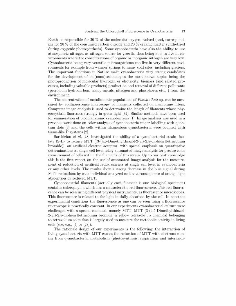
Fig. 1. The decrease of chlorophyll red fluorescence as a consequence of the accumulationinside the cell of MTT formazans crystal can be used to measure the intensity of MTTmetabolic, light -dependent, reduction by cyanobacteria
ary metabolism). The reduction of MTT further decrease the intensity of chloro-phyll fluorescence. The chemical reduction of MTT changes some of its properties,namely the color and the physical state of the molecule. The oxidized molecule isyellowish and water soluble whereas the reduced molecule (the so-called formazan)is dark brown, having a specific absorption spectrum, and it is insoluble in water(Fig. 1 (A)).
Fig. 1 (B) shows how MTT is reduced by enzymes called reductase (acting inphotosynthesis, respiration and intermediary metabolism) to formazan. MTT isyellowish, soluble in water, and reduced MTT (MTT formazan) is insoluble (not
Studying the Chlorophyll Fluorescence in Cyanobacteria 15
shown); when an appropriate chemical is added to dissolve the insoluble purpleformazan product into a colored solution, this colored solution can be quantifiedby measuring the optical density at a certain wavelength (usually between 500 and600 nm, as one can see, by a spectrophotometer).
Thus the reduction of MTT generate crystals of formazan which remain insidethe cyanobacterial cell covering the intracellular structures of cyanobacteria. Themost important intracellular structures of cyanobacteria involved in the reductionof MTT (as well as in the reduction of other artificial electron acceptors) are thethylakoides. Thylakoides are the sites where some of the light energy absorbed bythylakoids is converted in chemical stable energy found in molecules such as ATPand reduced form of chemical compounds (e.g NADPH etc.,); other part of someof the light energy absorbed by thylakoids is re-emitted as fluorescence.
During the process of MTT reduction by living cyanobacteria in light, thereduced MTT (formazan) accumulate inside the cell; this accumulation can beseen microscopically using light microscopy and even quantificated by automatedimage analysis [28]. This accumulation of reduced MTT inside the cell physicallycovers intracellular structures, including thylakoides thus acting as a shield whichblocks the access of light to thylakoides, thus decreasing the intensity of chlorophyllfluorescence.
Up to our best knowledge, this is for the first time when the decrease in chloro-phyll fluorescence produced by the reduction of MTT is monitored using an epiflu-orescence microscope. However, the inhibition of important metabolic processes incyanobacteria during tetrazolim salt reduction is documented in literature. Paerland Bland [23] show the effects of localized reduction of five tetrazolium salts hasstrong negative impact on three important metabolic processes in cyanobacteria :N2 fixation (acetylene reduction), CO2 fixation, and H2 consumption.
During short-term (within 30 min) exposures in the cyanobacterium A. oscil-larioides, salt reduction in heterocysts occurred simultaneously with inhibition ofacetylene reduction
Conversely, when salts failed to either penetrate or be reduced in heterocysts,no inhibition of acetylene reduction occurred. When salts were rapidly reduced invegetative cells, 14CO2 fixation and 3H2 utilization rates decreased [23] .
The type of experiment presented in this paper has a deeper biological theoret-ical significance and a stronger practical application than our previous work doneon color analysis of cyanobacteria under labelling with quantum dots [3] because ofmetabolic background. The decrease of chlorophyll fluorescence as a consequenceof the accumulation inside the cell of MTT formazans crystal can be used to (in-directly) measure the intensity of MTT reduction at the level of filaments or evenat the level of individual cells within each filament, cells which are subcomponentsof the biological individual (the filament in the case of filamentous cyanobacteria).
16 I. Ardelean et al.
4 Analyzing the Images
The study of the chlorophyll fluorescence in cyanobacteria has been split in severalstages. In each stage, an image is provided as input and it is processed by a tissueP system with promoters described above. The result is an automatized imageprocess performed by a sequence of P systems.
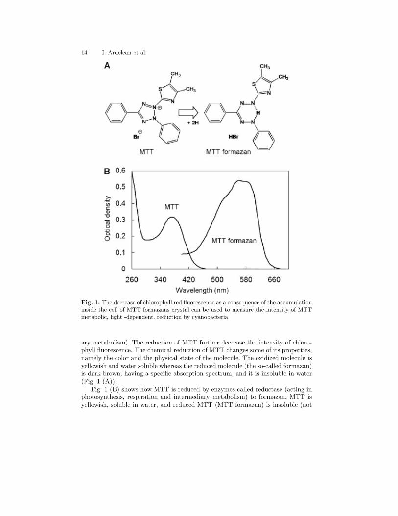
The target is to obtain information about the central cyanobacteria of theimage of Fig. 2 (a). To do that, the following stages are performed:
Stage 1: Grey Scale. The image is transformed into a grey scale one (Fig. 2(b)). We only keep the information on the red plane to do this.
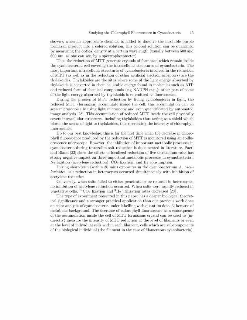
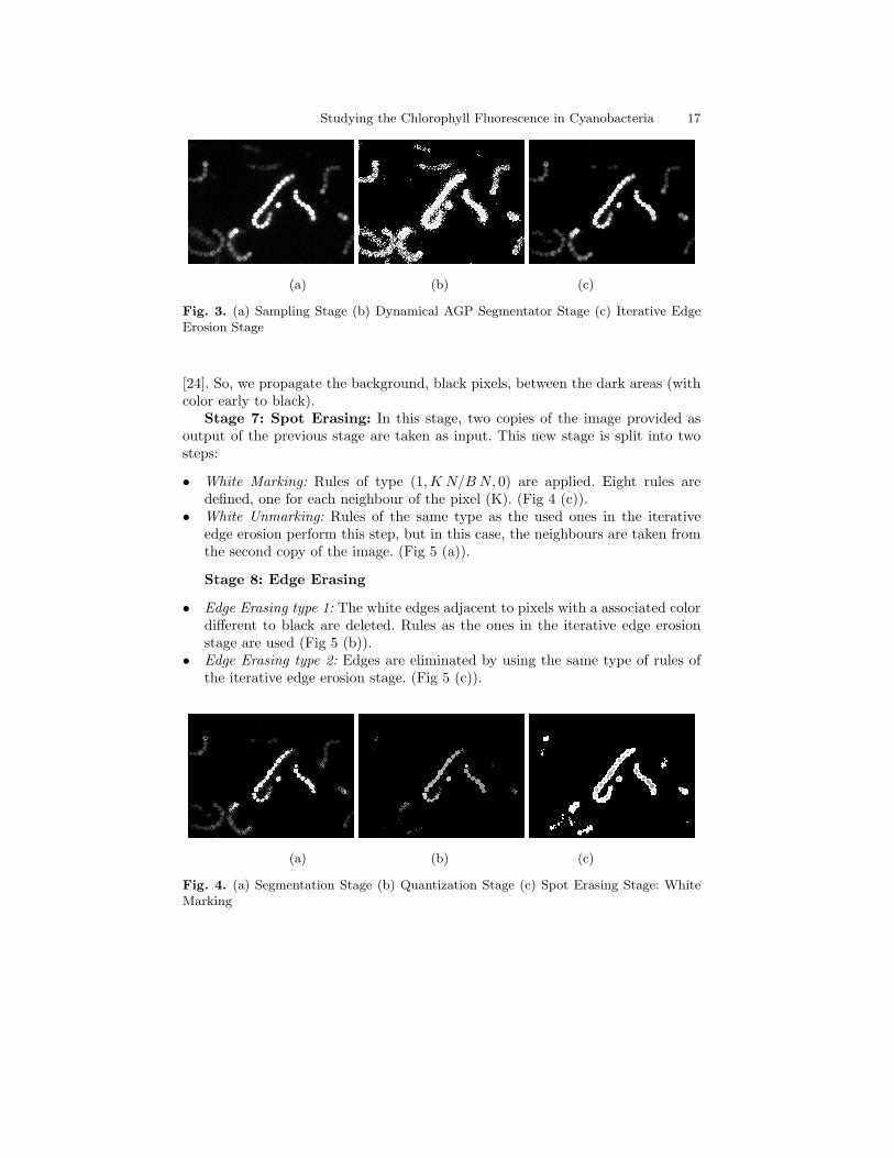
Stage 2: Sampling (Fig. 3 (a)). Before being processed by a computer, theimages greater than an specific size must be sampled. The aim of this is a basicprocess is to obtain images of the same size before comparing them.
Stage 3: Dynamical AGP Segmentator (First threshold, Fig 3 (b)). Thisis an iterative stage. We apply, in each iteration, a variant of the AGP segmentator(See [14]). In order to blur the image, each pixel on the boundary turns on whiteor it takes the smallest gray value of their neighbours. As usual in P systems, thisprocess finishes when no more segmentation rules can be applied.
Stage 4: Iterative Edge Erosion Fig 3 (c). In each iteration, rules of thefollowing type are applied: K1 K2 K3
1, K8 B K4
K7 K6 K5
/K1 K2 K3
K8 K′ K4 , 0
K7 K6 K5
where K ′ = min{Ki : i = 1, . . . , 8 ∧Ki 6= B}
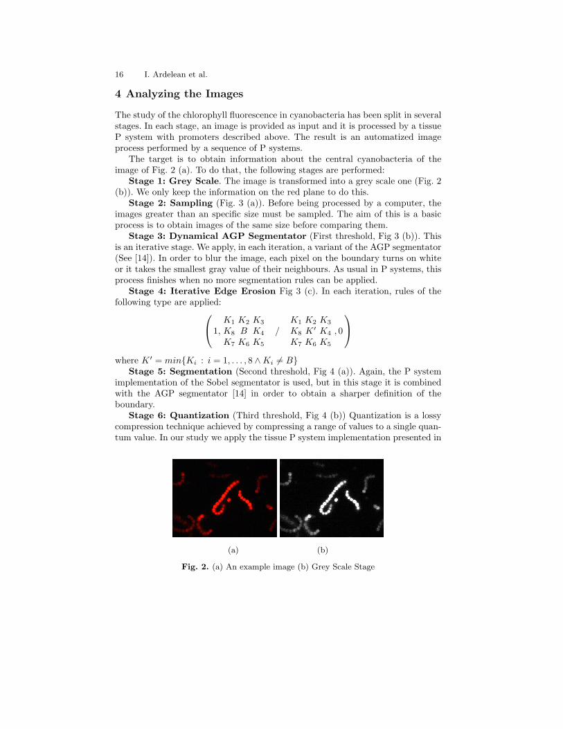
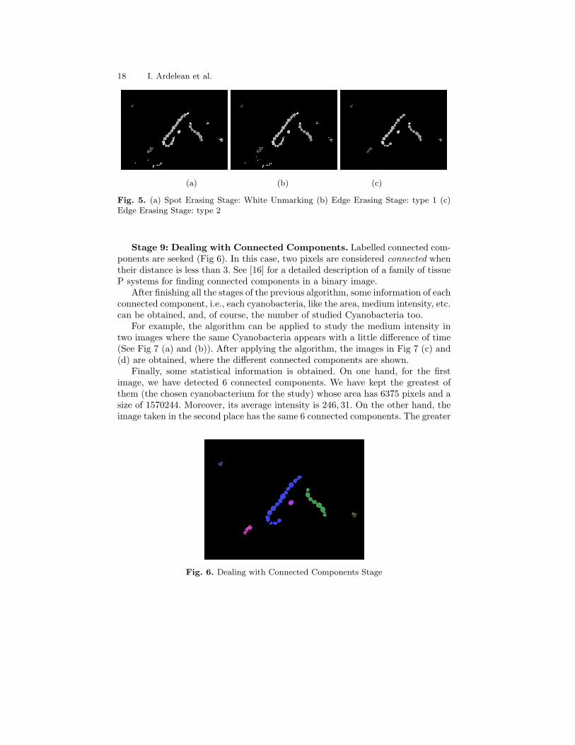
Stage 5: Segmentation (Second threshold, Fig 4 (a)). Again, the P systemimplementation of the Sobel segmentator is used, but in this stage it is combinedwith the AGP segmentator [14] in order to obtain a sharper definition of theboundary.
Stage 6: Quantization (Third threshold, Fig 4 (b)) Quantization is a lossycompression technique achieved by compressing a range of values to a single quan-tum value. In our study we apply the tissue P system implementation presented in
(a) (b)
Fig. 2. (a) An example image (b) Grey Scale Stage
Studying the Chlorophyll Fluorescence in Cyanobacteria 17
(a) (b) (c)
Fig. 3. (a) Sampling Stage (b) Dynamical AGP Segmentator Stage (c) Iterative EdgeErosion Stage
[24]. So, we propagate the background, black pixels, between the dark areas (withcolor early to black).
Stage 7: Spot Erasing: In this stage, two copies of the image provided asoutput of the previous stage are taken as input. This new stage is split into twosteps:
• White Marking: Rules of type (1,K N/BN, 0) are applied. Eight rules aredefined, one for each neighbour of the pixel (K). (Fig 4 (c)).
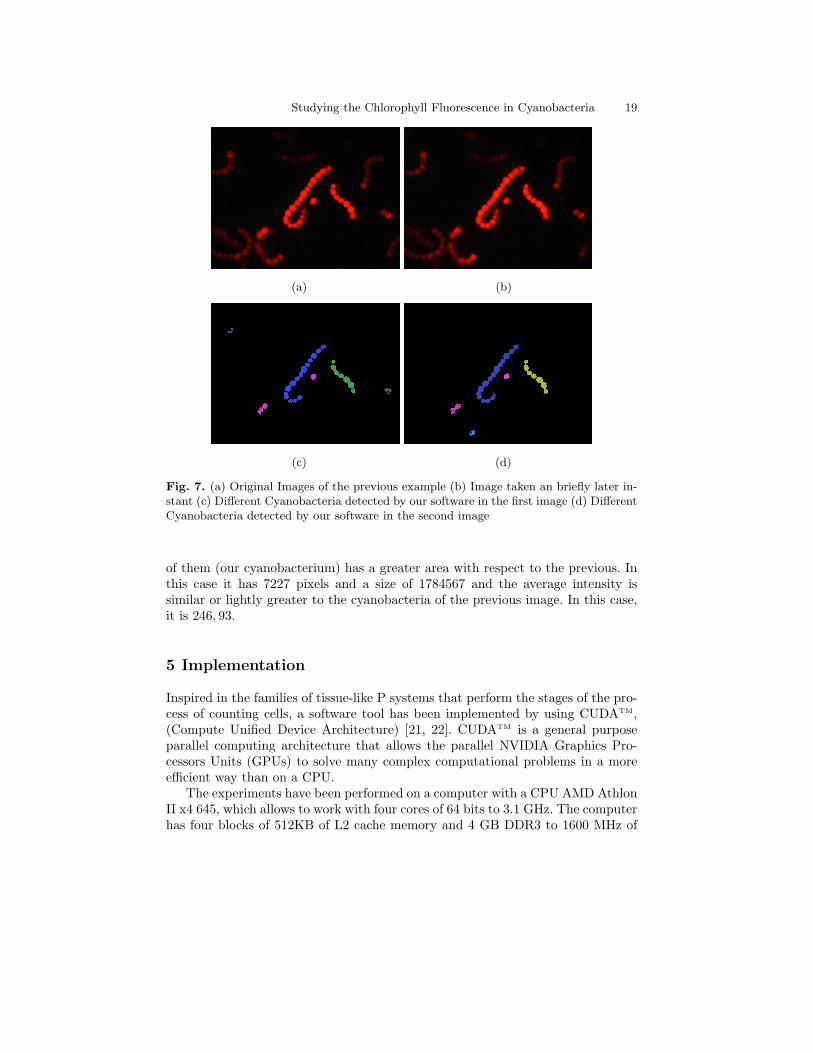
• White Unmarking: Rules of the same type as the used ones in the iterativeedge erosion perform this step, but in this case, the neighbours are taken fromthe second copy of the image. (Fig 5 (a)).
Stage 8: Edge Erasing
• Edge Erasing type 1: The white edges adjacent to pixels with a associated colordifferent to black are deleted. Rules as the ones in the iterative edge erosionstage are used (Fig 5 (b)).
• Edge Erasing type 2: Edges are eliminated by using the same type of rules ofthe iterative edge erosion stage. (Fig 5 (c)).
(a) (b) (c)
Fig. 4. (a) Segmentation Stage (b) Quantization Stage (c) Spot Erasing Stage: WhiteMarking
18 I. Ardelean et al.
(a) (b) (c)
Fig. 5. (a) Spot Erasing Stage: White Unmarking (b) Edge Erasing Stage: type 1 (c)Edge Erasing Stage: type 2
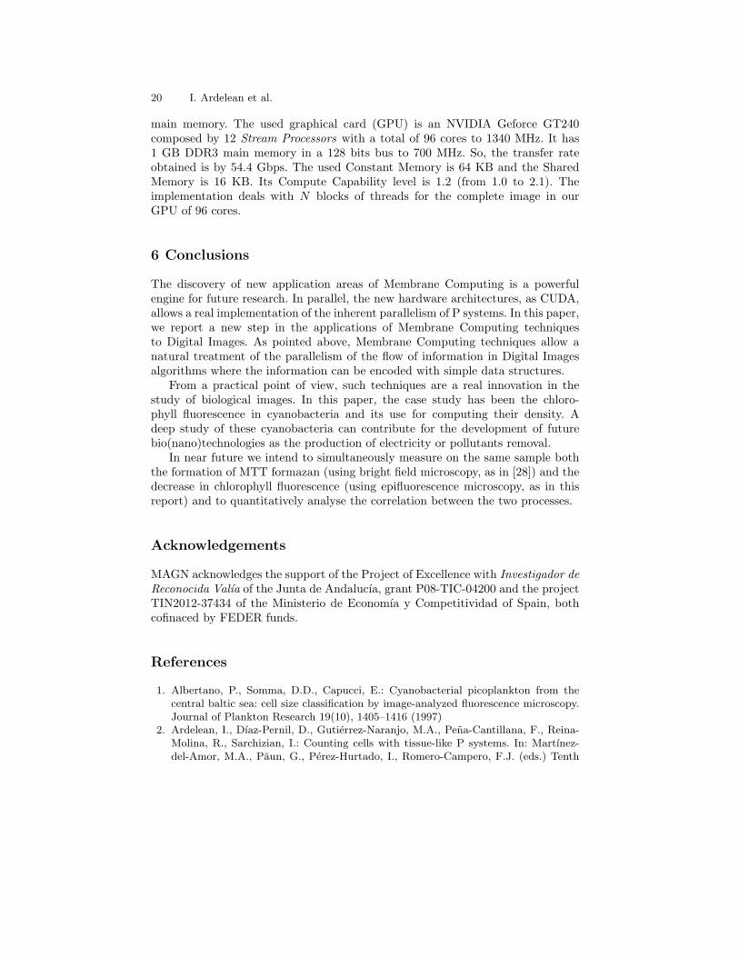
Stage 9: Dealing with Connected Components. Labelled connected com-ponents are seeked (Fig 6). In this case, two pixels are considered connected whentheir distance is less than 3. See [16] for a detailed description of a family of tissueP systems for finding connected components in a binary image.
After finishing all the stages of the previous algorithm, some information of eachconnected component, i.e., each cyanobacteria, like the area, medium intensity, etc.can be obtained, and, of course, the number of studied Cyanobacteria too.
For example, the algorithm can be applied to study the medium intensity intwo images where the same Cyanobacteria appears with a little difference of time(See Fig 7 (a) and (b)). After applying the algorithm, the images in Fig 7 (c) and(d) are obtained, where the different connected components are shown.
Finally, some statistical information is obtained. On one hand, for the firstimage, we have detected 6 connected components. We have kept the greatest ofthem (the chosen cyanobacterium for the study) whose area has 6375 pixels and asize of 1570244. Moreover, its average intensity is 246, 31. On the other hand, theimage taken in the second place has the same 6 connected components. The greater
Fig. 6. Dealing with Connected Components Stage
Studying the Chlorophyll Fluorescence in Cyanobacteria 19
(a) (b)
(c) (d)
Fig. 7. (a) Original Images of the previous example (b) Image taken an briefly later in-stant (c) Different Cyanobacteria detected by our software in the first image (d) DifferentCyanobacteria detected by our software in the second image
of them (our cyanobacterium) has a greater area with respect to the previous. Inthis case it has 7227 pixels and a size of 1784567 and the average intensity issimilar or lightly greater to the cyanobacteria of the previous image. In this case,it is 246, 93.
5 Implementation
Inspired in the families of tissue-like P systems that perform the stages of the pro-cess of counting cells, a software tool has been implemented by using CUDATM,(Compute Unified Device Architecture) [21, 22]. CUDATM is a general purposeparallel computing architecture that allows the parallel NVIDIA Graphics Pro-cessors Units (GPUs) to solve many complex computational problems in a moreefficient way than on a CPU.
The experiments have been performed on a computer with a CPU AMD AthlonII x4 645, which allows to work with four cores of 64 bits to 3.1 GHz. The computerhas four blocks of 512KB of L2 cache memory and 4 GB DDR3 to 1600 MHz of
20 I. Ardelean et al.
main memory. The used graphical card (GPU) is an NVIDIA Geforce GT240composed by 12 Stream Processors with a total of 96 cores to 1340 MHz. It has1 GB DDR3 main memory in a 128 bits bus to 700 MHz. So, the transfer rateobtained is by 54.4 Gbps. The used Constant Memory is 64 KB and the SharedMemory is 16 KB. Its Compute Capability level is 1.2 (from 1.0 to 2.1). Theimplementation deals with N blocks of threads for the complete image in ourGPU of 96 cores.
6 Conclusions
The discovery of new application areas of Membrane Computing is a powerfulengine for future research. In parallel, the new hardware architectures, as CUDA,allows a real implementation of the inherent parallelism of P systems. In this paper,we report a new step in the applications of Membrane Computing techniquesto Digital Images. As pointed above, Membrane Computing techniques allow anatural treatment of the parallelism of the flow of information in Digital Imagesalgorithms where the information can be encoded with simple data structures.
From a practical point of view, such techniques are a real innovation in thestudy of biological images. In this paper, the case study has been the chloro-phyll fluorescence in cyanobacteria and its use for computing their density. Adeep study of these cyanobacteria can contribute for the development of futurebio(nano)technologies as the production of electricity or pollutants removal.
In near future we intend to simultaneously measure on the same sample boththe formation of MTT formazan (using bright field microscopy, as in [28]) and thedecrease in chlorophyll fluorescence (using epifluorescence microscopy, as in thisreport) and to quantitatively analyse the correlation between the two processes.
Acknowledgements
MAGN acknowledges the support of the Project of Excellence with Investigador deReconocida Valıa of the Junta de Andalucıa, grant P08-TIC-04200 and the projectTIN2012-37434 of the Ministerio de Economıa y Competitividad of Spain, bothcofinaced by FEDER funds.
References
1. Albertano, P., Somma, D.D., Capucci, E.: Cyanobacterial picoplankton from thecentral baltic sea: cell size classification by image-analyzed fluorescence microscopy.Journal of Plankton Research 19(10), 1405–1416 (1997)
2. Ardelean, I., Dıaz-Pernil, D., Gutierrez-Naranjo, M.A., Pena-Cantillana, F., Reina-Molina, R., Sarchizian, I.: Counting cells with tissue-like P systems. In: Martınez-del-Amor, M.A., Paun, G., Perez-Hurtado, I., Romero-Campero, F.J. (eds.) Tenth
Studying the Chlorophyll Fluorescence in Cyanobacteria 21
Brainstorming Week on Membrane Computing. vol. I, pp. 69–78. Fenix Editora,Sevilla, Spain (2012)
3. Armaselu, A., Popescu, A., Apostol, I., Ardellean, I., Damian, V., Iordache, I.,Sarchizian, I., Apostol, D.: Passive nonspecific labeling of cyanobacteria in natu-ral samples using quantum dots. Optoelectronics and Advanced Materials - RapidCommunications 5(10), 1084 – 1090 (October 2011)
4. Berridge, M.V., Herst, P.M., Tan, A.S.: Tetrazolium dyes as tools in cell biology:New insights into their cellular reduction. Biotechnology Annual Review 11, 127 –152 (2005)
5. Carnero, J., Dıaz-Pernil, D., Gutierrez-Naranjo, M.A.: Designing tissue-like P sys-tems for image segmentation on parallel architectures. In: Martınez-del-Amor, M.A.,Paun, G., Perez-Hurtado, I., Romero-Campero, F.J., Cabrera, L.V. (eds.) NinthBrainstorming Week on Membrane Computing. pp. 43–62. Fenix Editora, Sevilla,Spain (2011)
6. Cecilia, J.M., Garcıa, J.M., Guerrero, G.D., Martınez-del-Amor, M.A., Perez-Hurtado, I., Perez-Jimenez, M.J.: Implementing P systems parallelism by meansof GPUs. In: Paun, G., Perez-Jimenez, M.J., Riscos-Nunez, A., Rozenberg, G., Sa-lomaa, A. (eds.) Workshop on Membrane Computing. Lecture Notes in ComputerScience, vol. 5957, pp. 227–241. Springer, Berlin Heidelberg (2009)
7. Cecilia, J.M., Garcıa, J.M., Guerrero, G.D., Martınez-del-Amor, M.A., Perez-Hurtado, I., Perez-Jimenez, M.J.: Simulating a P system based efficient solutionto SAT by using GPUs. Journal of Logic and Algebraic Programming 79(6), 317–325(2010)
8. Cecilia, J.M., Garcıa, J.M., Guerrero, G.D., Martınez-del-Amor, M.A., Perez-Hurtado, I., Perez-Jimenez, M.J.: Simulation of P systems with active membraneson CUDA. Briefings in Bioinformatics 11(3), 313–322 (2010)
9. Chavez de Paz, L.E.: Image analysis software based on color segmentation for char-acterization of viability and physiological activity of biofilms. Applied and Environ-mental Microbiology 75(6), 1734–9 (2009)
10. Christinal, H.A., Dıaz-Pernil, D., Gutierrez-Naranjo, M.A., Perez-Jimenez, M.J.:Thresholding of 2D images with cell-like P systems. Romanian Journal of Infor-mation Science and Technology (ROMJIST) 13(2), 131–140 (2010)
11. Christinal, H.A., Dıaz-Pernil, D., Real, P.: Segmentation in 2D and 3D image us-ing tissue-like P system. In: Bayro-Corrochano, E., Eklundh, J.O. (eds.) Progressin Pattern Recognition, Image Analysis, Computer Vision, and Applications 14thIberoamerican Conference on Pattern Recognition, CIARP 2009, Guadalajara,Jalisco, Mexico, November 15-18, 2009. Proceedings. Lecture Notes in ComputerScience, vol. 5856, pp. 169–176. Springer, Berlin Heidelberg (2009)
12. Christinal, H.A., Dıaz-Pernil, D., Real, P.: Region-based segmentation of 2D and 3Dimages with tissue-like P systems. Pattern Recognition Letters 32(16), 2206 – 2212(2011)
13. Dıaz-Pernil, D., Gutierrez-Naranjo, M.A., Molina-Abril, H., Real, P.: A bio-inspiredsoftware for segmenting digital images. In: Nagar, A.K., Thamburaj, R., Li, K.,Tang, Z., Li, R. (eds.) Proceedings of the 2010 IEEE Fifth International Conferenceon Bio-Inspired Computing: Theories and Applications BIC-TA. vol. 2, pp. 1377 –1381. IEEE Computer Society, Beijing, China (2010)
14. Dıaz-Pernil, D., Berciano, A., Pena-Cantillana, F., Gutierrez Naranjo, M.A.: Seg-menting Images with Gradient-based Edge Detection Using Membrane Computing.Pattern Recognition Letters 34(8), 846–855 (2013)
22 I. Ardelean et al.
15. Dıaz-Pernil, D., Gutierrez-Naranjo, M.A., Molina-Abril, H., Real, P.: Designing anew software tool for digital imagery based on P systems. Natural Computing pp.1–6 (2011)
16. Dıaz-Pernil, D., Gutierrez-Naranjo, M.A., Real, P., Sanchez-Canales, V.: Computinghomology groups in binary 2D imagery by tissue-like P systems. Romanian Journalof Information Science and Technology 13(2), 141–152 (2010)
17. Fero, M., Pogliano, K.: Automated quantitative live cell fluorescence microscopy.Cold Spring Harb Perspectives in Biology 2(8), a000455 (2010)
18. Gimel’farb, G., Nicolescu, R., Ragavan, S.: P systems in stereo matching. In: Real,P., Dıaz-Pernil, D., Molina-Abril, H., Berciano, A., Kropatsch, W. (eds.) ComputerAnalysis of Images and Patterns, Lecture Notes in Computer Science, vol. 6855, pp.285–292. Springer Berlin / Heidelberg (2011)
19. Gimel’farb, G.L.: Probabilistic regularisation and symmetry in binocular dynamicprogramming stereo. Pattern Recognition Letters 23(4), 431–442 (2002)
20. Ionescu, M., Sburlan, D.: On P systems with promoters/inhibitors. Journal of Uni-versal Computer Science 10(5), 581–599 (may 2004)
21. Nickolls, J., Buck, I., Garland, M., Skadron, K.: Scalable parallel programming withCUDA. Queue 6, 40–53 (March 2008)
22. Owens, J.D., Houston, M., Luebke, D., Green, S., Stone, J.E., Phillips, J.C.: GPUComputing. Proceedings of the IEEE 96(5), 879–899 (May 2008)
23. Paerl, H.W., Bland, P.T.: Localized tetrazolium reduction in relation to N2 fixation,CO2 fixation, and H2 uptake in aquatic filamentous cyanobacteria. Applied and En-vironmental Microbiology 43(1), 218–226 (January 1982)
24. Pena-Cantillana, F., Dıaz-Pernil, D., Berciano, A., Gutierrez-Naranjo, M.A.: A par-allel implementation of the thresholding problem by using tissue-like P systems. In:Real, P., Dıaz-Pernil, D., Molina-Abril, H., Berciano, A., Kropatsch, W.G. (eds.)CAIP (2). Lecture Notes in Computer Science, vol. 6855, pp. 277–284. Springer(2011)
25. Pena-Cantillana, F., Dıaz-Pernil, D., Christinal, H.A., Gutierrez-Naranjo, M.A.: Im-plementation on CUDA of the smoothing problem with tissue-like P systems. Inter-national Journal of Natural Computing Research 2(3), 25–34 (2011)
26. Peschek, G.A.: Structure-function relationships in the dual-function photosynthetic-respiratory electron-transport assembly of cyanobacteria (blue-green algae). Bio-chemical Society Transactions 24(3), 729–733 (August 1996)
27. Peschek, G.A., Obinger, C., Fromwald, S., Bergman, B.: Correlation betweenimmuno-gold labels and activities of the cytochrome-c oxidase (aa3-type) in mem-branes of salt stressed cyanobactria. FEMS Microbiology Letters 124(3), 431–437(1994)
28. Sarchizian, I., Cırnu, M., Ardelean, I.I.: Isolation of a heterocyts - forming cyanobac-terium and quantification of its biotechnological potential with respect to redox prop-erties at single cell level. Romanian Biotechnological Letters 16(6), 3–9 (2011)
29. Selinummi, J., Ruusuvuori, P., Podolsky, I., Ozinsky, A., Gold, E., Yli-Harja, O.,Aderem, A., Shmulevich, I.: Bright field microscopy as an alternative to whole cellfluorescence in automated analysis of macrophage images. PLoS ONE 4(10), 1 9(2009)
30. Selinummi, J., Seppala, J., Yli-Harja, O., Puhakka, J.A.: Software for quantificationof labeled bacteria from digital microscope images by automated image analysis.Biotechniques 39(6), 859–863 (2005)
Studying the Chlorophyll Fluorescence in Cyanobacteria 23
31. Sheeba, F., Thamburaj, R., Nagar, A.K., Mammen, J.J.: Segmentation of peripheralblood smear images using tissue-like P systems. Sixth International Conference onBio-Inspired Computing: Theories and Applications (BIC-TA), 2011, 257–261 (sept2011)
32. Walsby, A.E., Avery, A.: Measurement of filamentous cyanobacteria by image anal-ysis. Journal of Microbiological Methods 26(1-2), 11 – 20 (1996)
33. Zhang, G.X., Gheorghe, M., Li, Y.: A membrane algorithm with quantum-inspiredsubalgorithms and its application to image processing. Natural Computing 11(4),701–717 (2012)
